sublethal effects of petroleum hydrocarbons and …
TRANSCRIPT
SUBLETHAL EFFECTS OF PETROLEUM HYDROCARBONS AND TRACE METALS,
INCLUDING BIOTRANSFORMATIONS, AS REFLECTED BY MORPHOLOGICAL,
CHEMICAL, PHYSIOLOGICAL, AND BEHAVIORAL INDICES
by
Donald C. Malins, Harold O. Hodgins,Usha Varanasi, Sin-Lam Chan, Bruce B. McCain,
Douglas D. Weber, and Donald W, Brown
Other Contributing Authors:
Tracy K. Collier, Leroy C. Folmar, William D. Gronland,Edward H. Gruger, Jr., Joyce W. Hawkes, William L. Reichert,William T. Roubal, Herbert R. Sanborn, Michael H. Schiewe,
and John E. Stein
Environmental Conservation DivisionNorthwest and Alaska Fisheries Center
National Marine Fisheries Service, NOAA2725 Montlake Boulevard East
Seattle, Washington 98112
Final ReportOuter Continental Shelf Environmental Assessment Program
Research Unit 73
June 1982
1. SUMMARY OF OBJECTIVES,WITH
1.1
1.2
1.3
TABLE OF CONTENTS
Page
CONCLUSIONS, AND IMPLICATIONSRESPECT TO OCS OIL AND GAS DEVELOPMENT . . . . . . . . . .
Summary of Objectives . . . . . . . . . . . . . . . . . . .
Summary of Conclusions.. . . . . . . . . . . . ..O . .
1.2.1 Chemistry. . . . . . . . . . . , . . . . . . . . .1.2.2 Pathology. . . . . . . . . . . . . . . . . . . . .1.2.3 Behavior and Physiology . . . . . . . . . . . . . .
Implications with Respect to OCS Oiland Gas Development. ‘, . . . . . . . . . . . . . . . , . ,
1.3.1 Chemistry. . . . . . . . . . . . . . . . . . . . .1.3.2 Pathology. . . . . . . . . . . . . ., . . . , . .1.3.3 Behavior and Physiology . . . . . . . . . . . . . .
2. INTRODUCTION. . . . . . . . . . . . . . . . . . . . . . . . . .
3. BACKGROUND. . . . . . . . . . . . . . . . . . . . . . . . . . .
3.1 Chemistry. . . . . . . . . . . . . . ., . . . . . .. . .3.2 Pathology. . . . . . . . . . . . . . . . . . . .. . . . .3.3 Behavior and Physiology. . . . . . . . . . . . . . . . . .
4. STUDY AREA . . . . . . . . . . . . . . . . . . . . . . . . . . .
5. METHODS . . . . . . . . . . . . . . . . . . . . . . . . . . . .
5.1 Chemistry. . . . . . , . . . . . . , . . . . . . , . , . .
5.1.1 Accumulation and Biotransformation ofSpecific Aromatic Hydrocarbons in Salmonids . . . .
5.1.2 Accumulation of Petroleum Hydrocarbons byFish Exposed to Seawater Soluble Fraction ofPrudhoe BayCrudeOil . . . . . . . . . . . . . . .
5.1.3 Metabolism of NPH by Coho Salmon. . . . . . . . . .5.1.4 Naphthalene and its Metabolizes in Fish Skin
and Mucus. . . . . . . . . . . . . . . . . . . . ,5.1.5 Accumulation and Biotransformation of NPH
by Flatfish. . . . . . . . . . . .5.1.6 Effect of Temperature on Disposition of NPH “ “ “ “
and its Metabolizes in Fish . . . . . . . . . . . .5.1.7 Uptake and Metabolism of Sediment-Associated
Aromatic Hydrocarbons by Flatfish . . . . . . . . .
7
7
8
899
10
101112
13
14
141516
17
18
18
18
1818
20
20
21
22
Page
5.1.8 B a P Metabolism by English Sole. . . . . . . . . . .5.1.9 Binding of BaP Intermediates to DNA
Catalyzed by Liver Enzymes of Fish. . . . . . . . .5.1.10 AHM Activities in Different Species . . . . . . . .5.1.11 Uptake, Metabolism, and Toxicity of
Hydrocarbons in Invertebrates . . . . . . . . . . .5.1.12 Food Chain Transfer of 2,6-Dimethylnaphthalene
to Sea Urchins via Algae... . . . . . . . . . . .5.1.13 Biological Fate of Metals in Fish . . . . . . . . .
5.2 Pathology. . . . . . . . . . . . . . . . . . . . . . . . .
5.2.1 Effects of Petroleum on Disease Resistance. . . . .5.2.2 Pathological Changes in Flatfish from
Exposure to Oil-Contaminated Sediment . . . . . . .5.2.3 Cytopathology. . . . . . . . . . . . . . . . . . .
5.3 Behavior and Physiology. . . . . . . . . . . . . . . . . .
5.3.1 Chemical Analysis of Water, Sediment,and Tissue. . . . . . . . . . . . . . . . . . . . .
5.3.2 Preparation of Oil-Water Mixtures . . . . . . . . .5.3.3 Vertebrate Studies. . . . . . . . . . . . . . . . .5.3.4 Invertebrate Studies. . . . . . . . . . . . . . . .
6. RESULTS. . . . . . . . . . . . . . . . . . . . . . . . . . . .
6.1 Chemistry. . . . . . . . . . . . . . . . . . . . . . . . .
6.1.1
6.1.2
6.1.36.1.46.1.5
6.1.6
6.1.7
6.1.86.1.9
6.1.10
6.1.11
Uptake and Biotransformation of SpecificHydrocarbons in Salmonids . . . . . . . . . . . . .Accumulation of Petroleum Hydrocarbonsby Fish Exposed to the Seawater SolubleFraction of Prudhoe Bay Crude Oil . . . . . . . . .Metabolism of NPH by Coho Salmon. . . . . . . . . .NPH and its Metabolites in Fish Skin and Mucus. . .Accumulation and Biotransformation ofNPHby Flatfish. . . . . . . . . . . . . . . . . . . .Effect of Environmental Temperature onDisposition of NPH and its Metabolizes in Fish. . .Uptake and Metabolism of Sediment-AssociatedNPHand BaPby Flatfish . . . . . . . . . . . . . .BaP Metabolism by English Sole. . . . . . . . . . .Binding of BaP Intermediates to DNA Catalyzedby Liver Enzymes oafish... . . . . . . . . . . .Activities of Aryl Hydrocarbon Monooxygenasesin Different Species. . . . . . . . . . . . . . . .Uptake, Metabolism, and Toxicity ofHydrocarbons in Invertebrates . . . . . . . . . . .
23
2324
24
2526
27
27
3031
33
33343850
55
55
55
555961
67
68
7886
87
91
92
Page
6.1.12 Food Chain Transfer of 2,6-Diinethylnaphthaleneto Sea Urchins via Algae.. . . . . . . . . . . . . 98
6.1.13 Biological Fate of Metals . . . . . . . . . . . . . 100
6.2 Pathology. . . . . . . . . . . . . . . . . . . . . . . . . 106
6.2.1 Effects of Petroleum on Disease Resistance. . . . . 1066.2.2 Pathological Changes in Flatfish from
Exposure to Oil-Contaminated Sediment . . . . . . . 1106.2.3 Cytopathology. . . . . . . . . . . . . . . . . . . 119
6.3 Behavior and Physiology. . . . . . . . . . . . . . . . . . 129
6.3.1 Vertebrate Studies. . . . . . . . . . . . . . . . . 1296.3.2 Invertebrate Studies. . . . . . . . . . . . . . . . 161
7. DISCUSSION. . . . . . . . . . . . . . . . . . . . . . . . . . . 169
7.1 Chemistry. . . . . . . . . . . . . . . . . . . . . . . . . 169
7.1.1
7.1.2
7.1.37.1.47.1.57.1.6
7.1.7
7.1.87.1.9
Accumulation and Biotransformation ofSpecific Aromatic Hydrocarbons in Salmonids . . . . 169Accumulation of Petroleum Hydrocarbons byFish Exposed to the Seawater Soluble Fractionof Prudhoe BayCrudeOil. . . . . . . . . . . . . . 170Metabolism of NPH by Coho Salmon, . . . . . . . . . 170NPH and its Metabolizes in Fish Skin and Mucus. . . 170Uptake and Biotransformation of NPH by Flatfish . . 171Effect of Temperature on Disposition of NPHand its Metabolizes in Fish . . . . . . . . . . . . 173Uptake and Metabolism of Sediment-AssociatedNPHand BaPbyFlatfish . . . . . . . . . . . . . . 174BaP Metabolism by English Sole. . . . . . . . . . . 176Bindinq of BaP Intermediates to DNACatalyzed by Liver Enzymes. . . . . . . . . . .
7.1.10 Activities of Aryl Hydrocarbon Monooxygenasesin Different Species. . . . . . . . . . . . . .
7.1.11 Uptake, Metabolism, and Toxicity ofHydrocarbons in Invertebrates . . . . . . . . .
7.1.12 Food Chain Transfer of 2,6-Dimethylnaphthaleneto Sea Urchins via Algae. . . . . . . . . . . .
7.1.13 Biological Fate of Metals in Fish . . . . . . .
7.2 Pathology. . . . . . . . . . . . . . . . . . . . . . ,
7.2.1 Effects of Petroleum on Disease Resistance. . .7.2.2 Pathological Changes in Flatfish from
Exposure to Oil-Contaminated Sediment . . . . .7.2.3 Cytopathology . . . . . . . . . . . . . . . . .
. . 177
. . 177
. . 178
. . 179
. . 180
. . 182
. . 182
. . 182
. . 183
5
Page
7.3 Behavior and Physiology. . . . . . . . . . . . . . . . . . 186
7.3.1 Vertebrate Studies. . . . . . . . . . . . . . . . . 1867.3.2 Invertebrate Studies. . . . . . . . . . . . . . . . 190
8. CONCLUSIONS. . . . . . . . . . . . .- ● ● ““ O“ ● ● ● “O ● 192
8.lChemistry. . . . . . . . . . . . . ● s “ ● ● o “ o ● ● “ “ 192
8.1.1
8.1.2
8.1.3
8.1.4
8.1.58.1.6
8.1.7
8.1.8
8.1.9
Accumulation and Biotransformation ofAromatic Hydrocarbons by Marine Species . . . . . . 192Factors Influencing Uptake and Metabolismof Naphthalene by Fish.. . . . . . . . . . . . . . 193Naphthalene and its Metabolizes in FishSkinandMucus. . . . . . . . . . . . . . ● o ● ● “ 193Uptake and Metabolism of Sediment-AssociatedNaphthalene and Benzo(a)pyrene by Flatfish. . . . . 194BaP Metabolism by English Sole. . . . . . . . . . . 194Examination of Aryl Hydrocarbon MonooxygenaseActivity in Different Species and Binding of BaPIntermediates to DNA Catalyzed by Liver Enzymes . . 194Uptake, Metabolism, and Toxicity ofHydrocarbons in Invertebrates . . . . . . . . . . . 194Food Chain Transfer of 2,6-Dimethylnaphthalenefrom Algae to Sea Urchins . . . . . . . . . . . . . 195Fate ofMetals in Fish... . . . . . . . . ...* 195
8.2 Pathology. . . . . . . . . . . . . . . ● ● ● o “ ● “ ●
8.2.1 Effects of Petroleum on Disease Resistance. . .8.2.2 Pathological Changes in Flatfish from
Exposure to Oil-Contaminated Sediment . . . . .8.2.3 Cytopathology . . . . . . . . . ..*~”ooo
8.3 Behavior and Physiology. . . . . . . . . . . . . . . .
8.3.1 Vertebrate Studies. . . . . . . . . . . . . . .8.3.2 Invertebrate Studies. . . . . . . . . . . . . .
9. PAPERS PUBLISHED, DISSERTATIONS, THESES, AND PRESENTATIONSGIVEN UNDER RESEARCH UNIT 73 OCSEAP SPONSORSHIP . . . . . .
9.1 Papers Published. . . . . . . . . . . . . . . . . . .
9.2 Dissertations and Theses . . . . . . . . . . . . . . .
9.3 Presentations. . . . . . . . . . . . * . ● ● . “ ● “ ●
. . 195
. . 195
. . 196
. . 196
. . 196
. . 196
. . 197
. . 199
. . 199
. . 203
. . 204
ACKNOWLEDGEMENTS.
REFERENCES. . . .
209. . . . . ● . . . . ● .*** ● *~”” ““a””
210. . . . . . . . . . . . . . . ● .*** ● “”*”
6
1. SUMMARY OF OBJECTIVES, CONCLUSIONS, AND IMPLICATIONS WITH RESPECTTO OCS OIL AND GAS DEVELOP14ENT
1.1 Summary of Objectives
The overall objectives of this program were to assess the potentialeffects of petroleum and petroleum-related activities on marine organismsindigenous to Alaskan waters. The principal objectives addressed were to:
(a) Determine the uptake of polycycl ic aromatic hydrocarbons bysalmonids and pleuronectids exposed to these compounds in sediment,water or via their diet.
(b) Study the metabolism of aromatic hydrocarbons by fish andevaluate the potential of their metabolizes for interacting with DNAand other cellular constituents.
(c) Evaluate the uptake, disposition and toxicity of petroleumhydrocarbons in larval and adult invertebrates.
(d) Determine the activities of enzymes (aryl hydrocarbon mono-oxygenases) that metabolize aromatic hydrocarbons in a variety ofaquatic species.
(e) Stud.Y the factors (e.q., temperature. routes and lenqth ofexposure) that-influence the’up{a[e, metabolism and dispositio~ ofpetroleum hydrocarbons in marine fishes.
(f) Study the uptake and deputation of toxic trace metals byand flatfish.
(g) Determine the effects of petroleum hydrocarbons and themdispersants on disease resistance and host defense mechanisms ofmarine fish and shellfish.
sa
ca
mo n
(h) Evaluate pathological effects that can result from exposingadult and juvenile flatfish to sediments contaminated with petroleum.
(i) Assess the cytopathological changes in marine fish resultingfrom exposure to petroleum hydrocarbons.
(j) Evaluate the avoidance, homing, and predator-prey behavior ofsalmon exposed to water-borne hydrocarbons, and the avoidance of oil-contaminated sediment by juvenile flatfish.
(k) Determine the effect of ingested and water-borne crude oil onreproductive success and/or the early developmental stages of salmonids,flatfish, smelt, and invertebrates.
(1) Assess the effects of water-borne hydrocarbons on the chemosensory-mediated behavior of invertebrates.
7
1.2 Summary of Conclusions
The conclusions of this program are summarized according todiscipline; chemistry, pathology, and behavior and physiology.
1 .2 .1 Chemistry
Marine fish and invertebrates accumulate a broad spectrum ofaromatic hydrocarbons when exposed to these compounds in water, sediment,or via force feeding or intraperitoneal injection. In fish, the modeof exposure to aromatic hydrocarbons can, markedly influence the extentof accumulation of parent hydrocarbons and.their metabolizes as wellas the types of metabolizes formed. Also, increasing the number ofbenzenoid rings or the degree of alkyl substitution of aromatichydrocarbons results in increased accum~lation of these compounds byfish.
Measurement of aryl hydrocarbon monooxygenase activities show thatmost invertebrates and vertebrates investigated were capable ofmetabolizing aromatic hydrocarbons.
Regardless of species, mode of exposure or structure ofhydrocarbon, results show that metabolic products are retained in thetissues of animals for a longer time than are the parent hydrocarbons.
Fish brain appears to accumulate mainly parent aromatic hydrocarbons(e.g., naphthalene), whereas other sites, such as liver and bile,contained primarily conjugated and nonconjugated metabolizes of aromatichydrocarbons.
Lowering the environmental temperature increased both concentrationsand resident times of naphthalene in the tissues of fish perorallyexposed to naphthalene. Lowering the temperature also decreased thebioconversion of naphthalene and altered the proportions of individualmetabolizes in the tissues.
Themetabolite profiles of naphthalene in both adult and larvalshrimp were similar to those reported for fish, and demonstrate thatthe early developmental stages of shrimp have the enzyme systemsnecessary for converting naphthalene to both conjugated and nonconjugatedmetabolizes. In addition, exposure to low levels (1-100 ppb) ofnaphthalene in seawater impaired fertilization and early embryonicdevelopment of mollusc larvae, and survival of crustacea larvae.
The liver enzymes of pleuronectids convert benzo(a)pyrene intoreactive intermediates (such as epoxides) that bind to DNA and proteins.Such interactions are known to damage critical cellular constituentsin mammals.
Studies with both aromatic hydrocarbons and metals demonstrate thatthe skin and epidernal mucus of fish are involved in both the uptake anddischarge of these compounds.
8
Salmonid and pleuronectid fish accumulated water-borne lead andcadmium; the metals persisted in many organs and tissues for weeksafter the exposure was terminated. Cadmium was preferentially boundby low molecular weight proteins in liver cytosol; high accumulationof lead was found in the brains of fish.
Food chain transfer of aromatic hydrocarbons was demonstrated.Sea urchins feeding on 2,6-dimethylnaphthalene-exposed algae accumulateand metabolize this hydrocarbon.
1.2.2 Pathology
Juvenile and adult flatfish and adult spot shrimp exposed tooil-contaminated sediment did not show an altered resistance to bacterialinfection. Juvenile salmon exposed to seawater-accommodated crude oil for2 weeks, as well as salmon and trout perorally exposed to crude oil forup to 10 months, also showed no demonstrableor evidence of immune dysfunction. However,an adverse effect of a petroleum dispersant Iresistance of salmon.
Three species of flatfish exposed to oi-for up to 4 months differed substantially in
changes in disease resistancepreliminary tests suggestedCorexit 9527) on disease
-contaminated sedimentsthe degree to which they
accumulated parent aromatic hydrocarbons. Pathological changes occurredin the livers of all flatfish species tested, but similar abnormalitieswere frequently observed in controls. In addition, the physiochemicalcharacteristics of sediment greatly influenced its retention of petroleum.Retention of aromatic hydrocarbons was more than 10-fold greater inhigh-silt than in high-sand sediment.
Ultrastructural changes in the liver and lens tissues of adulttrout occurred after high doses of petroleum hydrocarbons were administeredperorally for 2-12 months. Also, both salmon and flatfish exposed towaterborne hydrocarbons exhibited gill lesions characterized by loss ofsurface cells.
Exposing embryonic smelt and flatfish to low ppb concentrationsof seawater-accommodated crude oil resulted in high mortality at hatching.The eye and brain of exposed smelt embryos appeared to be target organsand, in the later phases of embryonic development, exhibited extensivenecrocytosiso In flatfish there was evidence of disruption in bothepithelial cell mitochondria and in the olfactory epitheliums.
1.2.3 Behavior and Physiology
At low (15-150) ppb concentrations of waterborne hydrocarbons, spotshrimp overt feeding behavior and the sea urchin pedicellarial defenseresponse were reduced by half. At these hydrocarbon concentrationsnudibranchs failed to locate mating conspecifics and suffered impairedreproduction and embryological abnormalities. in addition, less than10% of the smelt eggs exposed throughout embryogenesis hatched; of thelarvae that hatched, only lVL survived. Flatfish embryos exposed to
9
80 ppb developed into normal larvae, but embryos exposed to more than130 ppb hatched into dead or grossly abnormal larvae.
At waterborne hydrocarbon concentrations of 150-500 ppb there wasa significantly increased consumption of exposed sea urchins by starfishpredators and of exposed salmon fry by salmon predators. Salmon predationdecreased sharply after the predators were exposed to oil for 3 or moredays. Exposing salmon eggs throughout embryo and alevin development atthese hydrocarbon concentrations resulted in a 400% increase (comparedto controls) in mortality; exposure as either embryos or alevins aloneincreased mortality by 100-150% over that of controls.
Exposing adult salmon to 1-2 ppm of aromatic hydrocarbons causeda 3 day delay in return from offshore in seawater to their “home” stream;concentrations of 2-3 ppm inhibited upstream spawning migration. However,short term exposure to a 4 ppm concentration did not discernibly affectadult salmon olfactory perception. Also, exposing adult salmon to 40 ppmof freshwater-accommodated crude oil did not alter their homing capabilityin freshwater or their rate of return.
Juvenile flatfish did not consistently avoid oil-sediment mixturescontaining 8,000-10,000 ppm total hydrocarbons, and maturing trout fedlarge amounts of crude oil (1,000 ppm added to food) for 7 months didnot show statistically significant changes in their hatching success.
1.3 Implications with Respect to OCS Oil and Gas Development
Research findings from this program have clear implications withrespect to petroleum effects on aquatic species and consequently to OCSoil and gas development. Most of the studies were designed as laboratoryexperiments with emphasis on exposures of aquatic organisms in flowing-seawater tanks. Controlled studies with experimental designs of thetype reported here are indispensable parts of a total program directedat understanding effects of petroleum on the marine environment. Thedegree to which laboratory results can be directly applied to naturalevents remains a considerable problem. However, lacking the opportunityfor testing target species directly under natural conditions, models,such as those used in present studies, and representative test situations,must be applied. -
1.3.1 Chemistrv
The results of stud”concentrations of metalsof both types of compoun(
es exposing a variety of fish species to ppband aromatic hydrocarbons imply that low levelss arisinq from Detroleum operations could
result in-substantial metal and h~drocarbon accumulation in fish.This is particularly notable for flatfish, which show a striking abilityto accumulate both types of pollutants. Also, the tendency of fish toaccumulate considerable amounts of the metabolic products of aromatichydrocarbons [e.g., metabolizes of benzene, naphthalenes, anthracene,
10
benzo(a)pyrene~ is a cause for concern because of the toxicity ascribedto certain metabolizes in other animal experiments. These studieshave clearly established that aromatic hydrocarbons are converted to avariety of oxidized products by marine organisms and that the metabolizestend to be retained in tissues for a longer time than the parenthydrocarbons. Thus, in assessing marine pollution, considerable biasmay arise from determining only the concentrations of parent hydrocarbonsin marine animals.
Results show that polycyclic aromatic hydrocarbons, such as benzo(a)-pyrene, can persist in sediment and are thus available for continualuptake by demersal fish. Benzo(a)pyrene is rapidly and extensivelymetabolized by flatfish into a number of mtagenic and carcinogeniccompounds. The extent of metabolisrll and retention times of metabolizesby flatfish are considerably greater for benzo(a)pyrene than for naphthalene.Although benzo(a)pyrene is a minor component of crude oil, these factorsraise serious concerns regarding benzo(a)pyrene and other high molecularweight polycyclic aromatic hydrocarbons in the marine environment.
Findings also show that lowering the water temperature resulted inan increased retention of petroleum hydrocarbons and their metabolizesin the tissues of exposed salmon and flatfish. These results suggestthat fish in colder regions may accumulate particularly heavy burdensof potentially damaging xenobiotics from prolonged petroleum exposure.This finding is of major importance when considering the environmentaleffects of arctic and subarctic petroleum operations.
The low concentrations (1 ppb) of aromatic hydrocarbons that produceadverse effects on the fertilization and early embryonic development ofmolluscs indicate the incompatibility of aromatic petroleum hydrocarbonsand gametes of these species in the water. This is of considerableimportance because the gametes of many commercially important speciesof molluscs are exuded directly into the water where fertilizationtakes place.
It was shown that dimethyl naphthalene can be accumulated by algaeand transferred to sea urchins feeding on the algae. Moreover, seaurchins and spot shrimp were shown to be capable of metabolizing aromatichydrocarbons and retaining both metabolizes and the parent compound,which raises serious concern about the transfer of potentially toxicmetabolizes through the food web.
1.3.2 Pathology
Exposing flatfish and spot shrimp to crude oil-contaminatedsediments, and feeding crude oil to salmonid species, produced nodemonstrable alterations in disease resistance. Preliminary testingsuggested that chemical dispersants may reduce disease resistance.However, additional research to verify and expand this observationwould be necessary before implications could be made.
Exposing juvenile and adult flatfish to oil-contaminated sedimentresulted in pathological changes which were considered reversible.But whether flatfish exposed to similarly contaminated sediments could,under natural conditions, successfully compete for food, reproduce,escape predators, and perform other vital functions remains unknown.However, the cytopathological changes observed in surf smelt embryos(e.g., necrosis of eye and brain tissue) were severe. It was concludedthese changes would clearly affect development and survival.
1.3.3 Behavior and Physiology
Behavioral studies indicate that salmon are likely to avoid acutelytoxic concentrations of petroleum hydrocarbons, and migrating salmonwhich encounter subavoidance levels would be unlikely to suffer adetrimental effect on the physiological processes involving homingcapability. An adverse effect on hatching success or survival ofoffspring as a result of crude oil ingestion is also unlikely. Itshould be noted, however, that the effect of oil exposure on otherimportant behavioral and physiological aspects of reproduction, suchas redd building, mate selection, and egg laying, were not investigated.For intertidally spawned salmon eggs, ppb concentrations of weatheredcrude oil resulted in a high mortality of embryos and alevins, butonly when exposure encompassed a considerable portion of the earlydevelopmental stage.
Studies on predator-prey reactions indicate that salmon fry exposedto ppb concentrations of petroleum hydrocarbons for 24 hr are much moresusceptible to predation than non-exposed fry. Conversely, salmonpredators exposed to similar ppb hydrocarbon concentrations did notstatistically significantly reduce prey consumption for at least 3 days.Thus, salmonid fry may be vulnerable to predation immediately after anoil spill, while a continued exposure may impair adult feeding.
It was concluded from these studies, however, that the overallprobability of salmon encountering concentrations of petroleum capable ofeliciting severely adverse effects is slight; only in unusual circumstanceswould substantial damage to Pacific salmonids be anticipated.
In contrast, low ppb concentrations of waterborne weatheredcrude oil resulted in a high mortality of fl atfish and smelt embryosand larvae. Oil-exposed eggs were often ruptured, with subsequent_fragmentation of the chorions and disintegration of the embryos, andaffected larvae were unable to swim normally. Low hydrocarbonconcentrations also had a pronounced effect on invertebrate chemosensorymediated behaviors~ such as feeding, defense, and reproduction.
Therefore, from the studies concerned with the early developmentalstages of fish and the chemosensory mediated behavior of invertebrates,the petroleum concentrations necessary to produce deleterious effectsobserved could realistically be expected to occur in the marine environment.Due to the subtle nature of behavioral changes, and the likelihood thatdead eggs and abnormal larvae would sink out of the water column, it
12
is doubtful that these effects could be detected during field evaluationof oil contamination. The result would be, therefore, that pollutanteffects would be substantially underestimated. Thus, the most usefulapplication of this data would be through systems modeling.
Experiments concerning the effect of oiled sediment on flatfishbehavior indicate that high levels of crude oil incorporated in thesediment were apparently accepted by juvenile flatfish without noticeablebehavioral effects. Although there is little direct evidence fromthese studies that oil-contaminated sediment is detrimental to the healthof juvenile and adult flatfish, it is still a reasonable assumptionthat long-term residence by these fish in a heavily oil-contaminatedenvironment is not compatible with survival.
2. INTRODUCTION
The responses of marine organisms to environmental contaminantsare reflected in a number of changes detectable at organismic, as wellas at tissular, cellular, subcellular, and molecular levels. Thegeneral purpose of this study was to detect these petroleum-relatedchanges in marine species and to evaluate their implications for thesurvival and health of the animals.
When petroleum is transported in, or obtained from, coastal oroffshore areas, petroleum hydrocarbons and associated trace metalsinevitably escape into the marine environment. These materials, atvarious levels, can produce critical damage to marine resources.Damage by crude oil components takes many forms (Blumer, M., Testimonybefore Subcommittee on Air and Water Pollution, Senate Comm. on PublicWorks, Machias, Naine, 8 Sept. 1970).
1. Direct kill of organisms through coating and asphyxiation,through contact poisoning, or through exposure to water-soluble toxiccomponents of oil at some distance in space and time from the accident.
2. Destruction of the generally more sensitive juvenile forms oforganisms.
3. Incorporation of sublethal amounts of oil and oil productsinto organisms resulting in reduced resistance to infection and otherstresses and in failure to reproduce.
4. Destruction of the food sources of higher species.
5. Exposure to long-term poisons, e.g., carcinogens.
6. Low level interruption of any of the numerous events necessaryfor the feeding, migration, and propagation of marine species and forthe survival of those species which stand higher in the marine food web.
7. Contamination of marine food resources, making them unfit forhuman consumption.
Studies by OCSEAP RU 73 were largely concerned with the indirect,long-term effects of petroleum such as those detailed in items 2, 3,5, and 6. These effects are much more difficult to detect and evaluatethan those related to acute exposures, but may over a period of timehave even more serious consequences for marine biota.
3. BACKGROUND
3.1 Chemistry
With increased exploration, production, and transportation ofpetroleum, and the inevitability of accidental oil release, petroleumhydrocarbons have become common contaminants of the marine environments.At the time our OCSEAP research was initiated, most studies concerningthe uptake and biochemical effects of oil on aquatic organisms focusedon accumulation of parent hydrocarbons in whole organisms, and, to alesser extent, in specific tissues (Lee et al. 1972, Anderson 1975,Varanasi and Malins 1977).
However, in the mid-1970’s, an increasing interest developed in theenzyme systems of aquatic organisms that convert aromatic hydrocarbonsto their electrophilic metabolizes (Payne 1976, Pedersen et al. 1976,Philpot et al. 1976, Gruger et al. 1977). The hepatic tissues of manyaquatic organisms contain enzymes, such as aryl hydrocarbon monooxygenases(AHPI), capable of metabolizing aromatic hydrocarbons (Malins 1977,Varanasi and Malins 1977, Bend and James 1978). Some aromatic hydrocarbonmetabolizes have been shown to be mutagenic and carcinogenic in mammals(Sims and Grover 1974). The early reviews clearly point out thatpetroleum hydrocarbons can induce or enhance AHM activity in aquaticspecies. Evidence is rapidly accumulating to suggest that all vertebratemarine organisms possess the AtlM system; there are conflicting reportson its presence in invertebrates. In contrast, studies on the uptakeand disposition of polycyclic aromatic hydrocarbons (PAH) were few andvirtually no information was available concerning the extent of PAHmetabolism or profiles of PAH metabolizes in either marine fish orinvertebrates.
This report describes results of studies conducted to assess theuptake, metabolism, and disposition of various hydrocarbons in marineorganisms, with special emphasis on tissue concentrations of hydrocarbonsand their metabolizes and the types of metabolizes formed in vivo.Studies conducted by other researchers during the course o~ourinvestigations are referred to in Section 7.1.
14
3.2 Pathology
Effects of Petroleum on Disease Resistance
Considerable evidence indicates that petroleum hydrocarbons andassociated trace metals affect host defense mechanisms in variousmammals (Kripke and Weiss 1970, Keller 1973, Keller and Kovacic 1974,Stjernsward 1974, Cook et al. 1975, Keller et al. 1!375, Keller andRoan 1980), and birds (Vengris and Mare 1974). In addition, a fewstudies suggest an immunosuppressive potential in fish (Robohm andNitkowski 1974, O’Neill 1981). Because disease is the result of acomplex interaction among the host, the pathogen, and the environment,any environmental perturbation which compromises host defense canprecipitate an outbreak of disease; particularly those diseases causedby the many opportunistic bacterial pathogens.
This report presents the results of experiments undertaken toassess the effects of various exposures of crude oil on the diseaseresistance of commercially important species of the Northeastern PacificOcean, including salmonid and flatfish species, and a crustacean, thespot shrimp (Pandalus platyceros). A preliminary investigation of theeffects of chemical dispersants on disease resistance is also presented.
Pathological Changes in Flatfish from Exposure to Oil-ContaminatedSediment
The considerable amount of literature which reports histopathologicalchanges in marine fish as a result of exposure to petroleum hydrocarbons(see !lalins 1982 for comprehensive review) primarily reflects exposures towaterborne hydrocarbons. These reported laboratory studies can, at best,only suggest the effects of oil exposures on bottom-dwelling fish comingin contact with contaminated sediment.
Only a few studies are available on the pathological effects of exposureto oil-contaminated sediment. In one of these, a field study, two speciesof flounder (Pseudopl euronectes americanus and Limanda ferruginea) werecollected from control stations and from statio~e to the ARGOMERCHANT oil spill; no correlation was established between petroleumfrom the spill and observed morphological damage (Sawyer 1978).
Early studies of the AMOCO CADIZ oil spill off the Coast of France,however, suggested a definite link between the spilled petroleum andgross pathological alterations, such as fin erosion in plaice (Pleuronectesplatessa) from Aber Benoit and Aber Wrac’h (Miossec 1981). A latersurvey of plaice from Aber Benoit and Aber Mrac’h between 1979 and1980 revealed fin and tail necrosis, extensive gill lesions, abdominalmuscle and gastric gland degeneration, and increased concentrations ofhepatic microphage centers (Haensly et al. 1982). A comparison ofthese findings to those from a reference area (Baie de Douarnenez andthe ports of Loctudy and Ile Tudy) suggested a likely associationbetween spilled oil and the observed biological alterations in plaice.
The studies presented in this report were designed to evaluatethe possible relationships between biological anomalies and crude oil-contaminated sediment under controlled laboratory conditions.
Cytopathology
Several reviews have discussed the results of histological andultrastructural studies of aquatic organisms exposed to environmentalcontaminants (Hawkes 1977, Hodgins et al. 1977, Gardner 1978, Hawkes1980, Malins 1982). This section concentrates on studies of embryonicand larval fish, an area that warrants special attention because evenlow levels of petroleum seemto have a particularly deleterious effecton the early life stages. Eggs, embryos, and larvae are very susceptibleto external environmental influences, for, in contrast to the adultorganism, the early development stages have few, if any metabolic“resting points,” a greater surface-volume ratio, fewer cells, andundeveloped or poorly developed defense and homeostatic mechanisms(LeGore 1974). Petroleum compounds capable of interacting with nucleicacids (see chemistry section of this report), or of interfering withcell migration , communication, or metabolic activity can alter normaldevelop]ilent.
Our studies of early developmental stages describe the effectsof the seawater-soluble fraction of crude oil on smelt embryos,particularly cytopathological changes in the brain and eye, and to alesser extent, on the timing of these changes. We also examined theeffects of waterborne crude oil on the morphology of flatfish larvae.
3.3 Behavior and Physiology
Chemical agents released by animals, and chemical signals in theenvironment itself, can influence a variety of activities: symbiosis(Ache and Davenport 1972); homing (Cook 1969); reproduction (Atema andEngstrom 1971, Kittredge et al. 1971, Ryan 1966); site selection andlarval settlement (Crisp 1974); evaluation of local habitat (van Weeland Christofferson 1966, Laverack 1974); and detection of both predatorsand prey (Phillips 1978). The importance of chemical sensing in aquaticorganisms has been long recognized, but only during the past decade hasthere been extensive research on the chemica] communication of aquaticspecies and the effects that man-induced contaminants may have ininterfering with this communication. There is clear evidence that oilproducts interfere with chemosensory-mediated behavior (Atema et al. 1973),and that aroiiratic hydrocarbons in particular are probably the mostactive petroleum components in this regard (Kittredge et al. 1974,Takahashi and Kittredge 1973). Behavioral disruptions at exposureconcentrations in the low parts-per-billion (ppb) range have been notedamong marine organisms as diverse as bacteria, algae, and invertebrates(Johnson 1977, Jacobson and Boylan 1973). Although disruption ofinvertebrate behavior may occur at low ppb hydrocarbon concentrations,vertebrate behavioral reponses and changes in activity patterns during
16
hydrocarbon exposure have been observed at only high ppb or low parts-per-million (ppm) concentrations (Pattern 1977).
Field observations have suggested that mobile marine organisms donot avoid areas of petroleum contamination. Cross et al. (1978) reporteddead fish and crustacea subsequent to the AMOCO CAIIIZ incident, andMacLeod et al. (1978) cited the presence of Bunker C oil in the stomachof codfish taken near the site of the ARGO MERCHANT spill. This projectis the first to report on the behavior of adult salmon exposed towaterborne hydrocarbons or flatfish exposed to oil-contaminated sediment.
Infertile gametes and teratogenic effects on progeny were demonstratedfor trout exposed to DDT (Burdick et al. 1964, Macek 1968), and in flatheadsole (Hippoglossoides elassodon) fed a single dose of benzo(a)pyrene (13aP)(Hose et al. 1981). he studies of trout reproduction discussed inthis report represent the first known investigation of the effects onthe reproductive processes of fish from long-term dietary exposure tocrude oil components.
In reviews of acute toxicity and sublethal biological effects ofpetroleun on arctic and subarctic marine fishes, Craddock (1977) andPatten (1977) presented evidence of lethargy, loss of appetite, andalterations in schooling behavior associated with exposure to variousseawater-soluble fractions of petroleum. However, no studies werereported on the influence of petroleum on predator-prey behavior whichhas been described as a sensitive indicator of perturbed environmentalconditions (Goodyear 1972, Hatfield and Anderson 1972, Sylvester ”1972,Coutant et al. 1974, Yocum and Edsall 1974, Sullivan et al. 1978,Weltering et al. 1978). The purpose of the present studies was todetermine the influence of crude oil in seawater on salmonid Predator-prey interactions. Coho salmon (Oncorhynchus kisutch) were chosen aspredaturs since this species has been identified as a primary predatorof juvenile salmonids in seawater (Parker 1!371).
4. STUDY AREA
Host experiments were performed in the laboratories at either theNorthwest and Alaska Fisheries Center (NWAFC) in Seattle, or at theNWAFC’S saltwater field station at Mukilteo, Washington. Field experimentswere conducted in the Puget Sound area.
Organisms used in experiments are representative of temperate,arctic, and subarctic species, and with few exceptions were eithercollected from Puget Sound or were indigenous artadromous fishes of thePuget Sound drainage.
5. METHODS
5.1 Chemistry
5.1.1 Accumulation and Biotransformation of Specific Aromatic Hydrocarbonsin Salmonlds
Fingerling coho salmon (ca 20 g; purchased from DomSea Farms,Bainbridge Island, WA) maintained in freshwater, were injected intra-peritoneally (i.p.) with 2.5NCi of 14C-labeled benzene (sp. act. 25mCi/mmole), naphthalene (NPI-1) (sp. act. 5mCi/mmole), or anthracene(sp. act. 23mCi/mmole) dissolved in 0.05ml of ethanol. The fishinjected with benzene were sampled (3 fish per time point) 6 and 24 hrafter injection. Anthracene and NPH-exposed fish (3 fish per timepoint) were sampled 24, 72, and 144 hr after the injection. Brain,liver, gallbladder, heart, muscle (flesh), and residual carcass wereanalyzed for parent hydrocarbons and metabolic products; tissues wereadded to 2-5 ml of 90% formic acid overlaid with 5-10 ml of hexane atroom temperature. After 12-24 hr, a saturated solution of sodiumhydroxide was added until the solution was strongly alkaline (pH>12).Hydrocarbons remained in hexane and metabolizes in the aqueous phase.(For further details see Roubal et al. 1977a. )
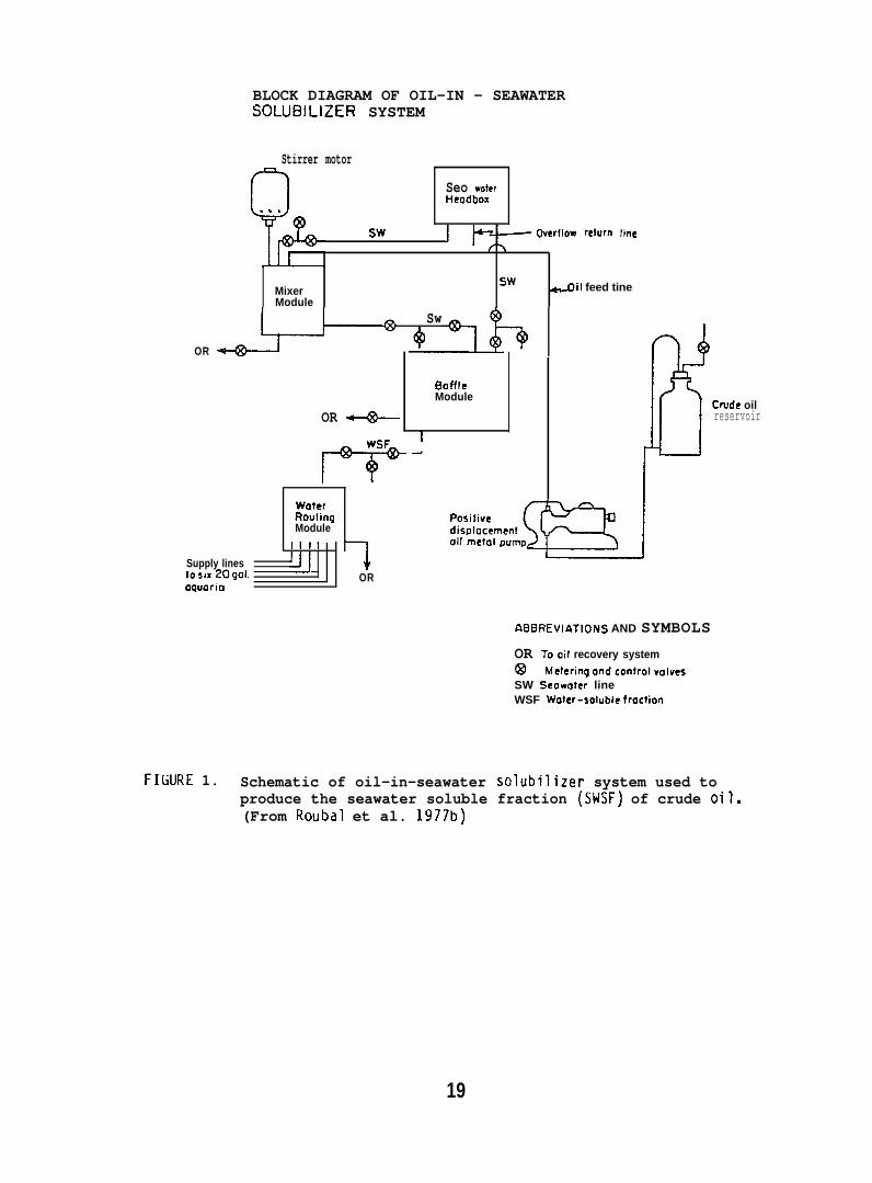
5.1.2 Accumulation of Petroleum Hydrocarbons by Fish Exposed to SeawaterSoluble Fraction (SWSF) of Prudhoe Bay Crude Oil (PBCO)
Coho salmon (11-19g; purchased from DomSea Farms, Bainbridge Island,WA) and starry flounder (Platichthys stellatus) (32-186g; captured fromPuget Sound) were exposed to a SWSF of PBCO in seawatera, at 10”C, undercontinuous flow-through bioassay conditions. The apparatus used isdepicted in Figure 1. The concentration of total soluble hydrocarbonsin flowing seawater delivered from the solubilizer was 5 ppm as measuredby capillary gas chromatography (GC). Hydrocarbons were analyzed bygas chromatography-mass spectrornetry (GC/MS). SWSF delivered from thesolubilizer was diluted with seawater to produce a final hydrocarboncontent of 0.9 + 0.1 ppm (Fig. 1). Coho salmon were exposed to the0.9 + ().1 ppm SESF for a 6-week period, followed by 6 weeks of holdingexpo~ed fish in oil-free seawater to evaluate deputation.
Muscle tissue of salmon was analyzed for concentrations of SWSFhydrocarbons starting one week after the beginning of the exposureperiod. Excised tissues were thoroughly rinsed with 3.5% saline andaliquots were digested at room temperatures in 4 N sodium hydroxide.The digests were analyzed for hydrocarbons using capillary GC. Similaranalyses were made on samples of starry flounder muscle gills and1 iver from 10 fish. (For further details see Roubal et al. 1978. )
5.1.3 Metabolism of NPH by Coho Salmon
Four coho salmon (16U + 35g; purchased from DomSea Farms, BainbridgeIsland, WA) maintained at 10°~0.50C were force-fed 74.6 uCi of 3H-NPH
a Seawater salinity was 27-30°/uo in all chemistry studies.
18
BLOCK DIAGRAM OF OIL-IN - SEAWATERSOLUBILIZER SYSTEM
Stirrer motor
Seo vfaterHeodbox
Sw J Gerflow re~um line
SwMixer ail feed tineModule
Sw
OR
OR ~
Wsl
m
WoterRttut ingModule
1111
Supply lines J[jto SIX 209a1. ORoquario
—
OoffleModule
I-4 (5 Crude oil
reservoir
ABBREVIATfONS AND SYMBOLS
OR Tooil recovery system@ hieteringondcontrol~olvesSW Seowoter lineWSF Woter-soluble froction
FILURE 1. Schematic of oil-in-seawater solubilizer system used toproduce the seawater soluble fraction (SWSF) of crude oil.(From Roubal et al. 1977b)
19
(sp. act. 85 mCi/mmole) dissolved in salmon oil. After 16 hr, thelivers and gall bladders were removed and pooled for analysis. Tissueswere homogenized in distilled water, saturated with sodium chloride,and proteins were precipitated with acetone. Metabolizes were extractedinto ethyl acetate, the extract dried under nitrogen, and the residuewas redissolved in methanol. Metabolize extracts were chromatographedby high-pressure liquid chromatography (HPLC). (For further detailssee Collier et al. 1978. )
5.1.4 Naphthalene and Its Metabolizes in Fish Skin and Mucus
Rainbow trout (Salmo gairdneri) (150 + 50g; purchased from TroutLodge, Puyallup, WA), held at 8°C were eit~er force-fed 74,6uCi of[1,4,5,8,-3H] NPH (SP. act. 83.3 mCi/mmole) dissolved in 250A11 ofsalmon oil or injected i.p. with 4UJ1 of oil containing 94.6~Ci of3H.NpH. Starry flounder (100 + 20g; captured from Puget Sound held
iat 12°C were force-fed a gelat~n capsule containing 87 flCi of H-NPH(sp. act. 198 mCi/mmole) dissolved in salmon oil. Concentrations ofNPtl and total metabolizes in skin and mucus were determined by digestionof the sample at room temperature in hexane and 4 N NaOH. Concentrationof the NPH was determined from radioactivity in the hexane layer, andconcentration of total metabolizes was determined from the aqueouslayer. Concentrations of NPH and its metabolizes in the skin werecompared with those in the liver. (For further details see Varanasiet al. 1978 and Collier 1973.)
5.1.5 Accumulation and Biotransformation of NPH by Flatfish
Sexually immature starry flounder and rock sole (both species:82 + 30g) were captured near the mouth of the Columbia River and atPoi~t Puny in Puget Sound, respectively, and were maintained atexperimental temperatures of 12 + 1°C in flowing seawater for a periodof 2 wk prior to treatment. Fisli were fed daily to satiation on amixture of earthworms and euphausiids.
Force-feeding study. Test fish of both species were force-feda gelatln capsule (No. 5) containing 56 ~Ci of [1-3H]-NPH (sp. act.198 mCi/mmole) dissolved in 25 ,u1 of salmon oil. The fish were thenplaced in aquaria supplied with flowing seawater at 12”C. Three tosix fish were analyzed at 24, 48, and 168 hr after the initiation ofexposure. Rock sole were analyzed also at 6 wk. Fish were not fedduring the first week after the initiation of NPH exposure.
Injection Study. Starry flounder were injected i.p., with 25 U1of salmon 011 containing 56 _uCi of the [1-3H]-NPH (sp. act. 198 mCi/mmole).The fish were held at 12°C, and sampled at 24 and 168 hr after injection.
Sample collection. Epidermal mucus and skin were collected, andsamples of muscle, liver, brain, gills, blood, kidney, stomach, intestine,and bile were also collected.
20
Analytical methods. Radioactivity associated with both NPH andtotal metabolizes in each tissue (A400 mg) was determined by digestionin hexane-sodium hydroxide. Dry weight of each tissue was obtained byfreeze-drying and the values expressed as percent of wet weight of tissue.Lipid content of liver, muscle, and skin of starry flounder and rock sole(Lepidopsetta bil ineata) were also determined.
Data were statistically analyzed using Student’s t-test. Also,rates of decline of NPH and metabolize concentrations in tissues wereobtained by assuming a lognormal distribution and describing the decayof concentration by y = ax-b.
NPH metabolizes were isolated from liver, muscle, skin, and bileof the exposed rock sole and starry flounder; the samples were homogenizedin methanol and then extracted twice with hexane to remove NPH, followedby extraction with a mixture of boiling methylene chloride :2-propanol:water(75:25:2, v/v/v) and twice with boiling ethanol :diethyl ether (50:50, v/v)to remove metabolizes.
Individual classes of metabolizes were separated via thin-layerchromatography (TLC). Nonconjugated metabolizes were separated usinga solvent system of ~-dioxane:benzene:acetic acid (25:90:4, v/v/v).Conjugated metabolizes were separated by TLC using a solvent systemconsisting of the upper phase of I-butanol :concentrated ammoniumhydroxide:water (80:20:100, v/v/v). Nonradioactive standards added toeach sample allowed visualization of individual classes of metabolizesafter staining with color producing reagents.
After the determination of the position of the various metabolizesthe adsorbant was scraped from the chromatograms in 5 mm bands andradioactivity in each band was determined. (For further details seeVaranasi et al. 1979, 1981.)
5.1.6 Effect of Temperature on Disposition of NPH and Its Metabolizes in Fish
(a) Coho salmon (150 + 50 g; purchased from DomSea Farms, BainbridgeIsldnd, WA) maintained at 4–and 10*C were force-fed 5.55NCi NPH (sp. act.5.1 mCi/mmole) to determine the effect of temperature on the amount of[1-14C] NPH incorporated into selected organs. After 8 and 16 hr thefish were sampled and the brain, liver, kidney, gallbladder and gutwere removed along with samples of blood, dark muscle and light muscle.To analyze for NPH and total metabolizes in the fish, the formic aciddigestion/hexane extraction method was used (see Section 5.1.1).
(b) Sexually immature starry flounder from the same group of fishused in Section 5.1.5 were held at 4 + l°C in flowing seawater for 2 wkbefore the experiment was conducted. ‘Fish were fed daily (to satiation)a mixture of earthworms and euphausiids.
21
mmoSix
out
e) d“fish
ined
The fish were force-fed 56~Ci of [1-3H]-NPH (sp. act. 198mCi/ssolved in 25 fil of salmon oil, but were not fed thereafter.were sampled at 24 and 168 hr after force-feeding.
NPH and its metabolizes were analyzed by the same proceduresin Section 5.1.5. (For further details see Varanasi et al. 1981a. )
5.1.7 Uptake and Metabolism of Sediment-Associated Aromatic Hydrocarbonsby Flatfish
English sole (62 + 22 g; captured from Puget Sound) were held inflowing seawater {12.0–+ 0.5”C) and fed a diet of minced clams for twoweeks. The feeding was–stopped three days prior to the initiation ofexperiments.
Oil-contaminated sediment (1%, v/v) was prepared as described byMcCain et al. ~)1978 except that 3.6 mCi 3H-BaP (sp. act. 0.83mCi/mmole)and 0.14 mCi 1 C-NPH (sp. act. 167 mCi/mmole) were dissolved in PBCOprior to mixing with the sediment. The oil-contaminated sediment wasplaced in a 17-1 glass aquaria to a depth of 5-6 cm where it was allowedto stand in flowing seawater (20 I/day) for 24 hr (day 1).
Six fish were placed in the experimental tank for a 24-hr exposureon day 2, three fish were sampled on day 3, and the remaining 3 fishwere placed on clean sediment in flowing seawater for 24 hr. Immediatelyafter the first six fish were removed on day 3, five additional fishwere placed in the experimental tank. These fish were exposed to theoiled sediment for 168 hr before sampling on day 10.
Samples of sediment and sediment-associated water (SAW) were takenfrom 2 cm below the sediment/water interface. The wide end of a glasspipette was vertically inserted into the sediment while the tip of thepipette was covered. After positioning the pipette, the tip wasuncovered to allow sediment and SAW to rise within the pipette. Thesample was carefully transferred to a vial andafter the suspended particles had settled.
Samples of wet sediment, unfiltered and f’SAW, and samples of gill, skin, muscle, blood,and intestine were analyzed for total radioact’
the SAW was decanted off
ltered (0.45A, millipore)liver, bile, stomach,vity (3H and 14C).
Ethyl acetate extracts of sediment, SAW, and liver and bi?e--before and after enzymatic hydrolysis with~-glucuronidase or sulfatase--were analyzed by TLC for parent BaP and its metabolizes. Four solventsystems were employed for TLC analyses: Solvent system A (toluene:ethanol, 9:1, v/v) was used for separation of nonconjugated BaP metabolizes;solvent system B (Plate was developed up to 6 cm in ethyl acetate andthen redeveloped in the same direction with toluene:ethanol, 100:3, v~v)was used for separation of nonconjugated NPtl metabolizes, solventsystem C (upper phase of concentrated ammonium hydroxi de:water:n-butanol ,“1O:50:4O, v/v/v) was used for separation of conjugated NPH metabolizes,
22
and solvent system D (hexane:diethyl ether, 95:5, v/v) was used forseparation of nonconjugated BaP metabolizes from BaP and liver lipids.Assessments of NPH and total NPH metabolizes in sediment, SAM, bile,and liver were also made by a solvent partitioning method using hexaneand sodium hydroxide. In addition, ethyl acetate-soluble metabolizesfrom bile before and after enzymatic hydrolyses were analyzed by HPLC.
Protein from the aqueous phase of liver homogenates was pelletedby centrifuyation, followed by extraction with acetone and diethylether, dryed, and then solubilized to determine radioactivity that wasnot extractable. (For further details see Varanasi and Gmur 1981. )
5.1.8 BaP Metabolism by English sole
English sole (Parophrys vetulus) (742 17g; captured from PugetSound), held at 12°C in flowing seawater , were force-fed 3H-BaP (2 mg/kgbody wt) and liver, muscle and bile were analyzed for BaP and itsrnetabolites by methods described above (Section 5.1.7). Three fishwere sampled at 8, 16, 24, 48 and 168 hr after force feeding BaP.(For further details see Varanasi and Gmur lY81b. )
5.1.9 Binding of BaP Intermediates to DNA Catalyzed by Liver Enzymesof t~sh
Starry flounder (131 + 42g; captured near the mouth of the ColumbiaRiver) and English sole (1~5 + 33g; captured from Puget Sound) wereinjected intraperitoneally wi~h 10 mg/kg of BaP, 3-methyl cholanthrene(MC), or PBCO, in corn oil. Control fish in these studies were untreated,because there was no detectable difference in the binding of metabolicallyactivated 3H-BaP to DNA when liver enzymes from untreated or corn oil-treated fish were used. The fish were sampled 24 hr after injectionand supernatants of liver homogenates (10,000 x g) were prepared. Ratliver supernatants were prepared in the same manner. The influence of anumber of parameters, such as substrate concentration, temperature, reactiontime, and concentrations of cofactors were tested to obtain optimumconditions for in vitro binding assays. The standard reaction mixturecontained: 2 m~o~added in 2.5 ml of 0.02 M phosphate buffer(pH 7.4); 0.75 mg NAL)PH added in 0.1 ml of 0.1 M EDTA (pH 7.4); and0.2 ml of the 10,000 x g supernatant (5 mg protein). The reaction wasstarted by adding 5 nmoles of BaP in 50 Ml of ethanol. The mixturewas incubated in the dark for 15 min at 250C when fish liver supernatantwas used, and at 37°C when rat liver supernatant was used. DNA wasisolated from the reaction mixture by extraction with phenol saturatedwith phosphate buffer followed by ethanol precipitation.
BaP metabolizes were formed by incubating liver superndtants with3H-BaP under the conditions described above, without the addition of DNA.The mixture was extracted with ethyl acetate (2 x 6 ml) and radioactivityin aqueous and organic phases was determined. Separation and quantificationof ethyl acetate-soluble metabolizes were carried out using both TLC andHPLC. (For further details see Varanasi and Grnur 1980 and Varanasi et al.1980. )
23
5.1.10 AHM Activities in Different Species
Fish, crabs, and snails were collected during NOAA cruise No.MF-77-l of the Miller Freernan. Livers from fish, visceral organs fromcrab, and whole-were frozen and held at -60°C during transit(from Alaska to Seattle) and in the laboratory prior to analyses. Thespecimens were collected from January 25 to February 10, 1977, inareas northeast of Kodiak, Alaska.
Analyses of AHtl activities were also carried out on livers ofPacific cod (Gadus macrocephalus) which were found to have pseucfobranchialtumors. The-were collected during a Miller Freeman cruise inAlaska, and were part of another OCSEAP pr~(Research Unit 332).
Specific activities of AHM were measured using 3H-BaP as thesubstrate for the AI-IN. The procedures employed were a modification ofthose by DePierre et al. (1975). The temperature and pH of the AHNassay mixtures were optimized for the fish (i.e., 25°C and pH 7.5) andassays were performed with NADPH (tetrasodium salt; Sigma Chemical Co.)rather than with a NADPH-generating system.
A typical assay reaction in 2.1 ml contained 0.67 mM NADPH, 1.4 mMMgC12, 50 U1 of enzyme source (25 mg protein/ml), 20 U1 of an acetonesolution of 3.2 mN tritiated benzo(a)pyrene (0.96 uCi), and 6U m~l TrisI-ICI buffer (pI-l 7.5). The enzyme source was a 20% (wt/vol) homogenateof tissue (e.g., liver) in cold 0.25 !4 sucrose solution that was separatedas a supernatant fraction from cellular debris by centrifugation at 9,000x9(or 10,OLNlxg) for 20 min. Duplicate reaction mixtures (in open culturetubes under subdued light) were shaken for 10 min at 25° before initiationof reactions by the addition of the 3H-BaP. Incubation time was 20 min.During work up, two hexane extractions were employed, in contrast to asingle extraction according to DePierre et al. (1975). This procedureresulted in better agreement among assays and less variation in blanks.
Additional analyses of hepatic AHM activities were conducted withthe use of NPH as the substrate (Nilsson et al. 1976). Protein contentsof the enzyme sources were determined by the method of Lowry et al. (1951).
5.1.11 Uptake, Metabolism and Toxicity of Hydrocarbons in Invertebrates
(a) Sperm and eggs from artificially spawned mussels, Mytilusedul is, (CO1 lected from Puget Sound) were placed in 400 ml o~tercontaining 3H-NPH (sp. act. 198mCi/mmole) at concentrations of 100, 10,and 1 ppIJ and in control seawater at 11 + l“C. Each test and controlcondition was run in triplicate. Sample~of eggs and/or larvae wereremoved from each corltainer at 0.5, 1, 2, 3, 6, 12, and 24 hr, andwere preserved in 5;: buffered formalin solution. Water samples weretaken at each time period for determinations of the NPH concentration.Specimens were counted and the developmental stages identified.
(b) Sperm and eggs of oysters, Crassostrea gigas, (collectedfrom Puget Sound) were separately exposed to a seawater solution of3H-NPH (sp. act. 198 mCi/mmole) at two concentrations (10 and 1 ppb)
24
for 15 min after which the complementing gametes were added. In addition,non-exposed gametes of both sexes were introduced into seawater containingNPH at concentrations of 10, 1, and 0.1 ppb and one control inuncontaminated sedwater. Larval samples were taken at intervals of upto 48 hr and were preserved in 5% buffered formalin. Water sampleswere taken at each time point to determine the concentration of NPH.The eggs and/or larvae in each sample were counted and the developmentalstages identified.
(c) One-year old spot shrimp (collected from Puget Sound) wereexposed in flow-through aquaria for 7 days to the SWSF of PBCO at anaverage concentration of 110 ppb as determined by GC. The SMSF wasobtained from the solubilizer (Fig. 1). The animals were washed,extracted, and the extracts were analyzed for accumulated hydrocarbonsby GC/llS. Abdomens were separated from thoracic segments and analyzedseparately. (For further details see Sanborn and Malins 1980. )
(d) Newly metamorphosed larval stages of spot shrimp and Dungenesscrab, Cancer magister, (collected from Puget Sound) were exposed in flowingseawat-~~o 8-12 ppb of [1-14C] NPH (sp. act. 5 mCi/mmole) or[1-14C] NPH c~mplexed with bovine serum albumin (BSA). The spot shrimpand Dungeness crab were hatched in the laboratory from ovigerous females.The shrimp larvae hatched each day were held in separate holding tanksand fed brine shrimp. Exposure periods varied from 12 to 24 hr anddeputation studies were carried out for periods of up to 132 hr. Larvaewere examined for both [1-~4C] NPH and its metabolizes. Total radioactivityin the animals was determined, and total metabolizes of [1-14C] NPH weredetermined by enploying formic acid/hexane extractions (see Section 5.1.1).(For further details see Sanborn and Malins 1978. )
(e) Adult spot shrimp were placed in an 80ppb seawater (10+ l“C)solution of 3H- (sp. act. 198 mCi/mmole) and 14C- (sp. act. 3.67 m~i/mole)labeled NPH; the 3H/14C ratio was 48:1. The concentration was maintainedunder flow-through conditions. The animals were removed after I(J hr;washed, the thorax and abdomen separated, and the tissues analyzed byHPLC. (For further details see Sanborn and Malins 1980. )
(f) Stage I spot shrimp larvae that had been hatched in the laboratorywere exposed to an 18 ppb seawater solution of 3H-NPH (sp. act. 198 mCi/mmole)at 10°-120C. After 10 hr the larvae were washed, weighed, extractedand analyzed for NPH metabolizes by HPLC.
!5.1.12 Food Chain Transfer of 2,6-Dimethylnaphthalene (2,6-DMN) toSea Urchins via Algae
The marine algae, Fucus distichus, and the green sea urchin(Strongylocentrotus dro=i~re collected local ly. The Fucuswas exposed to 2,6-UFIN as follows: Two hundred grams of the wetalgae was rinsed in seawater and then introduced-into an 68 1 aquariumcontaining seawater and 10 mCi of [2,6-3H]-u!IN (sp. act. 2 Ci/mmole)in 0.5 ml of ethanol. Samples of the seaweed were removed at 5 hrintervals over a period of 35 hr, rinsed well, divided into aliquots,then frozen at -2U”C until analyzed for incorporated tritium.
25
A fresh batch of the seaweed was harvested and added tocontaining the 3H-2,6-ilMN. After 25 hr, the time of maximumincorporation, the exposed Fucus was removed, rinsed well inand then placed in an aquar~ontaining 6 sea urchins (170new batch of the 2.6-DMN-treated Fucus was added 24 hr later
an aquariumtritium-seawater,:4g). Ato the
sea urchin aquarium, after remova~unconsumed Fucus. This sequenceof presenting exposed Fucus to the sea urchins at 24 hr intervals wasrepeated throughout the 14 day experiment.
After three days of feeding on the 2,6-UMN-treated Fucus, 3 seaurchins were removed, rinsed, and frozen at -20°C until analyzed. Theremaining 3 sea urchins were removed after 14 days of feeding on thetreated Fucus and handled similarly.
The exoskeleton of sea urchins was pulverized and extrdcted withhot methanol and filtered. Aliquots were analyzed for tritium byscintillation counting. Half-gram portions of the powdered exoskeletonresidue remaining after methanol extraction were also digested intissue solubilizer and then assayed for tritium.
Fucus samples and aliquots of homogenized gonadal and digestive tractwere analyzed for 2,6-INN by digestion in hexane-sodiurn hydroxide.Metabolizes of 2,6-CM were analyzed by extracting soft tissues withhot methanol-diethyl ether, followed by TLC of tnt? extracts. Sulfatefractions were removed from TLC plates and extracted with hot methanol.After removal, the sulfate fractions were treated with aryl sulfataseat 37°C in buffer (pH 5.0, 0.2 M acetate). The digests were extractedwith ethyl acetate and analyzed by TLC. The 3- and 4-hydroxy derivativesof 2,6-DNN, isolated as a single banddeveloped 5 times with toluene.
, were resolved on a TLC plate(For further details see Plalins and
Roubal 1982. )
5.1.13 Biological Fate of Metals in Fist~
Exposure conditions for metals studies
Nat(3U
Water-immersion Studies. Coho salmon (2U0 + 20 g; obtained fromonal Ilarine Fisheries Service, Manchester, WI) and starry flounder~ 15 g; captured from near tile mouth of the Columbia River) were he”d
at experimental temperatures of 4° or 10”C for a period of two weeks,and then exposed to either seawater-borne lead-210 or cadmium-10Y(150 ppb) under partial flow-through conditions.
The fish were exposed to either lead or cadmium for a period oftwo weeks at each temperature then three or four fish per exposuregroup were sampled. At the end of the two-week period, remaining fishwere placed in control seawater for deputation and sampled after 7 and37 days. A group of control fish of similar weight was kept underidentical conditions in control seawater. Concentrations of lead dndCddi.lium in control sedwater were less than 5 and 2 ppb, respectively.
Injection Studies. Coho salmon (80 + 5 g) were injected intravenously(i. v.~ with32 + 2 ug of metals as either–lead nitrate mixed with 210 Pbor cadmium chlo~ide mixed with log Cd dissolved in 250 ,u1 of tris buffer(pH 7.2). A similar experiment was carried out with fish which wereexposed to 150 ppb of nonradioactive cadmium or lead for 2 wk prior toiv. injection of radiolabeled metals to determine if prior exposureto metals caused any alterations in radioactivity associated with metalbinding proteins.
Analytical methods for metals studies.
Samples of mucus, skin (with scales), scales, skin blood, ills,liver, and kidney were obtained and concentrations of 210 Pb or !09 Cdwere determined in these tissues by liquid scintillation spectrometry.Concentrations of total lead or cadmium in these tissues, expressed ona wet weight basis, were obtained from ratios of 210 Pb/Pb and 1°9 Cd/Cdin stock solutions used in the exposures. Concentrations of nonradioactivelead and cadmium in seawater and fish tissues were determined by LaucksAnalytical Laboratories (Seattle, Washington).
Samples of liver and kidney of fish from the injection study werehomogenized and cytosol obtained. Three samples were chromatographed ona column packed with Sephadex G-75 superfine. The radioactivity in theeluted fractions was determined to assess the distribution of metalsbound to various protein fractions. The protein concentration in eacheluant was determined by a modified method of Lowry et al. (1951).(For further details see Varanasi and Markey 1978 and Reichert et al.1979. )
5.2 Pathology
5.2.1 Effects of Petroleum on Disease Resistance
Acquisition, Handling, and Exposure of Test Animals. Test animalsfor disease resistance studies were obtained from the followinci sources:juvenile coho salmon were from the Issaquah Hatchery of the WashingtonDepartment of Fisheries, from the Willard National Fish Hatchery,Cook, Washington and from Sashin Creek, Little Port Walter, Alaska.The Sashin Creek fish were received as eyed eggs and reared at theNorthwest and Alaska Fisheries Center (NWAFC), Seattle, Washington.
.
Juvenile rainbow trout were from a stock maintained at the NWAFC andwere the progeny of fish originally obtained from the Spokane Hatcheryof the Washington Department of Game. Adult flat.fish were captured byotter trawl and juvenile flatfish collected by beach seine in Puget Sound;adult spot shrimp were collected by pot fishing in Puget Sound.
All experimental animals were held under laboratory conditions forat least 2 wk before the start of testing. The salmonid fish were fedan Oregon moist pellet (OMP) diet at 2% of their body weight, and theflatfish were fed a mixture of minced clams and krill. Spot shrimpwere maintained on a diet of fish offal.
27

Several different methods were used to expose fish and crustaceato petroleum hydrocarbons. Salmonids were exposed both via the diet,by mixing (NIP with PBCO as described by Hodgins et al. (1977), andvia flow-through exposure to the SWSF of P13C0 (Roubal et al. 1977b).Flatfish and spot shrimp were exposed to P13C0 or Cook Inlet crude oil(CICO) by maintenance on oi 1 -cont~mi nated sediment (McCain et al. 1978).Sediment, water, and selected tissues from test animals were analyzedfor total extractable petroleum hydrocarbons (TEPH) by the methodsdescribed by Malins et al. (1980).
In vivo Assays of Irnmunocompetence.
(a) Throughout these studies oil-exposed animals were comparedto controls for their ability to survive a laboratory challenge by themarine fish pathogen Vibrio anguillarum. Several different bacterialisolates were used, the taxonofliic identity of each was confirmed byconventional cultural and biochemical tests, and by deoxyribonucleicacid hybridization.
As needed, bacteria were cultivated overnight in either Trypticasesoy broth (B13L) or brain heart infusion (Oifco) on a reciprocal shakerat room temperature (ca 23”C). Both media were supplemented with anadditional 10 my/l NaC1. Ten-fold serial dilutions were prepared in0.15 M NaCl and the numbers of viable bacteria were estimated by standardspread-plate technique. For challenge, fish were transferred from theexposure facilities at the I’lukilteo Field Station to the Disease IsolationLaboratory at the NWAFC. Groups of 5 to 20 test animals were placed inindividual 38 1 aquaria containing aerated seawater (salinity 26-30°/00)or freshwater maintained at the exposure temperature (10-15°C). Fishwere challenged with selected test concentrations of bacteria by eitheri.p. injection (flatfish) or by subcutaneous injection at the posteriorinsertion of the dorsal fin (salmonids). Shrimp were challenged byinjection of bacteria at the suture separating the thorax and abdomenon the dorsal surface. Aquaria were checked daily for mortalities fora minimum of 10 d after challenge. Tissues from dead fish and shrimpwere cultured for bacteria; only in those cases in which V. anguillarumwas reisolated in pure culture were the deaths attributed–to the bacterialchiillenge.
LD50 values (i.e., the number of bacteria that kill 5VL of theanimals) and their 95% confidence intervals (C.I.) were calculated bylogit analysis (Cox 1970). The LD50 values were statistically comparedusing a method similar to that described by Litchfield and Hilcoxon(1949). The 95% C.I. for the difference between any two LD50 valueswas calculated and the hypothesis that the two values were equal wasrejected (~=0.05) if this C.I. did not contain the point zero. Inthose cases in which inadequate partial kills prevented calculation ofan LD50 by the logit procedure, the method of Reed and Muench (1938)was used. In addition, statistical comparisons between the percentmortality occurring in oil-exposed and control groups challenged withthe same number of bacteria were made using Chi-square analyses.
(b) An additional test was conducted to assess the effect of oilexposure on the adaptive immune response of fish. Groups of P6C0-exposedand control rainbow trout were vaccinated with a heat-killed vaccineprepared from!. anguillarum and subsequently challenged with varyingconcentrations of the llvlng organisms.
In vitro Assays of Immunocornpetence.
(a) Numbers of antibody-forming cells in anterior kidney andsplenic tissues of PBCO-exposed and control rainbow trout were determinedby a modified Jerne plaque assay (Chiller et al. 1969). Fish wereimmunized against trinitrophenol conjugated with lipopolysaccharide(LPS) from Escherichia CO1 i B. Plaque formation was assayed on alawn of sheep red blood~ls (SRBC) which were coated with LPS, in asoft agarose matrix. Rainbow trout sera, frozen and thawed one time,were used as a source of complement.
(b) Serum agglutinating antibody levels were compared betweenoil-exposed and control fish by the microdilution technique (klicrotiter,Cooke Engineering Company). Fish were immunized against ~. anguillarumand 21 d later 2-fold serial dilutions of serum were tested for specificagglutinins. Fish were held at 15°C to facilitate antibody formation.
(c) For polyclonal lymphoid cell activation assays, peripheralblood leukocytes from oil-exposed and control fish were incubated withpurified protein derivative (PPll) prepared from the tubercle bacillus.Activity was quantified by measurement of plaque-forming ability onlawns of SRBC’S in agarose.
(d) The degree of mitogenic stimulation was compared between PBCO-exposed and control fish under assay conditions previously describedin detail by Etlinger et al. (1976). Briefly, leukocyte culturesprepared from splenic tissue were assayed for lymphocyte proliferationfollowing incubation in the presence of the plant-derived mitogenicsubstance concanavalin A-(Con A). Stimulation was quantified by measurementof the incorporation of ‘H-thymidine in the proliferating cellular DNA.
(e) Early in the course of these in vitro investigations ofimmunocompetence it was noted that the ~l~sed rainbow trout hadreduced spleen sizes compared to those of the controls. Since antibody-forming leukocytes have been previously identified as one of the principalcellular components of the spleen (Chiller et al. 1969), this conditionwas further examined. Spleen-weight to body-weight ratios were measuredand cellular composition with respect to total numbers of erythrocytesand leukocytes-thrombocytes were determined. These same hematologicalparmeters were also determined for anterior kidney tissue homogenatesand peripheral blood.
Effects of Corexit 9527 on Disease Resistance. Juvenile coho salmonwere exposed for 30 mln at 15 + o to 30 ppm (v/v) Corexit 9527 inseawater containing various coficentrations of V. anguillarum. Controlfish were similarly exposed in seawater contaiiiing either bacteria or
Corexit only. Fish were then transferred to individual 38 1 aquariacontaining aerated fresh seawater and mortality was monitored for 10 d.All dead fish were examined by bacterial culture and death was consideredto be due toV_. anguillarum only when the bacterium was reiso?ated inpure culture.
5.2.2 Pathological Changes in Flatfish from Exposure to Oil-ContaminatedSediment
Acquisition and Handling of Test Animals. English sole, rock sole,or starry flounder were captured with otter trawl or beach seine fromeither the mouth of the Columbia River or in Puget Sound. Fish weretransported to Mukilteo, Washington and held for 7-14 days prior to use.All fish were determined to be actively feeding before experiments wereinitiated. Two types of sediment (either high-silt or high-sand content)contaminated with 0.2, 0.5, or 1.0% (v/v) PBCO were tested; both juvenileand adult stages were examined and exposures ranged from 2 wk to 4 mo.The general approach for all experiments was similar and is outlinedbel OW.
Experimental Design. Fish were randomly assigned to a test orcontrol group, weighed, measured, and cold-branded (Fujihara and Nakatani1967) for individual identification. Prior to each test, samples forhematology and histopathology were collected from 6 to 10 controlfish. Hematological tests included hematocrit, hemoglobin, .and totalred blood cell and leucocyte counts using standard techniques asdescribed by Blaxhall and Daisley (1973). Tissue samples (gill, skin,fin, intestine, kidney, liver, and spleen) were placed in phosphate-buffered formalin. In addition, samples of liver, skin and musclewere placed either in glass vials or aluminum foil and stored at -20”Cfor subsequent chemical analysis. Sediment samples (400 g) for analysisof TEPH were collected and stored at -20°C.
At intervals of 1 to 4 wk, depending on the length of the experiment,sediment samples and samples of 3 to 6 test and control fish were collectedusing the same procedures. The remaining fish were weighed and measuredand returned to the aquaria.
Fish were fed to satiation, twice daily five days per week, amixture of clams, euphausids, and live earthworms. Tanks were monitoreddaily for mortality. Water flow in the 200 1 test and control aquariawas maintained at 3 l/rein; average water temperature, 8.5”C; averagesalinity, 27 O/OO; light was maintained on a 121:120 schedule.
Sediment was collected at 2 sites near Sequim, Washington. The high-sand type (approximately 99% of particles >0.07 mm diameter) was fromPort Williams; the high-silt type (48% of particles <0.07 mm), from alagoon adjacent to the Battelle Northwest Laboratory. PBCO and sedimentwere mixed together for 3(J min in fiberglass-lined cement mixer atconcentrations of 0.2, 0.5, or 1.0% v/v.
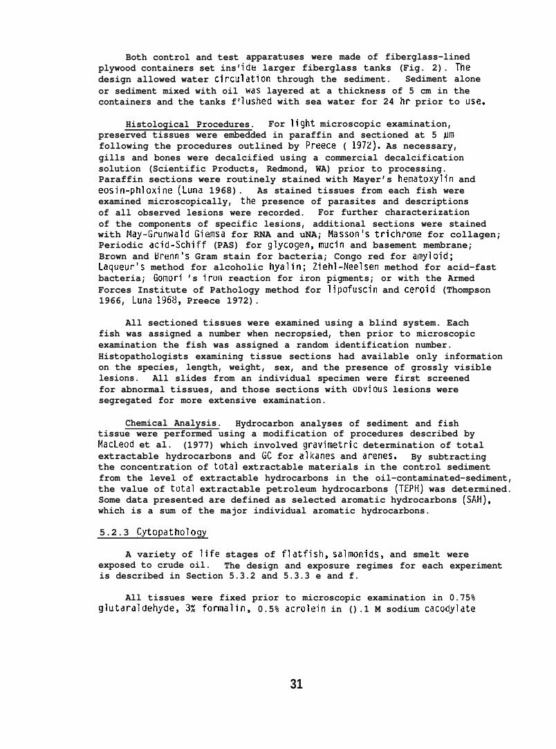
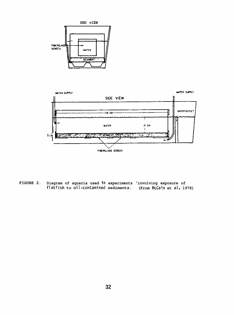
Both control and testplywood containers set ins’design allowed water circu”or sediment mixed with oilcontainers and the tanks f’
apparatuses were made of fiberglass-linedde larger fiberglass tanks (Fig. 2). Theation through the sediment. Sediment alonewas layered at a thickness of 5 cm in theushed with sea water for 24 hr prior to use.
Histological Procedures. For light microscopic examination,preserved tissues were embedded in paraffin and sectioned at 5 Nmfollowing the procedures outlined by Preece ( 1972). As necessary,gills and bones were decalcified using a commercial decalcificationsolution (Scientific Products, Redmond, WA) prior to processing.Paraffin sections were routinely stained with Mayer’s hematoxylin andeosin-phloxine (Luna 1968). As stained tissues from each fish wereexamined microscopically, the presence of parasites and descriptionsof all observed lesions were recorded. For further characterizationof the components of specific lesions, additional sections were stainedwith May-Grunwald Giemsa for RNA and uNA; Masson’s trichrome for collagen;Periodic acid-Schiff (PAS) for glycogen, mucin and basement membrane;Brown and Brenn’s Gram stain for bacteria; Congo red for amyloid;Laqueur’s method for alcoholic hyalin; Ziehl-Neelsen method for acid-fastbacteria; Gomori ’s irun reaction for iron pigments; or with the ArmedForces Institute of Pathology method for lipofuscin and ceroid (Thompson1966, Luna 1968, Preece 1972).
All sectioned tissues were examined using a blind system. Eachfish was assigned a number when necropsied, then prior to microscopicexamination the fish was assigned a random identification number.Histopathologists examining tissue sections had available only informationon the species, length, weight, sex, and the presence of grossly visiblelesions. All slides from an individual specimen were first screenedfor abnormal tissues, and those sections with o~vious lesions weresegregated for more extensive examination.
Chemical Analysis. Hydrocarbon analyses of sediment and fishtissue were performed using a modification of procedures described byMacLeod et al. (1977) which involved gravirnetric determination of totalextractable hydrocarbons and GC for alkanes and arenes. By subtractingthe concentration of total extractable materials in the control sedimentfrom the level of extractable hydrocarbons in the oil-contaminated-sediment,the value of total extractable petroleum hydrocarbons (TEPH) was determined.Some data presented are defined as selected aromatic hydrocarbons (SAH),which is a sum of the major individual aromatic hydrocarbons.
5.2.3 Cytopathology
A variety of life stages of flatfish, salmonids, and smelt wereexposed to crude oil. The design and exposure regimes for each experimentis described in Section 5.3.2 and 5.3.3 e and f.
All tissues were fixed prior to microscopic examination in 0.75%CJlutdrdldehyde, 3% formalin, 0.5% acrolein in ().1 M sodium cacodylate
31
END vIEWN
+—FlesSCRU N
WATER SUPPLY WAVER sl-=%Y
5 c;.
FIfk5RQASS SCSEOJ
FIGURE 2. Diagram of aquaria used in experiments ‘involving exposure offlatfish to oil-contamined sediments. (From McCain et al. 1978)
32
buffer with 0.02% CaC12.H20, and 5.5% sucrose (Hawkes 1974). Thetissues designated for light microscopy (LM) or for transmission electronmicroscopy (TEM) were post-fixed for 1 1/2 hours in 1% osmium tetroxidein the same buffer, dehydrated in an ethimol series, and embedded inplastic. For LM, sections were cut at either 1.0 or 0.5 Mm. The 1.0 urnsections were stained with Richardson’s mixture or with a PAS reagent(Nevalainen et al. 1972); the 0.5 urn sections were stained with toluidineblue or a trichrome (MacKay and Mead 1970). For TEM, sections werecut with a diamond knife and stained with lead citrate and uranyl acetateand examined with d Philips EM-3(J1 electron microscope. For scanningmicroscopy (SEM)$ samples were dehydrated after the initial fixationscritically point dried, coated with gold-palladiun, and examined withan AMR-1OOO scanning electron microscope.
5.3 Behavior and Physiology
5.3.1 Chemical Analysis of Water, Sediment and Tissue
Water and Sediment Analysis. Water samples for hydrocarbon an 1 sis?1were collected in 100 or 300 ml screw cap glass bottles with Teflon R -
lined lids. The samples were usually extracted on the day of collection;however, if they were to be processed later, 3 ml of concentratedhydrochloric acid was added to eliminate microbial activity and thesamples were stored at 2°C.
Two methods of extraction and analysis were used. In initialexperiments, a recovery standard was added to the 1OO-M1 water samplesand extracted using 5 ml of carbon disulfide. The organic phase wasseparated and evaporated to 0.8 ml, a GC internal standard added,and 8M1 of the extract was gas Ctlromatogrdphed on a l-m long by 2-mmID glass column packed with 0.2% carbowax 1500 on 59/80 mesh carbopack C(Supelco, Belleforte, PA). Nitrogen was used as a carrier gas with aflow rate of 46 ml/min and temperature programming was from 40 to215°C at 15°C/min.
In later studies, water samples were collected in 300 ml bottlescontaining 12 ml of methylene chloride. Immediately after collection,the samples were shaken for 3 min to partition hydrocarbons into themethylene chloride and the samples were stored at 2°C. At the time ofprocessing, recovery Stdndards were ddded and the water samples wereextracted three times with a total of 24 ml of methylene chloride.- Theextract was dried with sodium sulfate and reduced to 0.8 ml in aconcentrator tube. GC internal standards were added and the extractsanalyzed by glass capillary-gas chromatography (characteristics describedby MacLeod et al. 1977). The limit of detection for individual aromaticcompounds in the water sample was 1 ppb.
For both GC methods employed, identities of hydrocarbon componentswere confirmed by mass spectral analysis (for description] of equipmentand characteristics see Malins et al. 1980), and hydrocarbon concentrationwas determined from the total area under the chromatogram. All datawere corrected for extraction efficiency except where expressly stated.
33
For specific analysis of benzene, 1 ml of benzene-seawater mixturewas placed in a 2 ml GC vial, capped, and allowed to stand for 18 to 20 hr.Oneml of air was then removed from the vial headspace and injecteddirectly into a GC. These results were compared with results of benzenestandards prepared by injection of Ml amounts of benzene-methanolsolutions into GC vials containing 1 ml of seawater, which were thenprocessed in the same manner.1
Extraction and analysis of petroleum hydrocarbons in sedimentfollowed a procedure described by Brown et al. (1979).
Tissue Analysis. Analytical procedures for tissue analysis ontrout followed methods of Warner (1976) utilizing alkaline digestion,solvent extraction, and silica gel chromatography. To reduce losses ofvolatile compounds of PBCO, the alkaline digestion procedure was modifiedby adding 6 ml of 4 N NaOH to the 10 g of sample, and the sample digestedat 30”C for a minimum of 16 hr.
Silica gel chromatography Fraction 3 from the modified Warner method,containing triaromatic compounds, was concentrated to 2.0 ml and analyzedusing an Aminco-Bowman spectrofluorometer (American Instrument Company,Silver Spring, Maryland). Dilute solutions of PBCO (0.1 ,ug/ml to10.O~g/ml in methylene chloride:petroleum ether [ZU:8U V/V] were usedas standards for the spectrofl uorometric quantitation of the samples.The maximum excitation wavelength and maximum emission wavelength forPBCO were found to be 262 nm and 364 nm, respectively.
Chemical analysis of surf smelt embryos, salmon liver, and salmonbrain tissues for petroleum hydrocarbons was conducted using methodsdescribed by Malins et al. (1980).
5.3.2 Preparation of Oil-Water Mixtures
Four flow-through systems introduced petroleum hydrocarbons intowater:
(a) The system developed by Roubal et al. (1977b) provided a SWSFof crude oil composed almost entirely of monocyclic aromatic hydrocarbons(Roubal et al. 1978). For several studies a mixture of hydrocarbonsmodeled on the SWSF of fresh PBCO was formulated, and contained 95%(by weight) of those hydrocarbons detected in the SWSF (Table 1).
(b) A method of introducing the “model” hydrocarbon mixture intowater was devised using a water jet eductor (Schutte and Koerting,Cornwells Heights, PA, type 264, 1/2 in, capacity ratio of 0.36).Water was passed through the eductor at a flow rate of 16 l/rein, anda pressure of 5.5 kg/cm2. The hydrocarbon mixture and individual
1 An alternative method for analysis of benzene, especially when benzenewas a component of the total SWSF, was to use carbon disulfide as anextracting solvent. Carbon disulfide is not detected by GC and thusdoes not mask the GC response to benzene.
34
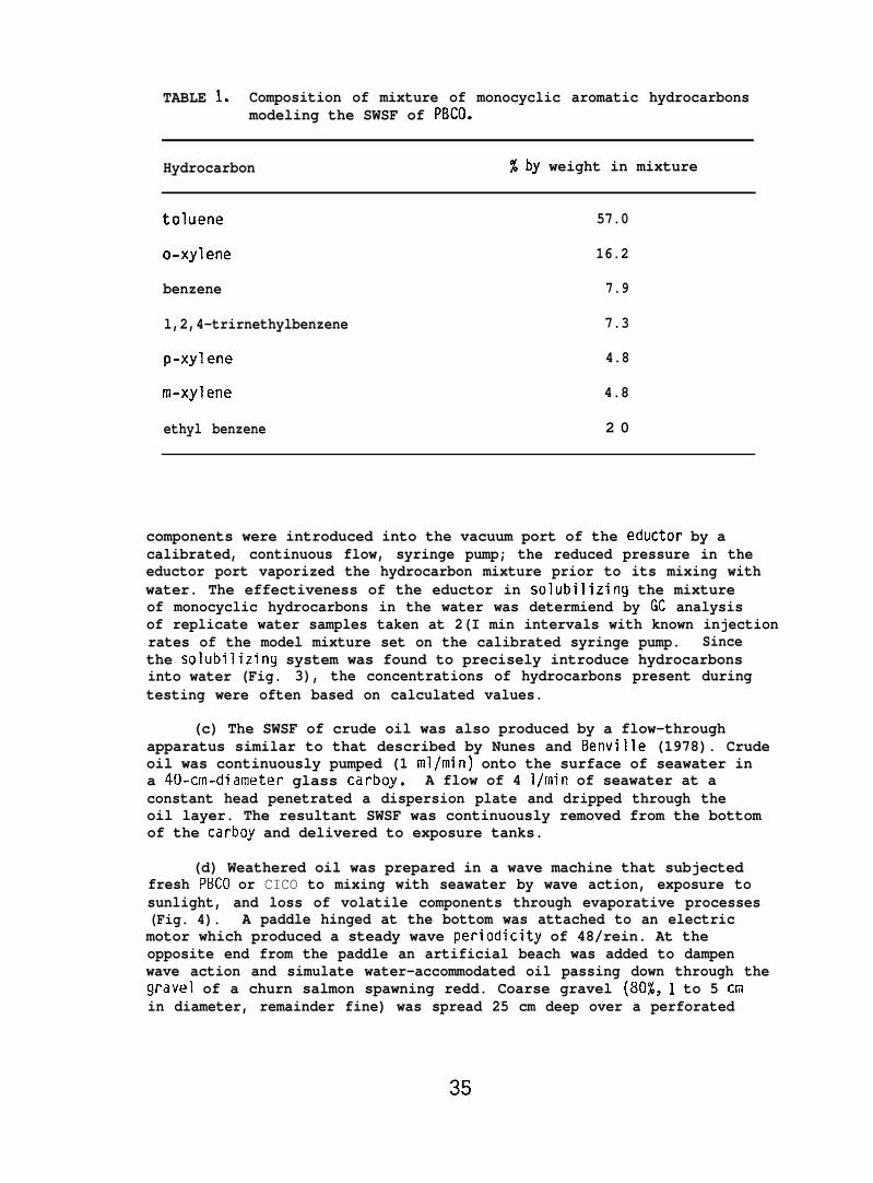
TABLE 1s Composition of mixture of monocyclic aromatic hydrocarbonsmodeling the SWSF of PBCO.
Hydrocarbon %by weight in mixture
toluene 57.0
o-xylene 16.2
benzene 7.9
l,2,4-trirnethylbenzene 7.3
p-xylene 4.8
m-xylene 4.8
ethyl benzene 2 0
components were introduced into the vacuum port of the eductor by acalibrated, continuous flow, syringe pump; the reduced pressure in theeductor port vaporized the hydrocarbon mixture prior to its mixing withwater. The effectiveness of the eductor in solubilizing the mixtureof monocyclic hydrocarbons in the water was determiend by GC analysisof replicate water samples taken at 2(I min intervals with known injectionrates of the model mixture set on the calibrated syringe pump. Sincethe solubilizing system was found to precisely introduce hydrocarbonsinto water (Fig. 3), the concentrations of hydrocarbons present duringtesting were often based on calculated values.
(c) The SWSF of crude oil was also produced by a flow-throughapparatus similar to that described by Nunes and Benville (1978). Crudeoil was continuously pumped (1 ml/min) onto the surface of seawater ina 4U-crn-diameter glass carboy. A flow of 4 l/rnin of seawater at aconstant head penetrated a dispersion plate and dripped through theoil layer. The resultant SWSF was continuously removed from the bottomof the carboy and delivered to exposure tanks.
(d) Weathered oil was prepared in a wave machine that subjectedfresh PBCO or CICO to mixing with seawater by wave action, exposure tosunlight, and loss of volatile components through evaporative processes(Fig. 4). A paddle hinged at the bottom was attached to an electricmotor which produced a steady wave periodicity of 48/rein. At theopposite end from the paddle an artificial beach was added to dampenwave action and simulate water-accommodated oil passing down through thegravel of a churn salmon spawning redd. Coarse gravel (80Z, 1 to 5 Cmin diameter, remainder fine) was spread 25 cm deep over a perforated
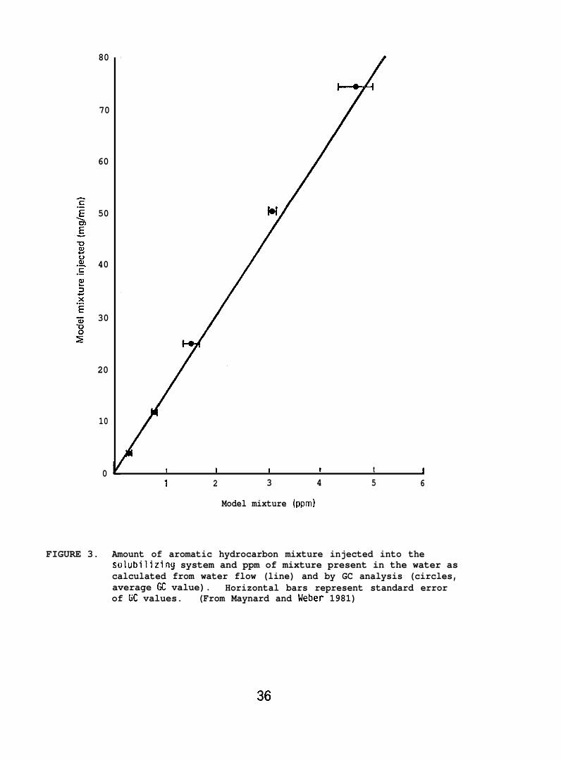
80
70
60
50
40
30
20
10
0 I I I 1 t !1 2 3 4 5 6
Model mixture (ppm)
FIGURE 3. Amount of aromatic hydrocarbon mixture injected into thesolubilizing system and ppm of mixture present in the water ascalculated from water flow (line) and by GC analysis (circles,average GC value). Horizontal bars represent standard errorof GC values. (From Maynard and Weber 1981)
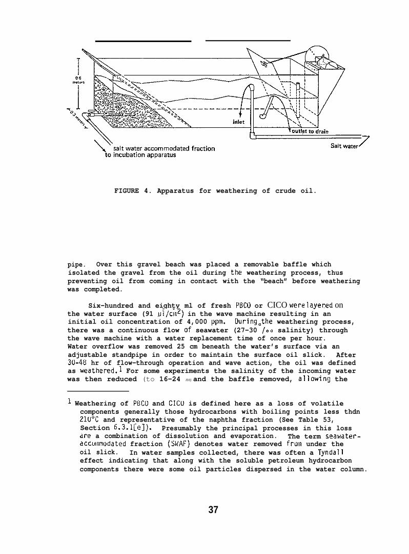
FIGURE 4. Apparatus for weathering of crude oil.
pipe. Over this gravel beach was placed a removable baffle whichisolated the gravel from the oil during the weathering process, thuspreventing oil from coming in contact with the “beach” before weatheringwas completed.
Six-hundred and eighty ml of fresh PBCU or CICO were layered onthe water surface (91 M1/cm2) in the wave machine resulting in aninitial oil concentration of 4,000 ppm. DuringQthe weathering process,there was a continuous flow of seawater (27-30 /.. salinity) throughthe wave machine with a water replacement time of once per hour.Water overflow was removed 25 cm beneath the water’s surface via anadjustable standpipe in order to maintain the surface oil slick. After30-48 hr of flow-through operation and wave action, the oil was definedas weathered.1 For some experiments the salinity of the incoming waterwas then reduced (to 16-24 /00) and the baffle removed, allowing the
1 Weathering of PBCO and CICU is defined here as a loss of volatilecomponents generally those hydrocarbons with boiling points less thdn21U°C and representative of the naphtha fraction (See Table 53,Section 6.3.l[e]). Presumably the principal processes in this lossdre a combination of dissolution and evaporation. The term seawater-accm?wdated fraction (SWAF) denotes water removed from under theoil slick. In water samples collected, there was often a Tyndalleffect indicating that along with the soluble petroleum hydrocarboncomponents there were some oil particles dispersed in the water column.
37
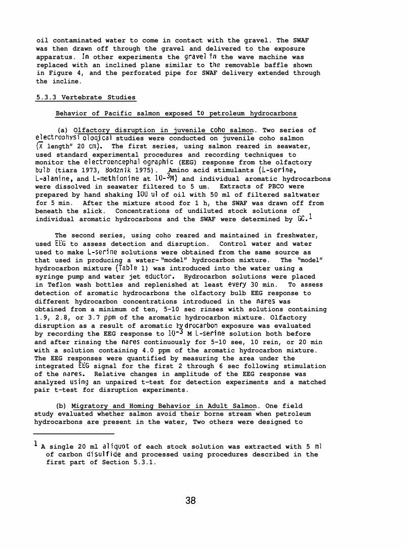
oil contaminated water to come in contact with the gravel. The SWAFwas then drawn off through the gravel and delivered to the exposureapparatus. In other experiments the gravel in the wave machine wasreplaced with an inclined plane similar to the removable baffle shownin Figure 4, and the perforated pipe for SWAF delivery extended throughthe incline.
5.3.3 Vertebrate Studies
Behavior of Pacific salmon exposed to petroleum hydrocarbons
(a) Olfactory disruption in juvenile coho salmon. Two series ofelectroDhYsl oloqical studies were conducted on juvenile coho salmon(~ length” 20 cmj. The first series, using salmon reared in seawater,used standard experimental procedures and recording techniques tomonitor the electroencephal ographic (EEG) response from the olfactorybulb (tiara 1973, &30dznik 1975). Amino acid stimulants (L-serine,L-alanine, and L-methionine at lU-3M) and individual aromatic hydrocarbonswere dissolved in seawater filtered to 5 um. Extracts of PBCO wereprepared by hand shaking 10U U1 of oil with 50 ml of filtered saltwaterfor 5 min. After the mixture stood for 1 h, the SWAF was drawn off frombeneath the slick. Concentrations of undiluted stock solutions ofindividual aromatic hydrocarbons and the SWAF were determined by GC.l
The second series, using coho reared and maintained in freshwater,used EEG to assess detection and disruption. Control water and waterused to make L-serine solutions were obtained from the same source asthat used in producing a water- “model” hydrocarbon mixture. The “model”hydrocarbon mixture (Table 1) was introduced into the water using asyringe pump and water jet eductor. Hydrocarbon solutions were placedin Teflon wash bottles and replenished at least every 30 min. To assessdetection of aromatic hydrocarbons the olfactory bulb EEG response todifferent hydrocarbon concentrations introduced in the nares wasobtained from a minimum of ten, 5-10 sec rinses with solutions containing1.9, 2.8, or 3.7 ppm of the aromatic hydrocarbon mixture. Olfactorydisruption as a result of aromatic h drocarbon exposure was evaluated
3by recording the EEG response to 10- M L-serine solution both beforeand after rinsing the nares continuously for 5-10 see, 10 rein, or 20 minwith a solution containing 4.0 ppm of the aromatic hydrocarbon mixture.The EEG responses were quantified by measuring the area under theintegrated EEG signal for the first 2 through 6 sec following stimulationof the nares. Relative changes in amplitude of the EEG response wasanalyzed usiny an unpaired t-test for detection experiments and a matchedpair t-test for disruption experiments.
(b) Migratory and Homing Behavior in Adult Salmon. One fieldstudy evaluated whether salmon avoid their borne stream when petroleumhydrocarbons are present in the water, Two others were designed to
1 A single 20 ml aliquot of each stock solution was extracted with 5 mlof carbon disulfide and processed using procedures described in thefirst part of Section 5.3.1.
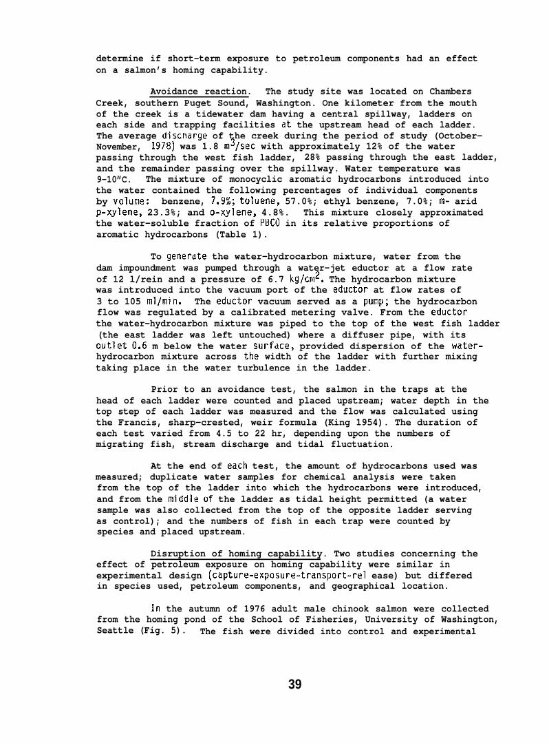
determine if short-term exposure to petroleum components had an effecton a salmon’s homing capability.
Avoidance reaction. The study site was located on ChambersCreek, southern Puget Sound, Washington. One kilometer from the mouthof the creek is a tidewater dam having a central spillway, ladders oneach side and trapping facilities at the upstream head of each ladder.The average discnarge of the creek during the period of study (October-November, 1978) was 1.8 m3/sec with approximately 12% of the waterpassing through the west fish ladder, 28% passing through the east ladder,and the remainder passing over the spillway. Water temperature was9-10”C. The mixture of monocyclic aromatic hydrocarbons introduced intothe water contained the following percentages of individual componentsby volune: benzene, 7.9%; toluene, 57.0%; ethyl benzene, 7.0%; m- aridp-xylene, 23.3%; and o-xylene, 4.8%. This mixture closely approximatedthe water-soluble fraction of PBCO in its relative proportions ofaromatic hydrocarbons (Table 1).
To generdte the water-hydrocarbon mixture, water from thedam impoundment was pumped through a water-jet eductor at a flow rateof 12 l/rein and a pressure of 6.7 kg/cn12. The hydrocarbon mixturewas introduced into the vacuum port of the eductor at flow rates of3 to 105 ml/min. The eductor vacuum served as a punp; the hydrocarbonflow was regulated by a calibrated metering valve. From the eductorthe water-hydrocarbon mixture was piped to the top of the west fish ladder(the east ladder was left untouched) where a diffuser pipe, with itsoutlet 0.6 m below the water surfdce, provided dispersion of the water-hydrocarbon mixture across the width of the ladder with further mixingtaking place in the water turbulence in the ladder.
Prior to an avoidance test, the salmon in the traps at thehead of each ladder were counted and placed upstream; water depth in thetop step of each ladder was measured and the flow was calculated usingthe Francis, sharp-crested, weir formula (King 1954). The duration ofeach test varied from 4.5 to 22 hr, depending upon the numbers ofmigrating fish, stream discharge and tidal fluctuation.
At the end of ea,ch test, the amount of hydrocarbons used wasmeasured; duplicate water samples for chemical analysis were takenfrom the top of the ladder into which the hydrocarbons were introduced,and from the middle of the ladder as tidal height permitted (a watersample was also collected from the top of the opposite ladder servingas control); and the numbers of fish in each trap were counted byspecies and placed upstream.
Disruption of homing capability. Two studies concerning theeffect of petroleum exposure on homing capability were similar inexperimental design (capture-exposure-transport-rel ease) but differedin species used, petroleum components, and geographical location.
In the autumn of 1976 adult male chinook salmon were collectedfrom the homing pond of the School of Fisheries, University of Washington,Seattle (Fig. 5). The fish were divided into control and experimental
39
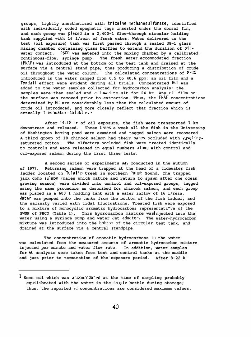
groups, lightly anesthetized with tricaine rnethanesulfonate, identifiedwith individually coded spaghetti tags inserted under the dorsal fin,and each group was placed in a 2,400-1 flow-through circular holdingtank supplied with 16 l/rein of fresh water. Water delivered to thetest (oil exposure) tank was first passed through a sealed 38-1 glassmixing chamber containing glass baffles to extend the duration of oil-water contact. PBCO was metered into the mixing chamber by a calibrated,continuous-flow, syringe pump. The fresh water-accommodated fraction(FWAF) was introduced at the bottom of the test tank and drained at thesurface via a central stand pipe, thus producing a distribution of crudeoil throughout the water column. The calculated concentrations of PllCOintroduced in the water ranged from 0.5 to 40.6 ppm; an oil film and aTyndall effect were evident during all trials. Concentrated HC1 wasadded to the water samples collected for hydrocarbon analysis; thesamples were then sealed and allowed to sit for 24 hr. Any oil film onthe surface was removed prior to extraction. Thus, the FHAF concentrationsdetermined by GC are considerably less than the calculated amount ofcrude oil introduced, and more closely reflect that fraction which isactually freshwater-solubl e.l
After 14-18 hr of oil exposure, the fish were transported 7 kmdownstream and released. Three times a week all the fish in the Universityof Washington homing pond were examined and tagged salmon were recovered.A third group of 18 chinook salmon had their nares occluded with vaseline-saturated cotton. The olfactory-occluded fish were treated identicallyto controls and were released in equal numbers along with control andoil-exposed salmon during the first three tests.
A second series of experiments was conducted in the autumnof 1977. Returning salmon were trapped at the head of a tidewater fishladder located on Tulalip Creek in northern Puget Sound. The trappedjack coho sdlmon (males which mature and return to spawn after one oceangrowing season) were divided into control and oil-exposed groups, taggedusing the same procedure as described for chinook salmon, and each groupwas placed in a 600 1 holding tank with a water inflow of 16 l/rein.Water was pumped into the tanks from the bottom of the fish ladder, andthe salinity varied with tidal fluctuations. Treated fish were exposedto a mixture of monocyclic aromatic hydrocarbons representati”ve of theSWSF of PBCO (Table 1). This hydrocarbon mixture waswater using a syringe pump and water Jet eductor. Themixture was introduced into the bottoril of the circulardrained at the surface via a central standpipe.
The concentration of aromatic hydrocarbons il
njected into thewater-hydrocarbontest tank, and
the waterwas calculated from the measured amounts of aromatic hydrocarbon mixtureinjected per minute and water flow rate. In addition, water samplesfor GC analysis were taken from test and control tanks at the middleand just prior to termination of the exposure period. After 8-22 hr
1 Some oil which was accormilodated at the time of sampling probablyequilibrated with the water in the sample bottle during storage;thus, the reported GC concentrations are considered maximum values.
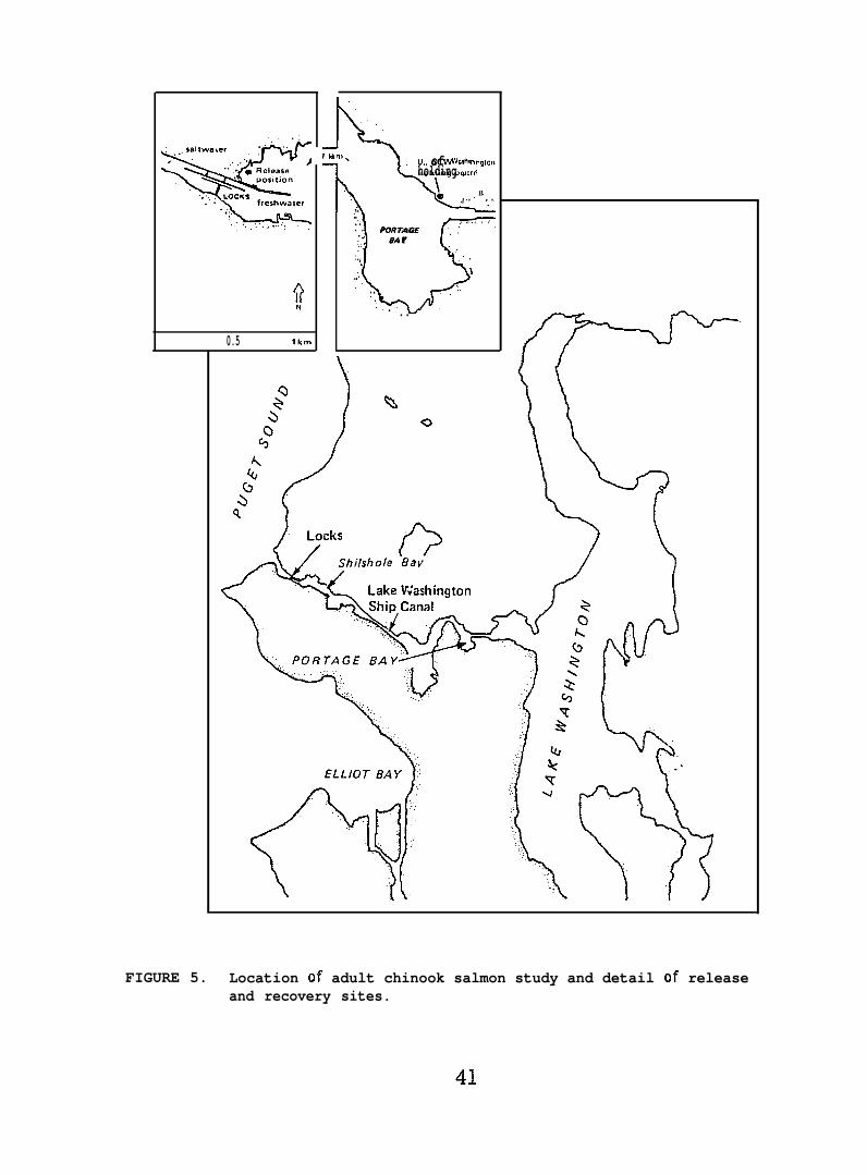
w‘“’’”’”=. . . . . . . .., .
f)N0.5 lkm
\
1 km% L). of Wash@ nglonholding pond\
b
,. ”....... . . . . .. .. . PORTAGE ,.,
,, 8A r. . .
\. .. .
., . .
) %o
FIGURE 5. Location of adult chinook salmon study and detail of releaseand recovery sites.
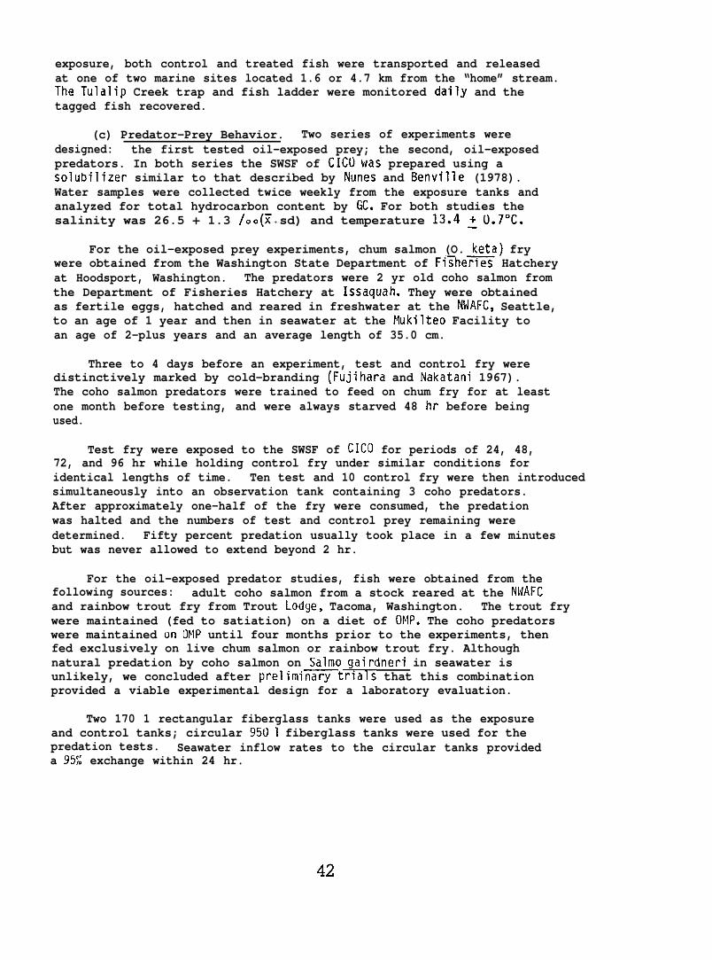
exposure, both control and treated fish were transported and releasedat one of two marine sites located 1.6 or 4.7 km from the “home” stream.The Tulalip Creek trap and fish ladder were monitored daily and thetagged fish recovered.
(c) Predator-Prey Behavior. Two series of experiments weredesigned: the first tested oil-exposed prey; the second, oil-exposedpredators. In both series the SWSF of CICOwas prepared using asolubilizer similar to that described by Nunes and Benville (1978).Water samples were collected twice weekly from the exposure tanks andanalyzed for total hydrocarbon content by GC. For both studies thesalinity was 26.5 + 1.3 /oo (1 + sd) and temperature 13.4 ~().7°C.
For the oil-exposed prey experiments, chum salmon (O. keta) frywere obtained from the Washington State Department of Fi~he= Hatcheryat Hoodsport, Washington. The predators were 2 yr old coho salmon fromthe Department of Fisheries Hatchery at Issaquah. They were obtainedas fertile eggs, hatched and reared in freshwater at the NWAFC, Seattle,to an age of 1 year and then in seawater at the Mukilteo Facility toan age of 2-plus years and an average length of 35.0 cm.
Three to 4 days before an experiment, test and control fry weredistinctively marked by cold-branding (Fujihara and Nakatani 1967).The coho salmon predators were trained to feed on chum fry for at leastone month before testing, and were always starved 48 hr before beingused.
Test fry were exposed to the SWSF of CICO for periods of 24, 48,72, and 96 hr while holding control fry under similar conditions foridentical lengths of time. Ten test and 10 control fry were then introducedsimultaneously into an observation tank containing 3 coho predators.After approximately one-half of the fry were consumed, the predationwas halted and the numbers of test and control prey remaining weredetermined. Fifty percent predation usually took place in a few minutesbut was never allowed to extend beyond 2 hr.
For the oil-exposed predator studies, fish were obtained from thefollowing sources: adult coho salmon from a stock reared at the NWAFCand rainbow trout fry from Trout Lodge, Tacoma, Washington. The trout frywere maintained (fed to satiation) on a diet of OMP. The coho predatorswere maintained on OMP until four months prior to the experiments, thenfed exclusively on live chum salmon or rainbow trout fry. Althoughnatural predation by coho salmon on Salmo gairdneri in seawater isunlikely, we concluded after prelimi~trlals that this combinationprovided a viable experimental design for a laboratory evaluation.
Two 170 1 rectangular fiberglass tanks were used as the exposureand control tanks; circular 950 1 fiberglass tanks were used for thepredation tests. Seawater inflow rates to the circular tanks provideda 95S exchange within 24 hr.
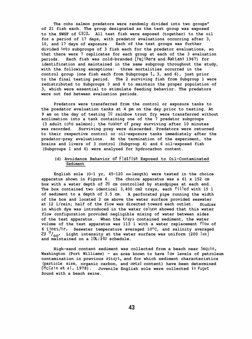
The coho salmon predators were randomly divided into two groups”of 21 fish each. The group designated as the test group was exposedto the SWSF of CICO. All test fish were exposed (together) to the oilfor a period of 17 days, with predator evaluations occurring after 3,10, and 17 days of exposure. Each of the test groups was furtherdivided into subgroups of 3 fish each for the predator evaluations, sothat there were 7 replicates for each group at each of the 3 evaluationperiods. Each fish was cold-branded (Fujihara and Naktani 1967) foridentification and maintained in the same subgroup throughout the study,with the following exceptions: three mortalities occurred in thecontrol group (one fish each from Subgroups 1, 3, and 6), just priorto the final testing period. The 2 surviving fish from Subgroup 1 wereredistributed to Subgroups 3 and 6 to maintain the proper population of3, which were essential to stimulate feeding behavior. The predatorswere not fed between evaluation periods.
Predators were transferred from the control or exposure tanks tothe predator evaluation tanks at 4 pm on the day prior to testing. At9 am on the day of testing 10 rainbow trout fry were transferred withoutacclimation into a tank containing one of the 7 predator subgroups(3 adult coho salmon); the numberof prey surviving after 10 minuteswas recorded. Surviving prey were discarded. Predators were returnedto their respective control or oil-exposure tanks immediately after thepredator-prey evaluations. At the termination of the experiment, thebrains and livers of 3 control (Subgroup 4) and 6 oil-exposed fish(Subgroups 1 and 6) were analyzed for hydrocarbon content.
(d) Avoidance Behavior of Fldtfish Exposed to Oil-ContaminatedSediment
English sole (O-1 yr, 45-120 mm length) were tested in the choiceapparatus shown in Figure 6. The choice apparatus was a 61 x 152 cmbox with a water depth of 20 cm controlled by standpipes at each end.The box contained two identical 3,400 cm2 trays, each filled with 15 1of sediment to a depth of 3.5 cm. A perforated pipe running the widthof the box and located 2 cm above the water surface provided seawaterat 12 l/rein; half of the flow was directed toward each outlet. Studiesin which dye was introduced in the water colur,ln showed that this waterflow configuration provided negligible mixing of water between sidesof the test apparatus. When the trays contained sediment, the watervolume of the test apparatus was 113 1 with a water replacement flow of6 times/hr. Seawater temperature averaged 10”C, and salinity averaged29 O/OO. Light intensity at the water surface was uniform (200 lUX)and maintained on a 10L:14U schedule.
High-sand content sediment was collected from a beach near Sequim,Washington (Port Williams) - an area known to have low levels of petroleumcontamination in previous assdys, and for which sediment characteristics(particle size, organic carbon, and metal content) have been determined(McCain et al. 1978). Juvenile English sole were collected in PugetSound with a beach seine.
43
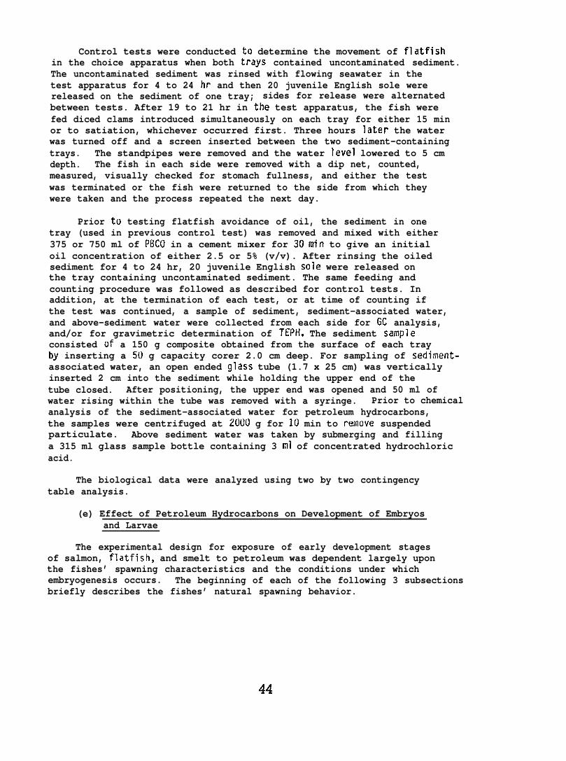
Control tests were conducted to determine the movement of flatfishin the choice apparatus when both trays contained uncontaminated sediment.The uncontaminated sediment was rinsed with flowing seawater in thetest apparatus for 4 to 24 hr and then 20 juvenile English sole werereleased on the sediment of one tray; sides for release were alternatedbetween tests. After 19 to 21 hr in the test apparatus, the fish werefed diced clams introduced simultaneously on each tray for either 15 minor to satiation, whichever occurred first. Three hours later the waterwas turned off and a screen inserted between the two sediment-containingtrays. The standpipes were removed and the water level lowered to 5 cmdepth. The fish in each side were removed with a dip net, counted,measured, visually checked for stomach fullness, and either the testwas terminated or the fish were returned to the side from which theywere taken and the process repeated the next day.
Prior to testing flatfish avoidance of oil, the sediment in onetray (used in previous control test) was removed and mixed with either375 or 750 ml of PBCO in a cement mixer for 30min to give an initialoil concentration of either 2.5 or 5% (v/v). After rinsing the oiledsediment for 4 to 24 hr, 20 juvenile English sole were released onthe tray containing uncontaminated sediment. The same feeding andcounting procedure was followed as described for control tests. Inaddition, at the termination of each test, or at time of counting ifthe test was continued, a sample of sediment, sediment-associated water,and above-sediment water were collected from each side for GC analysis,and/or for gravimetric determination of TEPH. The sediment sampleconsisted of a 150 g composite obtained from the surface of each trayby inserting a 50 g capacity corer 2.0 cm deep. For sampling of sediment-associated water, an open ended glass tube (1.7 x 25 cm) was verticallyinserted 2 cm into the sediment while holding the upper end of thetube closed. After positioning, the upper end was opened and 50 ml ofwater rising within the tube was removed with a syringe. Prior to chemicalanalysis of the sediment-associated water for petroleum hydrocarbons,the samples were centrifuged at 2000 g for 10 min to remove suspendedparticulate. Above sediment water was taken by submerging and fillinga 315 ml glass sample bottle containing 3 ml of concentrated hydrochloricacid.
The biological data were analyzed using two by two contingencytable analysis.
(e) Effect of Petroleum Hydrocarbons on Development of Embryosand Larvae
The experimental design for exposure of early development stagesof salmon, flatfish, and smelt to petroleum was dependent largely uponthe fishes’ spawning characteristics and the conditions under whichembryogenesis occurs. The beginning of each of the following 3 subsectionsbriefly describes the fishes’ natural spawning behavior.
1 .52m
FIGURE 6. Choice apparatus used in testing avoidanceof juvenile Englishsole to oil-contaminated sediment.
~ Saltwater accommodated fraction\
\
‘%)’% — u ’ - . . . . . . . . . . . ,----- . . .
Fresh water
FIGURE 7. Chum salmon egg incubation apparatus with delivery of brackishSWAF of weathered PBCU from the wave generator (Fig. 4) for3 hr/day and delivery of fresh water for 21 hr/day.
45
(1) Chum Salmon. Chum salmon frequently spawn in tidalareas at at the mouths of streams (Neave 1966, Bakkala 1970). Eggsare deposited in redds and then covered with gravel as a result ofupstream redd digging and stream flow. At least one high tide a dayinundates the redds with salt and brackish water; the length of exposureto water of high salinity depends upon redd location. After approximately50-90 days (development rate is a function of water temperature) theeggs hatch; however, the salmon alevins remain in the gravel for another30-50 days before emerging and migrating.
Chum salmon eggs were obtained from the U.S. Fish and WildlifeService National Fish Hatchery at Quilcene, Washington. Immediately afterfertilization, the eggs were transported to the Nukilteo laboratory.Subsequent sampling from control groups indicated that 97.5% of thesalmon eggs were fertilized. One day after fertilization, approximately70 chum salmon eggs were placed in each of 56 (30 x 75 cm) glasscylinders, and both ends covered with Teflon netting. A glass tray wasdivided longitudinally into eight troughs and pea gravel was layered2.5 cm deep on the bottom. Seven cylinders of eggs were placed horizontallyinto each trough (Fig. 7).
Eggs were exposed 3 hr/day to oil-contaminated brackish water(16-24 o/oo salinity, and 4.5-10.2 “C ambient temperature) at a flow rateof 400 ml/min per trough. For 21 hr/day the eggs received fresh dechlorinatedwater (5.5 to 10.5°C ambient temperature) at the same flow rate for awater replacement of 8 times per hr. Exposure to oil-contaminated wateroccurred 4 days per week for 16 consecutive weeks. On the other 3 dayseggs and alevins received uncontamin~ted brackish water for 3 hr/day.The oil exposure conditions (by group) designated for each of the 8 troughs,are expressed diagrammatically in Figure 8.
(2) Flatfish. Sand sole and English sole are pleuronectidflatfish with pelagic eggs and larvae. Flatfish eggs are released onor near the bottom; after about one day, depending upon depth of spawning,the eggs rise to float near the water’s surface (Ketchen 1956, Alderdiceand Forrester 1971). Eggs hatch after approximately one week and thelarvae emerge relatively undeveloped.
Gravid English sole and sand sole were obtained by trawling inPuget Sound. Eggs were stripped from ripe females and fertilizedimmediately with sperm from a ripe male on board the trawling vessel.Fertilization success and subsequent viability in flatfish eggs was high(approximately 94);$) as indicated by cell cap formation. Twenty-four hrsafter fertilization (early cell-cap stage) approximately 350 to 700flatfish eggs were introduced into 1,000 and 2,000 ml separator funnelscontaining 9(.)0 and 1800 ml, respectively, of uncontaminated seawater orthe SWAF of weathered PBCO obtained from the wave machine. The funnelswere attached to an air supply through the bottom and the water bubbledslowly, thus creating a current in the funnel which kept the eggs insuspension. Funnels were submerged in a water bath with 10”C flowingseawater.
46
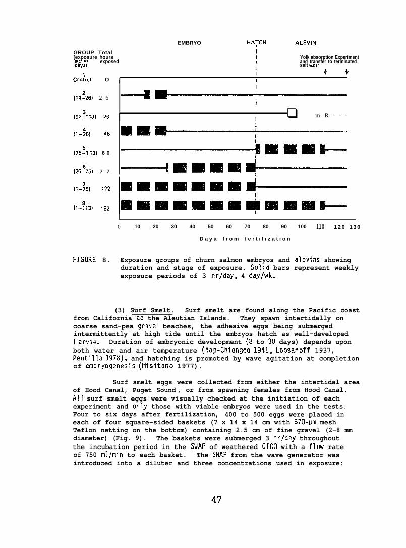
EMBRYO HATCH ALEVIN
GROUP Total(exposure hours ; Yolk absorption Experiment
exposed%%’
I and transfer to terminated1
salt waler
Conlrol O I
I ++iI
(14~26) 2 6I1tI
(92~?13) 29 1 ❑ m R - - -
46 ❑ m~
1
(1-:6)
(75:1 13) 6 0 ~ll#l#IlllIllll!III+
(26:75) 7 7 ------+ mtmmm d------i
o 10 20 30 40 50 60 70 80 90 100 110 1 2 0 1 3 0
D a y a f r o m f e r t i l i z a t i o n
FIGURE 8. Exposure groups of churn salmon embryos and alevins showingduration and stage of exposure. Solid bars represent weeklyexposure periods of 3 hr/day, 4 day/wk.
(3) Surf Smelt. Surf smelt are found along the Pacific coastfrom California to the Aleutian Islands. They spawn intertidally oncoarse sand-pea gravel beaches, the adhesive eggs being submergedintermittently at high tide until the embryos hatch as well-developed1 arvae. Duration of embryonic development (8 to 30 days) depends uponboth water and air temperature (Yap-Chiongco 1941, Loosanoff 1937,Pentilla 1978), and hatching is promoted by wave agitation at completionof embryogenesis (Plisitano 1977).
Surf smelt eggs were collected from either the intertidal areaof Hood Canal, Puget Sound , or from spawning females from Hood Canal.All surf smelt eggs were visually checked at the initiation of eachexperiment and only those with viable embryos were used in the tests.Four to six days after fertilization, 400 to 500 eggs were placed ineach of four square-sided baskets (7 x 14 x 14 cm with 570-#m meshTeflon netting on the bottom) containing 2.5 cm of fine gravel (2-8 mmdiameter) (Fig. 9). The baskets were submerged 3 hrjday throughoutthe incubation period in the SWAF of weathered CICO with a flow rateof 750 ml/min to each basket. The SWAF from the wave generator wasintroduced into a diluter and three concentrations used in exposure:
47
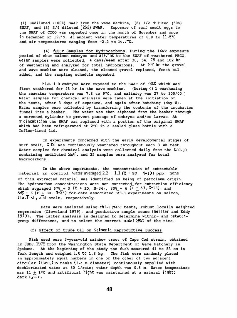
(1) undiluted (100%) SWAF from the wave machine, (2) 1/2 diluted (50%)SWAF, and (3) 3/4 diluted (25X) SWAF. Exposure of surf smelt eggs tothe SWAF of CICO was repeated once in the month of November and oncein December of 197’9, at ambient water temperatures of 8.8 to 11.5°Cand air temperatures ranging from -2.2 to 16.7°C.
(4) Water Samples for Hydrocarbons. During the 16wk exposureperiod of chum salmon embryos and alevins to the SWAF of weathered PBCO,~~ater samples were collected, 4 days/week after 30, 54, 78 and 102 hrof weathering and analyzed for total hydrocarbons. At 102 hr the graveland wave machine were cleaned, the cleaned gravel replaced, fresh oiladded, and the sampling schedule repeated.
Flatfish embryos were exposed to the SWAF of P13C0 which wasfirst weathered for 48 hr in the wave machine. (During oi 1 weatheringthe seawater temperature was 7.8 to 9“C, and salinity was 27 to 300/00.)Water samples for chemical analysis were taken at the initiation ofthe tests, after 3 days of exposure, and again after hatching (day 8).Water samples were collected by transferring the contents of the incubationfunnel into a beaker. The water was then siphoned from the beaker througha screened cylinder to prevent passage of embryos and/or larvae. Atmidincubation the SWAF was replaced with a portion of the original SWAFwhich had been refrigerated at 2°C in a sealed glass bottle with aTeflon-lined lid.
In experiments concerned with the early developmental stages ofsurf smelt, CICO was continuously weathered throughout each 3 wk test.Water samples for chemical analysis were collected daily from the troughcontaining undiluted SWAF, and 35 samples were analyzed for totalhydrocarbons.
In the above experiments, the concentration of extractablematerial in control water averaged 2.2 + 1.1 (~ + SD, N=1O) ppb; noneof this extracted material was identified as being of petroleum origin.The hydrocarbon concentrations were not corrected for extraction efficiencywhich averaged 67% + 9 @ + SD, N=34), 80% + 6 G~SD, N=16), and84% + 6 (~ + SD, N=75) for-data associated =ith experiments on salmon,flat~ish, afid smelt, respectively.
Data were analyzed using chi-square tests, robust locally weightedregression (Cleveland 1979), and predictive sample reuse (Geisser and Eddy1979)* The latter analysis is designed to determine within- and between-group differences, and to select the correct model >952 of the time.—
(f) Effect of Crude Oil on Salmonid Reproductive Success
Fish used were 3-year-old rainbow trout of Cape Cod strain, obtainedin June, 1975 from the Washington State Department of Game Hatchery inSpokane. At the beginning of the study the fish measured 41 to 53 cm infork length and weighed l.Oto 1.8 kg. The fish were randomly placedin approximately equal numbers in one or the other of two adjacentcircular fiberglas tanks (1.8 m diameter) continuously supplied withdechlorinated water at 30 l/rein; water depth was 0.8 m. Water temperaturewas 11 + 1°C and artificial light was maintained at a natural light:dark cy~e.
48

SALT WATER ACCOMMODATEDFRACTION (SWAF)
100% SWAF
OF wEATHERED CiCO k ~---+,, .,. . —— L-.., - RZ??I
50% SWAF !
Eggs and gravel
Waste overflow +1 I II II II
FIGURE 9. Surf smelt embryo exposure apparatusSWAF of weathered CIC(I from the wave
with delivery of thegenerator (Fig. 4].
Petroleum-coated food (1 part oil :1,000 parts food, by weight) wasroutinely prepared in the following manner: Two kg of l/4-inch diameterOregon moist pellets were placed in
74 liter glass beaker. Two g of
PBCO were mixed with 148 ml of FREON ‘) TF solvent (trichlorotrifluoroethane)and poured over the food. The food and oil were thoroughly mixed andthe food was spread over porcelain-covered metal trays for 90 min ofair drying in a fume hood. The food was then weighed into daily aliquots,sealed in plastic bags, and frozen until used. Food for control experimentswere prepared identically except that the crude oil was omitted. Fishwere fed these diets at an approximate rate of 2% (wet weight of food)of body weight each workday between July, 1975 and August, 1976.
In late November 1975, all fish were examined for degree of maturity.Subsequent biweekly and then weekly examinations were made. As femalesripened they were spawned and eggs were fertilized using standard trout-
49
culture methods (Leitritz 1959). Ripe males were consistently availablefor the duration of the spawning period from January through February 1976.All eggs from control fish were divided into equal aliquots and one aliquotwas fertilized with sperm from an oil-exposed test male and the otherwith sperm from one control male. Ten of the oil-exposed females weresimilarly treated; eggs from the remaining oil-exposed females werefertilized with sperm from oil-exposed males only. A total of 31 testand 10 control crosses were made. Eggs were incubated at 7° to 9°C.Mortality data were collected through the yolk-sac absorption stageand statistically analyzed usinq the Mann-uhitne.y modification ofWilcoxon’s sum o+ rank; test (L~ng-
At the time of spawning, samp”collected and frozen for later aria”
ey 1971). -
es of adult tissues and eggs wereysis of petroleum hydrocarbons.
5.3.4 Invertebrate Studies
Effect of Petroleum Hydrocarbons on Sea Urchin Defense Behavior
Seawater-soluble fractions of PBCO were prepared according to theflow-through method of Roubal et al. (1977b). Solutions containingsingle aromatic hydrocarbons were prepared by a modified method describedby Nunes and Benville (1978). In the latter, filtered seawater drippedinto a glass reservoir at a constant head from a perforated bucket.Hydrocarbons were introduced continuously near the top of the glassreservoir via a calibrated, repeating, syringe pump and the seawater-hydrocarbon mixture was drawn off at the bottom of the reservoir anddelivered to a 2-1 glass exposure beaker (for details see Johnson 1979).The flow rate of the seawater-hydrocarbon mixture through the exposurebeaker was 200 ml/min; average salinity was 28 O/OO; average watertemperature was 10”C.
The reported SWSF hydrocarbon concentrations produced by PBCO arethe summed concentrations of the monocyclic aromatic compounds. A gaschromatogram representative of the SWSF of PBCO is given in Figure 10,and represents the type of data used in determining the concentrationof hydrocarbons present in exposure water for all experiments withinvertebrates. The concentration of each component was found to be similarto that reported in detail by Roubal et al. (1978) with the monocyclicaromatic hydrocarbons accounting for 95% of the total SWSF; the remainderconsisted of naphthalenes and cyclohexanes.
Urchins are preyed upon by starfish, including the sunflowerstarfish (Pycnopodia helianthoides; Mauzey et al. 1968), and whenpresented with a chemical stimulus from their starfish predator, thesea urchins exhibit a defensive response that is consistent andrepeatable. The starfish can move faster than the urchin, but theurchin, if overtaken, possesses a compliment of globiferous pedicellariaethat are activated by a water soluble exudate from the starfish (Phillips1978). These globiferous pedicellaria are distributed liberally overthe aboral surface of urchins, and a typical 3 cm diameter urchin maypossess 100 or more. Each pedicellaria articulates on a peduncle andhas three jaws , each tipped with a hollow tooth capable of delivering
50
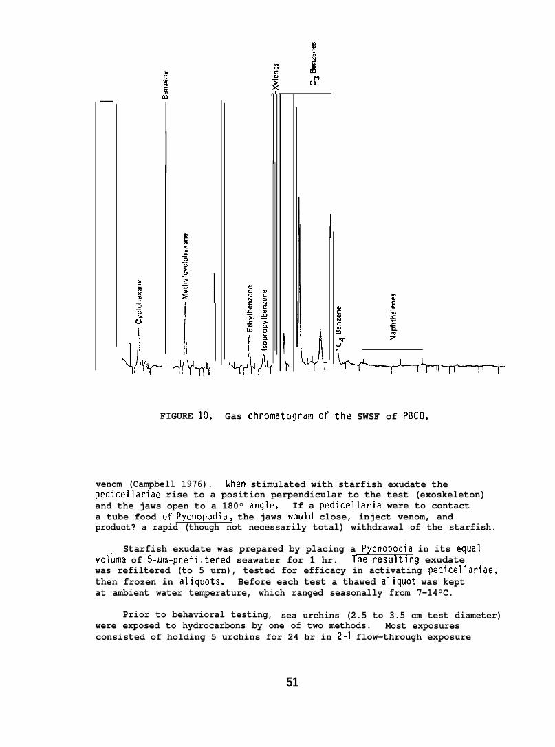
c.!’
1“%4-)-fFIGURE 10, Gas Chromatogrdm of the SWSF of PBCO.
venom (Campbell 1976). When stimulated with starfish exudate thepedicellariae rise to a position perpendicular to the test (exoskeleton)and the jaws open to a 180° angle. If a pedicellaria were to contacta tube food of Pycnopodia, the jaws would close, inject venom, andproduct? a rapid (though not necessarily total) withdrawal of the starfish.
Starfish exudate was prepared by placing a Pycnopodia in its equalvolume of 5-#jm-prefiltered seawater for 1 hr. l’he resultlng exudatewas refiltered (to 5 urn), tested for efficacy in activating pedicellariae,then frozen in aliquots. Before each test a thawed aliquot was keptat ambient water temperature, which ranged seasonally from 7-14°C.
Prior to behavioral testing, sea urchins (2.5 to 3.5 cm test diameter)were exposed to hydrocarbons by one of two methods. Most exposuresconsisted of holding 5 urchins for 24 hr in 2-I flow-through exposure
51
beakers containing the SWSF of PBCO or single aromatic hydrocarbons,and then placing them individually in 100-ml beakers containing 40 mlof the same hydrocarbon solution. The urchins were then left undisturbedfor approximately 10 min before stimulation with starfish exudate. Thesecond method was to expose the urchins individually in the 100-mlbeakers for only 10 min before testing. Following exposure, 1 ml ofstarfish exudate was slowly administered with a syringe in a circularmotion over each urchin. The defense reponse assay consisted of countingthe number of pedicellariae that opened to 180°.
If 75 or more pedicellariae responded to starfish stimulus, theresponse was considered total (100%); if less than 75 opened, a partialresponse was recorded (0-99%). For each test, the average response ofat least 5 hydrocarbon treated urchins was divided by the average responseof at least 5 controls to yield a response index for each treatment.This method of analysis takes into consideration variations in responseof controls for each group tested.
Reproductive Behavior of Dorid Nudibranchs Exposed to PetroleumHydrocarbons
(a) Chemosensory disruption. Mature dorid nudibranchs werecollected from the seawater system at the Mukilteo Biological Station.The adult nudibranchs were exposed to the SWSF of PBCO for 24 hr in2-1 flow-through aquaria with a seawater flow of 250 ml/min and atemperature and salinity of 13.6+ 0.6°C (x + range) and 27.0~2°/oo,respectively. The SWSF was produ~ed by a method described by Roubalet al. (1977 b); to obtain different hydrocarbon concentrations in eachaquarium the SWSF flows were diluted by mixing with seawater, Two watersamples were collected from each aquarium for GC analysis of hydrocarbonconcentration.
Following SWSF exposure, the nudibranchs were assayed in“untreated” seawater for chemotatic behavioral response.1 Individualnudibranchs were placed in the bottom arm of a “Y” choice chamber andallowed to move into either the “stimulus” arm or the non-stimulus(“blank”) arm. (For detai 1s of testing apparatus see Malins et al. 1978or Johnson 1979.) The “stimulus” arm contained four reproductive nudibranchs.The “blank” arm was of identical construction, but did not containnudibranchs. Clean seawater filtered to 5flm supplied each arm of thechoice chamber at a flow rate of 60 rnl/min. The “stimulus” and “blank”chambers were alternated and thoroughly washed between tests to removemucus. A positive response denoted movement toward either chamber;“no choice” meant the nudibranchs did not make a definite movementinto either arm within 10 min from the start of the test.
The percentage moving toward the “stimulus” chamber for the SWSFexposed group of nudibranchs was statistically compared with the controlpercentage using an arc sin transformation test for equality of twopercentages (Sokal and Rohlf 1969).
1 The reproductive behavior of the nudibranch is thought to be initiatedby a sex pheromone as has been implicated in mediating reproductiveaggregations in other gastropod (Dinter 1975, Audesirk 1977).
52
(b) Embryological development. The exposure system was identicalto that used above in testing chemosensory disruption of dorid nudibranchs.There was, however, a difference in dete~ining the petroleum hydrocarbonconcentration in each aquarium. Over the 18-day exposure period fourwater samples for GC analysis were taken from the oil solubilizer headbox . Hydrocarbon concentration in each aquarium was calculated fromthe average SWSF concentration in the head box and the amount of SWSFdiluted with untreated seawater.
Four qroups of 20 mature nudibranchs of approximately equalsize (totai weightflow-through aquar’were collected andaquaria; replicatemass were taken da”experiment termina(Hawkes 1974, with
2.1 g for each group) were placed in 4-separate 2-1a. The first egg masses laid by the dorid nudibranchsplaced in holding containers within each exposureegg aliquots of 2-3 mm3 from the center of each eggIy 5 days per week until hatching was complete or theed. The eggs were fixed in buffered preservativethe addition of 17 g of synthetic seawater per liter
of buffer to attain 800 mOsm). In addition, eggs laid each followingday were collected, weighed, and then discarded.
Embryonic development was studied using light microscopy. Eggsin aliquot samples were counted and each normal egg assigned to one ofthe following 7 categories: One-cell; two-cell; blastula; gastrula;early velure; late velure; and shell.l Abnormal eggs were categorizedas: moderate abnormality (a recognizable embryo but malformed, orwith extra cellular material in the capsule) or severe ahnorrnality (norecognizable embryo and often no capsule present). Photomicrographsof the normal and abnormal categories listed above are given in reportsby Malins et al. (1978) and/or Murnaw (1978).
Feeding Behavior of Shrimp Exposed to Petroleum Hydrocarbons
Spot shrimp were caught in commercial shrimp pots at depths of100 to 120 m in Puget Sound. Following capture the shrimp were maintainedin 1200-1 flow-through tanks at ambient seawater temperature of 10 to12°C and a salinity of 27 to 30 O/oo. Three days prior to testing,individual shrimp were moved to 3-1 glass exposure tanks, each with aflow rate of 300 ml/min. The shrimp were not fed for 3 days prior toexposure or during the 6 day exposure period. The number of shrimptested and the number of observations made using 4 SWSF concentrationsof PBCO are given in Table 2.
To obtain different concentrations of hydrocarbons in exposures,the SWSF from an oil solubilizer (Roubal et al. 1977b) was diluted justprior to its entry into the test chambers while maintaining a totalflow of 300ml/min (Fig. 11). The shrimp chambers were enclosed inblack plastic and observations on feeding behavior were made throughone-way mirrors. Each data point consisted of three, 3-rein observationsof background activity, response to seawater control, and response toa 1:10 dilution of artificial squid extract (Mackie 1973) . Seawater
1 Developmental stages used here are described in detail in severalreports involving nudibranch development McGowan and Pratt 1954,
&Perron and Turner 1977, Thompson 1960, 19 2, 1967).
53
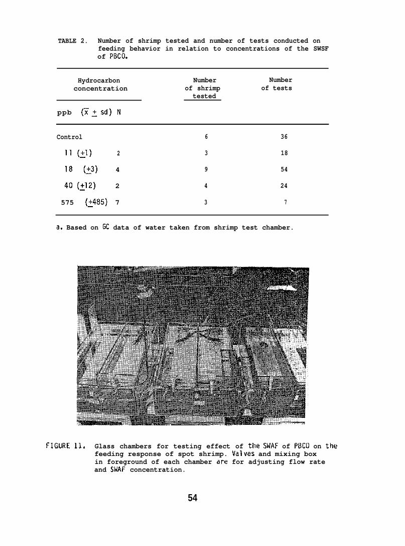
TABLE 2. Number of shrimp tested and number of tests conducted onfeeding behavior in relation to concentrations of the SWSFof PBCO.
Hydrocarbon Number Numberconcentration of shrimp of tests
tested
ppb (~jsd) N
Control 6 36
11 (:1) 2 3 18
18 (:3) 4 9 54
40(~12) 2 4 24
575 (3485) 7 3 7
a, Based on GC data of water taken from shrimp test chamber.
FIGuRE 11. Glass chambers for testing effect of the SWAF of PBCO on thefeeding response of spot shrimp. Valves and mixing boxin foreground of each chamber are for adjusting flow rateand SMAF concentration.
54
control and squid extract stimulants were introduced at the upstreamend of the test chamber at a flow rate of 10 ml/min.
Two behavioral indices were selected for evaluating feedingresponses to a given stimulant: (1) Perepod probing--Probing thesubstrate with the first perepods was recorded as the number of probingbouts during a test; this activity is indicative of higher ordersearching behavior. (2) Contact with stimulant inlet--Defined asgrasping or actively picking at the inlet with chelae or mouth parts;this behavior was considered representative of maximum feeding response.
6. RESULTS
6.1 Chemistry
6.1.1 Uptake and Biotransformation of Specific Hydrocarbons in Salmonids
Coho salmon injected with radiolabeled benzene, NPH, and anthracenedifferentially retained these hydrocarbons in liver, brain and muscleas indicated by the data in Figure 12, Table 3. In the tissues examined,the percent of administered dose at all time periods was the lowest infish exposed to benzene and the highest in those exposed to anthracene(Table 3).
The data in Table 3 also show that the administered hydrocarbonswere metabolized by coho salmon and metabolic products were presentin the liver and gall bladder. However, in the brain most of therecoverable radioactivity was in the form of the parent hydrocarbons24 hr (>98%) after injection.
To delineate the nature of metabolic products, solvent extractsof various tissues were analyzed by TLC. The results revealed a widespectrum of metabolic products in various proportions in the brain,liver, gall bladder, heart, and flesh of fish at 24 hr after administrationof NPH. Naphthol, a dihydrodiol derivative of NPH and their conjugates(e.g. , glucuronides, sulfates and mercapturic acids) were detected.
6.1.2 Accumulation of Petroleum Hydrocarbons by Fish Exposed to SWSF of PBCO
The hydrocarbon composition of SNSF of PBCO is given in Table 4which shows the presence of mono and diaroflatic hydrocarbons. Data arepresented in Table 5 for the accumulation of hydrocarbons in coho salmonexposed to a SWSF of PBCO equal to 0.8 ppm of aromatic hydrocarbonsin flow-through seawater. After 1 wk of exposure, no hydrocarbonsrepresentative of the SWSF were detected in muscle tissue. Exposuresfrom 2 to 6 wk, however, resulted in the accumulations of significant
1 Three additional behaviors were also recorded but not presented here;antennular flicks, antennular cleaning, and forward movement. Foreach of these behaviors there was often a response to seawater controlstimulus which resulted in inconclusive data. For details of theseobservations and results see Malins et al. (1977) and/or Miller (1980).
55

B
10,000
1,000
100
c1ANTNRACENE
nNAPNTNALEIU
uaENzENE
%------- ------- -
- \\--~ ------ -------’
*--
24 72 14NOURS FROM TIME OF INJECTION OF
LABELED HYOROCW80N
FIGURE 12. Radioactivity in tissues of coho salmon receiving lk-labeledhydrocarbons (2.5 uCi) by intraperitoneal injection. A, anthraceneradioactivity (—), naphthalene radioactivity (––––-)and benzene radioactivity ( -----); B, 24-hr values for radioactivityin the gall bladder of injected fish. Total carbon-14 in organsas % of administered dose is given in Table 3. (Fran Roubalet il. 1977a)
56
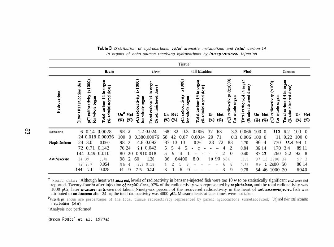
TabIe 3 Distribution of hydrocarbons, total aromatic metabolizes and total carbon-14in organs of coho salmon receiving hydrocarbons by intraperitoneal injection
Tissuea
Brain Liver Gall bladder Flesh Carcass
so0‘1
Benzene 6 0.14 0.002824 0.018 0,00036
Naphtha.lene 24 3.0 0.06072 0.71 0,142
144 0.49 0.010Anthracene 24 39 0.78
72 2.7 0.054144 1.4 0.028
98 2 1.2 0.024 68 32 0.3 0.006 37 63 3.3 0.066 100 0 310 6.2 100 0100 0 0.380.00076 58 42 0.07 0.0014 29 71 0.3 0.006 100 0 11 0.22 100 098 2 4.6 0.092 87 13 13 0.26 28 72 83 1.70 96 4 770 15.4 99 176 24 2.1 0.042 5 5 4 5 - c - - – 4 2 0.84 86 14 170 3.4 89 1180 20 0.910.018 5 9 4 1 - - - - 2 0 0.40 87 13 260 5.2 92 898 2 60 1.20 36 64400 8.0 10 90 580 11.6 87 13 1700 34 97 396 4 8.8 0.18 4 2 5 8 - - - - 6 8 1.36 99 1 2s00 50 86 1491 9 7.5 0.15 3 1 6 9 - - - - 3 9 0.78 54 46 1000 20 6040
U Heart data: Although heart was anaSyzed, levels of radioactivity in benzene-injected fish were too 10 w to be statistically significant and were notreported. Twenty-four hr after injection of napht.halene, 97% of the radioactivity was represented by naphthalcne, and the total radioactivity was1000 pCi; later m~suremcnts were not taken. Ninety-six percent of the recovered radioactivity in the heart of anthraeene-injected fish wasattributed to anthracenc after 24 hr; the total radioactivity was 4000 pCi. Measurements at later times were not taken
bPercentages shown are percentages of the total tissue radioactivity represented by parent hydrocarbons (unmetabolized; Un) and their total aromaticmetabolitcs (Met)
‘Analysis not performed
(From Roubal et al. 1977a)
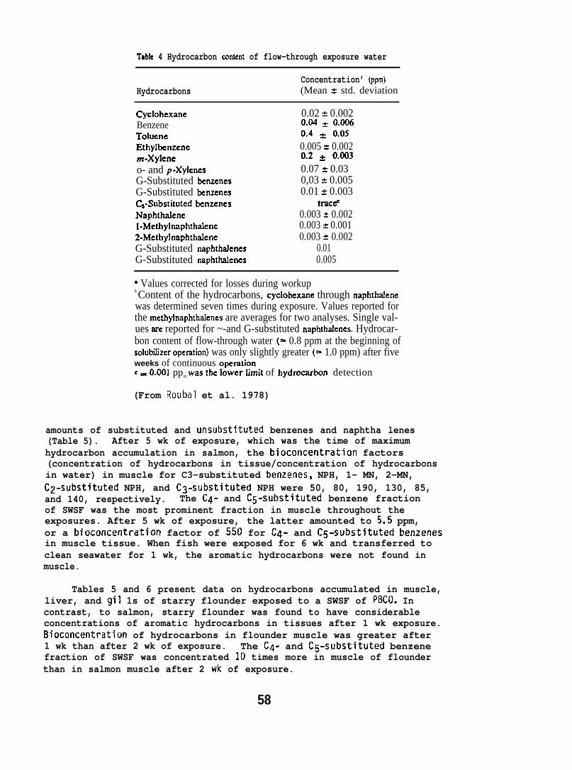
Table 4 Hydrocarbon mnten! of flow-through exposure water
Concentration’ (ppm)Hydrocarbons (Mean A std. deviation
Cyclohexane 0.02 & 0.002Benzene &@$ * 0.()()6Toluenc &4 = ().()5Ethylbenzene 0.005 * 0.002m-Xylcne 0.2 * 0.(303o- and p-Xylcncs 0.07 ? 0.03G-Substituted benzcncs 0,03 * 0.005G-Substituted benzenes 0.01 z 0.003(&-Substituted benzenes traceNaphthalene 0.003 = 0.002l-Methylnaphthalene 0.003 * 0.0012-Methylnaphthalene 0.003 * 0.002G-Substituted naphthrdenes 0.01G-Substituted naphthalenes 0.005
‘ Values corrected for losses during workupb Content of the hydrocarbons, cyclohexane through naphthalenewas determined seven times during exposure. Values reported forthe methylnaphthafenes are averages for two analyses. Single val-ues are reported for ~-and G-substituted naphthalcnes. Hydrocar-bon content of flow-through water (=- 0.8 ppm at the beginning ofsolubdizer opemtion) was only slightly greater (= 1.0 ppm) after fiveweeks of continuous opemtionG = ().00] ppm w= t~ ]Ower Iimi[ of hyd~hn detection
(From Roubal et al. 1978)
amounts of substituted and unsubstituted benzenes and naphtha lenes(Table 5). After 5 wk of exposure, which was the time of maximumhydrocarbon accumulation in salmon, the bioconcentration factors(concentration of hydrocarbons in tissue/concentration of hydrocarbonsin water) in muscle for C3-substituted benzenes, NPH, 1- MN, 2-MN,C2-substituted NPH, and C3-substituted NPH were 50, 80, 190, 130, 85,and 140, respectively. The C4- and C5-substituted benzene fractionof SWSF was the most prominent fraction in muscle throughout theexposures. After 5 wk of exposure, the latter amounted to 5.5 ppm,or a bioconcentration factor of 550 for C4- and C5-substituted benzenesin muscle tissue. When fish were exposed for 6 wk and transferred toclean seawater for 1 wk, the aromatic hydrocarbons were not found inmuscle.
Tables 5 and 6 present data on hydrocarbons accumulated in muscle,liver, and gil 1s of starry flounder exposed to a SWSF of PBC(). Incontrast, to salmon, starry flounder was found to have considerableconcentrations of aromatic hydrocarbons in tissues after 1 wk exposure.Bioconcentration of hydrocarbons in flounder muscle was greater after1 wk than after 2 wk of exposure. The C4- and C5-substituted benzenefraction of SWSF was concentrated 10 times more in muscle of flounderthan in salmon muscle after 2 wk of exposure.
58

I 300 -5
400 - L,”sy
300 -
EaU
200 -
100 -
5
I400 G(NI
300
bladder
x
kE
t eu
200
LIzXLL~ ‘m ‘ - ~ -0 10 20 30 40 50 ( o 10 20Tim. m i n T?me, min
FIGURE 13. HPLC Drofiles of derivatives of na~hthalene in liver and
3all bladder of coho salmon 16 hr after force-feeding ofH-naphthalene. Profiles represent single analysis of
pooled tissues from 4 fish. Identification and quantificationof peaks are given in Table 7: (---) represents backgroundlevel of 16 cpm; dpm = cpm/O.46. (From Collier et al. 1978)
found in the gall bladder as opposed to the liver. However, the liverhad a higher organ burden of dihydrodiol than did the gall bladder.These data represent the first use of high pressure liquid chromatography(HPLC) for analyzing aromatic hydrocarbon metabolizes in fish.
6.1.4 NPH and Its Metabolizes in Fish Skin and Mucus
Ski n. Skin of rainbow trout contained maxiinum concentrations ofNPH (~ppb) and of its metabolizes (17 ppb) 24 hr after the fishwere force fed 3H-NPH. The concentrations of both NPH and its metabolizessubsequently declined to 10.3 and 2.5 ppb, respectively, at 168 hr(Table 8). The data on changes in concentrations of NPH and its metabolizeswith time in the skin were statistically treated and both curves fit alognormal probability distribution function (Table 8). Statisticaltreatment of the data revealed that the rates of change in concentrationsof NPH and its metabolizes in the skin of the force-fed fish weresignificantly different (P<O.L15). Up to 24 hr after the treatment,NPH concentration increased more rapidly than that of the metabolizes(Table 8). Subsequently, NPH concentration declined more rapidly so that
61
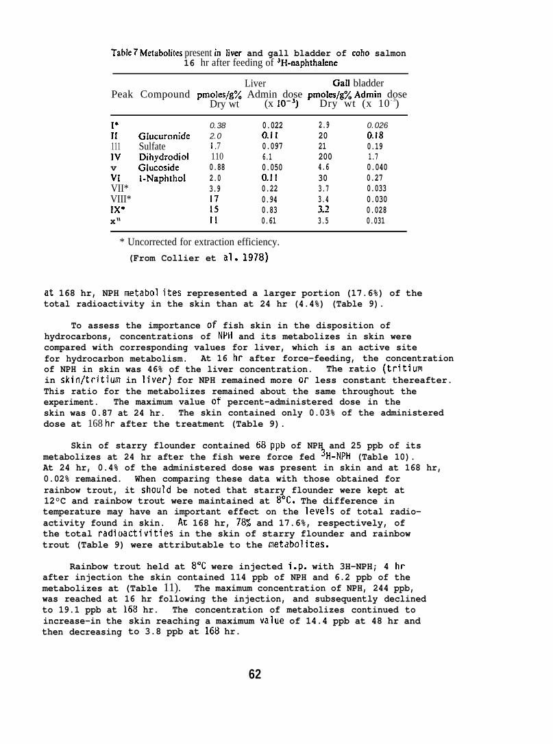
Table 7 Metabolites present in liver and gall bladder of coho salmon16 hr after feeding of 3H-napkthalene
Liver Gall bladderPeak Compound pmoles/g% Admin dose pmoles/g% ,4dmin dose
Dry wt (x 10-’) Dry wt (x 10 - 3)
1’11 Glucuronide111 Sulfatew Dihydrodiolv Glucosidew l-NaphtholVII*VIII*IX*x“
0.382.0i .71100.882.03.9171511
0.0220.110.0976.10.0500.110.220.940.830.61
2.920212004.6303.73.43.?3.5
0.0260.t80.191.70.0400.270.0330.0300.0280.031
* Uncorrected for extraction efficiency.
(From Collier et al. 1978)
at 168 hr, NPH metabol ites represented a larger portion (17.6%) of thetotal radioactivity in the skin than at 24 hr (4.4%) (Table 9).
To assess the importance of fish skin in the disposition ofhydrocarbons, concentrations of NPH and its metabolizes in skin werecompared with corresponding values for liver, which is an active sitefor hydrocarbon metabolism. At 16 hr after force-feeding, the concentrationof NPH in skin was 46% of the liver concentration. The ratio (tritiumin skin/tritium in liver) for NPH remained more or less constant thereafter.This ratio for the metabolizes remained about the same throughout theexperiment. The maximum value of percent-administered dose in theskin was 0.87 at 24 hr. The skin contained only 0.03% of the administereddose at 168 hr after the treatment (Table 9).
Skin of starry flounder contained 68 ppb of NPH and 25 ppb of itsmetabolizes at 24 hr after the fish were force fed 3H-NPH (Table 10).At 24 hr, 0.4% of the administered dose was present in skin and at 168 hr,0.02% remained. When comparing these data with those obtained forrainbow trout, it should be noted that starry flounder were kept at12°C and rainbow trout were maintained at 8“C. The difference intemperature may have an important effect on the levels of total radio-activity found in skin. Ax 168 hr, 78$ and 17.6%, respectively, ofthe total radioactivities in the skin of starry flounder and rainbowtrout (Table 9) were attributable to the netabolites.
Rainbow trout held at 8°C were injected i.p. with 3H-NPH; 4 hrafter injection the skin contained 114 ppb of NPH and 6.2 ppb of themetabolizes at (Table 11). The maximum concentration of NPH, 244 ppb,was reached at 16 hr following the injection, and subsequently declinedto 19.1 ppb at 168 hr. The concentration of metabolizes continued toincrease-in thethen decreasing
skin reaching a maximum value of 14.4 ppb at 48 hr andto 3.8 ppb at 168 hr.
62
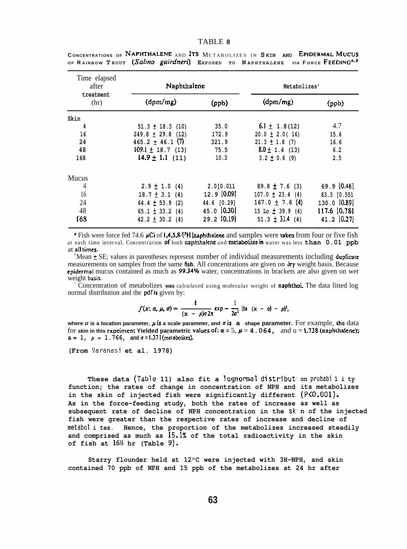
TABLE 8
C ONCENTRATIONS OF NAPHTHALENE AND ITS METABOLIZES IN S KIN AND EPIDERMAL MucusO F R A I N B O W T ROUT (Sa/mo guirchrer~ EXPOSED TO N APHTHALENE VIA F O R C E FEEDING”Cb
Time elapsedafter Naphthalenc Metabolizes’
treatment(hr) (dpm/mg) (ppb) (dpm/mg) (ppb)
Skin4
162448168
Mucus4
162448
S68
51.3 t 18.3 (10)249.8 t 29.8 (12)465.2 & 46.1 (7)109.1 t 18.7 (13)14.9t 1.1 (11)
2.9 t 1.0 (4)18.7 A 3.1 (4)64.4 t 53.9 (2)65.1 t 33.2 (4)42.2 t 30.2 (4)
35.0172.9321.975.510.3
2.0[0.01112.9 [0.09144.6 [0.29]45.0 [0.30129.2 fO.19]
6.1 t 1.8(12)20.0 A 2.0( 16)21.3 t 1.8 (7)8.Ok 1.4 (13)3.2 ~ 0.6 (9)
89.8 ~ 7.6 (3)107.0 t 23.4 (4)167.0 ~ 7.6 (4)15 Lo * 39.9 (4)51.3 * 31.4 (4)
4.715.616.66.22.5
69.9 [0.46183.3 [0.551130.0 [0.89]117.6 [0.78]41.2 [0.27J
* Fish were force fed 74.6 gCi of 1,4,5,8-[JH Inaphthalene and samples were taken from four or five fishat each time interval. Concentration of both naphthalene and metabotites in water was less than 0.01 ppbat all times.
b Mean t SE; values in parentheses represent number of individual measurements including dup!icatemeasurements on samples from the same fish.. All concentrations are given on dry weight basis. Becauseepidermal mucus contained as much as 99.34’?+ water, concentrations in brackets are also given on wetweight bask.
c Concentration of metabolizes was calculated using molecular weight of naphthol. The data fitted lognormal distribution and the pdfis given by:
f(xq~o)= ‘ - * Hn (x - a) - gjl,(x - &2R CXp
where a is a location parameter, L is a scale parameter, and u is a shape parameter. For example, the datafor skin in this expciment Yielded parametric values ofi u = 5, Y = 4.064, and u = 1.738 (naphthalene);a = 1, M = 1.766, and u = l_.37 I (m-etabolites).
(From Varanasi et al. 1978)
These data (Table 11) also fit a Iognormal distributfunction; the rates of change in concentration of NPH andin the skin of injected fish were significantly differentAs in the force-feeding study, both the rates of increasesubsequent rate of decline of NPH concentration in the sk”
on probabi 1 i tyits metabolizes(P<o.ool).as well asn of the injected
fish were greater than the respective rates of increase and decline ofmetabol i tes. Hence, the proportion of the metabolizes increased steadilyand comprised as much as 15.1% of the total radioactivity in the skinof fish at 168 hr (Table 9).
Starry flounder held at 12°C were injected with 3H-NPH, and skincontained 70 ppb of NPH and 15 ppb of the metabolizes at 24 hr after
63
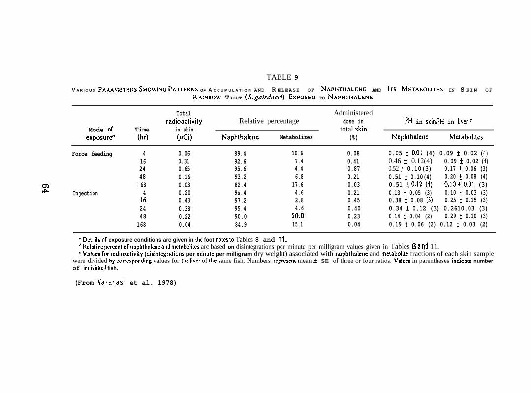
TABLE 9
V A R I O U S Pmwmxs SHOWiNG PA~ERNS OF A C C U M U L A T I O N AND R ELEASE OF NAPIITIIALENE AND ITS METAFIOLITES IN S KIN OFRAIN~OW TROUT (S. gairdl~ero EXPOSED TO NAPHTHALENE
Total Administeredradioactivky Relative percentage dose in [’H in skin/’H in liverl’
Mode Of Time in skin total skincxpOsurc” (hr) (pCi) Naphthalcne Metabolizes (%) Naphthalene Metabolhts
Force feeding 4162448
m I 68lb Injection 4
162448168
0.060.310.650.160.030.200.430.380.220.04
89.492.695.693.282.49s.497.295.490.084.9
10.67.44.46.8
17.64.62.84.6
10.015.1
0.080.410.870.210.030.210.450.400.230.04
0.05 f 0.01 (4) 0.09 f 0.02 (4)0.46 t 0.12(4) 0.09 i 0.02 (4)0.52 t 0.10(3) 0.17 t 0.06 (3)0.51 * 0.10(4) 0.20 f 0.08 (4)0.51 f0.12 (4) 0.10+0.01 (3)0.13 * 0.05 (3) 0.10 f 0.03 (3)0.38 + 0.08 (3) 0.25 t 0.15 (3)0.34 t 0.12 (3) 0.2610.03 (3)0.14 * 0.04 (2) 0.29 i 0.10 (3)0.19 t 0.06 (2) 0.12 t 0.03 (2)
u D~tiiil~ of exposure conditions arc given in Ihc foot no!cs to Tables 8 and It.h Relil[ivc pcrccnl of n:lpllth:dcnc ml mclabolilcs arc based on disintegrations pcr minute per milligram values given in Tables 8 a 11~ 11.v VAICS for rudionclivity (disin[cgrations per minulc per milligram dry weight) associated with naphthalcnc and mctabolitc fractions of each skin sample
were divided by corrqwnding values for the Iivcr of IIIC same fish. Numbers rcprcsenl mean ~ SE of three or four ratios. values in parentheses indicate numberof imlivitltull fish.
(From Varanasi et al. 1978)
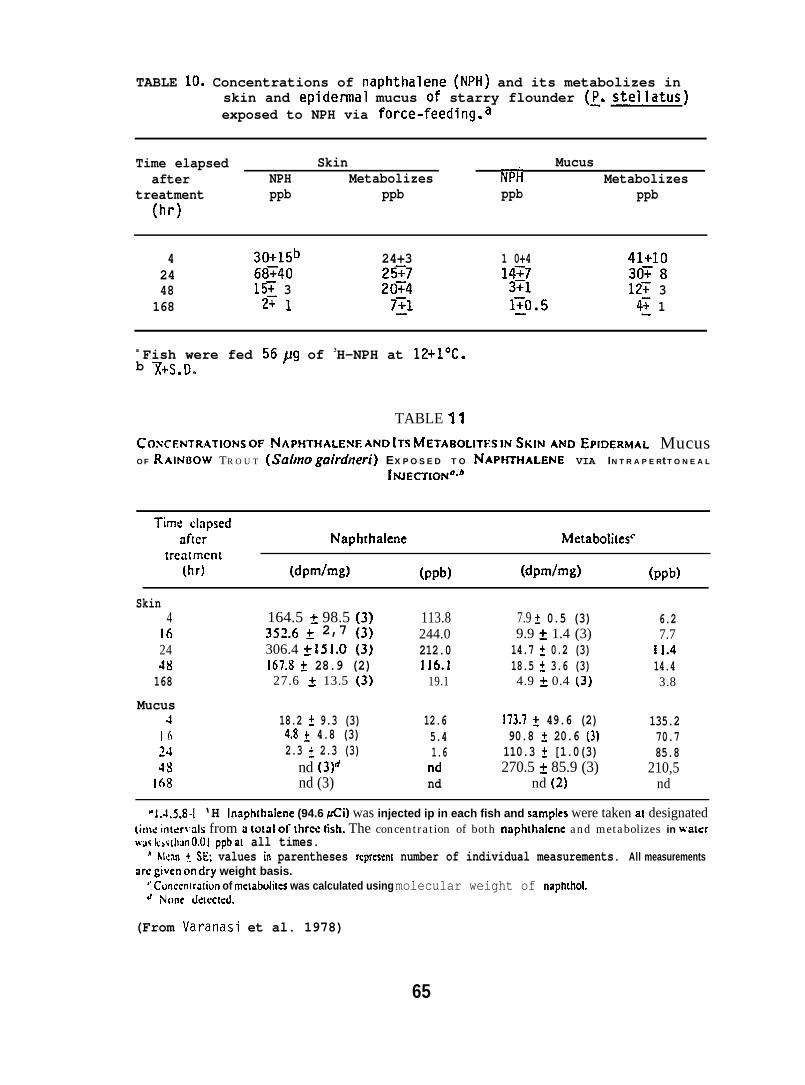
TABLE 10. Concentrations of naphthalene (NPH) and its metabolizes inskin and epidermal mucus of starry flounder (~. stellatus)exposed to NPH via force-feeding.a
Time elapsed Skin Mucusafter NPH Metabolizes NPti Metabolizes
treatment ppb ppb(hr)
ppb ppb
4 30+15b 24+3 1 0+4 41+1024 68T40 25T7 14T7 307848 15T 3 20~4 3T1 12T 3
168 2., I 7~1 1:0 ● 5 4~ 1
a Fish were fed 56~g of 3H-NPH at 12+1”C.b ~+S,D-
TABLE 11
COXF~TRATIOM OFNAPHTHALE~~ AND !Ts METABOLIZES IN$KINANDEPIDERMAL MucusO F RAIFJOOW TR O U T (Suhrm gairdneri) EX P O S E D T O NAPHTHALEFJE VIA IN T R A P E Rt T O N E A L
INJECTION”Sb
Time elapsedafter Naphthalene Metabolizes’”
trea[mcnt(hr) (dpm/mg) (ppb) (dpm/mg) (ppb)
Skin4 164.5 t 98.5 [3) 113.8 7.9 I 0.5 (3) 6.2
16 352.6 ~ 2,7 {3) 244.0 9.9 * 1.4 (3) 7.724 306.4 ~ 151.0 (3) 212.0 14.7 * 0.2 (3) 11.448 167.8 t 28.9 (2) 116.1 18.5 k 3.6 (3) 14.4168 27.6 A 13.5 (3) 19.1 4.9 * 0.4 (3) 3.8
Mucus4 18.2 t 9.3 (3) 12.6 173.7 ~ 49.6 (2) 135.2
I 6 4.8 ~ 4.8 (3) 5.4 90.8 ~ 20.6 (3) 70.7~~ 2.3 ~ 2.3 (3) 1.6 110.3 * [1.0(3) 85.848 nd (3)d nd 270.5 t 85.9 (3) 210,5
168 nd (3) nd nd (2) nd
“ 1..t.5,8-l ‘H lnaphthalene (94.6 Xi) was injected ip in each fish and sampIes were taken at designated[imc inter~als from u total 0[ three tish. The concentration of both naphthakne and metabolizes in walerwaslusi tilun O.ol ppbat all times.
A Nlcnn +. SE: values in parentheses rcprcsenl number of individual measurements. All measurementsarcgiven on dry weight basis.
‘. Conccn[ra[iun of mcLabcdites was calculated using molecular weight of naptnhol.~ None detected.
(From Varanasi et al. 1978)
65
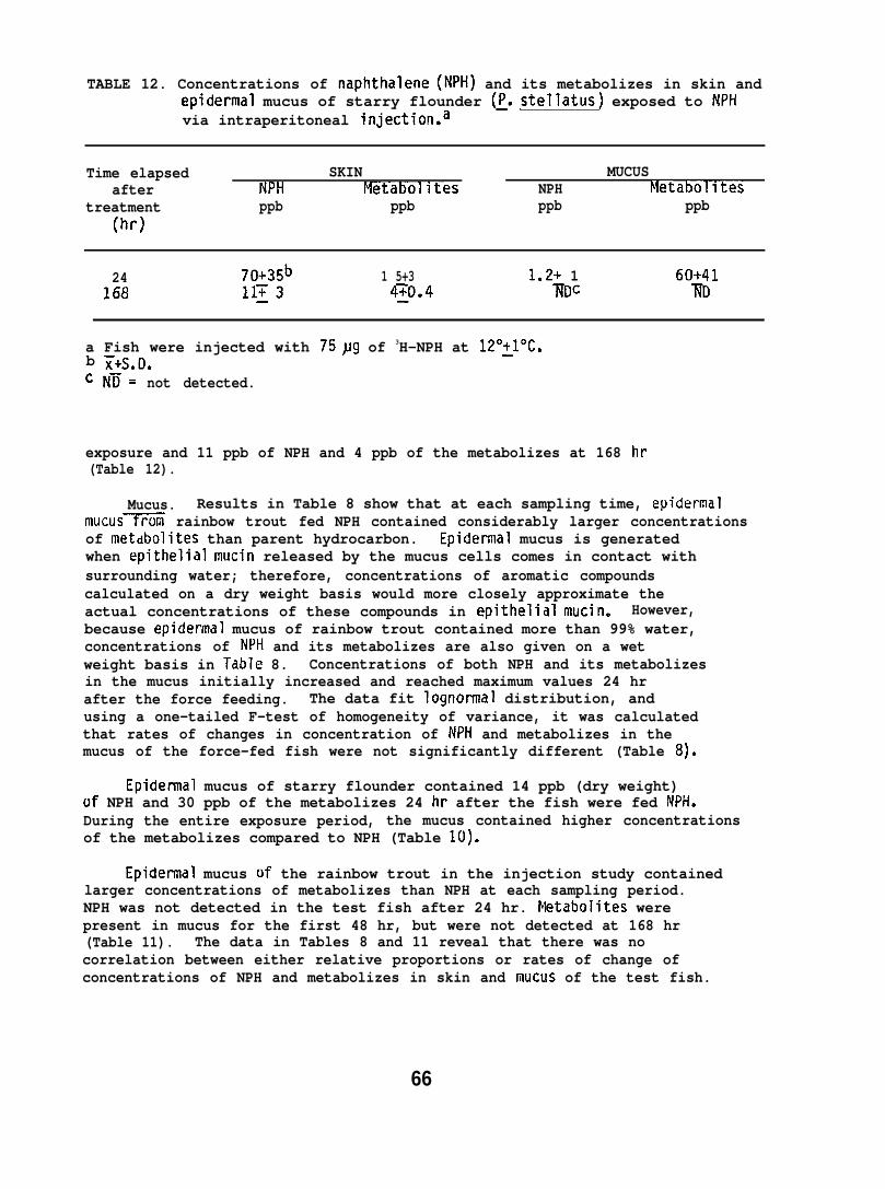
TABLE 12. Concentrations of naphthalene (NPH) and its metabolizes in skin andepidermal mucus of starry flounder (~. stellatus) exposed to NPHvia intraperitoneal injection.a
Time elapsed SKIN MUCUSafter NPI-I Metabolltes NPH Metabolltes
treatment ppb ppb ppb ppb(hr)
24 70+35b 1 5+3 1.2+ 1 60+41168 1113 ~o.4 ~DC ~D
a Fish were injected with 75~g of 3H-NPH at 12°~10C.b ~+S.D.cN~= not detected.
exposure and 11 ppb of NPH and 4 ppb of the metabolizes at 168 hr(Table 12).
Mucus. Results in Table 8 show that at each sampling time, epidermalmucus= rainbow trout fed NPH contained considerably larger concentrationsof metdbolites than parent hydrocarbon. Epidermal mucus is generatedwhen epithelial mucin released by the mucus cells comes in contact withsurrounding water; therefore, concentrations of aromatic compoundscalculated on a dry weight basis would more closely approximate theactual concentrations of these compounds in epithelial mucin. However,because epidermal mucus of rainbow trout contained more than 99% water,concentrations of NPH and its metabolizes are also given on a wetweight basis in Table 8. Concentrations of both NPH and its metabolizesin the mucus initially increased and reached maximum values 24 hrafter the force feeding. The data fit Iognormal distribution, andusing a one-tailed F-test of homogeneity of variance, it was calculatedthat rates of changes in concentration of NPH and metabolizes in themucus of the force-fed fish were not significantly different (Table 8).
Epidennal mucus of starry flounder contained 14 ppb (dry weight)of NPH and 30 ppb of the metabolizes 24 hr after the fish were fed NPH.During the entire exposure period, the mucus contained higher concentrationsof the metabolizes compared to NPH (Table 10).
Epidermal mucus of the rainbow trout in the injection study containedlarger concentrations of metabolizes than NPH at each sampling period.NPH was not detected in the test fish after 24 hr. Netabolites werepresent in mucus for the first 48 hr, but were not detected at 168 hr(Table 11). The data in Tables 8 and 11 reveal that there was nocorrelation between either relative proportions or rates of change ofconcentrations of NPH and metabolizes in skin and mucus of the test fish.
66
6.1.5 Accumulation and Biotransformation of NPI-I by Flatfish
Force-feeding Study (Starry Flounder) at 12°C. Tissues of starryflounder contained considerable concentrations of both NPH and itsmetabolic products 24 hr after force-feeding NPH (Fig. 14). Maxima inconcentrations of both the hydrocarbon and its metabolic productsoccurred during the first 48 hr,then began to decline in all tissuesexcept bile (Fig. 14). Statistical treatment of these data shows thatthe rates of decline of NPH concentrations in liver, skin, and bloodwere significantly greater than the respective rates of decline inmetabolize concentrations (Fig. 14). At 168 hr, all sites examined,except brain, contained considerably more radioactivity associatedwith netabolic products than with NPH (Fig. 14; Table 13); more than70% of the total radioactivity in blood and liver was associated withthe metabolic products. Throughout the experiment, radioactivity inbile of starry flounder was largely due to metabolizes.
Force-feeding Study (Rock Sole) at 12°C. Comparison of the datain Figures 14 and 15 revealed that the radioactivity in all tissues ofrock sole was considerably greater than the radioac~ivity in thecorresponding tissues of starry flounder at 24 hr. Moreover, valuesfor NPH and metabolize concentrations in tissues of rock sole weresignificantly (P<O.01) higher than the corresponding values for starryflounder at 168 hr. Liver, skin, and muscle of starry flounder contained11.6, 3.0, and 1.7 g of lipid, respectively, per 100 g of wet tissue;whereas, liver, skin, and muscle of rock sole contained 4.3, 1.5, and1.0 g of lipid, respectively, per 100 g of wet tissue.
Differences between rates of decline in NPH and the metabolizeconcentrations in liver, blood, and skin of rock sole were also measured.As with starry flounder, the rate of decline in NPH concentration ineach tissue was greater than the rate of decline in metabolize concentration(Fig. 15). It was also determined that the extent of decline of NPH ineach of the tissues was greater for starry flounder than for rock sole.From 24 to 168 hr, there was 12.4- and 2.3-fold decrease, respectively,in NPH dnd metabolize concentrations in the livers of rock sole;corresponding values for starry flounder were 63.0 and 2.6.
Stomach and intestine of rock sole contained more radioactivitythan those of starry flounder at both 24 and 168 hr. At 24 hr, stomachand intestine of the rock sole contained 0.3 and 6.0%, respectively, ofthe administered dose, whereas the corresponding values for starryflounder were 0.2 and 1.5.
Because considerable radioactivity was present in the rock soleafter 1 wk, some fish were sampled at 6 wk after the treatment. Gillsand kidney contained considerable concentrations of metabolic products,whereas bile and liver contained smaller concentrations, and in theother tissues examined radioactivity was barely detectable (Fig. 15).
67
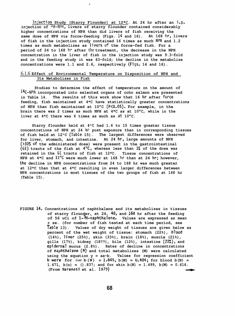
Injection Study (Starry Flounder) at 12*C. At 24 hr after an i.p.injection of JI-I-NPH, livers of starry flounder contained considerablyhigher concentrations of NPH than did livers of fish receiving thesame dose of NPH via force-feeding (Figs. 14 and 16). At 168 hr, liversof fish in the injection study contained 16 times as much NPtl and 1.2times as much metabolizes as Iive.rs of the force-fed fish. For aperiod of 24 to 168 hr after the treatment, the decrease in the NPHconcentration in the liver of fish in the injection study was 9.3-foldand in the feeding study it was 63-fold; the decline in the metabolizeconcentrations were 1.1 and 2.6, respectively (Figs. 14 and 16).
6.1.6 Effect of Environmental Temperature on Disposition of NPH andIts Metabolizes in Fish
Studies to determine the effect of temperature on the amount of14C-NPH incorporated into selected organs of coho salmon are presentedin Table 14. The results of this work show that 16 hr after forcefeeding, fish maintained at 4*C have statistically greater concentrationsof NPH than fish maintained at 10”C (P<O.05). For example, in thebrain there was 2 times as much NPH at 4*C as at 10”C, while in theliver at 4*C there was 6 times as much as at 10”C.
Starry flounder held at 4°C had 1.6 to 15 times greater tissueconcentrations of NPH at 24 hr post exposure than in corresponding tissuesof fish held at 12°C (Table 15). The largest differences were observedfor liver, stomach, and intestine. At 24 hr, large amounts of NPH(>30% of the administered dose) were present in the gastrointestinal(GI) tracts of the fish at 4°C, whereas less than 3% of the dose wasretained in the GI tracts of fish at 12*C. Tissue concentrations ofNPH at 4*C and 12*C were much lower at 168 hr than at 24 hr; however,the decline in NPH concentrations from 24 to 168 hr was much greaterat 12*C than that at 4*C resulting in even larger differences betweenNPH concentrations in most tissues of the two groups of fish at 168 hr(Table 15).
FIGURE 14. Concentrations of naphthalene and its metabolizes in tissuesof starry flounder, at 24, 48, and 168 hr after the feedingof 56 uCi of l-3H-naphthalene. Values are expressed as mean+ se. (for number of fish tested at each time period, seeTable 13). Values of dry weight of tissues are given below aspercent of the wet weight of tissue: stomach (22%), blood(14%), liver (25%), skin (33%), brain (18%), muscle (21%),gills (17%), kidney (18??), bile (12%), intestine (22%), andepidermal mucus (2.8%). Rates of decline in concentrationsof naphthalene (N) and total metabolizes (M) were calculatedusing the equation y = ax-b. Values for regression coefficientbwere for liver b(N) = 1.845$ b(M) = tl.404; for blood b(N) =1.671, b(n) = ().837; and for skin b(N) = 1.499, b(M) = 0.616.(From Varanasi et al. 1979)
68
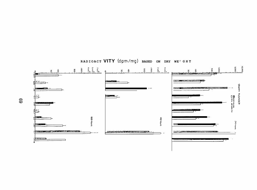
RADIOACT
1-~,.,
‘. :>-–”
VITY (dpm/mg) BASED ON DRY WE’
1 .-
kEEE!!54-
G H T
.- -.
,.L/JDn
La
—.
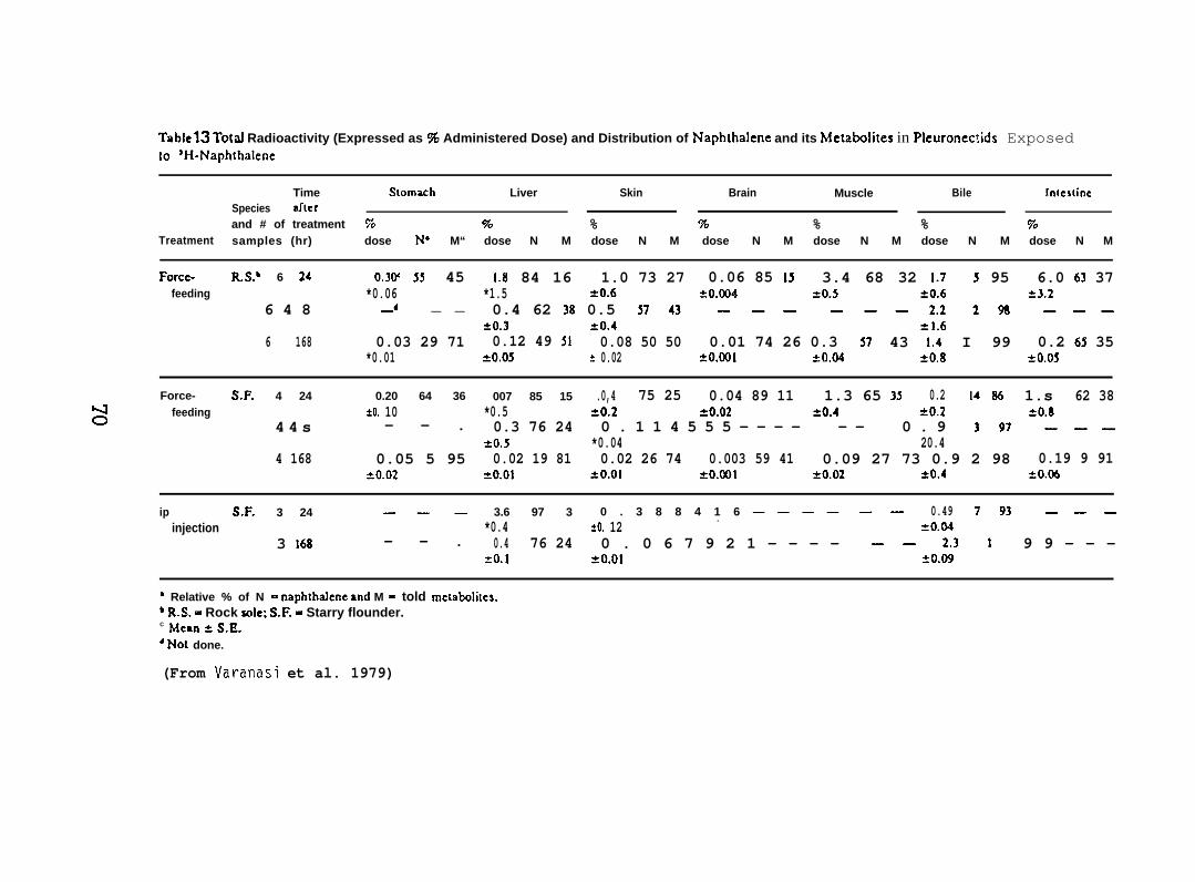
Tablets Total Radioactivity (Expressed as % Administered Dose) and Distribution of Naphthalene and its Mctabolites in Pleuronectids Exposedto ‘H-Naph[halene
Time S[omach Liver Skin Brain Muscle Bile !nteslincSpecies Sflcrand # of treatment 70 70 % % % %
Treatment70
samples (hr) dose N“ M“ dose N M dose N M dose N M dose N M dose N M dose N M
Forcc- R.S.b 6 24 0.3W 55 45 1.8 84 16 1.0 73 27 0.06 85 15 3.4 68 32 1.7 5 95 6.0 63 37feeding *0.06 *1.5 =0.6 20.(X)4 =0.5 ?0.6 23.2
6 4 8 _d _ _ 0.4 62 38 0.5 J743— ——— —— 2.2298———20.3 ?0.4 =1.6
6 168 0.03 29 71 0.12 49 51 0.08 50 50 0.01 74 26 0.3 57 43 1.4 I 99 0.2 65 35*0.01 *&os * 0.02 *0.001 *0.M 20.8 20.05
Force- S.F. 4 24 0.20 64 36 007 85 15 .0,4 75 25 0.04 89 11 1.3 65 35 0.2 14 86 1.s 62 38
s feeding *O. 10 *0.5 =0.2 20.02 20.4 50.2 SO.84 4 s — — . 0.3 76 24 0 . 1 1 4 5 5 5 — — — — — — 0 . 9 397———
*0.5 *0.04 20.44 168 0.05 5 95 0.02 19 81 0.02 26 74 0.003 59 41 0.09 27 73 0.9 2 98 0.19 9 91
20.02 *0.01 *o+ol *0.001 ?0.02 20.4 *0.06
ip S.F. 3 24 ——— 3.6 97 3 0 . 3 8 8 4 1 6 — — — — —— 0.49 793-——injection *0.4 .
*O. 12 =0.043 16s — — . 0.4 76 24 0 . 0 6 7 9 2 1 — — — — ——2.31 9 9 - - -
*0.1 *0.01 20.09
‘ Relative % of N = naphthalene and M = told mctaboli[cs.‘ R.S. = Rock sole; S.F. - Starry flounder.C Mean = S.E.4 ~01 done.
(From Varanasi et al. 1979)
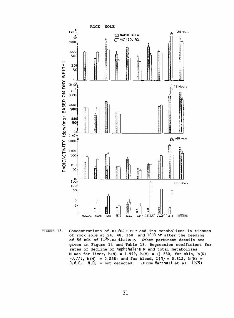
ROCK SOLE
FIGURE 15.
1 X1051
k
I ad
5000
ICOO ~
500 ;
100 ;50 ;
1 0
3K105JI do
1000
“k
500 j’
100 ;50 :;
10 ~5 XI(Y’
1500
7I 000
1. .. . .. .. . .:..:.:::.:.:. . ..:
EZ3NAPHTHALENEO ME7ABOLITES
J. . .. .::::::. . .:.. . .::.::::.:
h 24 Hours
& 48 Hours- -
11::. .:.. .:: . ..: ::.:. .:.
.-2 ursI
ST0i44CH ~0 LIVER SKIN @7AlN ML!5CLE GILLS KICNEY 81LE INTESTINE
Concentrations of naphthalene and its metabolizes in tissuesof rock sole at 24, 48, 168, and 1008 hr after the feedingof 56 uCi of l-3H-napthalene. Other pertinent details aregiven in Figure 14 and Table 13. Regression coefficient forrates of decline of naphthalene N and total metabolizesM was for liver, b(N) = 1.999, b(M) = ().530, for skin, b(N)=0.771, b(M) = 0.558; and for blood, b(N) = 0.812, b(M) =0.601. N.D. = not detected. (From Varanasi et al. 1979)
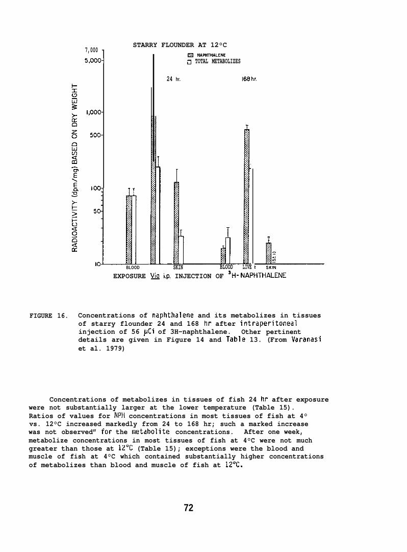
7,000 -5,000-
1,ooo-
5oo-
1oo-
50-
STARRY FLOUNDER AT 12°C
J:.:.::::.x
:.:.
::::
::::
.:
:::
:::
y
:::
:::
:::
:::
y
:.:.>
10 BLOOD
m NAPHTHALENEn TOTAL METABOLIZES
24 hr. 168hr.
SKIN BLOOD LIVE
T
L? SKINEXPOSURE VA i.p. INJECTION OF SH-NAPHTHALENE
FIGURE 16. Concentrations of naphthalene and its metabolizes in tissuesof starry flounder 24 and 168 hr after intraperitonealinjection of 56 ~Ci of 3H-naphthalene. Other pertinentdetails are given in Figure 14 and Table 13. (From Varanasiet al. 1979)
Concentrations of metabolizes in tissues of fish 24 hr after exposurewere not substantially larger at the lower temperature (Table 15).Ratios of values for NPH concentrations in most tissues of fish at 4°vs. 12°C increased markedly from 24 to 168 hr; such a marked increasewas not observed” for the met~holite concentrations. After one week,metabolize concentrations in most tissues of fish at 4°C were not muchgreater than those at 12°C (Table 15); exceptions were the blood andmuscle of fish at 4°C which contained substantially higher concentrationsof metabolizes than blood and muscle of fish at 12”C.
72
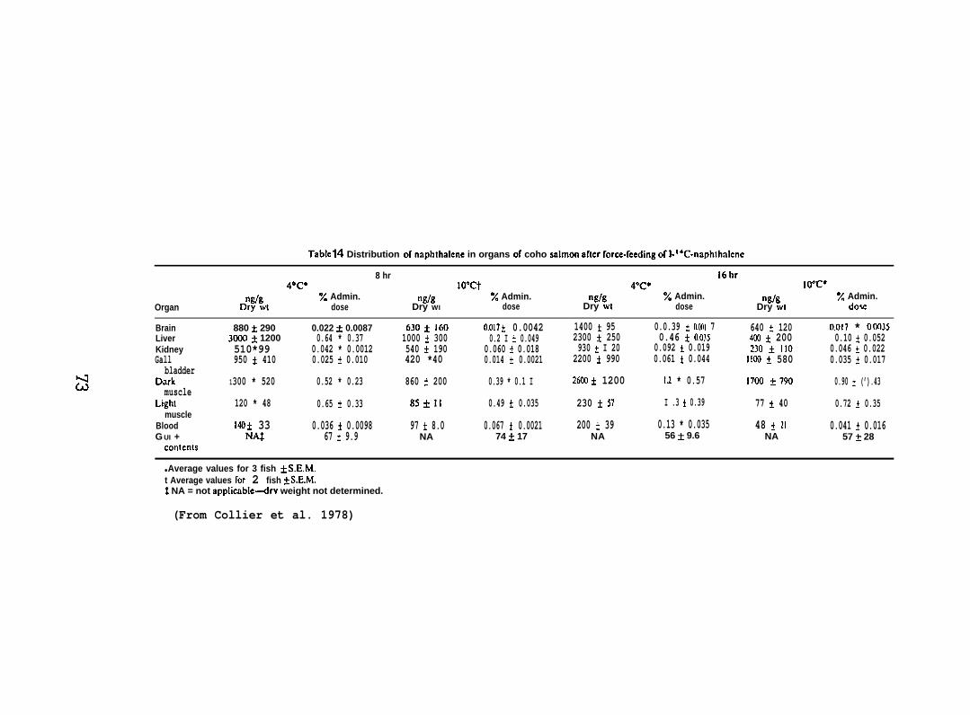
Tablcf14 Distribution of rmphtbalene in organs or coho wrlmon aflcr force-keding of l-’’n~phth~lcncnc
8 hr 16hr4@c* lo”ct 40C* Io”c”
Wg % Admin. ngfg % Admin. nglg % Admin. nglg ~ Admin.Organ Dry wl dose Dry WI dose Dry wt dose Dry WI Slosc
Brain 880 f 290 0.022 & 0.0087 630~ 160 0.017 * 0.0042 1400 * 95 0.0.39 * O.(MI 7 640 f 120 0.017 * oon35Liver 3(Kro* 1200 0.64 * 0.37 1000 * 300 0.2 I * 0.049 2300 ~ 250 0.46 t 0.035 4CX3 * 200 0.10 & 0.052Kidney 510*99 0.042 * 0.0012 540 * 190 0.060 * 0.018 930 * I 20 0.092 * 0.019 230 * 110 0.046 + 0.022Gall 950 * 410 0.025 + 0.010 420 *40 0.014 + 0.0021 2200 * 990 0.061 * 0.044 1100 ~ 580
bladder0.035 * 0.017
Dark 1300 * 520 0.52 * 0.23 860 * 200 0.39 * 0.1 I 2600* 1200 1.2 * 0.57 !700 +790muscle
0.90 * (’).43
Li~hl 120 * 48 0.65 t 0.33 85+11 0.49 + 0.035 230 + 57 I .3 * 0.39 77 * 40 0.72 ~ 0.35muscle
Blood 140 & 33 0.036 + 0.0098 97 & 8.0 0.067 k 0.0021 200 & 39 0.13 * 0.035 48 k 21 0.041 + 0.016G UI + NA$ 67 & 9.9 NA 74 * 17 NA 56 ~ 9.6 NA 57 f 28
conknls
● Average values for 3 fish ~ S.E.M.t Average values for 2 fish *S.E.M.t NA = not applicable-drv weight not determined.
(From Collier et al. 1978)
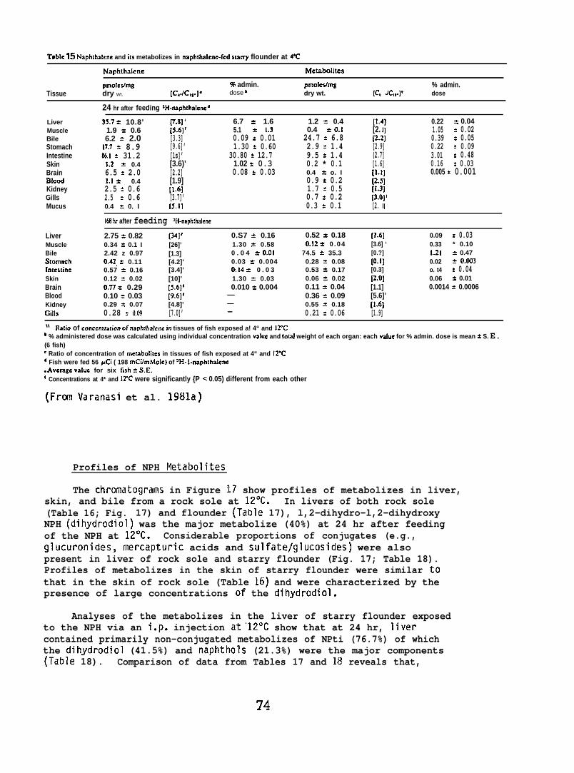
Table 15 Naphlhalene and its metabolizes in naphthalene-fed srarry flounder at 4’C
Naphthalene Metabolitcs
pmolestmg 9% admin.Tissue dry Wt.
pmolealmg % admin.[c,./c,,.]’ dose b dry wt. [c, Jc,*.]f dose
LiverMuscleBileStomachIntestineSkinBrainBloodKidneyGillsMucus
24 hr after feeding ‘H-naphlhalened
33.7 = 10.8’ p.tl] ‘ 6.7 & 1.6 1.2 * 0.4 [1.4] 0.22 = 0.041.9 : 0.6 [5.6]’ 5.1 z 1.3 0.4 = 0.16.2 = 2.0
[2. 1] 1.05 * 0.02[3.3] 0.09 * 0.01 24.7 % 6.8 p.2J 0.39 = 0.05
17.7 % 8.9 [9.6]’ 1.30 = 0.60 2.9 ? 1.486.1 = 31.2
[2.9] 0.22 z 0.09[1s]’ 30.80 z 12.7 9.5 = 1.4 (2.7] 3.01 * 0.48
1.2 = 0.4 [3.6)’ 1.02 * 0.3 0.2 * 0.1 [1.6] 0.16 * 0.036.5 = 2.0 [2.2] 0.08 * 0.03 0.4 z o. I1.1 = 0.4
[1.1] 0.005 * 0.001[1.9] 0.9 * 0.2
2.5 = 0.6[2.3]
[1.6] 1.7 = 0.5 [1.3)2.5 = 0.6 [3.7]’ 0.7 * 0.2 [3.0]’0.4 * 0. I rs.11 0.3 = 0.1 [2. 1]
168 hr after feeding ‘H-naphthaleneLiver 2.75 = 0.82 [34]’ 0.S7 = 0.16 0.52 = 0.18 [1.6] 0.09 z 0.03Muscle 0.34 = 0.1 I [26]’ 1.30 = 0.58 0,12 * 0 .04 [3.6] ‘ 0.33Bile
* 0.102.42 z 0.97 [1.3] 0 . 0 4 * O.o1 74.5 * 35.3 [0.?] 1.21 2 0.47
Stomnch 0.42. = 0.11 [4.2]’ 0.03 * 0.004 0.28 = 0.08 [0.1] 0.02 * 0.L133hrtestine 0.57 = 0.16 [3.4]’ 0.14 * 0 . 0 3 0.53 = 0.17 [0.3] o. t4 * 0.04Skin 0.12 = 0.02 [10]’ 1.30 * 0.03 0.06 * 0.02 p.o] 0.06 * 0.01Brain O.n z 0.29 [5.6]r 0.010 = 0.004 0.11 * 0.04 [1.1] 0.0014 t 0.0006Blood 0.10 = 0.03 [9.6]’ — 0.36 = 0.09 [5.6]’Kidney 0.29 : 0.07 [4.8]’ — 0.55 = 0.18 [1.6]Gills 0.28 : 0.C9 [7.0]’ — 0.21 * 0.06 [1.9]
“ Ratio of conccrwaticm of naplr!halcnc in tissues of fish exposed a! 4“ and 12”Cb % administered dose was calculated using individual concentration vaIue and !otal weight of each organ: each vafue for % admin. dose is mean = S. E.(6 fish)C Ratio of concentration of metabolitcs in tissues of fish exposed at 4“ and 12-Cd Fish were fed 56 pCi ( 198 mCi/mMole) of aH- I-naphthaknc● Aveiage value for six fish = S.E.r Concentrations at 4* and 12’C were significantly {P < 0.05) different from each other
(Fran Varanasi et al. 1981a)
Profiles of NPH Pletabolites
The chromatograms in Figure 17 show profiles of metabolizes in liver,skin, and bile from a rock sole at 12”C. In livers of both rock sole(Table 16; Fig. 17) and flounder (Table 17), l,2-dihydro-l,2-dihydroxyNPH (dihydrodiol) was the major metabolize (40%) at 24 hr after feedingof the NPH at 12°C. Considerable proportions of conjugates (e.g.,glucuronides, mercapturic acids and sulfate/glucosides) were alsopresent in liver of rock sole and starry flounder (Fig. 17; Table 18).Profiles of metabolizes in the skin of starry flounder were similar tothat in the skin of rock sole (Table 16) and were characterized by thepresence of large concentrations of the dihydrodiol.
Analyses of the metabolizes in the liver of starry flounder exposedto the NPH via an i.p. injection at’12°C show that at 24 hr, livercontained primarily non-conjugated metabolizes of NPti (76.7%) of whichthe dihydrodiol (41.5%) and naphthols (21.3%) were the major components(Table 18). Comparison of data from Tables 17 and 18 reveals that,
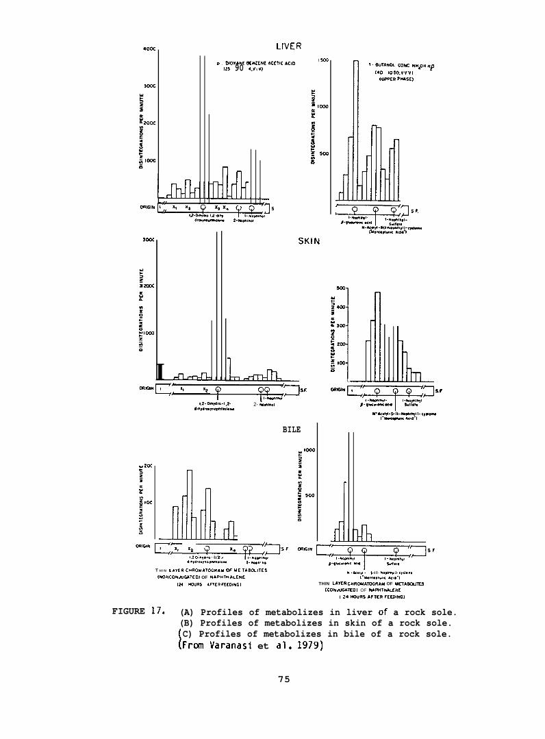
400C
Lh5
I SoOP . OIOXbNE 0EN2EN2 ACCTIC ACIO(25 90 4.V v v)
n
I mm-!-l
--n
1.2- Othydre-l ,2-*t”,d,u-,r,ahw
rldll _““” G- ‘2 -T=qTa”
!,2 0,”,4 !.. l.? . ,. *,*!*,
d..,am.,.. vhoto”g”g 2- W”. *O4
2- Iiara,mal
BILE
T H I N LAYER CMROUA?OGRAU W MC TAOCi.lTES
INOF+CONJUCMTEO) OF NAPHTMALENE[24 HOURS AFTER fEEO+UGl
J
9. 8UTANOL CONC NM40U H#
I*O 80 m,” ““,
iwER PIibsE)
L
h -a,.!,,”. s.{ I - N,dwq!l. c,,,,.”,I“IMK*W.OC AC,*-1
THIN LAYER CHRCUAWRAM OF METASWIES
(CONJUGS7’0) OF MMTMALCNC
I M MOURS AFTfR FEEO+NGI
FIGURE 17. (A) Profiles of metabolizes in liver of a rock sole.(B) Profiles of metabolizes in skin of a rock sole.
[C) Profiles of metabolizes in bile of a rock sole.Fr~ Varanasi et al. 1!w9)
75
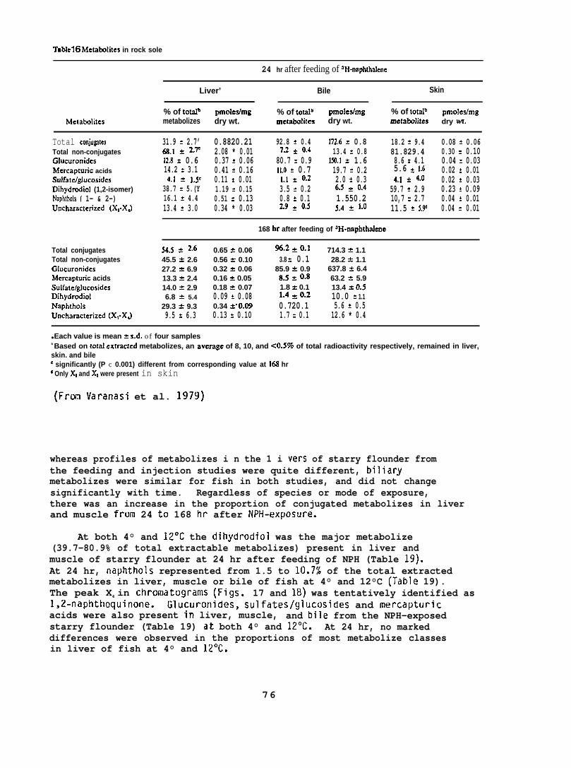
Tabte16 Metatdites in rock sole
24 hr after feeding of ‘H-naphthalene
Liver’ Bile Skin
% of totalb pmoIes/mg % of totalb pmoledmg % of totalbMetakolitcs
pmolcdmgmetabolizes dry wt. metabdites dry wt. metabdites dry wt.
Total conju@esTotal non-conjugatesGhscuronidesMcrcaptunc acidsSulfatdgJucosidesDihydrodiol (1,2-isomer)Naphthols { 1- & 2-)Uncharacterized (X,-XJ
31.9 = 2.7’ 0.8820.21~.1 * 2-7 2.08 * 0.0112.8 t 0.6 0.37 * 0.0614.2 % 3.1 0.41 * 0.164.j * l-se 0.11 * 0.01
38.7 = 5.(Y 1.19 * 0.1516.1 * 4.4 0.51 * 0.1313.4 * 3.0 0.34 * 0.03
92.8 = 0.4 872.6 Y 0.8 18.2 * 9.4 0.08 = 0.067.2 = o.4 13.4 = 0.8 81.829.4 0.30 * 0.10
80.7 = 0.9 150.1 = 1.6 8.6 * 4.1 0.04 * 0.0311.0 * 0.7 19.7 = 0.2 5.6 ~ 1.6 0.02 * 0.01l.! * 0.2 2.0 * 0.3 4.1 * 4.(3 0.02 * 0.033.5 * 0.2 6.5 ~ o.4 59.7 = 2.9 0.23 * 0.090.8 = 0.1 1.550.2 10,7 = 2.7 0.04 * 0.012.9 = o.5 5.4 * 1-() 11.5 * 5.9 0.04 = 0.01
168 hr after feeding of aH-naphthalene
Total conjugatesTotal non-conjugatesGlucuronidcsMcrcaptunc acidsSuifatc/glucosidesDihydrodiolNaphtholsUncharacterizcd (X,-XJ
%.5 ~ Z.(j 0.65 t 0.06 96,2 * 0.] 714.3 * 1.145.5 ? 2.6 0.56 = 0.10 3.8 = 0.1 28.2 * 1.127.2 = 6.9 0.32 = 0.06 85.9 * 0.9 637.8 & 6.413.3 A 2.4 0.16 A 0.05 8.5 ~ ().8 63.2 a 5.914.0 A 2.9 0.18 & 0.07 1.8 * 0.1 13.4 * 0.56.8 * 5.4 0.09 * 0.08 1.4 * ().2 10.0 A 1.1
29.3 * 9.3 0.34 *0.09 0.720.1 5.6 = 0.59.5 = 6.3 0.13 * 0.10 1.7 = 0.1 12.6 * 0.4
● Each value is mean = s.d. of four samplesb Based on IotaI cxtractcd metabolizes, an avccage of 8, 10, and <0.5Y0 of total radioactivity respectively, remained in liver,skin. and bilee significantly (P c 0.001) different from corresponding value at 168 hrd Only X, and X, were present in skin
(Fron Varanasi et al. 1979)
whereas profiles of metabolizes i n the 1 i vers of starry flounder fromthe feeding and injection studies were quite different, biliarymetabolizes were similar for fish in both studies, and did not changesignificantly with time. Regardless of species or mode of exposure,there was an increase in the proportion of conjugated metabolizes in liverand muscle from 24 to 168 hr after NPH-exposure.
At both 4° and 12°C the dihydrodiol was the major metabolize(39.7-80.9% of total extractable metabolizes) present in liver andmuscle of starry flounder at 24 hr after feeding of NPH (Table 19).At 24 hr, naphthols represented from 1.5 to 10.7% of the total extractedmetabolizes in liver, muscle or bile of fish at 4° and 12°C (Table 19).The peak X4 in chromatograms (Figs. 17 and 18) was tentatively identified as1,2-naphthoquinone. Glucuronides, sulfates/glucosides and mercapturicacids were also present in liver, muscle, and bile from the NPH-exposedstarry flounder (Table 19) at both 4° and 12°C. At 24 hr, no markeddifferences were observed in the proportions of most metabolize classesin liver of fish at 4° and 12°C.
76
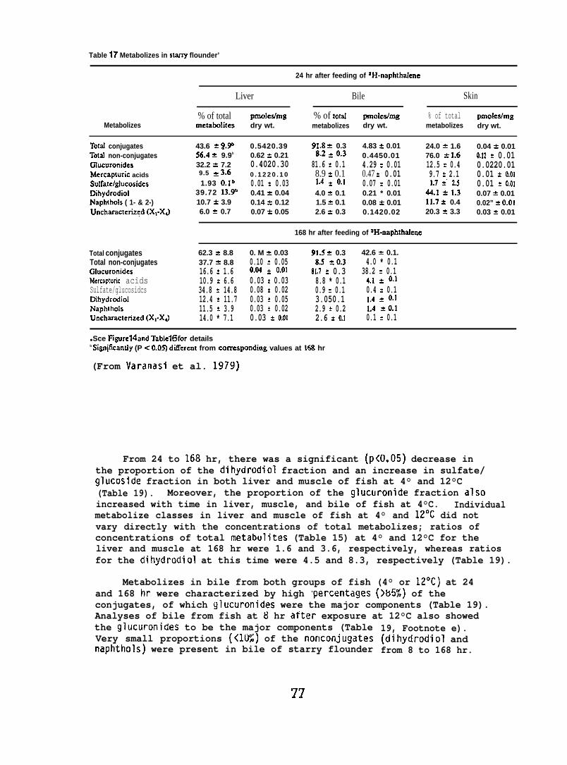
Table 17 Metabolizes in stany flounder’
24 hr after feeding of aH-naphthalene
Liver Bile Skin
% of total pmoledmg % of tom! pmo[ealmg % of total pmoles/mgMetabolizes mciabolites dry wt. metabolizes dry wt. metabolizes dry wt.
Total conjugates 43.6 A ~.~ 0.5420.39 91.8 = 0.3 4.83 = 0.01 24.0 = 1.6 0.04 * 0.01Totrd non-conjugates 56.4 = 9.9b 0.62 = 0.21 8.2 ~ o.3 0.4450.01 76.0 a 1.6 o.f2 * 0.01Glucuronides 32.2 e 7.2 0.4020.30 81.6 * 0.1 4.29 = 0.01 12.5 A 0.4 0.0220.01Mercapturic acids 9.5 = 3.6” 0 . 1 2 2 0 . 1 0 8.9 = 0.1 0.47 * 0.01 9.7 * 2.1 0.01 * 0.01Sulfa[e/glucosides 1.93 o.lb 0.01 = 0.03 1.4 * ().1 0.07 * 0.01 3.7 =“ 2.5 0.01 * 0.01Dihydrodiol 39.72 13.9” 0.41 = 0.04 4.0 * 0.1 0.21 * 0.01 44.1 * 1.3 0.07 = 0.01Naphthols ( 1- & 2-) 10.7 * 3.9 0.14 & 0.12 1.5 * 0.1 0.08 A 0.01 11.7 * 0.4 0.02” & 0.01Uncharactcnzed (X,-XJ 6.0 = 0.7 0.07 * 0.05 2.6 % 0.3 0.1420.02 20.3 z 3.3 0.03 * 0.01
168 hr after feeding of ‘H-naphthalene
Total conjugatesTotal non-conjugatesGlucuronidesMercaptunc acidsSulfate/glucosidcsDihydrodiolNaphthoisUncharactcrized (X,-XJ
62.3 A 8.8 0. M & 0.03 91-5 * 0.3 42.6 Y 0.1.37.7 * 8.8 0.10 * 0.05 g.s $0.3 4.0 * 0.116.6 = 1.6 o.~ * o.~1 81.7 A 0.3 38.2 * 0.110.9 * 6.6 0.03 * 0.03 8.8 * 0.1 4.1 * ().134.8 a 14.8 0.08 = 0.02 0.9 * 0.1 0.4 * 0.112.4 = 11.7 0.03 * 0.05 3.050.1 ].4 * 0.]11.5 = 3.9 0.03 * 0.02 2.9 k 0.2 L4= 0.114.0 * 7.1 0.03 a 0.01 2.6 = 0.1 0.1 * 0.1
● Sce Figurelhrd Table16for detailsb Sign@cantly (P < 0.05) ditTerent from comespondlng values at MS hr
(From Varanasi et al. 1979)
From 24 to 168 hr, there was a significant (P<O.05) decrease inthe proportion of the dihydrodiol fraction and an increase in sulfate/glucoside fraction in both liver and muscle of fish at 4° and 12°C(Table 19). Moreover, the proportion of the glucuronide fraction alsoincreased with time in liver, muscle, and bile of fish at 4°C. Individualmetabolize classes in liver and muscle of fish at 4° and 12°C did notvary directly with the concentrations of total metabolizes; ratios ofconcentrations of total metabulites (Table 15) at 4° and 12°C for theliver and muscle at 168 hr were 1.6 and 3.6, respectively, whereas ratiosfor the dihydrodiol at this time were 4.5 and 8.3, respectively (Table 19).
Metabolizes in bile from both groups of fish (4° or 12°C) at 24and 168 hr were characterized by high rpercentages (>65%) of theconjugates, of which glucuronides were the major components (Table 19).Analyses of bile from fish at 8 hr after exposure at 12°C also showedthe glucuronides to be the major components (TableVery small proportions (<lU:) of the nonconjugatesnaphthols) were present in bile of starry flounder
77
19, Footnote e).(dihydrodiol andfrom 8 to 168 hr.
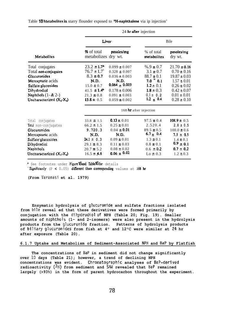
Table 18hierabolites in starry flounder exposed to 3H-naphthakne via ip injection’
24 hr idler injection
Liver Bile
% of total pznoles/mg % of total pmokfmgMetabdites metabolizes dry wt. xnetatdites dry wt.
Total conjugatesTotal non<onjugatesGlucuronidesMercaptunc acidsSulfateglucosidesDihydrodiolNaphthols (1- & 2-)Uncharacterized (X,-XJ
23.2 = 1.7b76.7 = L7b
8.3 & 0.7N.D.
15.0 * 0.741.5 = 1.4b21.3 = 0.813.8 * 0.5
0.099 = 0.0070.328 = 0.0070.036 z 0.003
N.D.0,06.4 * ().0030.178 = 0.0060.091 = 0.0030.059 * 0.002
%.9 * 0.73.1 & 0.7
88.7 ~ 0.17.0 * ().11.2 = 0.11.8 * 0.30.1 * 0.21.2 * (3.4
21.70 = 0.160.70 = 0.16
19.87 * 0.031.57 * 0.010.26 ~ 0.020.42 ~ 0.070.01 & 0.010.28 Y 0.10
Total conjugatesTotal non-conjugatesGlucuronidesMercapturic acidsSulfate’glucosidesDihydrodiolNaphtholsUnchamcterized (X,-XJ
168 hr after injection
33.8 * 1.5 0.13 * 0.01 97.5 % 0.4 108.9 & 0.566.2 * 1.5 0.25 * 0.01 2.520.4 2.8 z 0.59.720.3 0.04 A 0,01 89.5 = 0.5 100.0 = 0.6
N.D. N.D. 6.7 ~ (3.4 7.5 ~ 0.524.1 * 0.3 0.09 * 0.01 1.3 * 0.1 1.4 * 0.129.1 k 8.3 0.11 * 0.03 0.8 a 0.1 0.9 * 0.120.7 z 5.2 0.08 = 0.02 0.6 A 0,2 (37 = (J216.5 A $.6 ())6 = ().(32 Lo a 0.3 1.2 & 0.3
S See footnotes under Figurc16and TablclGfor detailsb Significandy (P < 0.05) diflkrent zhan comesponding values at 168 hr
(From Varanasi et al. 1979)
Enzymatic hydrolysis of glucuronide and sulfate fractions isolatedfrom bile reveal ed that these derivatives were formed primarily byconjugation with the dihydrodiol of NPH (Table 20; Fig. 19). Smalleramounts of naphthols (1- and 2-isomers) were also present in the hydrolysisproducts from the glucuronide fraction. Patterns of hydrolysis productsof biliary glucuronides from fish at 4° and 12°C were similar at 24 hrafter exposure (Table 20).
6.1.7 Uptake and Metabolism of Sediment-Associated NPH and BaP by Flatfish
The concentrations of BaP in sediment did not change significantlyover 10 days (Table 21); however, a trend of declining NPHconcentrations was evident. Chromatographic analyses of BaP-derivedradioactivity (3H) from sediment and SAN revealed that BaP remainedlargely (>93%) in the form of parent hydrocarbon throughout the experiment.
78
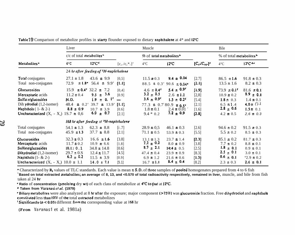
Tablel ~ Comparison of metabolize profiles in s[arry flounder exposed to dietary naphthalene at 4* and IT’C
Liver Muscle Bile
c/c of total mclabolitesl’ 70 of total metabolitesb % of total melabolitesh
Metaboliles” 4°C 120C,{ [c,./c,*.]’ 4°C 12°c [C,JC,,.]C 4°C I 2°C a“
24 Ar aftw feeding of ‘H-naplt[h(tlene
Total conjugates 27.1 A 1.8 43.6 & 9.9 [0,5]Total non-conjugates 72.9 * 1.8’ 56.4 * 9.9’ [1.1]
Glucuronides 15.9 z 0.41 32.2 z 7.2 [0,4]Mercapmric acids 11.2 * 0.4 9.5 z 3.6 [0.9]Sulfa! elglucosides N.D. ;.9 ~ (J. ]r _Dih ydrodiol (1,2-isomer) 48.4 a 0.2’ 39.7 & 13.9’ [1.1]Naphthols ( 1- & 2-) 8,8 * 0.9 10.7 t 3.9 [0.6]Uncharacterized (X, - X4) 1S.7 & 0,6 6.0 ~ ().7 [2.1]
168 ltr after $eeding of aH-naphthcdcne
Total conjugates 54.1 = 1.3 62.3 A 8.8 [i .7]Total non-conjugates 45.9 t 1.3 37.7 ? 8.8 [2.1]
Glucuronides 32.3 A 0.3 16.6 * 1.6 [3.8]Mercapturic acids 11.7 2 0.2 10.9 * 6.6 [1.8]Suifatelglucosides 10.1 2 0.1 34.8 T 14.8 [0.6]Dihydrodiol (1,2-isomer) 29.7 * 0.S 12.4 z 11.7 [4.5]Naphthols (1- & 2-) 6.2 z 0.2 11.5 * 3.9 [0.9]Uncharacterized (X, - X4) 10.0 = 1.1 14.0 * 7.1 [5.1]
11.5 * 0.3 9.4 * (),0488.5 k 0.3’ 90.6 a 0.04c
4.6 k 0.41 5.4 * 0.913.2 ~ 0.5 2.6 z I.13.6 z O*91 I.S ~ ~.2r
77.3 * 0.7 r 80.9 ~ 0,5r1.8 * 0.1 2.4 t 0.01 r
9.4 * 0.2 7.2 z Oog
28.9 k 0,5 46.1 * 0.371.1 * 0.5 53.9 t 0.3
13.1 * 1.3 23.7 k 1.47.2 = 0.2 8.0 a 0.98.7 t 2.1 1+4 & 0.5
47.4 2 0.4 23.9 = 0.96.9 A 1.2 21.6 * 0.6
16.7 & 1.1 8.4 ~ oa4
[2.7][2.1]
[1.9][2,8][5.4][2.1][1.6][2.8)
[2.6][5.5]
[2.3][3.8][2.5][8.3][1,3][8.2]
86.5 * !.613.5 A 1.6
73.9 t O.lr10.9 * 0.2
1.8 t 0.36.5 % ! .42.8 = o.64.2 = 0.5
94.6 z 0.25.5 2 0.2
85.1 a 0.27.7 z 0.2].8 t o-l2.5 = o.]oo6 = o.!2.3 x 0.3
91.8 * 0.38.2 z 0.3
81.6 * 0.18,9 ~ Ool1.4 * 0.14.-J ~ (’).1
1.5 * 0.12,6 % 0.3
91.5 * 0.38.5 = 0.3
81.7 = 0.38.8 * 0.10.9 * 0.13.0 % 0.1“2.9 t 0.22.6 = 0.1
m Characterized by R, values of TLC standards. Each value is mean * S.D. of three samples of pooled homogenates prepared from 4 to 6 fishb Based on total extracted irretabolites; an average of 8, 13, and <0.570 of total radioactivity respectively, remained in liver, muscle, and bile from fishtaken al 24 hr“ Ratio of concentration (pmole/mg dry WI) of each class of metabolize at 4°C to ~hal at 12”C.“ Taken from Varanasi c/ (//. (1979)r Biliary mctaboliles were also analyzed at 8 hr after the exposure; major component (>7S%) was glucuronidc fraction. Free dihydrodiol and naphtholsconsli!uted less than IOYC of Ihe total ex[racled metabolizes‘ Sirznificantly (~ < 0.05) different from the corresponding value at 168 hr
(From Varanasi et al. 1981a)
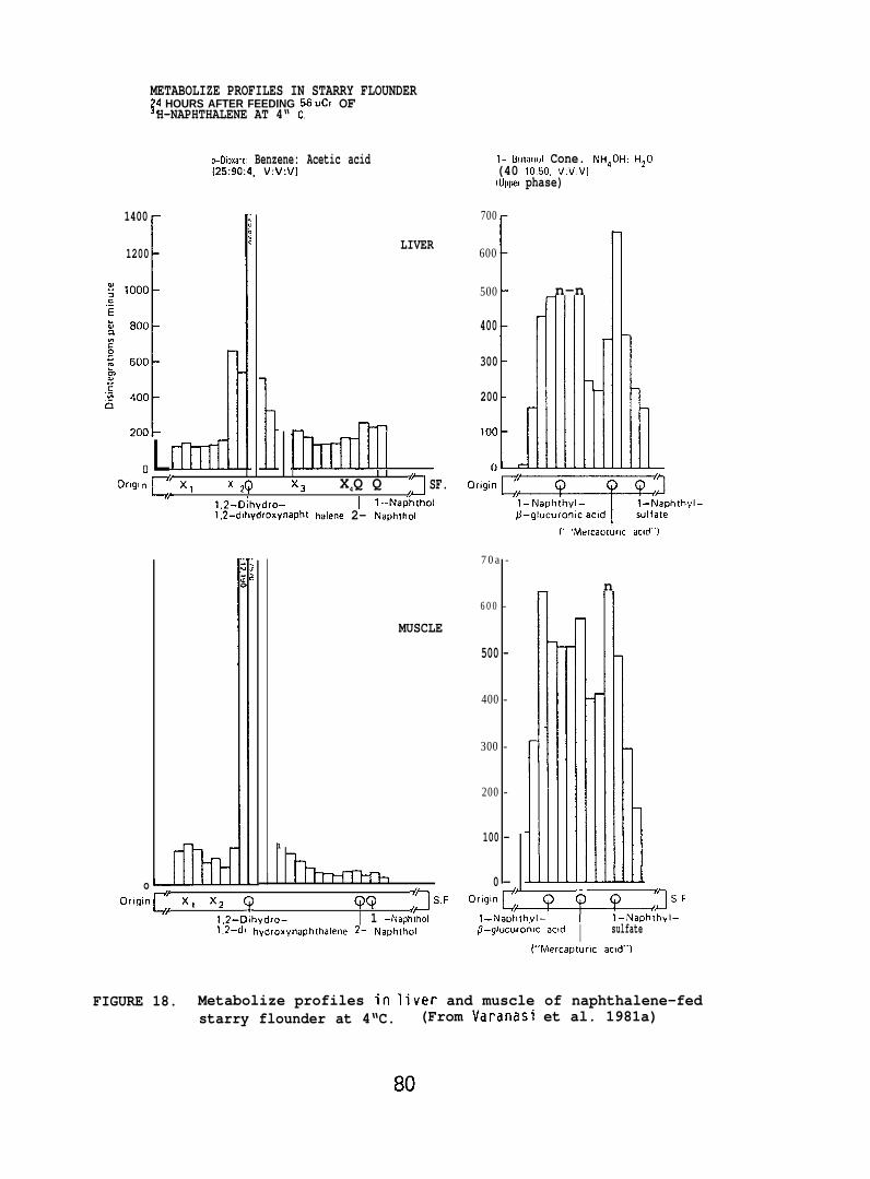
METABOLIZE PROFILES IN STARRY FLOUNDER34 HOURS AFTER FEEDING 56.Ci OFH-NAPHTHALENE AT 4“ C,
1400
1200[
n L
p-Dioxane: Benzene: Acetic acid[25:90:4, V:V:V]
LIVER
— 1-1-1
lhml-lL.Or,g, n r’ xl x 2Q X3 X4Q Q 2 SF.
oOrigin
,,1,2-D;hydro– I l--Naphthol1,2-dihydroxynaph! halene 2- Naphthol
MUSCLE
1
I
c xl X2 ? QQ 31.2-Dthydro– 1 –Naphthol1,2-d! hydroxynaphthalene 2- Naphthol
S.F
l- f.hItwIol Cone. NH40H: H20(40 10:50. V.v:v)(Upper phase)
700
600
500
400
300
200
100
0L
n-n
—Origin
cm~:hth”-
70a -
600 -
500 -
400 -
300 -
200 -
100 -
0 —
(“ Mercaoturlc acid”’)
n
—
U-glucufonic amd I sulfate(“Mercapturlc acid’”)
FIGURE 18. Metabolize profiles in liver and muscle of naphthalene-fedstarry flounder at 4“C. (From Varanasi et al. 1981a)
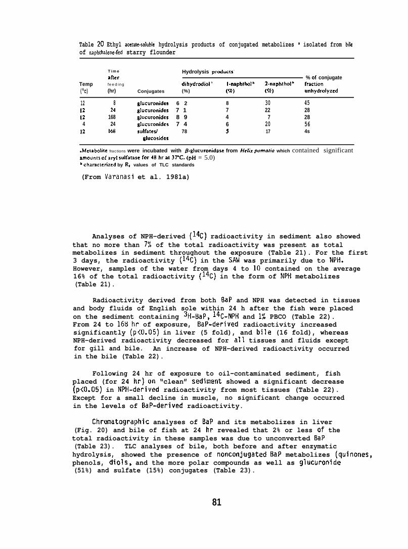
Table 20 Ethyl acetalc-soluble hydrolysis products of conjugated metabolizes M isolated from bileof naph[halene.fed starry flounder
Time Hydrolysis prcducts -
atler % of conjugateTemp feeding dihydrcdiol b I-rmphthol’ 2-naphthol’ fraclion(“c) (hr) Conjugates (%) (%) (%) unhydrolyzcd
12 8 glucuronides 6 2 8 30 4512 24 glucuronides 7 1 7 22 2812 168 8]ucuronidcs 8 9 4 7 284 24 ghscuronides 7 4 6 20 56
12 I& su!fatesl 78 5 17 4sgfucosides
● Mctabolite fractions were incubated with /3.gIucuro’nidase from Helix pwrratia which contained significantstmounls ofaryl sulfatase for48hra13TC. (pH = 5.0)bcharactenzed by Rt values of TLC standards
(From Varanasi et al. 1981a)
Analyses of NPH-derived (14C) radioactivity in sediment also showedthat no more than 7% of the total radioactivity was present as totalmetabolizes in sediment throughout the exposure (Table 21). For the first3 days, the radioactivity (14C) in the SAM was primarily due to NPH,However, samples of the water from days 4 to 10 contained on the average16% of the total radioactivity (14C) in the form of NPti metabolizes(Table 21).
Radioactivity derived from both BaP and NPH was detected in tissuesand body fluids of English sole within 24 h after the fish were placedon the sediment containing 3H-BaP, 14C-NPH and 1% PBCO (Table 22).From 24 to 168 hr of exposure, BaP-derived radioactivity increasedsignificantly (p<O.05) in liver (5 fold), and bile (16 fold), whereasNPH-derived radioactivity decreased for all tissues and fluids exceptfor gill and bile. An increase of NPH-derived radioactivity occurredin the bile (Table 22).
Following 24 hr of exposure to oil-contaminated sediment, fishplaced (for 24 hr) on “clean” sediinent showed a significant decrease(p<O.05) in NPH-derived radioactivity from most tissues (Table 22).Except for a small decline in muscle, no significant change occurredin the levels of BaP-derived radioactivity.
Chromatographic analyses of BaP and its metabolizes in liver(Fig. 20) and bile of fish at 24 hr revealed that 2% or less of thetotal radioactivity in these samples was due to unconverted BaP(Table 23). TLC analyses of bile, both before and after enzymatichydrolysis, showed the presence of nonconjugated BaP metabolizes (quinones,phenols, diols, and the more polar compounds as well as glucuronide(51%) and sulfate (15%) conjugates (Table 23).
81
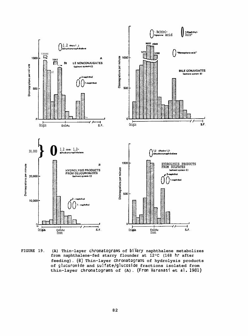
[0 1.2 d$hydro.l ,2.dihydroxvnwhthdme
I 73586
t1000 Q
~ ‘g B, LENONCONJ”GAT:$
~1-1 iia hd.m sysmm C);::::c ,,...;:: : ,,,.,.-E . . . . .;;::: ;:;:: :::::,.,.,.k ?’5’ ““”
,.,,,I
~-Origin EtOAc S.F.
30,000} 0 1.2 ddwdro. 1,2-dihydroxvnm+thdwn
“ +——&——ff—————fOrigin S,F.front
[0 1. Nw)hthvl- 0 1.NwMIIvI.B’@lcllronk acid Suits*
~ +--lFOrigin . .
[0 1.2 dihvdrwl.2.dihydroxvnaphlhalsrn
) T:.*
1000 :::::. . . . . :. HYDROLYSIS PRODUCTS::, . FROM SULFATES1:: g [advent rfsmm C)
1 on. . . . .:x # 2-na@Xhd:::::,. . . . . l-namhd
~~Origin S.F.
front
FIGURE 19. (A) Thin-layer chromatograms of bilary naphthalene metabolizesfrom naphthalene-fed starry flounder at 12°C (168 hr afterfeeding). (B) Thin-layer chromatograms of hydrolysis productsof glucuronide and sulfate/glucoside fractions isolated fromthin-layer chromatograms of (A). (Fran Varanasi et al. 1981)
82
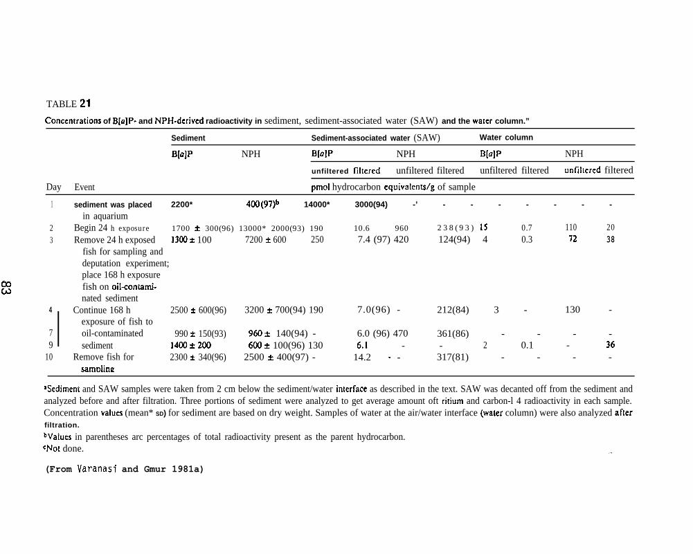
TABLE 21
~onccntrations of B[u]P- and NPH-derived radioactivity in sediment, sediment-associated water (SAW) and the water column.”
Sediment Sediment-associated water (SAW) Water column
B[a]P NPH B[a]P NPH B[a]P NPH
unfiltered fihered unfiltered filtered unfiltered filtered unfiltered filtered
Day Event pmol hydrocarbon equivalenWg of sample
mcd
1 sediment was placed 2200* 4oo(97)b 14000* 3000(94) -’ - - - - - - -in aquarium
2 Begin 24 h exposure 1700 * 300(96) 13000* 2000(93) 190 10.6 960 2 3 8 ( 9 3 ) 1S 0.7 110 203 Remove 24 h exposed 13(IO* 100 7200 t 600 250 7.4 (97) 420 124(94) 4 0.3 72 38
fish for sampling anddeputation experiment;place 168 h exposurefish on oil-contati-nated sediment
4
IContinue 168 h 2500 f 600(96) 3200 t 700(94) 190 7.0(96) - 212(84) 3 - 130 -
exposure of fish to7 oil-contaminated 990 k 150(93) 9643 & 140(94) - 6.0 (96) 470 361(86) - - - -9 sediment 1400*200 600* 100(96) 130 6.1 - - 2 0.1 - 36
10 Remove fish for 2300 t 340(96) 2500 * 400(97) - 14.2 “ - 317(81) - - - -samriimz
+.edimem and SAW samples were taken from 2 cm below the sediment/water inierface as described in the text. SAW was decanted off from the sediment andanalyzed before and after filtration. Three portions of sediment were analyzed to get average amount oft ritium and carbon-l 4 radioactivity in each sample.Concentration vahses (mean* SD) for sediment are based on dry weight. Samples of water at the air/water interface (water column) were also analyzed afterfiltration.
bValues in parentheses arc percentages of total radioactivity present as the parent hydrocarbon.+Jot done. -.,
(From Varanasi and Gmur 1981a)
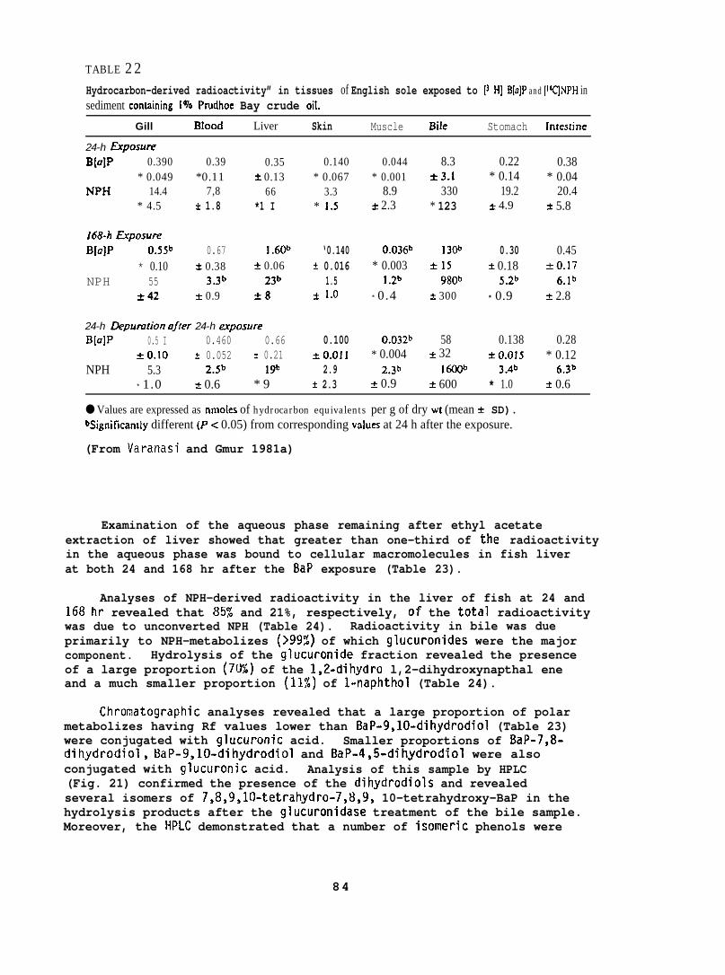
TABLE 22Hydrocarbon-derived radioactivity” in tissues of English sole exposed to [3 H] B[u]P and [W]NPH insediment containing 1070 Prudhoe Bay crude oil.
Gill BIood Liver Skin Muscle Bile Stomach lritesIjne
24-h &90SUtT!
B[a]P 0.390 0.39 0.35* 0.049 *0.11 * 0.13
NPH 14.4 7,8 66* 4.5 k 1.8 *1 I
f68-h fipOSUfI?
B[a]P o.55b 0.67 1.60* 0.10 k 0.38 k 0.06
NPH 55 J.Jb zJb*42 * 0.9 i8
24-h Depumtion offer 24-h e.rposureB[a]P 0.5 I 0.460 0.66
*0.10 ~ 0.052 * 0.21NPH 5.3 Z.sb 1 gb
* 1.0 ~ 0.6 * 9
0.140* 0.067
3.3* 1,5
‘0.140t 0.016
1.5* 1.()
0.100*0.011
2.9k 2.3
0.044* 0.001
8.9A 2.3
0.036b* 0.003
] .Zb
* 0.4
0.032b* 0.004
2.3b& 0.9
8.3*3.1
330* 123
1 3CP*15
980b* 300
58* 32
161Wk 600
0.22* 0.14
19.2* 4.9
0.30i 0.18
S.zb
* 0.9
0.138* 0.015
J.db* 1.0
0.38* 0.04
20.4k 5.8
0.45to.17
b.lbk 2.8
0.28* 0.12
6.3b
* 0.6
● Values are expressed as nmoles of hydrocarbon equivalents per g of dry wt (mean ~ SD).Uignificandy different (P< 0.05) from corresponding vaks at 24 h after the exposure.
(From Varanasi and Gmur 1981a)
Examination of the aqueous phase remaining after ethyl acetateextraction of liver showed that greater than one-third of the radioactivityin the aqueous phase was bound to cellular macromolecules in fish liverat both 24 and 168 hr after the BaP exposure (Table 23).
Analyses of NPH-derived radioactivity in the liver of fish at 24 and168 hr revealed that 85% and 21%, respectively, of the total radioactivitywas due to unconverted NPH (Table 24). Radioactivity in bile was dueprimarily to NPH-metabolizes (>99%) of which glucuronides were the majorcomponent. Hydrolysis of the glucuronide fraction revealed the presenceof a large proportion (7U%) of the 1,2-dihydro l,2-dihydroxynapthal eneand a much smaller proportion (11%) of l-naphthol (Table 24).
Chromatographic analyses revealed that a large proportion of polarmetabolizes having Rf values lower than BaP-9,10-dihydrodiol (Table 23)were conjugated with glucuronic acid. Smaller proportions of BaP-7,8-dihydrodiol, BaP-9,10-dihydrodiol and BaP-4,5-dihydrodiol were alsoconjugated with glucuronic acid. Analysis of this sample by HPLC(Fig. 21) confirmed the presence of the dihydrodiols and revealedseveral isomers of 7,8,9,1f)-tetrahydro-7,13,9, 10-tetrahydroxy-BaP in thehydrolysis products after the glucuronidase treatment of the bile sample.Moreover, the HPLC demonstrated that a number of isomeric phenols were
84
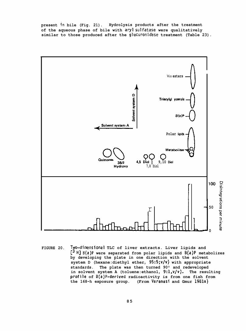
present in bile (Fig. 21). Hydrolysis products after the treatmentof the aqueous phase of bile with aryl sulfatase were qualitativelysimilar to those produced after the glucuronidase treatment (Table 23).
SolvantsystamA
OQ 9?CJuinones3i319 45 Diol
Hydroxy 7,8 Diol
Wax esters --oTriacylgl ycarols
-0
B(81P4
Polar iipids
Meta&oliti
o0
9,10 Diol
r
FIGURE 20. Tyo-dimensional TLC of liver extracts. Liver lipids and
100
50
0
-.2
[3 H] B(a)P were separated from polar lipids and B(a)P metabolizesby developing the plate in one direction with the solventsystem D (hexane:diethyl ether, 95:5;v/v) with appropriatestandards. The plate was then turned 90° and redevelopedin solvent system A (toluene:ethanol, 9:l,v/v). The resultingprOfile of B(a)P-derived radioactivity is from one fish fromthe 168-h exposure group. (From Varanasi and Gmur 1981a)
85
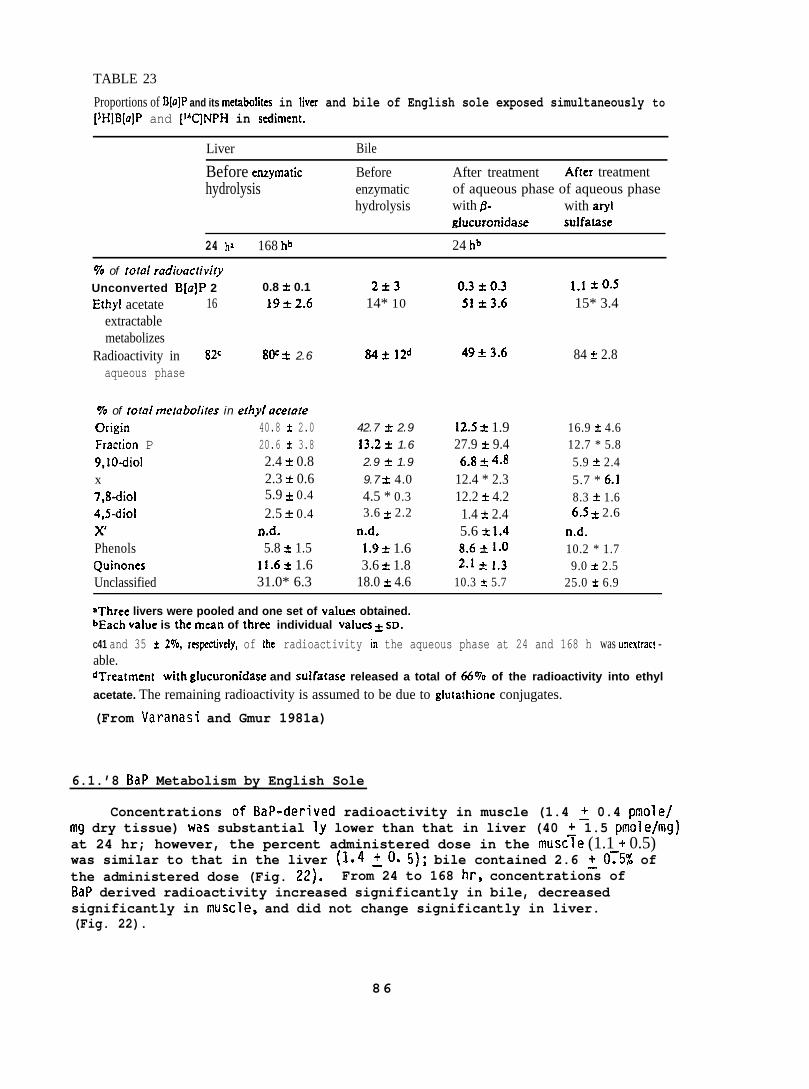
TABLE 23
Proportions of B@]P and its met.abolites in liver and bile of English sole exposed simultaneously to[JH]B[u]P and [14C_jNPH in sediment.
Liver Bile
Before enzymatichydrolysis
Before After treatment After treatmentenzymatic of aqueous phase of aqueous phasehydrolysis with & with aryl
glucuronidase sulfatase
24 h’ 168 hb 24 hb
70 of Iota/ radioacrivi(yUnconverted B[a]P 2 0.8 * 0.1
Ethyl acetate 16 19*2.6extractablemetabolizes
Radioactivity in 8ZC 8(Y t 2.6aqueous phase
To of total rne(abolites in ethyl ucetoteOrigin 40.8 f 2.0Fraction P 20.6 ~ 3.89,10diol 2.4 f 0.8x 2.3 k 0.67,8dioi 5.9 * 0.44,5-diol 2.5 k 0.4xl n.d.Phenols 5.8 ~ 1.5Quinones 11.6k 1.6Unclassified 31.0* 6.3
2*314* 10
84* 12d
42.7 ~ 2.913.2* 1.62.9 i 1.99.7 & 4.04.5 * 0.33.6 t 2.2
n.d.1.9 * 1.63.6 & 1.8
18.0 ~ 4.6
4953.6
!2.5 & 1.927.9 + 9.46.8 & 4.8
12.4 * 2.312.2 & 4.21.4 * 2.45.6 * 1.48.6 * 1.02.1 & 1.3
10.3 * 5.7
1.1 *0.515* 3.4
84 k 2.8
16.9 t 4.612.7 * 5.85.9 k 2.45.7 * 6.18.3 i 1.66.5 ~ 2.6
n.d.10.2 * 1.79.0 * 2.5
25.0 * 6.9
aThree livers were pooled and one set of valum obtained.bEach value is the man of three individual valu~ k sDO
C41 and 35 * 2L70, rmpectively, of the radioactivity in the aqueous phase at 24 and 168 h was urrextract -able.dTrea(ment with glucuronidase and sulfatase released a total of 66T0 of the radioactivity into ethyl
acetate. The remaining radioactivity is assumed to be due to glutathione conjugates.
(From Varanasi and Gmur 1981a)
6.1.’8 BaP Metabolism by English Sole
Concentrations of BaP-derived radioactivity in muscle (1.4 ~ 0.4 pmole/mg dry tissue) was substantial Iy lower than that in liver (40 ~ 1.5 pmole/mg)at 24 hr; however, the percent administered dose in the muscle (1.1 + 0.5)was similar to that in the liver (1.4 ~O. 5); bile contained 2.6 ~0~5% ofthe administered dose (Fig. 22). From 24 to 168 hr, concentrations ofBaP derived radioactivity increased significantly in bile, decreasedsignificantly in muscle, and did not change significantly in liver.(Fig. 22).
86
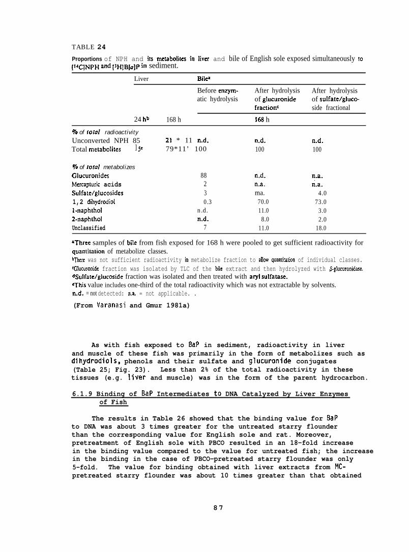
TABLE 24
Proportions of NPH and its mctabolites in liver and bile of English sole exposed simultaneously to[lfC]NpH and [JH]B[a]p in sediment.
Liver Blw
Before enzym- After hydrolysis After hydrolysisatic hydrolysis of glucuronide of sulfate/gluco-
fractionc side fractional
24 h~ 168 h 168 h
9’o of ro[al radioactivityUnconverted NPH 85Total metaboIites ] 5e
Yo of total metabolizesCducuronidesMercapturic acidsSulfate/glucosides1,2 dihydrodiolI-naphthol2-naphtholUnclassified
21 * 11 n.d.79*11’ 100
88230.3
n.d.n.d.
7
n.d.100
n.d.n.a.ma.70.011.08.0
11.0
n.d.100
n.a.n.a.
4.073.0
3.02.0
18.0
aThree samples of biIe from fish exposed for 168 h were pooled to get sufficient radioactivity forquamitation of metabolize classes.bThere was not sufficient radioactivity in metabolize fraction to allow quantitation of individual classes.~G!ucuronide fraction was isolated by TLC of the bile extract and then hydrolyzed with &glucuronidase.%ulfate/glucoside fraction was isolated and then treated with aryl sulfatase.eThis value includes one-third of the total radioactivity which was not extractable by solvents.n.d. = not detected: n.a. = not applicable. .
(From Varanasi and Gmur 1981a)
As with fish exposed to 8aP in sediment, radioactivity in liverand muscle of these fish was primarily in the form of metabolizes such asdihydrodiols, phenols and their sulfate and glucuronide conjugates(Table 25; Fig. 23). Less than 2% of the total radioactivity in thesetissues (e.g. liver and muscle) was in the form of the parent hydrocarbon.
6.1.9 Binding of BaP Intermediates to DNA Catalyzed by Liver Enzymesof Fish
The results in Table 26 showed that the binding value for BaPto DNA was about 3 times greater for the untreated starry flounderthan the corresponding value for English sole and rat. Moreover,pretreatment of English sole with PBCO resulted in an 18-fold increasein the binding value compared to the value for untreated fish; the increasein the binding in the case of PBCO-pretreated starry flounder was only5-fold. The value for binding obtained with liver extracts from MC-pretreated starry flounder was about 10 times greater than that obtained
87
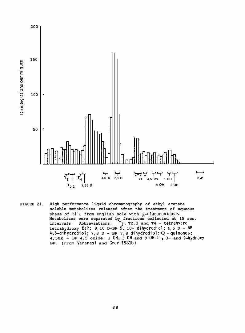
200
150
100
50
.
.
dw‘1 T’T‘ 4
T2?3 9,10 D
i
?4,5 D
IL.d/ I
7,8 D Cl 4,5 ox 1 OH I BaP
9 Oti 3 OH
FIGURE 21. High performance liquid chromatography of ethyl acetatesoluble metabolizes released after the treatment of aqueousphase of bile from English sole with p-glucuronidase.Metabolizes were separated by fractions collected at 15 sec.intervals. Abbreviations: Tl, T2,3 and T4 - tetrahydrotetrahydroxy BaP; 9,10 D-BP 9, 10- dihydrodiol; 4,5 D - BP4,5-dihydrodiol; 7,8 D - BP 7,8 dihydrodiol; Q - wiflofles;4,50X - BP 4,5 oxide; 1 OH, 3 OH and 9 OH-I-, 3- and 9-hydroxyBP. (From Varanasi and Gmur 1981b)
88
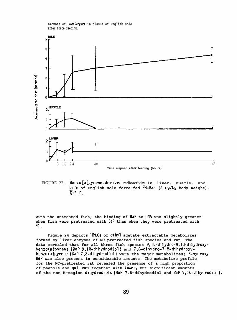
Amounts of 8enzo(a)pyrene in tissue of English soleafter force feeding.
6-BILE
5 “
4 -
3 “
2 -
1 -
0
2–MUSCLE
1 -
0
LIVER2?-
T
1 -
0 1 {8 16 24 48 168
Time elapsed after feeding (hours)
FIGURE 22. Benzo[a]pyrene-deri ved radioactivity in liver, muscle, andbile of English sole force-fed 3H-BaP (2 mg/kg body weight).~+S.D.
with the untreated fish; the binding of BaP to DNA was slightly greaterwhen fish were pretreated with BaP than when they were pretreated withMc .
Figure 24 depicts HPLCS of ethyl acetate extractable metabolizesformed by liver enzymes of MC-pretreated fish species and rat. Thedata revealed that for all three fish species 9,10-dihydro-9,10-dihydroxy-benzo(a)pyrene (BaP 9,10-dihydrodiol) and 7,8-dihydro-7,8-dihydroxy-benzo(a)pyrene (BaP 7,8-dihydrodiol) were the major metabolizes; 3-hydroxyBaP was also present in considerable amounts. The metabolize profilefor the NC-pretreated rat revealed the presence of a high proportionof phenols and quinones together with lower, but significant amountsof the non K-region dihydrodiols (BaP 7,8-dihydrodiol and BaP 9,10-dihydrodiol).
89
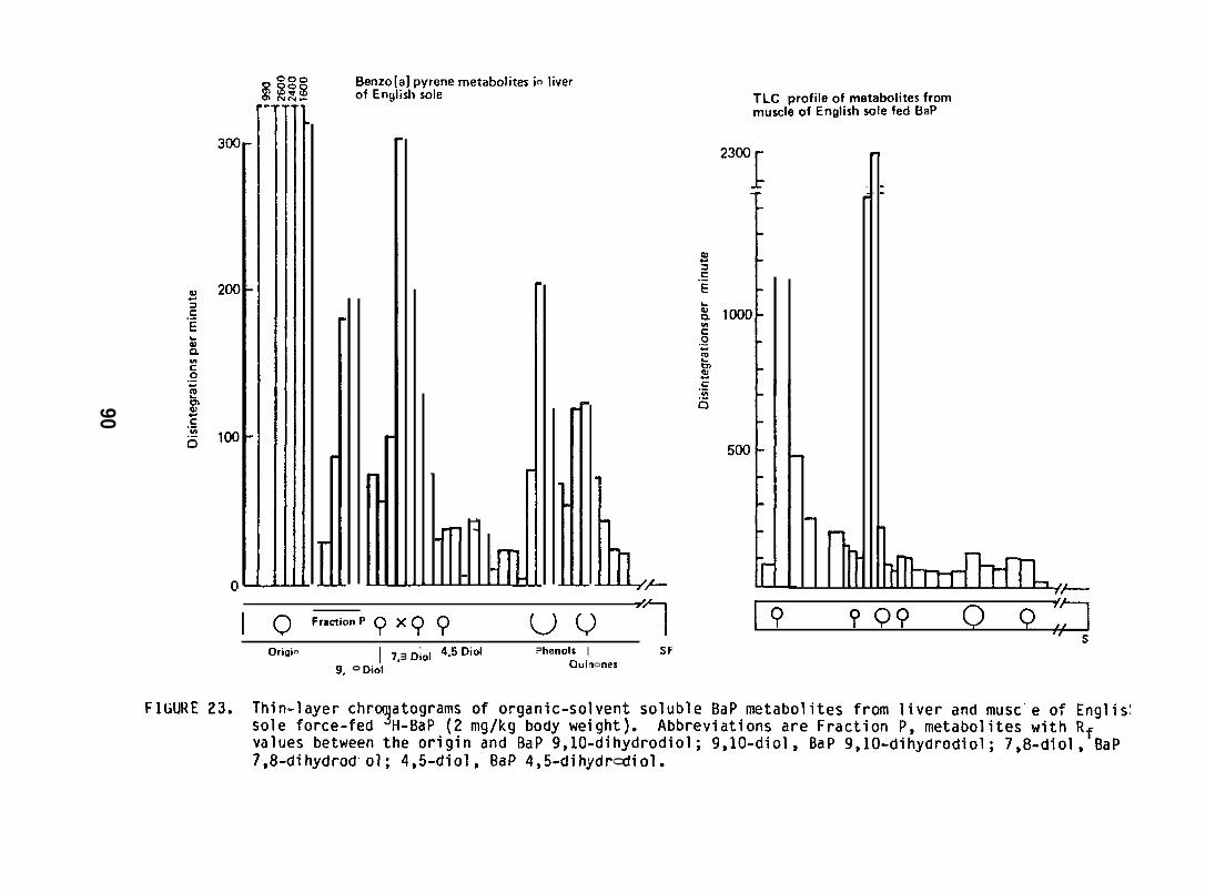
c.-
t#
1,,
T
1’t
i
—
alnu!uJ Xid suo!leJ6alu!s!Q
—
1’I
1,b
0091?
—
kIA
z50e—
.-3go0e2n.3.-nuY-
W
z. .-0m-
’—
-.-:0m“
c,-0!
,-6
.*.
-.-1
w-U0L.
alnu!w Jad
suo
!le16alu!s!Q
90
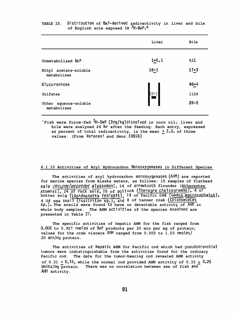
TABLE 25. Ilistribution of BaP-derived radioactivity in liver and bileof English sole exposed to 3H-13aP.a
Liver Bile
Unmetabolized BaP
Ethyl acetate-solublemetabolizes
Glucuronides
Sulfates
Other aqueous-solublemetabolizes
l-l83+1 1124
26+5
a Fish were force-fed 3H-BaP (2mg/kg)dissolved in corn oil; liver andbile were analyzed 24 hr after the feeding. Each entry, expressedas percent of total radioactivity, is the mean ~S.D. of threevalues. (From Varanasi and Gmur 1981b)
6.1.10 Activities of Aryl Hydrocarbon Monooxygenases in Different Species
The activities of aryl hydrocarbon monooxygenases (AHM) are reportedfor marine species from Alaska waters, as follows: 15 samples of flatheadsole (Hi poglossoides elassodon), 14stomia~sole, 15 of pollbutter sole (Le ido setta isolepis),4 Of sea snaihn~ndSII.). The snails were found to have. .whole body samples. The AHM activitipresented in Table 27.
of arrowtooth flounder (Atharestesock (Theragra chalcogr19 of Pacific8 of tanner crab (Cmecetesno detectable activity of AHM ines of the species exa~nined are
The specific activities of hepatic AHM for the fish ranged from0.006 to 0.927 nmoles of BaP products per 20 min per mg of protein;values for the crab viscera AHM ranged from 0.005 to 1.03 nmoles/20 min/mg protein.
The activities of hepatic AHM for Pacific cod which had pseudobranchialtumors were indistinguishable from the activities found for the ordinaryPacific cod. The data for the tumor-bearing cod revealed AHM activityof 0.31 ~0.16, while the normal cod provided AHM activity of 0.33 + 0.26Units/mg protein. There was no correlation between sex of fish and–AHM. activity.
91
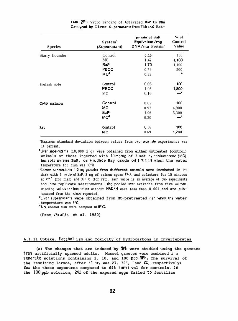
TABLE26111 Vitro Binding of Activated BaP to DNACattilyzed b~ Liver Supernatmts from Fish and Rat*
pnlole of BaP Ye ofSystem b Equivnkmt/mg
SpeciesControl
(Supernatant) DNA/mg Protein’ Value
Starry flounder ControlMCBaPPf3coMCd
English sole
Coho salmon
ControlPBCOMC
ConIrolMCBaPMC”
Rat ControlM C
0.151.621.700.740.53
0.061.050.16
0.020.971.060.30
Q.060.69
1001,1001,100
500d
1001,800—“
1004,9005,300
—“
1001,260
“Maximum standard deviation between values from two sepe rate experiments was14 percent.
bLiver supernatants (10,000 x g) were obtained from either untreated (control)animals or those injected with 10 mgikg of 3-met hylcholanthrene (MC),benzo(a)pyrene BaP, or Prudhoe Bay crude oil (PBCO) when the watertemperature for fish was 13”C.‘Liver supernafanls (=5 mg protein) from different animals were incubated in thedark with 5 nmole of BaP, 2 mg of salmon sperm DNA and cofactors for 15 minutesat 25°t2 (for fish) and 37° C (for rat). Each value is an average of two experimentsand lhree replicate measurements using pooled fiver extracts from five arlimals.Binding values for incuhalion without fdADPH were less than 0.001 and are sub-tracted from the values reported.
‘Li”~er supernatants were obtained from MC-pretreated fish whol) the watertemperature was 8“C.“No control fish were sampled at 8“C,
(From Varanasi et al. 1980)
6.1.11 Uptake, Netabol ism and Toxicity of Hydrocarbons in Invertebrates
(a) The changes that are induced by NPH were studied using the gametesfrcin artificially spawned adults. Mussel gametes were combined i nsmarate solutions containing 1. 10. and 100 D~b NPH. The survival oftheforthe
resulting larvae, after 24 hr, was 27, 32”, - and 2%, respectively>the three exposures compared to 69% survfi val for controls. In100 ppb solution, 24% of the exposed eggs failed to fertilize
92
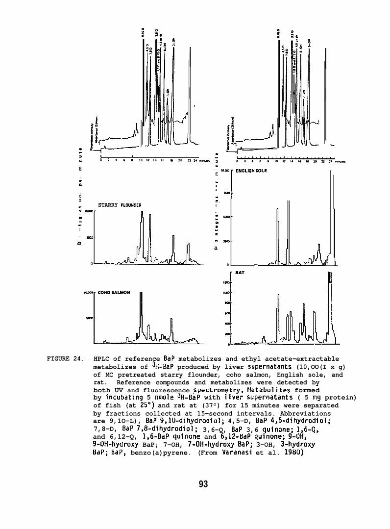
LI , t , , , , toa 46* 10 12 14 16 IS 20 22 24 ,.fwm,U,
E.ma.co“ STARRY FLOUNOERm N,Ma..mm“c
. - -a
0
a“
ac
- w.= EP4GLISHSOLEE
[.●
a
“c.0
m●“e
“
a
- - 1am -
0 — --’J -+A
FIGURE 24. HPLC of reference BaP metabolizes and ethyl acetate-extractablemetabolizes of 3H-BaP produced by liver supernatants (10,OO(I x g)of MC pretreated starry flounder, coho salmon, English sole, andrat. Reference compounds and metabolizes were detected byboth UV and fluorescence spectrometry. Hetabolites formedby incubating 5 nmole 3H-BaP with liver supernatants ( 5 mg protein)of fish (at 25”) and rat at (37°) for 15 minutes were separatedby fractions collected at 15-second intervals. Abbreviationsare 9,1O-L), BaP 9,10-dihydrodiol; 4,5-D, BaP 4,5-dihydrodiol;7,8-D, BaP 7,8-dihydrodiol; 3,6-Q, BaP 3,6 qutnone; 1,6-Q,and 6,12-Q, 1,6-BaP quinone and 6612-BaP qujnone; 9-OH,9-1.)H-hydroxy BaP; 7-OH, 7-OH-hydrox.y BaP; 3-OH, 3-hydroxyBaP; BaP, benzo(a)pyrene. (From Varanasi et al. 1980)
93
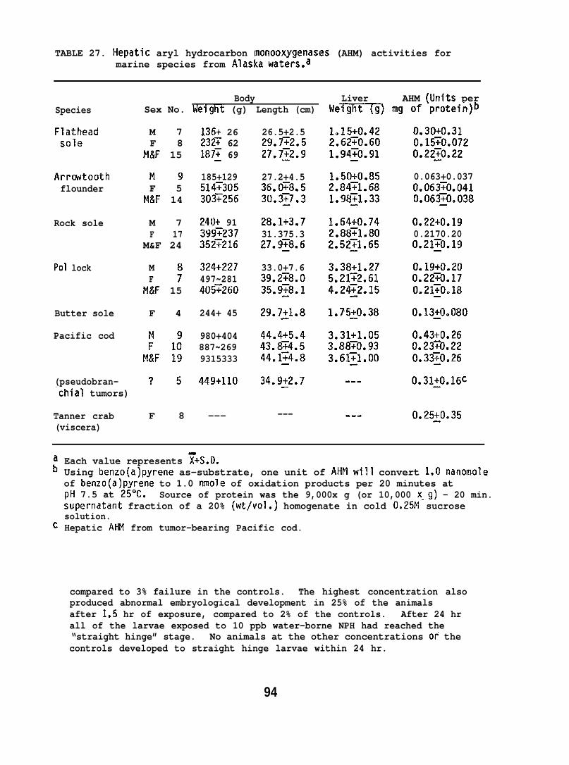
TABLE 27. Hepatic aryl hydrocarbon monooxygenases (AHM) activities formarine species from Alaska waters.a
Body Liver AHM (Units perSex No. ldelght (g) Length (cm) Were) mg of protein)bSpecies
Flatheadsole
Arrcwtoothflounder
Rock sole
Pol lock
Butter sole
Pacific cod
(pseudobran-
M 7F 8
M&F 15
136+ 26232~ 62187~ 69
26.5+2.529.fi2.527.752.9
27.2+4.536.0~8.530.3:7.3
28.1+3.731.375.327.9~8.6
33.0+7.639.2~8.O35.9~8.l
29.721.8
44*4+5.443.874.544.1~4.8
34.922.7
---
1.15+0.422.6270.601.94~o.91
0.30+0.310.15~Oe0720.22:0.22
185+129514T305303~256
1.50+0.852.84~1.681.!18~l.33
0.063+0.0370.06370.0410.063~0.038
M!JF 5
M&F 14
M 7F 17
M&F 24
240+ 913997237352~216
1.64+0.742.8871.802.5221.65
0,22+0.190.2170.200.21~0.19
MF 7
M&F 15
324+227497~281405~260
3.38+1.275.2172.614.24~2.15
0.19+0.200.2270.170.21~0.18
F 4 244+ 45 1.75~0.38 0.13j0.080
980+404887~2699315333
3.31+1.053.88~0.933.61~1.00
0.43+0.260.2370.220.33~0.26
449+110 ---
“chial tumors)
Tanner crab F 8 ---(viscera)
0.25~0.35---
Each value represents ~+S.D.Using benzo(a)pyrene as–substrate, one unit of AHM will convert 1.0 nanomoleof benzo(a)pyrene to 1.0 nmole of oxidation products per 20 minutes atpH 7.5 at 25°C. Source of protein was the 9,000x g (or 10,000 x-g) - 20 min.supernatant fraction of a 20% (wt/vol.) homogenate in cold 0.25M sucrosesolution.Hepatic AHM from tumor-bearing Pacific cod.
compared to 3% failure in the controls. The highest concentration alsoproduced abnormal embryological development in 25% of the animalsafter 1.5 hr of exposure, compared to 2% of the controls. After 24 hrall of the larvae exposed to 10 ppb water-borne NPH had reached the“straight hinge” stage. No animals at the other concentrations of thecontrols developed to straight hinge larvae within 24 hr.
94

Table z~ 11 ydrnctrbon conwnt of thoracic and abdominal segments of adult spot shrimp (P. platyc<ms)exposed to a water-soluble fraction of Pmdhoe Bity cmde oil.
Water-solublefraction~ Thorax$ Bio- Abdome~ Bio-
Hydrocarbons (rig/g) (rig/g) concn. (ng,/g) concn.
CyclohmtneBenzeneTolucneEthylbenzeneXylenesC~-substituted benzeneCJC,-substituted benzenes
Subtotui
2 9 –1S“5 –1
55 –107 –t
37”1 3204“ 1 220t “4 540
=:–1–%
9 350 954 150 37
386 180 129
lost-l 680
l~aphthalene 04 10 25 20 sol-~kthylnaphthalcne 0-$ 100 250 40 1002-h[ethylnaphthalene 0-4 110 275 60 150Cz-substkuwd naphthalene 240 171 100 71C,-substituted ntiphthalene :; 190 271 20 29
Subtotal: Cl-C, - —subs~itutcd nnphthalenes 640 220
Total 110 1730 920hydrocarbons
The shrimp were exposed for 7 days to a concn. of 100 p.p.b. of the water-soluble fraction of the oil.
~ Cidc. from representative analysis before dilution.$ Sot quantifiabk due to anidyticxd procedure.$ Concn. was determined as ntig wet wt., detectable limit was 10ng/g.
(From Sanborn and Malins 1980)
(b) The decrease in fertilization demonstrated in the musselexperiments and the effect of NPH on sperm and eggs were further exploredusing oyster gametes. The oyster sperm exposed to 10 ppb and 1 ppb ofNPH and then combined with uncontaminated eggs resulted in 14 and 9%unfertilized eggs, respectively, while no effect was seen at 0.1 ppbNPH. However, only 5% of the eggs exposed to 10 ppb and 1 ppb NPH andthen combined with uncontaminated sperm failed to fertilize. Thiscompares to a 4% failure to fertilize when uncontaminated eggs or eggsexposed to 0.1 ppb NPH and uncontaminated sperm were combined.
(c) The data on accumulation of SWSF hydrocarbons in one-year-oldspot shrimp show that detectable levels of low molecular weight aromatichydrocarbons were readily accumulated in thoracic segments (Table 28)(probably associated with the hepatopancreas). The abdominal segmentswere found to contain significant concentrations of identified aromatichydrocarbons. The data given in Table 28 represent hydrocarbon accumulationsin experimental animals with respect to data obtained from a control group.
95
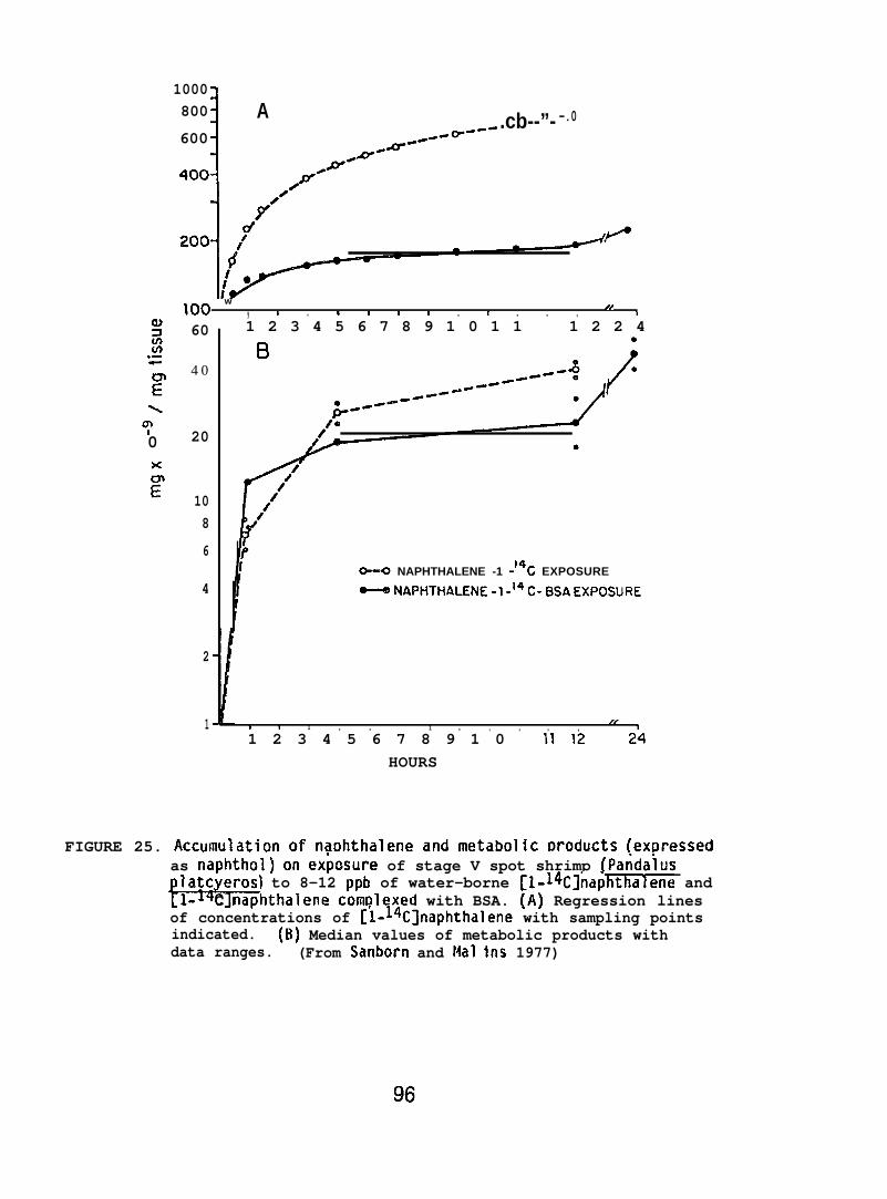
gIn(n.-+
?’\y0x
?’
1000800
1
A .cb--”- -.0600 .-0=--”
-a””
100 w , 1 , * 1 1 1 , I , , ,
1/t
1 2 3 4 5 6 7 8 9 1 0 1 1 1 2 2 460
4 0
20
108
6
4
2
1
--0 NAPHTHALENE -1 -14C EXPOSURE
O--@ NApHTHALENE -1.14 c- BsAExposuRE
8 n 1 , , 1 , , , , ,//
1
1 2 3 4 5 6 7 8 9 1 0 111224HOURS
FIGURE 25. Accumulation of naDhthalene and metabolic Droducts [exf)ressedas naphthol) on ex~osure of stage V spot shrimp (ParidaiusIatc eros) to 8-12 ppb of water-borne [1-14C]napkthalene and*C naphthalene complexed with BSA. (A) Regression linesof concentrations of [1-lxC]naphthalene with sampling pointsindicated. (B) Median values of metabolic products withdata ranges. (From Sanborn and Mal ins 1977)
96
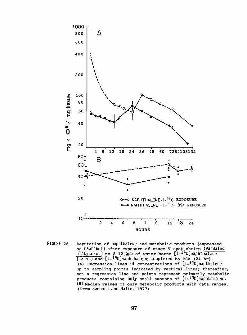
a)zLn.-+
?
\
m
0x
F
FIGURE 26.
800
600
400
200
10080
60
40
20
A
b 1 I I I 1 I , , ., I 1 14 8 12 18 24 36 48 60 7284108132
I20
1
0-- NAPHTHALENE-1-14C EXPOSUREx NAPHTHALENE -1-14 C- BSA EXPOSURE
lo~%—————l2 4 6 8 1 0 12 18 24
HOURS
Deputation of naphthalene and metabolic products (expressedas naphthol) after exposure of stage V spot shrimp (Pandaluslatyceros) to 8-12 ppb of water-borne [1-14C]naphthalene
{12 hr) and [1-14C]naphthalene complexed to BSA (24 hr).(A) Regression lines of concentrations of [1-~4C]napthaleneup to sampling points indicated by vertical lines; thereafter,not a regression line and points represent primarily metabolicproducts containing only small amounts of [1-14C]naphthalene.(B) Median values of only metabolic products with data ranges.(From Sanbornand Malins 1977)
97
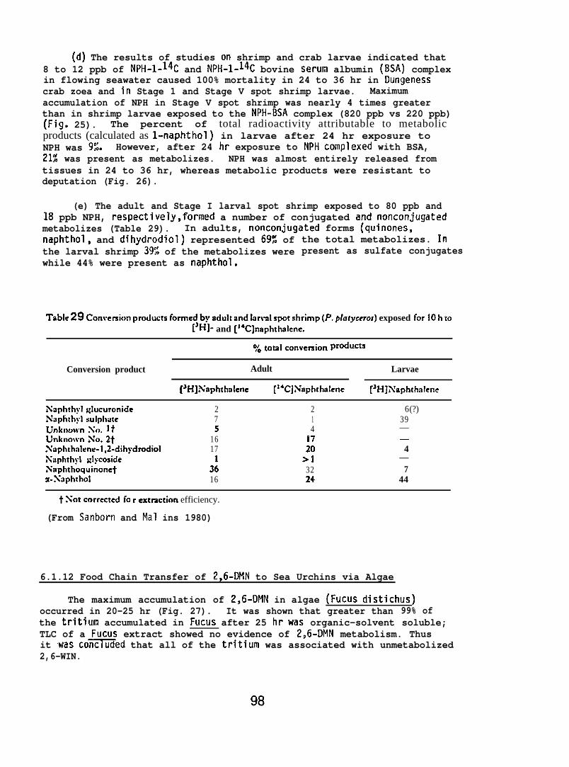
(d) The results of studies on shrimp and crab larvae indicated that8 to 12 ppb of NPH-I-14C and NPH-I-14C bovine serum albumin (BSA) complexin flowing seawater caused 100% mortality in 24 to 36 hr in Dungenesscrab zoea and in Stage 1 and Stage V spot shrimp larvae. Maximumaccumulation of NPH in Stage V spot shrimp was nearly 4 times greaterthan in shrimp larvae exposed to the NPH-BSA complex (820 ppb vs 220 ppb)(Fig. 25). The percent of total radioactivity attributable to metabolicproducts (calculated as l-naphthol) in larvae after 24 hr exposure toNPH was 9$. However, after 24 hr exposure to NPH complexed with BSA,21% was present as metabolizes. NPH was almost entirely released fromtissues in 24 to 36 hr, whereas metabolic products were resistant todeputation (Fig. 26).
(e) The adult and Stage I larval spot shrimp exposed to 80 ppb and18 ppb NPH, respectively,formed a number of conjugated and noncortjugatedmetabolizes (Table 29). In adults, nonconjugated forms (quinones,naphthol, and dihydrodiol) represented 69% ofthe larval shrimp 39% of the metabolizes werewhile 44% were present as naphthol.
the total metabolizes. Inpresent as sulfate conjugates
Tablezg Conversiort productsfortnedby aduhandlnrvalspot shrimp(P.p@wcros) exposed forlOhto[’HI- and [14C]naphthalene.
~~ ~oUlcOnvemion Pr~~cts
Conversion product Adult Larvae
[sH]Xaphthalene [14CJXaphthalene [3H]A’aphthalcnc
Xaphrhyl glucuronideXiiplithyl wlphttteUnknownXo. ltUnknownXo .2*Xaphthnlen~-1,2-dihydrod{olAXuphthyi ~lycosideXaphthoquinonetz-Xaphthol
275
1617
3:16
214
1720
>13224
6(?)39——4
—7
44
txotcorrectedfo rextraction efficiency.
(From Sanborn and Mal ins 1980)
6.1.12 Food Chain Transfer of 2,6-DMN to Sea Urchins via Algae
The maximum accumulation of 2,6-DMN in algae (Fucus distichus)occurred in 20-25 hr (Fig. 27). It was shown that greater than 99% ofthe tritium accumulated in Fucus after 25 hrwas organic-solvent soluble;TLC of a Fucus extract showed no evidence of 2,6-DMN metabolism. Thusit was co-ed that all of the tritium was associated with unmetabolized2,6-WIN.
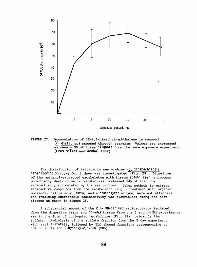
T
.
10 15 20 25 30 35
Exposure period, hrs
FIGURE 27. Accumulation of 3H-2,6-dimethylnaphthalene in seaweed(~. distichus) exposed through seawater. Values are expressedas mean + SD of three aliquots from the same exposure experiment.(From MaTins and Roubal 1982)
The distribution of tritium in sea urchins (~. droebachiensis)after feeding on Fucus for 3 days was investigated (Fig. 28). Digestionof the methanol-extracted exoskeleton with tissue solubilizer, a processpotentially destructive to metabolizes, released 64% of the totalradioactivity accumulated by the sea urchins. Other methods to extractradioactive compounds from the exoskeleton (e.g., treatment with organicsolvents, dilute acid, EDTA, and a proteolytic enzyme) were not effective.The remaining extractable radioactivity was distributed among the softtissues as shown in Figure 28.
A substantial amount of the 2,6-DMN-derived radioactivity isolatedfrom the digestive tract and gonadal tissue from the 3 and 14 day experimentswas in the form of conjugated metabolizes (Fig. 29), primarily thesulfate. Hydrolysis of the sulfate fraction from the 3 day experimentwith aryl sulfatase, followed by TLC showed fractions corresponding tothe 3- (80%) and 4-hydroxy-2,6-DhlN (20%).
99
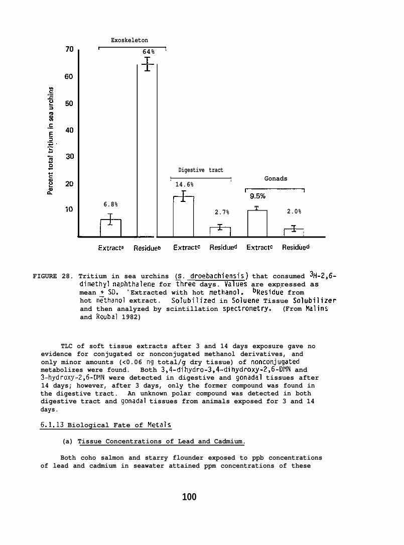
Exoskeleton1 64% t
6.8%
11”
+
—
Digestive tractI 1 Gonads14.6%
+-l ~
2.7% T 2.0%
Izl lx I
Extract’ Residueb ExtractC Residued ExtractC Residued
FIGURE 28. Tritium in sea urchins (S. droebachiensis) that consumed %1-2,6-dimethyl naphthalene for Three days. Values are expressed asmean +-SD.’ a Extracted with hot ~ethanol. bllesidue fromhot m~tttanol extract. Solubilized in Soluene Tissue Solubilizerand then analyzed by scintillation spectrometry. (From Flalinsand Roubal 1982)
TLC of soft tissue extracts after 3 and 14 days exposure gave noevidence for conjugated or nonconjugated methanol derivatives, andonly minor amounts (<0.06 ng total/g dry tissue) of nonconjugatedmetabolizes were found. Both 3,4-dihydro-3,4-dihydroxy-2,6-D}lN and3-hydroxy-2,6-DMN were detected in digestive and gonadal tissues after14 days; however, after 3 days, only the former compound was found inthe digestive tract. An unknown polar compound was detected in bothdigestive tract and gonadal tissues from animals exposed for 3 and 14days.
6.1.13 Biological Fate of Metals
(a) Tissue Concentrations of Lead and Cadmium.
Both coho salmon and starry flounder exposed to ppb concentrationsof lead and cadmium in seawater attained ppm concentrations of these
100
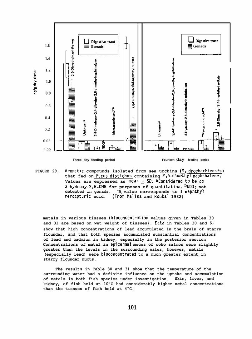
1.6
1.4
1.2
1.0
0.8
0.6
0.4
0.2
0.03
0.00
—
—
{ 1-
Three day feeding period
❑ Digestivewact~ Gonads
Fourteen day feeding period
FIGURE 29. Aranatlc compounds isolated from sea urchins (~. droebachiensis)that fed on Fucus distichus containing 2,6-dimethYl naphthalene.Values are expressed as mean ~SD. atonsidered t~ be”as3-hydroxy-2,6-DMN for purposes of quantitation. bNDG; notdetected in gonads. cRf value corresponds to l-naphthylmercapturic acid. (From Malins and Roubal 1982)
metals in various tissues (bioconcentration values given in Tables 30and 31 are based on wet weight of tissues). Oata in Tables 30 and 31show that high concentrations of lead accumulated in the brain of starryflounder, and that both species accumulated substantial concentrationsof lead and cadmium in kidney, especially in the posterior section.Concentrations of metal in epidermal mucus of coho salmon were slightlygreater than the levels in the surrounding water; however, metals(especially lead) were bioconcentrated to a much greater extent instarry flounder mucus.
The results in Table 30 and 31 show that the temperature of thesurrounding water had a definite influence on the uptake and accumulationof metals in both fish species under investigation. Skin, liver, andkidney, of fish held at 10”C had considerably higher metal concentrationsthan the tissues of fish held at 4“C.
101
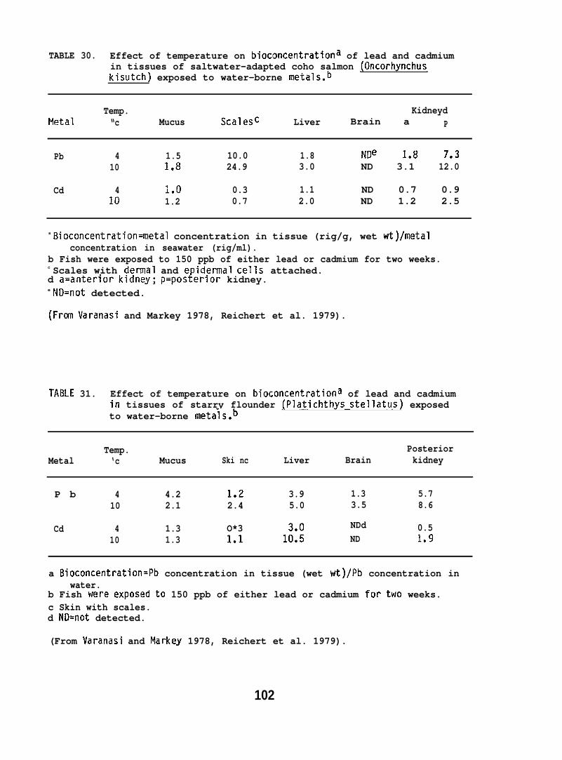
TABLE 30. Effect of temperature on bioconcentrationa of lead and cadmiumin tissues of saltwater-adapted coho salmon (Oncorhynchuskisutch) exposed to water-borne metals.b
Temp. KidneydMetal “c Mucus Scalesc Liver Brain a P
Pb 4 1.5 10.0 1.8 NDe 1.8 7e310 1.8 24.9 3.0 ND 3.1 12.0
Cd 4 1.0 0.3 1.1 ND 0.7 0.910 1.2 0.7 2.0 ND 1.2 2.5
a Bioconcentration=metal concentration in tissue (rig/g, wet wt)/metalconcentration in seawater (rig/ml).
b Fish were exposed to 150 ppb of either lead or cadmium for two weeks.c Scales with dermal and epidermal cells attached.d a=anterior kidney; p=posterior kidney.e ND=not detected.
(From Varanasi and Markey 1978, Reichert et al. 1979).
TABLE 31. Effect of temperature on bioconcentrationa of lead and cadmiumin tissues of starry flounder (Platichthys stellatus) exposedto water-borne metals.b
Temp. PosteriorMetal ‘c Mucus Ski nc Liver Brain kidney
P b 4 4.2 1.2 3.9 1.3 5.710 2.1 2.4 5.0 3.5 8.6
Cd 4 1.3 O*3 NDd 0.510 1.3 1.1 1;:: ND 1.!3
a Bioconcentration=Pb concentration in tissue (wet wt)/Pb concentration inwater.
b Fish were exposed to 150 ppb of either lead or cadmium for two weeks.c Skin with scales.d ND=not detected.
(From Varanasi and Markey 1978, Reichert et al. 1979).
102
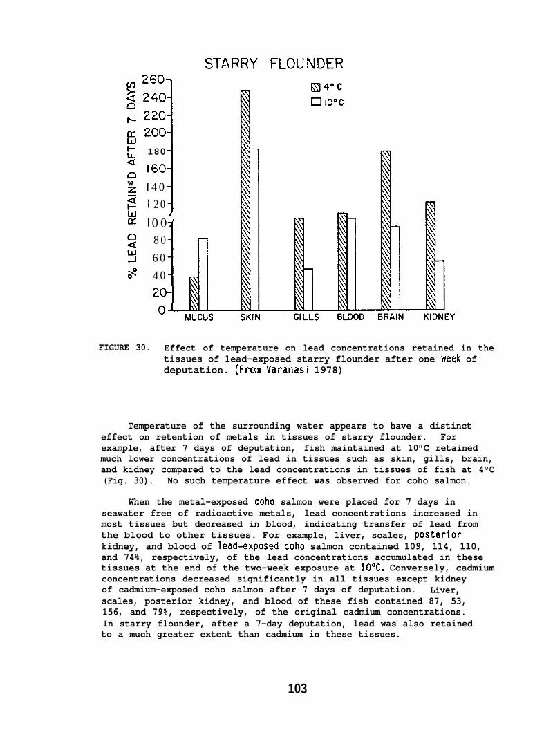
c1w~a
180
160
40
120
0 0
180
60
40
2oj
FIGURE 30. Effect of temperature on lead concentrations retained in thetissues of lead-exposed starry flounder after one week ofdeputation. (From Varanasi 1978)
Temperature of the surrounding water appears to have a distincteffect on retention of metals in tissues of starry flounder. Forexample, after 7 days of deputation, fish maintained at 10”C retainedmuch lower concentrations of lead in tissues such as skin, gills, brain,and kidney compared to the lead concentrations in tissues of fish at 4°C(Fig. 30). No such temperature effect was observed for coho salmon.
When the metal-exposed coho salmon were placed for 7 days inseawater free of radioactive metals, lead concentrations increased inmost tissues but decreased in blood, indicating transfer of lead fromthe blood to other tissues. For example, liver, scales, posteriorkidney, and blood of lead-exposedcoho salmon contained 109, 114, 110,and 74%, respectively, of the lead concentrations accumulated in thesetissues at the end of the two-week exposure at 10”C. Conversely, cadmiumconcentrations decreased significantly in all tissues except kidneyof cadmium-exposed coho salmon after 7 days of deputation. Liver,scales, posterior kidney, and blood of these fish contained 87, 53,156, and 79%, respectively, of the original cadmium concentrations.In starry flounder, after a 7-day deputation, lead was also retainedto a much greater extent than cadmium in these tissues.
103
After 37 days of deputation, substantial (>50%) concentrations ofboth lead and cadmium still persisted in most tissues of coho salmon.A notable finding was that metal concentrations in the posterior kidneyof coho salmon continued to increase even after the fish were placed inseawater far a period of 5 wk. Also, more than 50% of the lead andcadmium accumulated in the scales of coho salmon was still present afterfive weeks of deputation. Deputation of starry flounder tissues was notstudied for periods longer than 7 days, because after 7 days of deputationthe mortality rate increased in both control and exposed fish.
(b) Distribution of Cadmium and Lead in Subcell ular Fractions.
Samples of liver and kidney removed from the above coho salmonand starry flounder exposed to water-borne lead and cadmium were alsoused to investigate subcellular distribution of these metals. Cytosolfractions of liver and posterior kidney of cadmium-exposed starryflounder contained 72 and 80% of total tissue cadmium (109Cd),respectively, and cytosol fractions of these tissues from coho salmoncontained 62 and 48% of total tissue cadmium (lOgCd), respectively.Microsomal fractions of liver and kidney each contained 18% of thecadmium present in the tissue. Distribution of lead was somewhatdifferent: liver of lead-exposed coho salmon contained almost equalproportions of lead in mitochondria, microsomes, and cytosol.
Intravenous injections of 10gCd were employed to assess ifcadmium in gills, liver, and kidney of cadmium-exposed fish was boundto metalloproteins (M.W. 10,000 daltons). Results with coho salmonshow that similar to mammalian systems, a major fraction of the 109Cdwas bound to cadmium-binding proteins, CdBP (<8,900), in liver andkidney of coho salmon. Moreover, the percentage of the total 109Cdin cvtosol bound to CdBP was Qreater in those fish which had beenexpo~ed to 200 ppb Cd in seawiter for two weeks, compared to controlsin seawater only (Table 32). The studies with unexposed coho salmonindicate that CdBP concentrations in gills are low. Yet, within 24 hrafter metal challenge there is an appreciable increase in the accumulationof CdBP-bound cadmium indicating induction of CdBP.
(c) Effect of Metal Exposure on Synthesis of Epidermal Mucus
To assess the effect of water-borne metals on mucus production,weight of mucus was determined for each test and control coho salmon.The results show that at 10*C, 150 ppb of either lead or cadmium insurrounding water induced significantly higher (p<O.05) mucus productionin coho salmon (Table 33).
Our studies also show that when the fish were injected with eitherlead (2~0Pb) or cadmium (109Cd) salts substantial concentrationsof lead (164 ppb, dry wt) and cadmium (744 ppb, dry wt) were presentin the mucus for at least 2 days following the injection.
104
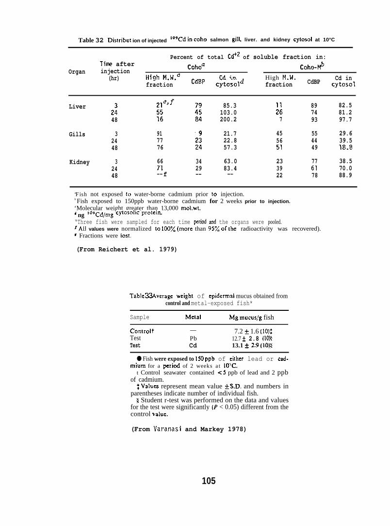
Table 32 Dis!ribut ion of injected ‘09Cd in coho salmon gill, liver. and kidney cytosol at 10°C
Percent of total Cd+2 of soluble fraction in:Tfme after
Organ injection Cohoa Coho-F$(hr) HirJh M.W.C CdBP Cd in High M.14. Cd in
fraction cytosol~ fraction CdBP Cytosol
Liver2:48
Gills 32448
Kidney 32448
917776
6671--f
3429--
85.3103.0200.2
21.722.857.3
63.083.4--
u7
455651
233922
897493
554449
776178
82.581.297.7
29.639.518.8
38.570.088.9
‘Fish not exposed 10 water-borne cadmium prior to injection.b Fish exposed to 150ppb water-borne cadmium for 2 weeks prior to injection.‘Molecular weight greater than 13,000 moLwt.‘ ng 109cd/rng cytOSO]iC prOtein-
“Three fish were sampled for each time period and the organs were pooled.fAll values were normalized to 100~0 (more than 95% of the radioactivity was recovered).f Fractions were lost.
(From Reichert et al. 1979)
Table~verage weight of epidermal mucus obtained fromcontrol and metal-exposed fish*
Sample Metal Mg mucus:g fish
Controlt — 7.2 ~ 1.6 (10)$Test Pb 12.7 A 2.8 (1OXTest Cd 13.1 *2.9 (ION
● Fish were exposed to 150ppb of either lead or cad-mium for a period of 2 weeks at 10:C.
t Control seawater contained <5 ppb of lead and 2 ppbof cadmium.
~ Values represent mean value ~S.D. and numbers inparentheses indicate number of individual fish.
$ Student r-test was performed on the data and valuesfor the test were significantly (P < 0.05) different from thecontrol va[ue.
(From Varanasi and Markey 1978)
105
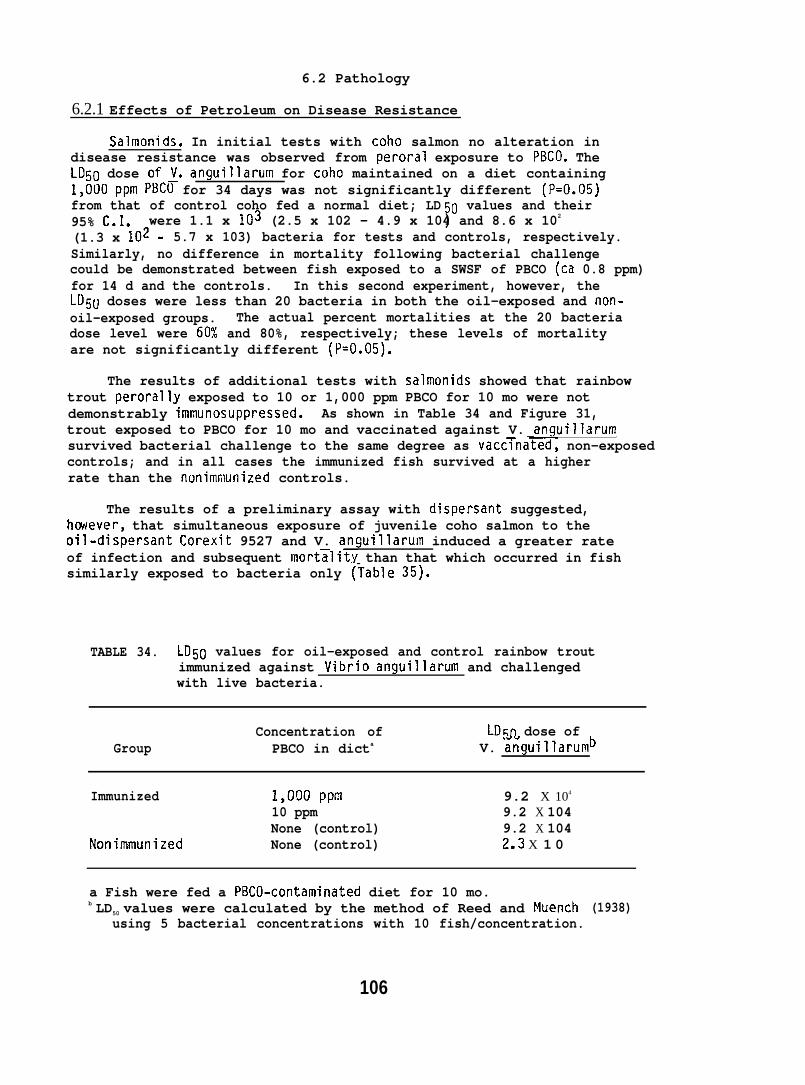
6.2 Pathology
6.2.1 Effects of Petroleum on Disease Resistance
Salmonids. In initial tests with coho salmon no alteration indisease resistance was observed from peroral exposure to PBCO. TheLD50 dose of~. anguillarum for coho maintained on a diet containing1,000ppm PBCO for 34 days was not significantly different (P=O.05)from that of control coho fed a normal diet; LD 0 values and their95% C.I. 7were 1.1 x 10³ (2.5 x 10² - 4.9 x 10⁴ and 8.6 x 10²
(1.3 x 10²- 5.7 x 10³) bacteria for tests and controls, respectively.Similarly, no difference in mortality following bacterial challengecould be demonstrated between fish exposed to a SWSF of PBCO (ca 0.8 ppm)for 14 d and the controls. In this second experiment, however, theLI)5U doses were less than 20 bacteria in both the oil-exposed and non-oil-exposed groups. The actual percent mortalities at the 20 bacteriadose level were 6~1 and 80%, respectively; these levels of mortalityare not significantly different (P=O.05).
The results of additional tests with salmonids showed that rainbowtrout perorally exposed to 10 or 1,000 ppm PBCO for 10 mo were notdemonstrably immunosuppressed. As shown in Table 34 and Figure 31,trout exposed to PBCO for 10 mo and vaccinated against V. anguillarurnsurvived bacterial challenge to the same degree as vacc~nated, non-exposedcontrols; and in all cases the immunized fish survived at a higherrate than the nonimmunized controls.
The results of a preliminary assay with dispersant suggested,hcwever, that simultaneous exposure of juvenile coho salmon to theoil-dispersant Corexit 9527 and V. anguillarum induced a greater rateof infection and subsequent mort~lit.y than that which occurred in fishsimilarly exposed to bacteria only (Table 35).
TABLE 34. LD50 values for oil-exposed and control rainbow troutimmunized against Vibrio anguillarum and challengedwith live bacteria.
Concentration of LD50 dose ofGroup PBCO in dicta V. anguillarumb
Immunized 1,000 ppr?l 9.2 X 10⁴
10 ppm 9.2 X 10⁴None (control) 9.2 X 10⁴
Nonimmunized None (control) 2.3 X 1 0
a Fish were fed a PBCO-contaminated diet for 10 mo.b LD50 values were calculated by the method of Reed and Muench
using 5 bacterial concentrations with 10 fish/concentration.
106
(1938)
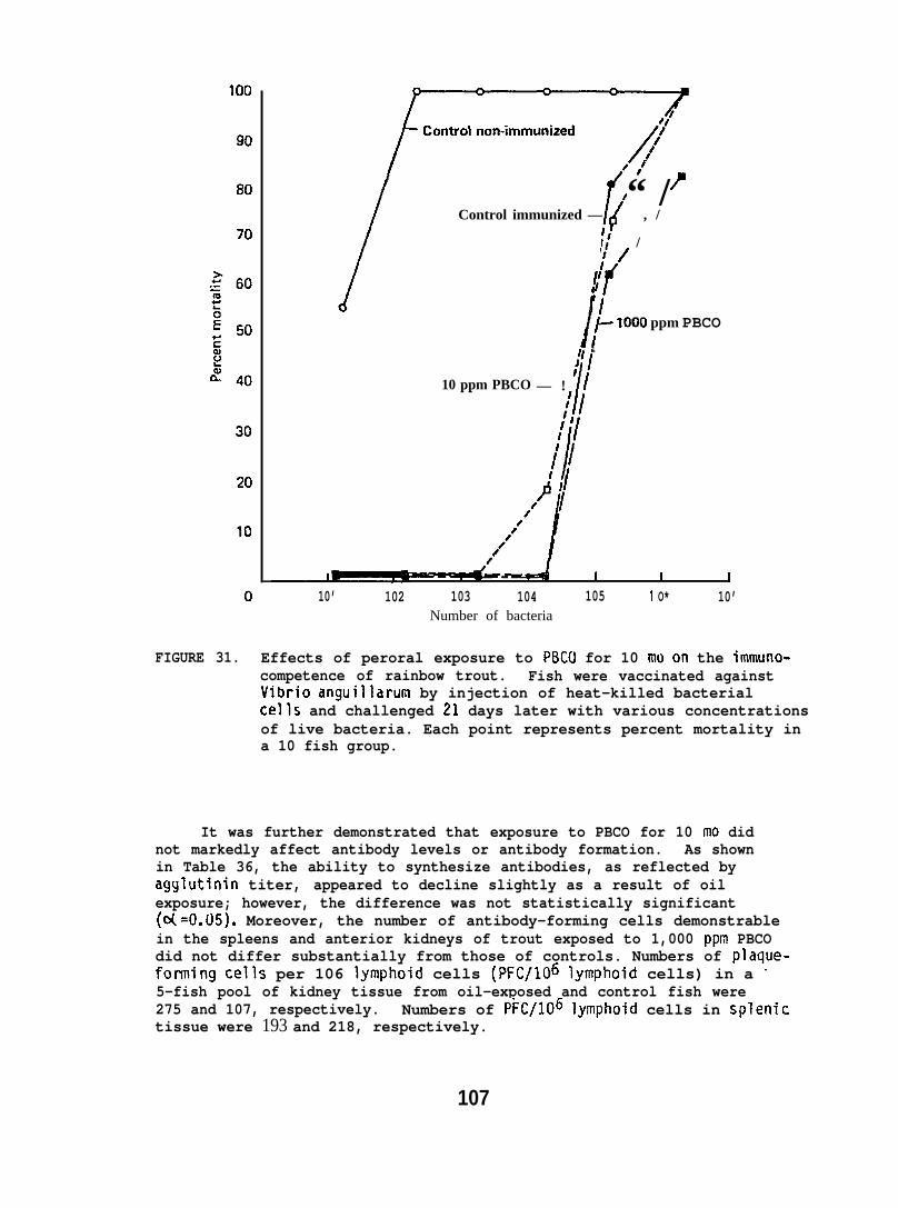
-?
[
7’
{“ /
Control immunized — ‘ , /
‘; /
I
;/
;
i
fI~ 1000 ppm PBCOI
~i10 ppm PBCO /— ! I
10’ 1021 I t
103 104 105 1 O* 10’Number of bacteria
FIGURE 31. Effects of peroral exposure to PBC(I for 10 mo on the immuno-competence of rainbow trout. Fish were vaccinated againstVibrio anguillarum by injection of heat-killed bacterialcells and challenged 21 days later with various concentrationsof live bacteria. Each point represents percent mortality ina 10 fish group.
It was further demonstrated that exposure to PBCO for 10 mo didnot markedly affect antibody levels or antibody formation. As shownin Table 36, the ability to synthesize antibodies, as reflected byagglutinin titer, appeared to decline slightly as a result of oilexposure; however, the difference was not statistically significant(oL=O.05). Moreover, the number of antibody-forming cells demonstrablein the spleens and anterior kidneys of trout exposed to 1,000 ppm PBCOdid not differ substantially from those of controls. Numbers of plaque-fonning cells per 106 lymphoid cells (PFC/106 Iymphoid cells) in a ‘5-fish pool of kidney tissue from oil-exposed and control fish were275 and 107, respectively. Numbers of PFC/106 lymphoid cells in splenictissue were 193 and 218, respectively.
107
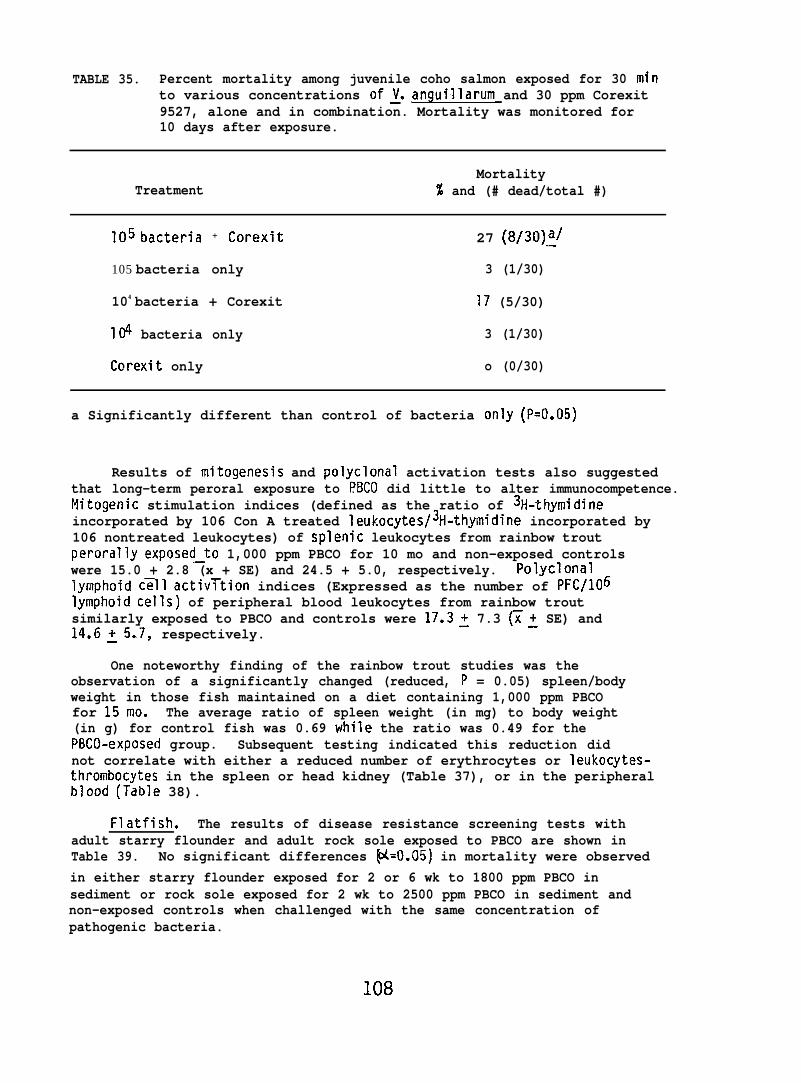
TABLE 35. Percent mortality among juvenile coho salmon exposed for 30 minto various concentrations of~. anguillarum and 30 ppm Corexit9527, alone and in combination. Mortality was monitored for10 days after exposure.
MortalityTreatment % and (# dead/total #)
105 ba~teria + ~orexit 27 (8/30)~/
105 bacteria only 3 (1/30)
104 bacteria + Corexit 17 (5/30)
104 bacteria only 3 (1/30)
Corexit only o (0/30)
a Significantly different than control of bacteria only (P=O.05)
Results of mitogenesis and polyclonal activation tests also suggestedthat long-term peroral exposure to f?BCO did little to alter immunocompetence.Mitogenic stimulation indices (defined as the ratio of 31i-thymidineincorporated by 106 Con A treated leukocytes/3H-thymidine incorporated by106 nontreated leukocytes) of splenic leukocytes from rainbow troutperorally exposed_to 1,000 ppm PBCO for 10 mo and non-exposed controlswere 15.0 + 2.8 (x + SE) and 24.5 + 5.0, respectively. Polyclonal?ymphoid cT1l activ~tion indices (Expressed as the number of PFC/106lymphoid cells) of peripheral blood leukocytes from rainbow troutsimilarly exposed to PBCO and controls were 17.3: 7.3 ~~ SE) and14.6:5.7, respectively.
One noteworthy finding of the rainbow trout studies was theobservation of a significantly changed (reduced, P = 0.05) spleen/bodyweight in those fish maintained on a diet containing 1,000 ppm PBCOfor 15mo. The average ratio of spleen weight (in mg) to body weight(in g) for control fish was 0.69 while the ratio was 0.49 for thePBCO-exposed group. Subsequent testing indicated this reduction didnot correlate with either a reduced number of erythrocytes or leukocytes-thrombocytes in the spleen or head kidney (Table 37), or in the peripheralblood (Table 38).
Flatfish. The results of disease resistance screening tests withadult starry flounder and adult rock sole exposed to PBCO are shown inTable 39. No significant differences ~=t).05) in mortality were observedin either starry flounder exposed for 2 or 6 wk to 1800 ppm PBCO insediment or rock sole exposed for 2 wk to 2500 ppm PBCO in sediment andnon-exposed controls when challenged with the same concentration ofpathogenic bacteria.
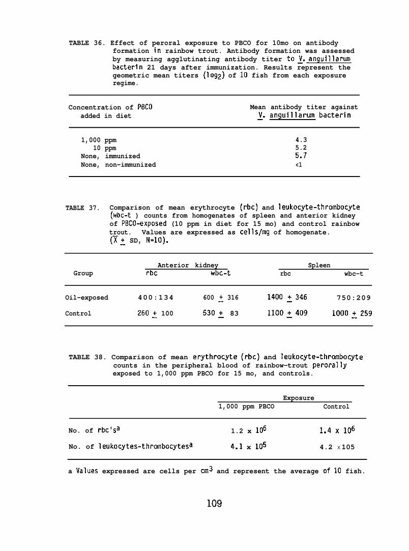
TABLE 36. Effect of peroral exposure to PBCO for 10mo on antibodyformation in rainbow trout. Antibody formation was assessedby measuring agglutinating antibody titer toy. anguillarumbacterin 21 days after immunization. Results represent thegeometric mean titers (log2) of 10 fish from each exposureregime.
Concentration of PBCO Mean antibody titer againstadded in diet ~. anguillarum bacterin
1,000 ppm 4.310 ppm 5.2
None, immunized 5.7None, non-immunized <1
TABLE 37. Comparison of mean erythrocyte (rbc) and leukocyte-thrombocyte(wbc-t ) counts from homogenates of spleen and anterior kidneyof PBCO-exposed (10 ppm in diet for 15 mo) and control rainbowtrout. Values are expressed as cells/mg of homogenate.(~: SD, N=1O).
GroupAnterior kidney
rbc wbc-tSpleen
rbc wbc-t
Oil-exposed 400:134 600 ~ 316 1400 ~346 750:209
Control 260~ 100 530~ 83 lloo~409 1000:259
TABLE 38. Comparison of mean erythrocyte (rbc) and Ieukocyte-thrombocytecounts in the peripheral blood of rainbow-trout perorallyexposed to 1,000 ppm PBCO for 15 mo, and controls.
Exposure1,000 ppm PBCO Control
No. of rbc’sa 1.2 x 10⁶ 1.4X 10⁶
No. of leukocytes-thrombocytesa 4.1 x 10⁵ 4.2 X 10⁵
a Values expressed are cells per cm3 and represent the average of 10 fish.

TABLE 39. Result of screening of flatfish for changes in diseaseresistance aft,er exposure to PBCO-contaminated sediment.
Test species and Number of Percent Mortalityexposure conditions bacteria oil-exposed control
Starry flounder exposed 1.1X10⁸ 802 wk on sediment 1.1X10⁷ 60 ::contaminated with 1800 ppm PBCOa 1.1X10⁶ o
1. 1X10⁵ o :
Starry flounder exposed 6 wk 1.3X10⁸ 80on sediment contaminated 1.3X10⁷ 60 ::with 1800 PBCO :.:;:;; o
● o :
Rock sole exposed 2 wk on 2.2X10⁸ 80 80sediment contaminated 2.2X1Q7 60 40with 2500 ppm PBCO 202X106 o 0
2.2X10⁵ o 0
a Concentration of PBCO was determined at the start of exposure period.b 5 fish per group
The results of testing of the effects of CICO on disease resistanceof juvenile flatfish are shown in Table 40. English sole exposed tooil-contaminated sediment for up to 2 wk showed no significant difference~=0.05) from controls in their ability to survive a laboratory bacterialchallenge. The accompanying analyses of sediment, water, and Iiver tissuedocumented both the availability and uptake of petroleum hydrocarbonsby the exposed fish.
Spot Shrimp. The results of tests in which adult spot shrimp wereexposed for up to 4 wk to sediment contaminated with CICO are summarizedin” Table 41. - LD50 values computed from mortality data followingchallenge with V. anguillarum indicated no significant differences(A=O.05) in di~ease resistance. Results of the analyses of sediment,water, and soft tissue for TEPH demonstrated that the-petroleum hydrocarbonswere both bioavailable and taken up by the exposed shrimp.
6.2.2 Pathological Changes in F?atfish from Exposure to Oil-ContaminatedSediment
Five experiments were conducted in 1977 through 1979; parametersof the individual experiments are listed in Table 42.
110
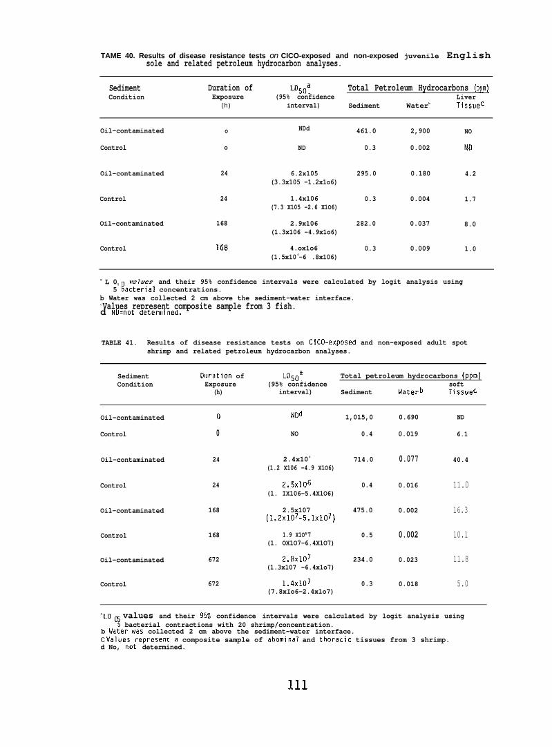
TAME 40. Results of disease resistance tests on CICO-exposed and non-exposed juvenile Englishsole and related petroleum hydrocarbon analyses.
Sediment Duration of L050a Total Petroleum Hydrocarbons (ppm~Condition Exposure (95% confidence Liver
(h) interval) Sediment Waterb Tissuec
Oil-contaminated o NDd 461.0 2,900 NO
Control o ND 0.3 0.002 Ntl
Oil-contaminated 24 6.2x10⁵ 295.0 0.180 4.2(3.3x10⁵ -1.2x1o6)
Control 24 1.4x10⁶ 0.3 0.004 1.7(7.3 X10⁵ -2.6 X1O6)
Oil-contaminated 168 2.9x10⁶ 282.0 0.037 8.0(1.3x10⁶ -4.9x1o6)
Control 168 4.ox1o6 0.3 0.009 1.0(1.5x10 6-6 .8x106)
a L 05 values and their 95% confidence intervals were calculated by logit analysis using5 Bacterial concentrations.
b Water was collected 2 cm above the sediment-water interface.c Values represent composite sample from 3 fish.d NU.not determined.
TABLE 41. Results of disease resistance tests on CICO-exposed and non-exposed adult spotshrimp and related petroleum hydrocarbon analyses.
Sediment Ouration of LD50a Total petroleum hydrocarbons (ppm)Condition Exposure (95% confidence
(h)soft
interval) Sediment Waterb Tissuec
Oil-contaminated
Control
l-l
0
Nod 1,015,0
NO 0.4
0.690
0.019
ND
6.1
40.4Oil-contaminated 2.4x10 6 714.0(1.2 X10⁶ -4.9 X1O6)
24
24
0.077
2.5x10~ 0.4(1. IX106-5.4X1O6)
Control 0.016 11.0
16.3
10.1
11.8
5.0
Oil-contaminated 168 2.5x10⁷ 475.0(1.2xI07-5.1xI07)
0.002
1.9 X1O”7 0.5(1. OX1O7-6.4X1O7)
Control
Oil-contaminated
Control
168 0.002
2.8x10⁷ 234.0(1.3x10⁷ -6.4x1o7)
672 0.023
1.4XI07 0.3(7.8xIo6-2.4x1o7)
672 0.018
a LO o values and their 95% confidence intervals were calculated by logit analysis using; bacterial contractions with 20 shrimp/concentration.
b Water was collected 2 cm above the sediment-water interface.C values represent a composite sample of abominal and thoracic tissues from 3 shrimp.d No, not determined.
111
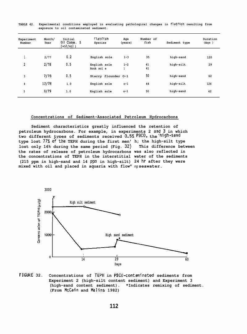
TABLE 42. Experimental conditions employed in evaluating pathological changes in flatfish resulting fromexposure to oil contaminated sediment.
Experiment Month/ Initial Flatfish Age Number of DurationNumber Year Oil Cone. % Species (years) fish Sediment type (days )
(vol/vol )
1 2/77 0.2 English sole 1-3 35 high-sand 120
2 2/78 0.5 English sole 1-2 41 high-silt 29Rock sol e 1 41
3 7/78 0.5 Starry flounder O-1 50 high-sand 62
4 12/78 1.0 English sole o-1 44 high-silt 126
5 9/79 1.0 English sole o-1 50 high-sand 42
Concentrations of Sediment-Associated Petroleum Hydrocarbons
Sediment characteristics greatly influenced the retention ofpetroleum hydrocarbons. For example, in experiments 2 and.3 in whichtwo different types of sediments received 0.5% pBCO, the h19h-sandtype lost 77% o~”the TEPH during the first men’lost only 16% during the same period (Fig. 32)the rates of release of petroleum hydrocarbonsthe concentrations of TEPH in the interstitial(215 ppm in high-sand and 14 ppm in high-silt)mixed with oil and placed in aquaria with flow”
h; the high-silt typeThis difference between
was also reflected inwater of the sediments24 hr after they wereng seawater.
3000
r~mm I High silt sediment
E.-Z.z~ 1000 - High sand sedimentc ●
G
o I I I14 29 60
Days
FItilJRE 32. Concentrations of TEPH in PBCU-contaminated sediments fromExperiment 2 (high-silt content sediment) and Experiment 3(high-sand content sediment). *Indicates remixing of sediment.(From McCain and Malins 1982)
112
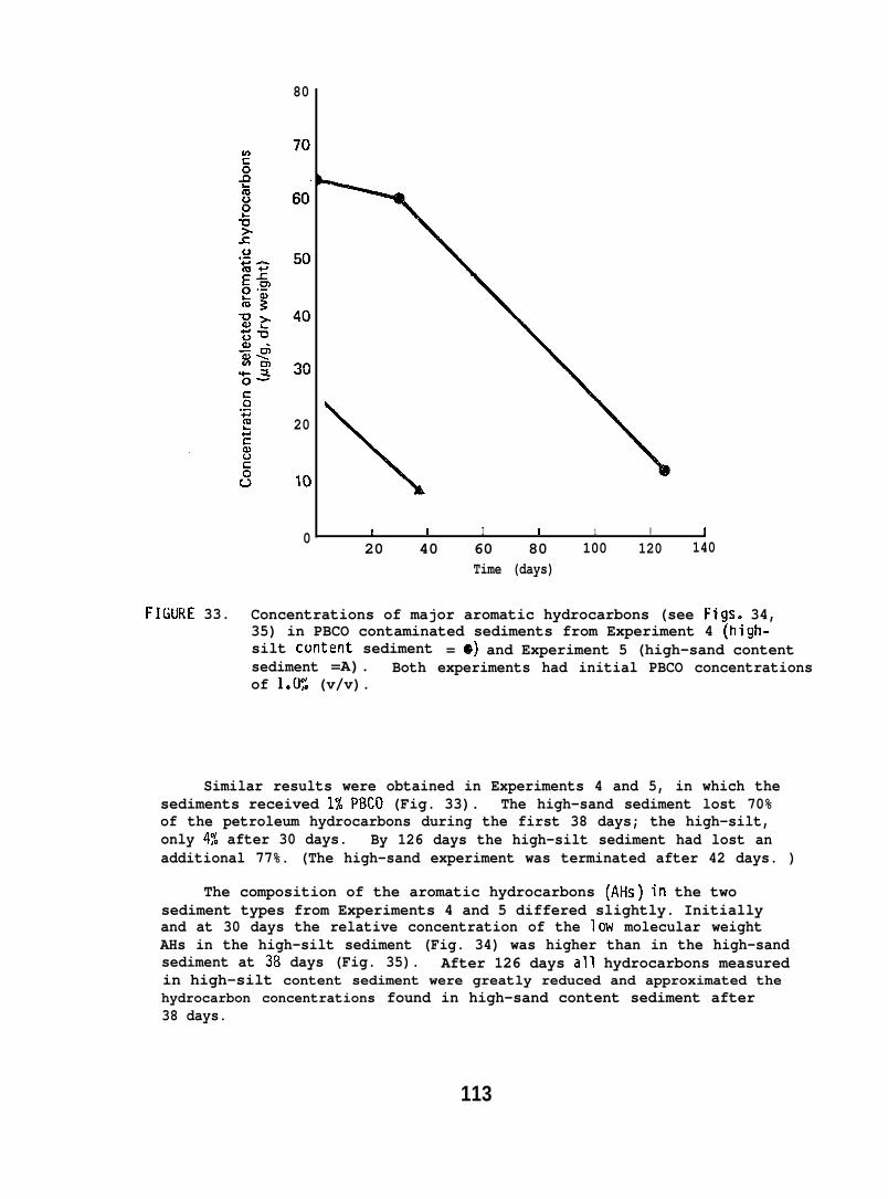
80
20
10
0 I I I I 1 I ~
20 40 60 80 100 120 140Time (days)
FI(IIRE 33. Concentrations of major aromatic hydrocarbons (see Figs. 34,35) in PBCO contaminated sediments from Experiment 4 (high-silt content sediment = e) and Experiment 5 (high-sand contentsediment =A). Both experiments had initial PBCO concentrationsof 1*W: (v/v).
Similar results were obtained in Experiments 4 and 5, in which thesediments received 1% PBCO (Fig. 33). The high-sand sediment lost 70%of the petroleum hydrocarbons during the first 38 days; the high-silt,only 4% after 30 days. By 126 days the high-silt sediment had lost anadditional 77%. (The high-sand experiment was terminated after 42 days. )
The composition of the aromatic hydrocarbons (AHs) in the twosediment types from Experiments 4 and 5 differed slightly. Initiallyand at 30 days the relative concentration of the low molecular weightAHs in the high-silt sediment (Fig. 34) was higher than in the high-sandsediment at 38 days (Fig. 35). After 126 days all hydrocarbons measuredin high-silt content sediment were greatly reduced and approximated thehydrocarbon concentrations found in high-sand content sediment after38 days.
113
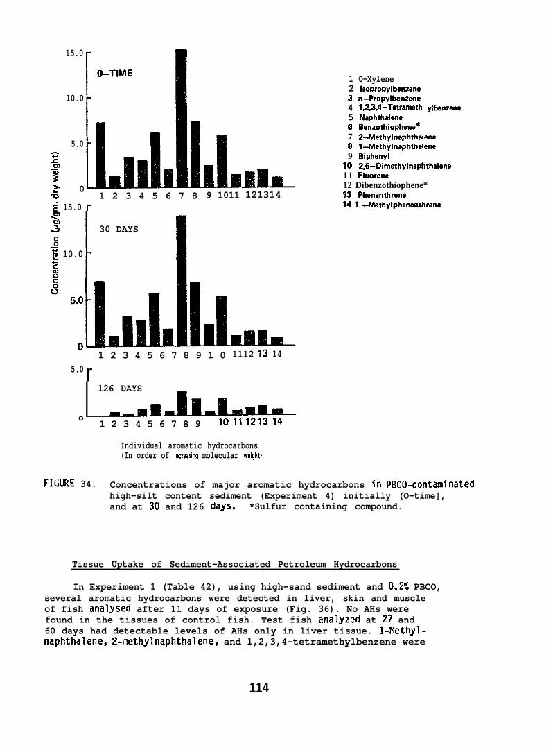
15.0
10.0
5.0~m.-%> 0G 1 2 3 4 5 6 7 8~ 15.0 f-ql~ I 30 DAYSco
“i? 10.0
A9 1011
111.1u
1 2 3 4 5 6 7 8 9 1 05.0 r
121314
1 O-Xylene2 Isopropylbenzene3 rr-propylbenzene4 1,2,3,4-Tewamethy Hx?nzene5 NaphthaIene6 Benzothiophene*7 2-Methylnaphthalene8 1+’lathylnaphthalene9 Biphenyl
30 2,6-Dimethylnaphthalene11 Fluorene12 Dibenzothiophene*13 Phenanthrerae14 1 -Methylphenenthrene
Sam1112
m14
126 DAYS
o -1 2 3 4 5 6 7 8 9 !011121314
Individual aromatic hydrocarbons(In order of increasing molecular weight)
FIGURE 34. Concentrations of major aromatic hydrocarbons in WNX.1-contaminatedhigh-silt content sediment (Experiment 4) initially (O-time],and at 30 and 126 clays. *Sulfur containing compound.
Tissue Uptake of Sediment-Associated Petroleum Hydrocarbons
In Experiment 1 (Table 42), using high-sand sediment and 0.2$% PBCO,several aromatic hydrocarbons were detected in liver, skin and muscleof fish analysed after 11 days of exposure (Fig. 36). No AHs werefound in the tissues of control fish. Test fish analyzed at 27 and60 days had detectable levels of AHs only in liver tissue. l-=Methyl-naphthalene, 2-methylnaphthalene, and l,2,3,4-tetramethylbenzene were
114
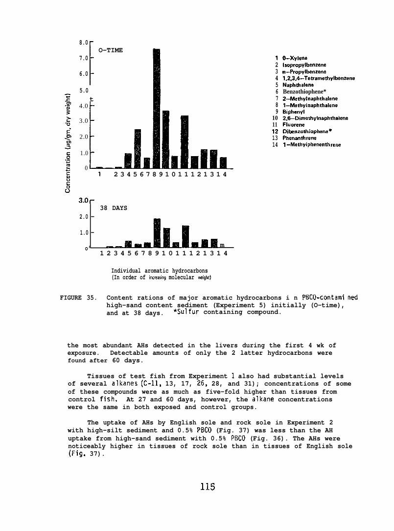
8.0
1
O–TIME7.0
6.0
5.0t
4.0
3.0[
2.0
1.0
0u
123456789
1011121314
O-XyleneIsopropylbenzenen-Propylbenzene1,2,3,4-TetramethylbenzeneNaphthaleneBenzothiophene*2-Methylnaphthalenel-MethyhraphthaleneBiphenyl2,6-DimethylnaphthaleneFluoreneDibenzothiophene”Phenanthrane1 -Methylphenenth rene
1 2 3 4 5 6 7 8 9 1 0 1 1 1 2 1 3 1 4
3.Or38 DAYS
2.0 -
1.0 -
o’ m1 2 3 4 5 6 7 8 9 1 0 1 1 1 2 1 3 1 4
Individual aromatic hydrocarbons(In order of increasing molecular weight)
FIGURE 35. Content rations of major aromatic hydrocarbons i n PBC()-contami nedhigh-sand content sediment (Experiment 5) initially (O-time),and at 38 days. *sulfur containing compound.
the most abundant AHs detected in the livers during the first 4 wk ofexposure. Detectable amounts of only the 2 latter hydrocarbons werefound after 60 days.
Tissues of test fish from Experiment 1 also had substantial levelsof several alkanes (C-11, 13, 17, 26$ 28, and 31); concentrations of someof these compounds were as much as five-fold higher than tissues fromcontrol fish. At 27 and 60 days, however, the alkane concentrationswere the same in both exposed and control groups.
The uptake of AHs by English sole and rock sole in Experiment 2with high-silt sediment and 0.5% PBCO (Fig. 37) was less than the AHuptake from high-sand sediment with 0.5% P13C0 (Fig. 36). The AHs werenoticeably higher in tissues of rock sole than in tissues of English sole(Fig. 37).
115
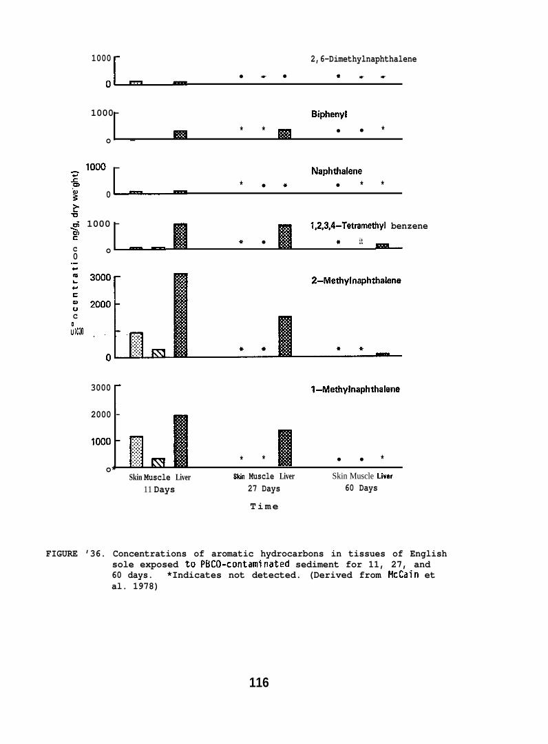
1000r
2,6-Dimethylnaphthalene
-● ☛ ● ☛ ☛ ☛
01 -
1000- Biphenyl* * ● ● *
o -
~ looo - Naphthalene.cm.- * ● + ● * *z 0~>+5~ 1000 - 1,2,3,4-Tetramethyl benzeneUIs * ● * itc o~—0.-t-
c tnfs: 1000 . . . . .. . . . .. . . . .. . . . .. . . . .. . . . .0
. ...+ L * *
2-Methylnaphthalene
-
3000
[
l-Methylnaphthalene
2000 -
* * ● ● *o*
Skin Muscle Liver Skin Muscle Liver Skin Muscle Liver
11 Days 27 Days 60 Days
Time
FIGURE ’36. Concentrations of aromatic hydrocarbons in tissues of Englishsole exposed to PBCO-contaminated sediment for 11, 27, and60 days. *Indicates not detected. (Derived from McCain etal. 1978)
116
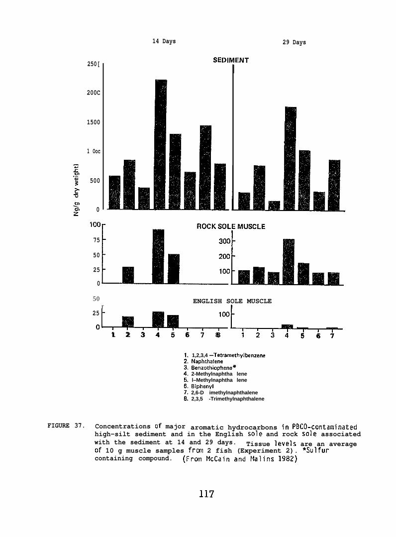
250[
200C
1500
1 Ooc
z.cm.-$ 5002u~
02100
75
50
25
0
14 Days
SEDllI[NT
29 Days
50r
ENGLISH SOLE MUSCLE1
25 -
0’~b1 2 3 4 5 6 7 8 5 6 7
FIGURE 37. Concentrations of major
1,2,3,4 -TetramethylbanzeneNaphthaleneBenzothiophene*2-Methylnaphtha lenel–Methylnaphtha leneBiphenyl2,6-D imethylnaphthalene2,3,5 -Trimethylnaphthalene
aromatic hydrocarbons in PBCO-contaminatedhigh-silt sediment and in the English sole and rock sole associatedwith the sediment at 14 and 29 days. Tissue levels are an averageof 10 g muscle samples frcal 2 fish (Experiment 2). ~Sulfurcontaining compound. (Fran McCain and Malins 1982)

Muscle tissue of English sole exposed for 91 andExperiment 4 (high-silt, 1.0% PBCO) had no detectablethe muscle tissue of the English sole exposed to 1.0%sediment (Experiment 5) had detectable concentrationsdry wt) of 1- and 2-methyl naphthalene after 38 days.
126 days inAHs. However,PBCO in high-sand(26 and 41 ppb
Biological Effects of Exposure to Oiled Sediments
Rock sole and starry flounder sustained high mortalities (30-50%)in both control and exposed groups after 30-60 days, whereas Englishsole had low mortalities in both of the 120 day tests (Experiments 1and 4). As a result English sole were used to assess the biologicaleffects of long-term exposure to oil-contaminated sediments.
In Experiment 1 both test and control fish lost weight during thefirst 2 mo, with the oil-exposed group losing more than the controls(Fig. 38). After this period, the control fish began to regain weightand at 4 mo, only 27;g of the animals weighed less than at 2 wk. Theoil-expqsed fish were slower in their recovery from the initial weightloss, and at 4 mo 69% weighed less than they did initially. The numbersof fish gaining and not gaining weight at 4 mo were significantlydifferent between the experimental and control groups (P=O.05). Theseresults are apparently related to reduced feeding in the oil-exposedgroup, as indicated by consistently more uneaten food removed from thetest aquarium than from the control aquarium.
TIME (months)
FIGURE 38. Weight changes in oil-exposed and control English sole over aperiod of 4 months (Experiment 1). Each observation at monthlyintervals represents the average change (g) from weights at2 wk for 16 test and 15 control fish. (From McCain et al. 1978)
118
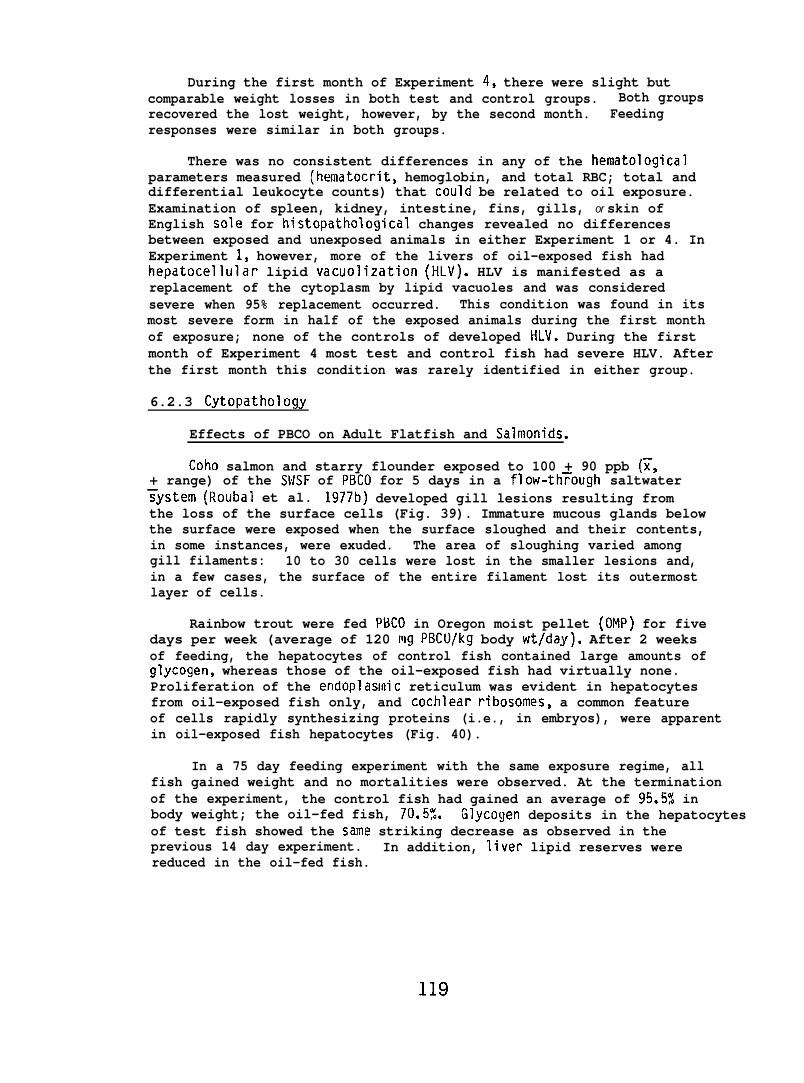
During the first month of Experiment 4, there were slight butcomparable weight losses in both test and control groups. Both groupsrecovered the lost weight, however, by the second month. Feedingresponses were similar in both groups.
There was no consistent differences in any of the hematologicalparameters measured (hematocrit, hemoglobin, and total RBC; total anddifferential leukocyte counts) that could be related to oil exposure.Examination of spleen, kidney, intestine, fins, gills, or skin ofEnglish sole for histopathological changes revealed no differencesbetween exposed and unexposed animals in either Experiment 1 or 4. InExperiment 1, however, more of the livers of oil-exposed fish hadhepatocellular lipid vacuolization (HLV). HLV is manifested as areplacement of the cytoplasm by lipid vacuoles and was consideredsevere when 95% replacement occurred. This condition was found in itsmost severe form in half of the exposed animals during the first monthof exposure; none of the controls of developed HI-V. During the firstmonth of Experiment 4 most test and control fish had severe HLV. Afterthe first month this condition was rarely identified in either group.
6.2.3 Cytopathology
Effects of PBCO on Adult Flatfish and Salmonids.
Coho salmon and starry flounder exposed to 100 + 90 ppb ~,+ range) of the $W.SF of PBCO for 5 days in a flow-th~ough saltwater~ystem (Roubal et al. 1977b) developed gill lesions resulting fromthe loss of the surface cells (Fig. 39). Immature mucous glands belowthe surface were exposed when the surface sloughed and their contents,in some instances, were exuded. The area of sloughing varied amonggill filaments: 10 to 30 cells were lost in the smaller lesions and,in a few cases, the surface of the entire filament lost its outermostlayer of cells.
Rainbow trout were fed PBCO in Oregon moist pellet (OMP) for fivedays per week (average of 120 mg PBCO/kg body wt/day). After 2 weeksof feeding, the hepatocytes of control fish contained large amounts ofglycogen, whereas those of the oil-exposed fish had virtually none.Proliferation of the end”oplasmic reticulum was evident in hepatocytesfrom oil-exposed fish only, and cochlear ribosomes, a common featureof cells rapidly synthesizing proteins (i.e., in embryos), were apparentin oil-exposed fish hepatocytes (Fig. 40).
In a 75 day feeding experiment with the same exposure regime, allfish gained weight and no mortalities were observed. At the terminationof the experiment, the control fish had gained an average of 95.5% inbody weight; the oil-fed fish, 70.5%. Glycogen deposits in the hepatocytesof test fish showed the same striking decrease as observed in theprevious 14 day experiment. In addition, liver lipid reserves werereduced in the oil-fed fish.
119
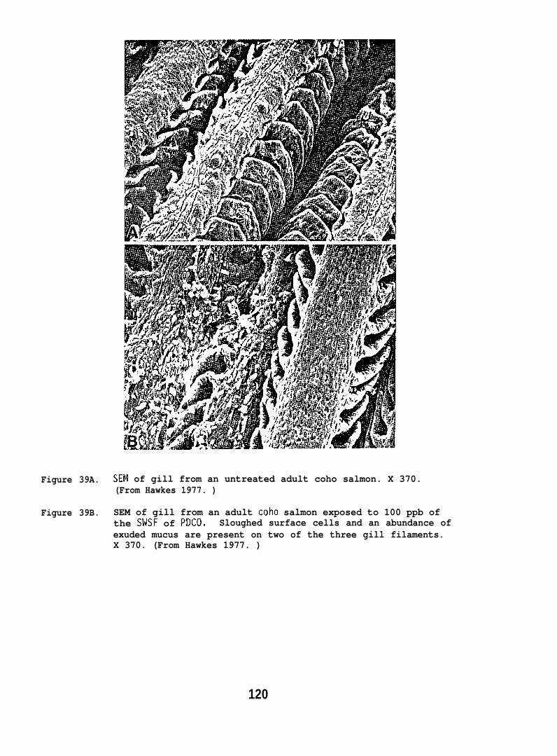
Figure 39A. SEN of gill from an untreated adult coho salmon. X 370.(From Hawkes 1977. )
Figure 39B. SEM of gill from an adult coho salmon exposed to 100 ppb ofthe SMSF of PDCO. Sloughed surface cells and an abundance ofexuded mucus are present on two of the three gill filaments.X 370. (From Hawkes 1977. )
120
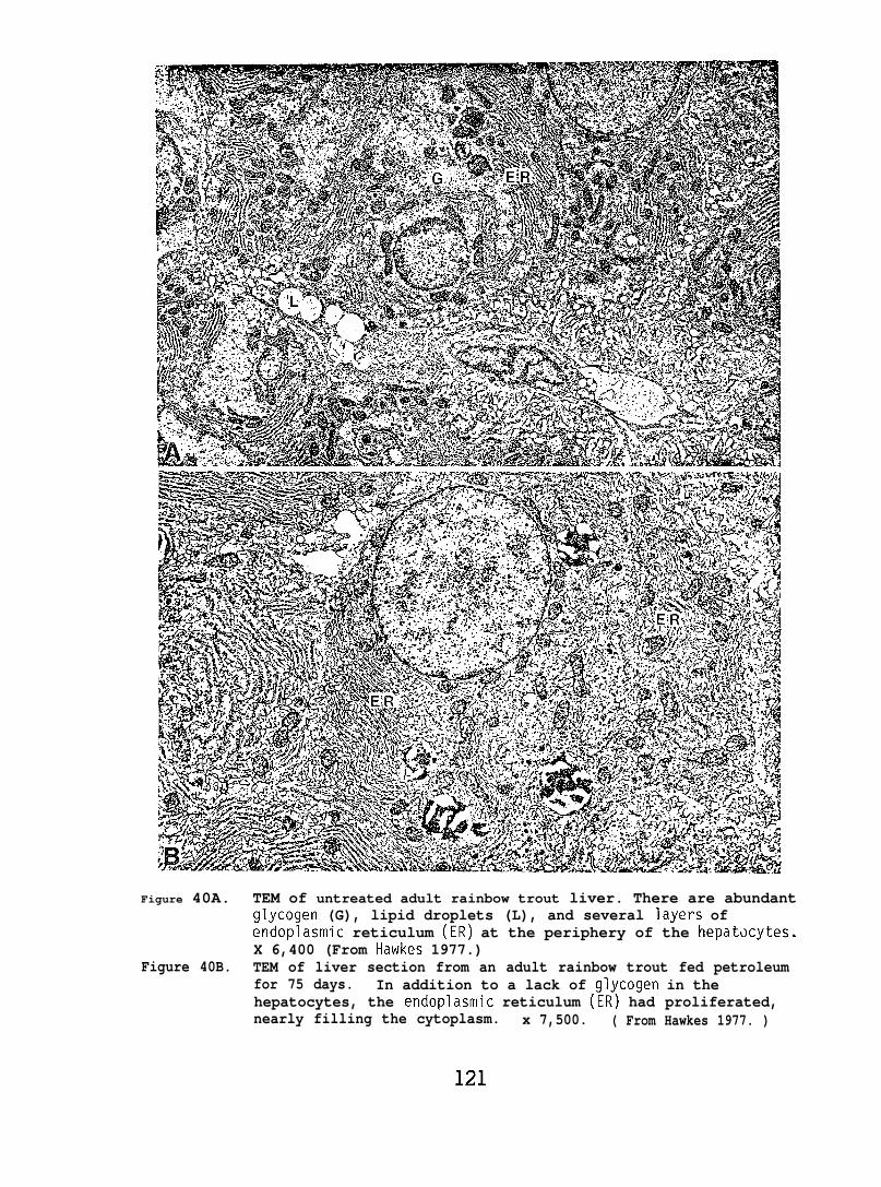
Figure 40A. TEM of untreated adult rainbow trout liver. There are abundantglycogen (G), lipid droplets (L), and several layers ofendoplasmic reticulum (ER) at the periphery of the hepatocytes.X 6,400 (From Hawkes 1977.)
Figure 40B. TEM of liver section from an adult rainbow trout fed petroleumfor 75 days. In addition to a lack of glycogen in thehepatocytes, the endoplasmic reticulum (ER) had proliferated,nearly filling the cytoplasm. x 7,500. ( From Hawkes 1977. )
121
Rainbm trout fed a PBCO-contaminated diet (1000 ppm added to food)for 8 mo were sampled for microscopy at the time of spawning. BothTEM and I-M analysis indicated an abnormal increase in collagen aroundthe liver sinusoids. Trout from this same study continued to be fedthe oil-contaminated diet for another 4 to 5 mo after spawning; thesefish exhibited abnormally soft and enlarged eye lenses. Of trout fedPBCO for 12-13_mo the eye lens volume was double that of controls;226~81 mm3 (x~SD) and lll~19mm3, respectively.
The lens is composed of ribbon-like filaments which interdigitateand form a sphere. The filaments have simple projections on theirbroad surfaces which plug into pits on the adjacent fiber; in addition,there are complex interlocking series of protuberances on their thinside (Fig. 41A). In trout exposed to a crude oil contaminated dietfor either 75 days or one year, the fiber structure changed: the surfacebecame distorted and the interdigitating projections were irregular. Ina longer experiment, rainbow trout were fed PBCO (120 mg/kg body wt/day)for 3 years; three of five petroleum-exposed fish developed cataracts,whereas all three control fish had normal lenses. In lenses withcataracts, the fiber surface was irregular and the lateral projectionswere severely deformed (Fig. 41B).
Effects of weathered PBCO on early development of sand sole.
SEN examination of 5 sand sole larvae exposed to 164 ppb of theSWSF of weathered PBCO during embryogenesis revealed 4 of 5 withseverely reduced (both numbers and length) olfactory cilia (Fig. 42);the fifth appeared normal (comparable to controls). In fish withreduced olfactory cilia, the epidermal microridges, a feature of normalkeratinocytes, were absent from the keratinocytes surrounding theolfactory epitheliums. In addition, the keratinocytes of these fourlarvae were rounded and protruded from the skin surface, an indicationof cellular hypertrophy.
TEM examination of sections from 8 sand sole larvae (4 exposed to164 ppb of the SWSF as embryos, and 4 controls) revealed ultrastructuralchanges in the mitochondria of epidermal cells of oil-exposed larvae.Although the sample size was too small to definitively correlate observedchanges to oil exposure, the abnormal mitochondria showed classichydropic changes, a reduction in cristae and a reduction in the electrondensity of the matrix. Separation between the outer and inner mitochondrialmembranes was quite pronounced and breaks in both membranes were observed.Other organelles such as the nuclei, Golgi assemblies, and microtubuleswere normal.
Effects of weathered CICO on early development of surf smelt.
Morphology of unexposed embryos. Sections of entire 21 or 27-day-oldcontrol em~ryos were examined with light microscopy and electron microscopyand all tissues appeared normal. In neurons of the brain the heterochromatinwas evenly distributed in small clusters throughout the nuclei. The
122
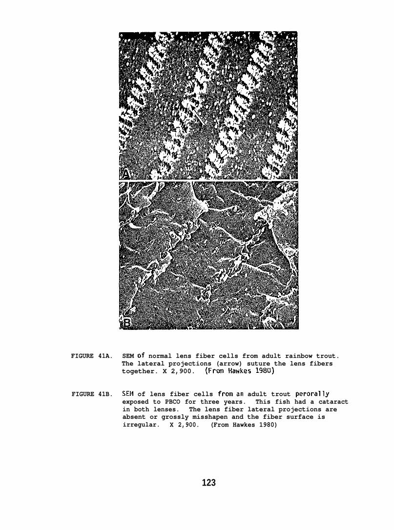
FIGURE 41A.
FIGURE 41B.
SEM of normal lens fiber cells from adult rainbow trout.The lateral projections (arrow) suture the lens fiberstogether. X 2,900. {From Hawkes 1980)
SEM of lens fiber cells froman adult trout perorallyexposed to PBCO for three years. This fish had a cataractin both lenses. The lens fiber lateral projections areabsent or grossly misshapen and the fiber surface isirregular. X 2,900. (From Hawkes 1980)
123
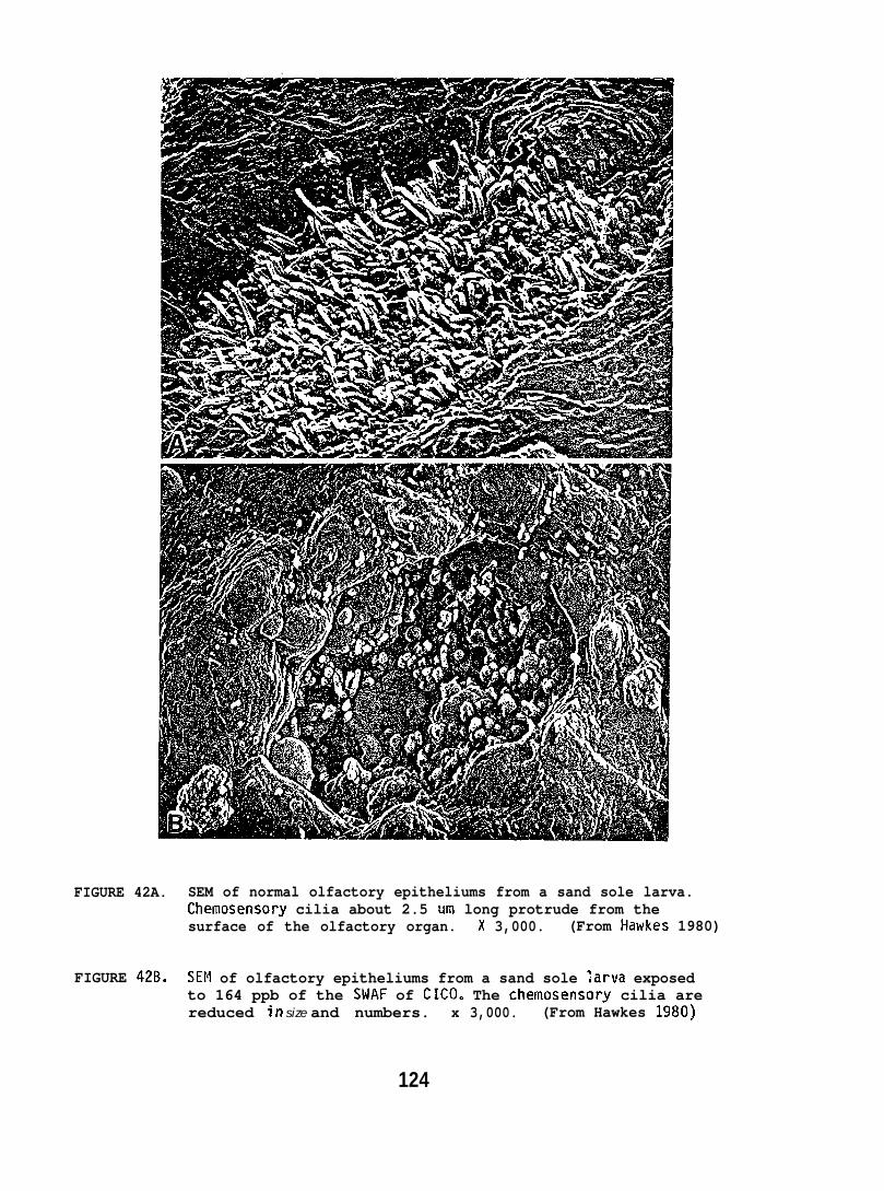
FIGURE 42A. SEM of normal olfactory epitheliums from a sand sole larva.Chemosensory cilia about 2.5 urn long protrude from thesurface of the olfactory organ. x 3,000. (From Iiawkes 1980)
FIGURE 42B, SEM of olfactory epitheliums from a sand sole ;arva exposedto 164 ppb of the SWAF of CICO. The chemosensory cilia arereduced in size and numbers. x 3,000. (From Hawkes 1980)
124
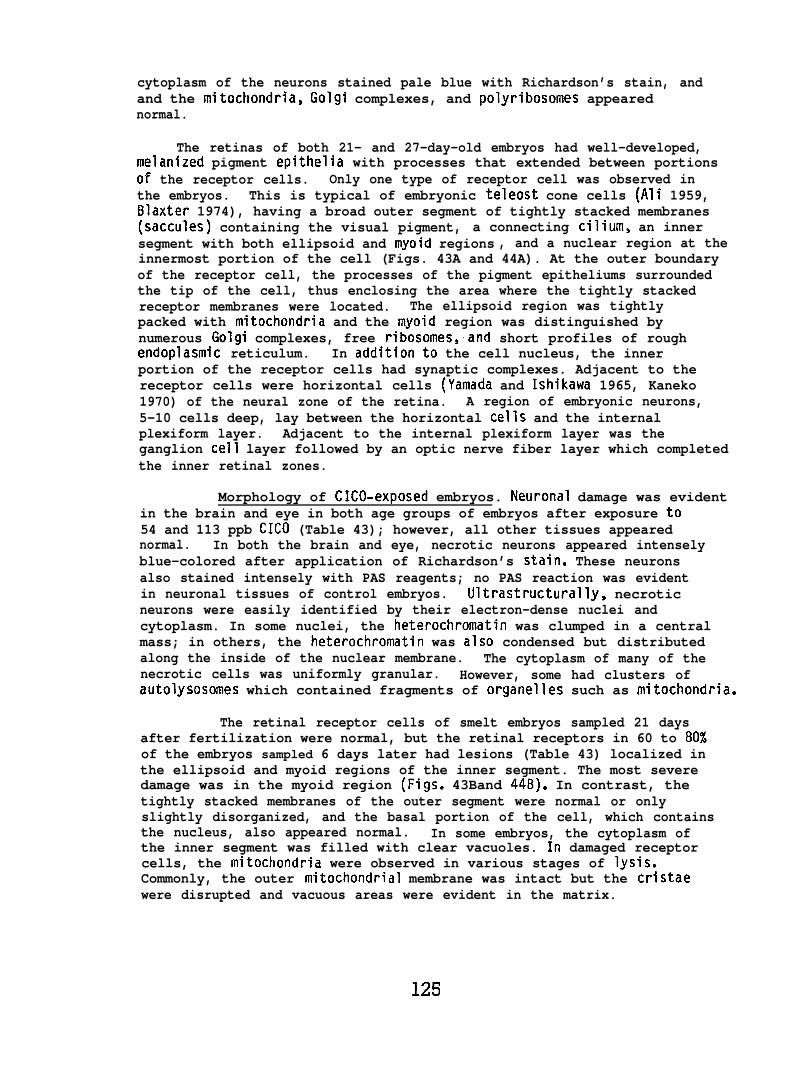
cytoplasm of the neurons stained pale blue with Richardson’s stain, andand the mitochondria, Golgi complexes, and polyribosomes appearednormal.
The retinas of both 21- and 27-day-old embryos had well-developed,melanized pigment epithelia with processes that extended between portionsof the receptor cells. Only one type of receptor cell was observed inthe embryos. This is typical of embryonic teleost cone cells (Ali 1959,Blaxter 1974), having a broad outer segment of tightly stacked membranes(saccules) containing the visual pigment, a connecting cilium, an innersegment with both ellipsoid and myoid regions , and a nuclear region at theinnermost portion of the cell (Figs. 43A and 44A). At the outer boundaryof the receptor cell, the processes of the pigment epitheliums surroundedthe tip of the cell, thus enclosing the area where the tightly stackedreceptor membranes were located. The ellipsoid region was tightlypacked with mitochondria and the tnyoid region was distinguished bynumerous Golgi complexes, free ribosomes,and short profiles of roughendoplasmic reticulum. In additionto the cell nucleus, the innerportion of the receptor cells had synaptic complexes. Adjacent to thereceptor cells were horizontal cells (Yamada and Ishikawa 1965, Kaneko1970) of the neural zone of the retina. A region of embryonic neurons,5-10 cells deep, lay between the horizontal cells and the internalplexiform layer. Adjacent to the internal plexiform layer was theganglion cell layer followed by an optic nerve fiber layer which completedthe inner retinal zones.
Morphology of CICO-exposed embryos. Neuronal damage was evidentin the brain and eye in both age groups of embryos after exposure to54 and 113 ppb CICO (Table 43); however, all other tissues appearednormal. In both the brain and eye, necrotic neurons appeared intenselyblue-colored after application of Richardson’s stain. These neuronsalso stained intensely with PAS reagents; no PAS reaction was evidentin neuronal tissues of control embryos. Ultrastructurally, necroticneurons were easily identified by their electron-dense nuclei andcytoplasm. In some nuclei, the heterochromatin was clumped in a centralmass; in others, the heterochromatin was also condensed but distributedalong the inside of the nuclear membrane. The cytoplasm of many of thenecrotic cells was uniformly granular. However, some had clusters ofautolysosomes which contained fragments of organelles such as mitochondria.
The retinal receptor cells of smelt embryos sampled 21 daysafter fertilization were normal, but the retinal receptors in 60 to 80%of the embryos sampled 6 days later had lesions (Table 43) localized inthe ellipsoid and myoid regions of the inner segment. The most severedamage was in the myoid region (Figs. 43Band 44B). In contrast, thetightly stacked membranes of the outer segment were normal or onlyslightly disorganized, and the basal portion of the cell, which containsthe nucleus, also appeared normal. In some embryos, the cytoplasm ofthe inner segment was filled with clear vacuoles. In damaged receptorcells, the mitochondria were observed in various stages of lysis.Commonly, the outer mitochondrial membrane was intact but the cristaewere disrupted and vacuous areas were evident in the matrix.
125
TAE4E 43. Percent surf smelt embryos with cellular abnormalities of the eye and bratn sampled 21 and 27days after fertilization (45 and 63 hr oil exposure)
Percent with Cellular abnormalitiesAverage 21 days post- fertil ization 27 days post-fertilization
hydrocarbon Vacuolated Necrotl c Necrotic Vacuolated Necrot~c Necrot lcconcentration retinal retinal forebrain retinal retinal forebrain
(ppb) receptor neurons neurons receptor neurons neuronscells cel Is
control o (4p o (4) o (5) o (4) o (4) o (5)
54 0 (6) 83 (6) 72 (7) 80 (5) 80 (5) 80 (5)
113 0 (4) 75 (4) 80 (5) 75 (4) 80 (5) 66 (6)
a Number of embryos examined.(Fran Hawkes and Stehr 1982)
FIGURE 43A. TEM of a portion of the retina from a norms? 28-day-old smeltembryo. Regions of the receptor cells include the outer segment(0), ellipsoid region (E), myoid region (M), nucleus (N), andsynaptic junctional complex (S). Neural cells of the retinashown in the micrograph include horizontal cells (H) andretinal neurons (RN). X 5,000. (From Hawkes and Stehr 1982)
FIGURE 43B. TEM of a portion of the retina of a 28-day-old exposed (113 ppbCICO) surf smelt embryo. Vesiculation is evident in the myoidregions (M) of the receptor cells and necrotic neurons arepresent (arrow). X 5,000. (From Hawkes and Stehr 1982)
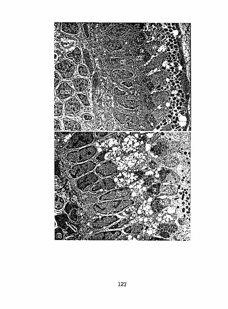
127
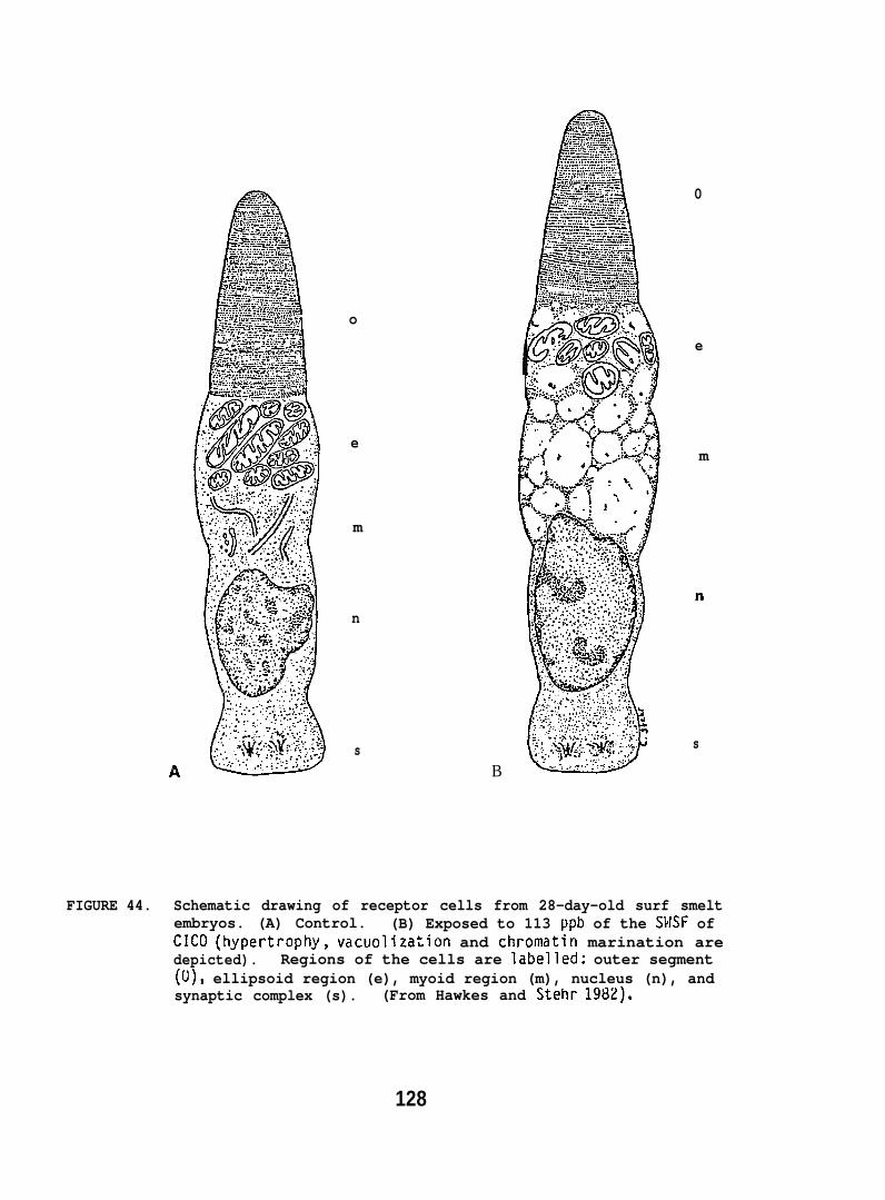
o
e
m
n
s
..:.4::7: ,:, ,..:,..
.- , , , . . . . .. .. . ..... . . ..-
--27: ,-...7.-., ; ,,. -, . .
.... ... ... ,..~ ..: ......~, ~.=. ,
- :.-.----? ,.-.., . . .. e.=- Q-.=--.-.-.:?avx .“:.=: ... ... .7.%1 : s A—=?.=
:=:&:=-:;-...:-::.,:&:,:A:+:A:+:----- ,<sa--, ..=-==----
- ---; .=.:-.-..2 :--- . . .--a-+,-=+ :.=,--_-.= .=
=’=: . . .... . . . . . . . .. --,.:,,..
;:$
B .-. .: .: .?-> .. :.? .:.
0
e
m
n
s
FIGURE 44. Schematic drawing of receptor cells from 28-day-old surf smeltembryos. (A) Control. (B) Exposed to 113 ppb of the SMSF ofCICO (hypertrophy, vacuolization and chromatin marination aredepicted). Regions of the cells are labelled: outer segment(O), ellipsoid region (e), myoid region (m), nucleus (n), andsynaptic complex (s). (From Hawkes and Stehr 1982).
128
6.3 Behavior and Physiolo~
6.3.1 Vertebrate Studies
Behavior of Pacific Salmon Exposed to Petroleum Hydrocarbons
(a) Olfactory Disruption in Juvenile Coho Salmon. The firstseries of electrophysiologi cal tests to evaluate the effect of petroleumhydrocarbons on the olfaction of seawater-adapted juvenile salmon wereof an exploratory nature, and the data were not evaluated statistically.Exposure of the olfactory epitheliums to both benzene (0.2 to 17.0 ppm)and NPH (0.2 to 2.0 ppm) for 25 seconds induced a rapidly diminishingoscillatory pattern from the olfactory bulb indicating stimulation,but no immediate injury to, or irritation of, the olfactory epitheliums.Following perfusion of the nares with either 2 ppm NPH or benzene for10 minutes, there was no reduction or inhibition of the immediatesubsequent response to 103 L-serine, L-methionine, or L-alanfne. Whenone part of 1.5 ppm NPH was mixed with an equal part of 103 L-serinethe EEG response approximated that of the L-serine solution alone.
There was no evidence of the salmon detecting 2,6-dimethylnaphthalene,or 2,3,6-trimethyl naphthalene at concentrations of 0.2 to 2.0 ppm. TheEEG response to these aranatics was similar to that of rinsing the nareswith filtered saltwater; that is, there was a slight temporary decreasein baseline activity. In contrast, the SWAF of PBCO (1.8 to 20.0 ppm)was detected by the fish; however, there was no decrease in subsequentresponses to amino acids. Threshold concentrations for discriminationwere not determined.
In the second series of electrophysiological tests, activity ofthe olfactory bulb (EEG responses) of coho salmon following exposureof the nares to a standard stimulant (10-3 M L-serine) were comparedwith EEG responses induced by different concentrations of an aromatichydrocarbon mixture representative of the SWAF of PBC(I (Table 44). Asignificant increase in the amplitude of the EEG response to the aromatichydrocarbon mixture occurred at concentrations between 1.9 and 2,8 ppm;this is less than the 2.8 to 3.7 ppm concentrations which resultedin statistically significant avoidance of the hydrocarbons by juvenilecoho salmon tested at the same time of year and at the same temperatures(Maynard and Weber 1981).
Coho salmon exposed to a 4.(I ppm aromatic hydrocarbon mixture forup to 20 min showed no significant change in EEG response to 10-3 ML-serine given before and after exposure (Table 45),
(b) Migratory and Homing Behavior in Adult Salmon
Avoidance reaction. Control observations (i.e., percentageof salmon mlgratlng up the east or west fish ladders when aromatichydrocarbons were not added) and tests with hydrocarbons were madeduring the peak of salrllon migration in Chambers Creek. More than1,400 salmon ascended the darn during control periods; 56% of these
129
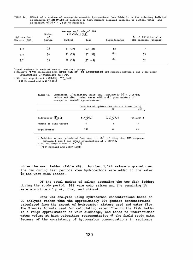
TABLE 44. Effect of a mixture of monocyclic aromatic hydrocarbons (see Table 1) on the olfactory bulb EEG
as percent of 10-s M L-serine response.as measured by am litude of response to test mixture compared response to control water, and
Average amplitude of EEGNumber response (SD)b
Hyd rota rbon of % of 10-3 M L-serineMixture (ppm) testsa Control Test Significance EEG response (average)
1.9 10 37 (27) 23 (24) NS 7
2.8 20 35 (24) 87 (52) *** 25
3.7 15 31 (19) 117 (49) *** 32
a Equal numbers in each of control and test groups.
b Relative values calculated from area (in cm2) of integrated EEG response between 2 and 6 Sec afterintroduction of stimulant to naris.
c NS, not significant (p>O.05); ***~<0.001(Frmn Maynard and Weber 1981)
TABLE 45. Comparisonbefore andmonocyclic
of olfactory bulb EEG response to 10-3 M L-serineafter rinsing nares with a 4.0 ppm mixture ofaromatic hydrocarbons.
Duration of hydrocarbon mixture rinse (rein)<1 10 20 -
Difference (~:SE) 6.4~16.7 42.7~17.5 -36.2334.1
Number of fish tested 6 6 5
Significance NSa NS NS
a Relative values calculated from area (in Chlz) of integrated EEG responsebetween 2 and 6 sec after introduction of L-serine.
b NS, not significant ( = 0.05).(Frcm Maynard and Weber 1981)
chose the west ladder (Table 46). Another 1,149 salmon migrated overthe dam during test periods when hydrocarbons were added to the waterin the west fish ladder.
Of the total number of salmon ascending the two fish laddersduring the study period, 99% were coho salmon and the remaining 1%were a mixture of pink, chum, and chinook.
Data was analyzed using hydrocarbon concentrations based onGC analysis rather than the approximately 40% greater concentrationscalculated from the amount of hydrocarbon mixture used and water flow.The Francis formula used for calculating water flow in the fish ladderis a rough approximation of weir discharge, and tends to underestimatewater volume at high velocities representative of the field study site.Because of the consistency of hydrocarbon concentrations in replicate
130
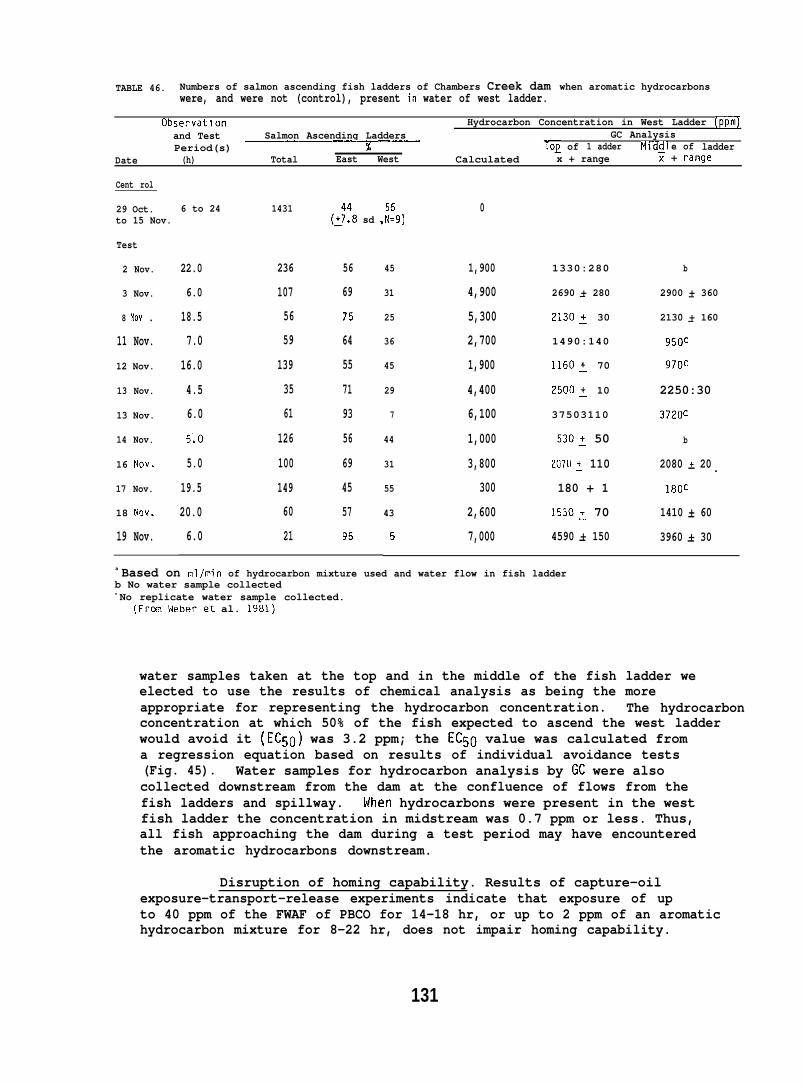
TABLE 46. Numbers of salmon ascending fish ladders of Chambers Creek dam when aromatic hydrocarbonswere, and were not (control), present in water of west ladder.
Observatlon Hydrocarbon Concentration in West Ladder (ppm]and Test Salmon Ascending Ladders GC AnalysisPeriod(s) ~ log of 1 adder M]dcll e of ladder
Date (h) Total East West Calculated x + range 1 + range
Cent rol
29 Oct. 6 to 24 1431 0to 15 Nov. (:::8 sd ,::9)
Test
2 Nov.
3 Nov.
8 fiov .
11 Nov.
12 Nov.
13 Nov.
13 Nov.
14 Nov.
16 NOV.
17 Nov.
18 NOV.
19 Nov.
22.0 236 56
6.0 107 69
18.5 56 75
7.0 59 64
16.0 139 55
4.5 35 71
6.0 61 93
5:0 126 56
5.0 100 69
19.5 149 45
20.0 60 57
6.0 21 95
45
31
25
36
45
29
7
44
31
55
43
5
1,900
4,900
5,300
2,700
1,900
4,400
6,100
1,000
3,800
300
2,600
7,000
1330:280
2690 + 280—
2130~ 30
1490:140
1160: 70
2500~ 10
37503110
530: 50
2070: 110
180 + 1
1530~ 70
4590 + 150—
b
2900 + 360—
2130 + 160—
950C
970~
2250:30
3720C
b
2080 + 20— .180C
1410 + 60—3960 + 30—
a Based on ral/min of hydrocarbon mixture used and water flow in fish ladderb No water sample collectedc No replicate water sample collected.
(Frm\4eberet al. 1981)
water samples taken at the top and in the middle of the fish ladder weelected to use the results of chemical analysis as being the moreappropriate for representing the hydrocarbon concentration. The hydrocarbonconcentration at which 50% of the fish expected to ascend the west ladderwould avoid it (EC50) was 3.2 ppm; the EC50 value was calculated froma regression equation based on results of individual avoidance tests(Fig. 45). Water samples for hydrocarbon analysis by GC were alsocollected downstream from the dam at the confluence of flows from thefish ladders and spillway. When hydrocarbons were present in the westfish ladder the concentration in midstream was 0.7 ppm or less. Thus,all fish approaching the dam during a test period may have encounteredthe aromatic hydrocarbons downstream.
Disruption of homing capability. Results of capture-oilexposure-transport-release experiments indicate that exposure of upto 40 ppm of the FWAF of PBCO for 14-18 hr, or up to 2 ppm of an aromatichydrocarbon mixture for 8-22 hr, does not impair homing capability.
131
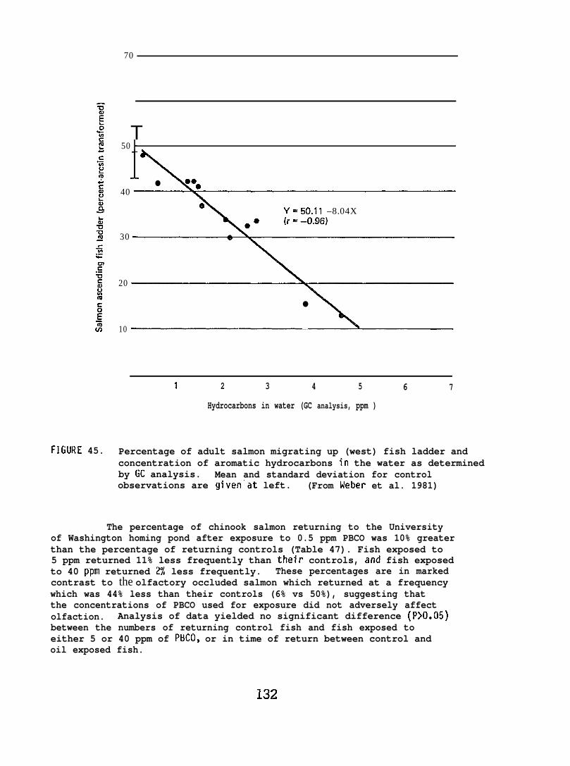
70
FIGURE 45.
T50 ~
●
40e
Y=50.11 –8.04X
30
20
●
10
1 2 3 4 5 6 7
Hydrocarbons in water (GC analysis, ppm )
Percentage of adult salmon migrating up (west) fish ladder andconcentration of aromatic hydrocarbons in the water as determinedby GC analysis. Mean and standard deviation for controlobservations are given’at left. (From kleber et al. 1981)
The percentage of chinook salmon returning to the Universityof Washington homing pond after exposure to 0.5 ppm PBCO was 10% greaterthan the percentage of returning controls (Table 47). Fish exposed to5 ppm returned 11% less frequently than their controls, and fish exposedto 40 ppm returned 2% less frequently. These percentages are in markedcontrast to the olfactory occluded salmon which returned at a frequencywhich was 44% less than their controls (6% vs 50%), suggesting thatthe concentrations of PBCO used for exposure did not adversely affectolfaction. Analysis of data yielded no significant difference (P>O.05)between the numbers of returning control fish and fish exposed toeither 5 or 40 ppm of PBCO, or in time of return between control andoil exposed fish.
132

TABLE 47. Release and recovery data for mature chinook salmon at University of Washington followingexposure to the FWAF of PBCO.
concentration Average daysRelease PPm Number released Number recovered (%)-
Exposureto return
date Calc. a Analyzedo Control Exposed Control Exposed Exposed-Control(hr) T+ranqe
Oct. 15 0.5 0.005+.004 7 6 4 (57) 4 (67) +8.530
—
Nov. 14-18 5.3 0.045+.02 19 .22 9 (47) 8 (36) +1.13-8C
—
Nov. 14-18 40.6 0.100:.05 30 32 10 (33) 10 (31) +0.410-16d
Total 56 60 23 (41) 22 (37) +1.9
abcd
Determined from volume of PBCO injected and water flow.Determined by GC (sum of arcinatic hydrocarbons).Four separate release groups.Five separate release groups.
TABLE 48. Release and recovery data for jack coho salmon at Tulalip following exposure to a mixture ofmonocyclic aromatic hydrocarbons (Table 1).
HydrocarbonRelease
Average daysNumber released Exposure Transported concentration Number recovered (%} to return
date Control Test (hr) (km) pprn)a Control Test Exposed-Control
Ott 21 23 23 22 4.7 1.0 14 (61) 16 (70) -0.4
Ott 28 55 44 22 1.6 1.0 24 (44) lB (41) -1. B
Ott 31 62 66 8 1.6 1.0 17 (27) 25 (3B) -5.1
Nov 3 26 25 B 1.6 1.0 15 (58) 9 (36) -1.9
Nov 4 64 65 8 1.6 2.(-I 28 (44) 29 (45) -4.5
Nov 6 52 52 8 1.6 2.0 23 (44) 19 (37) -2.3
Tot al 282 275 121 (46) 116 (45) -2.7
a Determined from volume of hydrocarbons injected and water flow.
In the first 4 tests at Tulalip Creek, jack coho salmon wereexposed for 8-22 hr at calculated concentrations of aromatic hydrocarbonsof 1 ppm, and for the last 2 tests the fish were exposed for 8 hr to 2 ppm.Analysis of individual tests indicated no significant difference (P>O.05)between the number of returning control fish versus number of hydrocarbonexposed fish (Table 48). However, the exposed salmon were delayed anaverage of 3 days in their return to Tulalip Creek (P~O.05).
133
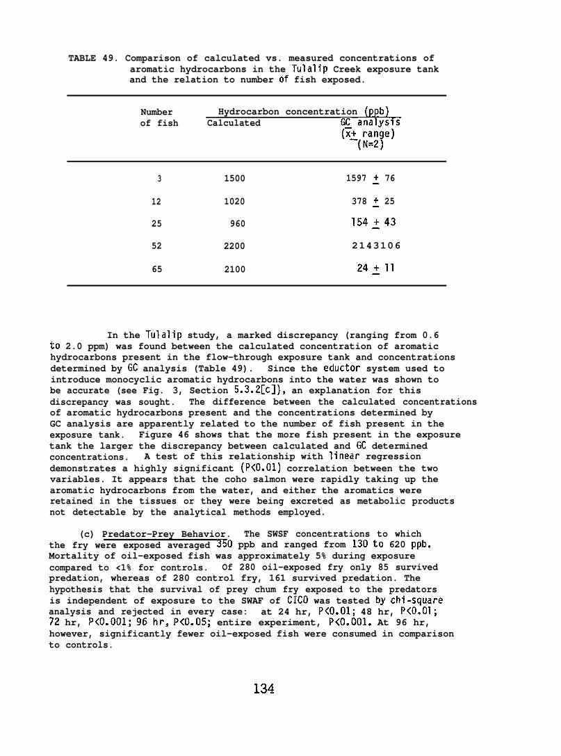
TABLE 49. Comparison of calculated vs. measured concentrations ofaromatic hydrocarbons in the Tulalip Creek exposure tankand the relation to number of fish exposed.
Number Hydrocarbon concentration (ppb)of fish Calculated G~ analysis
(x~{~:~e)=
3 1500 1597 ~ 76
12 1020 378 ~ 25
25 960 154:43
52 2200 2143106
65 2100 24211
In the Tulalip study, a marked discrepancy (ranging from 0.6to 2.0 ppm) was found between the calculated concentration of aromatichydrocarbons present in the flow-through exposure tank and concentrationsdetermined by GC analysis (Table 49). Since the eductor system used tointroduce monocyclic aromatic hydrocarbons into the water was shown tobe accurate (see Fig. 3, Section 5.3.2[c]), an explanation for thisdiscrepancy was sought. The difference between the calculated concentrationsof aromatic hydrocarbons present and the concentrations determined byGC analysis are apparently related to the number of fish present in theexposure tank. Figure 46 shows that the more fish present in the exposuretank the larger the discrepancy between calculated and GC determinedconcentrations. A test of this relationship with linear regressiondemonstrates a highly significant (P<O.01) correlation between the twovariables. It appears that the coho salmon were rapidly taking up thearomatic hydrocarbons from the water, and either the aromatics wereretained in the tissues or they were being excreted as metabolic productsnot detectable by the analytical methods employed.
(c) Predator-Prey Behavior. The SWSF concentrations to whichthe fry were exposed averaged 350 ppb and ranged from 130to 620 ppb.Mortality of oil-exposed fish was approximately 5% during exposurecompared to <1% for controls. Of 280 oil-exposed fry only 85 survivedpredation, whereas of 280 control fry, 161 survived predation. Thehypothesis that the survival of prey chum fry exposed to the predatorsis independent of exposure to the SWAF of CICO was tested by chi-squareanalysis and rejected in every case: at 24 hr, P<O.O1; 48 hr, P<O.O1;72 hr, P<0.001; 96hr, P<O.05; entire experiment, P<O.001. At 96 hr,however, significantly fewer oil-exposed fish were consumed in comparisonto controls.
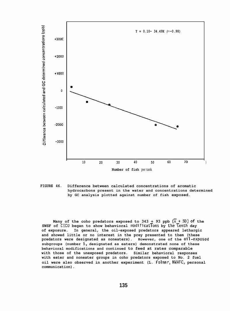
+3000
+2000
+1000
0
-1000
.2000
-3000
Y = 0.10- 34.49X (r= -0.98)
10 20 30 40 50 60 70
Number of fish per tank
)
FIGURE 46. Difference between calculated concentrations of aromatichydrocarbons present in the water and concentrations determinedby GC analysis plotted against number of fish exposed.
Many of the coho predators exposed to 343 + 93 ppb (~+ SD) of theSWSF of CICO began to show behavioral modificat~ons by the~enth dayof exposure. In general, the oil-exposed predators appeared lethargicand showed little or no interest in the prey presented to them (thesepredators were designated as noneaters). However, one of the o’il-exposedsubgroups (number 6, designated as eaters) demonstrated none of thesebehavioral modifications and continued to feed at rates comparablewith those of the unexposed predators. Similar behavioral responseswith eater and noneater groups in coho predators exposed to No. 2 fueloil were also observed in another experiment (L. Folmar$ NWAFC, personalcommunication).
135
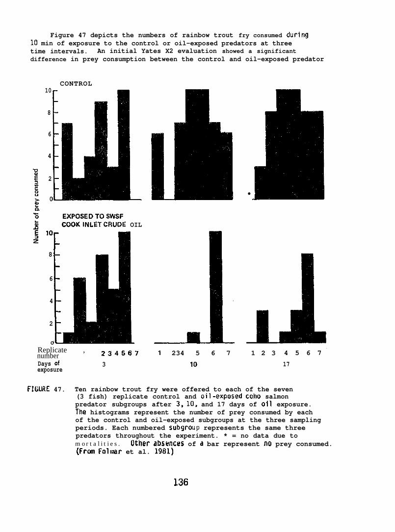
Figure 47 depicts the numbers of rainbow trout fry consumed during10 min of exposure to the control or oil-exposed predators at threetime intervals. An initial Yates X2 evaluation showed a significantdifference in prey consumption between the control and oil-exposed predator
CONTROL10
8
6
4
2
10~n‘6 EXPOSEDTOSWSF5 COOKINLET’CRUDE OIL
10
8
6
4
2
IoReplicate , z ~ d ~ ~ ~numberDays of 3exposure
●
�
1 234 5 6 7
10
1 2 3 4 5 6 7
17
FIGURE 47. Ten rainbow trout fry were offered to each of the seven(3 fish) replicate control and oil-expos@ ~oho salmonpredator subgroups after 3, 10, and 17 days of oil exposure.The histograms represent the number of prey consumed by eachof the control and oil-exposed subgroups at the three samplingperiods. Each numbered subgroiip represents the same threepredators throughout the experiment. * = no data due tom o r t a l i t i e s . Other absfmx?s of a bar represent no prey consumed.(FrcaI Folmar et al. 1981)
136
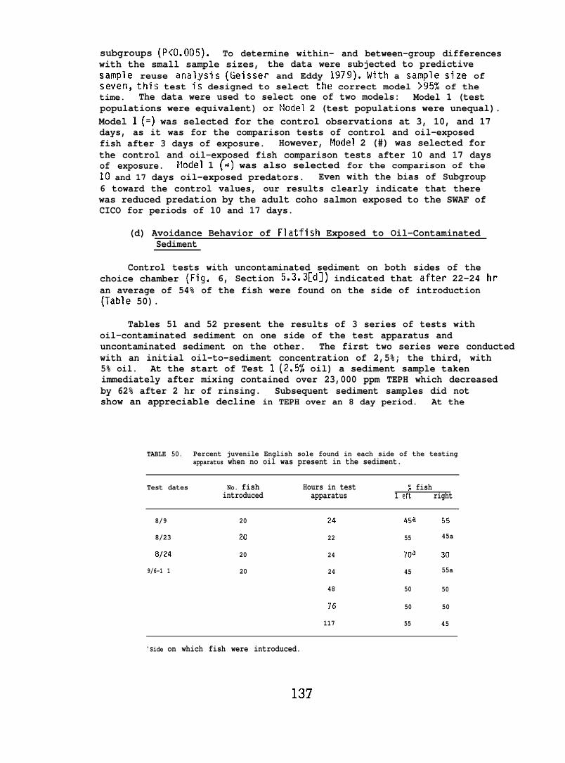
subgroups (P<O.005). To determine within- and between-group differenceswith the small sample sizes, the data were subjected to predictivesample reuse analysis (Geisser and Eddy 1979). With a sample size ofseven, this test Is designed to select the correct model >95% of thetime. The data were used to select one of two models: Model 1 (testpopulations were equivalent) or Model 2 (test populations were unequal).Model 1 (=) was selected for the control observations at 3, 10, and 17days, as it was for the comparison tests of control and oil-exposedfish after 3 days of exposure. However, Model 2 (#) was selected forthe control and oil-exposed fish comparison tests after 10 and 17 daysof exposure. Model 1 (=) was also selected for the comparison of the10 and 17 days oil-exposed predators. Even with the bias of Subgroup6 toward the control values, our results clearly indicate that therewas reduced predation by the adult coho salmon exposed to the SWAF ofCICO for periods of 10 and 17 days.
(d) Avoidance Behavior of Flatfish Exposed to Oil-ContaminatedSediment
Control tests with uncontaminated sediment on both sides of thechoice chamber (Fig. 6, Section 5.3.3[d]) indicated that after 22-24 hran average of 54% of the fish were found on the side of introduction(Table 50).
Tables 51 and 52 present the results of 3 series of tests withoil-contaminated sediment on one side of the test apparatus anduncontaminated sediment on the other. The first two series were conductedwith an initial oil-to-sediment concentration of 2,5%; the third, with5% oil. At the start of Test 1 (2.5% oil) a sediment sample takenimmediately after mixing contained over 23,000 ppm TEPH which decreasedby 62% after 2 hr of rinsing. Subsequent sediment samples did notshow an appreciable decline in TEPH over an 8 day period. At the
TABLE 50. Percent juvenile English sole found in each side of the testingapparatus when no oil was present in the sediment.
Test dates No. fish Hours in test % fishintroduced apparatus 1 eft right
8/9 20 24 4~a 55
8/23 20 22 55 45a
8/24 20 24 70a 30
9/6-1 1 20 24 45 55a
48 50 50
76 50 50
117 55 45
a Side on which fish were introduced.
137
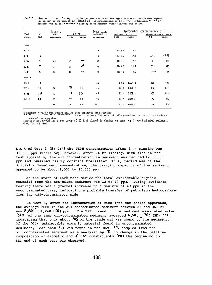
TA84E 51. Percent juvenile English sole on each side of the test apparatus when oil -contaminated sedimentwas present on one side at an initial oil concentration of 2.5% (v/v). Hydrocarbon analysi S ofsediment was by the gravimetric method; above-sediment water analysis was by GC.
Hours in Hours oiledTest
Hydrocarbon concentration (ppm)No. test $ fish sediment in sediment (dry wt. )
dates fishabove-sediment water
apparatus > eft right apparatus 1 eft right left right
Test 1
8/14 o Oa 23315.9 17.3
8/14 o 2 8974.8 17,5 .001 <.001
8/16 20 23 25 75b 46 8404.0 17.5 .003 .026
8/17 20C 21 60 40b 71 7140.0 34.1 .078 .040
alla 20C 22 45 55b 94 8056.9 43.2 NAd NA
Test 28/29 o 22 12.2 8194.3 .025 .026
8/30 20 22 75b 25 46 12.2 8496.0 .032 .037
8/31 2(F 22 7($ 30 69 13.3 5258.1 .050 .062
9/1-5 2@ 23 65b 35 93 14.7 6350.2 NA NA
98 35 65 192 15.9 6821.8 NA NA
a Sediment sample taken before filling test apparatus with seawater.b Sjd~ on which fish were introduced. In each instance fish were initially placed on the non-oil -contaminate
side of the apparatus.c Previous 20 fish removed and a new group of 20 fish placed in chamber on same non-oi 1 -contaminated sediment.d NA, not analyzed.
start of Test 3 (5% oil) the TEPH concentration after 4 hr rinsing was18,600 ppm (Table 52); however, after 26 hr rinsing, with fish in thetest apparatus, the oil concentration in sediment was reduced to 8,300ppm and remained fairly constant thereafter. Thus, regardless of theinitial oil-sediment concentration, the carrying capacity of the sedimentappeared to be about 8,000 to 10,000 ppm.
At the start of each test series the total extractable organicmaterial from the non-oiled sediment was 12 to 17 ppm. During avoidancetesting there was a gradual increase to a maximum of 43 ppm in theuncontaminated tray, indicating a probable transfer of petroleum hydrocarbonsfrom the oil-contaminated side.
In Test 3, after the introduction of fish into the choice apparatus,the average TEPH in the oil-contaminated sediment between 26 and 361 hrwas 8,860~ 1,240 (SD) ppm. The TEPH found in the sediment-associated water(SAW) of the same oil-contaminated sediment averaged 6,980:762 (SD) ppm,indicating that only about 2UJ of the crude oil was bound to the sediment.Of the total extractable organic material found in uncontaminatedsediment, less than 20% was found in the SAM. SAN samples from theoil-contaminated sediment were analyzed by GC; no change in the relativecomposition of aromatic and alkane constituents from the beginning tothe end of each test was observed.
138
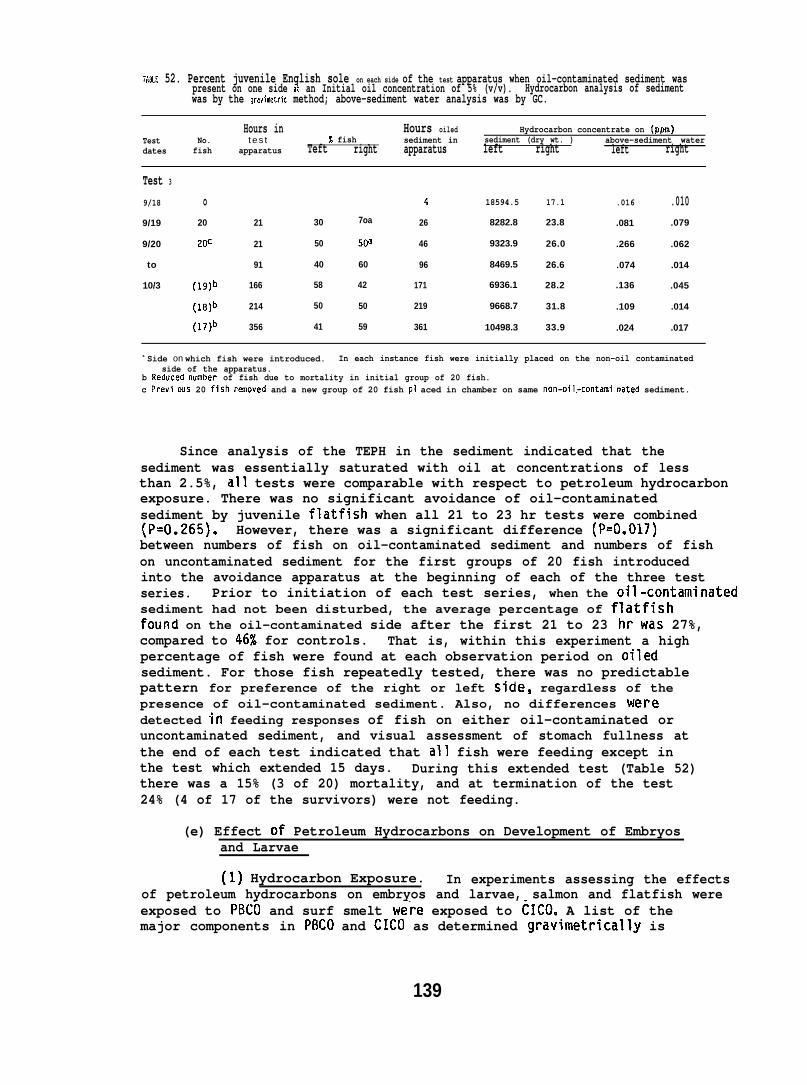
TAN-E 52. Percent juvenile English sole on each side of the test apparatus when oil-contaminated sediment waspresent on one side at an Initial oil concentration of 5% (v/v). Hydrocarbon analysis of sedimentwas by the gravimetric method; above-sediment water analysis was by GC.
Hours in Hours oiled Hydrocarbon concentrate on (Pm)Test No. t e s t % fish sediment in sediment (dry wt. )dates
above-sediment waterfish apparatus Teft right apparatus left right left right
Test 3
9/18 o 4 18594.5 17.1 .016 .0109/19 20 21 30 7oa 26 8282.8 23.8 .081 .079
9/20 20C 21 50 5W 46 9323.9 26.0 .266 .062
to 91 40 60 96 8469.5 26.6 .074 .014
10/3 (19)b 166 58 42 171 6936.1 28.2 .136 .045
(le)b 214 50 50 219 9668.7 31.8 .109 .014
(17)b 356 41 59 361 10498.3 33.9 .024 .017
a Side on which fish were introduced. In each instance fish were initially placed on the non-oil contaminatedside of the apparatus.
b Reduced number of fish due to mortality in initial group of 20 fish.c Previ ws 20 fish rsmoved and a new group of 20 fish P1 aced in chamber on same non-oil.-contami nated sediment.
Since analysis of the TEPH in the sediment indicated that thesediment was essentially saturated with oil at concentrations of lessthan 2.5%, all tests were comparable with respect to petroleum hydrocarbonexposure. There was no significant avoidance of oil-contaminatedsediment by juvenile flatfish when all 21 to 23 hr tests were combined(P=O.265). However, there was a significant difference (P=O.017)between numbers of fish on oil-contaminated sediment and numbers of fishon uncontaminated sediment for the first groups of 20 fish introducedinto the avoidance apparatus at the beginning of each of the three testseries. Prior to initiation of each test series, when the oil-con~aminateclsediment had not been disturbed, the average percentage of flatfishfound on the oil-contaminated side after the first 21 to 23 hrwas 27%,compared to 46% for controls. That is, within this experiment a highpercentage of fish were found at each observation period on oiledsediment. For those fish repeatedly tested, there was no predictablepattern for preference of the right or left side, regardless of thepresence of oil-contaminated sediment. Also, no differences weredetected in feeding responses of fish on either oil-contaminated oruncontaminated sediment, and visual assessment of stomach fullness atthe end of each test indicated that all fish were feeding except inthe test which extended 15 days. During this extended test (Table 52)there was a 15% (3 of 20) mortality, and at termination of the test24% (4 of 17 of the survivors) were not feeding.
(e) Effect of Petroleum Hydrocarbons on Development of Embryosand Larvae
(1] Hydrocarbon Exposure. In experiments assessing the effectsof petroleum hydrocarbons on embryos and larvae, salmon and flatfish wereexposed to PBCO and surf smelt we;e exposed to CICO. A list of themajor components in PBCO and CICO as determined gravimetrically is
139
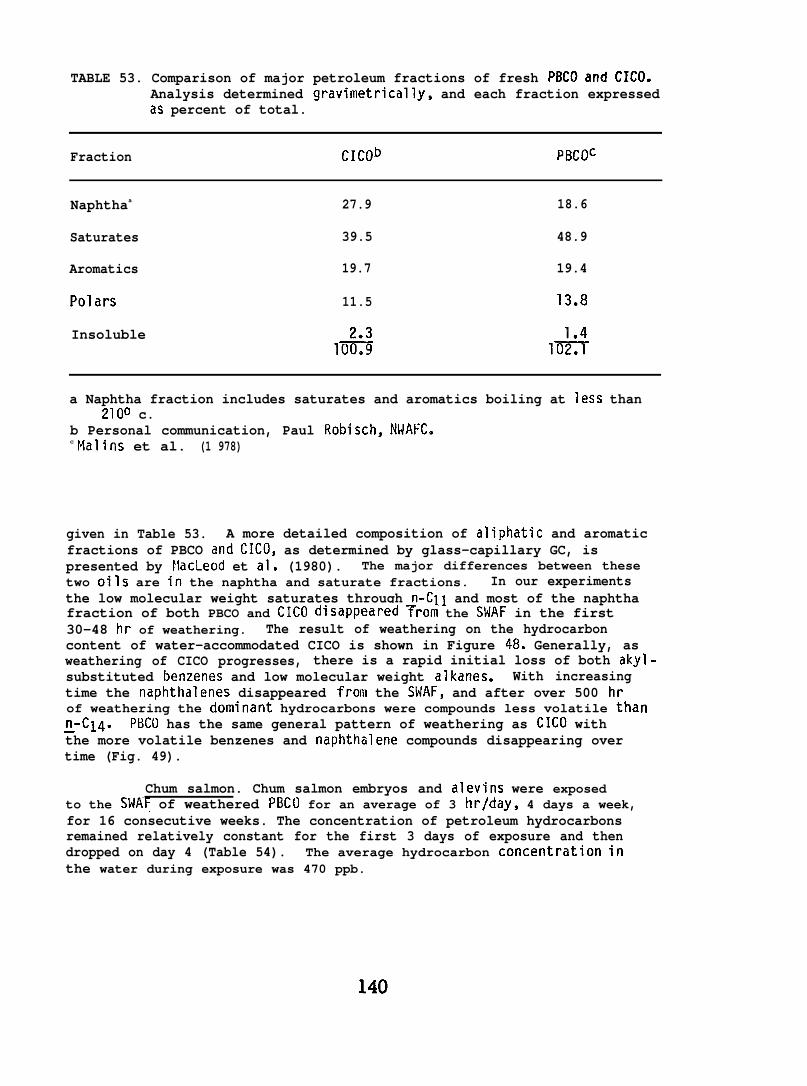
TABLE 53. Comparison of major petroleum fractions of fresh PBCOar?d CICO.Analysis determined gravimetrically, and each fraction expressedas percent of total.
Fraction CICOb PBCOC
Naphthaa 27.9 18.6
Saturates 39.5 48.9
Aromatics 19.7 19.4
Polars 11.5 13.8
Insoluble1% 1#
a Naphtha fraction includes saturates and aromatics boiling at less than2100 c.
b Personal communication, Paul Robisch, NWAFC.c Malins et al. (1 978)
given in Table 53. A more detailed composition of aliphatic and aromaticfractions of PBCO and CICO, as determined by glass-capillary GC, ispresented by MacLeod et al, (1980). The major differences between thesetwo oils are in the naphtha and saturate fractions. In our experimentsthe low molecular weight saturates through n-Cll and most of the naphthafraction of both PBCO and CICO disappeared ~rom the SWAF in the first30-48 hr of weathering. The result of weathering on the hydrocarboncontent of water-accommodated CICO is shown in Figure 48. Generally, asweathering of CICO progresses, there is a rapid initial loss of both akyl-substituted benzenes and low molecular weight alkanes. With increasingtime the naphthalenes disappeared “from the SWAF, and after over 500 hrof weathering the dominant hydrocarbons were compounds less volatile thann_-c14. PBCO has the same general pattern of weathering as CICO withthe more volatile benzenes and naphthalene compounds disappearing overtime (Fig. 49).
Chum salmon. Chum salmon embryos and alevins were exposedto the SWAt of weathered PBCO for an average of 3 hr/day, 4 days a week,for 16 consecutive weeks. The concentration of petroleum hydrocarbonsremained relatively constant for the first 3 days of exposure and thendropped on day 4 (Table 54). The average hydrocarbon concentration inthe water during exposure was 470 ppb.
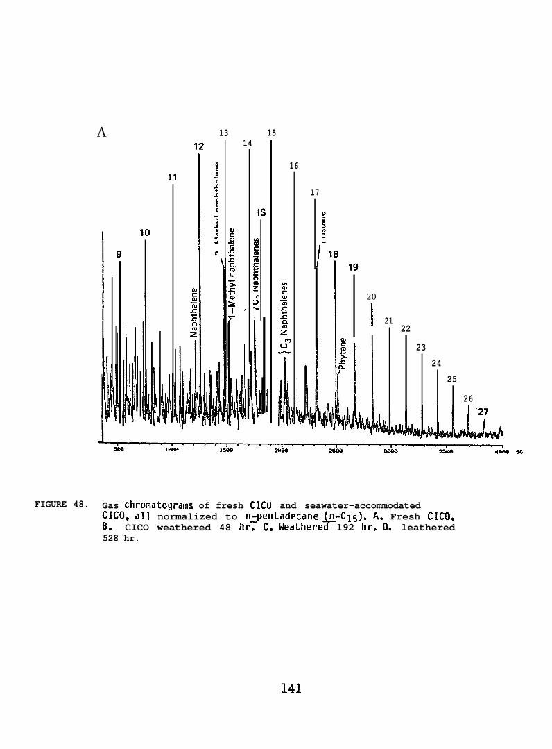
A 13 15
10
11
12 14
SC
16
17
20
2122
23
24
25
26
FIGURE 48. Gas chromatoyrams of fresh CIW and seawater-accommodatedCICO, all normalized to n-pentadecane (n-C15). A. Fresh ClCO.B. CICO weathered 48 hr~ C. Weathered 192 hr. L leathered528 hr.
141

I!!4k41i,:
20
21
22
23
LFIGURE 48. B. CICO weathered 48 hr.
25
142
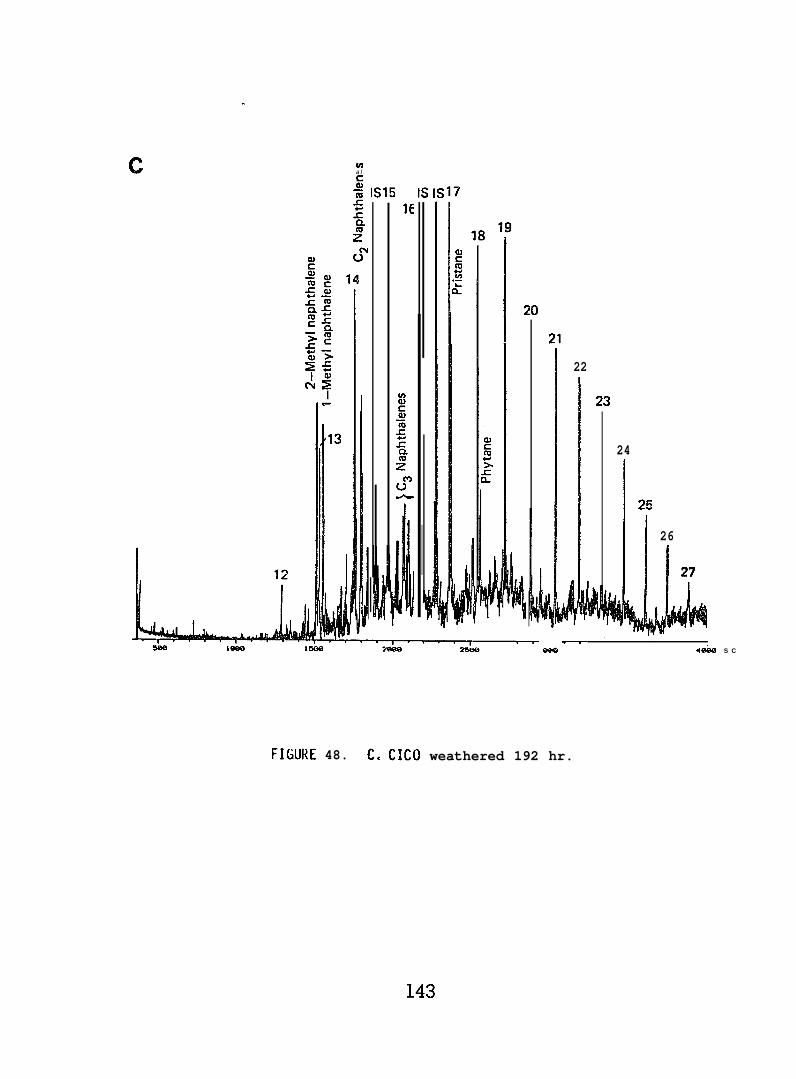
.
c L+lal
$w 1s15 1s1s17
14
22
24
25
26
b27
~l~URE 48. C.. CICO weathered 192 hr.
—4abLl S C
143
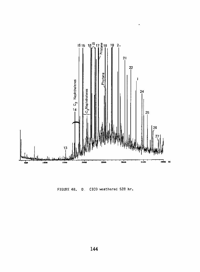
0 . m N m
)Z C2 Naphthalenes
E to 0
@=
——
Iu
=&
-
0
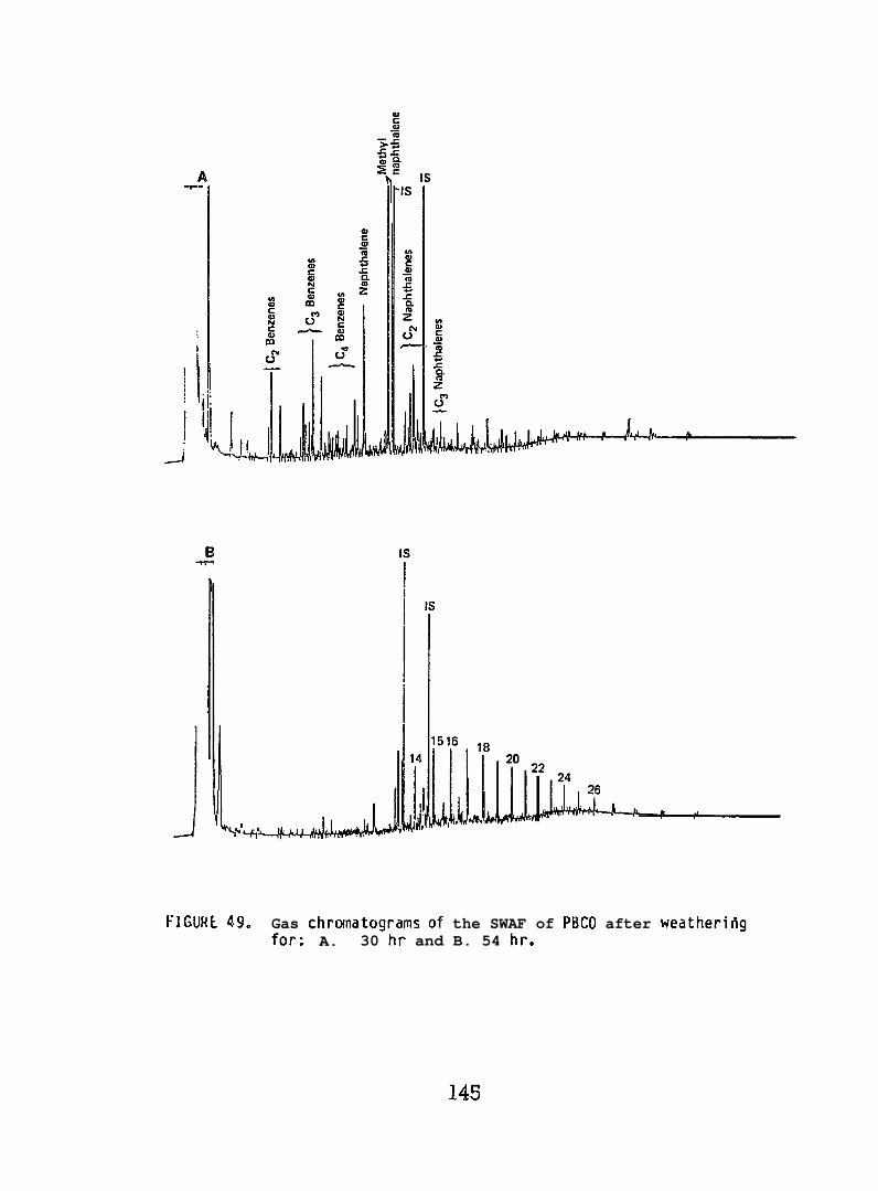
FIGURE 49. Gas chromatograms of the SWAF of PBCO after weatherifigfor: A. 30 hr and B. 54 hr.
145
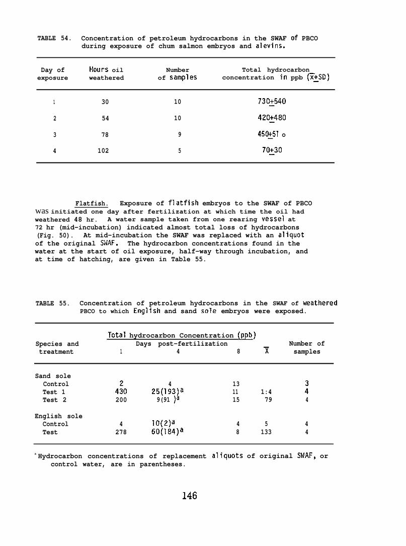
TABLE 54. Concentration of petroleum hydrocarbons in the SWAF of PBCOduring exposure of chum salmon embryos and alevins.
Day of HOUPS oil Number Total hydrocarbonexposure weathered of samples concentration in ppb (~~SD)
1 30 10 730~540
2 54 10 420~480
3 78 9 450~51 o
4 102 5 70:30
Flatfish. Exposure of flatfish embryos to the SWAF of PBCOwas initiated one day after fertilization at which time the oil hadweathered 48 hr. A water sample taken from one rearing vessel at72 hr (mid-incubation) indicated almost total loss of hydrocarbons(Fig. 50). At mid-incubation the SWAF was replaced with an aliquotof the original SWAF. The hydrocarbon concentrations found in thewater at the start of oil exposure, half-way through incubation, andat time of hatching, are given in Table 55.
TABLE 55. Concentration of petroleum hydrocarbons in the SWAF of weatheredPBCO to which English and sand sole embryos were exposed.
Total hydrocarbon Concentration (ppb)Species and Days post-fertilization Number oftreatment 1 4 8 T samples
Sand soleControl 4 13Test 1 4:0 25(193)a 11 1:4 :Test 2 200 9(91 )a 15 79 4
English soleControl 4 lo(2)a 4 5 4Test 278 60(184)a 8 133 4
a Hydrocarbon concentrations of replacementcontrol water, are in parentheses.
aliquots of original SWAF, or
146
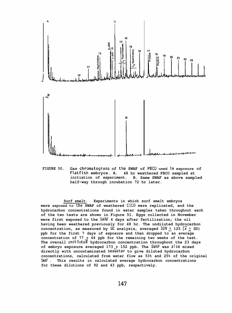
A
J13
Is
II Is
14 I
LFIGURE 50. Gas chromatogrdms of the SWAF of PBCLI used in exposure of
flatfish embryos. A. 48 hr weathered PBCO sampled atinitiation of experiment. B. Same SWAF as above sampledhalf-way through incubation 72 hr later.
Surf smelt. Experiments in which surf smelt embryoswere exposed to the SWAF of weathered CICO were replicated, and thehydrocarbon concentrations found in water samples taken throughout eachof the two tests are shown in Figure 51. Eggs collected in Novemberwere first exposed to the SHAF 4 days after fertilization; the oilhaving been weathered previously for 48 hr. The undiluted hydrocarbonconcentration, as measured by GC analysis, averaged 3242 125 (~: SD)ppb for the first 7 days of exposure and then dropped to an averageconcentration of 77 + 64 ppb for the remaining two weeks of the test.The overall undilute~ hydrocarbon concentration throughout the 23 daysof embryo exposure averaged 173 + 152 ppb. The SWAF was also mixeddirectly with uncontaminated sea~ater to give diluted hydrocarbonconcentrations, calculated from water flow as 53% and 25% of the originalSWAF . This results in calculated average hydrocarbon concentrationsfor these dilutions of 92 and 43 ppb, respectively.
147
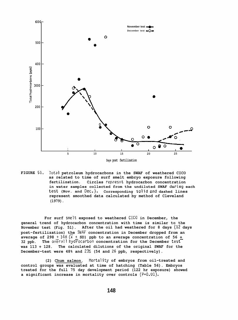
November test +December test 4-
600-
500 -
400 -z~Wc2 0kg 300 -+ &z>%zt-
200 -
0
0100 - 9
~o o@o1 # I I t o5 10 15 20 25
Days post fertilization
FIGURE 51. Total petroleum hydrocarbons in the SWAF of weathered CICOas related to time of surf smelt embryo exposure followingfertilization. Circles represnt hydrocarbon concentrationin water samples collected from the undiluted SWAF during eachtest (Nov. and Oec.). Corresponding solid dnd dashed linesrepresent smoothed data calculated by method of Cleveland(1979).
For surf smelt exposed to weathered CICO in December, thegeneral trend of hydrocarbon concentration with time is similar to theNovember test (Fig. 51). After the oil had weathered for 8 days (12 dayspost-fertilization) the SWAF concentration in December dropped from anaverage of 298 + 148 ~ + SD) ppb to an average concentration of 56 +32 ppb. The ov~rall hyd~ocarbon concentration for the December test–was 113 + 128. The calculated dilutions of the original SWAF for theDecember-test were 48% and 237i (54 and 26 ppb, respectively).
(2) Chum salmon. Nortality of embryos from oil-treated andcontrol groups was evaluated at time of hatching (Table 56). Embryostreated for the full 75 day development period (122 hr exposure) showeda significant increase in mortality over controls (P=O.01).
148
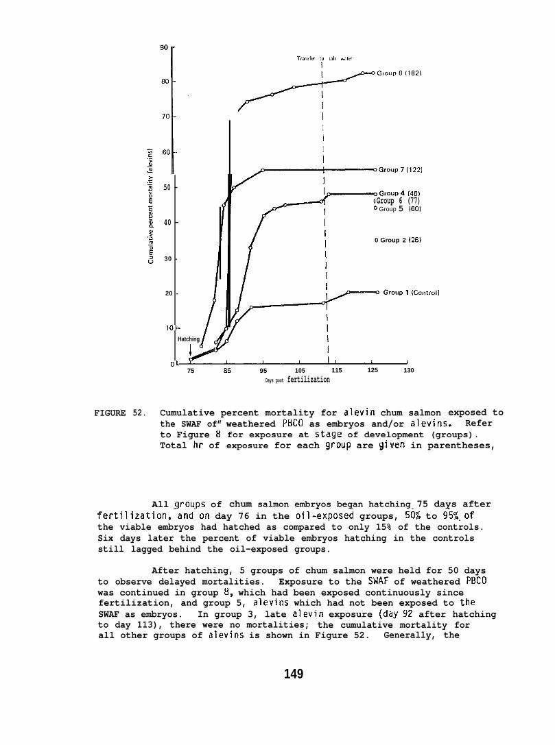
FIGURE 52.
Trandm m sail water
+Group’(’””
IIII
50 -0 Group 6 (77)O Group 5 160)
40 -
0 Group 2 (26}
30 -
I
20 -
1 0-
Hatching
i
OL75 E15 95 105 115 125 130
Days post fertilization
Cumulative percent mortality for alevin chum salmonthe SWAF of”to Figure 8Total hr of
All qroups of
exposed toweathered PBCO as embryos and/or alevins. Referfor exposure at stdge of development (groups).exposure for each group are given in parentheses,
chum salmon embryos began hatching 75 days afterfertilization,-and”on day 76 in the oil-~xpose~ groups, 5@L to 9ki%.ofthe viable embryos had hatched as compared to only 15% of the controls.Six days later the percent of viable embryos hatching in the controlsstill lagged behind the oil-exposed groups.
After hatching, 5 groups of chum salmon were held for 50 daysto observe delayed mortalities. Exposure to the SWAF of weathered P13C0was continued in group 8, which had been exposed continuously sincefertilization, and group 5, alevins which had not been exposed to theSWAF as embryos. In group 3, late alevin exposure (ddy 92 after hatchingto day 113), there were no mortalities; the cumulative mortality forall other groups of alevins is shown in Figure 52. Generally, the
149
TABLE 56. Percent mortality of chum salmon embryos at time of hatching(75 days post-fertilization), and percent of viable eggs hatchedon days 76 and 82.
Average Cumulative % hatch ofGroup(s) a Exposure period Hours percent viable eggs on days
(days post-f ert. ) exposed mortality 76 82
1,3,5 (control ) o 24 15 70
2 (14-26) 26 28 60 95
4 (1 -26) 46 25 95 99
6 (26-75) 77 19 80 98
7,8 (1 -75) 122 42 5(I 96
a See Figure 8, Section 5.3.3.(e)b Data adjusted for 2.6% of eggs which were unfertilized.
longer the duration of exposure to the SWAFthe lower the survival.Highest mortality occurred in group 8, which was exposed continuouslyfrom fertilization; in this group 70% of the alevins were dead 12 daysafter hatching as compared to a 12% mortality in controls. The secondhighest mortalities occurred in group 7. These were exposed continuouslyas embryos, but with no oil exposure as alevins; 504 of this groupdied within 12 days after hatching.
Alevins were removed from each trough 12 days after hatching .and examined for gross abnormalities. Three types of abnormalitieswere evident: malformed yolk sac, “dome” head, and kyphosis (angularcurvature of the notochord).
The normal yolk sac is elliptical compared with the foreshortenedand bulbous sac observed in some oil-exposed individuals. This abnormalityoccurred in the majority of oil-exposed groups, but was most frequent(61%) in those exposed continuously from fertilization (Table 57, group 8).Normally developing chum salmon alevins have heads with slight “dome”shape above the optic lobes of the brain (Distler 1954). Oil-exposedindividuals had single- or double-’’dome” heads which were much morepronounced than in Distler’s illustrations or in our controls, andresembled lake charr (Salvelinus namaycush) alevins reared in water withlow dissolved oxygen c~l~ Dissection of alevins withcephalic abnormalities revealed no evidence that the “dome” shape was aresult of hydrocephaly. Kyphotic notochords occurred only in alevinsexposed to oil continuously (143 hr); other groups did not exhibit anyobvious notochord deformities.
Sixteen days after hatching, samples of 10 to 15 alevins fromeach group were fixed and stained to examine development of fin rays.Fin ray development in alevins exposed to the SWAF of weathered PBCOonly during embryonic development was similar to controls; however,for those exposed to oil after hatching (groups 5 and 8) there appearsto be an inhibition of ray development, particularly of the dorsal fin(Table 58).
150
TABLE 57. Gross morphological abnormalities observed in chum salmonalevins 12 days after hatching (87 days post-fertilization).
Exposure period Hours Sample % abnormalitiesGroup (days post-fert.) exposed size Yolk sac head kyphosls
1 (Control ) o 204 0 0 0
2 (14-26) 26’ 15 0 9 0
3 (1 -26) 46 19 16 16 0
4 (75-87) 21 10 10 33 0
5 (26-75) 77 15 0 33 0
6 (l-75) 122 6 33 33 0
8 (1-87) 143 16 61 44 11
a Pictorial description of yolk sac and cephalic abnormalities are shown inMalins et al. (1980).
TABLE 58. Effect of weathered PBCO on fin ray counts of chum salmon alevins16 days after hatching (91 days post-fertilization).
Exposure period Hours ~Number of fin rays with calcificationGroup (days post -fert. ) exposed Caudal Anal Dorsal
1 (Control ) o 17 7.8 8.3
2 (14-26) 26 18.7 11.2 9.2
4 (l-26) 46 12.5 8.7 6.3
5 (75-91) 29 10.0 1.2 0
6 (26-75) 71 20.1 12.7 10.9
7 (l-75) 122 15.0 11.7 6.0
8 (1-91) 151 5.U 3.5 0
a Following staining with alizarin red S.
A minimum of 10 alevins from each group were measured attermination of the experiment (127 days after fertilization). Therewere no differences in growth, as measured by total length, betweencontrol and oil-exposed alevins.
(3) Flatfish. Effects of the SWAF of weathered PBCO onEnglish and sand sole embryos and larvae are given in Table 59 andsummarized in Figure 53. Flatfish eggs and larvae were categorizedusing the following nomenclature:
Nondeveloped eggs: Consisted of two types of eggs whichwere indistinguishable as to the cause of nondevelopment--(l) nonviableeggs which were not successfully fertilized; (2) embryos which died inearly cell division as a result of oil exposure or natural failure(embryo not formed).
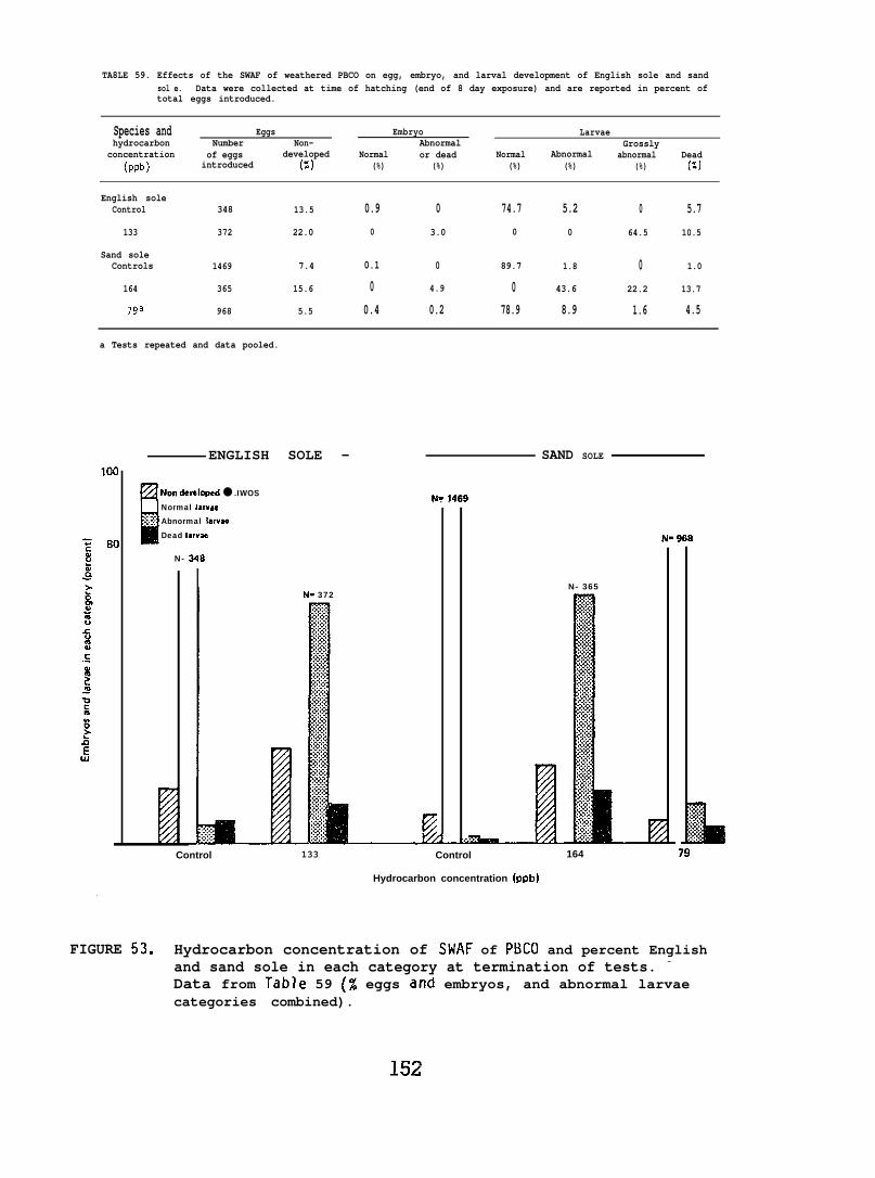
TA8LE 59. Effects of the SWAF of weathered PBCO on egg, embryo, and larval development of English sole and sandsol e. Data were collected at time of hatching (end of 8 day exposure) and are reported in percent oftotal eggs introduced.
Species and Eggs Embryo Larvaehydrocarbon Number Non- Abnormal Grossly
concentration of eggs developed Normal or dead Normal Abnormal(ppb)
abnormal Deadintroduced (5) (%) (%) (%) (%) (%) (%)
English soleControl 348 13.5 0.9 0 74.7 5.2 0 5.7
133 372 22.0 0 3.0 0 0 64.5 10.5
Sand soleControls 1469 7.4 0.1 0 89.7 1.8 0 1.0
164 365 15.6 0 4.9 0 43.6 22.2 13.7
7ga 968 5.5 0.4 0.2 78.9 8.9 1.6 4.5
a Tests repeated and data pooled.
ENGLISH SOLE — SAND SOLE
~Non davelqmci ● .IWOS N= lAri9---u Normal l-rum. . .
: j :: Abnormal larv~
Dead Iarvza
N- 248
I N= 372
I N- 968
N- 365
Control 133 Control 164
Hydrocarbon concentration (ppb)
79La..,.j~:.,,..,...::~. . .
FIGURE 53. Hydrocarbon concentration of SWAF of PBCO and percent Englishand sand sole in each category at termination of tests. -
Data from Table 59 (% eggs and embryos, and abnormal larvaecategories combined).
152
Normal embryo: Embryo transparent, with regular heart contractions.
Abnormal or dead embryo: Opaque, often scoliosis evident, andintermittent or no heart contractions.
Normal larvae: Regular heart contractions, notochord straight,finfold continuous, digestive tract complete, and pigmentation complete.
Abnormal larvae: Regular heart contractions, slight curvatureof the notochord and entire body, generally a lateral curvature of up to 45°.
Grossly abnormal larvae: Regular heart contractions, butwith body curvature exceeding 45° (some with notochord curvature of 180°and double 180° curvatures). Finfold deformed, digestive tract incompletepigmentation not in patches but scattered, lying motionless on bottom,opaque.
Dead larvae: No heart contraction, generally opaque, andusually contorted.
A single test with English sole eggs indicated that exposureto an average hydrocarbon concentration of 133 ppb resulted in a highpercentage of hatching, but all larvae were either abnormal or diedshortly after hatching.
In control tests with sand sole an average of 90~2% (~~range)of the eggs hatched into normal larvae. Exposure to the SWAF at aconcentration of 79 ppb resulted in an average of 79 + 2% normal larvaeand only 10 + 1% deformed larvae. At a SWAF concentration of 164 ppbembryos deveToped and hatched, but two-thirds of the larvae were deformed;the most common abnormality was scoliosis.
(4) Surf smelt. An accounting of all surf smelt embryosand larvae in the two replicate experiments was conducted when hatchingof live control embryos appeared complete. These data are presentedin Tables 60 and 61 along with the average hydrocarbon exposure concentrations.A majority of the eggs were unaccounted for at the end of both experiments,and as no intact embryos could be lost from the incubation baskets,this loss is attributed to early embryo death, and subsequent embryodisintegration; for controls this averaged 49~3?i (x: range).
In both experiments the control embryos and those exposed to thelower hydrocarbon concentrations produced an equal number of live larvae(controls, 44 + 4%; the 26 and 43 ppb exposure groups, 43 + l%). However,eggs exposed t~ the twu higher hydrocarbon concentrations ~roduced fewlive larvae with most dying in the embryonic stage {8919% as deadembryos or disintegrated eggs).
Cumulative hatching rates for both experiments are shown inFigures 54 and 55. Control larvae in the December experiment hatchedapproximately 6 days later than those in the November experiment, butthe total percent hatching was similar. At an average hydrocarbon
153

TA8LE 60. Effects of a 57 hr exposure to the SWAF of weathered CICO on hatchingsuccess of surf smelt embryos (November 1979 experiment). Oataccmpiled at completion of hatching in controls, and reported inpercent of total embryos introduced.
Hydrocarbon Embryos Larvaeconcentration Inltlal no. Allve Dead Disintegrated Alived Oead
(ppb) (N) (%) (%) (%) (%) (%)
Control 440 3.2 3.6 51.9 40.6 0.7
43 425 0 3.0 52.1 42.2 2.7
92 420 9.6 13.8 66.1 9.1 1.4
173 425 1.7 47.1 50.5 0.7 0
a Live larvae were collected daily immediately after hatching, and the percentpresented is cumulative.
50 “
43 ppbN= 191
40 -
N= 17930 -
20 -
10 -
0 ,19 20 21 22 23 24 25 26 27 28
Days from fertilization
FIGURE 54. Cumulative percent hatching of surf smelt embryos exposed to3 different concentrations of the SWAF of weathered CIC(I fora total of 57 hr (November 1979 experiment). Average hydrocarbonconcentration for undiluted SWAF (173 ppb) was determined byGC analysis. The two lower SWAF concentrations (92 and 43 ppb)were calculated from proportional dilutions of the undilutedSWAF.
154
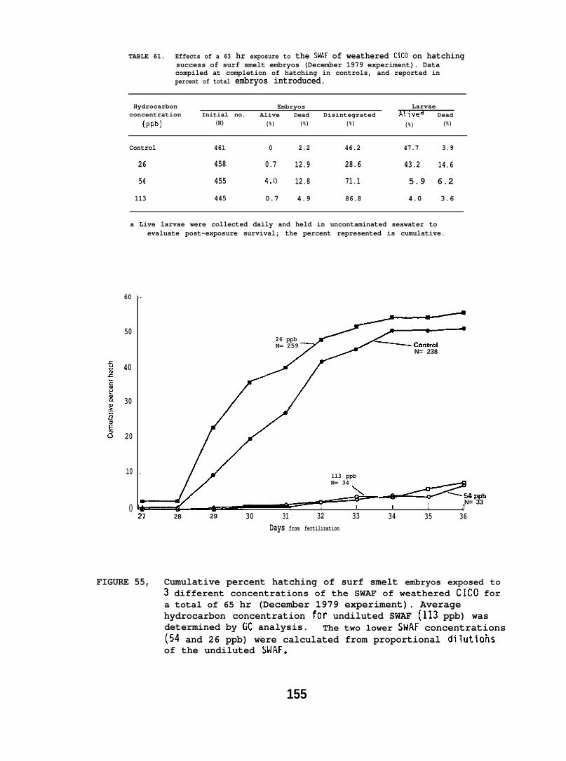
TABLE 61. Effects of a 63 hr exposure to the SklAF of weathered CICO on hatchingsuccess of surf smelt embryos (December 1979 experiment). Datacompiled at completion of hatching in controls, and reported inpercent of total embryos introduced.
Hydrocarbon Embryos Larvaeconcentration Initial no. Alive Dead Disintegrated Al ‘lved
(pPb) (N)Dead
(%) (%) (%) (%) (%)
Control 461 0 2.2 46.2 47.7 3.9
26 458 0.7 12.9 28.6 43.2 14.6
54 455 4.3 12.8 71.1 5.9 6.2
113 445 0.7 4.9 86.8 4.0 3.6
a Live larvae were collected daily and held in uncontaminated seawater toevaluate post-exposure survival; the percent represented is cumulative.
60
50
40
30
20
10
0
1-
26 ppbN= 259
.113 ppbN= 34
m
N= 238
~~ppbN= 33— I 1 1 1 J
27 28 29 30 31 32 33 34 35 36Days from fertilization
FIGURE 55, Cumulative percent hatching of surf smelt embryos exposed to3 different concentrations of the SWAF of weathered CICO fora total of 65 hr (December 1979 experiment). Averagehydrocarbon concentration for undiluted SWAF (113 ppb) wasdetermined by GC analysis. The two lower StiAF concentrations(50 and 26 ppb) were calculated from proportional dilutiohsof the undiluted SWAF.
155
concentration of 26 ppb (December experiment) there was an apparentacceleration in hatching similar to that observed for chum salmonembryos exposed to the SWAF of weathered PBCO.
Samples of developing embryos were collected at two pointsduring the December experiment (Table 62). On day 14 post-fertilization,mortality in control embryos was greater than In oil-exposed embryos;however, in embryos sampled one week later (approximately two-thirdsof the way through incubation), this was reversed. The most appropriatestatistical model (Geisser and Eddy 1979) for the 22 day post-fertilizationdata indicated that the percent abnomlal and dead embryos In the controlgroup and those exposed to 26 ppb petroleum hydrocarbons were similar,but differed from embryos exposed to 54 and 113 ppb. These latter twoin turn differed from each other.
In the December 1979 experiment, three consistent types of grossabnormalities were observed in the oil-exposed embryos: reduced coiling(an index of growth by length); absence of eye rotation and closure ofthe choroid fissure; and diffused pigment. The most frequent abnormalitywas reduced coiling. As embryos develop, the body coils inside thechorion attaining a maximum of 2.5 coils just prior to hatching. Al 1abnormal embryos, exposed to a hydrocarbon concentration of 113 ppbexhibited reduced growth, with only 1.5 coils after 22 days of development;the same number of coils was observed one week earlier in controls.Approximately 50% and 20% of abnormal embryos exposed to hydrocarbonconcentrations of 54 and 26 ppb, respectively, also exhibited reducedgrowth after 22 days incubation. The eyes of affected embryos did notdevelop beyond the 15-day post-fertilization stage. Arrested eyedevelopment was the predominate abnormality in embryos incubated in26 ppb total hydrocarbons.
Surf smelt embryos have a single row of contracted, dark,melanophores along the ventral midline, and smaller, stellate-shapedmelanophores scattered over the ventral surface of the yolk. Fiftypercent of the abnormal embryos exposed to 54 and 113 ppb had diffusedpigment in these melanophores.
In order to observe any latent effects of oil exposure, newlyhatched larvae from the December 1979 experiment were held in uncontaminatedwater, and fed rotifers (Brachionus plicatilus). After holding anaverage of 10 days (depending on time of hatching) survivors werecounted (Table 63). Although the percent of normal appearing larvaehatching in the 26 ppb concentration of the SWAF was nearly identicalto the control group, their survival as larvae was significantly less[P<0.001); only 8.7% survived compared with 42.5% in controls.
Surf smelt embryos from the December 1979 experiment were sampled14 days after fertilization and whole eggs (embryo and egg envelope)were analyzed for petroleum hydrocarbons by GS/MS. Aromatic hydrocarbonsfound in control eggs and eggs exposed to the SWAF of weathered CICOare given in Table 64. Alkanes are commonly found in marine organisms,and prominent differences were not observed in concentrations fromeither oil-exposed or control eggs.
156
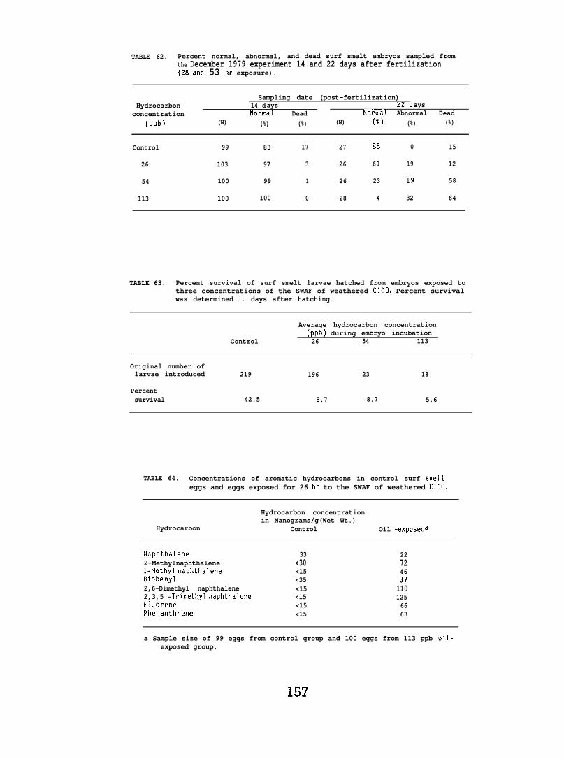
TABLE 62. Percent normal, abnormal, and dead surf smelt embryos sampled fromthe December 1979 experiment 14 and 22 days after fertilization(28and 53 hr exposure).
Sampling date (post-fertilization)Hydrocarbon 14 d ays 22 days
concentration Normal Dead Normal Abnormal Dead(ppb) (N) (%) (%) (N) (%) (%) (%)
Control 99 83 17 27 85 0 15
26 103 97 3 26 69 19 12
54 100 99 1 26 23 19 58
113 100 100 0 28 4 32 64
TABLE 63. Percent survival of surf smelt larvae hatched from embryos exposed tothree concentrations of the SWAF of weathered ClCO. Percent survivalwas determined 10 days after hatching.
Average hydrocarbon concentration(ppb) during embryo incubation
Control 26 54 113
Original number oflarvae introduced 219 196 23 18
Percentsurvival 42.5 8.7 8.7 5.6
TABLE 64. Concentrations of aromatic hydrocarbons in control surf smelteggs and eggs exposed for 26 hr to the SWAF of weathered CICO.
Hydrocarbon concentrationin Nanograms/g(Wet Wt.)
Hydrocarbon Control Oil -exposeda
Naphthalene 33 222-Methylnaphthalene <30 72l-klethy lnaphthalene <15 46Biphenyl <35 372,6-Dimethyl naphthalene <15 1102,3,5 -Trimethyl naphthalene <15 125Fluorene <15 66Phenanthrene <15 63
a Sample size of 99 eggs from control group and 100 eggs from 113 ppb oil-exposed group.
157
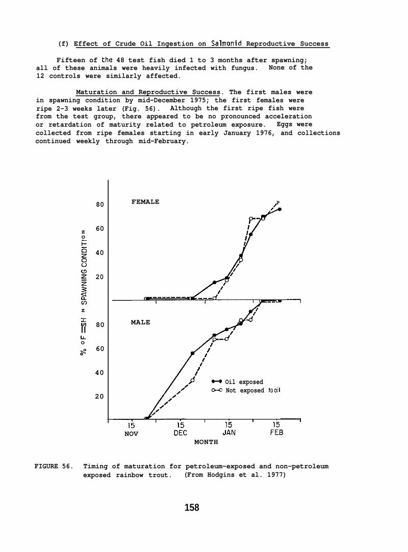
(f) Effect of Crude Oil Ingestion on Salmonid Reproductive Success
Fifteen of the 48 test fish died 1 to 3 months after spawning;all of these animals were heavily infected with fungus. None of the12 controls were similarly affected.
Maturation and Reproductive Success. The first males werein spawning condition by mid-December 1975; the first females wereripe 2-3 weeks later (Fig. 56). Although the first ripe fish werefrom the test group, there appeared to be no pronounced accelerationor retardation of maturity related to petroleum exposure. Eggs werecollected from ripe females starting in early January 1976, and collectionscontinued weekly through mid-February.
zoi=
z
5iiILo$?
80
60
40
20
80
60
40
20
FEMALE P
MALE
.-. Oil exposed--o Not exposed tooil
15NOV
MONTH
FIGURE 56. Timing of maturation for petroleum-exposed and non-petroleumexposed rainbow trout. (From Hodgins et al. 1977)
158
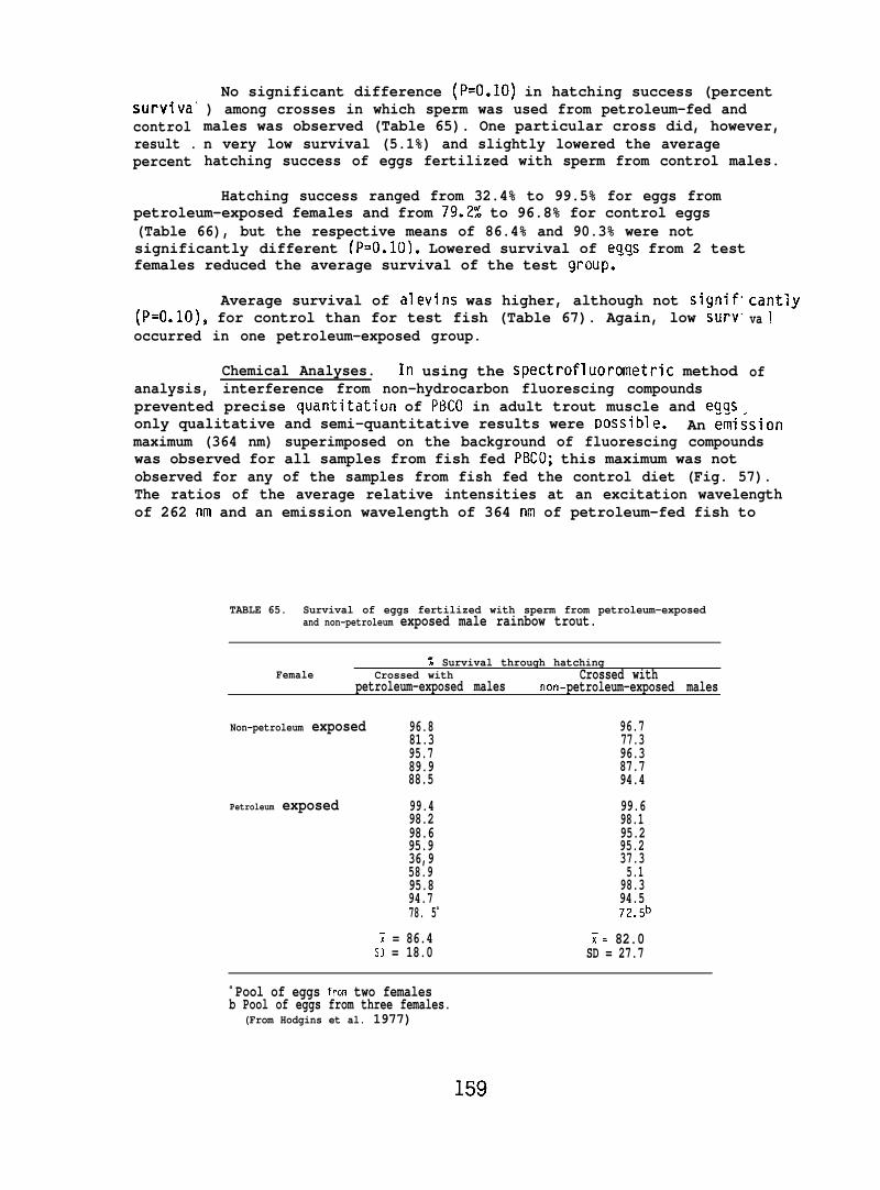
surviva”controlresult .percent
No significant difference (P=O.1O) in hatching success (percent) among crosses in which sperm was used from petroleum-fed andmales was observed (Table 65). One particular cross did, however,n very low survival (5.1%) and slightly lowered the averagehatching success of eggs fertilized with sperm from control males.
Hatching success ranged from 32.4% to 99.5% for eggs frompetroleum-exposed females and from 79.2% to 96.8% for control eggs(Table 66), but the respective means of 86.4% and 90.3% were notsignificantly different (P=O.1O). Lowered survival of ea~s from 2 testfemales reduced the average survival of the test group.””
Average survival of alevins was higher, although not signif(P=O.1O), for control than for test fish (Table 67). Again, low survoccurred in one petroleum-exposed group.
Chemical Analyses. In using the spectrofluorometric methodanalysis, interference from non-hydrocarbon fluorescing compoundsprevented precise quantitation of P13C0 in adult trout muscle and eqqs
cantlyva 1
of
only qualitative and semi-quantitative results were ~ossible. An ~~issionmaximum (364 nm) superimposed on the background of fluorescing compoundswas observed for all samples from fish fed PBCO; this maximum was notobserved for any of the samples from fish fed the control diet (Fig. 57).The ratios of the average relative intensities at an excitation wavelengthof 262 nm and an emission wavelength of 364 nm of petroleum-fed fish to
TABLE 65. Survival of eggs fertilized with sperm from petroleum-exposedand non-petroleum exposed male rainbow trout.
% Survival through hatchingFemale Crossed with Crossed with
petroleum-exposed males non- petroleum-exposed males
Non-petroleum exposed 96.8 96.781.3 77.395.7 96.389.9 87.788.5 94.4
Petroleum exposed 99.4 99.698.2 98.198.6 95.295.9 95.236,9 37.358.9 5.195.8 98.394.7 94.578. 5a 72.5b
~ = 86.4 ~= 82.0SD = 18.0 SD = 27.7
a Pool of eggs fro+n two femalesb Pool of eggs from three females.
(From Hodgins et al. 1977)
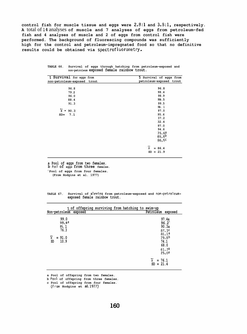
control fish for muscle tissue and eggs were 2.8:1 and 3.8:1, respectively.A total of 14 analyses of muscle and 7 analyses of eggs from petroleum-fedfish and 4 analyses of muscle and 2 of eggs from control fish wereperformed. The background of fluorescing compounds was sufficientlyhigh for the control and petroleum-impregnated food so that no definitiveresults could be obtained via spectrofluorometry.
TABLE 66. Survival of eggs through hatching from petroleum-exposed andnon-petroleum exposed female rainbow trout.
% Survival for eggs from % Survival of eggs fromnon-petroleum-exposed trout petroleum-exposed trout
96.8 96.879.2 98.496.0 98.988.4 9B.591.3 99.5
9B. 11= 90.3 97.0SD= 7.1 95.6
37.232.497.094.675.6a89.8b86.5c
T = 86.4SD = 21.9
a Pool of eggs from two females.b POO1 of eggs from three females.c Pool of eggs from four females.
(From Hodgins et al. 1977)
TA8LE 67. Survival of alevins from petroleum-exposed and non-petroleum-exposed female rainbow trout.
% of offspring surviving from hatching to swim-upNon-petroleum exposed Petroleum exposed
99.0 97.4a99.4a 96.2a
B9. 1 90.3a76.3 8i’.3c
81.1aY = 91.0 79.8bSD 10.9 74.1
68.061.7a25.oa
Y = 76.1SD = 21.4
a Pool of offspring from two females.b POO1 of offspring from three females.c Pool of offspring from four females.
(Fran Hodgins et al. 1977)
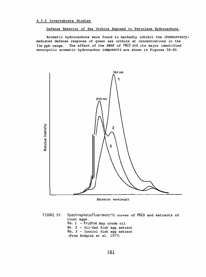
6.3.2 Invertebrate Studies
Defense Behavior of Sea Urchins Exposed to Petroleum Hydrocarbons.
Aromatic hydrocarbons were found to markedly inhibit the chemosensory-mediated defense response of green sea urchins at concentrations in thelow ppb range. The effect of the SWSF of PBCO and its major identifiedmonocyclic aromatic hydrocarbon canponents are shown in Figures 58-60.
364 nm
Emission wavelength
FI(NJRE 57. Spectrophotofluorometric curves of PBCO and extracts oftrout eggs.No. 1 - Prudhoe Bay crude oilNo. 2 - Oil-fed fish egg extractNo. 3 - Control fish egg extract(From Hodgins et al. 1977)
161
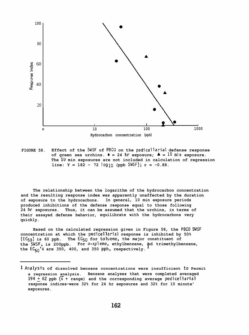
100
80
H 60g.-=c0~ 40
cc
20@
1 *I d Io 10 100 1000
Hydrocarbon concentration (ppb)
FIGURE 58. Effect of the SHSF of PBCLl on the pedicellarial defense responseof green sea urchins. o = 24 hr exposure; A = 10 min exposure.The 1(.) min exposures are not included in calculation of regressionline: Y = 182 - 72 log10 (ppb SWSF); r = -0.88.
The relationship between the logarithm of the hydrocarbon concentrationand the resulting response index was apparently unaffected by the durationof exposure to the hydrocarbons. In general, 10 min exposure periodsproduced inhibitions of the defense response equal to those following24 hr exposures. Thus, it can be assumed that the urchins, in terms oftheir assayed defense behavior, equilibrate with the hydrocarbons veryquickly.
Based on the calculated regression given in Figure 58, the PBCO SWSFconcentration at which the pedicellarial response is inhibited by 50%(EC50) is 60 ppb. The EC50 for toluene, the major constituent ofthe SWSF, is 200ppb. For o-xylene, ethylbenzene, nd trimethylbenzene,the EC50’S are 350, 400, and 350 ppb, respectively. !
1 Analysis of dissolved benzene concentrations were insufficient to Permita regression analysis. Benzene analyses that were completed averaged194262 ppb ~+ range) and the corresponding average pedicellarialresponse indices-were 32% for 24 hr exposures and 32% for 10 minute’exposures.
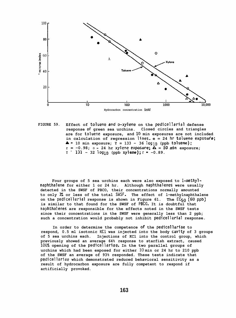
; 60 -.- A;c00.z 40 -
a
20 -
100
80
I
@
e
A
o
I * I I -0 10 100 1000 10,000
Hydrocarbon concentration (ppbl
FIGURE 59. Effect of toluene and o-xylene on the pedicellarial defenseresponse of green sea urchins. Closed circles and trianglesare for toluene exposure, and 10 min exposures are not includedin calculation of regression lines. e = 24 hr toluene exposu+e;A= 10 min exposure; Y = 133 - 36 log10 (ppb toluene);r = -0.98; O = 24 hr xylene exposure; ~ = 10 min exposure;Y = 131 - 32 Ioglo (ppb xylene); r = -0.89.
Four groups of 5 sea urchins each were also exposed to l-methyl-naphthalene for either 1 or 24 hr. Although naphtha?enes were usuallydetected in the SWSF of PBCO, their concentrations normally amountedto only 3% or less of the total SWSF. The effect of l-methylnaphthaleneon the pedicellarial response is shown in Figure 61. The EC50 (60ppb)is similar to that found for the SWSF of PBCO. It is doubtful thatnaphthalenes are responsible for the effects noted in the SWSF testssince their concentrations in the SWSF were generally less than 2 ppb;such a concentration would probably not inhibit pedicellarial response.
In order to determine the competence of the pedicellariae torespond, 0.5 ml isotonic KC1 was injected into the body cavity of 3 groupsof 5 sea urchins each. Injections of KC1 into the control group, whichpreviously showed an average 64% response to starfish extract, caused100% opening of the pedicellariae. In the two parallel groups ofurchins which had been exposed for either 10 min or 24 hr to 210 ppbof the SWSF an average of 93% responded. These tests indicate thatpedicellariae which demonstrated reduced behavioral sensitivity as aresult of hydrocarbon exposure are fully competent to respond ifartificially provoked.
163

Im -
60 -
e# 60c.-%c
~a ~m
20 - A
10 10 100 1000 10,000
Hydrocarbon concentration (ppb)
FIGURE 6CI. Effect of 1,2,4-trimethylbenzene and ethylbenzene on thepedicellarial defense response of green sea urchins. Closedcircles and triangles are for trimethylbenzene exposure, and10 min exposures are not included in calculation of regressionlines. ● = 24 hr toluene exposure; 4 = 10 min exposure;Y = 429 - 148 log~o (ppb trimethylbenzene); r = -0.88; o = 24 hrethylbenzene exposure; d$ = 10 min exposure; Y = 363 - 120 10910(ppb ethylbenzene); r = -0.96.
100 -
80 -
~ 60 -c.-%cEg 40 -
20 -
t I Io 10 100 1000
Hydrocarbon concentration (ppb)
FIGURE 61. Effect of l-methyl naphthalene on the pedicellarial defenseresponse of green sea urchins. e = 24 hr exposure; M = 1 hrexposure: Y = 128 - 44 loglo (ppb methyl naphthalene);r = -0.98.
164
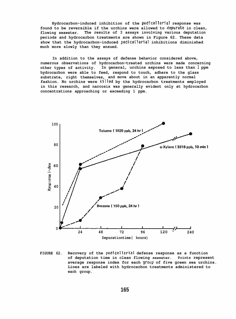
Hydrocarbon-induced inhibition of the pedicellarial response wasfound to be reversible if the urchins were allowed to depurate in clean,flowing seawater. The results of 3 assays involving various deputationperiods and hydrocarbon treatments are shown in Figure 62. These datashow that the hydrocarbon-induced pedicellarial inhibitions diminishedmuch more slowly than they ensued.
In addition to the assays of defense behavior considered above,numerous observations of hydrocarbon-treated urchins were made concerningother types of activity. In general, urchins exposed to less than 1 ppmhydrocarbon were able to feed, respond to touch, adhere to the glasssubstrate, right themselves, and move about in an apparently normalfashion. No urchins were killed by the hydrocarbon treatments employedin this research, and narcosis was generally evident only at hydrocarbonconcentrations approaching or exceeding 1 ppm.
100
80
: 60uc.-a)E0
5 40cc
20
024 48 72 96 120 ‘- 240
Depurationtime( hours)
10min)
FIGURE 62. Recovery of the pediCellarial defense response as a functionof deputation time in clean flowing seawater. Points representaverage response index for each group of five green sea urchins.Lines are labeled with hydrocarbon treatments administered toeach group.
165
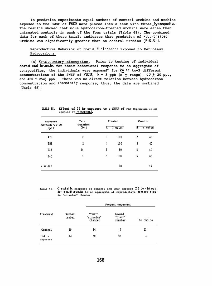
In predation experiments equal numbers of control urchins and urchinsexposed to the SWSF of PBCO were placed into a tank with three Pycnopodia.The results showed that more hydrocarbon-treated urchins were eaten thanuntreated controls in each of the four trials (Table 68). The combineddata for each of these trials indicates that predation of PBCO-treatedurchins was significantly greater than on control urchins (P=O.01).
Reproductive Behavior of Dorid Nudibranchs Exposed to PetroleumHydrocarbons
(a) Chemosensory disruption. Prior to testing of individualdorid nudi~ranchs for their behavioral response to an aggregate ofconspecifics, the individuals were exposed” for 2~hr to-3 differentconcentrations of the SWSF of PBCO; 15~ 3 ppb (x ~ range), 60~ 20 ppb,and 420 + 254) ppb. There was no direct relation between hydrocarbonconcentration and chemotatic response; thus, the data are combined(Table 69).
TABLE 68. Effect of 24 hr exposure to a SWAF of PBCO on predation of seaurchins by Pycnopodia.
Exposure Tr ia l Treated Control,concentration duration
(pPb) (hr) N % eaten N % eaten
470 2 7 100 7 43
359 2 5 100 5 40
233 24 5 60 5 40
145 2 5 100 5 60
Y = 302 88 49
TABLE 69. Chemotatic response of control and SWSF exposed (15to 420ppb)dorid nudibranchs to an aggregate of reproductive conspecificsin “stimulus” chamber.
Percent movement
Treatment Number Toward Towardtested “stimulus” “blank”
chamber chamber No choice
Control 19 84 5 11
24 hr 24 62 33 4exposure
166
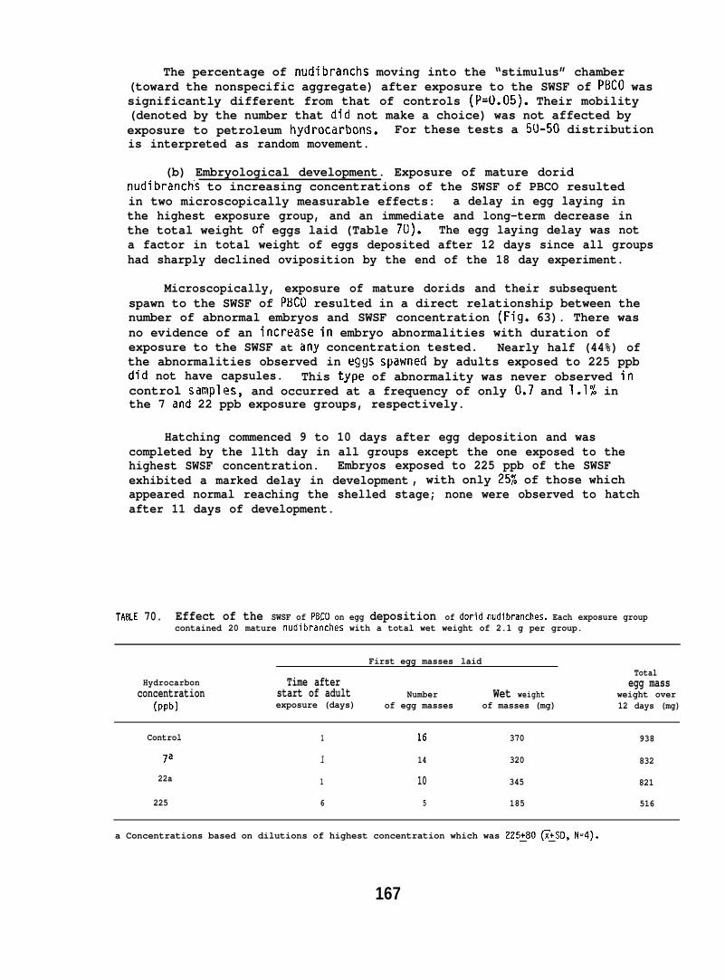
The percentage of nudibranchs moving into the “stimulus” chamber(toward the nonspecific aggregate) after exposure to the SWSF of PBCO wassignificantly different from that of controls (P=U.05). Their mobility(denoted by the number that did not make a choice) was not affected byexposure to petroleum hydrocarbons. For these tests a 50-50 distributionis interpreted as random movement.
(b) Embryological development. Exposure of mature doridnudibranchs to increasing concentrations of the SWSF of PBCO resultedin two microscopically measurable effects: a delay in egg laying inthe highest exposure group, and an immediate and long-term decrease inthe total weight of eggs laid (Table 70). The egg laying delay was nota factor in total weight of eggs deposited after 12 days since all groupshad sharply declined oviposition by the end of the 18 day experiment.
Microscopically, exposure of mature dorids and their subsequentspawn to the SWSF of PBCO resulted in a direct relationship between thenumber of abnormal embryos and SWSF concentration (Fig. 63). There wasno evidence of an increase in embryo abnormalities with duration ofexposure to the SWSF at iiny concentration tested. Nearly half (44%) ofthe abnormalities observed in eggs spawned by adults exposed to 225 ppbdid not have capsules. This type of abnormality was never observed incontrol samples, and occurred at a frequency of only 0.7 and l.1~ inthe 7 and 22 ppb exposure groups, respectively.
Hatching commenced 9 to 10 days after egg deposition and wascompleted by the llth day in all groups except the one exposed to thehighest SWSF concentration. Embryos exposed to 225 ppb of the SWSFexhibited a marked delay in development , with only 25% of those whichappeared normal reaching the shelled stage; none were observed to hatchafter 11 days of development.
TA8LE 70. Effect of the SWSF of PBCO on egg deposition of dorid nudibranches. Each exposure groupcontained 20 mature nudibranches with a total wet weight of 2.1 g per group.
First egg masses laidTotal
Hydrocarbon Time afterconcentration
egg massstart of adult Number Wet weight
(ppb)weight over
exposure (days) of egg masses of masses (mg) 12 days (mg)
Control 1 16 370 938
la 1 14 320 832
22a 1 10 345 821
225 6 5 185 516
a Concentrations based on dilutions of highest concentration which was 225:80 ~:SD, N=4).
167
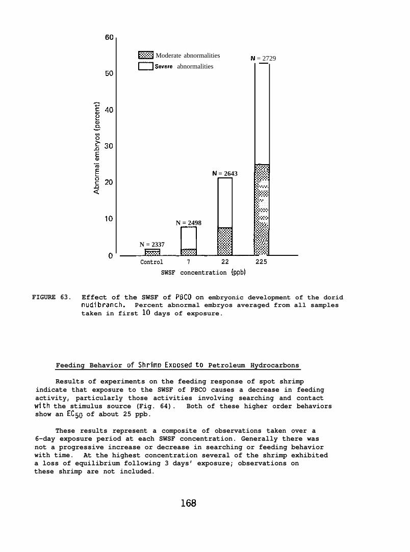
~ Moderate abnormalities N = 2729m Severe abnormalities
N = 2643
N = 2498
N = 2337
—
~. . .%%.,,......,..%...,,......,L* . . . . .. . . . . . . .,......,. . . . . . . .:.:.:.:.,.../.,. . . . . . . .,.*. . . .. . . . . . . .. . . .. . . . . . . .:,:.:,:.... ,. . . . . . . .,.:,:.:.. . .. .. .,.6.< . . .
:.-
. . .
:.:.:.:,
,......,
. .../..
. . . .
. . . . . . . .
y..:,:,
..+.,,.,
..+..,.!-.,., 0.!
Control 7 22 225SWSF concentration (ppb)
FIGURE 63. Effect of the SWSF of PLICO on embryonic development of the doridnudibranch. Percent abnormal embryos averaged from all samplestaken in first 10 days of exposure.
Feeding Behavior of Shrim~ Ex~osed to Petroleum Hydrocarbons
Results of experiments on the feeding response of spot shrimpindicate that exposure to the SWSF of PBCO causes a decrease in feedingactivity, particularly those activities involving searching and contactwith the stimulus source (Fig. 64). Both of these higher order behaviorsshow an EC50 of about 25 ppb.
These results represent a composite of observations taken over a6-day exposure period at each SWSF concentration. Generally there wasnot a progressive increase or decrease in searching or feeding behaviorwith time. At the highest concentration several of the shrimp exhibiteda loss of equilibrium following 3 days’ exposure; observations onthese shrimp are not included.
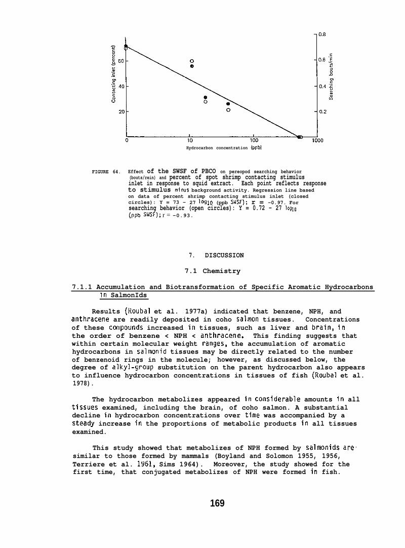
Hydrocarbon concentration (ppb)
FIGURE 64. Effect of the SWSF of PBCO on pereopod searching behavior(bouts/rein) and percent of spot shrimp contacting stimulusinlet in response to squid extract. Each point reflects responseto stimulus minus background activity. Regression line basedon data of percent shrimp contacting stimulus inlet (closedcircles): Y = 73 - 27 loglo (ppb SWSF); r = -0.97. Forsearching behavior (open circles): Y = 0.72 - 27 loglo(PPb SWSF); r = -0.93.
7. DISCUSSION
7.1 Chemistry
7.1.1 Accumulation and Biotransformation of Specific Aromatic HydrocarbonsIn SalmonIds
Results (Roubal et al. 1977a) indicated that benzene, NPH, andanthracene are readily deposited in coho salmon tissues. Concentrationsof these ccmpounds increased in tissues, such as liver and brain, inthe order of benzene < NPH < anthracene. This finding suggests thatwithin certain molecular weight ranges~ the accumulation of aromatichydrocarbons in salmonid tissues may be directly related to the numberof benzenoid rings in the molecule; however, as discussed below, thedegree of alkyl-group substitution on the parent hydrocarbon also appearsto influence hydrocarbon concentrations in tissues of fish (Roubal et al.1978).
The hydrocarbon metabolizes appeared in considerable amounts in alltissues examined, including the brain, of coho salmon. A substantialdecline in hydrocarbon concentrations over time was accompanied by asteady increase in the proportions of metabolic products in all tissuesexamined.
This study showed that metabolizes of NPH formed by salmonids aresimilar to those formed by mammals (Boyland and Solomon 1955, 1956,Terriere et al. 1961, Sims 1964). Moreover, the study showed for thefirst time, that conjugated metabolizes of NPH were formed in fish.
169
7.1.2 Accumulation of Petroleum Hydrocarbons by Fish Exposed to SWSFof PBCO
Roubal et al. 197S reported that substituted benzenes, NPH, andsubstituted NPH accumulated both in coho salmon and starry flounderwhen the fish were exposed to the SWSF of PBCO. Moreover, as the numberof alkyl substituents increased on benzene and naphthalene, so did theaccumulation of these compounds.
The C4- and C5-substituted benzene fraction of PBCO was the mostprcminent fraction accumulated in muscle of both coho salmon and starryflounder. However, substantially greater concentrations of this fractionaccumulated in the muscle of starry flounder than in muscle of coho salmon.Hydrocarbons of the SWSF were not detected in the liver and gills ofthe test coho salmon. However, substantial amounts of the substitutedbenzenes, NPH, and substituted NPHs were found in the liver and gillsof starry flounder. The ability of starry flounder to bioconcentratearomatic hydrocarbons suggests that starry flounder residing on sedimentscontaminated with petroleum hydrocarbons may accumulate substantialconcentrations of petroleum hydrocarbons in various tissues.
7.1.3 Metabolism of NPH by Coho Salmon
This study (Collier et al. 1978) represents the first use of HPLCfor characterizing NPH metabolizes in fish. The presence of both non-conjugated and conjugated derivatives in the liver and gall bladderindicates that coho salmon have a significant ability to metabolize NPHand excrete the metabolizes. However, the question still remains towhat extent low levels of these metabolizes are retained for extendedperiods in tissues of salmonid fish.
7.1.4 NPH and Its Metabolizes in Fish Skin and Mucus
Skin, The results (Varanasi et al. 1978) show that within a fewhwrs of NPli exposure, appreciable concentrations of both the hydrocarbonand its metabolizes were present in the skin of coho salmon, starryflounder, and rainbow trout, regardless of the mode of exposure or thesalinity of the medium. Moreover, comparison of these results with thoseobtained for liver of rainbow trout (Collier et al. 1980) and starryflounder (Varanasi et al. 1979) reveals that the pattern of uptakeand release of NPH and its metabolizes in skin was similar to thatobserved for the liver. NPH concentrations in skin declined morerapidly than the concentrations of NPH metabolizes, demonstrating atendency of skin to preferentially retain metabolizes. This findingis in agreement with results of Lee and coworkers (1976), Sanborn andt’lalins (1977), and Roubal et al. (1977a), which showed that the parenthydrocarbon was more rapidly discharged either directly or via biotrans-formation and that small but detectable concentrations of metabolizespersistedin tissues of crustaceans and fish over a long period.Whether metabolic products detected in the skin of our test fish arisefrom the direct transport of these compounds by blood to the skin and/orfrof;l biotransformation,of NPH by AHM that may be present in the skin
170
remains to be seen. It should be noted that maxima in concentrations ofboth NPH and itsmetabolites in the skin were preceded by the correspondingmaxima in the livers (Varanasi et al. 1978, Collier et al. 1980). Itappears therefore that injected or ingested NPH was first transported tothe liver and subsequently transported to the skin via the blood stream.
Mucus. Epidemlal mucus of fish serves as a physical and perhapsan im=ogical barrier to pathogenic organisms (Harris and Hunt 1973).It is also believed to be associated with osmoregulation (Jakowska 1963)and is an important factor in controlling the swimming speed of the fish(Rosen and Cornford 1971). The results (Varanasi et al. 1978) fromthe force-feeding and injection studies, which showed that NPH and itsmetabolic products were present in epidermal mucus of rainbow troutfor several days after the initial exposure to NPH, strongly suggestthat epidermal mucus of salmonids is involved in excretion of hydrocarbonsand their metabolizes. Epidermal mucus is in constant flux; its relativeimportance in the excretory mechanisms of fish would depend on the rateof discharge of these compounds from the mucus into the surroundingwater as well as on the turnover or sloughing rate of the mucus itself.Varanasi and Markey (1978) reported that the presence of certain metalsin water induces increased mucus production in fish, thereby acceleratingthe turnover of the mucus layer. Whether hydrocarbons exert such aninfluence on mucus production in fish is unknown; however, copiousamounts of mucus were produced by soft-shell clams exposed to No. 2fuel oil in water (Stainken 1975).
7.1.5 Uptake and Biotransformation of NPH by Flatfish
The results (Varanasi et al. 1979) show that pleuronectids, likesalmonids (Roubal et al. 1977a, Varanasi et al. 1978, Collier et al.1978), are able to absorb and metabolize dietary NPI-1, and that NPHand its metabolic products are broadly distributed in tissues and bodyfluids of the exposed fish. Rock sole contained statisticallysignificant larger concentrations of NPtl and its metabolizes thandid starry flounder and almost 4 times as much radioactivity remainedin the digestive tract of rock sole compared to that in the starryflounder at 24 hr after feeding of 3H-NPH. The digestive tract ofrock sole has pyloric caeca, whereas that of starry flounder does not;in addition to other species-specific differences, perhaps structuraland functional variations in the digestive tracts (Barrington 1957) ofthese fish are responsible for the observed differences in the levelsof accumulated radioactivity. In fact, a major fraction of the administeredr~dioactivity may have been directly discharged from the gastrointestinaltract of starry flounder.
Support for this observation comes from results showing that whenthe same dose of NPH was administered to starry flounder via i.p. injection,subsequent concentrations of NPH in tissues {e.g., liver) were comparableto those obtained for NPH exposed rock sole and were much higher thanthose for starry flounder force-fed NPH. It may be that absorption ofNPH over a longer period, from the gastrointestinal (GI) tract in thecase of rock sole in the feeding study and from the abdominal cavity
in the case of starry flounder in the injection study, was responsiblefor the results showing that the decline in the concentrations of NPHfrom the tissues (e.g., liver, skin, and blood) of these fish was muchslwer than that for starry flounder force-fed NPH.
In terrestrial mammals, the route of administration can alter thepharmacokinetics of a given compound (Gibaldi and Perrier 1974). Themode of hydrocarbon-exposure did influence concentrations and relativeproportions of NPH and its metabolic products as well as the types ofmetabolizes accumulated in pleuronectid fish. Metabolism of NPH inthe GI tract may be responsible for the observed differences in thepattern of metabolizes accumulated in livers of starry flounder in theinjection and feeding experiments at 12”C. Thus, it can be speculatedthat differences in patterns of metabolizes may occur when the majorroute of uptake of PAH is other than the GI tract (e.g., gills andskin when PAH is present in water or sediment).
Dietary aromatic hydrocarbons are cleared from terrestrial animalsprimarily via biliary and to a lesser extent, via renal excretion(Daniel et al. 1967, Guarino et al . 1972). As with salmonids (Roubalet al. 1977a, Melancon and Lech 1978, Statham et al. 1978, Collier et al.1978), bile of flatfish in both dietary and i.p. exposures accumulatedlarge amounts of NPH metabolic products. This suggests that biliaryexcretion was one of the major routes by which the hydrocarbon was clearedfrom these pleuronectids. Metabolizes in the bile of the NPti-exposedflatfish were characterized by the preponderance of glucuronides (>80%).Radioactivity excreted in urine was not measured, but considerableradioactivity was present in the kidney of the test fish over a longperiod (up to 6wk). Excretion of both NPH and metabolic products viaepidermal mucus (Vdranasi et al. 1978) and gills (Thomas and Rice 1981)seems to occur in salmonids. Epidermal mucus and gills of pleuronectidsexposed to NPH contained detectable concentrations of NPH and, moreimportantly, its metabolic products, which suggests that in addition tobiliary and renal excretion, clearance via epidermal mucus and gillsmay also take place in these fish. The relative importance of each ofthese pathways in clearance of xenobiotics in fish remains to be assessed.
The concentrations of NPH and its metabolic products in musclewere one-tenth of those for the li’ver of the individual species,indicating that no marked tendency was shown by muscle to accumulatethe hydrocarbon or its metabolizes. Liver and various extra-hepatictissues of marine organisms are shown to possess AHM (Varanasi andMalins 1977, Bend and James 1978), but the presence of this enzymesystem in the muscle of fish has not yet been demonstrated. Our resultsshow that the pattern of metabolizes accumulated in muscle were qualitativelysimilar to that in the liver of the same fish. However, in both species,the proportion of non-conjugates, specifically 1,2-dihydro-1,2-di hydroxy-NPH(dihydrodiol ), was higher in the muscle than in the liver. If the presenceof metabolic products in muscle was prim~rily due to their transport fromthe liver, then selection seems to take place in the types of metabolizesthat are deposited in muscle.
172
In agreement with the results with salmonids (Roubal et al.1977a), proportions of metabolizes relative to NPH increased with timeafter exposure in tissues (e.g., liver) of flatfish. Moreover, thefindings show that the types of metabolizes accumulated in flatfishliver were dependent on the time elapsed after the administration ofNPH. With time, the pattern of metabolizes changed in the liver oftest fish, regardless of species or mode of administration of NPH;there was an increase in the proportion of conjugates--specificallysulfate/glucoside fraction--and a decrease in the proportion of thedihydrodiol. Because certain dihydrodiols of PAH are known to bemutagenic and carcinogenic (Swaisland et al. 1974, Levin et al. 1976),a decreased proportion of the dihydrodiol may imply increased detoxificationof the hydrocarbon.
7.1.6 Effect of Temperature on Disposition of NPH and Its Metabolizes in Fish
Water temperature had a pronounced effect on NPH accumulation andretention in the major organs of coho salmon exposed to NPH via forcefeeding (Collier et al. 1978). An inverse relationship between environmentaltemperature and NPH retention was observed for brain, liver, kidney andblood. The work of Fucik and Neff (1977) with clams, Rangia cuneata,and Harris et al. (1977) with copepods, Calanus helgolandicus, indicateda similar relationship between temperature and NPH retention in marineinvertebrates. Thus, it seems that the nature and severity of anytoxic effects of NPH in marine organisms may be different in colderenvironments; cold-adapted organisms may retain NPH longer than thosein warmer waters.
In another study, Varanasi et al (1981a) reported that a decreasein water temperature from 12° to 4°C resulted in the retention ofsubstantially higher concentrations of NPH in tissues of NPH-exposedstarry flounder. The increase in NPH concentrations could be due toseveral factors: Differences in rates of absorption of the ingested dose,differences in rates of excretion of NPH from tissues, and differentialrates of biotransformation of NPH at these two temperatures.
Environmental temperature is known to have a marked effect on theactivity of the alimentary canal of fish; both the rate of passage offood and the rate of its absorption are much slower in fish at lowertemperatures (Barrington 1957). The results of Varanasi et al (1981a)indicate that both absorption and elimination of the ingested dosefrom the alimentary canal were slower for fish at 4°C than at 12°C.This may explain the slower decline in NPH concentrations from 24 to168 hr in tissues of starry flounder at the lower temperature. Loweringthe water temperature may also influence the rate of elimination ofNPH from tissues of fish. For example, Collier et al. (1978) did notobserve any effect of lowered water temperature on the retention ofingested NPH in the gut of coho salmon, but did report statisticallysignificant higher concentrations of NPtl in tissues of fish at 4QCcompared to those at 10°C.
173
The effect of decreased temperature on metabolize concentrationsin starry flounder tissues was not as marked as that observed for theNPf-1 concentrations, which suggests that a much smaller proportion ofNPH was biotransformed by liver of starry flounder held at the lowertemperature. However, such a result may also be due to altered ratesof excretion of NPH and its metabolizes at the lower temperature.Nevertheless, when considering consequences (e.g., toxicity) of increasedconcentrations and increased residence times of NPH in cold-acclimatedfish, the decreased bioconversion of NPH should be taken into account.
A change in environmental temperature is also known to alter theactivities of certain enzymes in fish (Hochachka and Somero 1971).Stegeman (1979) reported that in vitro activities of hepatic BaP hydroxylasemeasured at 25°C were signific~t~eater for Fundulus heteroclitusmaintained at 6.5°C than those at 16.5”C. Egaas and Varanasi (1982)reported that lowering the environmental temperature increased both theinitial hydrocarbon metabolism and the time needed for the effect of thechemical inducer to be evident. A change in the water temperature ofa fish brings about changes in proportions of polyunsaturated fatty acids(PUFA) in phospholipids associated with membranes (Hazel 1979a, 1979 b),which may result in altered rates of absorption and excretion of lipophilicPAHs and their metabolizes. Wills (1980) reported a linear relationbetween the rate of BaP oxidation and PUFA content in endoplasmic reticulumin rat. Thus, environmental temperature can alter concentrations ofindividual metabolizes formed and retained in tissues of fish exposedto PAHs. A decrease in water temperature brought about marked changesin individual metabolize classes in flatfish, which would have goneundetected if only total metabolize concentrations were determined.
Virtually no information is available on the toxicity of differentNPH metabolizes on fish. However, in mammals phenols and dihydrodiolsof certain PAHs [e.g., benzo(a)pyrene] are more toxic than the parentcompound, and some of these metabolizes also interact with cellularmacromolecules (Burke et al. 1977, Kapitulnik et al. 1977, Varanasi etal. 1981 b). Certain hydroxylated metabolizes of NPH also irreversiblybind to cellular protein (Hesse and Mezger 1979). Accordingly, theresults showing a statistically significant increase in the concentrationsof the dihydrodiol derivative of NPH in liver and muscle of cold-maintainedstarry flounder is noteworthy. Moreover, it should be noted that 1 wkafter NPH-exposure, fish held at 12°C contained very low concentrationsof NPH, whereas tissues (e.g., liver and muscle) of fish at 4°C stillretained substantial concentrations of NPH, a potential source ofmetabolic products.
7.1.7 Uptake and Metabolism of Sediment-Associated NPH and BaP by Flatfish
Several studies report on the uptake and metabolism of NPH by fish(Lee e~ al. 1972, Roubal et al. 1977a, 1978, Melancon and Lech 1978,Collier et al. 1978, 1980, Varanasi et al. 1979, 1981a). Studies onuptake of RaP are fewer (Lee et al. 1972, Lu et al. 1977, Gerhart andCarlson 1978, Martin 1980) and to our knowledge, until the work ofVaranasi and Gmur (l!181a), no inforrllation was available on the extent
of in vivo metabolism of BaP by fish liver. In this study, when Englishsol~w~exposed simultaneously to 14C-NPH and 3H-BaP in sediment,a substantial percentage of NPH-derived radioactivity in the liver offish was present in the form of the parent hydrocarbon, whereas mostof the BaP in the liver of the same fish was biotransformed. Moreover,the results show that the ratio of radioactivity in bile vs liver wassubstantially higher for BaP than that for NPH, demonstrating that BaPwas more extensively metabolized in liver and transferred to bile. Alarge proportion of NPH may have been excreted, prior to metabolism,via gill (Thomas and Rice 1981) and skin/mucus (Varanasi et al. 1978).
These results (Varanasi and Gmur 1981a), together with our previousresults (Varanasi et al. 1979), show that whether the fish were exposedto NPH or BaP, singly or in the presence of other hydrocarbons, theliver metabolized BaP more extensively than NPH. This probably explainswhy BaP is detected in very low concentrations or not detected at allin liver of fish (Veldre et al. 1979, Malins et al. 1980) sampled fromareas containing considerable concentrations of BaP, since the currentlyavailable techniques can detect only the parent hydrocarbon. It isobvious from the results that if fish tissues are examined for hydrocarbonsalone to assess whether the organisms have been exposed to these compounds,grossly misleading results may be obtained; the magnitude of error willbe dependent on a number of factors (e.g., tissue type, duration ofexposure, type of hydrocarbon). Therefore, metabolize analyses togetherwith hydrocarbon analyses should be included in order to assess properlythe levels of xenobiotics in marine organisms.
One of the major difficulties in tissue analyses of PAHs, such asBaP, has been the presence of lipids. A new method using two-dimensionalTLC was developed to separate BaP and its metabolizes from each otherand from lipids present in liver (Varanasi and Gmur 1981a). This methodmay be adapted to separate PAHs and their metabolizes from liver andmuscle of fish sampled in field studies.
Metabolizes of NPH in the bile of English sole in the present studywere similar to those reported for starry flounder and rock sole exposedto dietary NPH (Varanasi et al. 1979, 1981a). The glucuronide of 1,2-dihydro-l,2-dihydroxynaphthalene was the major metabolize. The tendencyof pleuronectid fish to produce a greater proportion of glucuronideconjugates than sulfate conjugates was also evident when bile sampleswere analyzed for both NPH and BaP metabolizes. Glucuronidation is themajor detoxification pathway in hamster embryo cells (Baird et al. 1977)and rat hepatocytes (Burke et al. 1977); however, marine invertebratestend to produce large proportions of sulfate conjugates (Corner et al.1960, Malins and Roubal 1982). Moreover, a preliminary report (Von Hofeet al. 1979) has shown that a higher proportion of sulfate conjugatesthan glucuronide conjugates was present in bile of Fundulus heterocl itusinjected (i,p.) with BaP. Species specific differences as well asdifferences in mode of exposure to hydrocarbons may be responsible forthe observed differences in types of conjugates formed by pleuronectidsand Fundulus.
175
A number of metabolizes known to be toxic to mammals were detectedin liver and bile of English sole exposed to sediment-asociated BaP.Studies with mammals have demonstrated that BaP-7,8-dihydrodiol isfurther metabolized to BaP-7,8-dihydroxy-9,10-epoxy-7,8,9, 10-tetrahydrobenzo(a)pyrene, which is known to bind covalently with DNA(Sims et al. 1974). The diolepoxide is hydrolyzed to 7,8,9,10-tetrahydroxy-7,8,9, 10-tetrahydrobenzo (a)pyrene. HPLC analyses of polarmetabolizes in the present study revealed the presence of tetrols inbile of English sole, indicating that the diolepoxide, which is implicatedas the ultimate carcinogen of BaP, was produced in liver of thesefish. Recently Varanasi et al. (1981b) have demonstrated that Englishsole metabolizes BaP into reactive metabolizes that bind to liver DNA.Such interaction of BaP with DNA is believed to be an important stepin chemical-induced neoplasia.
In contrast to the extensive conversion of BaP by English soleliver, BaP in sediment and sediment-associated water remained primarilyas the parent compound over a period of several days. The resultsalso show that while BaP concentrations in sediment did not changemarkedly, NPH concentrations declined steadily. Greater susceptibilityto microbial degradation (Herbes and Schwall 1979, Lee 1977) and higherwater volubility (Brown and Weiss 1978) of NPH compared to BaP are twolikely causes for the observed decline in NPH concentrations in sedimentduring the experiment.
The statistically significant decrease in concentrations of NPH-derivedradioactivity in most tissues of English sole from 24 to 168 hr wasattributed to the net release of NPH and/or its metabolizes from tissuesand not to decreased availability of NPH because NPH concentrations inSAW did not decline markedly. In contrast to NPH, BaP-derived radioactivitywas continually accumulated and retained by English sole. This tendencyto retain and bioconcentrate BaP-derived radioactivity is not unexpected.Roesijadi et al. (1978) reported that clams bioconcentrated radioactivitydue to PHN, chrysene and BaP in the order of increasing molecular weight ofPAH. Roubal et al. (1977a) with coho salmon fed benzene, NPH, or anthracenefound that the degree of retention of hydrocarbon-derived radioactivityin fish tissues was in direct correlation with increasing molecularweight of the parent hydrocarbon.
7.1.8 BaP Metabolism by English Sole
thatof bsuchfind”signwere
English sole force-fed BaP converted it into a number of metabolizeshave been characterized as mutagenic and carcinogenic in a varietyological assays. Both liver and muscle contained toxic metabolizesas BaP 7,8-dihydrodiol, BaP 9,10-dihydrodiol and phenols. The twongs showing that concentrations of BaP metabolizes decreasedficantly with time in muscle and that a number of toxic metabolizespresent in edible tissue of flatfish are imoortant from a human
health point of view. Further studies on the characterization of,mutagenic potency of, and rate of release of PAH metabolizes from muscleof fish are needed to gain a better understanding of the fate andeffects of these hydrocarbons in edible tissue of fish.
7.1.9 $Inding of BaP Intermediates to DNA Catalyzed by Liver Enzymes
The results show that liver enzymes of coho salmon, starry flounderand English sole converted BaP into reactive intermediates that bindto DNA (Varanasi and Gnur 1980, Varanasi et al. 1980). The metabolizesformed by fish liver extracts were characterized by a preponderance ofnon K-region dihydrodiols (e.g., BaP 7,8-dihydrodiol and BaP 9,10-dihydrodiol). It should be noted that mainly non K-region metabolizesof PAH covalently bind to DNA of PAH-exposed mammals.
Many environmental and species-specific differences may influencethe xenobiotic metabolizing capabilities of aquatic animals; it wouldbe imprudent to attribute the marked difference observed in the bindingof values for the three fish species to any particular factor. Moreover,wide variations in AHM activity have been reported to occur amongdifferent strains of a single species of fish or mice. However, it ispossible that starry flounder and English sole, being demersal fish,are habitually exposed to a multitude of xenobiotics (some of whichare inducers of AHM) present in bottom sediments, which could resultin an initially high value for the in vitro covalent binding of BaP toDNA. This would also explain why p~--ure of flatfish to 3-methyl-cholanthrene (MC) or BaP did not result in an increase in binding asgreat as that for coho salmon. The results (Varanasi et al. 1980)showing that the increase in the formation of reactive DNA bindingintermediates was much greater for English sole after treatment withPBCO than that for starry flounder suggest that English sole was moresensitive to petroleum exposure than starry flounder. This can becompared with results described in the Pathology Section 6.2.2 showingthat English sole were sensitive to PBCO when exposed to PBCO contaminatedsediment.
Our results (Varanasi and Gmur 1980, Varanasi et al. 1980) andthose of others (Ahoka et al. 1979) show that the magnitude of increasein in vitro binding of BaP to DNA on pre-exposure of fish or rat to PAti(MC~~~ was relatively greater than the increase in AHM activity.Covalent binding of BaP to DNA may prove to be more sensitive than AH14activity as an index of pre-exposure of fish to certain toxic chemicals.Moreover, the binding value serves as a useful index for correlatingmetabolism with the carcinogenicity of a PAH. The carcinogenic potent”of a compound to mammals is roughly correlated with the extent of cova”binding to DNA {Buty et al. 1976, Pelkonen et al. 1978). Accordingly,studies to assess the extent of covalent binding of a variety of xenobto cellular DNA in various target tissues of marine organisms may giveuseful information. In a recent study (Varanasi et al. 1981a) bindingBaP intermediates to DNA in English sole liver was reported to be ashigh as that for mammalian tissues susceptible to BaP carcinogenesis.
alent
otics
of
7.1.10 Activities of Aryl Hydrocarbon Monooxygenases (AHM) in Different-
Investigations of the biotransformation of aromatic hydrocarbonsand the fate of PAH metabolizes in marine organisms should also include
177
analysis for the presence or absence of aryl hydrocarbon monooxygenaseenzyme systems, e.g., benzo(a )pyrene monooxygenase (BPMO). In the lastdecade (Philpot et al. 1976, Payne 1977, Gruger et al. 1977, Kurelec etal. 1977, Bend et al. 1977, Bend and James 1978) it has been demonstratedthat many freshwater and marine fish species possess hepatic and extrahepaticAHi4 activity. The results of Sanborn and Malins (1977, 1980) andMalins and Roubal (1982) discussed elsewhere in this report show theability of several marine invertebrates to metabolize NPH and 2,6-DMNin vivo. Bend and James (1978) pointed out that those marine invertebrates~a~metabolize xenobiotics in vivo by cytochrome P-450-dependentmixed-function oxidases do so a~a=wer rate than do fish. Microsomalmixed-function oxidation and aryl monooxygenation in vitro are alsovery slow in crustacean hepatopancreas, relative t~f~epatic microsomes.The present research adds eight more species to the growing list ofaquatic organisms which possess aryl hydrocarbon monooxygenase activity.
7.1.11 Uptake, Metabolism and Toxicity of Hydrocarbons in Invertebrates
Exposure of Mollusc Larvae and Gametes to NPH, Mussel and oystergametes and the resultant larvae were sensltlve to NPH at concentrationsas low as 1 ppb. The major effect at low concentrations was a reductionin survival of the larvae. At higher concentrations, a decrease infertilization of eggs resulted when NPH exposed sperm was used. Embryo-logical abnormalities also were more prevalent at concentrations greaterthan 1 ppb. All of these factors indicate a reduction in the ecologicalfitness of petroleum-contaminated mollusc gametes and their resultinglarvae.
Exposure of Crustaceans to Hydrocarbons. Larval forms of spotshrimp and Dungeness crabs were found to be extremely sensitive towater-borne NPH and NPH bound to protein (Sanborn and Malins 1978).The compounds were lethal in the low ppb range to Stage I and Stage Vspot shrimp and newly hatched Dungeness crab zoea. These results areconsonant with the generally accepted belief that early developmentalstages are more sensitive to petroleum than adult marine organisms(Malins and Hodgins 1981). The findings also suggest that Stage I andStage V spot shrimp are capable of accumulating from 25 to 100 times theenvironmental concentrations of NPH, depending upon whether the NPH isfree or protein-bound (Sanborn and Malins 1978). The findings showingthat NPH is readily released from Stage I and Stage V spot shrimp andthemetabolites retained for several days (Sanborn and Malins 1978)are similar to those observed with fish (Roubal et al. 1977a, Varanasiet al. 1979, Collier et al. 1980).
The findings with P. ~latyceros suggest that the mature animalsreadily accumulate hydr~carbon components of the SWSF of crude oil(Sanborn and Mali ns 1980). The thoracic segments contained both lowmolecular weight benzenes and NPHs. It is likely that the hepatopancreaslocated in the thorax is a major site of hydrocarbon accumulation andmetabolism. Abdominal segments also contained low molecular weightbenzenes and NPHs indicating that shrimp sequester water-soluble hydrocarbonsin edible tissue. Bioconcentration values are low in comparison to
178
values observed with fish (Roubal et al. 1978). The bioconcentrationof substituted benzenes was in direct relationship to the degree ofalkylation, a finding that corresponds to results obtained with fish(Roubal et al. 1978).
Both adult and larval spot shrimp are capable of forming a widevariety of metabolic compounds (Sanborn and Malins 1980). The profileof metabolizes in adult shrimp was not markedly dissimilar from thatobserved in NPH-exposed fish (Varanasi et al. 1979, Collier et al.1980). The fact that larvae formed metabolizes indicates that earlydevelopmental stages have well developed enzyme systems capable ofconverting NPH to both conjugated and nonconjugated metabolizes.
7.1.12 Food Chain Transfer of 2,6-DMN to Sea Urchins via Algae
The present work (Malins and Roubal 1982) shows that 3H-2,6-DMNreadily accumulates in Fucus exposed to this hydrocarbon through thewater column. Moreover, the 2,6-DMN is not metabolized in the algaeand thus was transferred to the echinoderms as the parent compound(Malins and Roubal 1982).
It was not expected that the sea urchins would accumulate a largeproportion of the ingested tritium in the exoskeleton, particularlybecause the 2,6-DNN was administered through the diet and no evidencewas found for radioactive hydrocarbons and metabolizes in surroundingsea water. However, this finding raised the question of whether tritiumexchange occurred with the result that tritiated polar compounds (e.g.,water) accumulated in the exoskeleton. This possibility was dismissedbecause thelabel expersignificantthe tightlyinterfere wanswered.
3H/14c rat-es in exoskeleton and soft tissues in the dua~-ment indicated that such an exchange did not occur to adegree (Malins and Roubal 1982). The question of whetherbound aromatic fraction in the exoskeleton is available toth normal cellular functions of soft tissues remains to be
The preferential formation in echinoderms of 2,6-DMN metabolizesarising from oxidation of the aromatic ring is an interesting finding.In rat liver, 7,12-dimethylbenzanthracene is converted primarily tomethanol derivatives, although a shift to ring oxidation occurs afterpretreatment with other aromatic hydrocarbons (Jellinck and Goudy 1967).Moreover, Kaubisch et al. (1972) demonstrated that in rat liver microsomesoxidation of 1- and Z-nethyl naphthalene and 1,2-DMN leads principallyto naphthoic acids and methylnaphthoic acids, respectively. Littlecomparable infomlation is available on marine organisms; however, Grugeret al. (1981) showed that starry flounder accumulate primarily methanolderivatives in bile after oral administration of 2,6-DMN. Thus, themetabolism of methyl-substituted NPHs in this species of echinodermappears to be similar to that by aromatic hydrocarbon-pretreated rat.
Sulfotransferases are responsible for the conjugation of a varietyof compounds (Pasternak et al. 1963, Wortman 1961). Little informationexists on these metabolic transformations in marine invertebrates;
179
however, the lobster (Homarus americanus) is reported to have a poorability to convert aro~ydrocarbons to oxygen-containingderivatives” (Elmamlouk and Gessner 1978). Yet, if hydroxy compounds(e.g., p-nitrophenol) are administered, they are readily conjugatedthrough the sulfotransferases (Elmamlouk and Gesser 1978). The presentwork indicates that echinoderms are different from lobsters in havingactive enzyme systems for biosynthesizing the hydroxy compounds, aswell as for converting these oxidation products to aryl sulfates.
7.1.13 Biological Fate of Metals in Fish
The results (Varanasi and Markey 1978, Varanasi 1978, Reichert etal. 1979) demonstrated that fish held at lower temperatures accumulatedrelatively smaller concentrations of metals. These findings suggestthat the effect of temperature on the turnover and retention of metalsshould be taken into account when considering the overall effect of tracemetals on arctic biota. These studies also show that fish exposed tolow levels (ppb) of water-borne metals readily accumulate substantialconcentrations (ppm) in tissues. High levels of lead, a neurotoxin,were found in the brain of starry flounder, which suggests possibleneurological damage that could have behavioral consequences (Varanasi1978). Moreover, the results indicate that coho salmon exposed toeither water-borne lead or cadmium accumulate about twice as much metalin the posterior kidney as in the anterior kidney (Reichert et al. 1979).These results cannot be adequately explained at present; however, itis suggested that this tendency may be associated with the excretoryfunction of the posterior kidney in salmonids (Smith and Bell 1976).
Termination of exposures to metals does not automatically meanthat metal concentrations will necessarily remain constant or declinein the kidney and possibly other tissues. Even when a decline incadmium or lead concentrations was observed, e.g., in gills, considerablelevels still remained in gills after 5 wk deputation in control seawater(Reichert et al. 1979). The strong tendency to sequester cadmium andlead in gills (IIeichert et al. 1979) during uptake and deputationperiods raises questions about possible interference with osmoregulationand oxygen consumption (Thurberg et al. 1973).
More than 50% of lead and cadmium accumulated in the scales ofcoho salmon was still present after 5 wk of deputation (Varanasi andMarkey 1978). It is known that in salmonids, skin and scales play animportant role in calcium transport and regulation (Podoliak and Holden1965). The persistence of high concentrations of lead and smallerconcentrations of cadmium may interfere with transport and perhapsregulation of calcium in fish. Moreover, scales of salmonids areknown to be resorbed during maturation and the mineral components ofthe scales are utilized for general metabolism and production of gametes(Wallin 1957). In the metal-exposed fish, resorption of scales mayresult in the release of toxic metals in the bloodstream at the timeof stress. The consequences of release of such toxic metals on eggsand sperm remains to be assessed. It should be noted that increasedlevels of environmental or dietary calcium (Varanasi and Cinur 1978)
significantly reduces uptake and retention, and presumably toxicity,of lead in coho salmon.
The finding (Varanasi and Markey 1978) that coho salmon had lowerepidermal mucus concentrations of metals than did starry flounder, alongwith the results on rainbow trout (Varanasi et al. 1975) indicatedthat the level of metals accumulated in epidermal mucus are dependentlargely on a particular species of fish rather than the salinity ofthe medium. In addition, the results (Varanasi and Markey 1978) showthat short-term exposure to pollutants triggers increased synthesis ofmucus; however, the consequence of long-term exposure on mucus productionremains to be assessed. Sherwood and Bendele (1975) reported that inDover sole (Microstomus pacificus) collected from Pales Verdes, an areaof known metal contamination, fin erosion was accompanied by reductionin epidermal mucus (visual observation). Whether or not there existsa relationship between altered rate of mucus production and pathologicalsurface conditions is not known.
Epidermal mucus of salmonids exists in a state of continuous flux(Pickering 1976)--that is ~ small amounts of mucus are continuouslysloughed off and renewed. Thus, our results showing that metals werepresent in the mucus of the test fish for several days after terminationof exposure suggested that metals were discharged in epithelial mucinvia the mucous cells and excreted in the mucus (Varanasi and Markey1978, Varanasi 1978). The intriguing possibility exists that a certaincritical concentration of a pollutant in blood at a given temperaturemay trigger increased synthesis of epithelial mucin, resulting in rapidturnover of mucus which would expedite discharge of the pollutant. Withrespect to this hypothesis, it should be noted that NPH and its metabolicproducts are also shown to be released in epidermal mucus of NPH-exposedsalmonids (Varanasi et al. 1978) and flatfish.
Heavy metal-binding proteins in gills of marine species have beenreported previously (Ilarafante 1976, Bouquegneau et al. 1975, Noel-Lambotet al. 1978). Noel -Lambot et al. ( 1978) found extremely 1 ow concentrationsof cadmium bound to a cadmium binding protein (CdBP) in the gills ofeels that had been exposed to 200 ppm of cadmium for 5 hr. Ours tudieswith unexposed coho salmon indicate that CdBP concentrations in gillsare low. Yet within 24 hr after metal challenge there is an appreciableincrease in the accumulation of CdBP-bound cadmium, indicating inductionof CdBP. Accordingly, it is likely that CdBP may play an important rolein binding cadmium in the salmonid gill; this reaction may serve toprotect vital physiological processes (e.g., osmoregulation) from thepotentially destructive influences of this metal.
In rat liver, the proportion of cytosolic cadmium bound toCdBP (identified as metal lothionein) increases steadily for at least48 hr after injection (Frazier and Puglese 1978, Cempel and Webb 1976).Similar to studies with rat, induction of CdBP in the liver andkidney of salmonids was observed after the fish were exposed to cadmium(Reichert et al. 1979). Also, unexposed coho salmon apparently haveappreciable concentrations of CdBP present in liver. However, there
181
appears to be no specific low molecular weight protein like metal lothioneinin the cytosolic fraction of the kidney, liver, and gills of coho salmonthat has a high affinity for lead. Similar observations have been made inmammals. Consequently, this metal binds with a variety of proteins inthe cytosol and may alter protein structures.
In conclusion, the biochemical fate and biological effects ofmetals are probably influenced by temperature, calcium and other metalions in the marine environment. Synergistic and antagonistic effectsassociated with multiple systems of metals on marine organisms areimportant subjects for future study.
7.2 Pathology
7.2.1 Effects of Petroleum on Disease Resistance
The results of a series of tests designed to assess the effectsof petroleum hydrocarbons on disease resistance of salmonids, juvenileand adult fl atfish, and adult spot shrimp failed to identify a markedimpairment. Whether or not a lowered host resistance could occurunder other stress and exposure conditions and to different diseasescannot be predicted from the present data. These data do, however,strengthen an argument that exposure to environmentally realisticconcentrations of petroleum does not markedly impair disease resistancein the tested and related species.
In contrast, a preliminary assay with a representative petroleumdispersant (Corexit 9527) suggested the potential for an adverse effectof oil-dispersant mixtures on disease resistance of salmon.
7.2.2 Pathological Changes in Flatfish from Exposure to Oil-ContaminatedSediment
Flatfish were shown in these series of experiments to take up petroleumhydrocarbons in cwscle and liver tissue following contact with PBCO-contaminated sediments. Bioavailability and bioaccumulation appearsto be dependent on a number of factors including aromatic hydrocarbonstructure, sediment characteristics, and biotransformation mechanismsin the flatfish tissues.
The differential uptake and retention of 1- and 2-methyl naphthalenesand tetramethylbenzene suggests that aromatic structure influencesbioavailability. Such selectivity has been reported for starry flounderby Roubal et al. (1978), and has also been inferred from other studies(see Section 6.1.2). The reduction of tissue levels of petroleumhydrocarbons over time, despite a persistently high sediment concentrationof hydrocarbons, further suggests that increased clearance rates and/orincreased metabolic transformation of petroleum hydrocarbons to oxidationproducts are occurring (see Section 6.1.7).
182
The silt content of the sediments clearly influence the dynamicsof release of sediment-associated petroleum. There appears to be a slowerand more continuous release of hydrocarbons from high-silt sedimentthan from high-sand sediment.
The significance of severe hepatocellular lipid vaculoization(HLV) is unknown. Although this condition has been observed to occurat a higher frequency in crude oil-exposed English sole than in controls,it was observed at approximately the same frequency in both oil-exposedand control rock sole and starry flounder. HLV can be induced by avariety of causes, including nutritional deficiencies (Snieszko 1972),exposure to pesticides (Couch 1975), and by contact with pollutedenvironments (Pierce et al. 1978). Also, under the experimental conditionsemployed severe HLV appears to be reversible.
7.2.3 Cytopathology
(a) Adult Fish Exposures
Gill. Both salmon and flatfish exposed to 100 ppb concentrationsof the SWSF of PBCO exhibited gill lesions resulting from loss of surfacecells. Similar conditions have been observed in a number of fish speciesexposed to waterborne contaminants. Trace metals such as cadmium,nickel, and lead damage gills by causing sloughing of epithelial cells(Voyer et al. 1975, Schwiger 1957, Haider 1964). Excessive mucusproduction is also characteristic of exposure of fish to trace metals(Varanasi et al. 1975, and Section 6.1.13 of this report) and is thoughtto cause death by suffocation (Gardner 1975, Haider 1964). Similarly,epithelial sloughing and discharge of mucous glands was observed inmarine fish captured near an oil slick off the Texas and Louisiana coast(Blanton and Robinson 1973). The secretory cells of the pseudobranchof the Atlantic silverside (Menidia menidia) degenerated after exposureto either 0.14 ppm Texas-Lou~crude oil or 100 ppm waste motor oil(Gardner 1975, Gardner et al. 1975); light micrographs revealed severevacuolization of the entire pseudobranch but no data on adjacent gilltissue was presented.
The effects of phenol on gill tissue have been documented inseveral freshwater specie”s. Rainbow trout sloughed epithelial cellsand developed a general inflammation of the gills after exposure to6.5 to 9.6 ppm phenol (Mitrovic et al. 1968). Another freshwaterfish, the bream (Abramis brama), was exposed to lower concentrationsof phenol for a longer du~n and suffered similar destruction ofthe gill epithelial cells; in addition test fish suffered damage tothe blood vessels and extravasation of blood in the gill lamellae(Waluga 1966). Fourteen species of fish sampled from phenol-pollutedportions of the Rhine and Elbe Rivers revealed discharged mucous glandsand generalized gill inflammation (Reichenbach-Kl inke 1965).
Liver. Increases in rough endoplasmic reticulum in thehepatocytes of rainbow trout were observed after exposing the fish tohigh peroral doses of PBC(J for 8 months. In one of the few field studies
183
that included ultra structural data, a similar increase in rough endoplasmicreticulum was reported in the hepatocytes of Fundulus heteroclitussampled from an area which had received an oil spil 1 8 years earlier(Sabo and Stegeman, 1977). Also, the hepatocytes of channel catfish(Ictalurus punctatus) showed proliferation of endoplasmic reticulumand bizarre” whorls of both the smooth and rough endoplasmic reticulumafter chlorobiphenyl exposure via gastric incubation for 21 days(Hinton et al. 1978, Klaunig et al. 1979). Circular arrays of smoothendoplasmic reticulum in liver cells have been reported followingdietary exposure of rainbow trout to aflatoxin B (Scarpelli 1976), andto phenylbutazone (Scarpelli 1977). Hawkes et al. (1980) showedthat after 28 days exposure to a mixture of 5 ppm chlorobiphenyls indiet, the liver cells of chinook salmon had vesiculated rough endoplasmicreticulum and circular arrays of smooth surfaced membranes that closelyresembled the arrays of smooth endoplasmic reticulum reported in catfish(Hinton et al. 1978, Klaunig et al. 1979). Such changes in the endoplasmicreticulum, and particularly the changes resulting in circular arrays ofsmooth endoplasmic reticulum, have been suggested to be a response tosubstances that induce the mixed-function oxidase system (Klaunig et al.1979).
The process that results in changes in endoplasmic reticulumhas been hypothesized to include the disruption of lipoprotein synthesis.Triglycerides norms’lly utilized for lipoprotein formation are thoughtto accumulate in the hepatocytes (Tanikawa 1979). An increase inhepatocyte lipid content has been reported in English sole exposed tooiled-sediment (McCain et al. 1!378), in channel catfish (Hinton et al.1978) and rats (Kasza et al. 1978) exposed to dietary chlorobiphenyls,and in humans and other mammals exposed to a variety of toxic substances(Lombardi 1966; Tanikawa 1979). In contrast, our studies showed exposureof rainbow trout to dietary crude oil resulted in depletion of lipidin the hepatocytes, and although there was an increase in rough endoplasmicreticulum, no vesiculation of the cisternae was found (Hawkes 1977).
Lens. Opacities in lenses of mammals are preceded by hydrationof the len~ber cells (Hollwich et al. 1975); lens changes observedin rainbow trout perorally exposed to petroleum also appear to includehydration. Generally, long-term peroral exposure of trout to P13C0caused progressive degeneration of lens tissue characteristic of earlycataract formation. Payne et al. (1978) also reported tissue changes inthe lens of cunner (Tautogolabrus adspersus) exposed to an oil slick for6 RIO. In mammals, lens abnormali.t~iated with exposure to naphthalenehave been well documented (Hollwich et al. 1975). The sequence of metabolicevents that precedes cataract formation in rats and rabbits after exposureto naphthalene appears to include enzymatic oxidation reactions that leadto the formation of 1,2-dih.ydroxynaphthalene, a compound which may beautooxidized to 1,2-naphthoqu’components. The enzymes thatbeen found in the eye of rabb’Pirie 1967) and, recently, incommunication).
no~e;” this compound may react with lensperform some of the oxidation reactions havets (Van Heyningen 1979, Van Heyningen andthe eyes of fish (Stegeman, personal
(b) Embryonic Fish Exposures
Olfactory epitheliums. Scanning electron microscopy of sandsole larvae exposed to 164 ppb of the SWAF of weathered PBCO duringembryogenesis revealed degenerative changes in the chemosensory ciliaand a loss of the microridges that circumscribe the perimeter of theepithelial cells surrounding the olfactory organs. Whether olfactionwas impaired in these larvae is not known; however, the degree ofstructural alteration observed indicated severe damage to the receptororganelles.
These findings are in accordance with those of Gardner (1978), whoidentified the olfactory epitheliums of fish as a site of damage frompetroleum hydrocarbons. Gardner exposed the Atlantic silverside.~enidia men~dia, to whole crude oil; the water-soluble fraction ;f crudeoil, and the water-insoluble fraction of crude oil for 7 days. Pathologicalchanges in the olfactory organ differed with exposure regim~; whole cru~eoil induced a marked hyperplasia; epithel ial metaplasia developed afterexposure to the water-soluble faction; and submucosal blood vessels weredilated and congested after exposure to the water-insoluble fraction.
Brain and Eye. The late appearance of cytopathological changesin the retinal receptor cells of surf smelt exposed to the SWAF of CICOmay be related to the differentiation times of these cells. Typically,teleost retinas are relatively undifferentiated throughout embryogensisand, in some species (Ali 1959, Blaxter 1974), do not begin to matureuntil late in larval development. In surf smelt, the receptor cellsdevelop outer segment membranes (a necessary structure before thecells can function as photon receptors) between day-15 and day-21 ofembryogenesis. The first eye damage was observed in 21-day old embryos;this suggests that receptor cell damage from exposure to petroleumhydrocarbons is not evident cytologically until these cells are fullydifferentiated.
In addition to morphological alterations of the receptor cells,the effects of exposure to the SWAF of CICO on surf smelt embryos werereflected by necrosis in the neurons of both the brain and eye. In somefish and mammals, neural tissue appears to be particularly vulnerableto certain petroleum hydrocarbons; brain tissue has been reported tobioaccumulate relatively large amounts of these compounds. For example,naphthalene was sequestered in the brain of salmonids in amounts comparableto that in the liver (Roubal et al. 1977a, Collier et al. 1980, andSection 6.1.6 of this report). Neurotoxic effects of low-molecular-weighthydrocarbons in mammals have been attributed to accumulation of eitherthe parent hydrocarbons or their metabolizes in brain tissue (Savolainen1977). Although the effect of changes in the forebrain of the surfsmelt is difficult to assess, the observed neuronal damage may havebeen sufficient to account for the five-fold increase in mortality oflarvae exposed to the SWAF of CICLl as embryos as compared to unexposedcontrols (See Section 6.3.1 [e]).
7.3 Behavior and Physiology
7.3.1 Vertebrate Studies
Behavior of Pacific Salmon Exposed to Petroleum Hydrocarbons
(a) Olfactory disruption. The waterborne aromatic hydrocarboncanponents of petroleum are thought to be a likely cause of chemosensorydisruption in aquatic organisms (Takahashi and Kittredge 1973). Althoughthe mechanism of disruption is unknown, it has been suggested that suchcontaminants may mask the chemoreceptive sites, thus blocking incomingchemical signals at the receptor level (Sutterlin 1974). It is alsopossible that olfactory lesions, such as those observed by Gardner (1975)in Atlantic silversides exposed for 7 days to the SWSF of Texas-Louisianacrude oil, might account for disruption. Our data indicate that theolfactory receptor sites of salmon responsive to certain amino acidsare not masked by aromatic hydrocarbons; further, comparison of our EEGdata with results of similar studies using other pollutants indicatesthat short-term exposure (20 rein) to a 4 ppm aromatic hydrocarbon mixturedoes not result in disruption of the olfactory epitheliums. In contrast,when Hara (1972) infused the nares of juvenile coho and sockeye (~. nerka)salmon for 10 sec with either 2.7 ppm HgCIZ or 1.6 ppm CuSOo, theolfactory response to all stimulants was eliminated. Also, Hara et al.(1976) found a significant decrease in rainbow trout EEG response tostimulants after 30 min exposure to 0.3 ppm HgClz.
A statistically significant increase in olfactory bulb EEG responsewas observed at hydrocarbon concentrations of 2.8 ppm and greater.However, an olfactory EEG response does not preclude the fishes’ useof other sensory modalities (such as taste) for identification andconcomitant behavioral responses to hydrocarbons. Also, the olfactorybulb EEG response is the result of integrated neural activity and doesnot necessarily reflect the minimal olfactory detection of aromatichydrocarbons.
(b) Migratory and Homing Behavior in Adult Salmon
Avoidance reaction. Fifty percent of the mature salmonmigrating up Chambers Creek during the peak of the run avoided a mixtureof hydrocarbons in the water at concentrations greater than 3.2 ppm.These tests represent avoidance of hydrocarbons under estuarine ratherthan strictly freshwater conditions since movement of adult salmoninto Chambers Creek occurred predominantly at high tide when seawaterintrusion covered 1/2 to 3/4 of the fish ladders. Whether or notsimilar relationships between hydrocarbon concentration and avoidancehold for higher salinity waters is not known.
Disruption of homing capability. The concentrations ofhydrocarbons used in our homing tests are well below the acutely toxiclevels reported for Pacific salmon (Morrow 1973, Bean et al. 1974,Moles et al. 1979). Sublethal concentrations of petroleum, however,have been shown to cause a number of behavioral, physiological, and
histological changes in salmonids (Bean et al. 1974, Rice et al. 1’376,Cardwell 1973, Hawkes 1977). Thus, the 3 day delay of coho returningto Tulalip Creek following exposure to monocyclic aromatics may beattributed to a variety of causes. Several lines of evidence suggestthat the most likely cause is central nervous system disruption in theform of narcosis. The three major components of the aromatic hydrocarbonmixture (toluene, xylene’s, and benzene) have all been established asvertebrate narcotics. Other investigators have noted a similar “narcosis”in fish exposed to monocyclic aromatics (Strushaker 1977, Strushakeret al. 1974, Pickering and Henderson 1966), and there is strong evidencethat central nervous system tissue concentrates monocyclic aromatics(Kern et al. 1976, Roubal et al. 1977b, and Section 6.1.2 of thisreport). Also, observations made during hydrocarbon exposure and attime of release in our studies indicated a consistently reduced levelof activity of salmon exposed to petroleum hydrocarbons.
In the Tulalip study the difference between the calculatedconcentration of aromatic hydrocarbons in the exposure tank and theconcentrations determined by GC analysis (Table 9, Section 6.3.l[b])would indicate that the coho salmon were not exposed to 1-2 ppmhydrocarbons, but to a series of lower concentrations ranging from 25to 375 ppbe Analysis of the data using the latter hydrocarbon exposureconcentrations suggests that the percent returning to Tulalip Creekwas inversely related to prior exposure concentration. (For detailsof this method of analysis see Malins et al. 1978.) However, theoverall results of our studies indicate that, outside of a possibleslight delay, short-term exposure of Pacific salmon to petroleumhydrocarbons does not impair homing ability.
(c) Predator-Prey Behavior
Exposure of chum salmon fry to low levels of SWAF of CICO forperiods of 24 to 72 hr resulted in a statistically significant increasein their consumption by non-oil-exposed coho predators. However, thelongest exposure period (96 hr) did not cause the greatest loss topredation. Predation by coho predators was impaired only after thepredators were exposed to the SWAF of CICU for longer than 72 hr.
An interesting observation associated with predation by oil-exposedpredators was that levels of the parent hydrocarbons were markedly higherin the tissues of predators actively eating compared to those that werenot eating. Brain and liver hydrocarbon concentration differencesbetween the eater and noneater subgroups suggest differential uptake,excretion and/or metabolism of these chemicals. These results are inapparent contrast with reported findings that acute neurotoxic effects(Savolainen 1!377) and behavioral changes (Dixit and Anderson 1977)were related to accumulation of the parent hydrocarbon compounds.
Avoidance Behavior of Flatfish Exposed to Oil-Contaminated Sediment
Twenty-five to 60% of the juvenile English so’sediment contaminated with 8,000-10,000 ppm PBCO.concentration is high , it is environmentally atta’
e tested did not avoidAlthough this oilnable. In a review
187
of the literature on observed concentrations of petroleum in the marineenvironment, Clark and MacLeod (1977) presented data which show thathydrocarbon levels in sediment of polluted coastal areas are usuallyless than 1,000 ppm; however, they have been measured at concentrationsexceeding 12,000 ppm.
The significance of avoidance or non-avoidance cannot be definitivelyresolved in a laboratory situation because of numerous factors in thenatural environment that cannot be duplicated. For example, Rice (1973)has shown that pink salmon fingerlings will avoid the seawater-solublefractions of PBCO and suggests that this may cause them to move intooffshore waters where food supplies are less abundant and predationmay occur -- a situation which may promote return to an oil-contaminatedarea. It is evident, in any event, that without consideration of behavioralparameters, predictions of effects of petroleum hydrocarbons on manymarine organisms may be grossly exaggerated or, conversely, overlooked.
Effect of Petroleum Hydrocarbons on Development of Embryos and Larvae
(a) Chum salmon. Embryos and alevins were exposed 3 hr/dayin a flow-through system to an average of 470 ppb of the SWAF of weatheredPtlco. Survival of embryos exposed during either the first third orlast two-thirds of development was similar to controls, but continuousexposure throughout development reduced embryo survival. The primaryeffect of oil exposure during embryonic development was observed inalevins in the first 10 to 15 days after hatching. Of embryos andalevins exposed continuously, only 20% of those actually hatchingsurvived through yolk sac absorption; 85% of the control alevins survivedduring a comparable period. Oil exposure of newly hatched alevins, notpreviously subjected to oil contamination, resulted in mortality doublethat of controls.
A somewhat comparable study was conducted by Rice et al. (1975)assessing the effect of PBCO on pink salmon embryos and alevins. Theyfound that, generally, embryos and alevins were equally resistant tothe SWAF of PBCO. These findings are similar to ours in that chumsalmon alevin mortality was 44-48% regardless of whether they wereexposed to the SWAF of PBCO as embryos (46 to 77 hr exposure) or asalevins (60 hr exposure). In contrast to our studies, Rice et al.(1975) noted a difference in growth between control pink salmon alevinsand those exposed for 10 days to sublethal hydrocarbon concentrations(average of 725 ppb measured by infrared spectrophotometry). Althoughwe did not observe any differences in total length of oil-exposed andcontrol chum alevins, the lack of calcification of fin rays in chumalevins exposed to the SWAF of PBCO may be a reflection of developmentalretardation.
(b) Flatfish. Eggs were exposed to the SWAF of weathered PBCO usinga static-relacement regime with the SWAF being renewed at mid-incubation.Incubation of embryos at a concentration of 80 ppb resulted in a percentageof normal larvae comparable to that of controls. At hydrocarbonconcentrations of 130 to 165 ppb embryo survival and percent hatching
188
was high, but all hatched larvae were either abnormal or died shortlyafter hatching. Similar events were observed by Kuhnhold (1972) duringexposure of cod embryos to slightly weathered Iranian crude oil. Eventhough the percent hatching of oil-exposed cod embryos was high, mostof the larvae were deformed, unable to swim normally, and died withinone day.
(c) Smelt. In duplicate experiments, surf smelt embryoswere exposed In a flow-through system to the SWAF of weathered CICOfor 3 hr/day during embryonic development. At the highest hydrocarbonconcentrations (55 to 175 ppb) less than 10% of the embryos hatchedinto normal larvae. At the lowest hydrocarbon concentrations (25 to45 ppb), 43% of the exposed embryos hatched into apparently normallarvae -- a percentage similar to that of controls. The hatched larvaewere held in uncontaminated water and the survivors counted 10 dayslater. Over 40% of the control larvae survived, whereas survival wasless than 10$ for any group of larve exposed to the SWAF of weatheredoil as embryos.
One day prior to hatching, normal appearing surf smelt embryosfrom the above experiment were sampled and examined microscopically.Ultrastructural analysis indicated that up to 80% of the oil-exposedembryos had cellular abnormalities of the eye and brain; no abnormalitieswere observed in the controls (see Section 6.2.3). In a similar studyCameron and Smith (1980) exposed Pacific herring embryos to the SWAFof slightly weathered PBCO at a concentration of 680 ppb and examinedthe newly hatched larvae for cytopathological changes. Although theoil-exposed larvae showed no gross abnormalities, there were ultra-structural disruptions which the investigators believed would haveseverely decreased larval survival.
(d) Mortality of Controls. In all our experiments there werevarying levels of mortallty In controls, ranging from a low of 10% forsand sole to a high of 50% for embryos of surf smelt. These mortalitiesare attributed, in part, to stresses induced by simulating naturalenvironmental conditions. For example, chum salmon embryos and alevinswere subjected daily to wide fluctuations in salinity, and surf smeltembryos were subject to desiccation and thermal stress. Thus, the effectswe observed are most likely the result of stress from a combination ofchanical, p h y s i c a l , a n d b i o l o g i c a l f a c t o r s , and not from petroleumacting alone.
Effect of Crude Oil Ingestion on Salmonid Reproductive Success
The quantities of petroleum components consumed by experimental fishin the reproduction study clearly exceeded that which would be encounteredin natural food supplies; however, it was our intention to examine anextreme case of exposure. The fish readily consumed the petroleum-impregnated food and continued to grow and develop. Although therewere no mortalities of petroleum-fed fish prior to spawning, the post-spawning mortality of petroleum-exposed trout with fungus infectionssuggests some possible interaction betweeen petroleum exposure and recovery
from spawning. It is also possible, however, that the differentialpost-spawning mortality between the test and control groups may havebeen related to a greater density of fish in the test tank compared tothat in the control tank.
There was no statistically significant impairment of hatchingsuccess related to the petroleum exposure. Survival percentages of 86to 90% compare well with survivals of 90 to 95% for the hatchery programfrom which the fish were obtained (M. Albert, Hatchery Manager, personalcommunication, 1976), as well as with published values for other studiesusing rainbow trout (Anon., 1973). However, eggs from two of the testfemales had low survivals, and it may be that certain individual fishwere adversely affected by the petroleum exposure. There is no indicationthat the dietary petroleum exposure had any effect on male fertility;in fact, the lowest survival was associated with a control male.
Many other behavioral and physiological aspects of natural reproductionwere not examined in these studies. Clearly, activities such as homing,mate selection, redd-building behavior, and territoriality could bedisrupted by petroleum consumption and contribute to poor reproductivesuccess in the natural environment.
The fluorescence spectra associated with the trout muscle indicatedthat certain fluorescing compounds were mobilized from the food throughthe circulatory system in the fish, and localized in the tissues.Similarly, the evidence suggests that trout are capable of transportingcertain hydrocarbons into eggs when the fish are exposed to petroleumin food.
There is no ~vidence from these studies to suggest that chronicdietary exposure to concentrations of the less volatile components of PBCOthat are likely to occur in the environment would result in reproductivefailure of rainbow or steelhead trout; however, the histologicalabnormalities of eye lenses and livers observed in some of these fish(see Section 6.2.3) exposed to petroleum are potentially deleterious.
7.3.2 Invertebrate Studies
In marine organisms the chemosensory system plays a prominent rolein behavioral activities related to feeding, avoidance and escaperesponses$ and to reproduction. These responses can be induced inlaboratory-maintained animals by specific compounds at levels of parts-per-trillion (Kittredge et al. 1971) and have been abolished by water-soluble oil fractions at levels of 1 ppb (Jacobson and Boylan 1973).In our studies the SMSF of WC(I was found to disrupt, at low ppbconcentrations, chemosensory mediated behavior in three diverse invertebrateorganisms: the green sea urchin (an echinoderm); the dorid nudibranch(a mol lUSC); and the spot shrimp (an arthropod).
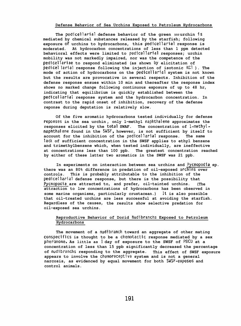
Defense Behavior of Sea Urchins Exposed to Petroleum Hydrocarbons
The pedicellarial defense behavior of the green sea urchin ismediated by chemical substances released by the starfish; followingexposure of urchins to hydrocarbons, this pedicellarial response ismoderated. At hydrocarbon concentrations of less than 1 ppm detectedbehavioral effects were limited to pedicellarial responses; urchinmobility was not markedly impaired, nor was the competence of thepedicellariae to respond eliminated (as shown by elicitation ofpedicel Iarial response following the injection of isotonic KCI ). Themode of action of hydrocarbons on the pedicellarial system is not knownbut the results are provocative in several respects. Inhibition of thedefense response ensues within 10 min and thereafter the response indexshows no marked change following continuous exposure of up to 48 hr,indicating that equilibrium is quickly established between thepedicellarial response system and the hydrocarbon concentration. Incontrast to the rapid onset of inhibition, recovery of the defensereponse during deputation is relatively slow.
Of the five aromatic hydrocarbons tested individually for defensereponses in the sea urchin , only l-methyl naphthalene approximates theresponses elicited by the total SWAF. The concentration of l-methyl-naphthalene found in the SldSF, however, is not sufficient by itself toaccount for the inhibition of the pedicellarial response. The samelack of sufficient concentration in the SWSF applies to ethyl benzeneand trimethylbenzene which, when tested individually, are ineffectiveat concentrations less than 100 ppb. The greatest concentration reachedby either of these latter two aromatics in the SWSF was 21 ppb.
In experiments on interaction between sea urchins and Pycnopodia sp.there was an 80% difference in predation of oil-exposed urchins overcontrols. This is probably attributable to the inhibition of thepedicellarial defense response, but there is the possibility thatPycnopodia are attracted to, and prefer, oil-tainted urchins. (Theattraction to low concentrations of hydrocarbons has been observed insome marine organisms, particularly crustacean.) It is also possiblethat oil-treated urchins are less successful at avoiding the starfish.Regardless of the causes, the results show selective predation foroil-exposed sea urchins.
Reproductive Behavior of Dorid Nudibranchs Exposed to PetroleumHydrocarbons
The movement of a nudibranch toward an aggregate of other matingconspecifics is thought to be a chemotactic response mediated by a sexpheranone. As little as 1 day of exposure to the SWSF of PBCU at aconcentration of less than 15 ppb significantly decreased the percentageof nudibranchs responding to the aggregate. This effect of SWSF exposureappears to involve the chemoreceptive system and is not a generalnarcosis, as evidenced by equal movement for both SWSF-exposed andcontrol animals.
191
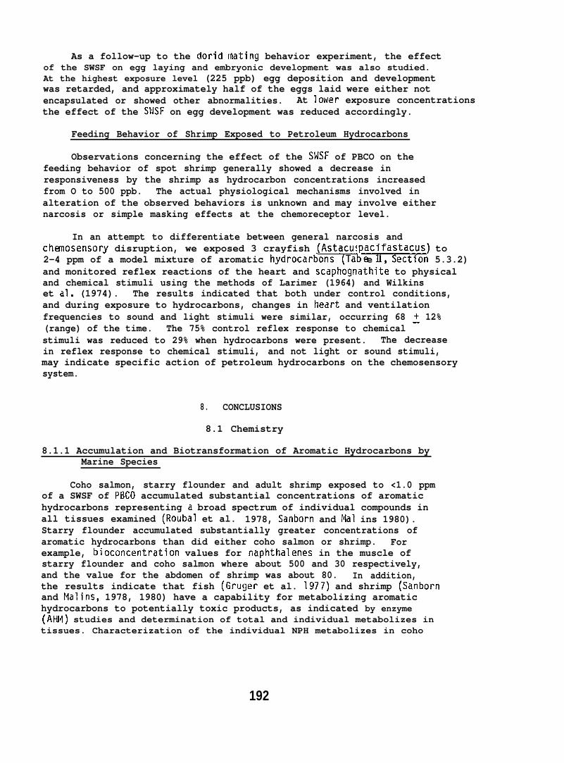
As a follow-up to the dorid [hating behavior experiment, the effectof the SWSF on egg laying and embryonic development was also studied.At the highest exposure level (225 ppb) egg deposition and developmentwas retarded, and approximately half of the eggs laid were either notencapsulated or showed other abnormalities. At lower exposure concentrationsthe effect of the SWSF on egg development was reduced accordingly.
Feeding Behavior of Shrimp Exposed to Petroleum Hydrocarbons
Observations concerning the effect of the SMSF of PBCO on thefeeding behavior of spot shrimp generally showed a decrease inresponsiveness by the shrimp as hydrocarbon concentrations increasedfrom O to 500 ppb. The actual physiological mechanisms involved inalteration of the observed behaviors is unknown and may involve eithernarcosis or simple masking effects at the chemoreceptor level.
In an attempt to differentiate between general narcosis andchanosensory disruption, we exposed 3 crayfish (Astacus acifastacus) to
~2-4 ppm of a model mixture of aromatic hydrocarb~b e 1, SectIon 5.3.2)and monitored reflex reactions of the heart and scaphognathite to physicaland chemical stimuli using the methods of Larimer (1964) and Wilkinset dl. (1974). The results indicated that both under control conditions,and during exposure to hydrocarbons, changes in heart and ventilationfrequencies to sound and light stimuli were similar, occurring 68 ~ 12%(range) of the time. The 75% control reflex response to chemicalstimuli was reduced to 29% when hydrocarbons were present. The decreasein reflex response to chemical stimuli, and not light or sound stimuli,may indicate specific action of petroleum hydrocarbons on the chemosensorysystem.
8. CONCLUSIONS
8.1 Chemistry
8.1.1 Accumulation and Biotransformation of Aromatic Hydrocarbons byMarine Species
Coho salmon, starry flounder and adult shrimp exposed to <1.0 ppmof a SWSF of P13C0 accumulated substantial concentrations of aromatichydrocarbons representing a broad spectrum of individual compounds inall tissues examined (Roubal et al. 1978, Sanborn and Mal ins 1980).Starry flounder accumulated substantially greater concentrations ofaromatic hydrocarbons than did either coho salmon or shrimp. Forexample, bioconcentration values for naphthalenes in the muscle ofstarry flounder and coho salmon where about 500 and 30 respectively,and the value for the abdomen of shrimp was about 80. In addition,the results indicate that fish (Gruger et al. 1977) and shrimp (Sanbornand Malins, 1978, 1980) have a capability for metabolizing aromatichydrocarbons to potentially toxic products, as indicated by enzyme(AHM) studies and determination of total and individual metabolizes intissues. Characterization of the individual NPH metabolizes in coho
192
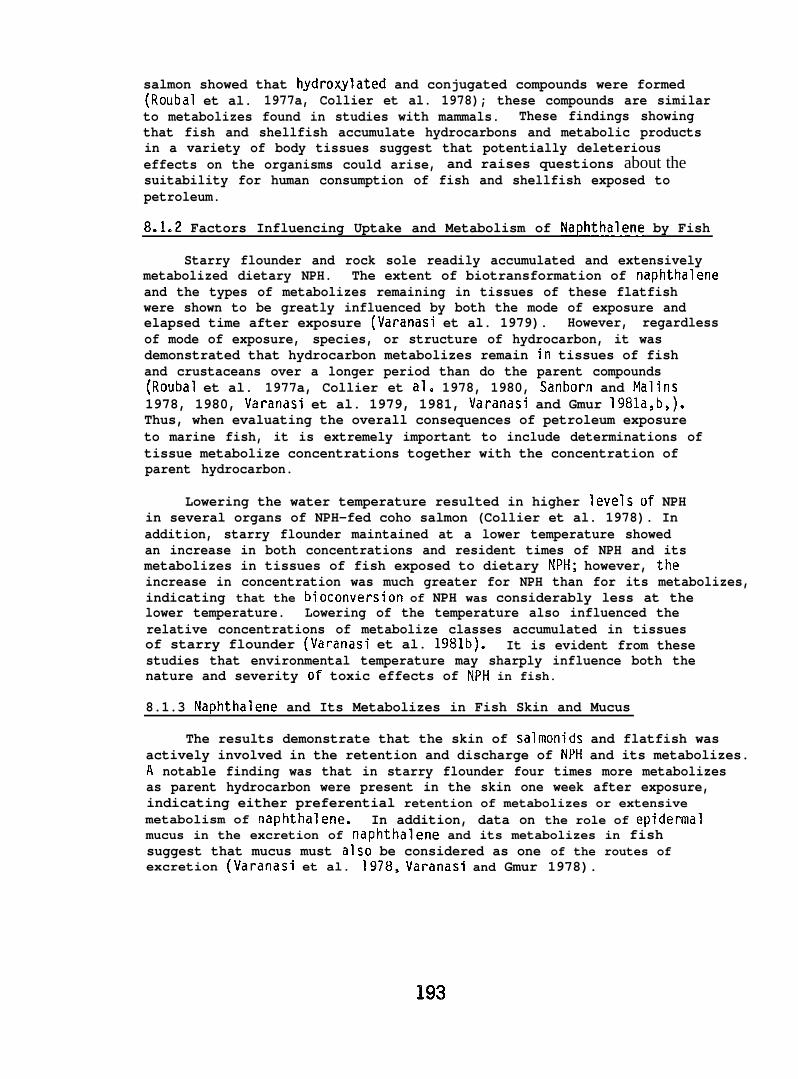
salmon showed that hydroxylated and conjugated compounds were formed(Roubal et al. 1977a, Collier et al. 1978); these compounds are similarto metabolizes found in studies with mammals. These findings showingthat fish and shellfish accumulate hydrocarbons and metabolic productsin a variety of body tissues suggest that potentially deleteriouseffects on the organisms could arise, and raises questions about thesuitability for human consumption of fish and shellfish exposed topetroleum.
8.1.2 Factors Influencing Uptake and Metabolism of Naphthalene by Fish
Starry flounder and rock sole readily accumulated and extensivelymetabolized dietary NPH. The extent of biotransformation of naphthaleneand the types of metabolizes remaining in tissues of these flatfishwere shown to be greatly influenced by both the mode of exposure andelapsed time after exposure (Varanasi et al. 1979). However, regardlessof mode of exposure, species, or structure of hydrocarbon, it wasdemonstrated that hydrocarbon metabolizes remain in tissues of fishand crustaceans over a longer period than do the parent compounds(Roubal et al. 1977a, Collier et al. 1978, 1980, Sanborn and Malins1978, 1980, Varanasi et al. 1979, 1981, Varanasi and Gmur 1981a,b,).Thus, when evaluating the overall consequences of petroleum exposureto marine fish, it is extremely important to include determinations oftissue metabolize concentrations together with the concentration ofparent hydrocarbon.
Lowering the water temperature resulted in higher levels of NPHin several organs of NPH-fed coho salmon (Collier et al. 1978). Inaddition, starry flounder maintained at a lower temperature showedan increase in both concentrations and resident times of NPH and itsmetabolizes in tissues of fish exposed to dietary NPH; however, theincrease in concentration was much greater for NPH than for its metabolizes,indicating that the bioconversion of NPH was considerably less at thelower temperature. Lowering of the temperature also influenced therelative concentrations of metabolize classes accumulated in tissuesof starry flounder (Varanasi et al. 1981b). It is evident from thesestudies that environmental temperature may sharply influence both thenature and severity of toxic effects of NPti in fish.
8.1.3 Naphthalene and Its Metabolizes in Fish Skin and Mucus
The results demonstrate that the skin of salmonids and flatfish wasactively involved in the retention and discharge of NPH and its metabolizes.A notable finding was that in starry flounder four times more metabolizesas parent hydrocarbon were present in the skin one week after exposure,indicating either preferential retention of metabolizes or extensivemetabolism of naphthalene. In addition, data on the role of epidermalmucus in the excretion of naphthalene and its metabolizes in fishsuggest that mucus must also be considered as one of the routes ofexcretion (Varanasi et al. 1978, Varanasi and Gmur 1978).
193
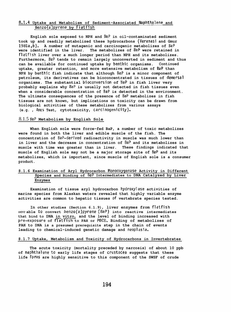
8.1.4 Uptake and Metabolism of Sediment-Associated Naphthalene andBenzo(a)pyrene by Flatfish
English sole exposed to NPH and BaP in oil-contaminated sedimenttook up and readily metabolized these hydrocarbons (Varanasi and Gmur1981a,b). A number of mutagenic and carcinogenic metabolizes of BaPwere identified in the liver. The metabolizes of BaP were retained inflatfish liver over a much longer period than NPH and its metabolizes.Furthermore, BaP tends to remain largely unconverted in sediment and thuscan be available for continued uptake by benthic organisms. Continueduptake, greater retention, and more extensive metabolism of BaP thanNPH by benthic fish indicate that although BaP is a minor component ofpetroleum, its derivatives can be bioconcentrated in tissues of demersalorganisms. The substantial bioconversion of BaP in fish liver veryprobably explains why BaP is usually not detected in fish tissues evenwhen a considerable concentration of BaP is detected in the environment.The ultimate consequences of the presence of BaP metabolizes in fishtissues are not known, but implications on toxicity can be drawn frombiological activities of these metabolizes from various assays(e.g. , I%nes Test, cytotoxicity, carcinogenicity).
8.1.5 BaP Metabolism by English Sole
When English sole were force-fed BaP, a number of toxic metabolizeswere found in both the liver and edible muscle of the fish. Theconcentration of BaP-derived radioactivity in muscle was much lower thanin liver and the decrease in concentration of BaP and its metabolizes inmuscle with time was greater than in liver. These findings indicated thatmuscle of English sole may not be a major storage site of BaP and itsmetabolizes, which is important, since muscle of English sole is a consumerproduct.
8.1.6 Examination of Aryl Hydrocarbon Monooxygenase Activity in DifferentSpecies and Binding of BaP Intermediates to DNA Catalyzed by LiverEnzymes
Examination of tissue aryl hydrocarbon hydroxylase activities ofmarine species from Alaskan waters revealed that highly variable enzymeactivities are common to hepatic tissues of vertebrate species tested.
In other studies (Section 6.1.9), liver enzymes from flatfishwere able to convert benzo(a)pyrene (t3aP) into reactive intermediatesthat bind to DNA in vitro, and the level of binding increased withpre-exposure of f~tfish to PAH or PBCO. Binding of metabolizes ofPAH to DNA is a presumed prerequisite step in the chain of eventsleading to chemical-induced genetic damage and neoplasia.
8.1.7 Uptake, Metabolism and Toxicity of Hydrocarbons in Invertebrates
The acute toxicity (mortality preceded by narcosis) of about 10 ppbof naphthalene to early life stages of crustacea suggests that theselife form are highly sensitive to this component of the SWSF of crude
oil. The tendency of early developmental stages of shrimp to metabolizeand retain NPH metabolizes at unchanged concentrations while concentrationsof the parent hydrocarbon decline is of concern because evidence linksarcmatic hydrocarbon metabolizes to toxicity in mammals, The highsusceptibility of larval and other developmental stages to aromatichydrocarbons must be considered an important factor in the environmentaleffect of arctic and subarctic petroleum operations.
8.1.8 Food Chain Transfer of 2,6-DMN from Algae to Sea Urchins
Sea urchins feeding on 2,6-dimethylnaphthalene-exposed algaeaccumulated and metabolized this hydrocarbon to hydroxy compounds andtheir conjugates, thus demonstrating a food chain transfer of an aromatichydrocarbon (Malins and Roubal 1982).
8.1.9 Fate of Metals in Fish
Salmon and starry flounder readily accumulate lead and cadmiumfrom seawater (Reichert et al. 1979). The metals in liver and kidneywere stored to a substantial degree in cellular fluids where biochemicaland physiological processes occur (e.g., cytosol). Cadmium waspreferentially bound by low molecular weight proteins (9,000 daltons),but was also associated with high molecular weight proteins of theCytosol. Lead showed a strong preference for neural tissue (e.g.,brain). The findings imply that low concentrations of metals enteringmarine waters are likely to increase the metal burden of key tissuesof fish and thereby possibly alter normal physiological processes.
Epidermal mucus of coho salmon is involved to some extent inexcretion of lead and cadmium (Varanasi and Markey 1978). The importanceof this route in relation to other excretory tissues (e.g., kidney),however, remains to be assessed. Exposure to sublethal levels of leadand cadmium for periods of up to two weeks resulted in increasedproduction of epidermal mucus in coho salmon. This increased rate ofproduction may induce alterations in physiochemical and theologicalproperties of mucus.
Our results also show that skin and scales act as storage andperhaps detoxification sites for metals in both salmonids and flatfish.Substantial amounts of metals persisted in theweeks after fish were returned to clean water.high concentrations of toxic metals would havestructure remains to be seen.
8.2 Pathology
skin and scales severalMhether persistence ofadverse effects on skin
8.2.1 Effects of Petroleum on Disease Resistance
Laboratory studies evaluating the effects of petroleum hydrocarbonson immunocompetence and disease resistance of selected fish and shellfish
195
failed to demonstrate a marked impairment. Preliminary assays did,however, suggest petroleum dispersants may enchance the incidence ofinfectious disease in salmon.
8.2.2 Pathological Changes in Flatfish from Exposure to Oil-ContaminatedSediment
Experimental exposures of 3 species of flatfish (English sole, rocksole, and starry flounder) to PBCO-contaminated sediments indicatesthat English sole are the most likely to be adversely affected. Althoughthe pathological changes observed appeared to be reversible and notdirectly life-threatening, they do reflect alterations that may reducefitness, and thereby survival, of flatfish in heavily oiled environments.
8.2.3 Cytopathology
Cytopathological changes occurred in flatfish and smelt embryosexposed throughout embryogenesis to low ppb concentrations of the SWAFof crude oil. The olfactory cilia of flatfish developed abnormally andextensive necrosis was evident in the eye and brain of smelt embryos.Structural changes were also observed in liver and lens tissues ofadult rainbow trout perorally exposed to high levels of petroleumhydrocarbons for 2 to 12 mo.
8.3 Behavior and Physiology
8.3.1 Vertebrate Studies
Behavior of Pacific Salmon Exposed to Petroleum Hydrocarbons
Mature Pacific salmon migrating upstream during the peak of therun substantially avoided a mixture of monocyclic aromatic hydrocarbonsin the water at concentrations of 3.2 ppm (EC50) and higher. Also,electrophysiological recordings from the olfactory bulb of juvenilesalmon indicated that short-term exposure to these high concentrationsof aromatic hydrocarbons would not be likely to disrupt the olfactorySystan.
Mark-recapture experiments were conducted to assess the ability ofadult chinook and coho salmon to repeat their spawning migrationfollowing exposure to petroleum hydrocarbons. Chinook salmon exposedfor 14 to 18 hr to FMAF of PBCO showed no alteration of homing capability.Similarly, the spawning migration of coho salmon was not disrupted by8 to 22 hr exposure to a mixture of monocyclic aromatic hydrocarbons.However, the return of the coho to their captive site was delayed by3 days.
Effects of Petroleum Hydrocarbons on Predator-Prey Behaviorof Salmon
Exposure of chum salmon fry to the SbJSF of CICO at a hydrocarbonconcentration of 350 ppb for periods of 24 to 72 hr resulted in a
196
statistically significant greater consumption of the oil-exposed preyby coho predators compared to the consumption of controls.
Similarly, exposure of coho salmon predators to 340 ppb of theSWSF of CICO for over 3 days resulted in a reduction in numbers ofsalmonid fry eaten. Concentrations of parent hydrocarbons were higherin both liver and brain of actively eating oil-exposed predators comparedto those not eating. This suggests that the parent hydrocarbons werenot the compounds primarily influencing a decline in feeding behavior.
Avoidance Behavior of Flatfish Exposed to Oil-Contaminated Sediment
Although there is evidence of initial avoidance of oil-contaminantsediment by juvenile English sole, generally the fish did not avoidoil-sediment mixtures at concentrations up to 10,000 ppm - the sediment’sapparent oil-carrying capacity. At these concentrations, some mortalitydid occur over a 15-day period, but the majority of flatfish were activeand fed readily.
Effect of Petroleum Hydrocarbons on Development of Embryos and Larvae
Embryos of chum salmon, English sole, and sand sole exposed towater-borne hydrocarbon concentrations ranging from 100 to 500 ppbshowed either high embryonic mortality, abnormal development, or delayedlarval mortality. In addition, hydrocarbon concentrations of lessthan 100 ppb severely reduced the larval survival of surf smelt.
Effect of Crude Oil Ingestion on Salmonid Reproductive Success
Long-term peroral exposure of maturing male and female rainbowtrout to high levels of PBCO had no marked effects on reproduction,as measured by hatching success and alevin survival.
8.3.2 Invertebrate Studies
Defense Behavior of Sea Urchins Exposed to Petroleum Hydrocarbons
Results of studies on the sea urchins’ defense behavior followingexposure to the SWSF of PBC(I and its major aromatic components indicatethat no single principal component of the SWF is responsible for thetotal inhibition observed by exposure to the SWSF alone; that at lowppb concentrations the pedicellarial defense inhibition resulting fromhydrocarbon exposure is apparently a chemosensory specific reaction andnot general narcosis; and that SWSF-exposed sea urchins are preyed uponby Pycnopodia to a greater extent that non-exposed urchins.
Reproductive Behavior of Dorid Nudibranchs Exposed to PetroleumHydrocarbons
Interference with the reproductive processes of dorid nudibranchsby the SWSF of PBCO occurred at several different levels. First, therewas disruption of the chemotatic responses necessary to form mating
197
aggregations. Second, there was disruption of the egg laying process asnoted by the lack of egg encapsulation, and delay in egg deposition.Third, eggs exposed to the SWSF showed retarded development and increasesin abnormalities. Disruptive effects on the reproductive biology ofdorid nudibranchs occurred at hydrocarbon concentrations as low as15 ppb.
Feeding Behavior of Shrimp Exposed to Petroleum Hydrocarbons
Exposure of spot shrimp to the SWSF of PBCO resulted in a reductionin feeding activity. At 25 ppb there was a 50% reduction in overtbehavioral “activity elicited in response to food stimuli, and at higherconcentrations of the SWSF there was a still further decline in feedingresponses, with some loss of equilibrium and mortality occurring at500 ppb.
198
9. PAPERS PUBLISHED, DISSERTATIONS, THESES, AND PRESENTATIONSGIVEN UNDER RU73 OCSEAP SPONSORSHIP
9.1 Papers Published
Collier, T.K., L.C. Thomas, and D.C. Malins (1978). Influence ofenvironmental temperature on disposition of dietary naphthalene incoho salmon (Oncorhynchus kisutch): Isolation and identificationof individual metabolltes. Comp. Biochem. Physiol. 61C(1 ),23-28.—
Collier, T.K., D.W. Brown and D.C. Malins (1979). Naphthalene metabolizesin brain and other organs of rainbow trout (Salmo gairdneri). Fed.Proc. 38(3),658.—
Collier, T.K., M.M. Krahn and D.C. Malins (1980). The disposition ofnaphthalene and its metabolizes in the brain of rainbow trout (Salmogairdneri). Environ. Res. 23,35-41.—
Folmar, L.C., D.R. Craddock, J.W. Blackwell, G. Joyce and H.O. Hodgins(1981). Effects of petroleum exposure on predatory behavior ofcoho salmon (Oncorhynchus kisutch). Bull. Environm. Contain. Toxicol.27,458-462.—
Gmur, D.J. and U, Varanasi (1982). Characterization of benzo(a)pyrenemetabolizes isolated from muscle, liver, and bile of a pleuronectidfish. Carci nogenesis (submitted).
Hawkes, J.W. (1980). The effects of xenobiotics on fish tissues:Morphological studies. Fed. Proc. 39,3230-3236..
Hawkes, J.W. (1977). The effects of petroleum-hydrocarbon exposure onthe structure of fish tissues. In: Fate and Effects of PetroleumHydrocarbons in Marine Ecosystems and Organisms, (Edited by D.A.Wolfe). Pergamon Press, New York. pp. 115-128.
Hawkes, J.W. and C.M. Stehr (1982). Cytopathology of the brain and retinaof embryonic surf smelt (Hypomesus pretiosus) exposed to crude oil.Environ. Res. 27,164-178.—
Harbell, S.C., H.O. Hodgins, and M.H. Schiewe (1979). Studies on thepathogenesis of vibriosis in coho salmon Oncorhynchus kisutch(Walbaum). J. Fish Dis. ~,391-404.
Hodgins, H.O., W.D. Gronlund, J.L. Mighell, J.W. Hawkes and P.A. Robisch(1977). The effect of crude oil on trout reproduction. In: Fateand Effects of Petroleum Hydrocarbons in Marine Ecosystem~andTlrganlsms, Ldited by D.A. Wolfe). Permagon press, New York, pp143-150.
199
Malins, D.C. (1977). Metabolism of aromatic hydrocarbons in marineorganisms. Ann. N.Y. Acad. Sci. 298,482-96.
Malins, D.C., T.K. Collier, L.C. Thomas, andW.T. Roubal (1979). Metabolicfate of aromatic hydrocarbons in aquatic organisms: Analysis ofmetabolizes by thin-layer chromatography and high-pressure liquidchromatography. Intl. J. Environ. Anal. Chem. ~,55-66.
Malins, D.C., M.M. Krahn, D.W. Brown, W.D. MacLeod, Jr., and T.K. Collier(1980). The analysis of petroleum products in marine environments.Helgol. Wiss. Meeresunters. 33,257-271.—
Malins, D.C., P. Fraser and W.T. Roubal (1980). Formation of a sulfatemetabolize of 2,6-dimethyl naphthalene in the sea urchin(Strongylocentrotus droebachiensis). Fed. Proc. 39,1753.—
Malins, D.C. and H.O. Hodgins (1981). Petroleum and marine fishes:A review of u~take. deposition. and effects. Environ. Sci. Tech.&1272-1280.’ - “
Malins, D.C. and H.O. Hodgins (1981). Petroleum and marine fishes: Areview of uptake, deposition, and effects. Environ. Sci. Tech.15: 1272-1280.
Malins, D.C. (1982). Alterations in cellular and subcellular structureof marine teleosts and invertebrates exposed to petroleum in thelaboratory and field: A critical review. Can. J. Fish. Aquat.Sci. 39. (In press)—
Malins, D.C. and H.O. Hodgins (1982). Correspondence - Response to:“Critique of ‘Petroleum and marine fishes: A review of uptake,deposition, and effects’ by Jerry F. Payne”. Environm. Sci. Tech.16,372-373.—
Malins, D.C. and W.T. Roubal (1982). Aryl sulfate formation in seaurchins (Strongylocentrous droebach~ensis) ingesting marine algae(Fucus distichus) containing 2,6-dimethylnaphth~lene. Environ.Res. 2/.290-297.
Haynard, D.J. and D.D. Weber (1981). Avoidance reactions of juvenilecoho salmon (Oncorhynchus kisutch) to monocyclic aromatics. Can.J. Fish. Aquat. Sci. 38,772-778.—
McCain, B.B., H.O. Hodgins, W.D. Gronlund, J.W. Hawkes, D.W. Brown,M.S. Myers, and J.H. Vandermeulen (1978). Bioavailability ofcrude oil from ex~erimentally oiled sediments to Enqlish sole(Parophyrs vetulus), and pathological consequences.- J. Fish. Res.Bd. Can. 35,657-664.—
McCain, B.B. and D.C. Malins (1982). Effects of petroleum hydrocarbonson selected demersal fish and crustaceans. In: New York Bight.Science and Management. (Edited by G. Mayer). Published by NewYork Bight MESA (In press).
200
Reichert, W.L., D.A. Federighi, “and D.C. Malins (1979). Uptake andmetabolism of lead and cadmium in coho salmon (Oncorhynchus kisutch).Comp. Biochem. Physiol. ~C,229-234.
Roubal, W.T., T.K. Collier, and D.C. Malins (1976). Metabolism of aromatichydrocarbons in coho salmon. Fed. Proc. 35(7),1785.—
Roubal, W.T., Collier, T.K., and Malins, D.C. (1977a). Accumulation andmetabolism of carbon-14 label led benzene, naphthalene, and anthraceneby young coho salmon (Oncorhynchus kisutch). Arch. Environ. Contain.Toxicol. 5,513-529.
Roubal, W.T,, D. Bovee, T.K. Collier, and S.1. Stranahan (1977b). Flow-through system for chronic exposure of aquatic organisms to seawater-soluble hydrocarbon from crude oil: Construction and applications.In: Proceedings of 1977 Oil Spill Conference (Prevention, Behavior,~ntrol, Cleanup), American Petroleum Institute, Washington, D.C.pp. 551-555.
Roubal, W.T., S.1. Stranahan, and D.C. Malins (1978). The accumulationof low molecular weight aromatic hydrocarbons of crude oil by cohosalmon (Oncorhynchus kisutch) and starry flounder (Platichthysstellatus). Arch. Environ. Contain. Toxicol. ~,237-244.
Sanborn, H.R. and D.C. Malins (1977). Toxicity and metabolism ofnaphthalene: A study with marine larval invertebrates. Proc, Sot.Exper. Biol. Med. 154,151-155.
Sanborn, H.R. and D.C. Malins (1980). The disposition of aromatichydrocarbons in adult spot shrimp (Pandalus platyceros) and theformation of metabolizes of naphthalene in adult and larval spotshrimp. Xenobiotica. ~,193-200.
Schiewe, M.H., J.H. Cross, and E.J. Ordall (1977). Deoxyribonucleicacid relationships among marine vibrios pathogenic to fish. Can. J.Microbiol. 23,954-958.—
Varanasi, U. and D. Markey (1977). Effect of calcium on retention oflead in fish skin. Fed. Proc. 36.722.
Varanasi, u. (1978). Sublethal effects of petroleum hydrocarbons andtrace metals: biological fate of metals in fish. In: MarineBiological Effects OF OCS Petroleum Development (Ed~ed~. Wolfe)NOAA Tech. Memo. ERL OC SEAP-1, pp. 41-53
Varanasi, U. and D.J. Gmur (1978). Influence of environmentaluptake and retention of lead in coho salmon (OncorhynchusToxicol. Appl. Pharmacol. 46,65-75.—
Varanasi, U. and D. Markey (1978). Uptake and release of leadin skin and mucus of coho salmon (Oncorhynchus kisutch).Physiol. QC,187-191.
clacium onkisutch).
and cadmiumComp. Biochem.
201
Varanasi, U., M. Uhler, and S.1. Stranahannaphthalene and its metabolizes in sk”Toxicol. Appl. Pharmacol. 44,277-285.—
Varanasi, U., D.J. Gmur, and P.A. Treseler
(1978). Uptake and release ofn and epidermal mucus of salmonids.
(1979). Influence of timeand mode of exposure on biotransformation of naphthalene by juvenilestarry flounder (Platichthys stellatus) and rock sole (Lepidopsettabilineata), Arch. Environ. Contain. Toxicol. 3,673-692.
Varanasi, U. (1980). Influence of temperature on hydrocarbon metabolismin flatfish. Research update. Coastal Oceanography and ClimatologyNews. ~,17-18.
Varanasi, U. and D.J. Gmur (1980). Metabolic activation and covalentbinding of benzo[a]pyrene to deoxyribonucleic acid catalyzed by liverenzymes of marine fish. Biochem. Pharmacol. 29,753-761.—
Varanasi, U., D.J. Gmur, and M.M. Krahn (1980). Metabolism and subsequentbinding of benzo[a]pyrene to ONA in pleuronectid and salmonid fish.
: Polynuclear aromatic hydrocarbons: Chemistry and biological%fects (Edited by A. Bjorseth and A.J. Dennis), Battelle Press,Columbus, Ohio. pp. 455-470.
Varanasi, U.. and D.J. Gmur (1981). Hydrocarbons and metabolizes inEnglish sole (ParopljCys vetulus) exposed simultaneously to 3H-benzo[a]pyrene and 4C-n-ene in oil contaminated sediment.Aquat. Toxicol. 1,49-67.
Varanasi, U. and D.J. Gmur (1981). In vivo metabolism of naphthalene andbenzo[a]pyrene by flatfish. In7C~cal analysis and biologicalfate: Polynuclear Aromatic Hydrocarbons. Proc. Fifth Internatl.Synlp . (Edited by A.J. Dennis and M. Cooke), Battelle Press, Columbus,~. pp. 367-376.
Varanasi, U., D.J. Gmur, and W.L. Reichert (1981). Effect of environmentaltemperature on naphthalene metabolism of juvenile starry flounder(Platichthys stell atus). Arch. Environ. Contain. Toxicol. 10$203-214.—
Weber, D.D., O.J. Maynard, W.D. Gronlund, and V. Konchin (1981). Avoidancereactions of migrating adult salmon to petroleum hydrocarbons. Can.J. Fish. Aquat. Sci. 38,799-781.—
202
9.2 Dissertations and Theses
Collier, T.K. (1978). I)isposition and metabolism of naphthalene inrainbow trout (Salmo gairdneri). M.S. Thesis, University ofWashington, Sea~Mash. 43 pp.
Johnson, F.G. (1979). The effects of aromatic petroleum hydrocarbonson chemosensory behavior of the sea urchin’, Strongylocentrotusdroebachiensis and the nudibranch, Ortchidoris bilamellata.Ph.D. dissertation, University of Washington, Seattle, Wash.110 pp.
tlaynard, D.J. (1980). Behavioral and sensory aspects of petrochemicalpollution on Pacific salmon. M.S. Thesis, University of Washington,Seattle, Mash. 175 pp.
Miller, S.E. (1980). Feeding behavior Of marine decapod crustaceans,and observations of behavioral responses of the spot shrimp,Pandalus platyceros (Brandt) to food stimulants and petroleum~ydrocarbons. ?I..S. Thesis, University of Washington, Seattle,Wash. 169 pp.
Mumaw, L.M. (1978). Effects of a saltwater soluble fraction of PrudhoeBay crude oil on the embryonic development of the nudibranch,Onchidoris bilamellata. ~4.S. Thesis,” University of Washington,Seattle, Nash. 63 pp.
Schiewe, M.H. (1980). Studies on the pathogenesis of vibriosis insalmonid fish. Ph.D. dissertation, University of Washington,Seattle, Wash. 158pp.
203
9.3 Presentations
Clark, R.C., Jr. and D.C. Malins (1980). Presented written testimonyand gave cross examination testimony on the biological effects ofpetroleum on marine organisms and on laboratory studies of theimpact of petroleum on marine organisms, respectively, at Boston,Massachusetts, in adjudicatory hearings (30 January to 1 February1980) on a proposed refinery site at Eastport, Maine, involvingthe Pittston Company.
Clark, R.C., Jr. (1980). Occurrence and impact of petroleum on arcticenvironments. Invited review paper at the international conferenceon: The Arctic Ocean; The Hydrographic Environment and Fate ofPollutants. London, England, on 11-12 March 19f10 under the auspicesof the Comite’ Arctique of the Centre Scientifique de Monaco andthe Royal Geographical Society.
Collier, T.K. (1978). Disposition and metabolism of dietary naphthalenein coho salmon (Oncorhynchus kisutch). Presented at 62nd AnnualMeeting of FASEB, Atlantic City, N.J. April 1978.
Collier, T,K. (1978), Occurrence of naphthalene metabolizes in brainof rainbow trout (Salmo gairdneri). Presented at 33rd NorthwestMeeting of the Amem Chemlcal Society. Seattle, MA June, 1978.
Collier, T.K. (1978). Disposition and metabolism of naphthalene inrainbow trout (Salmo gairdneri). Seminar presented at Universityof Washington, College of Fisheries, Seattle, WA. June, 1978.
Collier, T.K. (1979). Naphthalene metabolizes in brain and otherorgans of rainbow trout (Salmo gairdneri). Presented at 63rdAnnual Meeting of FASEB, Dallas, Texas, April 1979.
Foster, C.A. and J.W. Hawkes (1979). A histopathologic study of mussels(Mytilus edulis) exposed to petroleum from the AMOCO CADIZ Spill.Presented June 1979, National Shellfisheries Society, Victoria, B.C.
Gmur, D.J. (1979). Influence of environmental temperature on metabolismof dietary naphthalene in starry flounder (Platichthys stellatus).Presented at the 63rd Annual Meeting of FASEB, Dallas, Texas,.April 1979.
Hawkes, J.W. (1976). Effects of petroleum hydrocarbon exposure onthe structure of fish tissue. Plenary session speaker: Symposiumon Fate and Effects of Petroleum Hydrocarbons in Marine Ecosystemsand Organisms, November 10-12, 1976, Seattle, Washington.
Hawkes, J.W. (1977). The morphological effects of petroleum on fishtissues. Cordova Fisheries Institute, Cordova, Alaska, April 1977;also presented at a joint seminar: University of Alaska and AukeBay, NMFS Laboratory, Juneau, Alaska.
Hawkes, J.W. (1977). The morphological effects of petroleum andchlorobiphenyls on fish tissues. Poster session: Symposium onEnvironmental Pollutants, National Academy of Sciencies, June 1977,Storrs, Connecticut.
Hawkes, J.W. (1977). The effects of petroleum on fish tissues. Seminarspeaker: Washington State University, November 1977, Pullman,Washington.
Hawkes, J.W. (1977). Morphology. OCSEAP Program Review, Nov. 29-Dec. 2, 1977, Seattle, Washington.
Hawkes, J.W. (1977). Histopathology Panel for Oil Spill Workshop,Environmental Protection Agency, Mitre Corp.; Panel chairperson.November 1977, Seattle, Washington.
Hawkes, J.W. (1979). The effects of xenobiotics on fish tissues:Morphological studies. Lecture given at Dept. of Microbiology,Cell Biology, Biophysics, and Biochemistry, Pennsylvania StateUniversity, November 9, 1979.
Hawkes, J.W. (1979). The effects of xenobiotics on fish tissues:Ultrastructure studies. Symposium: Aquatic Animals as Models inBiomedical Research for the 1979 meeting of the American Societyfor Pharmacology and Experimental Therapeutics, Portland, Oregon,August 20-23, 1979.
Hodgins, H.O. (W.D. Gronlund, J.L. Mighell, J.W. Hawkes, and P.A. Robisch)(1976). Effect of crude oil on trout reproduction. Symposium onFate and Effects of Petroleum Hydrocarbons in Marine Ecosystems andOrganisms, November 10-12, 1976, Seattle, Washington.
Hodg’
Hodg”
ns, H.O. (1977). Physiological and behavioral effects. OCSEAPProgram Review, Nov. 29-Dee. 2,1977, Seattle, Washington.
ns, H.O. (1977). Morphological, behavioral, physiological , andpathological studies of effects of petroleum on fish. Workshop onThe Effects of Environmental Stress on Fish Health, sponsored bythe National Fisheries Research Center (U.S. Fish and WildlifeService), November 1-3, 1977, Seattle, Washington.
Hodgins, H.O. (1978). Current research on the effects of petroleum onmarine organisms at the Northwest and Alaska Fisheries Center.The Second Annual Department of Energy (DOE) West Coast MarineScience Contractors Program Review/Information Exchange Meeting,April 11-12, 1978, La Jolla, Calif.
Hodgins, H.O. (1980). OCSEAP Program Review, Anchorage, Alaska,March 1980.

Hodgins, H.O. (1980). Effects of crude oil. Presented at the Fateand Effects of Petroleum Program Review, Boulder, Colorado,September 21-23, 1980.
MacLeod, W.D., Jr. (1980). The nature and extent of petroleum pollution.Presented at the Fate and Effects of Petroleum Program Review,Boulder, Colorado, September 21-23, 1980.
Malins, D.C. (1975). Recent studies on the metabolism, fate, andeffects of petroleum hydrocarbons in marine organisms. Symposiumon Biological Effects from Oil in the Environment, March 1975,Tiburon, California.
Malins, D.C. (1976). Metabolism of aromatic hydrocarbons in coho salmon.Presented at 60th Annual FASEB Meeting. San Francisco, California.June, 1976.
14alins, D.C. (1976). The metabolic fate of aromatic hydrocarbons inmarine organism. Symposium on Aquatic Pollutants and BiologicalEffects with Emphasis on Neoplasia, September 1976, New YorkAcademy of Sciences, New York.
Malins, D.C. (1976). Biotransformation of petroleum hydrocarbons inmarine organisms indigenous to the Arctic and Subarctic. Symposiumon Fate and Effects of Petroleum Hydrocarbons in Marine Ecosystemsand Organisms, November 10-12, 1976, Seattle, Washington.
Malins, D.C. (1977). Multidisciplinary studies on the biological effectsof petroleum on Arctic and Subarctic marine environments. Semi na rat Terry Research Station, Aberdeen, Scotland, September 1977.
Malins, D.C. (1977). Biological fate and effects of petroleum on marineorganisms. Seminar at Institute of Marine Research, Bergen, Norway,October 1977.
Malins, D.C. (1977). Disposition and metabolism of petroleumhydrocarbons. OCSEAP Program Review, Nov. 29-Dee. 2, 1977,Seattle, Washington.
Malins, D.C. (1978). Metabolic fate of hydrocarbons in marine fish:Evidence for rnetabolite formation. Presented at the 8th AnnualSymposium of the Analytical Chemistry of Pollutants, Geneva,Switzerland, April 1978.
Malins, D.C. (1978). Disposition of aromatic hydrocarbons in marineorganisms. Presented at the American Chemical Society meeting,Miami, Florida, September 1978,
Malins, D.C. (1978). Hydrocarbon pollution in marine environments:A biochemical and toxicological dilemma. Presented to CanadianInstitute of Chemists, Victoria, B.C., December 1978.
Malins, D.C. (1979). Hydrocarbon pollution in marine environments:A biochemical and toxicological dilemma. Seminar given at theUniversity of California, Davis, February 1979.
Malins, D.C. (1979). The analysis of petroleum products in marineenvironments. Presented at the 14th European Marine BiologySymposium, Island of Helgoland, Germany, September 1979.
Malins, D.C. (198(I). Toxicity of oil in marine habitats. Presentedat the Norton Sound Synthesis Meeting, Anchorage, Alaska,October 27-28, 1980.
Malins, D.C. (1980). Overview of NWAFC/NhlFS Oil Pollution Research.Presented at the Fate and Effects of Petroleum Program Review,Boulder, Colorado, September 21-23, 1980.
McCain, B.B. (H.O. Hodgins, W.O. Gronlund, J.W. Hawkes, D.W. Brownand M.S. Myers) (1977). Effects of long-term exposure of flatfishto sediment contaminated with Alaskan crude oil. Symposium onRecovery Potential of Oiled Marine Northern Environments,October 10-14, 1977, Halifax, Nova Scotia, Canada.
McCain, B.B. (1980). OCSEAP Program Review, Anchorage, Alaska,March 1980.
Roubal, W.T. (1977). Flow-through system for chronic exposure ofaquatic organisms to seawater-soluble hydrocarbons from crude oil:Construction and applications. 1977 Oil Spill Conference(Prevention, Behavior, Control, Cleanup), American PetroleumInstitute. New Orleans, March 1977,
Stehr, C. (1978). A comparative study of the chorion and associatedpore structure of starry flounder, Platichthys stellatus and pinksalmon, Oncorhynchus gorbuscha eggs. Presented at the 29th AlaskaScience Conference, AAAS, Fairbanks, Alaska, August 1978.
Varanasi, U. (1976). Biophysical alterations in salmonid epidermalmucus induced by metal exposure. Northwest Meeting of the Societyfor Experimental Biology and Iledicine, October 1976, Portland,Oregon.
Varanasi, U. (and D. Markey) (1976). Bioaccumulations of trace metalsin the epidermal mucus and skin of marine fish. Symposium on theBiological Aspects of Inorganic Chemistry, University of BritishColumbia, Vancouver, B.C., Canada, June 1976.
Varanasi, U. (and D. Markey) (1977). Effect of calcium on retention oflead in skin. 61st Annual Meeting of Federation of AmericanSocieties for Experimental Biology, April 1-6, 1977, Chicago, Illinois.
Varanasi, u. (1977). 6iological fate of metals in fish. OCSEAP ProgramReview, Nov. 29-Dee. 2, 1977, Seattle, Washington.
207
Varartasi , U. (1978). Metabolism and disposition of naphthalene instarry flounder (Platichthys stellatus). Presented at the 62ndAnnual Meeting of FASE13, Atlantic City, N.J., April 1978.
Varanasi, U. (1978). Naphthalene metabolism in Pleuronectidae:Characterization of individual metabolizes in the bile. Presentedat the 69th Annual Meeting of American Society of BiologicalChemists, Atlanta, Georgia, June 1978.
Varanasi, U. (1978). Xenobiotic metabolism in marine organisms. Seminargiven at Skidaway Institute of Oceanography, Savannah, Georgia,June 1978.
Varanasi, U. (1978). Influence of environmental conditions on metabolismand disposition of aromatic hydrocarbons in marine organisms.Presented at 33rd American Chemical Society’s Northwest RegionalMeeting, Seattle, Washington, June 1978.
Varanasi, U. (1979). Binding of metabolically activated benzo[a]pyreneto DNA catalyzed by starry flounder (Platichthys stellatus) liverextract. Presented at 63rd Annual Meeting of FASE13, Dallas, Texas,April 1979.
Varanasi, U. (1979). Metabolism and subsequent binding of benzo[a]pyreneto DNA catalyzed by liver extracts of marine fish. The FourthInternational Symposium on Polynuclear Aromatic Hydrocarbons,Battelle Memorial Institute, Columbus, Ohio, October 1979.
Varanasi, U. (1980). OCSEAP Program Review, Anchorage, Alaska,March 1980.
Varanasi, U. (1980). Effects of petroleum components. Presented atthe Fate and Effects of Petroleum Program Review, Boulder, Colorado,September 21-23, 1980.
Varanasi, u. (1980). Metabolism of benzo[a]pyrene and naphthalenein vivo by flatfish. Presented at Fifth International Symposium~~nuclear Aromatic Hydrocarbons, Battelle, Columbus, Ohio,October 29, 1980.
Weber, D.D., (W. t). Gronlund, T. Federighi and D.W. Brown) (1979).Non-avoidance of oil contaminated sediment by juvenileEnglish sole (Parophrys vetulus). Presented at Symposium onPollution and Physiology~ine Organisms, Milford, Corm.,November 1979.
ACKNOWLEDGEMENTS
We wish to acknowledge the following scientific staff (past* andpresent) who contributed to the studies presented in this report.
Warren E. AmesJohn W. Blackwell*Donald H. Bovee*Robert C. Clark, Jr.Donovan R. Craddock*Donald D. Dungan*David A. Federighi*Teresa Federighi*John S. FinleyAndrew J. FriedmanDonald D. Gennero*Kirsten M. Gleim*Dennis J. Gmur*Lucinda Grant*Victor D. HenryEric Henderson*Tom HornDr. Frederick G. Johnson*Dr. Russell F. KappenmanNeva L. Karrick*Kate King*Dr. Vyacheslav Konchin*Daniel Lazuran*Dr. William L). MacLeod, Jr.
Dennis Markey*Desmond J. Maynard*Mark S. MeyersJames L. MighellStephen E. Miller*David A. MisitanoLaura M. Mumaw*Frank J. Ossiander*Kenneth V. Pierce*Patty G. Prohaska*L. Scott Ramos*Linda D. RhodesPaul A. RobischPhilip Scordelis*George F. Slusser*Carla M. StehrSusan I. Stranahan*Dr. Lawrence C. Thomas*Patrick A. Treseler*Michael Uhler*Marianne Y. Uyeda*Ellen M. Warinsky*Dr. Marleen M. Wekell*Keith K. Wong*
We also thank the following personnel who provided administrative,editorial , and clerical support.
Coleen M. Annis Gloria D. SergneriSusan M. DeBow Gail E. SianiElaine Eldridge Ethel F. TerashitaCarol E. Hastings Marianne TomitaM. Annette Hodgson Marylyn WestJames Peacock Marci Worlund
209
REFERENCES
Ache, B.W, and D. Davenport (1972). The sensory basis of host recognitionby symbiotic shrimps, Genus Betaeus. Biol. Bull. 143,94-111.— — —
Ahokas, J.T., H. Saarni, D.W. Nebert and 0. Pelkonen (1979). The invitro metabolism and covalent binding of benzopyrene to DNA c=alyzedby trout liver microsomes. Chem. Biol. Interact. 25,103-111.— . —
Alderice, D.R. and C.R. Forrester (1971). Effects of salinity andtemperature on embryonic development of the petrale sole (Eopsettajordani). ~. Fish. Res. Board Can. 28,727-744.— — —. .
Ali, M.A. (1959). The ocular structure, retinomotor and photobehavioralresponses of juvenile Pacific salmon. Canad. J. Zool. 37,965-996.— —— —
Anderson, J.W. (1975). Laboratory studies on the effects of oil onmarine organisms: An overview. Am. Petrol. Inst. Publ. 4249, pp. 1-70.— — — —
Anon (1973). Effects of oil and chemically dispersed oil on selectedmarine biota. A laboratory study. Battelle Pacific NorthwestLaboratories. Report for the American Petroleum Institute onEnvironmental Affairs, A.P.I. Publ. No. 4191, pp. 2.4-2.5.— — . — — —
Atema, J. and D.G. Engstrom (1971). Sex pheromone in the lobster Homarusamericanus. Nature. 232,261-263.— —
Atema, J., S. Jacobson, and J. Todd (1973). The importance of chemicalsignals in stimulating behavior of marine organisms; effects ofaltered environmental chemistry on animal communication. In:Bioassay Techniques and Environmental Chemistry (Edited by G.E.~nn Arbor sci~u~l”s I n to PP. ~77-1970
Audesirk, T.E. (1977). Chemoreception in Aplysia californica. 111.Evidence for pheromones influencing reproductive behavior. Behav.Biol. 20,235-243.— .
Baird, W.M., C.J. Chern and L. Diamond (1977). Formation of benzo[a]-pyrene-glucuronic acid conjugates in hamster embryo cell cultures.Cancer Res. 37,3190-3197.— .
Bakkala, R.G. (1970). Synoposis of biological data on the chum salmon(Oncorhynchus keta) Walbaum 1792. ~Species Synopsis No. Q. 89 pp.
Balon, E.K. (1980). Early ontogeny of the lake charr, (SalvelinusPerspectives in Vertebrate Science, Charrs, Salmonid fishes ofthe genus Salvelinus [Cristivomer] namaycush). Perspectives inVertebrate Science, Vol. 1. (Edited by E.K. Balon), Dr. W. JunkPublisher, The Hague. pp. 485-562.
210
Barrington, E.J.14. (1957). The alimentary canal and digestion. .In:The Physiology of Fishes. Vol. 1, (Edited by M.E. Brown). AcademicPress, New York. pp. 109-161.
Bean, R.M., J.R. Vanderhorst, and P. Wilkinson (1974). Interdisciplinarystudy of the toxicity of petroleum to marine organisms. BattellePacific Northwest Laboratories, Richland, Wash. 31 pp.
Bend, J.R., M.O. James, and P.M. Dansette (1977). In vitro metabolism ofxenobiotics in some marine animals. Ann. N.Y.~c~ci. 298,505-521.— — — — .
Bend, J.R., andM.O. James (1978). Xenobiotic metabolism in marine andfreshwater species. In: Biochemical and Biophysical Perspectivesin Marine Biology Vol. 4, (Edited by D.C. Malins and J.R. Sargent].Academic Press, New York. pp. 125-188
Blanton, W.G. and M.C. Robinson (1973). Some acute effects of low-boilingpetroleum fractions in the cellular structures of fish gills underfield conditions. In: The Microbial Degradation of Oil Pollutants,(Edited by D.G. Ahearn a~L. P. Meyers). Publ. No. LSU-SG-73-01,Louisiana State University, Center for Wetland Resources, BatonRouge. pp. 265-273.
Blaxhall, P.C. and K.W. Daisley (1973). Routine haematological methodsfor use with fish blood. ~. Fish Biol. ~,771-781.——
Blaxter, J.H.S. (1974). The eyes of larval fish. In: Vision in Fishes--New Approaches in Research, (Edited by M.A. Ali). Plenum Press,New York. pp. 427-443.
Bodznik, D. (1975). The relationship of the olfactory EEG evoked bynaturally occurring stream waters to the homing behavior ofsockeye salmon (Oncorhynchus nerka Walbaum). Comp. Biochem. Physiol.52A,487-495.
Bouquegneau, J.M., C. Gerday and A. Disteche (1975). Fish mercury-bindingthionein related to adaptation mechanism. FEBS Lett. 55,173-177.—
Boyland, E., and J.B. Solomon (1955). Metabolism of polycyclic compounds.8. Acid labile precursors of naphthalene produced as metabolizesof naphthalene. Biochem. J. 59,518-522.— .—
Boyland, E., and J.B. Solomon. (1956). Metabolism of polycyclic compounds.10. Estimation of metabolizes of naphthalene by paper chromatography.Biochem. J. 63,679-683.— ——
Brown, D.W., A.J. Friedman, D.G. Burrows, G.R. Snyder, B.G. Patten,W,E. Ames, L.S. Ramos, P.G. Prohaska, D.D. Gennero, D.D. Dungam,M.Y. Uyeda, and W.D. MacLeod, Jr. (1979). Investigation ofpetroleum in the marine environs of the Strait of Juan de Fuca andNorthern Puget Sound. EPA Series No. 600/7-79-164. 107 pp.
211
Brown, R.A. and F.T. Weiss (1978). Fate and effects of polynucleararomatic hydrocarbons in the aquatic environment. American PetroleumInstitute, Washington, D.C. pp. 1-23.
Burdick, G.E., E.J. Harris, li.J. Dean, T.M. Walker, J. Skea, and D. Colby(1964), The accumulation of DDT in lake trout and the effect onreproduction. Trans. h. Fish Sot. 93,127-136.— . —. —
Burke, M.D., H. Vadi, B. Jernstrom, and S. Orrenius (1977). Metabolism ofbenzo[a]pyrene with isolated hepatocytes and the formation anddegradation of DNA-bincfing derivatives. ~. Biol. Chem. 252,6424-6431.—— .
Buty, S.G., S. Thompson, and T.J. Slaga (1976a). Role of epidermal arylhydrocarbon hydroxy?ase in the covalent binding of polycyclichydrocarbon to DNA and its relationship to tumor initiation.Biochem. Biophys. Res. Comm. 70,1102-1108.— —. .
Caldwell, R.D. (1973). Acute toxicity of No. 2 diesel oil to selectedspecies of marine invertebrates, marine sculpins and juvenile salmon.Ph.D. dissertation, Univ. of Washington, Seattle, Wash., 114 pp.
Cameron, J.A., and Smith, R.L. (1980). Ultrastructural effects of crudeoil in early life stages of Pacific herring. Trans. Amer. Fish. Sot.109,224-228.
—— —.
Campbell, A.C. (1976). Observations on the activity of echinoid pedicellaria.III. Jaw responses of globiferous pedicellaria and their significance.Mar. Behav. Physiol. 3,25-39.——
Cempel, M. and M. Webb (1976). The time course of cadmium-thioneinsynthesis in the rat. Biochem. Pharmacol. 25,2067-2071.—
Chiller, J.M., H.O. Hodgins and R.S. Weiser (1969). Antibody response inrainbow trout (Salmo gairdneri). II. Studies on the kinetics ofdevelopment of =ody-producing cells and on complement and naturalhemolysins. _.J Immunol. 102,1202-1207.
Clark, R.C., and W.D. MacLeod, Jr. (1977). Inputs, transport mechanismsand observed concentrations of petroleum in the marine environment.In: Effects of Petroleum on Arctic and Subarctic Marine Environmentsand Organisms (Edited by D.C. Malins), Vol. 1, Academic Press.,New York. pp. 91-223.
Cleveland, W.S. (1979). Robust locally weighted regression and smoothingscatterplots., ~. Amer. Statistical Assoc. 74,829-836.——
Collier, T.K., L.C. Thomas, and D.C. Malins (1978). Influence ofenvironmental temperature on disposition of dietary naphthalene incoho salmon (Oncorhynchus kisutch): Isolation and identificationof individual metabolizes. Comp. Biochem, Physiol. 61C,23-28.—
Collier, T.K., M.M. Krahn, D.C. Malins (1980). The disposition ofnaphthalene and its metabolizes in the brain of rainbow trout (Salmogairdneri). Environ. Res. 23,35-41.——
212
Cook, J.A., E.O. Hoffman and N.R. DiLuzio (1975). Influence of lead andcadmium on the susceptibility of rats to bacterial challenge. Proc.Soc , Exp. Biol. Med. 150,748-754.— — —— —
Cook, S.6. (1 969). Experiments on homing in the limpet Siphonarianormalus. , Anim. Behav. 17,679-.682.——
Corner, E.D.S., Y.A. Leon and R.D. Bulbrook (1960). Steroid sulphatase,aryl sulfatase, and B-glucuronidase in marine invertebrates. J. Mar.Biol. Assoc. U.K. 39,51-61.
——— — ——
Couch, J.A. (1975). Histopathological effects of pesticides and relatedchemicals on the livers of fishes. In: Pathology of Fishes(Edited by W.E. Ribelin and G. Migaki ), Univ. Wisconsin Press,Madison, Wisconsin, pp. 559-84.
Coutant, C.C., H.M. Ducharme, and J.R. Fisher (1974). Effects of coldshock on vulnerability of juvenile channel catfish (Ichalurus punctatus)and largemouth bass (Micro terus salmonides) to predation.&
J. Fish.Res, Board Can. 31,25 -
——— — —
Cox, D.R. (1970). The Analysis of Binary Data. Methuen, London, 142 pp.
Craddock, D.R. (1977). Acute toxic effects of petroleum on arctic andsubarctic marine organisms. In: Effects of Petroleum on Arcticand Subarctic Marine Environments and Organisms, Vol. II, Edited~D.C. Malins) Academic Press, New York. pp. 1-94.
Crisp, D.J. (1974). Factors influencing the settlement of marineinvertebrate larvae. In: Chemoreception in Marine Organisms, (Editedby P.T. Grant and A.M. Mackie) Academic Press, New York. pp. 177-265.
Cross, F.A., W.P. Davis, D.E. l-loss, and D.A. Wolfe (1978). BiologicalObservations. In: The Amoco Cadiz Oil Spill, (Edited by W.A. Hess),NOAA/EPA Special Report, pp. 19/-215.
Daniel, P.M., O.E. Pratt, and M.M.L. Prichard (1967). Metabolism oflabeled carcinogenic hydrocarbons in rats. Nature 215,1142-1146.— —
DePierre, J.W., M.S. Moron, K.A.M. Johannesen, and L. Ernster (1975). Areliable, sensitive, and convenient radioactive assay for benzopyrenemonooxygenase. Anal. Biochem. 63,470-84.—
Dinter, I. (1974). Pheromonal behavior in the marine snail Littorinalittorea Linnaeus. Veliger. 17,37-39.—
Distler, N.N. (1954). Development of autumn chum salmon in the AmurRiver. Akad. Nauk SSSR Tr. Soveshch. Ikhtiol. Komm 4., 129-143(Translation avail. Clearinghouse Fed. Ser. Tech. Inform. , Springfield,VA. 22151 as TT63-111OO).
213
Dixit, D. and J.W. Anderson (1977). Distribution of naphthalenes withinexposed Fundulus similus and correlations with stress behavior. In:Proceedings 1977 =11 Conference (Prevention, Behavior, Control,e)” American Petroleum Institute, Washington, D.C. pp. 633-636.
Egaas, E. and U. Varanasi (1982). Effects of polychlorinated biphenyls andenvironmental temperature on in vitro formation of benzo(a)pyrenemetabolizes by liver of trout~S~gai rdneri). Biochem. Pharmacol.31,561-566.—
Elmamlouk, T.H., and T. Gessner (1978). Carbohydrate and sulfateconjugation of ~-nitrophenol by hepatopancreas of Homarus americanus.Comp. Biochem. Physiol. 61 C,363-367.
Etlinger, H. M., H.O. Hodgins and J.M. Chiller (1976). Evolution of thelymphoid system. I. Evidence for lymphocyte heterogeneity in rainbowtrout revealed by the organ distribution of mitogenic responses.~. Immunol. ~,15fi7-1553.
Folmar, L.C., D.R. Craddock, J.M. Blackwell, G. Joyce, and H.O. Hodgins (1981).Effects of petroleum exposure on predatory behavior of coho salmon(Oncorhynchus kisutch). Bull. Environm. Contain. Toxicol. Z7_,458-462.
Fucik K.W. andJ.M, Neff (1977). Effects of temperature and salinity onnaphthalene uptake in the temperate clam, Rangia cuneata, and theboreal clam, Protothaca staminea. In Fate and fects ofPetroleum Hydrocarbons in Marine Organisms and Ecosystems, (Edited byD.A. Wolfe), Pergamon Press, New York. pp. 305-312.
Fujihara, M.P. and R.E. Nakatani (1967). Cold and mild heat marking offishes. Prog. Fish. Cult. 2,172-174.——
Frazier, J.M. and J. Puglese (1978). Dose dependence of cadmium kineticsin the rat liver following intravenous injection. Toxic. ~. Pharmac.
43,461-474.—
Gardner, G.R. (1975). Chemically induced lesions in estuarine or marineteleosts. ‘ In:”The Pathology of Fishes, (Edited by W.E. J?ibelin andG. Migaki), University of Wisconsin Press, Madison, Wisconsin.pp. 657-693,
Gardner, G.R., P.O. Yevich , and P.F. Rogerson (1975). Morphologicalanomalies in adult oyster, scallop, and Atlantic silversides exposedto waste motor oil. Conference on Prevention and Control of OilPollution. pp. 473-477.
Gardner, G.R. (1978). A review of histopathological effects of selectedcontaminants on some marine organisms. Mar. Fish Rev. 40,51-52.—
Geisser, S. and W.F. Eddy (1979). A predictive approach to model selection.~. Amer. Statistical Assoc. 74,153-160.——
214

Gerhart, E.H. and R.F!. Carlson (1978). Hepatic mixed-function oxidaseactivity in rainbow trout exposed to several polycyclic aromatichydroca~bons. Environ. Res.’l7,284-295.— — .
Gibaldi, M. and D. Perrier (1974). Route of administration and drugdisposition, Drug Metab. Rev. 3,185-199.—— — .
Goodyear, C.P. (1972). A simple technique for detecting effects oftoxicants or other stresses on a predator-prey interaction.Trans. Am. Fish. Sot. 101,367-370.— — — ——
Gruger, E,H., Jr., M.M. Idekell, and P.A. Robisch (1977). Effects ofchlorinated biphenyls and petroleum hydrocarbons on the activities ofhepatic aryl hydrocarbon hydroxylase of coho salmon (Oncorhynchuskisutch) and chinook salmon (O. tshawytscha). In: Proceedings of-urn on Fate and Effects–of Petroleum Hydrocarbons in MarineEcosystems and Organisms (Edited by D. Wolfe). Pergamon Press,New York. pp. 323-331.
Gruger, E.H., Jr. J.V. Schnell, P.S. Fraser, D.Ii. Brown, D.C. Malins(1981). Metabolism of 2,6-dimethylnaphthalene in starry flounder(Platichthys stellatus) exposed to naphthalene and p-cresol. Aquat.Toxlcol. 1.3 7-48.—-
Gaurino, A.M., P.M. Briley, J.B. Anderson, M.A. Kinter, S. Schneiderman,L.11. Klipp, and R.H. Adamson (1972). Renal and hepatic excretion offoreign compounds by Squalus acanthias. Bull. Mt. Desert Island Biol.Lab. 12$ p.41,
— — — — —— .
Haensly, W.E., J.M. Neff, J.R. Sharp, A.C. Morris, M.F. Bedgood, andP.D. 130ehm (1982), I-iistopathology of Pleuronectes Platessa fromAber Wrac’h and Aber Benoit, Brittany, trance: Long term effectsof the AMOCO CADIZ crude oil spill. ~. Fish Disease, (In press).——
Haider, G. (1964). Heavy metal toxicity to fish. 1. Lead poisoning ofrainbow trout (Salmo gairdeneri) and its symptoms, Z. Angew Zool,51.347-368.—-
Hara, T.J. (1972). Electrical responses of the olfactory bulb of Pacificsalmon Uncorhynchus nerka and Uncorhynchus kisutch. ~. Fish. Res.Board Can. 29.1351-1355.
——
Hara, T.J. (1973). Olfactory responses to amino acids in rainbow trout,Salmo gairdneri. Comp. 13iochem. Physiol. 44,407-416.— —
Hara, T.J., Y.M.C. Law, and S. MacDonald (1976). Effects of mercuryand copper on the olfactory response in rainbow trout Salmo gairdneri.~. Fish. Res. 130ard Can. ~,1568-1573,— . — —
Harris, J. and S. Hunt (1973). Epithelial muclns of the Atlantic salmon(Salmo salar L.). Biochem. ~. Trans. ~,p.153.—— —
215
Harris, R. P., V. Berdugo, E.D. S. Corner, C.C. Kilvington, and S. C.M. O’Hara(1977). Factors affecting the retention of a petroleum hydrocarbonby ma~ine planktonic copepods. In: Fate and effectse~~etroleumhydrocarbons in marine ecosystems and~a~m’’lted by D.A.Wolfe), Perga~n Press, New York. ~. 286-304.
Hatfield, C.T. and J.M. Anderson (1972). Effects of two insecticideson the vulnerability of Atlantic salmon (Salmo salar) parr to brooktrout (Salvelinus fontinalis) predation. ~ish. ~. Board Can.~,27-29.
— .
Hawkes, J.M. (1980). The effects of xenobiotics on fish tissues:Morphological studies. Fed. Proc. 39,3230-3236.——
Hawkes, J.W. (1977). The effects of petroleum-hydrocarbon exposure onthe structure of fish tissues. In: Fate and effects of petroleumhydrocarbons in rilarine ecosystems and organisms. (Edited by D.A.Wolfe), Pergamon Press, New York. pp. 115-128.
Hawkes, J.W. (1974). The structure of fish skin. 1, General organization,Cell and Tissue Res. 149,147-158.— . — .
Hawkes, J.W., E.H. Gruger, Jr., and O.P. Olson (1980). Effects of petroleumhydrocarbons and chlorinated biphenyls on the morphology of theintestine of chinook salmon (Oncorhynchus tshawytscha). Environ.Res. 23,149-161.——
Hawkes, J.W. and C.M. Stehr (1982). Cytopathology of the brain and retinaof embryonic surf smelt -(Hypomesus pretiosus) exposed to crude oil.Environ. Res. 27,164-178.——
Hazel, J.Il. (1979a). Influence of thermal acclimation on membrane lipidcomposition of rainbow trout liver. Am. J. Physiol. 236,p.R91.——
Hazel, J.R. (1979b). Influence of temperature adaptation on compositionof the neutral lipid fraction of rainbow trout (Salmo gairdneri)
. ZOO1. 207,p.33.
Herbes, S.E. and L.R. $chwall (1978). Microbial transformation ofpolycyclic aromatic hydrocarbons in pristine and petroleum-contaminatedsediments. Appl. Environ. Microbiol. 35,306-316.—
Hesse, S. and M. Mezger (1979). Involvement of phenolic metabolizes inthe irreversible protein-binding of aromatic hydrocarbons: Reactivemetabolizes of [14C] naphthalene and [14C] l-naphthol formed byrat liver microsomes. Mol. Pharmacol. 16,665-667.—
Hinton, D.E., J.E. Klaunig andM.M. Lipsky (1978). PCB-induced alterationsin teleost liver; A model for environmental disease in fish. Mar.Fish. Rev. 40,47-50.— .
216
Hochachka, P.W., and G.N. Somero (1971). Biochemical adaptation to theenvironment. In: Fish Physiology. Vol. VI, (Edited by W.S. Hoarand P.J. Randall), Academic Press, New York. p. 99.
Hodgins, H.O., W.D. Gronlund, J.L. Wghell, J.W. HawKes and P.A. Robisch(1977). The effect of crude oil on trout reproduction. In: Fateand Effects of Petroleum H drocarbons in Marine Ecosystems and
+-—~a~(E~ted by D.A. Wolfe , Pergarnon Press, New York.—pp. 143-150.
tlollwich, F. A. Boateng, B. Kolck (1975). Toxic cataract. In: Cataractand abnormalities of the lens. (Edited by J.G. Bellows)$ Gruneand Stratton, New York. Dp. 230-243.
Hose, J.E., J.B. Hannah, M.L. Landolt, B.S. Miller, S.P. Felton, andW.T. Iwaoka (1981). Uptake of benzo[a]pyrene by gonadal tissue offlatfish (family Pleuronectidae) and its effects on subsequent eggdevelopment. ~. Tox. Environ. Health, ~,991-1000.
Jacobson, S.M. and il.B. Boyland (1973). Effect of seawater solublefraction of kerosene on chemotaxis in a marine snail, Nassariusobsoletus. Nature, 241,213-215.
Jakowska, S. (1963). Mucus secretion in fish. Ann. N.Y. Acad. ~.106.458-462.
— — —
Jellinck, P.H. and B, Goudy (1967). Effect of pretreatment with polycyclichydrocarbons on the metabolism of dimethylbenzanthacene-12-14C by ratliver and other tissues. Biochem. Pharmacol. 16,131-141.—
Johnson, F.G. (1977). Sublethal biological effects of petroleum hydrocarbonexposures: Bacteria, algae, and invertebrates. In: Effects ofPetroleum on Arctic and Subarctic Marine Environments and Organfsms,vol. 11$ Edited by D,C. Mallns), Academic Press, New York. pp. 271-318.
Johnson, F.G. (1979). The effects of aromatic petroleum hydrocarbonson chemosensory behavior of the sea urchin Strongylocentrotusdroebachiensis and the nudibranch, Onchidoris bilamellata, Ph.D.dissertation, University of Washington, Seattle, Wash. llopp.
Kaneko, A. (1970). Physiological and morphological identification ofhorizontal, bipolar and amacrine cells in goldfish retina. J. Physiol,207,623-633.
Kapitulnik, J. W. Levin, A.H. Conney, H. Yagi, and D.M. Jerina (1977). ~Benzo(a)pyrene 7,8-dihydrodiol is more carcinogenic than benzo(a)pyrenein nwborn mice. Nature 266,378
Kasza, L., M.A. Weinberger, D.E. Hinton, B.F. Trump, C. Patel, L. Friedman,L.H. Garthoff (1978). Comparative toxicity of polychlorinatedbiphenyl and polybrominated biphenyl in the rat liver: Light andelectron microscopic alterations after subacute dietary exposure.~ Environ. Pathol. Toxicol. l_,241-257.
217
Kaubisch, N., J.W. Daly and D.M. Jerina (1972). Arene oxides and intermediatesin the oxidative metabolism of aromatic compounds. Isomerizationof methyl-substituted arene oxides. Biochemistry 11,3080-3088.—
Ketchen, K.S. (1956). Factors influencing the survival of English sole(Parophrys vetulus) in Hecate Strait, British Columbia. Q. Fish. Res.Board Can. =694.
— .— — —
King, H.W. (1954). Handbook of Hydraulics (Revised by E.F. Brater),McGraw-Hill Book Co., I=., New York, Sect. 4, 21pp.
Kittredge, J-S., M. Terry and F.T. Takahashi (1971). Sex pheromoneactivity of the molting hormone, crustecdysone, on male crabs, Fish.Bull. 69,337-343.— .
Kittredge, J.S., F.T. Takahashi and F.O. Sarinana (1974). Bioassaysindicative of some sublethal effects of oil pollution. In: ProceedingsMarine Technological Society, Washington D.C., Sept. 23-25, pp. 891-897.
Klaunig, J.E., M.M, Lipsky, B.F. Trump and D.E. Hinton (1979). Biochemicaland ultrastructural changes in teleost liver following subacuteexposure to PCB. J. Environ. Pathol. Toxicol. ~,953-963.—
Keller, L.D. (1973). Immunosuppression produced by lead, cadmium, andmercury. Am. J. Vet. Res. 34,1457-1458.——— ——
Keller, L.D., J.H. Exon and J.G. Roan (1975). Antibody suppression bycadmium. Arch. Environ. Health. 30,598-601.—
Keller, L.D. and S. Kovacic (1974). Decreased antibody formation in miceexposed to lead. Nature 250,148-149— —
Keller, L.11. and J.G. Roan (1980). Response of lymphocytes from lead,cadmium, and methymercury exposed mice in the mixed lymphocytesculture. ~. Environ. Path. Toxicol. ~,393-398.
Kern, S., N. Hirsh, and J.W. Strushaker (1976). Uptake, distribution,and deputation of 14C-benzene in northern anchovy, Engraulis mordax,and striped bass, Morone saxatilis. Fish. Bull. _,74 545-551.
Kripe, M.L. and D.W. Weiss (1970). Studies on the immune response ofBAL8/c mice during tumor induction by mineral oil. Int. J. Cancer~,422-430.
— — —
Kuhnold, W.W. (1972). The influence of crude oils on fish fry. In:Marine Pollution and Sea Life, (Edited by M, Ruvio), Fishing NewsBooks (Ltd.) Surry, Engl. pp. 315-318.
Kurlec, B., S. Britvic, M. Rijavec, W.E.G. Miller, and R.K. Zahn (1977).Benzo(a)pyrene monooxygenase induction in marine fish-molecularresponse to oil pollution. Marine Biology 44,211-216.—
218
Langley, R. (1974). Practical Statistics. Dover Publications, New York,pp. 166-178.
Larimer, J.L. (1964). Sensory induced modifications of ventilation andheart rate in crayfish. Comp. Biochem. Physiol. ~,25-36.
Laverack, M.S. (1974). The structure and function of chemoreceptorcells. In: Chemoreception in Marine Organisms, (Edited by P.T.Grant and A.M. Mackie ), Academic Press, New York. pp. 1-48.
Lee,
Lee,
Lee,
R.R., R. Sauerheber and G.H. Dobbs (1972). Uptake, metabolism anddischarge of polycyclic aromatic hydrocarbons by marine fish.
(Berl .) 17,201-208.— .
R. F., C. Ryan, andM.L. Neuhauser (1976). Fate of petroleum hydrocarbonstaken up from food and water by the blue crab, Callinectes sapidus.Mar. Biol. (Berl. ) 37,363-370.——
R.F. (1977)0 Fate of petroleum components in estuarine waters ofthe s~utheastern United States. Proceedings from 1977 Oil SpillConference, New Orleans, Louisiana, pp. 611-616.
Levin, W., A.W. Wood, H. Yagi, D.M. Jerina, and A.H. Conney (1976).(~)-trans-7,8-di hydrobenzo(a)py rene: A potent skin carcinogen when
applied topically to mice. Proc. Natl. Acad. Sci. USA. 73,3867— — — — .—
Legore, R.S. (1974). The effect of Alaskan crude oil and selectedhydrocarbon compounds on embryonic development of the Pacific oyster,Crassostrea gigas. Ph.D. dissertation, Univ. of Washington, Seattle,WA, 189 pp.
Leitritz, E. (1959). Trout and salmon culture (hatchery methods). Calif.Fish and Game. Fish. Bull. 107,169 pp.—— .
Litchfield, J.T., Jr. and F. Wilcoxon (1949). A simplified method ofJ Pharm. Ex. Ther. 96,99-113.evaluating dose-effect experiments. _. _ _
Lombardi, B. (1966). Considerations on the pathogenesis of fatty liver.Lab. Invest. @l-20.——
Loosanoff, V.L. (1937). The spawning of Pacific surf smelt, Hy omesuspretiosus (Girard). +Internat. Revue der Gesmater Hydroblo ogle undHydrogrophie. 36,170-183. ‘——
Lowry, O.H., N.J. Rosebrough, A.L. Farr, and R.J. Randall (1951). Proteinmeasurement with the Folin phenol reagent. ~. Biol. Chem. 193,265-75.— — .
Lu, P., R.L. Metcalf, N. Plummer and l). Mandel (1977). The environmentalfate of three carcinogens: benzo(a)pyrene, benzidine and vinyl chlorideevaluated in laboratory model ecosystems. Arch. Environ. Contain. Toxicol.~,129-142.
219
Luna, L.G. (1968). Manual of Histologic Staining Methods of the ArmedForces Institut~afiology. 3~ Edition. ~~l~o~,New York. 258 pp.—
Macek, K.J. (1968). Reproduction of brook trout (Salvelinus fontinalis)fed sublethal concentrations of DDT. ~. Fish. Res. Board Can.25,17!37-1796.—
McGowan, J. A., and I. Pratt (1954). The reproductive system and earlyembryology of the nudibranch Archidoris montereyensis (Cooper).Bull. t’!us. CCTIIII. ZOO1. 2,261-276.— . ——
MacKay, G.R. and M.L. Mead (1970). A simple dichromatic stain for plasticembedded tissues, Proc. 28th EMSA Meeting, pp. 296-297.——
Mackie, A.M. (1973). The chemical basis of food detection in the lobsterHomarus grammarus. Mar. Biol. 21,103-108.— .
MacLeod, W.D., JR., D.W. Brown, R.G. Jenkins, L.S. Ramos, and V.D. Henry(1977). Petroleum hydrocarbons in the northern Puget Sound area-A pilot design study, NOAA Tech. Memo. ERL MESA-8, 53 pp.
MacLeod, W.D., JR., L.C. Thomas, M.Y. Uyeda, and R.ti. Jenkins (1978).Evidence of Argo Merchant cargo oil in marine biota by glass capillaryGC analysis. In: Proceedings of Conference on Assessment of EcologicalImpacts of oil SpilTs, June 14-U, Keystone, ~. pp.138-15~——
Malins, D.C. (1977). Metabolism of aromatic hydrocarbons in marineorganisms. Ann. N.Y. Acad. Sci. 298,482-96.— — — ——
Malins, D.C., E.H. Gruger, Jr., H.O. Hodgins, and D.D. Weber (1977).Sublethal effects of petroleum hydrocarbons and trace metals, includingbiotransformations, as reflected by morphological, chemical, physiological,pathological, and behavioral indices. Annual Report for ResearchUnit 73/74, Outer Continental Shelf Environmental AssessmentProgram, April 1977. 171 pp.
Malins, D.C., E.H. Gruger, Jr., H.O. Hodgins, N.L. Karrick, and D.D. Weber(1978). Sublethal effects of petroleum hydrocarbons and trace metals,including biotransformations, as reflected by morphological, chemical,physiological, pathological, and behavioral indices. Annual ReportResearch Unit 73, Outer Continental Shelf Environmental AssessmentProgram, April 1978. 134 pp.
Malins, 11.C., B.B. llcCain, D.W. Brown, A.K. Sparks, and H.O. Hodgins (1980).Chemical contaminants and biological abnormalities in central andsouthern Puget Sound. NOAA Tech. Memo. OMPA-2. 295 pp.
Malins, D.C. and H.O. Hodgins (1981). Petroleum and marine fishes: areview of uptake, disposition, and effects. Environ. Sci. Technol.15,1272-1279.—
220
Malins, D.C. (1982). Alterations in cellular and subcellular structureof marine teleosts and invertebrates exposed to petroleum in thelaboratory and field: A critical review. Can. J. Fish. Aquat.Sci. 39 (In press).
— — ———
Malins, D.C., W.T. Roubal (1982). Aryl sulfate formation in sea urchins(Strongylocentrous droebachiensis) ingesting marine algae (Fucusdistichus) containing 2,6-dimethyl naphthalene. Environ. Res. 27,290-297.
— .
Marafante, E. (1976). Binding of mercury and zinc to cadmium bindingprotein in liver and kidney of goldfish (Carassius auratus L.).Experiential 32,149-150.—
Martin, B.J. (1980). Effects of petroleum compounds on estuarine fishes.In: Ecological Research Series, EPA-600/3-80-019, pp. 1-31.
Mauzey, K.P., C. Birkeland and P.K. Dayton (1968). Feeding behaviorof asteroids and escape responses of their prey in the Puget Soundregion. Ecology 49,603-619.—
Maynard, D.J. and D.D. Weber (1981). Avoidance reactions of juvenilecoho salmon (Oncorhynchus kisutch) to monocyclic aromatics. Can.~. Fish. Aquat. Sci. 38,772-778.——
McCain, B.B., H.W. Hodgins, .S.D. Gronlund, J.W. Hawkes, D.W. Brown,M.S. Myers, and J.H. Vandermeulen (1978). Bioavailability ofcrude oil from experimentally oiled sediments to English sole(Parophyrs vetulus), and pathological consequences ~.c Fish. Res.Bd. Can. 35,657-664.— ——
McCain, B.B. and D.C. Malins (1982). Effects of petroleum hydrocarbonson selected demersal fish and crustaceans. In: New York Bight:
ed bv NPW YnrkScience and Management, (Edited by G. Mayer). Pu~i_ -. . . . . . . . . .%lght MESA (In press).
Melancon, M.J., and J.J. Lech (1978). Distribution and elimination ofnaphthalene and 2-methyl naphthdlene in rainbow trout during short-and long-term exposures. Arch. Environ. Contain. Toxicol. ~,207.
Miller, S.E. (1980). Feeding behavior of marine decapod crustaceans,and observations of behavioral responses of the spot shrimp, Pandalusplatyceros (Brandt) to food stimulants and petroleum hydrocarbons.M.S. Thesis, Univ. of Washington, Seattle, Wash. 169 pp.
Miossec, L. (1981). Effects de la pollution de 1’AMOCO CADIZ sur lamorphologic et sur la reproduction des plies (Pleuronectes platessa)clans l’aber wrac’h et l’aber benoit. AMOCO CADIZ, Consequencesd’une pollution accidentelle par les hydrocarbures, Actes duColloqueInternational Centre oceanologique de Bretagne Brest (FRANCE)19-22 Novembre 1979. Public par le Centre National Pour ”L’ExploitationDes Oceans, Paris. pp. 737-747.
221
Misitano, D. (1977). Technique for incubating and holding eggs of surfsmelt for bioassay. Prog. Fish Cult. 39,187.—— —
Mitrovic, V.U., V.M. Brown, D.B. Shurben, and M.H. Berryman (1968). Somepathological effects of sub-acute and acute poisons of rainbow troutby phenol in hard water. Water Res. 2,249-254— .
Moles, A., S.D. Rice, and S. Kern (1979). Sensitivity of Alaskan freshwaterand anadromous fishes to Prudhoe Bay crude oil and benzene. Trans.Amer. Fish. Sot. 108,408-414.—— . .
Morrow, J.E.. (1973). Oil induced mortalities in juvenile coho and sockeyesalmon. ~. Mar. Res.’’~,l314343.——
Nevalainen, Y.J., M. Laitio, and I. Lingren (1972). Periodic acid-Schiff(PAS) staining of epon-embedded tissues for light microscopy.Acta~ Histochem. ~,230-233.
Neave, F. (1966). Salmon of the North Pacific Ocean-Partof the life history of North Pacific salmon. ChumColumbia. International North Pacific Fish. Comm.——
Nilsson, R., E. Peterson and G. Dallner (1976). A simple
111. A reviewsalmon in BritishBull. 18,81-85.——
procedure forthe assay of low levels of aryl hydrocarbon hydroxylase activityby selective adsorption on polyethylene. Anal. Biochem. 70,208-17.—
Noel-Lambot, T., C. Gerday and A. Disteche (1978). Distribution of Cd,Zn aid CU in liver-and gills of the eel Anguilla anguilla withspecial reference to metallothioneins. Comp. Blochem. Physiol.64C, 177-178.
Nunes, P. and P.E. Benville, Jr. (1978). Acute toxicity of watersoluble fraction of Cook Inlet crude oil to the Manila clam.Mar. Poll. Bull. 2,326-331.— — —
OJNeill, J.F. (1!381). Effects of intraperitoneal lead and cadmium onthe humoral immune response of Salmo trutta. Bull. Environ.Contain. Toxicol. 27,42-48.—
Pasternak, C.A., S.K. Humphries, and A. Pirie (1963). The activationof sulfate by extracts of cornea and colonic mucosa from normaland vitamin A-deficient animals. Biochem. J. 86,382-384.——
Parker, R.R. (1971). Size selective predation among juvenile salmonidfishes in a British Columbia inlet. ~. Fish Res. Board Can.28,1503-1510.
— — — ——
Patten, B.J. (1977). Sublethal biological effects of petroleum hydrocarbonexposures: Fish. In: Effect of Petroleum on Arctic and SubarcticHarine Environments and Organisms. Vol. II. Edited by D.C. Malins),Academic Press, New York. pp 319-332.
222
Payne, J.F. (1976). Field evaluation of benzopyrene hydroxylase inductionas a monitor for marine petroleum pollution. Science 191,945-6.
Payne, J.F., J.W. Kiceniuk, W.R, Squires and G.L. Fletcher (1978).Pathological changes in a marine fish after a 6-month exposure topetroleum. J. Fish. Res. Bd. Can. 35,665-667.— — — . ——
Pedersen, M.G., W.K. Hershberger, P.K. Zachariah, andM.R. Juchau (1976).Hepatic biotransformation of environmental xenobiotics in six strains ofrainbow trout (Salmo gairdneri). ~. Fish. Res. Board Can. 33,666-75.— . —. —
Pelkonen, 0., A.R. Boobis, and D.W. Nebert (1978). Geneticbinding of reactive carcinogenic metabolizes to DNA.A Comprehensive Survey (Edited by P.M. Jones and R.I.vol. 3. Raven Press, New York. pp. 383-400.
Pentilla. D. (1978). Studies of the surf smelt (H!mornesus
differences in the
~~~ud-:
Dretiosus) inPug;t So~nd. ‘Wash, Dep. Fish. Tech. Rep. 4~,~7pp. ‘
,— —— — ——
Perron, F.E. and R.D. Turner (1977). Development, metamorphosis andnatural history of the nudibranch Doridella obscura Verril (CorambidaeOpisthobranchia ). ~. Exp. Mar. Biol. Ecol. -185.— . — — —
Phillips, D.W. (1978). Chemical mediation of invertebrate defensivebehaviors and the ability to distinguish between foraging and inactivepredators. Mar. Biol. 49,237-243.——
Philpot, R.M., M.O. James, and J.R. Bend (1976). Metabolism of benzo(a)-pyrene and other xenobiotics by microsomal mixed function oxidasesin marine species. In: Sources, Effects, and Sinks of Petroleum inthe Marine Environment. American Institute of Biological Sciences,Washington, D.C. pp. 184-199.
Pickering, A.D. (1976). Synthesis of N-acetyl neuraminic acid from (14C)glucose by the epidermis of the brown trout (Salmotrutta ~.). Comp.Biochem. Physiol. 54,325-328.—
Pickering, Q.H. and C. Henderson (1966). Acute toxicity of some importantpetrochemicals to fish. J.Water jollut. Control Fed. 38,1419-1429.——
Pierce, K.V., B.B.hepatomas andvetulus) fromJ. Fish. Res.— — —
Podoliak, H.A. andin the skin and skeleton of rainbow trout. Fish. Res. Bull. No. 29,State of New York Cons. Dept., Albany. pp. 66-71.
— . —
McCain, and S.R. ldellings (1978). The pathology ofother liver abnormalities in English sole (Parophrysthe estuary of the Duwamish River, Seattle, Washington.Board Can. 33,2577-2586.— — .
H.K. Holden, Jr. (1965). Storaqe of dietar.v calcium
Preece, A, (1972). A Manual for Histologic Technicians. 3rd edition.Little, Brown and Co., Boston. 428 pp.
223
Reed, L.J. and Muench (1938). A simple method of estimating fifty percentendpoints. An. J. Hyg. 27,493-497.— . — .
Reichenbach-Klinke, H. (1965). Der Phenolgehalt des Wassers in seinerAuswirkung auf den Fishorganismus. Arch. Fischereiwiss. 16,P.176.—
Reichert, W.L., D.A. Federighi and D.C. Malins (1979). Uptake andmetabolism of lead and cadmium in coho salmon (Oncorhynchus kisutch).=. Biochem. Physiol. 63J,229-234.
Rice, S.D. (1973). Toxicity and avoidance tests with Prudhoe’ Bay crudeoil and pink salmon fry. In: Proceedings of 1973 Joint Conferenceon Prevention and Control of Oil Spills, American Petroleum Institute,Washington, D.C. pp. 667-670.
Rice, S.I)., A. Moles, and J.W. Short (1975). The effect of prudhoe Bay‘crude-oil on su~vival and growth-of eggs, alevins, and fry of pinksalmon (Oncorhynchus gorbuscha). In: Proceedings of 1975 Conferenceon Prevention and Control of Oil Pollution,
——American Petroleum Institute,.—
~shington, L1.~pp. 503-507.
Rice, S.I)., J.W. Short, and J.F. Karinen (1976). Toxicity of Cook Inletcrude oil and No. 2 fuel oil to several Alaskan marine fishes andinvertebrates. In: Sources, Effects, and Sinks of Hydrocarbons inthe Aquatic Environment. American Inst~t~Bfilogical Scienc~Washington D.C. pp 395-406.
Robohm, R.A. and M.F. Nitkowski (1974). Physiological response of thecunner, Tautogolabrus adspersus, to cadmium. IV. Effects on theimmune system. NOAA Tech. Rep. NMFS SSRF 681:1520.
Roesijadi, G., J.W, Anderson and J.W. Blaylock (1978). Uptake ofhydrocarbons from marine sediments contaminated with Prudhoe Bay crudeoil: influence of feeding type of test species and availability ofpolycyclic aromatic hydrocarbons. ~. Fish Res. Board Can. 35,608-614.— . — .—
Rosen, M.ld., and N.E. Cornford (1971). Fluid friction of fish slimes.Nature. 234,49-51.
Roubal, W.T., Collier, T.K., and Malins, D.C. (1977a). Accumulation andmetabolism of carbon-14 labelled benzene, naphthalene, and anthraceneby young coho salmon (Oncorhynchus kisutch). Arch. Environ. “Contain.Toxicol. 5.513-529.--
Roubal, W.T., D.H. Bovee, T.K. Collier, and S.1. Stranahan (1977b). Flow-through system for chronic exposure of aquatic organisms to seawater-soluble hydrocarbons from crude oil: construction and applications.In: 1977 Oil Spill Conference (Prevention, Behavior, Control, Clean-Up),Am. Petrol. Inst., Washington, D.C. pp. 279-287.
224
Roubal, W.T., S.1. Stranahan, and D.C. Malins (1978). The accumulationof lW molecular weight aromatic hydrocarbons of crude oil by cohosalmon (Oncorhynchus kisutch) and starry flounder (Platichthysstellatus). Arch. Environ. Contain. Toxicol. ~,237-244.
Ryan, A.E.P. (1966). Pheromone: evidence in a decapod crustacean.Science. 151,340-3410
Sabo, D.J. andJ.J. Stegeman (1977). Some metabolic effects of petroleumhydrocarbons in marine fish. In: Physiological responses of marinebiota to pollutants. (Edited by Vernberg, F.J., Calabrese, A.,Thurberg, F.P., Vernberg, W.B.) Academic Press, New York. pp.279-287.
Sanborn, H.R. and L).C. Malins (1977). Toxicity and metabolism of naphthalene:a study with marine larval invertebrates. Proc. Sot. ~. Biol. Med.154. 151-155.
—— ———-
Sanborn, H.R. and O.C. Malins (1980). The disposition of aromatic hydrocarbonsin adult spot shrimp (Pandalus platyceros) and the formation ofmetabolizes of naphthalene in adult and larval spot shrimp. Xenobiotica.10,193-200.—
Savolainen, H. (1977). Some aspects of the mechanisms by which industrialsolvents produce neurotoxic effects. Chem. Biol. Interact. 18,1-10.—
Sawyer, T.K. (1978). Microscopic observations on vertebrates andinvertebrates collected near the ARGO MERCHANT oil spill. In thewake of the ARGO MERCHANT. University of Rhode Island, Center forOcean Management Studies. pp. 93-95.
Scarpelli, D.G. (1976). Drug metabolism and aflatoxin-induced hepatomas inrainbow trout (Salmo gairdneri). Prog. Exp. Tumor Res. 20,339-350.—— —. .
Scarpelli, D.G. (1977). General discussion to Part IV. Biological effectson marine animals: Health implications for man. Aquatic pollutantsand biologic effects with emphasis on neoplasia. Ann. N.Y. Acad. Sci.298,463-481.
— — — —
Schwiger, G. (1957). The toxic action of heavy metals salts on fish andorganisms on which fish feed, Arch. Fischereiwiss. ~,54-78.
Sherwood, M.J. and R.A. Bendele (1975). Mucus production in clover sole.In: Annual Report, Southern Calif. Coastal Water Res. Proj., El Segundo,=lifornia. pp. 51-53.
— ——
Sims, P. (1964). Metabolism of polycyclic compounds. The metabolism ofanthracene and some related compounds in rats. Biochem. ~. 92,621.—
Sims, P. and P.L. Grover (1974). Epoxides in polycylic aromatic hydrocarbonsmetabolism and carcinogenesis. Adv. Cancer Res. 20,165-274.— ——
225
Sims, P., P.L. Grover, A. Swaisland, K. Pal and A. Hewer (1974). Metabolicactivation of benzo(a)pyrene proceeds by a diol-epoxide. nature252,326-328.
Smith, L.S. and G.R. Bell (1976). A practical guide to the anatomy andphysiology of Pacific salmon. Fish. Mar. Serv. Misc. Spec. Publ. ~,14pp.
— — . —
Snieszko, T.K. (1972). Lipoid liver degeneration and viral hemorrhagicsepticemia. In: Fish Nutrition (Edited by J,E. Halver), AcademicPress, New York. pp. 422-439.
Stainken, D.M. (1975). Preliminary observations on the mode of accumulationof No. 2 fuel oil by the soft shell clam Mya arenaria. In Proceedings,1975 Joint Conference on Prevention and C~rol of Oil Pollution,?Unerlcan Petroleum Institute, Washington, D.C. pp. 63-468.
Statham, C.N., C.R. Elcombe, S.P. Szyjka and J.J. Lech (1978). Effect ofpolycyclic aromatic hydrocarbons on hepatic microsomal enzymes anddisposition of methylnaphthalene in rainbow trout in vivo.Xenobiotica ~,65-71.
.—
Stegeman, J.J. (1979). Temperature influence on basal activity andinduction of mixed function oxygenase activity in Fundulus heteroclitus.~. Fish. Res. Board Can. 36,1400-1405— . — . .
Stjernsward, J. (1974). Imunosuppression by carcinogens. Antibiotics etChemotherapia. 15,213-233.
——
Strushaker, J.W. (1977). Effects of benzene (a toxic component ofpetroleum) on spawning Pacific herring (Clupea harengus pallasi).Fish. Bull. 75,43-49.— —
Sullivan, J.F., G.T. Atchinson, D.J. Kolar, and A.W. McIntosh (1978).Changes in predator-prey behavior of fathead minnows (Pimephales
~~~~1~) ~ndF~~~g~e~Ou~~a~~ s~a~MicrOPte~uslsalmoides) caused by— “ — —0 — .—“ —~
Sutterlin, A.M. (1974). Pollutants and the chemical senses of aquaticanimals- perspective and review. Chem. Sens. Flavor. ~,167-178.——
Swaisland, A.J., A. Hewer, K. Pal, G.R. Keysell, J. Booth, P.L. Groverand P. Sims (1974). Polycyclic hydrocarbon epoxides: The involvementof 8,9-dihydro-8,9-di hydroxybenz (a)anthracene 10,11-oxide in reactionswith the DNA of benz(a)anthracene-treated Hamster embryo cells.FEBS Lett. 47,p.34.—
Sylvester, J.P. (1972). Effect of light on vulnerability of heat-stressedsockeye salmon to predation by coho salmon. Trans. Am. Fish. Sot.102,139-141.
— — —
Tanikawa, K. (1979). Ultrastructural Aspects of the Liver and its Disorders.Igaku-Shoin, Tokyo. 357 pp.
226
Takahashi, F.T. and J.S. Kittredge (1973). Sublethal effects of thewater-soluble components of oil: Chemical communication in themarine environment. In: The Microbial Degradation of Oil Pollutants,(Edited by D.G. Ahearu and S.P. Meyers), LOulSldna State llnlv.,Center for Wetland Resources, pp 259-264.
Terriere, L.C., R.B. Boose, and W.T. Roubal (1961). The metabolism ofnaphthalene and l-naphthol by houseflies and rats. Biochem. ~. 79,620.
—
Thomas, R.E. and S.il. Rice (1981). Excretion of aromatic hydrocarbonsand their metabolizes by freshwater and saltwater Dolly Varden char.In: Biological Monitoring of Marine Pollutants, (Edited by F.J.Vernberg, F.P. Thurberg, A. Calabrese and W.B. Vernberg), AcademicPress, New York. pp. 425-448
Thompson, T.E. (1960). The natural history, embryology, larval biology,and post-larval development of Adalaria proxima (Alder and Hancock)(Gastropoda Opistholoranchia). Phil. fr~y. Sot. B 242,3-58.— — — — —
Thompson, T.E. (1962). Studies on the ontogeny of Tritonia hombergiCuvier (gastropoda Opisthobranchia). Phil. Trans. Roy. Sot.B 245,171-218.
— — ————
Thompson, S.W. (1966). Selected Histochemical and Histopathological Methods.Charles C. Thomas, Springfield. 1639 pp.
ThomDson. T.E. {1967]. Direct develot)ment in a nudibranch. Cadlinalae~is, wi~h a discussion of dev~lopmental processes {nmobranchia.~. ~. Biol. Assoc. U.K. 47,1-22.— — ——
Thurberg, F.P., M.A. Dawson, and R.S. Collier (1973). Effects of copper
Van
Van
van
and cadmium on osmoregulation and oxygen consumption in two speciesestuarine crabs. Mar, Biol. 23,171-175.——
Heyningen, R. and A. Pirie (1967). The metabolism of naphthalene andits toxic effect on the eye. Biochem. ~. 102,842-852
Heyningen, R. (1979). Naphthalene cataract in rats and rabbits: aresume. Exp. Eye Res. 28,435-439.—
Iieel, P.B. and J,P. Christofferson (1966). Electrophysiologi-calstudies on perception in certain crabs. Physiol. ZOO1. 39,317-325.——
Varanasi, U. and D.J. Gmur (1978). Influence of water-borne and dietarycalcium on uptake and retention of lead by coho salmon (Oncorhynchuskisutch). Toxicol. Appl. Pharmacol. 46,65-75.—
of
Varanasi, U.. and C).J. Gmur (1981a). Hydrocarbons and metabolizes inEnglish sole (Paroph~ys vetulus) exposed simultaneously to 3H-benzo(a)pyrene and 4C-naphthalene in oil contaminated sediment.Aquat. Toxicol. l_,49-67.
227
Varanasi, U. and D.J. Gmur (1981b). In vivo metabolism of naphthaleneand benzo(a)pyrene by flatfish. ~n~roc. Fifth Internatl.Symp. Polynuclear Aromatic Hydrocarbons. Edited by A.J. Dennisand M. Cooke, Battelle Press, Columbus. (In press).
Varanasi, U., P.A. Robisch, and D.C. Malins (1975). Structural alterationsin fish epidermal mucus produced by water-borne lead and mercury.Nature 258,431-432.— —
Varanasi, U. and D.C. Malins (1977). Metabolism of petroleum hydrocarbons:Accumulation and biotransformation in marine organisms. In: Effectsof Petroleumog Arctic ~ Subarctic Mar~ne Environments ~mms.~l~lted by D.C. Ma_ademlc Press, New York. pp. 175-270.
Varanasi, U. (1978). Biological fate of metals in fish. In: MarineBiological Effects of OCS Petroleum Development, (Edited by DouglasA. Wolfe), U.S. Dep. Comber. , Natl. Oceanic Atmos. Admin., Environ.Res. Lab., NOAA Tech. Memo. ERL OCSEAP-1. pp. 41-53.
Varanasi, U., and D. Markey (1978). Uptake and retention of lead andcadmium in mucus and skin of coho salmon (Oncorhynchus kisutch).Comp. Biochem. Physiol. 60C,187-191.
Varanasi, U., M. Uhler, and S. I. Stranahan (1978). Uptake and releaseof napthalene and its metabolizes in skin and epidermal mucus ofsalmonids. Toxicol. Appl. Pharmacol. 44,277-288.—
Varanasi, U., D.J. Gmur and P.A. Treseler (1979). Influence of timeand mode of exposure on biotransformation of naphthalene by juvenilestarry flounder (Platichthys stellatus) and rock sole (Lepidopsettabilineata). Arch. Environ. Contain. Toxicol. ~,673-692.
Varanasi, U. and D.J. Gmur (1980). Metabolic activation and covalentbinding of benzo(a)pyrene to deoxyribonucleic acid catalyzed by liverenzymes of marine fish. Biochem. Pharmacol. 29,753-761.—
Varanasi, U., D.J. Gmur, and M.M. Krahn (1980). Metabolism and subsequentbinding of benzo[a]pyrene to DNA in pleuronectid and salmonid fish.In: Polynuclear aromatic hydrocarbon: Chemistry and biologicaleffects, 4th International Symposium, (Edited by A. Bjorseth andA.J. Dennis), Battelle Press, Columbus, pp. 455-470.
Varanasi, U., D.J. Gmur and W.L. Reichert (1981a). Effect of environmentaltemperature on naphthalene metabolism by juvenile starry flounder(Platichthys stellatus). Arch. Environ. Contain. Toxicol. 10,203-214.—
Varanasi, U., J.E. Stein, and T. Horn (1981b). Covalent binding ofbenzo(a)pyrene to DNA in fish liver. Biochem. 13iophys. Res.Commun. 103,780-787.— .
Veldre, I.A., A.R. Itra and L.P. Paalme (1979). Levels of benzo(a)pyrenein oil shale industry wastes, some bodies of water in the EstonianS.S.R. and water organisms. Environ. Health Perspect. 30,211-216.—
Vengris, V.E. and C.J. Mare (1974). lead poisoning in chickens and theeffect of lead on interferon and antibody production. Can. J. Comp.Med. 38,328-355.
—. ———
Von Hofe, E.H., K.L. Duncan, M.L. Beal, S. Mondal and H.W. Puffer (1979).Hydrocarbon pollution of the oceans: secondary metabolizes of acarcinogen in fish. Proc. West. Pharmacol. Sot. 22,281-284.—— ——
Voyer, R.A. (1975). Effect of dissolved oxygen concentration on theacute toxicitv of cadmium to the mummichoa. Fundulus heteroclitus(L), at variofis salinities. Trans. Am. F~~h. Sot. 104,129-134.— . — ——
Wallin, O. (1957). On growth structure and developmental physiology ofthe scale of fishes. Fish Board Sweden, Rep. Inst. Freshwater Res.Drottingholm 38,385-447.—
Waluga, D. (1966). Phenol induced changes in the peripheral blood of thebreams, Abramis brama (L). Acts Hydrobiol. ~,87-95.
Warner, J.S. (1976). Determination of aliphatic and aromatic hydrocarbonsin marine organisms. Anal. Chem. 48,578-583.——
Weber, D.D., D.J. Maynard, W.D. Gronlund, and V. Konchin (1981). Avoidancereactions of migrating adult salmon to petroleum hydrocarbons. Can. J.Fish. Aquat. Sci. 38,79-781.
— .——
Wilkens, J.L., L.A. Wilkens and M. McMahon (1974). Central control ofcardiac and scaphognathite pacemakers in the crab Cancer magister.~. Comp. Physiol. 90,89-104.—
Wills, E.D. (1980). The role of polyunsaturated fatty acid compositionof the endoplasmic reticulum in the regulation of the rate ofoxidative drug and carcinogen metabolism. In: Microsornes, DrugOxidations, and Chemical Carcinogenesis (Edited by M.J. Coon,A.H. Conney,~W. Estabrook, H.V. Gelboin, J.R. Gillette, andP.J. O’Brien). Academic Press, New York. pp 545-562.
Weltering, D.M., J.L. Hedtke, and L.J. Weber (1978). Predator-preyinteractions of fishes under the influence of ammonia. Trans. Am.Fish. Sot. 107,5(.)0-504.
——— — —
Wortman, B. (1961). Enzymatic sulfation of corneal mucopolysaccharidesby bee cornea epithelial extract. ~. Biol. Chem. 236,974-978.—— —
Yamada, E., and Ishikawa, T. (1965). The fine structure of the horizontalcells in some vertebrate retinae. Cold Spring Harbor Symp. Quant.Biol. 30,383-392.——
Yap-Chiongco, J.V. (1941). I-lypomesus pretiosus, its development andearly life history. Ph.D. dissertation, University of Washington,Seattle, Wash. 123 pp.
Yocom, T.G. and T.A. Edsall (1974). Effect of acclimation temperatureand heat shock on vulnerability of fry of lake whitefish (Coregonusclupeafornis) to predation. ~. Fish. Res, Board Can. 3J13W-1 bUb.——
229