subdural hygroma versus atrophy on mr brain scans: the ... · subdural hygroma versus atrophy on mr...
TRANSCRIPT
Subdural Hygroma versus Atrophy on MR Brain Scans: "The Cortical Vein Sign"
Kerry W. McCiuney,1 Joel W. Yeakley,1 Marc J . Fenstermacher,1 Samuel H. Baird ,1 and Carmen M. Bonmati 1
PURPOSE: To determine if the position of the superficial cerebral cortical veins can be used to distinguish subdural hygroma from atrophy on MR brain scans. METHODS: Retrospective review
of MR scans obtained in cases of extracerebral fluid collections, separating these into two groups, ie, subdural hygroma or atrophy. FINDINGS: All cases of atrophy in this study showed cortical veins and their branches traversing widened cerebrospinal fluid spaces over the cerebral convexities. None of the subdural hygroma patients showed this finding. Cortical veins in hygroma patients
were seen only at the margin of the displaced cortex, and did not traverse the fluid collections over the cerebral convexities. CONCLUSIONS: The authors call the visualization of cortical veins and their branches within fluid collections at the cerebral convexities "the cortical vein sign. " They believe this sign to be prima facie evidence of atrophy; its presence rules out the diagnosis of subdural hygroma in the region of interest.
Index terms: Cerebral hematoma, magnetic resonance; Cerebral atrophy, magnetic resonance;
Veins, cerebral
AJNR 13:1335-1339, Sep/ Oct 1992
Distinguishing between subdural hematoma and subdural hygroma by magnetic resonance (MR) imaging is usually not a problem since subdural hematomas usually contain blood products that alter the signal characteristics of the extracerebral fluid collection sufficiently that it may be recognized as different from cerebrospinal fluid (CSF) (1-4). This is true even in subdural hematomas of various ages that may be isodense to brain or CSF by computed tomography (CT) (2). However, when an extracerebral fluid collection is isodense to CSF on CT, various criteria have been utilized for predicting whether it represents the results of widening of the subarachnoid space due to atrophy versus a subdural fluid collection. These criteria usually are based on "mass effect" on the brain such as flattening of the cortical surface with obliteration of sulci with subdural hygroma, as opposed to abnormal prominence of the cortical sulci with atrophy.
Received May 2, 1990; revision requested June 5 and received July 25, 1991; final acceptance on November 18.
1 Department of Radiology, The University of Texas Health Science
Center, 6431 Fannin Street, Houston, TX 77030. Address reprint requests
to Joel W. Yeakley, MD.
AJNR 13:1335-1339, Sep/ Oct 1992 0195-6108/ 92/ 1305-1335
© American Society of Neuroradiology
These observations may be very subjective and inconclusive in many cases. The same difficulty arises on MR images when the extracerebral fluid collection is isointense to spinal fluid on all pulse sequences. This could occur in true "hygromas" that are the result of arachnoid tear or insufficiency allowing spinal fluid to enter the subdural space, or, theoretically, in subdural hematomas of sufficient age that blood products or their proteinaceous residual are not present in amounts adequate to allow differentiation of hygroma versus atrophy on the basis of differences in signal intensity.
We describe a method of distinguishing between subdural hygroma and atrophy that depends on objective observation of whether the cortical veins cross the fluid collection over the convexities at some distance from the sagittal sinus (atrophy) versus whether the cortical veins reach the brain surface immediately adjacent to the sagittal sinus and are not seen crossing the fluid collection over the convexities (hygroma).
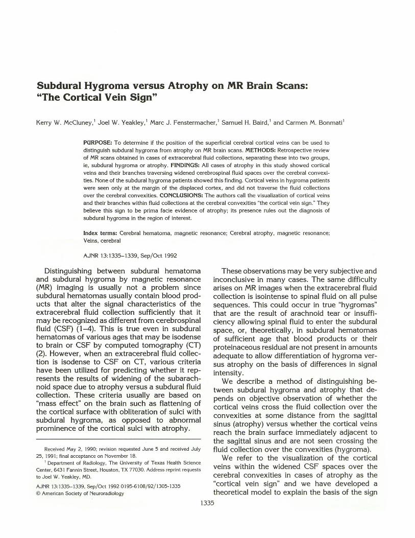
We refer to the visualization of the cortical veins within the widened CSF spaces over the cerebral convexities in cases of atrophy as the "cortical vein sign" and we have developed a theoretical model to explain the basis of the sign
1335
1336 McCLUNEY
RETRACTED CORTEX N ,4U"AAPHY
AJNR: 13, September /October 1992
+-PIA
Fig. 1. Theoretical model showing the basis of the cortical vein sign. The cortical veins and their branches are tubular structures draining into the sagittal sinus (black bordered inverted triangle). Notice that with subdural hygroma the arachnoid membrane has carried all venous structures below it onto the cortical surface of the brain, closing the subarachnoid space. The converse is true in atrophy. The arachnoid membrane remains closely applied to the dura on the "anticortical" (calvarial) surface of the CSF collection until the arachnoid is penetrated, opening the subarachnoid space. Veins course normally through the widened subarachnoid space in atrophy (double-headed arrows). In subdural hematoma, the position of the veins may be similar to that in subdural hygroma or may be ruptured as shown.
(Fig. 1). The purpose of this retrospective study was to verify our conclusion drawn from the model, namely, that the cortical vein sign is specific for atrophy and excludes a diagnosis of subdural hygroma.
Materials and Methods
We retrospectively reviewed MR scans of patients with extracerebral fluid collections isointense to CSF utilizing standard radiographic criteria to separate these into two groups, ie, subdural hygroma or atrophy. We accumulated cases until a total of 10 cases in each group was achieved. Criteria for inclusion in the subdural hygroma group were as follows (5, 6) : 1) isointense semilunar or crescent shaped extracerebral fluid collection limited to a cerebral convexity; 2) mass effect manifested by flattening of cortical surface of the brain and relative lack of visualization of cortical sulcal spaces adjacent to the fluid collection; 3) unilaterality where applicable; and 4) ventricular compression.
Criteria for inclusion in the atrophy group were as follows (7, 8) : 1) symmetrical enlargement of ventricles, basal cisternal spaces, and cortical sulcal spaces; and 2) lack of mass effect on the brain adjacent to the fluid collection.
We purposely eliminated historical data such as history of trauma or dementia, or the age of the patent, as criteria in an effort to be more objective on the basis of radiologic criteria alone.
We realize that this selection process is imperfect because of the fact that the diagnoses were not generally confirmed by surgery or autopsy in our series.
The relative location of where the cortical veins cross the fluid collection was reviewed in each case. That is, we analyzed whether the veins randomly crossed the CSF space even over the convexities and then followed a juxtacortical location, or whether the veins crossed the fluid collection in an immediate parasagittal location and assumed a juxtacortical location lateral to that point. In the latter group, the veins were not seen crossing the fluid space over the convexities.
Several additional cases of subdural hematoma at various stages of blood product degradation were reviewed to confirm the inwardly displaced cortical veins over the convexities in that condition previously described by Fobben et al (4).
The majority of the examinations were performed on a 1.5-T Signa scanner (GE, Milwaukee, WI) utilizing the standard head coil. Sagittal T1-weighted spin-echo images (800/20/1) and axial proton-density and T2-weighted images (2000/20,80/1) were utilized. Other parameters included 24.0-cm field of view, 5-mm section thickness with 2.5-mm gap, and 256 X 192 matrix. One scan performed outside this institution on a Picker 0.5-T scanner is also included in the illustrations.
Results
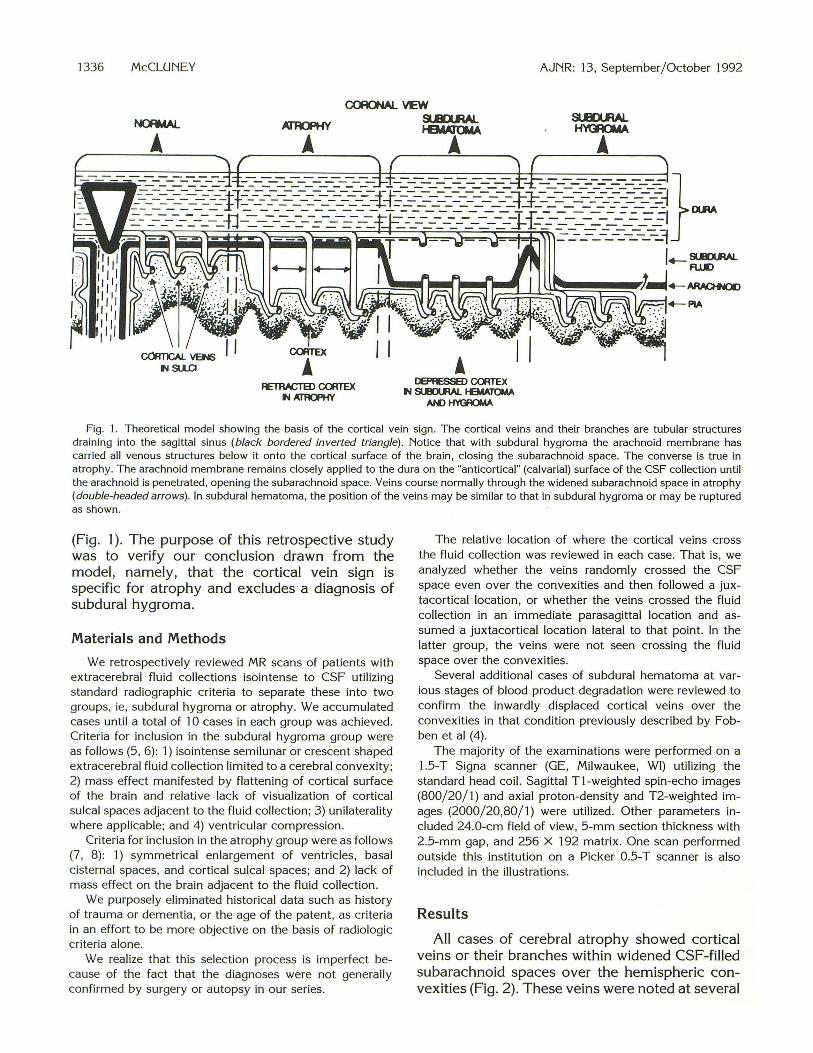
All cases of cerebral atrophy showed cortical veins or their branches within widened CSF-filled subarachnoid spaces over the hemispheric convexities (Fig. 2). These veins were noted at several
AJNR: 13, September / October 1992
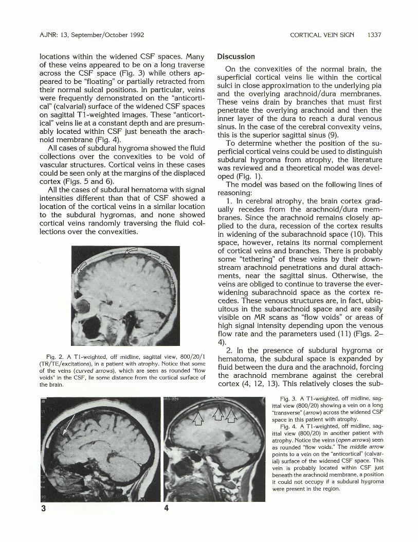
locations within the widened CSF spaces. Many of these veins appeared to be on a long traverse across the CSF space (Fig. 3) while others appeared to be "floating" or partially retracted from their normal sulcal positions. In particular, veins were frequently demonstrated on the "anticortical" (calvarial) surface of the widened CSF spaces on sagittal T1-weighted images. These "anticortical" veins lie at a constant depth and are presumably located within CSF just beneath the arachnoid membrane (Fig. 4).
All cases of subdural hygroma showed the fluid collections over the convexities to be void of vascular structures. Cortical veins in these cases could be seen only at the margins of the displaced cortex (Figs. 5 and 6).
All the cases of subdural hematoma with signal intensities different than that of CSF showed a location of the cortical veins in a similar location to the subdural hygromas, and none showed cortical veins randomly traversing the fluid collections over the convexities.
Fig. 2. A T1-weighted , off midline, sagittal view, 800/ 20/ 1 (TR/ TE/ excitations), in a patient with atrophy. Notice that some of the veins (curved arrows), which are seen as rounded "flow voids" in the CSF, lie some distance from the cortical surface of the brain.
3 4
CORTICAL VEIN SIGN 1337
Discussion
On the convexities of the normal brain, the superficial cortical veins lie within the cortical sulci in close approximation to the underlying pia and the overlying arachnoid/ dura membranes. These veins drain by branches that must first penetrate the overlying arachnoid and then the inner layer of the dura to reach a dural venous sinus. In the case of the cerebral convexity veins, this is the superior sagittal sinus (9).
To determine whether the position of the superficial cortical veins could be used to distinguish subdural hygroma from atrophy, the literature was reviewed and a theoretical model was developed (Fig. 1).
The model was based on the following lines of reasoning:
1. In cerebral atrophy, the brain cortex gradually recedes from the arachnoid/ dura membranes. Since the arachnoid remains closely applied to the dura, recession of the cortex results in widening of the subarachnoid space ( 1 0). This space, however, retains its normal complement of cortical veins and branches. There is probably some "tethering" of these veins by their downstream arachnoid penetrations and dural attachments, near the sagittal sinus. Otherwise, the veins are obliged to continue to traverse the everwidening subarachnoid space as the cortex recedes. These venous structures are, in fact , ubiquitous in the subarachnoid space and are easily visible on MR scans as "flow voids" or areas of high signal intensity depending upon the venous flow rate and the parameters used (11) (Figs. 2-4).
2. In the presence of subdural hygroma or hematoma, the subdural space is expanded by fluid between the dura and the arachnoid, forcing the arachnoid membrane against the cerebral cortex (4, 12, 13). This relatively closes the sub-
Fig. 3. A T1-weighted , off midline, sagittal view (800/ 20) showing a vein on a long "transverse" (arrow) across the widened CSF space in this patient with atrophy.
Fig. 4. A T1-weighted, off midline, sagittal view (800/ 20) in another patient with atrophy. Notice the veins (open arrows) seen as rounded "flow voids. " The middle arrow points to a vein on the "anticortical '" (calvarial) surface of the widened CSF space. Th1s vein is probably located within CSF just beneath the arachnoid membrane, a position it could not occupy if a subdural hygroma were present in the region .

1338 McCLUNEY
Fig. 5. A, Tl -weighted, off midline sagittal view (800/20) in a patient with subdural hygroma. Unlike atrophy, the cortical veins are not seen crossing the fluid collection but are identified only on the displaced brain surface (arrows). Note post-traumatic skull defect with bra in herniation.
B, Confirmation of findings on Tl weighted coronal view (800/20).
A
arachnoid space, trapping the cortical veins within it and displacing the arachnoid membrane and cerebral cortex as a unit away from the overlying dura. This leaves a homogenous fluid collection between the dura and arachnoid that is void of visible vascular structures (Figs. 5 and 6). The cortical veins that are visualized following the cortical surface of the brain at the cerebral convexities (Figs. 5 and 6) still have to reach the sagittal sinus, but they do so only far medially in the parasagittal area as may be observed on the venous phase of arteriograms with subdural fluid collections of any type (13). In addition, in sub-
Fig. 6. Chronic subdural fluid collection (hygroma) isointense to CSF. This axial T2-weighted scan (2000/ 80) demonstrates that the extracerebral fluid collection on the right is void of visible vascular structures and that the corti cal veins (curved arrows) can be seen only at the cortical margins.
AJNR: 13, September / October 1992
B
dural hematoma, but not usually in subdural hygroma, some of bridging veins may be ruptured as they pass from the brain surface to the sagittal sinus, further contributing to the relative lack of venous structures within fluid collections over the convexities.
Following the development of the theoretical model, our study was conducted comparing MR brain scans of patients with subdural hygroma to those with atrophy. All subdural hygromas were free of venous structures within the fluid collections over the convexities and all cases of atrophy showed venous structures within the overlying CSF fluid collections. Of particular interest was the finding of collections of veins on the "anticortical" (calvarial) surface of the fluid collections in patients with atrophy. These were best seen on T1-weighted sagittal images near the sagittal sinus (Fig. 4). These veins probably lie within CSF just beneath the arachnoid membrane.
It is to be noted that subdural fluid collections isointense to extraventricular CSF are uncommon. Chronic subdural hematomas usually have a significantly greater protein content than extraventricular CSF and are easily distinguished on MR due to their T1-shortening effect (1-4, 14). However, subdural hygromas that are generally isointense to extraventricular CSF can present diagnostic difficulties. Hygromas may be the result of tears, or insufficiency, in the arachnoid membrane that allow CSF to enter the subdural space producing an extracerebral fluid collection with signal intensities identical to CSF on all imaging parameters. A subdural hematoma of sufficient age that products of blood degradation
AJNR : 13, September /October 1992
are no longer present could theoretically produce a similar appearance.
Morphologically , the recognition of veins along the margin of the displaced cortex in extracerebral fluid collections is a valuable sign long known to angiographers (13) and reported by others (4). However, the true significance of this sign is that the veins are seen only at the cortical margins in extracerebral fluid collections (Figs. 5 and 6) . In atrophy, they are not only at the margins but also within the fluid and at the anticortical surface of the fluid.
Conclusion
The conclusion to be drawn from the model and this study is that, if cortical veins can be demonstrated deep within a convexity fluid collection, then that fluid must be CSF within dilated subarachnoid spaces and not subdural hygroma.
We have called the visualization of cortical veins and their branches deep within low signal intensity convexity fluid collections "the cortical vein sign." We believe this sign to be prima facie evidence of atrophy and that the sign excludes the diagnosis of subdural hygroma in the region of interest.
Acknowledgments
We credit Margaret Young for the physical production of the manuscript. Jay Johnson for the illustrations, and Juan Cabrera (posthumously) and Bob Boeye for Figure 1.
CORTICAL VEIN SIGN 1339
References
1. Hosoda K, Tamaki N, Masmura M, Matsumoto S, Maeda F. Magnetic
resonance images of chronic subdural hematomas. J Neurosurg
1987;67:677-683
2. Moon KL, Brant-Zawadzki MR, Pitts LH , Mills CM. Nuclear magnetic
resonance imaging of CT-isodense subdural hematomas. AJNR
1984;5:3 19-322
3. Young IR , Bydder GM, Hall AS, et al. Extracerebra l collections. AJNR
1983;4:833- 834
4. Fobben ES, Grossman Rl, A tlas SW, et al. MR characteristics of
subdural hematomas and hygromas at 1.5 T . AJNR 1989; I 0:
687-693
5. Weisberg L, Nice C. Cerebral computed tomography: a text-atlas. 3rd
ed. Philadelphia: Saunders, 1989:264, 403
6. Williams AL, Haughton VM. Cranial computed tomography. St. Louis:
Mosby, 1985:78
7. Weisberg L, Nice C. Cerebral computed tomography: a text-atlas. 3rd
ed. Philadelphia: Saunders, 1989:255-259
8. Williams AL, Haughton VM. Cranial computed tomography. St. Louis:
Mosby, 1985:250-256
9. Williams DPL, Warwick R, Dyson M , Bannister LH. Gray 's anatomy.
37th ed. Edinburgh: Churchill Livingstone, 1989:798
10. Geremia GK, Huckman MS. Degenerative diseases of the cerebral
hemisphere, brainstem and cerebellum. In: Taveras JM, Ferrucci JT,
eds. Radiology: diagnosis-imaging-intervention. Vo l. 3 Philadelphia:
Lippincott, 1990: chap 39
11 . Stark DD, Bradley WG. Magnetic resonance imaging. St. Louis:
Mosby, 1988:126
12. Kandel ER , Swartz JH. Principles of neuroscience. 2nd ed. New York :
Elsevier, 1985:247
13. Ramsey RG . Neuroradiology. 2nd ed. Philadelphia: Saunders,
1987:472
14. Brant-Zawadzki M, Kelly W, Kjos B, et al. Magnetic resonance imaging
and characterization of normal and abnormal intracranial cerebrospi
nal fluid (CSF) spaces. Neuroradiology 1985;27:3-8