su2012 quantitative in vivo assessment of colorectal length in human adults over time: direct...
TRANSCRIPT

AG
AA
bst
ract
sAdditionally, differentiated IMFEN-iRFP neurons were triggered by electrical field stimulationto produce transient relaxation of bioengineered IAS constructs. Conclusion: Our resultssuggest that IM-FEN-iRFPs are detectable in the near IR range (710nm), and that iRFPexpression does not inhibit IM-FEN ability to differentiate into neurons. This is the firstinstance where NIR proteins have been used to label neural cells. By labeling progenitorcells with NIR proteins, in vivo imaging can be optimized in the biologically clear window(700-1000nm). This work was supported by NIH R01DK071614 and the Wake ForestInstitute for Regenerative Medicine "Team Pilot" grant.
Su2011
Diagnostic Yield and Clinical Impact of a Low-Cost Microendoscope in theEarly Diagnosis of Barrett's Associated Neoplasia: A Prospective, Single-CenterRandomized Controlled TrialMichelle H. Lee, Neil Parikh, Alexandros D. Polydorides, Justin Louie, Marion-AnnaProtano, Josephine Mitcham, Rebecca Richards-Kortum, Sharmila Anandasabapathy
Background: Surveillance endoscopy with random four-quadrant biopsy (flat mucosa) andtargeted biopsy (irregular mucosa) is recommended for patients with BE. This procedure,however, is both inefficient, costly, and inaccurate: multiple normal biopsies are obtainedand yet the miss rate for neoplasia is as high as 57%. High-resolution microendoscopy(HRME) is a low-cost "optical biopsy" technology that provides sub-cellular magnified imagesof the esophageal mucosa, with real-time visualization of the cell nuclei when used withtopical fluorescent contrast. Objective: The purpose of this study was to compare thediagnostic yield and accuracy of high resolution white light endoscopy (WLE) +HRMEfollowed by targeted biopsies to WLE alone with targeted and random biopsies for thedetection of BE-associated neoplasia. Methods: Patients with histologically proven BE ≥1cmin length undergoing endoscopic surveillance for metaplasia, dysplasia or neoplasia (highgrade dysplasia and intramucosal carcincoma) were enrolled. Patients were randomized toeither WLE (Group A) or WLE+HRME (Group B). In Group A, WLE was performed withbiopsies of suspicious lesions, as well as random four quadrant biopsies. In Group B, WLEwas performed and suspicious lesions were noted. After staining with proflavine, WLEsuspicious areas were imaged first and non-neoplastic (Barrett's metaplasia, low grade dyspla-sia) or neoplastic (high grade dysplasia and intramucosal carcinoma) mucosa was identifiedand biopsied. Real-time HRME reads were performed using previously established criteriabased on glandular architecture, nuclear morphology and nuclear-to-cytoplasmic ratio. Next,four quadrant, random "optical biopsies" were obtained using HRME and read as eithernon-neoplastic or neoplastic, confirmed by biopsies taken from the same locations. A single,blinded, expert gastrointestinal pathologist interpreted all biopsies. Results: Thirty-sevenconsecutive patients were enrolled: 29 undergoing routine surveillance and 8 with suspectedintraepithelial neoplasia. The sensitivity of HRME is superior to WLE for detecting BE (100%vs. 60%, , p<0.05). Using a per-biopsy analysis, the overall diagnostic yield for BE associatedneoplasia with HRME was 66.7% compared to 12.1% with WLE alone. Per biopsy analysisdemonstrates that HRME could negate the need for biopsies in 89.8% (132/147) of sampleswhile per patient analysis indicates that 73.7% (14/19) of patients may not have requiredbiopsies. Discussion: The HRME platform can enhance diagnostic yield in endoscopic surveil-lance of Barrett's associated neoplasia and may reduce the number of biopsies needed toscreen for BE associated dysplasia and neoplasia, reducing the overall costs of screening.This may be an attractive, low-cost, high quality option for non-tertiary care hospitals andambulatory surgery centers.Performance Characteristics for Detection of Barrett's associated neoplasia (per biopsy anal-ysis)
Figure 1. Comparison of high resolution white light endoscopy, high resolution microendos-copy, and histopathology. Areas within the Barrett's segment on white light endoscopy (A)were determined to be neoplastic under HRME imaging (B) with histopathologic confirmationof esophageal adenocarcinoma(C).
Su2012
Quantitative In Vivo Assessment of Colorectal Length in Human Adults OverTime: Direct Evidence of Elongation in Intraperitoneal Segments At Serial CTColonographyJames L. Patrick, Perry J. Pickhardt
Background There is indirect evidence to suggest that colon length increases with age inadults, but serial in vivo comparison with colonoscopy is limited due to pleating andtelescoping effects, which foreshorten and obscure the actual length of the large intestine.The advent of advanced imaging tools at CT colonography (CTC) allows for more accurateand reproducible length assessment, including change over time at serial evaluation. Aim
S-522AGA Abstracts
The aim of this study was to assess for intra-patient changes in colorectal length over timein adults undergoing serial CTC investigation at a 5-year interval. In addition to changesin total length, the lengths of the intraperitoneal segments (ie, the transverse and sigmoidcolon) were compared with extraperitoneal remainder to determine potential contributorsto overall length changes. Methods Colorectal length measurements at serial CTC examina-tions were obtained directly from the automated centerline derived using dedicated software(Viatronix V3D Colon). Total, transverse, and sigmoid colon lengths were specificallyrecorded for each case. Total colonic volume was also recorded to assess degree of distention.Cases with inadequate luminal distention or an imprecise centerline were excluded. Thefinal cohort consisted of 80 adults (mean age at index study, 58.1 years), with a meaninterval between CTC examinations of 6.0 years (range, 5.0-8.2). Results The mean totalcolorectal length increased from 186.6 cm on the initial CTC to 190.4 cm at follow-upCTC; this increase of 3.8 cm (0.63 cm/year) was statistically significant (p <0.01). Theintraperitoneal colonic segments (transverse and sigmoid) appeared to largely account forthe increased length. Mean transverse colon length increased from 56.5 cm to 58.1 cm, anet increase of 2.3 cm (p <0.01) and sigmoid length increased from 64.9 cm to 66.2 cm,an increase of 1.3 cm (p <0.05). The changes in length of the remaining extraperitonealcolon were not statistically significant. Mean colonic volume was greater at initial CTCcompared with follow-up, arguing against improved distention as a potential cause forlengthening. Conclusions 1. A small but measurable increase in colon length (0.63 cm/yearincrease) was demonstrated in adults at serial CTC. We believe this to be the first directdemonstration of in vivo colonic lengthening in human adults. 2. The increase in colonlength over time appears to be pimarily due to increase in the peritoneal segments on amesentery (ie, the transverse and sigmoid colon).
Example of CTC case with automated segmentation of bowel. Colon is selected and smallbowel is ignored.
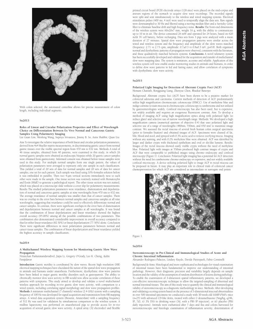
With colon selected, the automated centerline allows for precise measurement of colonlength, including individual segments.
Su2013
Roles of Linear and Circular Polarization Properties and Effect of WavelengthChoice on Differentiation Between Ex Vivo Normal and Cancerous GastricSamples Using Polarimetry ImagingLee Guan Lim, Wenfeng Wang, Supriya Srivastava, Jimmy B. So, Asim Shabbir, Quan Liu
Aim To investigate the relative importance of both linear and circular polarization parameters,derived from 4x4 Mueller matrix measurements, in discriminating gastric cancer from normalgastric tissues over the visible spectral region from 470 nm to 632 nm. Methods A total of46 tissue samples, obtained from 40 patients, were examined in this study, in which 26normal gastric samples were obtained as endoscopic biopsies while 20 gastric cancer sampleswere obtained from gastrectomy. Informed consent was obtained before tissue samples wereused in this study. For multiple normal samples from one single patient, the values ofpolarization parameters were averaged to represent only one sample in each classification.This yielded a total of 20 sets of data for normal samples and 20 sets of data for cancersamples, one set for each patient. Each sample was fixed using 10%-formalin solution beforeit was embedded in paraffin. Then two 4-μm vertical sections immediately next to eachother were made in the sample. One tissue section was routinely stained with Hematoxylinand Eosin (H&E) to generate a pathological report. The other tissue section was not stained,which was placed on a microscope slide without a cover slip for polarimetry measurements.Results The studied polarization parameters were retardance, diattenuation and depolariza-tion of normal and cancerous gastric samples at nine wavelengths from 470 nm to 632 nm.The average retardance of normal samples was smaller than that of cancer samples. Therewas no overlap in the error bars between normal samples and cancerous samples at all ninewavelengths, suggesting that retardance could be used to effectively differentiate normal andcancer samples. In contrast, there were significant overlaps in the error bars of diattenuationand depolarization between normal and cancer samples at all wavelengths. It was foundthat the combination of linear depolarization and linear retardance showed the highestoverall accuracy (95.00%) among all the possible combinations of two parameters. Thiscombination also demonstrated considerable improvement in overall accuracy compared tothat for either linear retardance (82.50%) or linear depolarization (77.50%) alone. ConclusionThere were significant differences in most polarization parameters between normal andcancer tissue samples. The combination of linear depolarization and linear retardance yieldedthe highest accuracy in sample classification.
Su2014
A Multichannel Wireless Mapping System for Monitoring Gastric Slow WavePropagationNiranchan Paskaranandavadivel, Jiajia Li, Gregory O'Grady, Leo K. Cheng, AydinFarajidavar
Introduction Gastric motility is coordinated by slow waves. Recent high resolution (HR)electrical mapping techniques have elucidated the nature of the spatial slow wave propagationin animals and humans under anaesthesia. Furthermore, dysrhythmic slow wave patternshave been linked to major gastric motility disorders such as gastroparesis. The ability tochronically monitor slow wave activity in HR would now allow accurate correlation of thesepatterns with symptoms. This study aimed to develop and validate a novel multichannelwireless approach for recording in-vivo gastric slow wave activity, with comparison to awired system, including correlating signal morphology and slow wave propagation profiles.Methods A miniature multichannel (7 channels) wireless (2.4 GHz) system with a samplingfrequency of 100 Hz was developed for signal acquisition and transmission from HR mappingarrays. A wired data acquisition system (Biosemi, Amsterdam) with a sampling frequencyof 512 Hz was used for validation by simultaneous comparison to the wireless system. Amidline laparotomy was performed on anaesthetized pigs to provide access for in-vivoacquisition of serosal gastric slow wave activity. A spiral array (32 electrodes) and flexible
S-523 AGA Abstracts
printed circuit board (PCB) electrode arrays (128 sites) were placed on the mid-corpus andantrum regions of the stomach to acquire slow wave recordings. The recorded signalswere split and sent simultaneously to the wireless and wired mapping systems. Electricalstimulation pulses (400 ms, 4 mA) were used to temporally align the data sets. Raw signalswere downsampled to 30 Hz and filtered using a moving median filter and a Savitzky Golayfilter to eliminate baseline drift and high frequency noise. Results The front-end dimensionsof the wireless system were 60×20×7 mm, weight 16 g, with the ability to communicateup to 10 m in air. The device consumed 26 mW and operated for 24 hours, based on 620mAh 3V cell battery, before recharging. Data sets from 3 pigs were analyzed with a meanduration of 27 minutes. Spatial slow wave propagation patterns were similar across thewired and wireless system and the frequency and amplitude of the slow waves matched(frequency: 2.71 vs 2.73 cpm, amplitude: 0.7±0.3 vs 0.8±0.3 mV, p>0.8). Both organisednormal and dysrhythmic patterns of propagation were observed, consistent with the literature,and these qualitatively matched between systems. Conclusions A novel telemetry systemhas been successfully developed and validated for the acquisition and transfer of multichannelslow wave mapping data. The system is miniature, accurate and reliable. Application of thiswireless system will now enable awake monitoring studies in animals and humans, in orderto define slow wave patterns in fed and fasting states, and allow correlation of symptomswith dysrhythmic slow wave activity.
Su2015
Polarized Light Imaging for Detection of Aberrant Cryptic Foci (ACF)Hemant Chatrath, Rongguang Liang, Zhenyue Chen, Bhaskar Banerjee
Background: Aberrant cryptic foci (ACF) have been shown to be a putative marker forcolorectal adenoma and carcinoma. Current methods of detection of ACF predominantlyutilize high magnification chromoscopic colonoscopy (HMCC). Use of methylene blue andindigo carmine to stain mucosa in chromoscopic colonoscopy is cumbersome and not utilizedby gastroenterologists widely. Confocal microscopy has also been used, but is expensive,not widely available and requires an exogenous fluorescent contrast. Aim: To develop amethod of imaging ACF using high magnification optics along with polarized light (toreduce glare) and selective use of narrow wavelength range. Methods: We developed a highmagnification camera (numerical aperture of objective 0.4) that uses polarized light andselective use of a range of wavelengths (480nm, 550nm, and 650 nm) to maximize imagecontrast. We assessed the rectal mucosa of several fresh human colon surgical specimens(prior to formalin fixation) and obtained images of ACF. Specimens were cleaned of fat,bile, blood and stool, and sprayed with 0.3% acetic acid to remove overlying mucus. Imagingwithout any staining, and with 0.2% methylene blue were obtained. ACF was identified bylarger and darker crypts with thickened epithelium and oval or slit-like lumens. Results:Images of the rectal mucosa showed easily visible crypts without the need of methyleneblue. Polarized light with images at 550nm produced high contrast images of crypts andACF that were comparable to published images using chromo endoscopy and confocalmicroscopy (Figure 1). Conclusion: Polarized light imaging has a potential for ACF detectionwithout the need for cumbersome chromo endoscopy or expensive, and not widely availableconfocal microscopy. A device utilizing polarized light to image ACF in rectal mucosa canbe proposed in future. It may play an important role in clinical trials of colorectal cancerchemoprevention for which ACF are considered as intermediate or surrogate end points
Figure 1.
Su2016
Stereomicroscopy in Pre-Clinical and Immunological Studies of Acute andChronic Intestinal InflammationAlexander Rodriguez-Palacios, Lindsey Kaydo, Davide Pietropaoli, Fabio Cominelli
Background & Aims: Histological and more sophisticated technologies for visual examinationof intestinal tissues have been fundamental to improve our understanding of intestinalpathology. However, their diagnostic precision and variability largely depends on samplelocation and the validity of the assumption of random distribution of lesions during pathology.To enable the examination of 3-dimension spatial inflammatory patterns, we developed acost-effective stereomicroscopic technique to allow the targeted-sampling of abnormal andnormal intestinal tissues. The aim of this study was to quantify the clinical and immunologicalvalidity of stereomicroscopy as a diagnostic methodology in mice. Methods: After developingand validating a scoring system based on the presence of 3-dimensional mucosal abnormalitiesin over 500 intestinal specimens we conducted a study with 30-week-old SAMP1/YitFc mice(n=25) with advanced CD-like ileitis, treated with either i) dexamethasone (5mg/kg, q24h,IP, 7d), ii) 3% DSS in drinking water (5d, with a PBS IP injection), or iii) placebo (PBSdaily injections). Animals were euthanized after 7 days and ilea and colons harvested forstereomicroscopic and histologic examination of inflammation severity, determination of
AG
AA
bst
ract
s