study ofcardiac rhythm in healthy newborninfants
TRANSCRIPT

Br Heart J 1980; 43: 14-20
Study of cardiac rhythm in healthy newborn infantsD P SOUTHALL, JEAN RICHARDS, PRUDENCE MITCHELL, D J BROWN,P G B JOHNSTON, E A SHINEBOURNE
From the Department of Paediatrics, Brompton Hospital and Cardiothoracic Institute, London
suMMARY Twenty-four-hour electrocardiograms were recorded in the first 10 days of life on 134healthy full-term infants with birthweights greater than 2-5 kg. The highest heart rate a minute,measured over nine beats, was 175±19 (SD). The lowest rates, measured over three, five, and ninebeats were 82± 12, 87+12, and 93±12, respectively. At their lowest rates 109 infants had sinusbradycardia and 25 had junctional escape rhythms. Thirty-three infants showed changes in P wave
configuration with or without pronounced variation in PR interval. Atrial premature beats were presentin 19 infants but only one had more than 12 per hour.
In a randomly selected subgroup of 71 infants sinus pauses were found in 51 (72%). Five (7%)had electrocardiographic patterns and rhythm disturbances that could not be differentiated from thosepreviously described as complete sinuatrial exit block or sinus arrest, eight (11%) had patternsindistinguisable from 2:1 sinuatrial block, and 23 (32%) had patterns indistinguishable from sinuatrialWenckebach block.
This study shows that normal infants have variations in heart rate and rhythm hitherto consideredto be abnormal.
The range of heart rate and nature of heart rhythmsare poorly documented in normal healthy infants.Twenty-four-hour recordings of the electrocardio-gram provide an accurate means of measuringheart rate and rhythm over long periods. The tech-nique does not seriously interfere with normalbehaviour and therefore documentation of changesin rhythm and rate is possible during activities suchas sleeping, feeding, or crying. This report describesthe results of single 24-hour recordings of theelectrocardiogram on 134 healthy full-term neo-nates who weighed over 2-5 kg at birth.
Patients and methods
Studies were made between birth and 10 days of age(the normal period of postnatal hospitalisation inthis area) on 134 randomly selected infants born inWest Dorset between January 1977 and June 1978.They were all healthy and recordings were per-formed in the maternity ward before dischargehome. All infants studied had normal standard 12lead electrocardiograms with a 10-second rhythmstrip. They were studied under standard maternityward environmental conditions and were fed andhandled normally by their mothers during therecording period.Received for publication 5 July 1979
The recordings were made onto a Medilogcassette recorder* and the tapes were analysed forthe presence of arrhythmias using a playbacksystem* and arrhythmia detector*, by a trainedtechnician. The Medilog recorder and analyserincorporated a synclock mechanism which, byproviding and processing a reference time signal,ensured that possible variations in tape speed didnot produce artefactual rhythm or rate disorders.Mothers were encouraged to handle their babies inthe normal way while recordings were taken.
Heart rates during activity and rest were mea-sured by a histogram module in the analyser*.Histograms of the distribution of different heartrates (as obtained by continuous measurement ofall intervals) were made on 79 babies duringapproximately two hours each of activity and rest.The maximum heart rates, measured by direct
electrocardiographic analysis, over nine consecutivebeats were recorded. The minimum rates foundover three, five, and nine consecutive beats werealso measured in this way and the heart rhythm atthis time (whether sinus or junctional) was noted.The number of premature beats over 24 hours was
measured by counting them in four different,randomly selected, hours of recording and comput-ing the total.* Oxford Instrument Company.
14
on February 9, 2022 by guest. P
rotected by copyright.http://heart.bm
j.com/
Br H
eart J: first published as 10.1136/hrt.43.1.14 on 1 January 1980. Dow
nloaded from

Study of cardiac rhythm in healthy newborn infants
Values for maximum and minimum heart rateswere analysed and compared for ages 1 to 3 daysand ages 4 to 10 days.
Seventy-one randomly selected recordings wereanalysed to determine if PP intervals were presentwhich exceeded the previous PP interval by morethan 50 per cent. If the QRS complex by which this
480 460 14601 1580ms ms ms ms
Fig. 1 Extract from 24-hour electrocardiogram showingsudden lengthening of PP interval by more than 110 percent of previous PP interval. This electrocardiographicpattern and rhythm disturbance cannot be distinguishedfrom complete sinuatrial exit block or sinus arrest.
interval was terminated was not preceded by a
P wave the PQ interval was measured. When thePP or PQ interval exceeded the previous PPinterval by more than 110 per cent this was classedas a pattern indistinguishable from completesinuatrial block or sinus arrest (Fig. 1); intervals ofbetween 90 and 110 per cent were classed as patternsof 2:1 sinuatrial block (Fig. 2) and intervalsbetween 50 and 90 per cent were classed as sinuspauses (Fig. 3). This latter group was also assessedwith respect to the pattern of PP intervals (aminimum of three) by which they were preceded.
4UU 4W 400 UU
ms ms ms ms
Fig. 2 Extract from 24-hour electrocardiogram showingsudden lengthening of PP interval by between 90 and110 per cent of previous PP interval. This electrocardio-graphic pattern and rhythm disturbance cannot bedistinguished from 2:1 sinuatrial block.
(1) Progressive increase in PP intervals.(2) Regular PP intervals.(3) Irregular PP intervals.(4) A progressive decrease in PP intervals, that is a
sinuatrial Wenckebach block pattern (Fig. 3cand 4).1To differentiate sinuatrial Wenckebach block
patterns from those of sinus arrhythmias we usedthe calculations of Schamroth and Dove' (see legendto Fig. 4) and also plotted the preceding PPintervals against their succeeding PP intervals(Fig. 5). The percentages used in our calculationswere chosen empirically to try and match the labelsand character of sinuatrial block pattern used byprevious workers.
Results
All infants were predominantly in sinus rhythmand showed a minimal, continuous irregularity ofrate previously termed phasic variation.2The distribution of heart rates (derived from RR
intervals) at rest and during activity measured bythe histogram module in the analyser showed amaximum heart rate of 250/minute and a minimumrate of 85/minute. Direct electrocardiographicanalysis of these higher and lower heart ratesshowed them to be spuriously high because of theinterpretation of movement artefact and large Twaves as R waves.The histogram of the highest rate achieved over
24 hours measured by direct electrocardiographicanalysis over nine beats did not differ significantlyfrom a curve of normal distribution* with a meanrate of 175 ±19 (SD). The highest heart rate foundwas 225/minute. The histograms of the lowest ratesmeasured by direct electrocardiographic analysisover three, five, and nine beats duration over 24hours also did not differ from a normal distribu-tion.* The mean lowest heart rate when measuredover three beats was 82/minute, over five beats was87/minute, and over nine beats was 93/minute.The standard deviation in each case was 12.Of 134 infants, 109 at their lowest heart rates
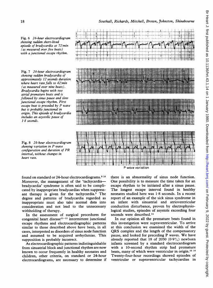
had sinus bradycardia and 25 had junctional escaperhythms. All infants showed sudden short episodesof bradycardia (Fig. 6).The three infants with the lowest heart rates are
described in more detail. One aged 8 days had anepisode of bradycardia of 10 beats duration whenthe rate fell to 42/minute. This began with twoatrial premature beats, was followed by a sinuspause of 1-8 seconds, and continued with a slowjunctional escape rhythm (Fig. 7). Four otherepisodes of bradycardia occurred during the*Confirmed by Kolomogorov-Smirnov test.3
15
on February 9, 2022 by guest. P
rotected by copyright.http://heart.bm
j.com/
Br H
eart J: first published as 10.1136/hrt.43.1.14 on 1 January 1980. Dow
nloaded from

Southall, Richards, Mitchell, Brown, Jtohnston, Shinebourne
recording. The second infant aged 2 days showedan episode of 14 beats duration where the rate fellto 59/minute. His only sib had previously died fromthe sudden infant death syndrome. The third infantwas 4 hours old when the 24-hour recording was
started. Subsequent analysis showed seven episodesof bradycardia of between 59 and 63/minute and of
between 12 and 18 beats duration. The first threeepisodes coincided with cyanotic attacks, observedby the nursing staff and treated by stimulation andoronasal suction. No cause was found for theseattacks. Though there were four further episodesof bradycardia during the night no cyanotic attackswere detected.
II iL. i 1;. 1;.:TThPI41;hiA:M L- pt-~
Fig. 3 Four extracts from24-hour electrocardiograms on
different infants showing sinuspauses. In each case there issudden lengthening of PP or PQintervals exceeding previous PPintervals by 50 to 90 per cent.In (A) preceding PP intervals are
regular; in (B) they are irregular,and in (D) they are increasingbefore the pause. In (C) there isprogressive decrease in PPintervals before the pause and thiselectrocardiographic patterncannot be distinguished fromsinuatrial Wenckebach block.
1 640 1 600 1 520 1 780 I
ms ms ms ms
Decreasing PP(S A Wenckebach)
I...;. ..B ... l...l. : 1 .
1472 1 580 1 500 1 836 1ms ms ms ms
Irregular PP...........-.....1. ~... ...
1528 1608 1 636 1 960ms ms ms ms
Increasing PP
is
Fig. 4 Twenty-four-hour recording showing a series of sinus pauses. Labelledcentral pause is preceded by progressive reduction in PP intervals. Usingcalculations of Schamroth and Dove' sinus cycle
1990= 610 + 520 + 860 = = 498
3+1
Pl - P2 increment = 610 - 498 = 112P2 - P3 increment = 520 - 498 = 22
Total increment = 134860 + 134 = 994 which is approximately twice sinus cycle of 498.Sinus extrasystole before pause is unlikely because pause exceeds -sinus cycle by362 ms.
v ..... ..{ .,T.7;.7 .7,',.E --- . - -.:: ;7....-:,:.1440 1440 1440 1 660ms ms ms ms
Regular PP
2i
K77XKL
16
A
is,% t A-it; l I 1lm1 wilk"I-ooua
1:L
1, I ".
j: l; It
I
* _s.
on February 9, 2022 by guest. P
rotected by copyright.http://heart.bm
j.com/
Br H
eart J: first published as 10.1136/hrt.43.1.14 on 1 January 1980. Dow
nloaded from

Study of cardiac rhythm in healthy newborn infants
Table 1 Analysis of extrasystoles from single 24-hourelectrocardiograph recordings on 134 infants
No. of extrasystoles in 24 hours No. of infants
ltol2 1212to24 124 to 48 296 to 120 2144 to 168 1480 to 600 1
Changes in P wave configuration and PR intervalvariation occurred in 33 (25%) infants (Fig. 8).
Nineteen infants (14%) had atrial prematurebeats but only one had more than 12 per hour(Table 1).There was no significant difference between the
two age groups, 1 to 3 days and 4 to 10 days, formeasurements of maximum and minimum heartrates (Table 2).Of the subgroup of 71 infants, five (7%) had
electrocardiographic patterns, ie sinus pauses, thatcould not be differentiated from those described inall the other articles studied as complete sinuatrialexit block or sinus arrest (Fig. 1). Eight infants(11%) had sinus pauses indistinguishable from 2:1sinuatrial block (Fig. 2).The longest interval between an escape beat and
the previous sinus beat was 1 8 seconds (Fig. 6).Sinus pauses representing increases in PP
intervals of 50 to 90 per cent were found in 51subjects (72%) with preceding PP interval patternsas follows (Fig. 3). (1) Progressive increase 23subjects (32%); (2) regular PP intervals 14 subjects(20%); (3) irregular PP intervals 35 subjects (50%);and (4) progressive decrease (sinuatrial Wenckebachpattern) 23 subjects (32%) (Fig. 3c and 4). Thecalculations of sinus cycle increments (Fig. 4) andgraphs of succeeding PP interval measurements
Second interval50 60
A
70 80 90
(Fig. 5) supported the presence of Wenckebachconduction rather than sinus arrhythmia in all 23subjects.Some subjects showed more than one pattern.
Discussion
The range of heart rates found in these healthyneonates is wider than previously reported onstandard electrocardiograms.4 The mean lowestrate measured by direct electrocardiographicanalysis over nine beats was 83/minute, with astandard deviation of 12. Heart rates as low as60/minute for short periods of time are therefore tobe expected in the normal neonatal population.Though 25 per cent of infants at their lowest rateshad short episodes of junctional escape rhythm,prolonged periods of junctional rhythm withretrograde P wave activity were not found in ourstudy. Episodes of sudden bradycardia are a distinctfeature of the cardiac rhythm at this age and havepreviously been reported in association with suddendeep breaths, hiccoughs, bowel movements, andvomiting.2 5 6 We are at present evaluating therelation of apnoea and other physiological functionsto these episodes.7
Electrocardiographic patterns that we could notdifferentiate, without invasive electrophysiologicaltests, from those described in previous studies ascomplete, 2:1, Wenckebach sinuatrial block, orsinus arrest were found in 50 per cent of infantsstudied. They were also found in our recentlycompleted investigation of 24-hour electrocardio-grams on 92 randomly selected healthy childrenaged 7 to 11 years.8 These electrocardiographicpatterns have often been interpreted as disordersof sinus node function and clinical events such asfaints, 'funny turns', or blackouts ascribed to thiscause when these electrocardiographic patterns are
Second interval50 60 70 80 90
Fig. 5 A plot of PP intervalson the Y axis against succeedingPP intervals on X axis. The lineshows position of PP intervals ofequal duration. (A) Pattern ofsinus arrhythmia. (B) Pattern ofsinuatrial Wenckebach block.
It'*X"
* q
* Xs
50.
-6' 60
0 70.
80
90
I "'.I .
*
17
on February 9, 2022 by guest. P
rotected by copyright.http://heart.bm
j.com/
Br H
eart J: first published as 10.1136/hrt.43.1.14 on 1 January 1980. Dow
nloaded from
Southall, Richards, Mitchell, Brown, J7ohnston, Shinebourne
Fig. 6 24-hour electrocardiogramshowing sudden short-livedepisode of bradycardia at 72/min(as measured over five beats)with a junctional escape rhythm.
Fig. 7 24-hour electrocardiogramshowing sudden bradycardia ofapproximately 12 seconds durationwhere heart rate falls to 42/min(as measured over nine beats).Bradycardia begins with twoatrial premature beats and isfollowed by sinus pause and slowjunctional escape rhythm. Firstescape beat is preceded by P wavebut is probably junctional inorigin. This episode of bradycardiaincludes an asystolic pause of1-8 seconds.
LI I - -, ' ,! , : t -, ' - 1i { i ', ', , l - .; i -- - - ! .1 '.
Fig. 8 24-hour electrocardiogramshowing variation in P waveconfiguration and duration of PRinterval, without changes inheart rate.
.... .TMt >i Xf[3t eaXF4
Tt n \- 11 L* ~;,I, T fl1 t4*7ST4THH11111111 1HIM*lt4 i
P wQve vQriation
found on standard or 24-hour electrocardiograms.9 10Moreover, the management of the 'tachycardia-bradycardia' syndrome is often said to be compli-cated by inappropriate bradycardias when suppress-ant therapy is given for the tachycardia.9 Thedegree and patterns of bradycardia regarded asinappropriate must also take normal data intoconsideration and not lead to the unnecessarywithholding of therapy.
In the assessment of surgical procedures forcongenital heart diseasell-'3 intermittent junctionalescape rhythms and electrocardiographic patternssimilar to those described above have been, in allcases, interpreted as disorders of sinus node functionand assumed to be acquired arrhythmias. Thissupposition is probably incorrect.As electrocardiographic patterns indistinguishable
from sinuatrial block and junctional rhythm are nowknown to occur frequently in healthy neonates andchildren, other criteria, on standard or 24-hourelectrocardiograms, are necessary to determine if
there is an abnormality of sinus node function.One possibility is to measure the time taken for anescape rhythm to be initiated after a sinus pause.The longest escape interval found in healthyneonates studied here was 1-8 seconds. In a recentreport of an example of the sick sinus syndrome inan infant with sinuatrial and atrioventricularconduction disturbance, proven by electrophysio-logical studies, episodes of asystole exceeding fourseconds were described.'4
In our opinion all the premature beats found inthis investigation were supraventricular. To arriveat this conclusion we examined the width of theQRS complex and the length of the compensatorypause, and looked for preceding P waves. We havealready reported that 18 of 2030 (09%) newborninfants screened by a standard electrocardiogramwith a 10-second rhythm strip had prematurebeats, many of which were ventricular in origin.15 16
Twenty-four-hour recordings showed episodes ofventricular or supraventricular tachycardias in
18
on February 9, 2022 by guest. P
rotected by copyright.http://heart.bm
j.com/
Br H
eart J: first published as 10.1136/hrt.43.1.14 on 1 January 1980. Dow
nloaded from

Study of cardiac rhythm in healthy newborn infants
Table 2 Comparison of lowest and highest rates over 24 hours in age group 1 to 3 days and age group 4 to 10 days
Highest rate/minute Lowest rate/minute Lowest rate/minute(measured over 9 beats) (measured over 3 beats) (measured over 9 beats)
Age (d) Min. Max. Mean SD Min. Max. Mean SD Min. Max. Mean SD
1-3 120 220 173 19 53 105 81 11 59 119 93 12
4-10 140 205 179 16 36 110 82 14 42 120 94 13P value <0 10 Not significant Not significant
Table 3 Comparison of reports on long-term electrocardiographic monitoring in the neonate
Southall et al.7 Valimikil" Viiimdki and Tarlo"l Morgan and Guntherothl"
Duration of recording (hr) 24 36 30 5Number studied 134 50 68 50Age at recording (d) 1-10 1-2 1-2 1-7Maximum heart rate per min 220 260 260 170Minimum heart rate per min 42 86 80 75Junctional escape rhythms (%) 19 10 13P wave and PR interval variation(%) 25 20 15 -
Premature beats (%) 14 8 9 2 plus 'a few'
five of this latter group. Premature beats, frequentenough to be detected on standard electrocardio-grams, are therefore more likely to be associatedwith tachyarrhythmias than occasional prematurebeats found only on 24-hour electrocardiographicrecordings. The latter are likely to be a normalfeature of the newborn heart rhythm.
In Table 3 the findings of our study are comparedwith those of three other reports describing long-term electrocardiographic monitoring of healthyfull-term infants.'7-19 The maximum and minimumheart rates found by VdlimAki'8 19 are higher thanthose found by the direct electrocardiographicanalysis method used in our study. Their techniqueof measuring rate was, however, similar to theautomatic histogram module of our 'Oxford'analyser. We have shown that movement artefactsand large T waves are often interpreted as extraQRS complexes by this automatic histogrammodule, thus giving spuriously high heart rates.This could explain the higher results in their study.The shorter time of recording used by Morgan andGuntheroth'7 may explain the absence of junctionalescape rhythms or the absence of P wave and PRinterval variation in their study.Twenty-four-hour recordings have shown that
electrocardiographic rhythm patterns previouslydescribed as representing disorders of sinus nodefunction occur in the healthy neonate. We havealso found differences between the values of highestand lowest heart rates measured in this study andthose previously reported on long-term electro-cardiographic recordings. The interpretation of
heart rhythm and rate in clinical situations musttake normal data into consideration.
Dr Southall has a grant from the Board of Governorsof the Brompton Hospital, and Jean Richardsfrom the Foundation for the Study of Infant Death.The project was supported by grants from theBritish Heart Foundation and the Wessex RegionalHealth Authority. Generous donations were alsogiven by the League of Friends of Dorchester andWeymouth Hospitals, the Weymouth Round Table,Weymouth Lions, and Weymouth Auxiliary Leagueof the Licensed Trade.
The Oxford Instrument Company gave invaluablesupport to this work.
References
'Schamroth L, Dove E. The Wenckebach phenomenonin sino-atrial block. Br Heart J 1966; 28: 350-8.2Urbach J R, Phuvichit B, Zweizig H, et al. Instantan-eous heart rate patterns in newborn infants. Am JObstet Gynecol 1965; 93: 965-74.3Siegel S. Non parametric statistics. New York: McGraw-Hill, 1956: 47-52.4Liebman J. Electrocardiography. In: Moss AJ, AdamsFH, eds. Electrocardiography in heart disease in infants,children and adolescents. Baltimore: William & Wilkins,1968: 183-99.'Vallbona C, Desmond MM, Rudolph AJ, et al.Cardiodynamic studies in the newborn. II. Regulationof the heart rate. Biol Neonate 1963; 5: 159-99.
19
on February 9, 2022 by guest. P
rotected by copyright.http://heart.bm
j.com/
Br H
eart J: first published as 10.1136/hrt.43.1.14 on 1 January 1980. Dow
nloaded from

Southall, Richards, Mitchell, Brown, J7ohnston, Shinebourne
"Lipton EL, Steinschneider A, Richmond JB. Auto-nomic function in the neonate. VIII. Cardiopulmonaryobservations. Pediatrics 1964; 33: 212-5.7Southall DP, Richards J, Brown DJ, Johnston PGB,de Swiet M, Shinebourne EA. Twenty-four hour taperecordings of ECG and respiration in the newborninfant with findings related to sudden death andunexplained brain damage in infancy. Arch Dis Child1980; 55: 7-16.8Southall DP, Johnston F, Richards JM, ShinebourneEA, Johnston PGB. Study of heart rate and rhythm innormal schoolchildren (abstract). Br Heart J 1979; 42:234."Scott 0, Macartney FJ, Deverall PB. Sick sinussyndrome in children. Arch Dis Child 1976; 51: 100-5.°0Yabek SM, Swensson RE, Jarmakani JM. Electro-cardiographic recognition of sinus node dysfunctionin children and young adults. Circulation 1977; 56:235-9.
"'Greenwood RD, Rosenthal A, Sloss LJ, LaCorte M,Nadas A. Sick sinus syndrome after surgery forcongenital heart disease. Circulation 1975; 52: 208-13.
1EI-Said GM, Gillette PC, Cooky DA, Mullins CE,McNamara DG. Protection of the sinus node inMustard's operation. Circulation 1976; 53: 788-91.
13Saalouke MG, Rios J, Perry LW, Shapiro SR, ScottLP. Electrophysiologic studies after Mustard's opera-tion for d-transposition of the great vessels. Am JCardiol 1978; 41: 1104-9.
14Onat A, Domanic N, Onat T. Sick sinus syndrome inand infant. Eur J Cardiol 1974; 2: 79-83.
',Southall DP, Vulliamy DG, Davies J, Anderson RH,Shinebourne EA, Johnson AM. A new look at theneonatal electrocardiogram. Br Med 7 1976; 2: 615-8.
"Southall DP, Orrell MJ, Talbot JF, et al. Study ofcardiac arrhythmias and other forms of conductionabnormality in newborn infants. Br Med Jf 1977; 2:597-9.
1Morgan BC, Guntheroth WG. Cardiac arrhythmias innormal newborn infants. J Pediatr 1965; 67: 1199-202.'Vdlimaki I. Tape recordings of the electrocardiogramin newborn infants. Section II Long-term ECG taperecordings of newborn infants. Acta Paediatr Scand[Suppl] 1969; 199: 1-75.
"Valimaki I., Tarlo PA. Heart rate pattern and apneain newborn infants. Am J Obstet Gynecol 1971; 110:343-9.
Requests for reprints to Dr D P Southall, Cardio-thoracic Institute, Fulham Road, London SW3 6HP.
20
on February 9, 2022 by guest. P
rotected by copyright.http://heart.bm
j.com/
Br H
eart J: first published as 10.1136/hrt.43.1.14 on 1 January 1980. Dow
nloaded from