study guide 2nd year mbbs - ckmc.edu.pk
TRANSCRIPT

1
STUDY GUIDE
2nd YEAR MBBS
Compiled by: Department of Medical Education CMH Kharian Medical College (CKMC), Kharian Cantt.

2
Learning Outcomes Knowledge:
Describe the gross anatomical features of Cerebrum, Midbrain, Pons, Medulla and Spinal cord
Describe the sensory and motor parts of nervous system
Describe the major levels of central nervous system along with their functions
Describe the integrative function of nervous system
Describe the blood cerebrospinal fluid and blood brain barriers
Describe the structure of Nerve and explain the myelination of nerve fiber
Describe the ascending and descending tracts of brain stem
Describe analgesia system in brain & spinal cord
Describe the mechanism of consolidation of memory
Describe the functions of autonomic nervous system
Explain the Physiology, anatomy and pathogenesis of Head & neck and special sense problems.
Apply basic sciences to understand the causes of common Head & neck and special sense problems.
Explain the structural & developmental organization of GIT.
Explain the composition, functions, mechanism & control of following gastrointestinal secretions: salivary, gastric, pancreatic, biliary, small & large intestines.
Describe the mechanism of absorption of various nutrients and their role in malabsorption syndrome.
Explain the physiological anatomy, biochemistry functions and dysfunctions of Liver.
Explain the GIT hormones (structure, function) & their role in secretion and motility.
Describe the chemical nature, biosynthesis and the physiological functions of hormones on their target organs.
Skill: Draw a labeled diagram of the identified structures with the help of eosin and
hematoxylin pencils on the histology notebooks
Mark the main anatomical land marks on skull
Dissect various parts of head and neck and special senses, and related structure
Demonstrate their gross Anatomy and relationship to each other.
Identify the histological features of all the endocrine glands under microscope.
To perform all the steps of blood glucose estimation in the lab.
Dissect various parts of GIT, and related structures including peritoneum, to demonstrate their gross Anatomy and relationship to each other.
Identify different organs of GIT under microscope and on model.
Attitude: Demonstrate the effective attitude towards the colleagues
Demonstrate a professional attitude, team building spirit and good communication skills
Observe lab safety rules

3
1. INTRODUCTION
1.1 BLOCK COMMITTEE
Chief Coordinator Block 1: Dr. Hammad Ahmed Butt M.Phil. HOD Pharmacology, CKMC Coordinator: Dr. Noman Sadiq Assistant Professor, Physiology CKMC
Resource Persons S No Name Designation Deptt Contact
1. Dr. Ahmed Murtaz Khalid
Assistant Prof
Physiology [email protected]
2. Dr. Shaista Noor Demonstrator Biochemistry [email protected]
For any information regarding block 1, contact in CKMC: Faculty members: Anatomy Dr. Aneeqa Chughtai Physiology Dr. Noman Sadiq Biochemistry Dr. Sadaf Saleem Uppal Surgery Dr. Ahmed Raza Medicine Dr. Aamir Habib Community Medicine Dr. Iffat Naiyar Behavioral Sciences Dr. Asif Azeem Bajwa Pharmacology Dr. Hammad Ahmed Butt Pathology Dr. Nasira Shaheen Radiology Dr. Humaira Saleem Gynae/ OB Dr. Jasia Jabeen Paediatric Medicine Dr. Saeed Zaman Department of Medical Education Dr. Noman Sadiq

4
1.2 What is a study guide? It is an aid to:
Inform students how student learning program of the block has been organized. Help students organize and manage their studies throughout the module. Guide students on assessment methods, rules and regulations.
1.3The study guide:
Informs about organization and management of the block.
Defines the objectives which are expected to be achieved at the end of the block.
Identifies the learning strategies such as lectures, small group teachings, clinical skills, demonstrations, tutorials and case based learning that will be implemented to achieve the block objectives.
Provides a list of learning resources such as books, computer assisted learning programs, web-links and journals, for students to consult in order to maximize their learning.
Highlights information on the contribution of continuous assessment and annual examinations on the student’s overall performance.
Includes information on the assessment methods.
Focuses on information pertaining to examination policy, rules and regulations. 2. Curriculum framework:
Students will experience integrated curriculum of the block. The time table is adjusted so that related topics within subjects are scheduled at the same time, with similar topics being taught on the same day or week. In addition to subject based teaching, integrated teaching sessions are introduced in clinical basic science lectures and CBL sessions that brings together clinical and basic sciences. Students will be able to have better understanding of basic sciences when they repeatedly learn in relation to clinical examples. 2.1 Organizing system: Medical college curriculum shall be organized in blocks of modules. The modules are named after body system for example a module of blood in a block. The key details are as follows: 1. There shall be three blocks in first year MBBS comprising modules. 2. The blocks shall be labeled as1, 2 and 3. 3. Each module in a block shall have a title. The name of the module shall represent the content taught and learned the majority of time in that module. Module shall be named after body systems.
4. The duration of three blocks shall vary between 8–11 weeks according to syllabus. 5. The syllabus shall be integrated horizontally around systems of the body. 6. There shall be vertical integration to the extent decided by the curriculum coordination committee. 7. Vertical integration shall be in case based learning sessions and in clinical lectures of basic sciences, scheduled in the structured training program.

5
3. Teaching and Learning methods (MIT): Following modes of information transfer (MIT) shall be used
Interactive Lectures
Clinical lectures of basic sciences
Case based Learning
Tutorials
Laboratory Work 3.1 Interactive lectures: In large group, the lecturer introduces basic science concepts through common clinical conditions and explains the underlying phenomena through questions, pictures, videos of patients’ interviews, exercises, etc. Students are actively involved in the learning process. 3.2 Self-study: Students’ assume responsibilities of their own learning through individual study, sharing and discussing with peers, seeking information from Learning Resource Center, teachers and resource persons within and outside the college. Students can utilize the time within the college scheduled hours of self- study. 3.3 Case based learning (CBL): 3.3.1 What is case based learning? Case based learning is a form of small group learning that involves the use of learning
activities commonly based on patient cases associated with real life situations. A case is used to stimulate learning and acquisition of knowledge, skills and attitudes. It is structured so that students develop the skills of clinical reasoning and critical thinking.
In CBL, the learning objectives for the case are explicitly stated at the beginning of the case, and the learners can focus their learning on attaining the specific outcomes. The students learning through CBL case is supported by other teaching modalities scheduled during the week (for example lectures, e-modules, videos, seminars, etc.). All of these learning activities and resources have been designed and organized to allow students to approach their learning as “Discovery Learning” and to create a learning environment that brings together related content from the different domains of medical knowledge. The Goal of CBL is for student to integrate knowledge and apply it to clinical situations from the start of their medical education. 3.3.2Self-Assessment and Reflection in CBL When you are preparing for the case you should reflect and answer the following questions to help you focus your learning. 1. What resources should I use to understand the case? 2. Do I understand how the parts of the case are connected? 3. How has my previous learning experience shaped my approach to this case? 4. What do I know to solve the case and achieve the learning objectives? 5. How do I seek and use feedback from others? 6. How do I seek and use data from other sources?
o External sources (e.g. text books/journal articles/lectures/role models) It is also important for students to consider their emotional reaction to feedback and how they deal with this. What steps should I take or resources should I use to respond to my areas in need of improvement?
6
You may consider various resources including:
Role models (tutor, other students who they find particular effective in their approach to learning)
Literature:(journal articles/texts) 3.3.3The CBL process 1. Pre- Tutorial: self-study by students 2. CBL session: a. Introduction and group rules
Tutor Introductions: Tutor introduce herself/himself
Student Introductions: Tutor asks the students introduce themselves to the group.
Group Rules: Every group needs to establish their own accepted rules for group behavior. These would likely include that only one person speaks at once; everyone listens attentively, comes to tutorial prepared and contributes to the discussion. Other ground rules might relate to checking internet, dictionary, notes or taking phone calls during the session.
b. Tutorial Discussions
The group will determine any roles people will adopt during the session (scribe, time keeper, leader etc.).
A student will volunteer to verbally read the first component of the case. The reading of the case provides the students an opportunity to formalize themselves with the pronunciation of unfamiliar terms, and provides auditory learning opportunities.
Discussion might begin with learners identifying any terms or concepts they did not understand.
The tutor will use active listening skills, and open-ended questions to promote discussion, and probing questions to prompt learners to explore topics more deeply. For Example, can a “normal” Lab value be abnormal? What do we expect it to be in this case? The teacher will not deliver the content. Tutor will only facilitate your learning.
c. Feedback: Feedback is an interactive process between two or more people that allows the parties to share information with the intention of guiding future performance. Feedback helps an individual to keep their behavior “on target”, thus, it helps a person to better achieve their goals.
o Feedback is specific rather than general. Provide specific information and examples. To make a general statement about another person’s work as a whole does not tell a person which part of his/her performance or actions need changing and which might serve as models.
o Feedback is descriptive rather than judgmental. Respond with observations rather than assumptions. Avoiding judgmental language reduces the other’s need to respond defensively. Share information – rather than give advice or tell learners what to do:
Offer suggestions
Generate alternatives
Share insight & experience

7
o Feedback is both positive and negative. A balanced description of a person’s behavior or action takes both the strong and weak points into account.
o Feedback takes into account the needs of both the receiver and the giver of
feedback. What you say to a person about his/her performance not only reflects his/her work or actions, but also how you feel about them at the moment.
o Feedback is directed at behavior that the receiver can do something about.
When a person is reminded of some shortcoming over which he/she has no control, the major change is in terms of an increased frustration level.
o Feedback is solicited rather than imposed. Feedback is most useful when the
receiver has formulated the kind of questions he/she most wants an answer to. o Feedback is directed primarily at the person’s performance or behavior rather
than at the person themselves. o Avoid defensiveness. Use less confrontational language; ask “what, when, where or
how” rather than “why”. d. Assessment: Tutor completes formal assessment for the student in the CBL tutorial at the end of tutorial process. The tutor meets with each student in person to encourage self- assessment, discuss the assessment, and provides additional feedback. e. At the end of a Session By the end of each session, the students need to clarify any outstanding questions. 3.3.4 Expectations from the learner in CBL Case based learning is a student-centered process and it is the responsibility of individual student to participate fully, not only for his or her own learning, but also to aid the learning of others in the group. Although much time is spent alone in the library or at the computer, the full benefits of CBL cannot be realized in isolation. 3.3.5 Role of learner during CBL sessions: During every CBL session, group chooses a group leader, a scribe and rest of the students become group members. One of the member acts as time keeper. Role of group leader
Keep the group on task.
Ensure deadlines are met.
Refine for the group the problem statement as the group learns more.
Ensure that each member of the group understands his or her role and responsibilities.
Help generate possible solutions.
Help diffuse group conflict.
Help ensure that rules are followed not by being dictatorial but by taking everyone along.

8
Role of scribe
Summarize the group’s discussions/ decisions in the various CBL steps.
Write everything that is being said on the white board/flip chart.
Organize information on the white board/flip chart.
Summarize and clarify. Role of group members
Be an active participant in the process of learning.
Share information.
Assist in the maintenance of group dynamics.
Identify gaps in self-knowledge.
Search for information from various sources.
Clarify & Summarize.
Resolve conflicts.
Reflect on group dynamics.
Reflect on learning that is taking place individually and in group.
Provide feedback.
Assist in the establishment of rules for group dynamics.
Follow established rules.
Be regular and punctual. 3.3.6 Guide to professional behavior during CBL session (Courtesy of McMaster University and CPSP DME) Respect
o Listens, and indicates so with appropriate verbal or non-verbal behavior. o Verbal and non-verbal behaviors are neither rude, arrogant nor patronizing. o Allows others to express opinions and give information without “putting down”
anyone. o Participates in discussion of differences in moral values. o Differentiates value of information from value of person. o Acknowledges others’ contributions. o Apologizes when late or gives reason for being so.
Communication Skills o Speaks directly to group members. o Presents clearly. o Uses words that others understand o Uses open-ended questions appropriately. o Identifies misunderstanding between self and others or among others. o Attempts to resolve misunderstanding. o Tests own assumptions about group members. o Accepts and discusses emotional issues. o Able to express own emotional state in appropriate situations. o Non-verbal behavior indicates that statements have been understood. o Recognizes and responds to group member’s non-verbal communication.
9
Responsibility
o Punctual completes assigned tasks. o Presents relevant information. o Identifies irrelevant or excessive information. o Takes initiative or otherwise helps to maintain group dynamics. o Advances discussion by responding to or expanding on relevant issues. o I own emotional or physical state when relevant to own functioning or group
dynamics. o Describes strengths and weaknesses of group members in a supportive manner.
Self-Awareness/ Self-Evaluation o Acknowledges own difficulty in understanding o Acknowledges own lack of appropriate knowledge o Acknowledges own discomfort in discussing or dealing with a particular issue o Identifies own strengths o Identifies own weaknesses o Identifies means of correcting deficiencies or weaknesses o Responds to fair negative evaluative comment with reasonable proposals for behavioral
change
3.3.7 Using learning resources In CBL, one of the objectives is self-directed learning. Students search for
literature, based on the individual learning goals developed as a result of self-assessment and reflection question provided earlier in the guide.
Bring books and previous notes and use them in tutorial, if necessary, to clarify concepts and terminology. It is helpful to have a good dictionary to check the meaning of terms. We often use words as if we knew what they meant; it may be helpful to challenge your colleagues to define key terms.
To obtain additional information, provided to you, you may be directed to a specific resource or asked how you might find a good resource (journal article, book, expert, etc.). It is important to avoid “guessing games” or wasting time tracking down an obscure reference. But, on the other hand, it is important to develop skill in finding good information.
You are encouraged to discuss matters of interests pertaining to specific case with your peers. Develop a specific list of references for each case considered. Part of the overall learning experience implicit in CBL is the development of skills that will facilitate access to learning resources throughout your future professional career.
4. Examinations: No student will be allowed to sit in the annual examination if attendance is
below 75% in theory and practical separately. 4.1 Assessment types The assessment will be continuous. The purpose of continuous assessment is formative and summative.
Summative Assessment: The marks of this type of assessment contribute in the final university result through internal assessment. It comprises:
o CBL/tutorial assessment o Scheduled tests o Sub-stages o End of block exam o Pre-annual exam
10
Scheduled tests and sub-stages will be conducted intermittently throughout the block. Their schedule will be intimated through the time tables.
The end of the block exam will be conducted after 8 weeks of instruction. It will comprise one theory paper and one practical exam for Anatomy, Physiology and Biochemistry. (Table of specifications (TOS) for exam has been provided)
Formative Assessment: Tests may be quizzes, surprise tests/written assignments/self-reflection by students during the teaching time but their marks will not be added to internal evaluation marks. The purpose of formative assessment is to provide feedback to the students, for the purpose of improvement and to teachers to identify areas where students need further guidance.
4.2 Internal Assessment (Will be submitted to the university before professional exam)
1. The weightage of internal assessment shall be 10 % in theory paper and 10 % in practical, in the annual professional examination (or 10 marks for a 100 marks in theory and practical each)
2. Scheduled tests, sub-stages, CBLs/tutorials, block examinations and pre-annual examinations, conducted by the college shall contribute towards internal assessment for professional examination.
4.3 Annual Professional Examination: 1. The professional examinations schedule will be provided by NUMS.
2. There will be two components of the final result
(i) Examination-90 % (ii) Internal Assessment- 10 %
3. There will be one theory paper and one Practical exam for Anatomy, Physiology and Biochemistry each. For practical the class will be divided into batches. Each batch will have practical exam of one subject on the specified day, according to schedule.
4. Theory & Practical assessment shall be of 100 marks each in Anatomy, Physiology and Biochemistry, making a total of 200 marks for each subject. 5. The Annual Theory paper shall be of 90 marks. 10 marks of internal assessment of theory papers, conducted throughout the year will be added to it, to make annual theory assessment of 100 marks.
6. Similarly, the annual practical examination will be of 90 marks. 10 marks of internal evaluation of practical exams, conducted throughout the year will be added to it, to make annual practical assessment of 100 marks.
7. The pass score shall be 50 out of 100, in theory and practical separately. 4.4 Schedule of examinations: a) Continuous assessments schedule Schedule provided by each department in Time table. b) Formative tests: Throughout the block
11
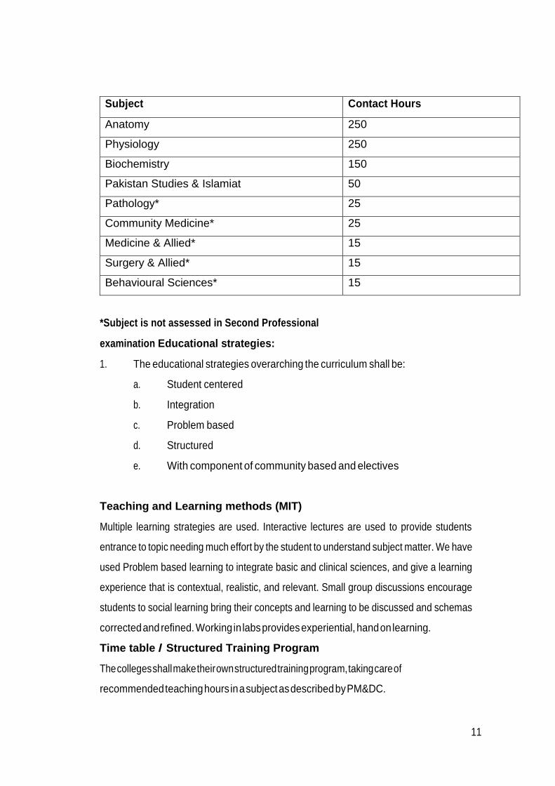
Subject Contact Hours
Anatomy 250
Physiology 250
Biochemistry 150
Pakistan Studies & Islamiat 50
Pathology* 25
Community Medicine* 25
Medicine & Allied* 15
Surgery & Allied* 15
Behavioural Sciences* 15
*Subject is not assessed in Second Professional
examination Educational strategies:
1. The educational strategies overarching the curriculum shall be:
a. Student centered
b. Integration
c. Problem based
d. Structured
e. With component of community based and electives
Teaching and Learning methods (MIT)
Multiple learning strategies are used. Interactive lectures are used to provide students
entrance to topic needing much effort by the student to understand subject matter. We have
used Problem based learning to integrate basic and clinical sciences, and give a learning
experience that is contextual, realistic, and relevant. Small group discussions encourage
students to social learning bring their concepts and learning to be discussed and schemas
corrected and refined. Working in labs provides experiential, hand on learning.
Time table / Structured Training Program
The colleges shall make their own structured training program, taking care of
recommended teaching hours in a subject as described by PM&DC.
12
Internal Assessment.
During the module the students shall be continually formatively assessed. The weightage of internal
assessment shall be 10 % in 2nd professional MBBS Examination. There shall be three modular and one
pre -annual examination. The scores of tests at the end of each modular assessment and pre-
annual examination shall be used for calculation of the internal assessment.
Module and Pre-Annual Examination
1. There will be three module examinations, one at the end of each module.
2. There will be only one Pre-annual examination.
3. The structure of the paper of all the module examinations and pre-annual will be same as
that for annual examination though syllabus will be different.
4. The syllabus for modular examination will be announced by the department at least 02 weeks
prior to examination.
5. Pre-annual examination will be from whole syllabus.
6. The date sheet for Module and pre-annual examinations will be published by
Examination branch of college while the examinations will be conducted by respective
department.
The result will be submitted to NUMS examination branch for incorporation in internal
assessment before annual examination
Annual Professional Examination.
The University shall take the 2nd professional Examination as per PM&DC guidelines at the end of the
academic year. Each subject section has table of specification of Module, Pre- annual and Annual
examination. Annual Theory & Practical Examination shall be of 200 marks each in; Anatomy, Physiology
and Biochemistry. The pass score shall be 50% in theory and practical separately. The detail marked
distribution of 2ND year is as under.

13
S
/
N
Subject
MCQs
PBQs
SAQs
SEQs
Int
Assess Sub
Total
Oral &
Practical
Int
Assess
Sub
Total
Grand
Total
1 Anatomy 25 65 10 100 90 10 100 200
2 Physiology 25 65 10
100 90 10 100 200
3 Biochemistry 25 65 10 100 90 10 100 200
600
Islamiat &
Pak Studies
There will be a 100 marks written paper and students need to pass this
subject securing minimum 33% marks for the award of bachelor degree
later on.
14
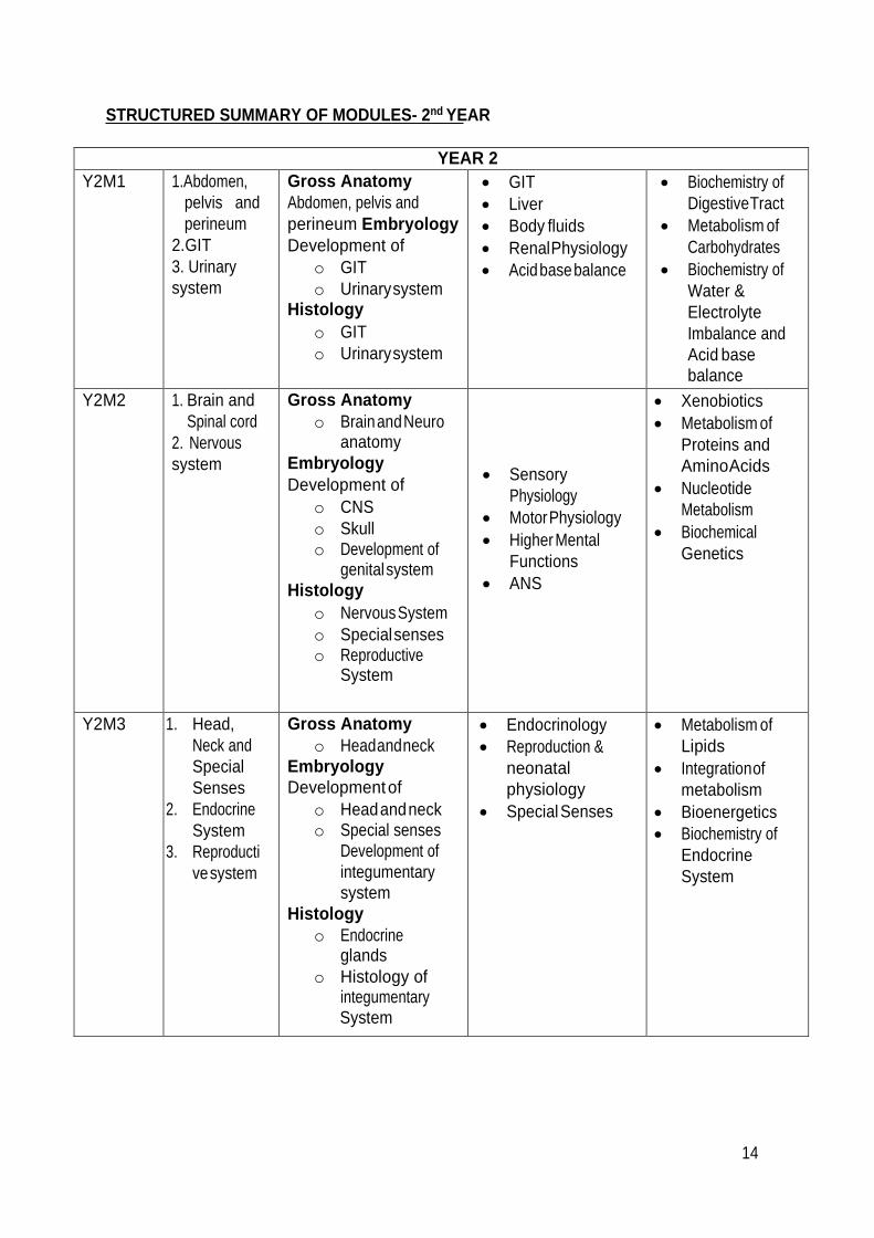
STRUCTURED SUMMARY OF MODULES- 2nd YEAR
YEAR 2
Y2M1 1.Abdomen,
pelvis and
perineum
2.GIT
3. Urinary
system
Gross Anatomy
Abdomen, pelvis and
perineum Embryology
Development of
o GIT
o Urinary system Histology
o GIT
o Urinary system
GIT
Liver
Body fluids
Renal Physiology
Acid base balance
Biochemistry of
Digestive Tract
Metabolism of
Carbohydrates
Biochemistry of
Water &
Electrolyte
Imbalance and
Acid base
balance
Y2M2 1. Brain and
Spinal cord
2. Nervous
system
Gross Anatomy
o Brain and Neuro anatomy
Embryology
Development of
o CNS
o Skull o Development of
genital system
Histology
o Nervous System
o Special senses o Reproductive
System
Sensory
Physiology
Motor Physiology
Higher Mental
Functions
ANS
Xenobiotics
Metabolism of
Proteins and
Amino Acids
Nucleotide
Metabolism
Biochemical
Genetics
Y2M3 1. Head,
Neck and
Special
Senses
2. Endocrine
System
3. Reproducti
ve system
Gross Anatomy
o Head and neck
Embryology
Development of
o Head and neck o Special senses
Development of
integumentary
system
Histology
o Endocrine glands
o Histology of integumentary System
Endocrinology
Reproduction &
neonatal
physiology
Special Senses
Metabolism of
Lipids
Integration of
metabolism
Bioenergetics
Biochemistry of
Endocrine
System

15
BLOCK-I
Section-I
Anatomy
16
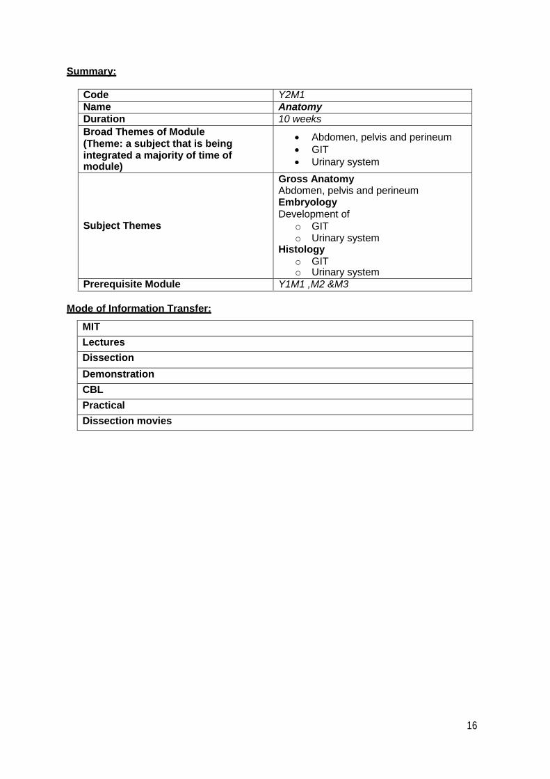
Summary:
Code Y2M1
Name Anatomy
Duration 10 weeks
Broad Themes of Module (Theme: a subject that is being integrated a majority of time of module)
Abdomen, pelvis and perineum
GIT
Urinary system
Subject Themes
Gross Anatomy Abdomen, pelvis and perineum Embryology Development of
o GIT o Urinary system
Histology
o GIT o Urinary system
Prerequisite Module Y1M1 ,M2 &M3
Mode of Information Transfer:
MIT
Lectures
Dissection
Demonstration
CBL
Practical
Dissection movies

17
GROSS ANATOMY:
Topic Learning Objectives MITs
Abdomen Division of abdomen into regions and quadrants and their contents
Describe the Division of abdomen into regions and quadrants
Enlist the contents of abdominal regions
Anterior abdominal wall
Describe the details of anterior abdominal wall.
Identify the layers of abdominal wall.
Identify the superficial and deep fascia and muscles of abdominal wall.
Describe the formation of rectus sheath and its importance.
Nerves of abdomen
Describe nerve supply of anterior and posterior abdominal wall.
Identify & create a visual representation of nerves supplying the abdomen.
Sequence and categorize information on the segmental sympathetic supplies and referred pain.
Explain the basic structure of paravertebral plexuses.
Describe somatic nervous supply of abdomen
Inguinal Canal
Describe Walls of Inguinal Canal
Describe Deep Inguinal Ring & Superficial Inguinal Ring
Identify Structures passing through the inguinal canal
Enlist Coverings of spermatic cord
Explain Mechanics of the inguinal Canal
Define hernia and describe its types
Discuss Direct & indirect Inguinal Hernia
Discuss Surface marking of inguinal canal
Peritoneal Cavity & Peritoneal Relationships
Define peritoneum
Understand the different folds of peritoneum, i.e., peritoneal ligaments, omenta and mesenteries.
Discuss the pouches, recesses and gutters formed by peritoneal enfoldings
Describe greater and lesser sacs
Enlist the intraperitoneal and retroperitoneal viscera
Discuss vertical tracings of peritoneum
LGIS/ Demos
18
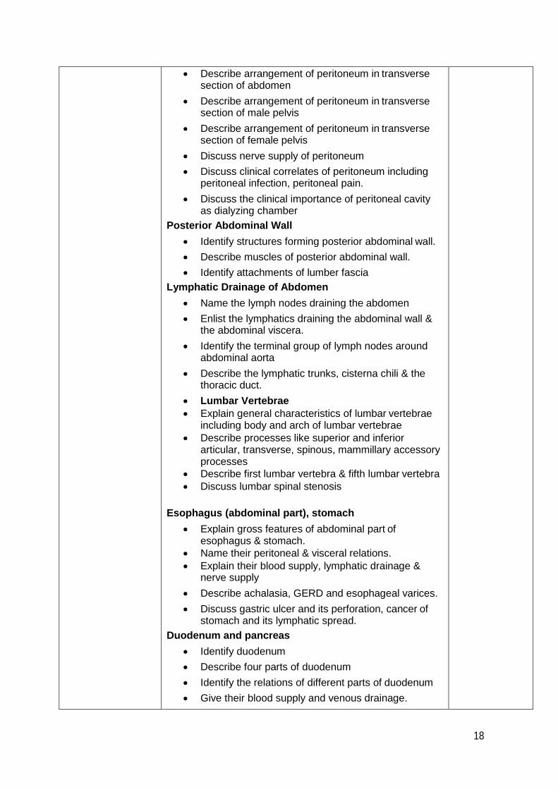
Describe arrangement of peritoneum in transverse section of abdomen
Describe arrangement of peritoneum in transverse section of male pelvis
Describe arrangement of peritoneum in transverse section of female pelvis
Discuss nerve supply of peritoneum
Discuss clinical correlates of peritoneum including peritoneal infection, peritoneal pain.
Discuss the clinical importance of peritoneal cavity as dialyzing chamber
Posterior Abdominal Wall
Identify structures forming posterior abdominal wall.
Describe muscles of posterior abdominal wall.
Identify attachments of lumber fascia
Lymphatic Drainage of Abdomen
Name the lymph nodes draining the abdomen
Enlist the lymphatics draining the abdominal wall & the abdominal viscera.
Identify the terminal group of lymph nodes around abdominal aorta
Describe the lymphatic trunks, cisterna chili & the thoracic duct.
Lumbar Vertebrae
Explain general characteristics of lumbar vertebrae including body and arch of lumbar vertebrae
Describe processes like superior and inferior articular, transverse, spinous, mammillary accessory processes
Describe first lumbar vertebra & fifth lumbar vertebra
Discuss lumbar spinal stenosis
Esophagus (abdominal part), stomach
Explain gross features of abdominal part of esophagus & stomach.
Name their peritoneal & visceral relations.
Explain their blood supply, lymphatic drainage & nerve supply
Describe achalasia, GERD and esophageal varices.
Discuss gastric ulcer and its perforation, cancer of stomach and its lymphatic spread.
Duodenum and pancreas
Identify duodenum
Describe four parts of duodenum
Identify the relations of different parts of duodenum
Give their blood supply and venous drainage.
19
Small Intestine & large intestine (comparison of two)
Describe the basic anatomy of small &large intestine
Identify the important gross features of large intestine
Explain the basic gross features which differentiate large intestine from small intestine
Identify the appendix on the basis of its distinguished features
Give relations of small and large intestine.
Describe the characteristics of ano-rectal regions
Discuss the blood supply, nerve supply and venous and lymphatic drainage of small and large intestine.
Discuss clinical correlates of small and large intestines and appendix
Discuss meckels diverticulum, resection of different parts of gut and its clinical effect
Discuss clinical problems occurring due to occlusion of GIT blood vessels
Abdominal aorta+ blood supply of abdomen
Describe the position and the vertebral levels of aorta in the abdomen.
Enlist the main branches of the aorta and their territories.
Explain the applied anatomy of the aorta.
Inferior vena cava + venous drainage of abdomen
Describe the formation of inferior vena cava
Enlist the tributaries of inferior vena cava
Explain abdominal and thoracic relations of this vein
Discuss clinical importance of inferior vena cava.
Liver
Describe the anatomical structure of liver.
Identify lobes, surfaces and ligaments
of liver.
Discuss its relations
Identify bare area of liver on a model of liver.
Give its blood supply lymph drainage and nerve supply
Discuss its clinical correlations
Gall bladder and biliary tract
Describe the location, size, relation and blood supply of gallbladder
Explain differences between Intra & Extra Hepatic Biliary Systems
List different components of Extra-hepatic biliary System

20
Identify the right & left hepatic ducts, common hepatic duct, cystic ducts, bile duct
Describe clinical conditions related to gallbladder
Hepatic portal system
Describe the hepatic portal circulation.
Explain the anatomy of hepatic vein.
Describe the Portal -Caval anastomosis.
Explain the clinical correlation of hepatic portal system.
Kidney
Describe the gross features of kidney and its coverings
Differentiate the anterior and posterior surfaces and relations of kidney.
Identify the internal structure of kidney
Describe the blood Supply of Kidney
Describe the Lymph nodes draining the kidney
Explain the Nerve supply of Kidney
Identify ureter, urinary bladder and urethra
Describe the course constrictions and relations of ureter
Discuss the blood supply and venous drainage of ureter.
Give location and description of suprarenal glands
Discuss their gross features and relations
Discuss their blood supply lymph drainage and nerve supply
Give clinical correlations of kidney ureter and suprarenal glands
Identify surface marking of stomach, spleen, liver, gall bladder, kidney & appendicular orifice.
Identify the surface anatomy of
Kidney,
Ureter and
Urinary bladder
Perform the Surface anatomy of the kidney on human bony landmarks
Pelvis
Bones and joints
Define bony pelvis, true and false pelvis
Describe surfaces of sacrum.
Explain articulation.
Identify muscles associated with sacrum.
Differentiate between male and female sacrum.
Enlist various types of joints of pelvis.
Explain type, articulations, ligaments and relations of joints.
Enlist factors providing stability to joint.
Describe blood supply , nerve supply & movements of joint
Differentiate the greater & lesser pelvis. Describe the superior & inferior circumference and
LGIS/ Demos

21
their boundaries.
Describe the anatomical position of pelvis.
Differentiate the shapes of female pelvis regarding childbirth.
Differentiate between male & female pelvis.
Pelvic diaphragm
Vessels and nerve supply of pelvis
Describe the anatomy of the pelvic walls.
Discuss the muscles of pelvic floor and formation of pelvic diaphragm
Develop an understanding of blood supply, nerve supply, and lymphatic drainage of muscles.
Describe actions of pelvic diaphragm
Identify pelvic nerves.
Describe Sacral plexus.
Identify coccygeal plexus.
Describe pelvic hypogastric plexus.
Discuss main arteries of pelvis common iliac artery external iliac artery internal iliac artery arteries of true pelvis.
Describe main veins of the pelvis and their tributaries.
Identify area of drainage of these veins.
Describe different groups of lymph nodes.
Explain the role of lymphatics and common route and spread of malignancies of pelvis.
LGIS/ Demos
Sigmoid colon and rectum
Describe sigmoid colon.
Describe rectum.
Explain relations, blood supply and innervation of these pelvic organs
Discuss their important clinical correlations
LGIS/ Demos
Urinary bladder Discuss urinary bladder, its peritoneal covering and internal structure
Discuss blood supply venous drainage and lymph drainage of urinary bladder
Describe nerve supply and mechanism of micturition
Discuss clinical correlates of urinary bladder
including urinary retention, difficulty with micturition after spinal cord injury, bladder injuries
LGIS/ Demos
Male genital organs
Explain male genital organs, their structure, position, function and important relations
Discuss vas deferens, seminal vesicle, and ejaculatory ducts
Give their blood supply and lymphatic drainage
Discuss prostate, its lobes and its relations
Describe its blood supply and lymphatic drainage
Discuss its clinical correlates including benign prostatic hyperplasia and CA prostate.
LGIS/ Demos
Ovaries fallopian tubes and uterus
Identify ovaries and fallopian tubes.
Describe the parts of ovaries and fallopian tubes. Identify the ligaments of ovaries
LGIS/ Demos
22
Enumerate the clinical correlates of ovaries and uterine tubes.
Explain the details of uterus, cervix and vagina.
Enumerate the parts of uterus, ligaments, relations and support of uterus.
Discuss the role of uterus in labour
Identify the clinical correlates of uterus, cervix and vagina
PERINIUM Identify borders and relations of the perineum.
Describe divisions of the perineum.
Explain superficial and deep perineal pouch and their contents
Explain cutaneous nerves of the perineum. Define perineal body.
LGIS/ Demos
Anal canal Explain the gross anatomy of Anal Canal
Identify the relations of the anal canal with the surrounding structures.
Describe the blood supply, venous and lymphatic drainage of anal canal.
Explain innervations of anal canal.
Discuss clinical conditions of anal canal.
Describe hemorrhoids and their types
Discuss perianal hematoma, fissure, abscess and fistula
Discuss incontinence after trauma and spinal cord injury
Ischiorectal fossa Identify the boundaries and recessess of ischiorectal fossa
Describe the contents of ischiorectal fossa Describe ischiorectal fossa infection
Testis Describe the coverings of testis.
Recognize the internal features of testis.
Explain the significance of pampiniform plexus.
Justify the location of testis outside the body
Integrate the knowledge of descent of testis to its vessels, lymphatics and nerves.
Recall the different clinical conditions associated with testis.
Male Urogenital Triangle
Describe gross anatomy of male external genitalia.
Describe the gross structure of penis
Explain its arterial, venous drainage & nerve supply.
Describe scrotum and its walls
Discuss its blood supply and lymphatic drainage
Describe the nerve supply of anterior and posterior walls of scrotum.
Explain anatomy of male urethra, its arterial, venous drainage & nerve supply.
Discuss injury to different parts of male urethera and extravastion of urine.
LGIS/ Demos
Female Urogenital Triangle
Enlist the names and anatomical location of female external genitalia.
LGIS/ Demos
23
Explain function, arterial supply, venous drainage and nerve supply of female external genitalia.
Discuss clinical importance of female external genitalia.
Explain course & relations of female urethra.
Describe arterial supply, venous drainage and nerve supply of female urethra.
Discuss clinical importance of female urethra.
EMBROYOLOGY
GIT Describe the divisions of primitive gut.
Describe the derivatives of foregut.
Describe the development of the derivatives of foregut.
Describe the applied anatomy of derivatives of foregut
Describe the derivatives of midgut.
Explain physiological herniation of midgut
Explain the rotation of midgut.
Describe derivatives of hindgut.
Discuss the formation of dorsal and ventral mesentry and structures taking origin from them
Describe the clinical correlation of mid and hind gut.
Describe the congenital anomalies of gut
Enlist the special features associated with common anomalies related to gut including:
Cleft lip or cleft palate Hernias
Esophageal atresia
Describe the congenital anomalies of gut
Enlist the special features associated with common anomalies related to gut including:
Mal-rotation of gut Anorectal malformations
Explain the hepatic and cystic buds
Discuss the site and source of parenchymal and stromal tissue of liver and gall bladder.
Explain the origin of pancreatic buds and their derivatives in adult pancreas
Explain congenital anomalies of liver, gall bladder and pancreas.
LGIS
Urinary System Describe the development of kidneys, their collecting system and excretory system
Discuss the congenital anomalies of kidneys, renal agenesis, horse shoe kidney, and wilms tumor
Describe the development of urinary bladder.
Explain the development of urethra. Describe the congenital anomalies related to them.
LGIS
HISTOLOGY
GIT Give overview of digestive system
Describe structure of the gastrointestinal tract, GIT Explain histological features of layers of GIT
LGIS / Lab
24
Describe histological features of structure of each layer of esophagus
Describe different regions of stomach, grossly and histologically
Explain various layers of the wall of stomach
Describe different glands and the various kind of cells present in them
Identify the parts of small intestine.
Describe the histological features of different parts of small intestine.
Briefly review the gross anatomy of pancreas
Discuss the histological components of pancreas
Discuss the histological details of Parenchyma and Lobules (acini) of Pancreas
Discuss the Duct System of Pancreas
Describe the endocrine component of pancreas
Describe the basic anatomy of large intestine
Identify the important histological features of large intestine
Explain the basic histological features which differentiate large intestine from small intestine
Identify the appendix on the basis of its distinguished features
Describe the characteristics of anorectal regions
Identify histology of liver
Explain common liver disorders
Explain clinical manifestations of liver disorders.
Describe Gall bladder histology
Describe the histological architecture of liver
Identify the structural details of hepatocytes, portal triad, hepatic sinus & hepatic lobule
Describe the different components of biliary tract Identify the histological appearance of gall bladder
Urinary System Describe the detailed microscopic features of nephron and collecting ducts
Describe the location of the ureter & urinary bladder
Explain the histology of Ureter, Urinary bladder and Urethra
LGIS / Lab
25
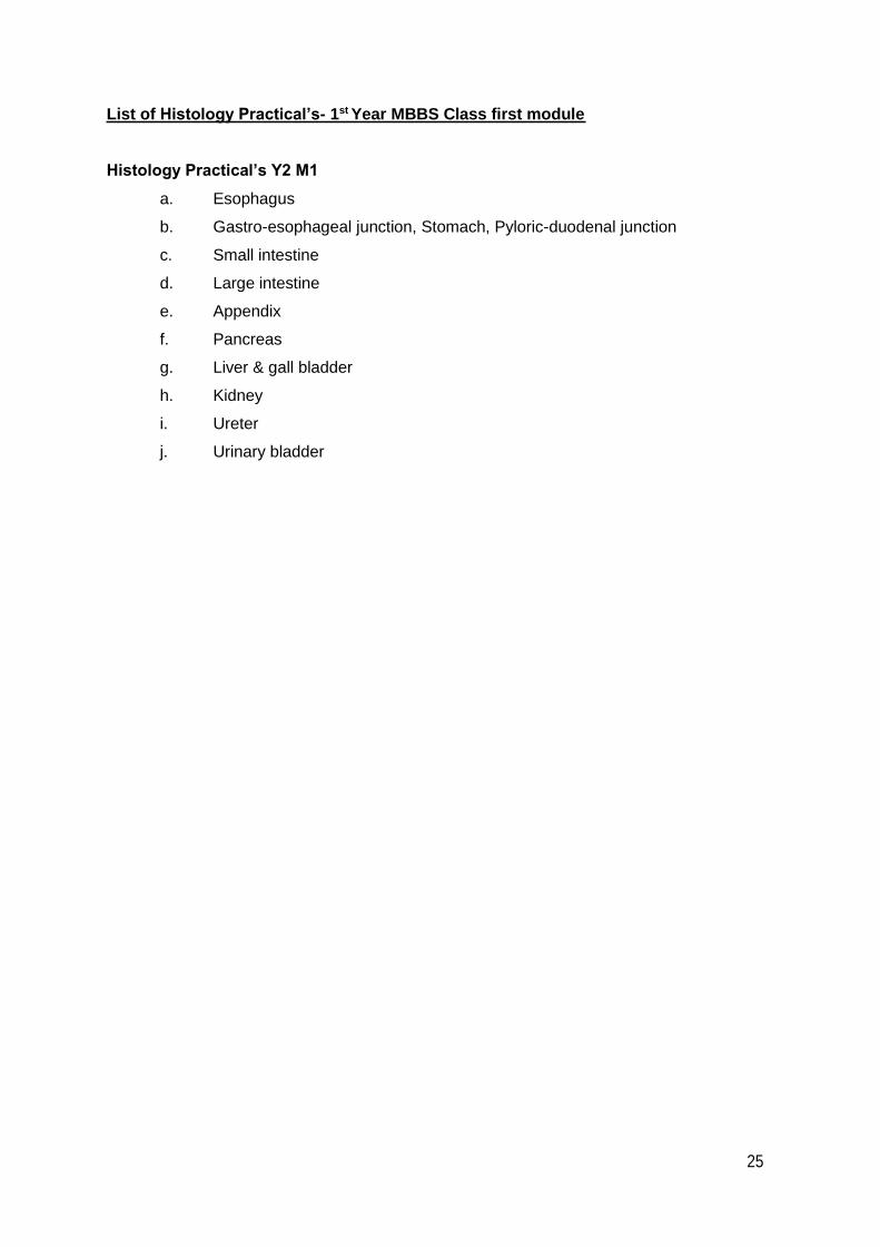
List of Histology Practical’s- 1st Year MBBS Class first module
Histology Practical’s Y2 M1
a. Esophagus
b. Gastro-esophageal junction, Stomach, Pyloric-duodenal junction
c. Small intestine
d. Large intestine
e. Appendix
f. Pancreas
g. Liver & gall bladder
h. Kidney
i. Ureter
j. Urinary bladder

26
Case Base Learning Scenarios:
CBL 1 - Anatomy A case of abdominal wall hernia
Learning objectives:
1. Revise the structure of the anterior abdominal wall, its muscles, nerve supply, blood
supply and lymphatic drainage.
2. What are the different abdominal planes and regions? What is the significance of
dividing the abdomen into regions?
3. Define and learn to mark the inguinal rings.
4. Learn the walls, function and mechanics of inguinal canal.
5. What is hernia? What are the parts of hernia and various types of abdominal wall
hernias?
6. Which type of hernia is present in this patient? And how did you make your diagnosis?
7. Learn to differentiate between the direct and indirect inguinal hernia.
8. What is processus vaginalis? And how is it related to inguinal hernia?
9. Why is it necessary to repair a hernia?
10. Which nerve is susceptible to injury during the surgical repair of inguinal hernia? And
what are its effects?
Reading References:
1. Snell’s Clinical Anatomy, 8th Edition.
2. Clinically Oriented Anatomy, Keith L.Moore, 6th Edition.
3. www.med.umich.edu/lrc/coursepages/.../anatomy/.../clinical_index.ht...
CBL 2 - Anatomy
A case of gastric ulcer
Learning objectives:
1. Learn the parts of stomach, its relations, stomach bed, blood supply, nerve supply and
lymphatic drainage.
2. To learn the histological structure of stomach and know the factors which protect the
gastric mucosa from acid and enzymes.
3. Define peritoneum and learn its functions.
4. To understand the terms intraperitoneal and retroperitoneal.
5. Understand and locate the peritoneal ligaments, omenta, mesenteries, pouches,
recesses and spaces.
6. What are the greater and lesser sacs of peritoneum?

27
7. How does the greater peritoneal sac communicate with the omental bursa?
8. Account for the chronic ulcer pain (heart burn, diffuse pain epigastrium) that the patient
complained of in his history from the acute, sharp, stabbing pain after the perforation
has taken place.
9. What is the cause of the rigidity of the abdominal wall and costal character of the
patient’s respiration?
10. Which major branch of the celiac trunk would be subject to erosion and possible fatal
hemorrhage if an ulcer on the posterior wall of the stomach perforates through the
gastric mucosa?
Reading References:
1. Snell’s Clinical Anatomy, 8th Edition.
2. Clinically Oriented Anatomy, Keith L.Moore, 6th Edition.
3. www.med.umich.edu/lrc/coursepages/.../anatomy/.../clinical_index.ht...
CBL 3 - Anatomy A case of cirrhosis liver
Learning objectives:
1. To learn the gross anatomy of liver, its lobes (anatomical and physiological), peritoneal
reflections, blood supply and relations.
2. Revise the functions of liver.
3. What are the bile ducts of liver?
4. What is the microanatomy and circulation of the liver?
5. What is the hepatic portal system?
6. To learn the location and tributaries of portal vein.
7. What are the connections between the portal circulation and the systemic circulation?
Recall the sites of portal - systemic anastomoses.
8. What goes wrong in cirrhosis of the liver?
9. Why was this patient jaundiced?
10. What is portal hypertension? Why does portal hypertension occur in cirrhosis?
11. Which signs in this patient indicate the portal hypertension?
Reading References:
1. Snell’s Clinical Anatomy, 8th Edition.
2. Clinically Oriented Anatomy, Keith L.Moore, 6th Edition.
3. www.med.umich.edu/lrc/coursepages/.../anatomy/.../clinical_index.ht...

28
CBL 4 - Anatomy A case of prolapse of uterus Learning objectives:
1. To know the meaning of uterine, prolapse, stress incontinence and cystocele.
2. To learn the gross features of the uterus and vagina, along with the relations, blood
supply, nerve supply and the lymphatic drainage.
3. To revise the peritoneal reflections of the pelvis and the uterus.
4. What are the supports of uterus?
5. What are ligaments of the uterus?
6. Describe the anatomy of the pelvic diaphragm? What muscles make it up?
7. To learn the significance of pelvic diaphragm.
8. The cystocele caused a bulge in the anterior wall of the vagina. What structures could
prolapse and cause a bulge in the posterior wall of the vagina?
Reading References:
1. Snell’s Clinical Anatomy, 8th Edition.
2. Clinically Oriented Anatomy, Keith L.Moore, 6th Edition.
3. www.med.umich.edu/lrc/coursepages/.../anatomy/.../clinical_index.ht...
CBL 5- Anatomy Patient with bleeding per rectum Learning Objectives:
1. Recognize the features of the rectum that differentiate it from the colon.
2. Describe the development of Rectum and Anal canal
3. Describe the development of cloacae and urogenital sinus
4. Describe ano-rectal anomalies
5. Describe the blood supply, venous and lymphatic drainage of anal canal
6. Enlist causes of bleeding per rectum
7. Describe the boundaries of ischiorectal fossa
8. Demonstrate History taking on a SP with lower GIT bleed
9. Demonstrate examination of a SP with lower GIT bleed.
Reading References:
1. Snell’s Clinical Anatomy, 8th Edition.
2. Clinically Oriented Anatomy, Keith L.Moore, 6th Edition.
3. www.med.umich.edu/lrc/coursepages/.../anatomy/.../clinical_index.ht...

29
Learning Resources:
Anatomy
1. Clinical Anatomy for Medical Students by Richard Snell (8th edition).
2. Basic Histology Text and Atlas by Luiz Carlos and Junqueira (12th latest edition).
3. Medical Embryology by Langman (11th edition).
4. Essential Clinical Anatomy by Keith Moore (6th edition).
5. The Developing Human (clinical oriented embryology) by Keith Moore (8th edition).

30
BLOCK-I
Section-II
Physiology

31
Summary:
Code Y2M1
Name Physiology
Duration 10 weeks
Broad Themes of Module
(Theme: a subject that is being integrated a
majority of time of module)
Abdomen, pelvis and perineum
GIT
Urinary system
Subject Themes
GIT
Liver
Body fluids
Renal Physiology
Acid base balance
Prerequisite Module Y1M1 ,M2 &M3
Mode of Information Transfer:
MIT
Lectures
Tutorials (PTT)
CBL
Practicals
Class tests
Physiology learning outcomes and MITs:
S.No. Topic Learning objectives
GIT Physiology
1. GIT physiology (LGIS, SGD/tutorial, Practical)
To know the physiologic anatomy of gastrointestinal wall
To understand the role of intestinal cells of cajal in the
electrical activity of G.I smooth muscle
To know the enteric nervous system and its role in
control of G.I function
To be able to differentiate between myenteric and sub
mucosal plexuses
To be able to explain the autonomic control of G.I tract
2. Chewing/swallowing
reflex
(LGIS)
To be able to explain importance of chewing
To know the mechanism of chewing reflex
To be able to describe the process of swallowing
To understand different phases of swallowing reflex
To understand different steps occurring in the
involuntary phase of swallowing
32
To know the effects of pharyngeal phase of
swallowing on respiration
To know how different types of peristalsis in
esophagus are taking place
To understand the importance of esophageal sphincter
3. Functions of
stomach & gastric
emptying
(LGIS)
To be able to categorize different functions of
stomach
To know the role of basic electrical rhythm in
regulation of G.I motility
To understand the process of stomach emptying
To be able to explain the different factors regulating
stomach emptying
To know secretion of different hormones taking place in
stomach
To be able to explain different steps taking place in the secretion of hydrochloric acid in stomach
4. Functions of small
intestine (LGIS) To be able to categorize different types of movements taking
place in small intestine
To understand role of ileocecal valve
To understand secretory functions of small intestine
5. Functions of large
intestine (LGIS) To be able to categorize different functions of large
intestine
To be able to explain different types of movements taking
place in colon
To understand the role of gastrocolic and
duodenocolic reflexes in regulation of mass
movements
To know the secretory functions of large intestine and its nervous control
7. Defecation reflex (LGIS)
To be able to explain the process of defecation
To understand the pathway of defecation reflex
To know different types of defecation reflex
To know the pathophysiological basis of megacolon
8. Vomiting reflex (LGIS)
To understand the factors leading to the process of
vomiting
To be able to explain the location of vomiting center in the
brain
To be able to explain the vomiting reflex
To understand the role of chemoreceptor trigger zone for
initiating vomiting
9. Hormones of GIT (LGIS)
To be able to categorize the different types of G.I
hormones
To understand the secretion of different hormones
from G.I.T and their regulation
10. Functions of liver To be able to categorize different functions of liver

33
To understand the role of liver in the
bilirubin
To know the synthetic functions of liver
metabolism of
Body fluids
1. Body fluid
compartments
(LGIS)
To be able to explain total body water content and its
distribution in different body compartments
To be able to quantify daily intake and output of water from
body
To understand the fluid present in the potential spaces and
mechanism of their collection in these spaces
To know the ionic composition of ECF and ICF
2. Water balance
(LGIS) To understand the basic principles of osmosis and
osmotic pressure
To know the mechanism of maintenance of osmotic
equilibrium between ICF and ECF
To be able to explain what would be the effect on ICF
and ECF compartments when isotonic, hypotonic and
hypertonic solution are added to ECF
3. Edema (LGIS)
To understand the role of starling forces in the
development/ prevention of edema
To describe role of lymphatics in prevention of edema
To be able to understand safety factor and its role in the
prevention of edema
To be able to describe the causes of intracellular edema
To be able to describe the causes of extracellular
edema
Renal Physiology
1. Renal physiology (LGIS, SGD/tutorial)
To know the functional anatomy of urinary system
To understand the multiple functions of kidneys
To know the physiology of micturition
To understand the processes involved in urine formation
resulting from glomerular filtration, tubular reabsorption,
and tubular secretion
2. GFR and
regulation
(LGIS,SGD/
tutorial)
its To know the composition of the glomerular filtrate and
glomerular capillary membrane
To understand the determinants of the GFR
To understand the physiological control of glomerular
filtration and renal blood flow
To know the autoregulation of GFR and renal blood
flow
3. Processing of
glomerular filtrate
(LGIS,
SGD/tutorial)
To be able to describe reabsorption and secretion by the
renal tubules
To understand the passive and active mechanisms
involved in tubular reabsorption

34
To understand the reabsorption and secretion along
different parts of the nephron
To learn about the regulation of tubular reabsorption
To know use of clearance methods to quantify kidney
function
4. Regulation of
Potassium
(LGIS)
To know about the regulation of internal potassium
distribution
To understand the potassium secretion by principal cells of
late distal and cortical collecting tubules
To be able to explain different factors that regulate
potassium secretion: plasma potassium concentration,
aldosterone, tubular flow rate, and hydrogen ion
concentration
5. Regulation of B.P (LGIS)
To know about the role of kidneys in pressure
natriuresis and diuresis
To understand the renal regulation of body fluid
volumes and arterial pressure
To understand role of nervous and hormonal factors in renal-body fluid feedback control
6. Renal regulation
of osmolarity
(LGIS)
To know the control of extracellular fluid osmolarity and
sodium concentration by kidneys
To know the osmoreceptor-ADH feedback system
To understand the role of thirst in controlling
extracellular fluid osmolarity and sodium
concentration
To understand the role of angiotensin II and
aldosterone in controlling extracellular fluid osmolarity and
sodium concentration
7. Micturition reflex (LGIS, SGD/tutorial)
To learn the physiologic anatomy and nervous
connections of the bladder
To understand the filling of the bladder and bladder wall
tone; the cystometrogram
To be able to explain the micturition reflex and
facilitation or inhibition of micturition by the brain
To know about the abnormalities of micturition
8. Formation of
concentrated
urine
(LGIS,
SGD/tutorial)
To understand the obligatory urine volume
To know about the requirements for excreting a
concentrated urine—high ADH levels and hyperosmotic
renal medulla
To understand the countercurrent mechanism producing
a hyperosmotic renal medullary interstitium
To know the role of distal tubule and collecting ducts in
excreting a concentrated urine
To understand the role of urea in hyperosmotic renal
medullary interstitium and formation of concentrated urine

35
To understand the countercurrent exchange in the vasa recta
in preservation of hyperosmolarity of the renal medulla
To be able to explain the concentrating mechanism and
changes in osmolarity in different segments of the tubule
To be able to quantify renal urine concentration and
dilution: “Free Water” and osmolar clearances
To know about the disorders of urinary concentrating ability
9. Plasma clearance (LGIS, SGD/tutorial)
To know the use of clearance methods to quantify
kidney function
To know about estimation of GFR by inulin clearance, and
plasma creatinine clearance
To understand PAH clearance for estimation of renal
plasma flow
To understand the calculation of filtration fraction, tubular
reabsorption and secretion from renal clearance
10. Acid base balance (LGIS, SGD/tutorial)
To know the defenses against changes in hydrogen ion
concentration: buffers, lungs, and kidneys
To know the buffering of hydrogen ions in the body fluids
To understand the bicarbonate buffer system and
quantitative dynamics of the bicarbonate buffer system
To understand the phosphate buffer system, proteins:
important intracellular buffers
To be able to explain the respiratory regulation of acid- base
balance
To understand renal control of acid-base balance and
secretion of hydrogen ions and reabsorption of
bicarbonate ions by the renal tubules
To understand the combination of excess hydrogen ions
with phosphate and ammonia buffers in the tubule—A
mechanism for generating new bicarbonate ions
11. Acid base
Disorders
(LGIS, SGD/tutorial)
To know the Renal Correction of acidosis—increased
excretion of hydrogen ions and addition of bicarbonate ions to
the extracellular fluid
To know the renal correction of alkalosis—decreased
tubular secretion of hydrogen ions and increased
excretion of bicarbonate ions
To understand causes of acid base disorders
To understand concept of anion gap

36
Approved List of Practical
Module 1
1. Consultation of research paper at under graduate level
2. Examination of Vital Signs
3. Blood glucose estimation
4. Determination of urine specific gravity
5. Examination of body Temperature
6. Examination of Body Mass Index (BMI)
7. Any other practical relevant to that Module
List of Case Based Learning (CBL)
CBLs Y2M1
1. Gastrointestinal physiology (4)
2. Renal physiology (4)
Total number of CBLs = 8
Gastrointestinal physiology
CBL 1
A 49 yrs old man presented in OPD with complaints of digestive problems. He had difficulty in
swallowing both solids and liquids and occasionally regurgitates. The problem is more noticeable
when he is under stress or when he eats too fast. He had the feeling that food is stuck in his esophagus
and is not going down. He lost 10 lb in last 2 months.
After physical examination the physician advised barium swallow. The report suggested that he has
achalasia. He was advised procedure to physically dilate LES.
Learning objectives
1. To know about enteric nervous system.
2. To know about peristalsis, law of gut and its significance.
3. Role of esophageal peristalsis in normal swallowing.
4. What events occur at LES and at what timing.
5. To know about innervations of lower esophageal sphincter.

37
CBL 2
An 18 years old boy came to OPD of a hospital with complaints of vomiting and diarrhea since last night. He
gave history of taking lunch at a local restaurant a day before yesterday. About a day after that lunch, he
developed nausea followed by profuse vomiting and watery diarrhea. On general physical
examination, moderate dehydration was noticed. Laboratory investigations revealed presence of E. coli
in stool, TLC 12,000/µl with neutrophils 75% and serum levels of Na+ (138 meq/l) and Cl- (105 meq/l).
Patient was given anti emetic drug along with oral rehydration salt (ORS) and appropriate antibiotics.
He was advised to observe hygienic eating habits to avoid such incidence in future.
Learning objectives:
1. To know the role of enteric and autonomic nervous system in GI functions.
2. To know the functions of stomach and factors affecting gastric emptying.
3. To know the functions of small & large intestines.
4. To learn about defecation and vomiting reflexes.
5. To learn pathophysiology of diarrhea.
CBL 3
A 67 years old man reported to the emergency department with the complaints of acute onset
abdominal pain that aggravated over the past 7 hours. He also complained of uncontrollable
stool voiding, which were mucoid and blood stained. He had smoked more than 30 cigarettes a day,
for the last 40 years and gave a 3 yrs history of claudication.
Plain abdominal radiograph did not show any abnormality, however, a diagnosis of acute mesenteric
ischemia, consequent to thromboembolism of mesenteric vessels was established on the result of contrast
enhanced CT scan. The patient was prepared for total colectomy and the surgery went uneventful.
The surgeon advised the ward staff to ensure IV hydration therapy of the patient and daily blood
sampling for blood CP and electrolytes along with regular monitoring of his vital signs and 6 hourly ECG
recording. His vitamin K levels were also advised to be checked on every third day.
38
Learning objectives:
1. Know the functions of large intestine.
2. Understand the mechanisms of electrolyte homeostasis related to colon.
3. Co-relate the effects of electrolyte disturbances consequent to colectomy on ECG.
4. Describe the relation of vitamin K absorption and large intestine.
5. Elaborate the significance of colectomy with probable development of anemia in
this patient.
CBL 4
A 12-year-old boy brought to the hospital by his parents with complains of large amounts of watery
diarrhea and vomiting for 1 day. Diarrhea was sudden in onset and painless. The stool passed having
characteristic "rice-water" appearance with few flecks of mucus in it. He vomited large amount of
clear fluid 4-6 times in one day and having continuous feeling of nausea. He was thirsty and has
complains of intense cramps in his calf muscles. His vitals include: BP 90/70 mm Hg, Pulse 110
beats/min, RR 18 breaths/min& Temp 99 ○F. On general physical examination he was looking restless
and irritable with dry tongue, decreased skin turgor and sunken eyes. Lab investigations revealed
severe electrolyte imbalance with particularly decreased plasma concentrations of Na+, Cl− & HCO3−.
Microscopic examination of stool revealed teeming of diarrheal fluid with motile comma-shaped bacteria.
On the basis of history and clinical findings he was diagnosed as a case of cholera.
Learning objectives:
1. Discuss mechanism of development of diarrhea in cholera infection.
2. What should be your treatment plan for this patient?
3. Define diarrhea and classify it.
4. Understand various levels of dehydration and their associated signs & symptoms.
5. What are possible complications of severe dehydration?
6. Discuss pathway of vomiting reflex.
7. Elucidate steps of act of vomiting.
8. Differentiate between vomiting center and chemoreceptor trigger zone.
9. Discuss basic movements of small intestine.
10. Discuss regulation of peristaltic activity in the small intestine.
11. Discuss defecation reflexes.
12. Discuss important functions of normal flora (colonic bacteria) of colon.
39
Renal Physiology
CBL 5
A 5-year-old boy presents to the urgent care clinic because his mother noticed that the child is not going to
the bathroom and his feet are swollen. Two weeks earlier the child was treated for streptococcal throat
infection with penicillin. The mother admits to ceasing administering the medication to the child after a
couple of days because she thought the child felt better. PHYSICAL EXAMINATION
Vital Signs: Temp 37°C, Pulse 78/min, Resp rate 15/min, BP 120/90 mm Hg
Physical Examination: The patient is oliguric and has a slight fluid accumulation in the lower extremities
and peri-orbital region. There is palpable peripheral edema in both feet.
LABORATORY STUDIES
Urinalysis: Very dark urine, presence of red blood cells, red blood cell casts, and protein (>3 g/day)
Serum complement C3, C4, CH50 levels: Low
ASO titer: 250 units /mL (normal: < 160 units/mL) Anti D
Nase-B level: > 60 units
BUN: 32 mg/dL (normal: 7-18 mg/dL)
Creatinine: 2.0 mg/dL (normal: 0.6-1.2 mg/dL)
DIAGNOSIS Post streptococcal glomerulonephritis
Learning objectives:
1. After discussion you should be able to:
2. Describe the morphology of a typical nephron and its blood supply.
3. Define autoregulation and list the major theories advanced to explain autoregulation in the
kidneys.
4. Define glomerular filtration rate, describe how it can be measured, and list the
major factors affecting it.
5. Outline tubular handling of Na+ and water.
6. Discuss tubular reabsorption and secretion of glucose and K+.
7. Describe how the countercurrent mechanism in the kidney operates to produce
hypertonic or hypotonic urine.
8. List the major classes of diuretics; understand how each operates to increase urine flow.
9. Describe the voiding reflex and draw a cystometrogram.

40
CBL 6
Salar, a 20 years old teenager is fond of fast food and insists on drinking soft drinks whenever he feels
thirsty. On a recent trip to a fast food outlet he suspected eating stale burger and returned home
feeling nausea, light headedness and lasitude. During the night he had two episodes of vomiting and
had to go to washroom thrice for passing stools which were watery in consistency. He insisted on drinking
soft drinks when he felt thirsty and weak after vomiting with little relief to symptoms. On next day medical
examination, he was diagnosed with mild gastroenteritis and prescribed appropriate medication and
fluids. He recovered within 24 hours.
Learning objectives:
1. To understand the concept of ECF osmolarity and Na+ concentration.
2. To elaborate mechanism of excreting concentrated urine.
3. To know definitions of osmolar clearance and free water clearance.
4. To understand thirst mechanism and describe thirst centre and osmo-receptor ADH feedback
mechanism.
5. To elaborate regulation of blood volume.
6. To elaborate regulation of ECF volume.
CBL 7
A 16-year-old high school student is brought to the emergency department by the emergency medical
service after being found lying in the front yard of a neighbor’s house, where he was mowing the lawn.
The patient has a regular yard service and has been mowing for several months without problems.
The patient was finishing his sixth yard for the day during a summer month with temperatures
exceeding 37.8°C. His mowing partner noticed that the patient had been complaining of fatigue, light-
headedness, nausea, and profuse sweating in the previous yard. While mowing the last yard, he became
very confused and behaved oddly before finally losing consciousness. In the emergency department, he
is tachycardic, with a temperature of 40.6°C. He is lethargic, and his skin is dry. He is diagnosed with
heat stroke, and therapy is begun immediately.
Learning objectives:
1. To understand concept of fluid osmolarity.
2. To understand systemic effects of ECF osmolarity.
3. To develop knowledge of renal regulation of fluid osmolarity.
4. To develop an understanding of fluid regimens available for hydration.
5. To gain insight into fluid overload and edema formation in body.
41
CBL 8
A 21-year-old man with insulin-dependent diabetes presents to the emergency center with mental
status changes, nausea, vomiting, abdominal pain, and rapid respirations. On examination, the
patient is noted to be hypotensive, breathing rapidly (tachypneic), and febrile. A fruity odor is detected
on his breath. A random blood sugar is significantly elevated at 600 mg/dL. The patient also has
hyperkalemia, hypomagnesemia, and elevated serum ketones. An arterial blood gas reveals a
metabolic acidosis. The patient is diagnosed with diabetic ketoacidosis (DKA) and is admitted to the
intensive care unit for intravenous (IV) hydration, glucose control, and correction of metabolic
abnormalities.
Learning objectives:
1. To understand basis of acid base regulation in human body.
2. To list various renal and respiratory mechanisms for acid base regulation.
3. To develop an understanding of electrolyte buffer systems.
4. To develop and understanding of acid base disorders.
5. To interpret laboratory findings of various acid base disorders.
6. To gain insight into concept of anion gap.
7. To develop an understanding of treatment options available for acid base disorders.

42
BLOCK -I
Section-III
Biochemistry
43
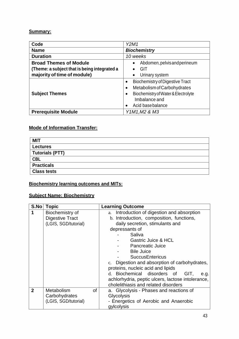
Summary:
Code Y2M1
Name Biochemistry
Duration 10 weeks
Broad Themes of Module
(Theme: a subject that is being integrated a
majority of time of module)
Abdomen, pelvis and perineum
GIT
Urinary system
Subject Themes
Biochemistry of Digestive Tract
Metabolism of Carbohydrates
Biochemistry of Water & Electrolyte
Imbalance and
Acid base balance
Prerequisite Module Y1M1,M2 & M3
Mode of Information Transfer:
MIT
Lectures
Tutorials (PTT)
CBL
Practicals
Class tests
Biochemistry learning outcomes and MITs:
Subject Name: Biochemistry
S.No Topic Learning Outcome
1 Biochemistry of Digestive Tract (LGIS, SGD/tutorial)
a. Introduction of digestion and absorption b. Introduction, composition, functions, daily secretion, stimulants and depressants of
- Saliva - Gastric Juice & HCL - Pancreatic Juice - Bile Juice - SuccusEntericus
c. Digestion and absorption of carbohydrates, proteins, nucleic acid and lipids d. Biochemical disorders of GIT, e.g. achlorhydria, peptic ulcers, lactose intolerance, cholelithiasis and related disorders
2 Metabolism of Carbohydrates (LGIS, SGD/tutorial)
a. Glycolysis - Phases and reactions of Glycolysis - Energetics of Aerobic and Anaerobic gylcolysis
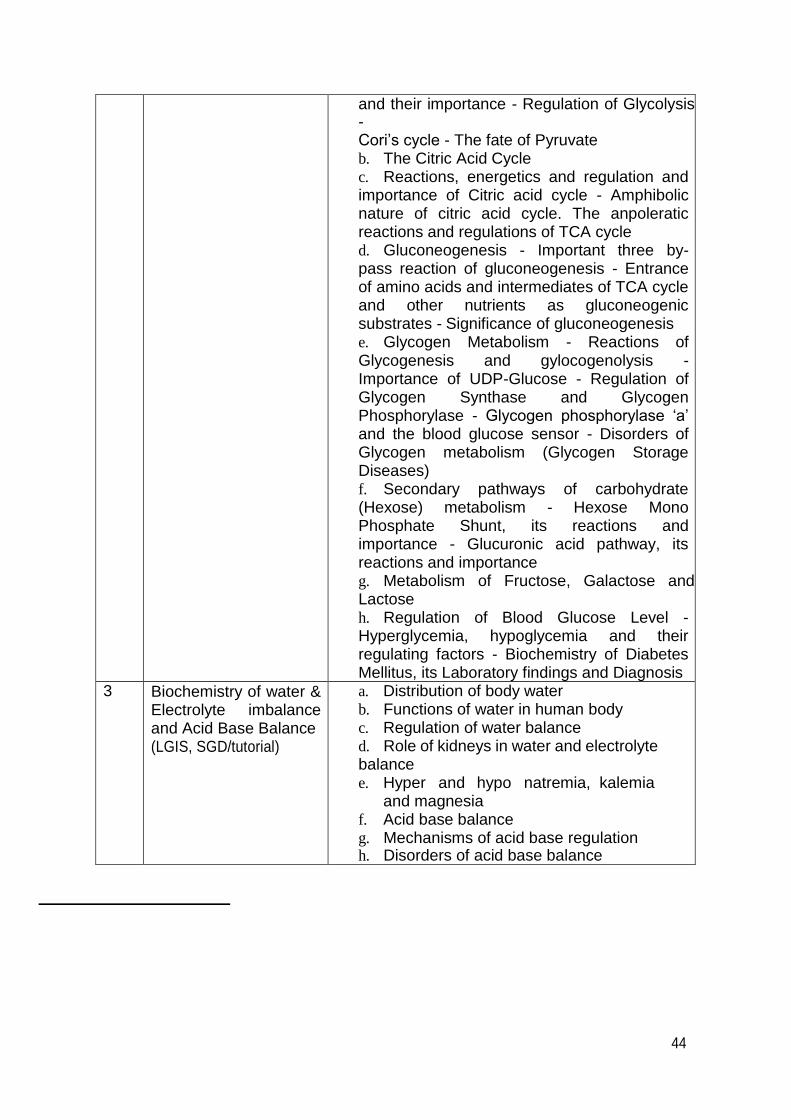
44
and their importance - Regulation of Glycolysis - Cori’s cycle - The fate of Pyruvate b. The Citric Acid Cycle c. Reactions, energetics and regulation and importance of Citric acid cycle - Amphibolic nature of citric acid cycle. The anpoleratic reactions and regulations of TCA cycle d. Gluconeogenesis - Important three by-pass reaction of gluconeogenesis - Entrance of amino acids and intermediates of TCA cycle and other nutrients as gluconeogenic substrates - Significance of gluconeogenesis e. Glycogen Metabolism - Reactions of Glycogenesis and gylocogenolysis - Importance of UDP-Glucose - Regulation of Glycogen Synthase and Glycogen Phosphorylase - Glycogen phosphorylase ‘a’ and the blood glucose sensor - Disorders of Glycogen metabolism (Glycogen Storage Diseases) f. Secondary pathways of carbohydrate (Hexose) metabolism - Hexose Mono Phosphate Shunt, its reactions and importance - Glucuronic acid pathway, its reactions and importance g. Metabolism of Fructose, Galactose and Lactose h. Regulation of Blood Glucose Level - Hyperglycemia, hypoglycemia and their regulating factors - Biochemistry of Diabetes Mellitus, its Laboratory findings and Diagnosis
3 Biochemistry of water & Electrolyte imbalance and Acid Base Balance (LGIS, SGD/tutorial)
a. Distribution of body water b. Functions of water in human body c. Regulation of water balance d. Role of kidneys in water and electrolyte balance e. Hyper and hypo natremia, kalemia and magnesia f. Acid base balance g. Mechanisms of acid base regulation h. Disorders of acid base balance
45
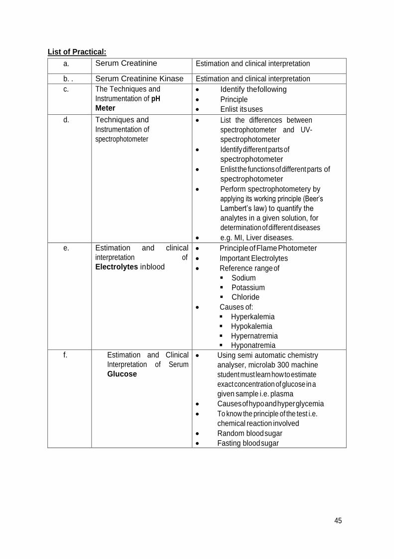
List of Practical:
a. Serum Creatinine Estimation and clinical interpretation
b. . Serum Creatinine Kinase Estimation and clinical interpretation
c. The Techniques and
Instrumentation of pH
Meter
Identify the following
Principle
Enlist its uses
d. Techniques and
Instrumentation of
spectrophotometer
List the differences between
spectrophotometer and UV-
spectrophotometer
Identify different parts of
spectrophotometer
Enlist the functions of different parts of
spectrophotometer
Perform spectrophotometery by
applying its working principle (Beer’s
Lambert’s law) to quantify the
analytes in a given solution, for
determination of different diseases
e.g. MI, Liver diseases.
e. Estimation and clinical
interpretation of
Electrolytes in blood
Principle of Flame Photometer
Important Electrolytes
Reference range of
Sodium
Potassium
Chloride
Causes of:
Hyperkalemia
Hypokalemia
Hypernatremia Hyponatremia
f. Estimation and Clinical
Interpretation of Serum
Glucose
Using semi automatic chemistry
analyser, microlab 300 machine
student must learn how to estimate
exact concentration of glucose in a
given sample i.e. plasma
Causes of hypo and hyper glycemia
To know the principle of the test i.e.
chemical reaction involved
Random blood sugar
Fasting blood sugar

46
g. Oral Glucose Tolerance Test
and its Clinical
Interpretation
To learn the method, principle and
significance of the test
To learn its interpretation
To know its indications
h. Estimation and Clinical
Interpretation of plasma
enzyme ALP
Using semi-automatic chemistry
analyzer, micro lab 300 machine
student must learn how to estimate exact
concentration of ALP in a given sample
i.e. plasma
To know the principle of the test i.e.
chemical reaction involved
Use of the test in clinical setting
Reference range
47
List of Case Based Learning (CBL):
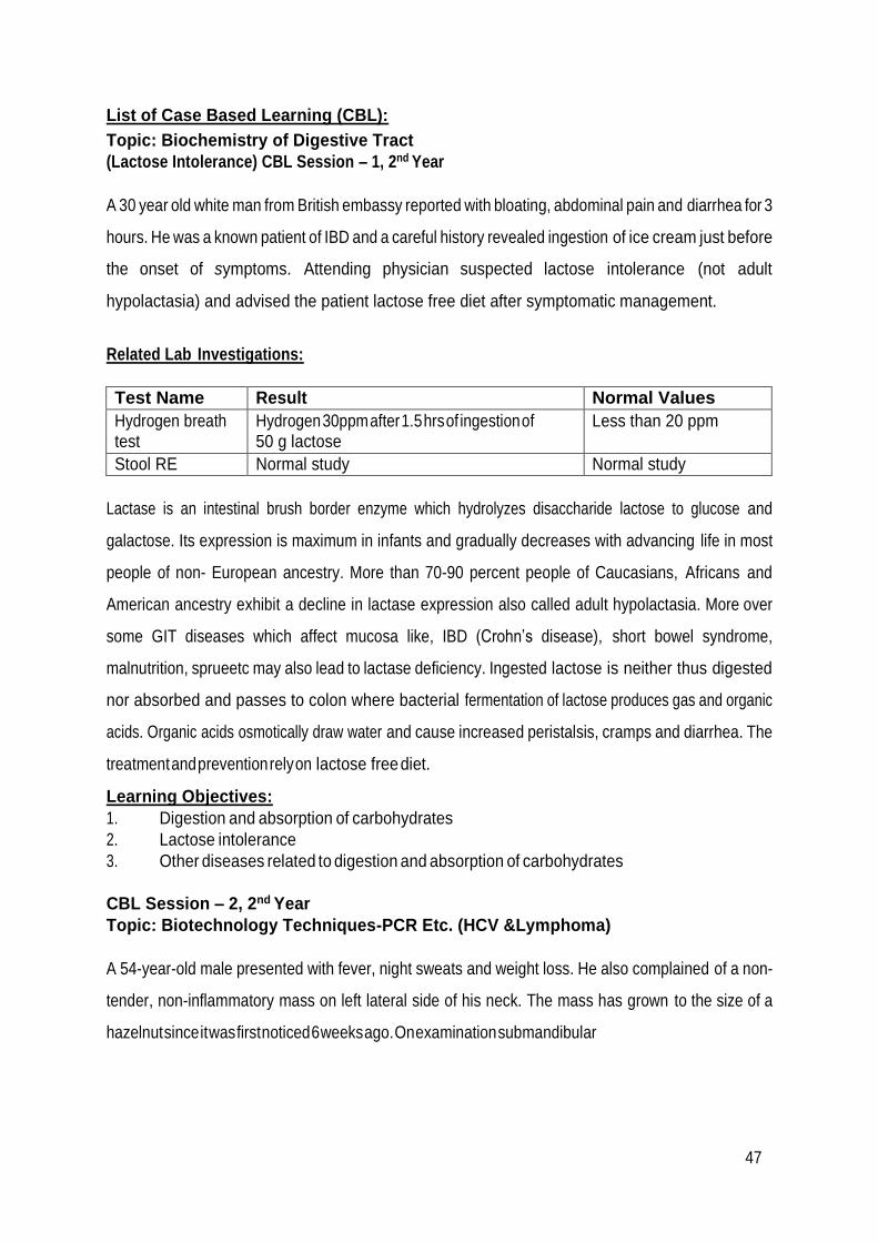
Topic: Biochemistry of Digestive Tract
(Lactose Intolerance) CBL Session – 1, 2nd Year
A 30 year old white man from British embassy reported with bloating, abdominal pain and diarrhea for 3
hours. He was a known patient of IBD and a careful history revealed ingestion of ice cream just before
the onset of symptoms. Attending physician suspected lactose intolerance (not adult
hypolactasia) and advised the patient lactose free diet after symptomatic management.
Related Lab Investigations:
Test Name Result Normal Values
Hydrogen breath test
Hydrogen 30ppm after 1.5 hrs of ingestion of 50 g lactose
Less than 20 ppm
Stool RE Normal study Normal study
Lactase is an intestinal brush border enzyme which hydrolyzes disaccharide lactose to glucose and
galactose. Its expression is maximum in infants and gradually decreases with advancing life in most
people of non- European ancestry. More than 70-90 percent people of Caucasians, Africans and
American ancestry exhibit a decline in lactase expression also called adult hypolactasia. More over
some GIT diseases which affect mucosa like, IBD (Crohn’s disease), short bowel syndrome,
malnutrition, sprueetc may also lead to lactase deficiency. Ingested lactose is neither thus digested
nor absorbed and passes to colon where bacterial fermentation of lactose produces gas and organic
acids. Organic acids osmotically draw water and cause increased peristalsis, cramps and diarrhea. The
treatment and prevention rely on lactose free diet.
Learning Objectives:
1. Digestion and absorption of carbohydrates
2. Lactose intolerance
3. Other diseases related to digestion and absorption of carbohydrates
CBL Session – 2, 2nd Year
Topic: Biotechnology Techniques-PCR Etc. (HCV &Lymphoma)
A 54-year-old male presented with fever, night sweats and weight loss. He also complained of a non-
tender, non-inflammatory mass on left lateral side of his neck. The mass has grown to the size of a
hazelnut since it was first noticed 6 weeks ago. On examination submandibular

48
and supraclavicular nodes were palpable. Blood complete picture was normal but patient was diagnosed
as a case of B Cell Non Hodgkin lymphoma with the help of tissue histology and bone marrow
aspiration cytology. During the laboratory investigation phase he was incidentally found to be a
patient of HCV infection as well.
In addition to commitment about the treatment and prognosis of the lymphoma, attending physician was
also concerned about the status of his HCV infection. He ordered abdominal ultrasound to check for
liver cirrhosis and Quantitative PCR to check for HCV viral load.
Related Lab Investigations:
Test Name Result Normal Values
Hemoglobin 14 g/dL Adult Male 13-18
g/dL
Adult Female 12-16
g/dL
WBC Count 11.5 x 10 9 / L 4.0 x 11.0 x 10 9 / L
(Tumor/mass )Lymph Node Biopsy B Cell lymphoma cells seen Normal cytology
Lymphoma cells in peripheral blood smear
Not seen Not seen
Bone marrow biopsy for histological examination
Monoclonal lymphoid aggregates seen
No such aggregates
LDH 630 U/L 225-450 U/L
Anti HCV Antibody test Positive Negative
USG Abdomen Nodular and shrunken
appearance of liver but mild changes
Normal echo-
texture
Test Name Result Normal Values
Quantitative PCR (for viral RNA) 700,000 IU/Ml Normally it is negative
Viral Load is expressed as
copies/mL: Low: less than 2
M/mL • High: more than 2
M/mL
Or Expressed as
International Units (IU/mL):
• Low: less than 800,000
IU/mL • High: more than 800,000 IU/mL
Cytogenetic testing in culture sample (2 -3
weeks)
No abnormal
chromosomal
pairing or less or
Normal chromosomal
pairing and normal number
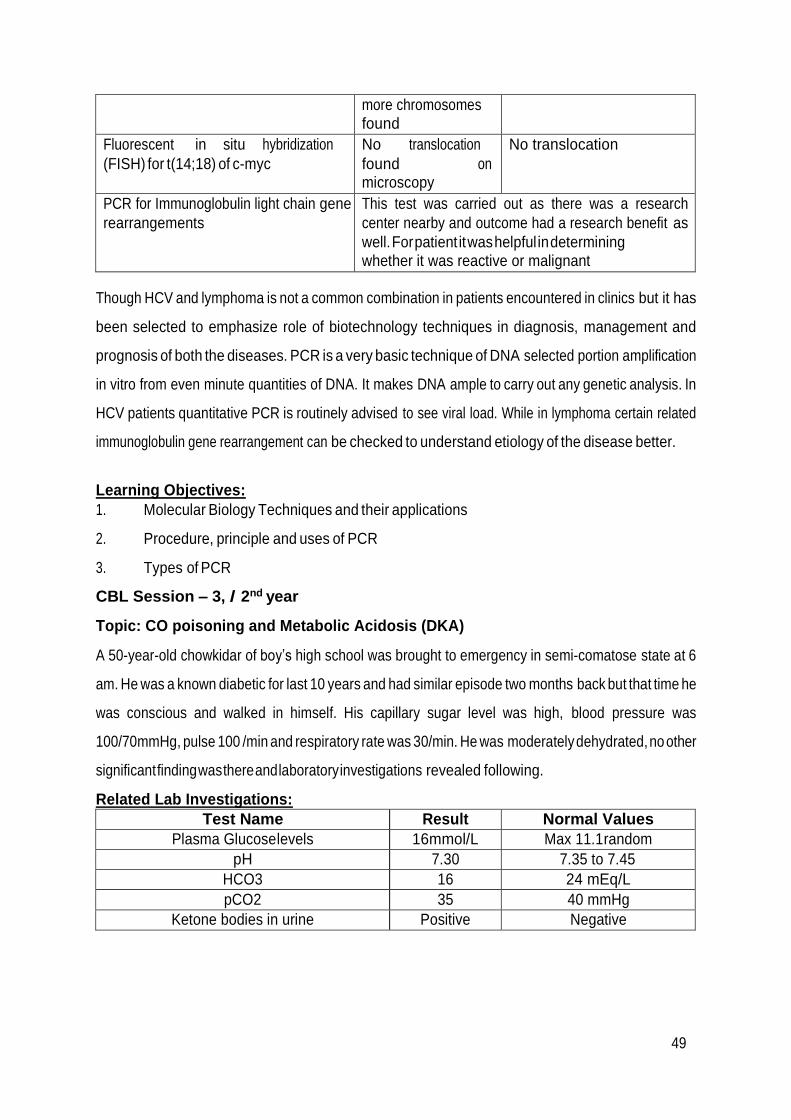
49
more chromosomes found
Fluorescent in situ hybridization
(FISH) for t(14;18) of c-myc
No translocation
found on microscopy
No translocation
PCR for Immunoglobulin light chain gene
rearrangements
This test was carried out as there was a research
center nearby and outcome had a research benefit as
well. For patient it was helpful in determining whether it was reactive or malignant
Though HCV and lymphoma is not a common combination in patients encountered in clinics but it has
been selected to emphasize role of biotechnology techniques in diagnosis, management and
prognosis of both the diseases. PCR is a very basic technique of DNA selected portion amplification
in vitro from even minute quantities of DNA. It makes DNA ample to carry out any genetic analysis. In
HCV patients quantitative PCR is routinely advised to see viral load. While in lymphoma certain related
immunoglobulin gene rearrangement can be checked to understand etiology of the disease better.
Learning Objectives:
1. Molecular Biology Techniques and their applications
2. Procedure, principle and uses of PCR
3. Types of PCR
CBL Session – 3, / 2nd year
Topic: CO poisoning and Metabolic Acidosis (DKA)
A 50-year-old chowkidar of boy’s high school was brought to emergency in semi-comatose state at 6
am. He was a known diabetic for last 10 years and had similar episode two months back but that time he
was conscious and walked in himself. His capillary sugar level was high, blood pressure was
100/70mmHg, pulse 100 /min and respiratory rate was 30/min. He was moderately dehydrated, no other
significant finding was there and laboratory investigations revealed following.
Related Lab Investigations:
Test Name Result Normal Values
Plasma Glucose levels 16mmol/L Max 11.1 random
pH 7.30 7.35 to 7.45
HCO3 16 24 mEq/L
pCO2 35 40 mmHg
Ketone bodies in urine Positive Negative

50
As the compensation of the primary acidosis was not appropriate so he was diagnosed as a case of
mixed acid base disorder (Metabolic acidosis and respiratory acidosis). A careful history revealed
that he used a stove for heating in the room which used wood fire. This lead the physician to suspect CO
poisoning as well. CO inhibits ETC and hampers ATP synthesis moreover it reduces the availability of
active Hb for normal tissue oxygenation and CO2 removal. The treatment included oxygen support,
HCO3 administration slow iv, sc insulin for hyperglycemia and iv fluids.
Learning Objectives:
1. How to diagnose acid base disorders
2. ETC and oxidative phosphorylation
3. Inhibitors of ETC and Oxidative phosphorylation including CO
Primary Disorder Change Compensation
Metabolic acidosis ↓ HCO3 1 ↓ 1.3 CO2
Metabolic alkalosis ↑ HCO3 1 ↑ 0.7 CO2
Respiratory acidosis
Acute ↑ pCO2 10 ↑ 1 HCO3
Chronic ↑ pCO2 10 ↑ 3.5 HCO3
Respiratory alkalosis
Acute ↓ pCO2 10 ↓ 2 HCO3
Chronic ↓ pCO2 10 ↓ 5 HCO3
CBL Session – 4, / 2nd year
Topic: Cholera(Water & Electrolytes)
A 21-years-old female law student working in a developing country suddenly began to pass profuse
watery stools almost continuously. She soon started to vomit. Her general condition declined abruptly,
and she was rushed to the local village hospital. On admission, she was cyanotic, skin turgor was poor,
blood pressure was 70/50 mmHg (normal 120/80 mm Hg), and her pulse was rapid and weak. The doctor
on duty diagnosed cholera, took a stool sample, and started treatment immediately. Patient was
isolated and given normal saline with 20mmol/L K+ added (ringers lactate was not administered).
Doxycycline 500mg every 6 hours was started and plenty of ORS was advised after initial IV fluids. The
relatives were called for
51
counseling. As cholera can become epidemic or pandemic, relatives were given following advice to
follows and teach others.
1. Drink only boiled or treated water.
2. Cook well and eat hot.
3. Avoid shellfish.
4. Peel all vegetables and fruits.
Related Lab Investigations:
Test Name Result Normal Values
Stool Microscopy Vibrio cholera seen
Nil
Stool culture Yielded growth
of Vibrio
cholera
Nil/ normal flora
Serum electrolytes Low Na+,K+, Cl- and HCO3-
Normal
Vibrio Cholera is a gram negative rod, comma shaped bacterium transmitted through faecal- oral route. Its
incubation period is few hours to 5 days and it causes profuse watery stools, fever, vomiting and rapid
dehydration which may cause death. One may lose up to 1L/hour water in stools. Heat killed vaccine is
not that effective and is not required for international travel. Prevention of epidemic spread is
important. The key to effective treatment and prevention of death is meticulous and timely water
and electrolyte replacement.
Learning Objectives:
1. Mechanisms of water regulation
2. Mechanisms of electrolyte regulation
3. Disturbances in water and electrolyte regulation and their correction
4. Role of ORS in rehydration
CBL Session – 5, / 2nd year:
Topic: Carbohydrate Metabolism (G6PD deficiency)
A23 year old boy was prescribed Septran (sulphamethoxazole and trimethoprim) and
paracetamol for urinary tract infection and fever (10 hrs. history of symptoms). After two days the
boy presented again with subsiding initial symptoms but lethargy fatigue, dyspnea

52
and slight pallor. His lab investigations are given in the table below. Doctor suspected G6PD
deficiency which was confirmed by genetic analysis later.
Related Lab Investigations:
Test Name Result Normal Values
Hemoglobin 9 g/Dl 12 – 17 g/dL
RBCs 3.2 x 10⁶ 4.1 – 5.6 x 10⁶/µL
PCV (hematocrit) 28 35-50 %
MCV 84 80-98 fL
MCH 28 27-34pg
MCHC 30 32-36g/dL
WBCs 13 x 103/µL 4-11 x 103/µL
Fluorescent spot test (Butler
test)
Positive Negative
Glucose 6-phosphate dehydrogenase (G6PD) deficiency is an inherited disease characterized by
hemolytic anemia caused by the inability to detoxify oxidizing agents. G6PD deficiency is the most
common disease-producing enzyme abnormality in humans. DiminishedG6PDactivity impairs the
ability of the cell to form the NADPH that is essential for the maintenance of the reduced glutathione
pool. These results in a decrease in the cellular detoxification of free radicals and peroxides
formed within the cell. Glutathione also helps maintain the reduced states of sulfhydryl groups in
proteins, including hemoglobin. Oxidation of those sulfhydryl groups leads to the formation of denatured
proteins that form insoluble masses (called Heinz bodies) that attach to the red cell membranes.
Learning Objectives:
1. HMP shunt and its importance
2. Sources of NADPH
3. Uses of NADPH in body
CBL Session-6 / 2nd year
Topic: Pyruvate Kinase Deficiency
A two year old girl was referred to hematologist after her pediatrician found her to be severely anemic with
splenomegaly and jaundice. Her mother gave a possible history of “blood problem” in her family but
did not know for sure. Her complete blood count revealed normal hemoglobin with normocytic anemia.
The platelet and white cell count was normal. On the

53
peripheral smear there were many bizarre erythrocytes including speculated cells. A diagnosis of Pyruvate
Kinase deficiency was made.
Explanation
The normal erythrocyte lacks mitochondria and is completely dependent on glycolysis for production
of ATP.ATP is required to meet the metabolic needs of the RBCs and to fuel the pumps necessary for
the maintenance of the biconcave, flexible shape of the cell, which allows it to squeeze through
narrow capillaries. The anemia observed in glycolytic enzyme deficiencies is a consequence of the
reduced rate of glycolysis, leading to decreased ATP production. The resulting alterations in the red
blood cell membrane lead to changes in the shape of the cell and, ultimately, to phagocytosis by the
cells of the reticuloendothelial system, particularly macrophages of the spleen. The premature death
and lysis of red blood cells results in hemolytic anemia.
Learning Objectives:
1. Reactions of Glycolysis
2. Reduction of Pyruvate to Lactate
3. Fates of pyruvate and hormonal regulation of glycolysis
4. Glucose Transporters
CBL Session –7, / 2nd year
Topic: Obesity
A 40 year old house wife presented with chronic pain in both knees and back. Her weight was 93 kg and
height was 5’3”. Her waist to hip ratio was measured to be 1.1. She also complained of inguinal hernia. She
was diagnosed as a case of android obesity with BMI almost 36. Her doctor advised her to change
dietary and life style habits and gave some symptomatic relief medicines after seeing the report of
tests ordered empty stomach.
Related Lab Investigations:
Test Name Result Normal Values
TC 6.2 mmol/L Less than 5.6 mmol/L
TGs 1.8 mmol/L Less than 1.6 mmol/L
LDL 3.6 mmol/L Less than 2.6 mmol/L
HDL 90 mmol/L More than 1.04 mmol/L
BSF 120 mg/dL Less than 110 mg/dL
54
Obesity is a disorder of body weight regulatory systems characterized by an accumulation of excess body
fat. The BMI (weight in kg)/(height in meters)2 provides a measure of relative weight, adjusted for
height. This allows comparisons both within and between populations. The healthy range for the BMI is
between 18.5 and 24.9. Individuals with a BMI between 25 and 29.9 are considered overweight, those
with a BMI equal to or greater than 30 are defined as obese, and a BMI over 40 is considered extremely
obese. Anyone more than 100 pounds overweight is considered severely obese. These cutoffs are
based on the studies examining the relationship of BMI to premature death, and are similar in men
and women.
Learning Objectives:
1. Assessment of Obesity
2. Metabolic Changes in Obesity
3. Molecules that influence Obesity
4. Body weight regulation
5. Weight reduction
6. Fast & Feed Cycle
CBL Session – 8, / 2nd year
Topic: Nutrition (Kwashiorkor)
An 11 months old infant was brought to children OPD with retardation in growth, apathy and lethargy. On
examination his face was edematous and moonlike, brittle and thin hair, pitting edema and
hepatomegaly. Physician diagnosed him as a case of kwashiorkor and advised changes in diet in
addition to vitamin supplements.
Explanation
Kwashiorkor occurs when protein deprivation is relatively greater than the reduction in total calories.
Protein deprivation is associated with severely decreased synthesis of visceral protein. Kwashiorkor
is frequently seen in children after weaning at about one year of age, when their diet consists
predominantly of carbohydrates. Typical symptoms include stunted growth, edema, skin lesions,
depigmented hair, anorexia, enlarged fatty liver, and decreased plasma albumin concentration. Edema
results from the lack of adequate plasma proteins to maintain the distribution of water between blood and
tissues. Edema may mask muscle loss.

55
Learning Objectives:
1. Dietary reference intakes
2. Energy requirement in humans
3. Balanced diet
4. Diet in pregnancy & lactation
5. Protein Energy Malnutrition
Reference Books:
1. Lippincott’s illustrated reviews and Harper’s text book of Biochemistry
2. Davidson’s Practice of Medicine
3. Internet sources

56
BLOCK-II
Section-I
Anatomy
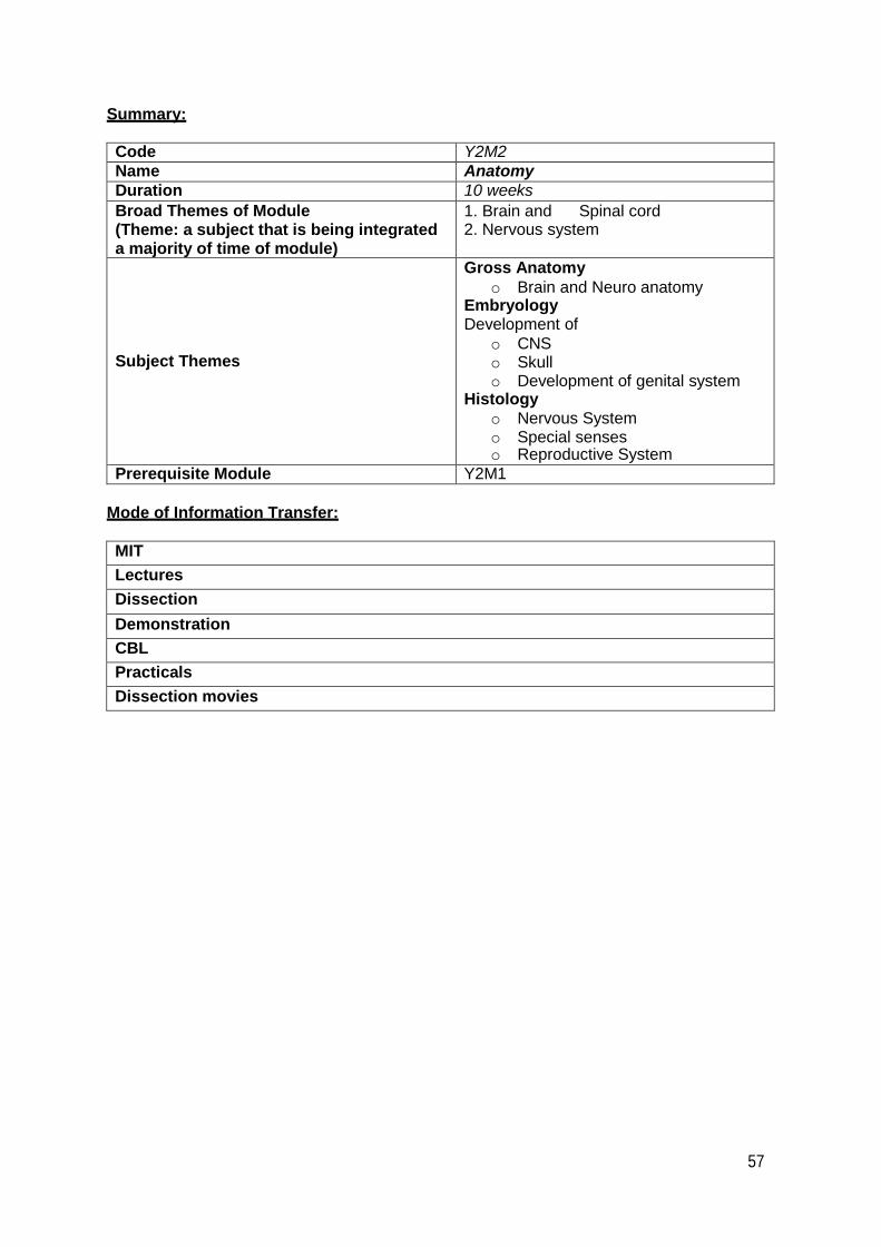
57
Summary:
Code Y2M2
Name Anatomy
Duration 10 weeks
Broad Themes of Module (Theme: a subject that is being integrated a majority of time of module)
1. Brain and Spinal cord 2. Nervous system
Subject Themes
Gross Anatomy
o Brain and Neuro anatomy Embryology Development of
o CNS o Skull o Development of genital system
Histology
o Nervous System o Special senses o Reproductive System
Prerequisite Module Y2M1
Mode of Information Transfer:
MIT
Lectures
Dissection
Demonstration
CBL
Practicals
Dissection movies
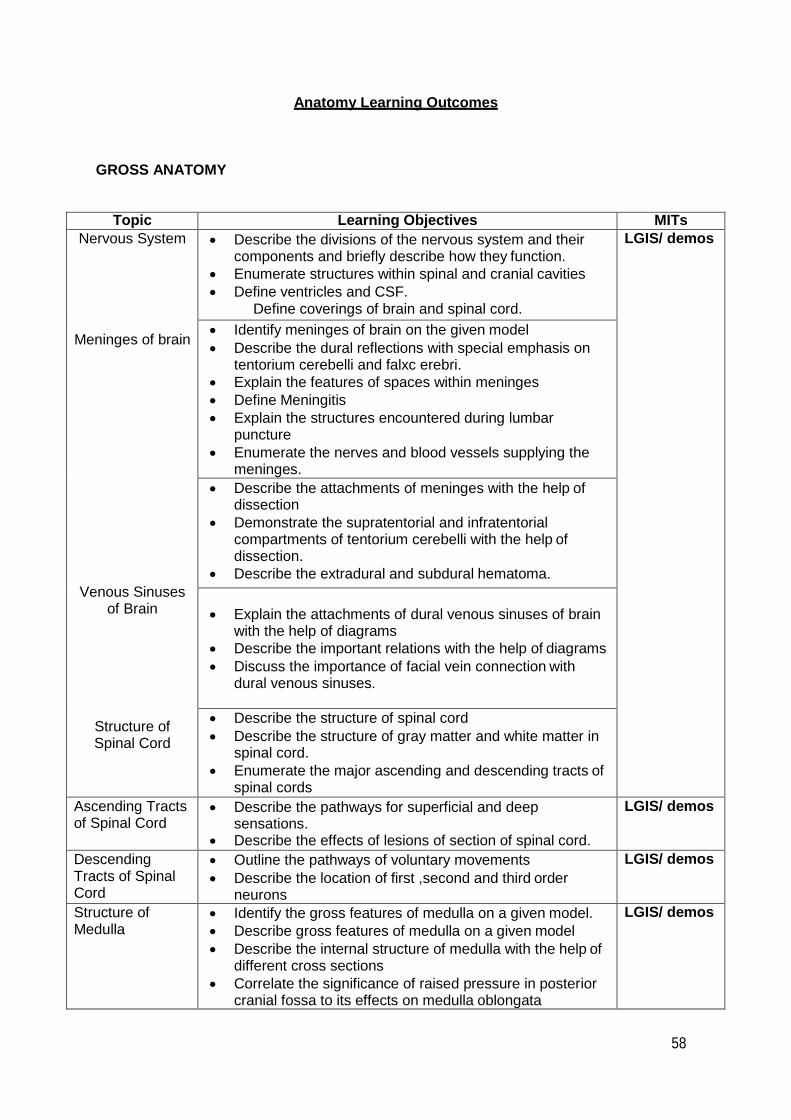
58
Anatomy Learning Outcomes
GROSS ANATOMY
Topic Learning Objectives MITs
Nervous System Describe the divisions of the nervous system and their components and briefly describe how they function.
Enumerate structures within spinal and cranial cavities
Define ventricles and CSF. Define coverings of brain and spinal cord.
LGIS/ demos
Meninges of brain Identify meninges of brain on the given model
Describe the dural reflections with special emphasis on tentorium cerebelli and falxc erebri.
Explain the features of spaces within meninges
Define Meningitis
Explain the structures encountered during lumbar puncture
Enumerate the nerves and blood vessels supplying the meninges.
Venous Sinuses of Brain
Describe the attachments of meninges with the help of dissection
Demonstrate the supratentorial and infratentorial compartments of tentorium cerebelli with the help of dissection.
Describe the extradural and subdural hematoma.
Explain the attachments of dural venous sinuses of brain with the help of diagrams
Describe the important relations with the help of diagrams
Discuss the importance of facial vein connection with dural venous sinuses.
Structure of Spinal Cord
Describe the structure of spinal cord
Describe the structure of gray matter and white matter in spinal cord.
Enumerate the major ascending and descending tracts of spinal cords
Ascending Tracts of Spinal Cord
Describe the pathways for superficial and deep sensations.
Describe the effects of lesions of section of spinal cord.
LGIS/ demos
Descending Tracts of Spinal Cord
Outline the pathways of voluntary movements
Describe the location of first ,second and third order neurons
LGIS/ demos
Structure of Medulla
Identify the gross features of medulla on a given model.
Describe gross features of medulla on a given model
Describe the internal structure of medulla with the help of different cross sections
Correlate the significance of raised pressure in posterior cranial fossa to its effects on medulla oblongata
LGIS/ demos

59
Discuss nuclei of cranial nerves present in medulla.
Describe clinical correlations of medulla
Structure of Pons Identify the gross features of Pons on a given model.
Explain the internal structure of Pons with cross sections at different levels
Discuss nuclei of cranial nerves lying in pons
Discuss the anatomical structures involved in Pontine hemorrhage
LGIS/ demos
Gross Anatomy of Midbrain
Identify the gross structure of midbrain on a given model.
Describe the internal structure of midbrain with cross sections at different levels
Discuss nuclei of cranial nerves lying in midbrain.
Enumerate the clinical consequences of trauma to midbrain
LGIS/ demos
Cerebellum Describe the gross features of cerebellum on the given model
Explain the cerebellar nuclei and their connection with other parts of brain. afferent fibers and efferent fibers
Discuss the effect of lesions of cerebellum on voluntary movements
LGIS/ demos
Gross anatomy of Interior cerebrum
Describe the features of parts of following on a given model Cerebrum Diencephalon Telencephalon
Identify gross features of parts of diencephalon. Thalamus hypothalamus
Correlate the anatomical lesions of nuclei of thalamus and hypothalamus with the clinical conditions like diabetes inspidus and obesity
LGIS/ demos
Reticular formation and limbic system
Discuss reticular formation and its afferent and efferent projections
Describe functions of reticular formation
Discuss components of limbic system
Discuss connecting pathways of the limbic system
Discuss afferent and efferent pathways of hippocampus
Discuss clinical correlations of reticular formation and limbic system
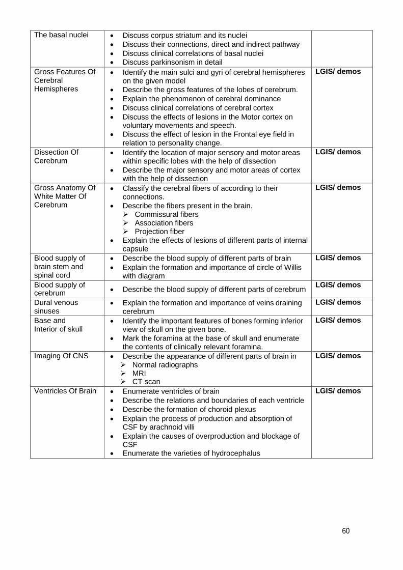
60
The basal nuclei Discuss corpus striatum and its nuclei
Discuss their connections, direct and indirect pathway
Discuss clinical correlations of basal nuclei Discuss parkinsonism in detail
Gross Features Of Cerebral Hemispheres
Identify the main sulci and gyri of cerebral hemispheres on the given model
Describe the gross features of the lobes of cerebrum.
Explain the phenomenon of cerebral dominance
Discuss clinical correlations of cerebral cortex
Discuss the effects of lesions in the Motor cortex on voluntary movements and speech.
Discuss the effect of lesion in the Frontal eye field in relation to personality change.
LGIS/ demos
Dissection Of Cerebrum
Identify the location of major sensory and motor areas within specific lobes with the help of dissection
Describe the major sensory and motor areas of cortex with the help of dissection
LGIS/ demos
Gross Anatomy Of White Matter Of Cerebrum
Classify the cerebral fibers of according to their connections.
Describe the fibers present in the brain. Commissural fibers Association fibers Projection fiber
Explain the effects of lesions of different parts of internal capsule
LGIS/ demos
Blood supply of brain stem and spinal cord
Describe the blood supply of different parts of brain
Explain the formation and importance of circle of Willis with diagram
LGIS/ demos
Blood supply of cerebrum
Describe the blood supply of different parts of cerebrum LGIS/ demos
Dural venous sinuses
Explain the formation and importance of veins draining cerebrum
LGIS/ demos
Base and Interior of skull
Identify the important features of bones forming inferior view of skull on the given bone.
Mark the foramina at the base of skull and enumerate the contents of clinically relevant foramina.
LGIS/ demos
Imaging Of CNS Describe the appearance of different parts of brain in Normal radiographs MRI CT scan
LGIS/ demos
Ventricles Of Brain Enumerate ventricles of brain
Describe the relations and boundaries of each ventricle
Describe the formation of choroid plexus
Explain the process of production and absorption of CSF by arachnoid villi
Explain the causes of overproduction and blockage of CSF
Enumerate the varieties of hydrocephalus
LGIS/ demos

61
EMBROYOLOGY
Development of Spinal cord
Describe the development of neural tube.
Describe the differentiation of neural tube into different parts of brain.
Describe the development of spinal cord.
Describe the positional changes of the cord
LGIS
Developmental Anomalies Of Spinal Cord
Explain the causes of neural tube defects
Explain the process of development of spin bifida
Describe the clinical conditions relevant to the development of neural tube defects Like Spina bifida spina bifida with meningomyelocele.
LGIS / Lab
Development Of Cerebrum
Describe the development of cerebral hemispheres and ventricles.
Explain the relation of congenital aqueduct stenosis and hydrocephalus.
Enumerate and briefly describe the congenital anomalies associated with development of cerebrum.
LGIS / Lab
Development of Brainstem
Describe the development of medulla, Pons, midbrain and cerebellum
Describe the developmental changes in alar and basal plates in brainstem
LGIS / Lab
LGIS / Lab
Development Of Skull
Describe the stages of development of neurocranium
Describe the stages of development of viscerocranium
Describe the stages of differentiation of neurocranium into Membranous Neurocranium and chondrocranium
Describe the importance of fontanelle of skull in relation to normal ossification of the skull changes in intracranial pressure
Describe the features of Newborn Cranium
LGIS / Lab
Development of external genital organs
Describe the development of cloacal folds
Describe the development of genital swellings
Describe the differentiation of male and female external genitalia from genital swellings
Enlist common anomalies
LGIS / Lab
Development of uterus and ovary
Describe development of
ovary Enlist common anomalies
LGIS / Lab
Development of fallopian tubes and vagina
Describe development of
Uterus
fallopian tubes
vagina
Enlist common anomalies
LGIS / Lab
Development of male reproductive system
Describe the formation of indifferent gonad
Describe the development of testis from indifferent gonad
Enumerate the developmental anomalies of male genital organ
LGIS / Lab
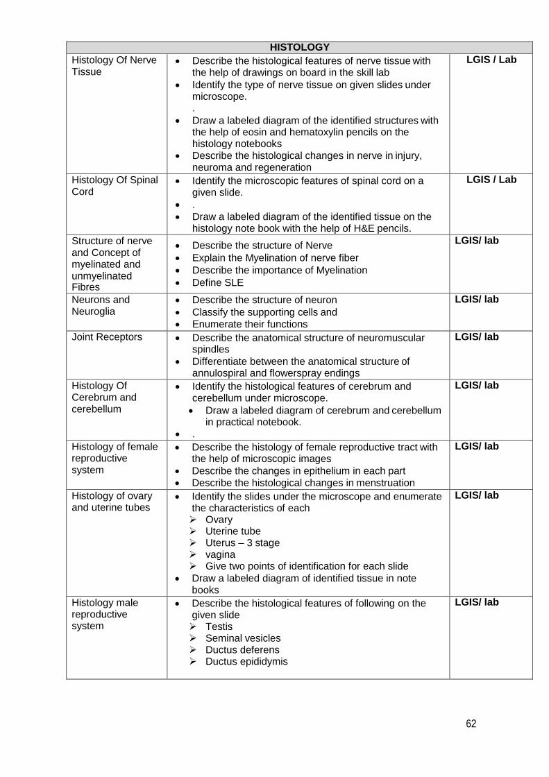
62
HISTOLOGY
Histology Of Nerve Tissue
Describe the histological features of nerve tissue with the help of drawings on board in the skill lab
Identify the type of nerve tissue on given slides under microscope. .
Draw a labeled diagram of the identified structures with the help of eosin and hematoxylin pencils on the histology notebooks
Describe the histological changes in nerve in injury, neuroma and regeneration
LGIS / Lab
Histology Of Spinal Cord
Identify the microscopic features of spinal cord on a given slide.
.
Draw a labeled diagram of the identified tissue on the histology note book with the help of H&E pencils.
LGIS / Lab
Structure of nerve and Concept of myelinated and unmyelinated Fibres
Describe the structure of Nerve
Explain the Myelination of nerve fiber
Describe the importance of Myelination
Define SLE
LGIS/ lab
Neurons and Neuroglia
Describe the structure of neuron
Classify the supporting cells and
Enumerate their functions
LGIS/ lab
Joint Receptors Describe the anatomical structure of neuromuscular spindles
Differentiate between the anatomical structure of annulospiral and flowerspray endings
LGIS/ lab
Histology Of Cerebrum and cerebellum
Identify the histological features of cerebrum and cerebellum under microscope.
Draw a labeled diagram of cerebrum and cerebellum in practical notebook.
.
LGIS/ lab
Histology of female reproductive system
Describe the histology of female reproductive tract with the help of microscopic images
Describe the changes in epithelium in each part
Describe the histological changes in menstruation
LGIS/ lab
Histology of ovary and uterine tubes
Identify the slides under the microscope and enumerate the characteristics of each Ovary Uterine tube Uterus – 3 stage vagina Give two points of identification for each slide
Draw a labeled diagram of identified tissue in note books
LGIS/ lab
Histology male reproductive system
Describe the histological features of following on the given slide Testis Seminal vesicles Ductus deferens Ductus epididymis
LGIS/ lab
63
Draw a labeled diagram of identified tissue in practical note book
Histology Of Eye Identify these structures under microscope
Draw a labeled diagram of the identified structure on histology notebook
Describe the histological features of lens, cornea & retina
LGIS/ lab
Organ Of Corti Identify the histological features of organ of corti under microscope
Identify the cells and spaces present in the cochlea on the given slide
Draw a labeled diagram of identified tissue in histo note books
LGIS/ lab
Taste buds Describe the structure of the taste buds and their cells
LGIS/ lab
Olfactory epithelium Describe the structure of the olfactory receptors and other cells involved in olfaction
LGIS/ lab
64
List of Practical:
Histology Practical’s
1. Testis & Epididymis
2. Vas deferens, Seminal vesicle
3. Prostate
4. Ovary
5. Fallopian tube, Uterus
6. Cervix, Vagina
7. Histology of Nervous Tissue, Neurons, Neuroglia , Structure of nerve and ganglia
8. Histology of Spinal cord
9. Histology of cerebellum
10. Histology of cerebral cortex
11. Histology of retina, cornea .lens, inner ear, Olfactory epithelium and taste buds
ANATOMY CBL
A PATIENT WITH COMMINUTED FRONTAL COMPLEX SKULL FRACTURE CBL 1: Learning objectives 1. To learn the osteology of exterior & interior of skull.
2. To learn the development of neurocranium & viscerocranium
3. To learn the complications associated with skull fractures.
4. To learn the various clinical modalities used to investigate a case of fracture skull.
CBL 2: Case of spinal shock
Learning objectives:
1. Correlate the structure of components of nervous tissue to their function.
2. Compare topographic anatomy of spinal cord in adult and newborn.
3. Localize grey and white matter in spinal cord.
4. Interpret neurological presentation of spinal cord injuries at different levels.
5. Describe neurulation and transformation of neural tube into central nervous system.
6. State embryological basis and presentation of neural tube defects.
7. Correlate the development of vertebral column to structure and function.
8. What is the basis of correlation between spinal cord segment and vertebrae?

65
PATIENT WITH INTENTION TREMOR AND TENDENCY TO FALL ON ONE SIDE CBL 3 (Spinocerebellar Ataxia) Learning objectives
1. Correlate structure of cerebellum on specimen and model to imaging (CT/MRI).
Correlate microstructure of cerebellum to function.
2. Correlate functional anatomy of cerebellum to various clinical presentations in
cerebellar diseases.
3. Perform clinical examination of cerebellar functions.
4. How do you anatomical divisions of cerebellum correlate with functional division?
5. How does cerebellum receive its input and from where?
6. How does cerebellum affect motor activities?
7. What is the basis of signs seen in cerebellar disorders?
8. Why this patient falls on one side of the body?
9. How is balance maintained? How cerebellum performs inhibitory functions?
CBL 4 :Case of parkinsonism Learning objectives:
1. Localize the various Anatomical & clinical basal ganglia.
2. Correlate the connections of basal nuclei to their functions.
3. Describe extra-pyramidal pathways.
4. Relate basis of disorders associated with basal nuclei.
5. Describe the synthesis, functions and degradation of various neurotransmitters
involved in functioning of basal nuclei.
6. Identify drugs modulating the functions of neurotransmitters in basal nuclei.
CBL 5 Patient with Stroke Learning objectives:
1. Describe the topographic anatomy of cerebrum and associate sulci and gyri with cortical functions. Identify various sulci, gyri and cortical areas on the specimen/model/imaging modalities.
2. Identify various fiber bundles in the cerebrum.
3. State the functional losses associated with the lesions of fiber bundles in cerebrum.
4. Correlate the effects of cerebral vascular occlusion to clinical presentations.
5. What functions are performed by frontal lobe?
6. Which areas of cerebral cortex are affected by occlusion of middle cerebral artery?
7. How is cerebral dominance determined?
8. How does the cerebral hemisphere coordinate in production of meaningful speech?
9. Where do collaterals from corticospinal tract go? What is their significance?
10. Which visual field is affected by the stroke?

66
BLOCK-II
Section-II
Physiology
67
Summary:
Code Y1M2
Name Physiology
Duration 10 weeks
Broad Themes of Module
(Theme: a subject that is being integrated a
majority of time of module)
1. Brain and Spinal cord
2. Nervous system
Subject Themes
Sensory Physiology
Motor Physiology
Higher Mental Functions
ANS
Prerequisite Module Y1M1
Mode of Information Transfer:
MIT
Lectures
Tutorials (PTT)
CBL
Practicals
Class tests
Physiology learning outcomes and MITs:
CNS
1. Processing of information in neuronal pool (LGIS, SGD/tutorial)
To be able to explain types of synapses—chemical and electrical and physiologic anatomy of the synapse and chemical substances that function as synaptic transmitters
To know the electrical events during neuronal excitation and electrical events during neuronal inhibition
To know about the transmission and processing of signals in neuronal pools ,relaying of signals through neuronal pools prolongation of a signal by a neuronal pool, after discharge, synaptic fatigue
2. Sensory receptors & receptor Potential (LGIS, SGD/tutorial)
To understand types of sensory receptors and the sensory stimuli and differential sensitivity of receptors
To understand the transduction of sensory stimuli into nerve impulses
To know the local electrical currents at nerve endings— receptor potentials, adaptation of
68
receptors
To know the nerve fibers that transmit different types of signals, and their physiologic classification
To understand the transmission of signals of different intensity in nerve tracts—spatial and temporal
summation 3. Sensory tracts
(LGIS, SGD/tutorial, Practical)
To know the functional anatomy of dorsal column medial leminiscal system and anterolateral pathway
To understand the sensations carried by different sensory tracts
To differentiate between different sensory tracts
4. Sensory cortex (LGIS, SGD/tutorial, Practical)
To know about the somatosensory cortex and somatosensory association areas
5. Physiology of pain (LGIS, SGD/tutorial, Practical)
To understand the types of pain and their qualities— fast pain and slow pain
To know about the pain receptors and their stimulation
To understand the dual pathways for transmission of pain signals into the central nervous system
To understand the referred pain and visceral pain 6. Brain analgesia
system (LGIS)
To know the pain suppression system in the brain and spinal cord
To understand the brain’s opiate system—endorphins and enkephalins
7. Clinical abnormalities/ thermal sensations (LGIS)
To know the clinical abnormalities of pain and other somatic sensations
To know the thermal sensations, thermal receptors and their excitation and transmission of thermal signals in the nervous system
69
8. Inner ear / auditory pathways (LGIS, SGD/tutorial, Practical)
To be able to explain central auditory mechanisms and auditory nervous pathways
To understand the function of the cerebral cortex in hearing and determination of the direction from which sound comes
To understand the hearing abnormalities and types of deafness
9. Motor system / Spindle / stretch reflex (LGIS, SGD/tutorial, Practical)
To be able to explain organization of the spinal cord for motor functions
To understand the muscle sensory receptors—muscle spindles and golgi tendon organs—and their roles in muscle control
To understand the receptor function of the muscle spindle and muscle stretch reflex
To understand the role of the muscle spindle in voluntary motor activity
To know the clinical applications of the stretch reflex and golgi tendon reflex
To know the function of the muscle spindles and golgi tendon organs in conjunction with motor control from higher levels of the brain
10. Motor functions of spinal cord (LGIS, SGD/tutorial, Practical)
To understand the flexor reflex and the withdrawal reflexes and crossed extensor reflex
To be able to explain the reciprocal inhibition and reciprocal innervation
To understand the reflexes of posture and locomotion and postural and locomotive reflexes of the cord
To know the spinal cord reflexes that cause muscle spasm
11. Spinal shock (LGIS) To be able to explain spinal cord transection and spinal shock
12. Pyramidal tract/ extra pyramidal tract (LGIS)
To be able to understand primary motor cortex premotor area, supplementary motor area
To understand the transmission of signals from the motor cortex to the muscles
To understand extrapyramidal system and excitation of the spinal cord motor control areas by the primary motor cortex and red nucleus To know the role of the brain stem in controlling motor function and support of the body against gravity
70
13. Cerebellum (LGIS, SGD/tutorial, Practical)
To be able to explain the contributions of the cerebellum and Its motor functions
To know anatomical functional areas of the cerebellum
To understand neuronal circuit of the cerebellum
To know the clinical abnormalities of the cerebellum in these spaces
14. Basal ganglia (LGIS)
To understand function of the basal ganglia in executing patterns of motor activity
To know the role of the basal ganglia for cognitive control of sequences of motor patterns
To understand function of the basal ganglia to change the timing and to scale the intensity of movements
To be able to explain the functions of specific neurotransmitter substances in the basal ganglia
15. Vestibular system (LGIS, SGD/tutorial, Practical)
To understand the vestibular apparatus and function of the utricle and saccule in the maintenance of static equilibrium
To understand the detection of head rotation by the semicircular ducts
To be able to explain the vestibular mechanisms for stabilizing the eyes
To understand other factors concerned with equilibrium
16. Speech (LGIS, SGD/tutorial, Practical)
To know the functions of specific cortical areas and association areas
To understand the comprehensive interpretative function of the posterior superior temporal lobe- Wernicke’s Area
To understand the functions of the parieto- occipitotemporal cortex in the nondominant hemisphere
To understand higher intellectual functions of the prefrontal Association areas
17. Memory (LGIS, SGD/tutorial, Practical)
To know the memory—roles of synaptic facilitation and synaptic inhibition
To know about the short-term memory, intermediate long-term memory and consolidation of memory
71
18. Sleep (LGIS, Tutorial)
To be able to describe slow-wave sleep and REM Sleep (paradoxical sleep, desynchronized sleep)
To know the basic theories of sleep and physiologic effects of sleep
To understand the origin of brain waves
19. EEG/epilepsy (LGIS)
To know about the effect of varying levels of cerebral activity on the frequency of the EEG
To understand the changes in the EEG at different stages of wakefulness and sleep
To understand epilepsy, grand mal epilepsy petit mal epilepsy, focal epilepsy
20. Functions of hypothalamus / temperature regulation (LGIS)
To know about the normal body temperatures
To understand mechanisms of heat production and heat loss
To be able to explain the regulation of body temperature—role of the hypothalamus
To understand the neuronal effector mechanisms that decrease or increase body temperature
To know the concept of a “set-point” for temperature control
To understand the behavioral control of body temperature
To know the abnormalities of body temperature regulation and fever

72
List of Practicals:
Module 2
1. Examination of cranial Nerves
2. Examination Sensory System
3. Examination of Motor System
4. Examination of Deep Tendon Reflexes
5. Examination of Superficial Reflexes
6. Examination of Cerebellar Functions
7. Any other practical relevant to that Module
List of Case Based Learning Scenerios:
CNS Physiology Including Autonomic Nervous System
CBL 1
A 35 year old female was brought to hospital after a gunshot wound in her abdomen. Her laparotomy
was performed with the midline incision, bullet was removed and organs involved were repaired. She was
discharged after 7 days on wound healing. After 3 days of removal of stitches, the patient observed
numbness and lack of thermal sensation in the area of her scar. The doctor reassured her for complete
recovery in few days. After one month she reported back with hyperaesthesia in the same area. On
examination the doctor observed the loss of pressure; two points discrimination and vibration
sensations in the same area. She was reassured that her sensations will very likely come back after
sometime. After 6 months she had partially recovered from most of the lost sensations.
Learning objectives:
The learning objectives for the students are to:
1. Know the outline of sensory component of nervous system and levels of sensory
processing.
2. Comprehend the functional unit of nervous system (the neuron), its types, functions and
properties.
3. Classify the various receptors and list their functions.
4. Differentiate between afferent and efferent, myelinated and unmyelinated never fibers.

73
5. Outline General and sensory classifications of never fibers, their functional significance
and association with the effects of anesthetics.
6. Explain the processes of nerve degeneration and regeneration.
7. Understand the morphology and types of synapses, their role in processing of
information by the nervous system.
8. Conceive electrical events involved in excitation and inhibition of synapse including pre and
postsynaptic inhibition.
9. Comprehend the process of synaptic delay, fatigue and summation of inputs.
10. Evolve the concept of receptor adaptation, process of spatial and temporal summation.
11. Comprehend the concept of sensory receptors, their types, characteristics distribution in the body.
To understand as to how sensory receptors, detect different modalities of stimuli or
sensations.
12. Describe the mechanism of generation of receptor potential and transduction of sensory
stimuli into nerve impulses.
13. Understand coding of intensity, location and quality of stimuli.
CBL 2
A 30 years old motor cyclist met a road side accident. He was brought to the hospital in fully conscious
state with immobile legs. On examination; right leg was found paralyzed with exaggerated deep
reflexes and hypertonia, while left leg was found normal. The tactile discrimination, position and
vibration senses of right leg were lost, whereas crude touch, pain & temperature sensations were intact on
right side. The examination of left leg revealed loss of pain, temperature & crude touch. Patient’s
bladder was catheterized. The diagnosis of Brown-Sequard syndrome due to injury to the lumbar
spine was made.
Learning objectives:
1. To know functional anatomy of spinal cord.
2. To learn ascending and descending spinal tracts.
3. To differentiate between various sensations.
4. To understand regulation of muscle tone.
5. To learn physiology of deep tendon reflexes.
6. To understand pathology of spinal shock & Brown- Sequard syndrome.
74
CBL 3
A 30-year-old female has had several episodes, each separated by several months, in which she
experiences brief periods of physical weakness, clumsiness of the legs and hands, visual disturbances,
and mood swings. Her weakness is most prominent in her lower extremities. CT scans show evidence of
multiple lesions in the white matter of the brain and spinal cord. During her last episode, there were
increasing neurological deficits due to an increasing number of disseminated lesions. The attending
physician made a diagnosis of multiple sclerosis based on history and investigations. Tab. prednisone
(60 mg/day for five to seven days) was prescribed to help her return to as normal and active a life as
possible.
Learning objectives:
1. To understand conceptual basis of cortical and brainstem control of motor function.
2. To identify specialized areas of motor control in cerebral cortex.
3. To distinguish between pyramidal and extra-pyramidal motor tracts.
4. To critically reflect on the integrative motor control of spinal cord, cerebellum, basal ganglia and
cerebral cortex.
5. To elaborate spinal cord reflexes underpinning motor control of spinal cord.
6. To gain insight into pathophysiology and clinical manifestations of demyelinating nerve
diseases.
CBL 4
An elderly woman of 56 yrs of age reported to the emergency dept of hospital with severe chest pain
which was radiating to left arm and left side of neck & face. Her past history revealed that she was
known hypertensive for past 20 yrs for which she was taking medicine. She also gave history of occasional
chest pain. Her G.P.E revealed that she was well oriented, afebrile, pulse = 90 min, B.P 150/110mm Hg,
E.C.G = T wave inversion in lead II, III, AVF. She was given one tab of nitroglycerine (vasodilator)
sublingually. She was diagnosed as case of ANGINA and was treated accordingly.
Learning objectives:
To develop a detailed working knowledge of:
1. Different types of pain.
2. Pathway of different pains.
3. Centers of pain in brain.
4. Brain analgesia system.

75
CBL 5
A 60 year old male, known case of hypertension was brought into emergency dept with H/O sudden
onset of unconscious and urinary incontinence. On examination patient had spastic paralysis of right half
of the body; hyper-reflexia and presence of Babinski’s sign. On regaining consciousness, patient was also
found to have motor dysphasia & facial nerve palsy (upper motor neuron type). He was diagnosed
to be suffering from cerebrovascular accident/cerebral stroke (CVA) of left side. Supportive
treatment & physiotherapy was advised.
Learning objectives:
1. To know about the functions of motor cortex and descending tracts.
2. To know about physiology of speech.
3. To know about upper & lower motor neuronal lesions in cranial nerve palsies.
4. To know about physiological basis of Babinski’s sign.
5. To know about physiology and pathology of deep tendon reflexes.
6. To learn the pathophysiology of cerebrovascular accident.
CBL 6
A 58 years old female presented in emergency department with h/o sudden loss of
consciousness which was persistent. There was hypertonia on Rt half of body & reflexes were
exaggerated. Urgent CT scan brain showed hemorrhage in the area of Lt internal capsule of brain.
There was h/o uncontrolled hypertension for the last 15 years. She was given supportive
treatment and after regaining consciences after 10 days, she had difficulty in speech. She was able to
understand written and spoken words but was unable to utter even simple words. There was spastic
paralysis of Rt half of the body; ankle clonus was demonstrable and Babinski’s sign was present.
Sensory system was intact. A diagnosis of haemerrhagic CVA (Lt) was made.
Learning objectives:
1. To know the functional anatomy of motor cortex.
2. To know the pathophysiolgy of cerebrovascular accident.
3. To comprehend the physiological significance of internal capsule.
4. To understand the pathphysiological basis of signs & symptoms of the patient.
5. To appreciate the different speech areas and physiological role of each.

76
6. To compare the upper motor & lower motor neuron lesions.
7. To understand different types of aphasia & compare these with dysarthria.
CBL 7
Zahid, a 53 yrs old businessman was brought to Neurology OPD by his younger brother with complaints
of his inability to speak since last morning. He is a known hypertensive for last 20 yrs, but not using his
medicines regularly. On physical examination his blood pressure was 190/130 mm of Hg. There was no
motor deficit except for inability to speak but he was unable to understand the spoken words as well as the
written words. His antihypertensive therapy was readjusted and blood pressure was brought to
140/90 mm of Hg. CT scan of brain revealed infracted Wernicke’s area of brain involving angular
gyrus, lower parts of temporal lobe and superior borders of Sylvian fissure. Diagnosis of Global
aphasia was made and management plan was handed over by the physician and family was
counseled about the prognosis.
Learning objectives:
1. To learn the physiology of language input and output in brain.
2. To learn the role of Wernicke’s area and Broca’s area in speech.
3. To learn the role of cerebellum, basal ganglia and sensory cortex in speech.
4. To learn the role of auditory and visual association areas in speech.
5. To learn the physiology of articulation.
6. To know various types of abnormalities of speech.
7. To know definition of aphasias.
8. To know various types of aphasias.
9. To know evaluation, treatment and prognosis of aphasia.
CBL 8
Zeeshan, a 6 yrs child, was brought to an ophthalmologist because his parents observed that his eyes are
not properly aligned and he is using only left eye to look at various objects. His external ocular
examination revealed healthy eye lids, palpebral fissures, conjunctiva and cornea. Pupils were equal in
size and light and accommodation response was normal on both sides. Ocular motility was tested and
unpairing of conjugate movements in the horizontal plane along with saccadic dysfunction was
observed. Visual acuity was 6/6 in left eye and 6/24 in right eye. Intra ocular pressure was 12mm of Hg on
both sides. Ophthalmoscopy and slit lamp examination revealed no abnormality. The ophthalmologist
covered the normal eye

77
with eye patch. He also counseled the parents about the conservative management plan and available
surgical options and advised review after 01 month.
Learning objectives:
1. To know the normal physiology of vision and visual pathways.
2. To learn the physiological basis of image formation on retina.
3. To know the optics of vision.
4. To learn the basis of ocular reflexes.
5. To learn the types of eye movements and their role in vision (locking-unlocking
movements, vestibular movements, saccadic movements, pursuit movements).
6. To learn the basis of interference excitation and fusion of visual images.

78
BLOCK-II
Section-III
Biochemistry

79
Summary:
Code Y2M2
Name Biochemistry
Duration 10 weeks
Broad Themes of Module
(Theme: a subject that is being integrated a
majority of time of module)
1. Brain and Spinal cord
2. Nervous system
Subject Themes
Xenobiotics
Metabolism of Proteins and Amino
Acids
Nucleotide Metabolism
Biochemical Genetics
Prerequisite Module Y2M1
Mode of Information Transfer:
MIT
Lectures
Tutorials (PTT)
CBL
Practicals
Class tests
Biochemistry learning outcomes and MITs:
S/N Topic Los
Xenobiotic (LGIS) Cytochrome P450
Xenobiotic metabolism
1 Metabolism of
Proteins and Amino
Acids
(LGIS, SGD/tutorial,
Practical)
Amino acid oxidation, metabolic fates of amino acid,
transamination, deamination decarboxylation,
deamidation and transamination
Transport of amino group, role of Pyridoxal
phosphate, Glutamate, Glutamine, Alanine
Ammonia intoxication, Nitrogen excretion and Urea
formation, Urea cycle and its regulation, genetic
defects of Urea cycle
Functions, pathways of amino acid degradation and
genetic disorders of individual amino acids
Integration and regulation of Metabolic
Pathways in Different Tissues
2 Nucleotide Metabolism Chemistry of nucleotides
Typical and atypical nucleotides

80
Synthesis of nucleotides
Salvage pathway of nucleotides
Degradation of purines and related diseases
Degradation of pyrimidines and related
diseases
3 Biochemical Genetics (LGIS) DNA Structure
Organization of Eukaryotic DNA
Replication of prokaryotic DNA
Replication of Eukaryotic DNA
Structure of three types of RNA
Prokaryotic transcription
Eukaryotic transcription
Post transcriptional modifications
Translation of genetic code
Regulation of gene expression
4 Cancer biology (LGIS) DNA repair mechanisms
XerodermaPigmentosum
Retroviruses and cancer
5 Genetic disorders (LGIS) Thalassemia
Sickle cell anemia
Defects of metabolism
6 Molecular biology technology (LGIS) PCR
DNA Cloning
Southern blotting
Probes
Restriction fragment length polymorphism
Prenatal Diagnosis
Analysis of gene expression
Gene therapy
7 Neurotransmitters (Synaptic
transmission) (LGIS) Catecholamines chemistry synthesis and
degradation
Serotanin
Histamine
List of Practicals:
1. DNA Extraction
2. PCR
3. Estimation and clinical interpretation of Uric Acid in blood
4. Determination of Amino acids in Urine by Paper Chromatography
5. Estimation and clinical interpretation of plasma enzyme Amylase
6. Estimation and clinical interpretation of Bilirubin in blood
7. Estimation and clinical interpretation of plasma enzyme LDH
8. Estimation and clinical interpretation of plasma enzyme ALT
81
List of Case Based Learning Scenerios:
CBL-1
Topic: Metabolism of Nucleic acids (Acute Gout)
A moderately obese 54-year-old male appeared at the emergency department complaining of severe
pain of 10 hours duration in his left big toe. He stated that he was a regular consumer of meat and
soda (alcohol and sea food consumption are also risk factors). He had no other significant medical
history. On examination, his left big toe was found to be red and markedly swollen around the
metacarpophalangeal joint, and exquisitely sensitive. There was no evidence of arthritis elsewhere.
Because of the history and location of the affected joint, the attending physician suspected that the
patient was having an attack of acute gout. She ordered a number of lab tests, including a white cell
count, determination of serum uric acid, and x-ray examination of the affected joint. The x-ray findings were
non-specific; no indication of chronic arthritis was evident. Findings of other tests are tabulated below.
Under local anesthesia, arthrocentesis was performed on the affected joint and a small amount of
synovial fluid withdrawn and sent to the laboratory for detection of cells and crystals. Typical needle-
shaped crystals of MSU showing negative birefringence were detected in the synovial fluid.
LAB INVESTIGATIONS:
Test Name Result Normal Values
Serum Uric acid 680 µmol/L Children 120-330 µmol/L
Adult Male 210-430 µmol/L
Adult Female 150-360 µmol/L
ESR 60 mm 1 – 10 mm in 1st hour
WBC Count 11.0 x 10 9 / L 4.0 x 11.0 x 10 9 / L
RA Factor Negative Negative
Gout is a disease caused by hyperuricemia mostly due to genetic factors while diet and lifestyle play
a minor role in its causation. Uric acid is an end product of purine metabolism and as it is already near
its saturation limit in plasma, minor increase due to mostly under- excretion from kidney or
overproduction leads to its deposition in crystal form mostly where the solvent is stagnant like synovial
fluid of relatively immobile joints. This crystallization appears first at the most immobile and coldest
fluid body, typically big toe joint space and typically at night because temperature is further lower at
night time and due to sleep and
82
mobility is also further decreased. Crystals in a smooth lubricated environment play havoc and cause
acute inflammatory response leading to severe pain, redness, warmth and loss of function locally.
Moreover, uric acid deposition in other soft tissues leads to formation of tophi.
LEARNING OBJECTIVES:
1. Structure and chemistry of nucleosides and nucleotides.
2. Functions of nucleotides.
3. The biochemical basis of various clinical features
REFERENCE BOOKS:
1. Harper’s text book of Biochemistry.
2. Davidson’s Practice of Medicine.
3. Lippincott’s textbook of Biochemistry
CBL-2
Nucleic Acids (ADA Deficiency)
A little girl aged 11 months was brought by her parents to a children's hospital. She had a number of
attacks of pneumonia and thrush (oral infection usually due to a fungus Candida albicans) since birth.
The major findings of a thorough workup were very low levels of circulating lymphocytes (i.e.
severe lymphopenia) and low levels of circulating immunoglobulins. The attending
pediatrician suspected SCID. Analysis of a sample of red blood Cells revealed a low activity of ADA
and very high level (about 50 times normal) of dATP. This confirmed the diagnosis of SCID due to
deficiency of ADA, the enzyme that converts adenosine to inosine.
The deficiency of ADA is inherited as autosomal recessive and accounts for almost 15% cases of SCID. T
lymphocytes express high activity of enzyme normally. Lack of ADA activity leads to accumulation of
adenosine and dATP which is toxic to T cells. Secondarily B lymphocytes are also affected and lead to
impaired humoral immunity. Defective immune system allows different opportunistic
infection to occur and recur. An example of acquired immunodeficiency is AIDS. Such conditions
can be treated by, antibiotics, fortifying immune system by immunoglobulins and treating the root
cause.
83
LEARNING OBJECTIVES:
1. Ingestion and fate of nucleotides in human body
2. Role of Nucleotides in DNA synthesis
3. The biochemical basis of various clinical features
REFERENCE BOOKS:
1. Harper’s text book of Biochemistry. (Page 616)
2. Davidson’s Practice of Medicine.
3. Lippincott’s textbook of Biochemistry
CBL-3
Topic: DNA Damage and Repair (XerodermaPigmentosum)
An 8-year-old boy, an only child, presented at a dermatology clinic with a skin tumor on his right cheek.
He had always avoided exposure to sunlight because it made his skin blister. His skin had scattered
areas of hyperpigmentation and other areas where it looked mildly atrophied. There was no family
history of a similar disorder. Because of the presence of a skin tumor at such a young age, the history of
avoidance of sunlight, and the other milder skin lesions, the dermatologist made a provisional
diagnosis of XP.
The patient's fibroblasts and control fibroblasts were exposed to UV light, and cell samples were taken
at 8 hour intervals for a total of 32 h post irradiation. Extracts of DNA were prepared and the
numbers of dimers remaining at each time point indicated were determined. Only 24% of the
dimers formed persisted in DNA extracted from the normal cells at 32 h, whereas approximately 95%
were found in the extract from the patient's cells at 32
h. This showed that the UV-induced lesions had not been repaired, and thus confirmed the diagnosis of
XP.
Exposure to unfiltered UV light causes formation of pyrimidine dimers in both prokaryotic and eukaryotic
cells. This adduct formation prevents replication of DNA beyond this point. If these persist, it can lead to
abnormal replication product and numerous mutations and multiple skin cancers. There are specific
proteins in humans called XP proteins (uvr ABC in prokaryotes) which are continuously correcting such
adduct formations by excision repair of damaged DNA. If there is a defect in one of these proteins
(genetic), it leads to abnormal repair mechanism and disease resulting is called Xeroderma
pigmentosum. It must be differentiated from porphyrias.
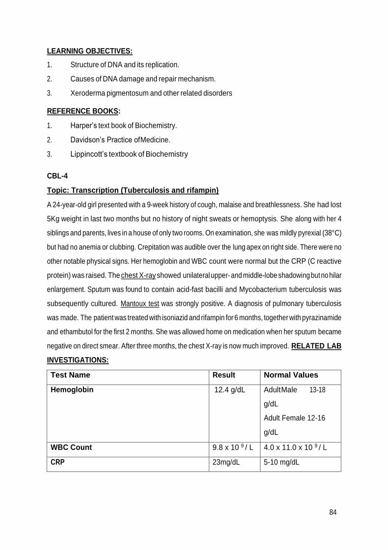
84
LEARNING OBJECTIVES:
1. Structure of DNA and its replication.
2. Causes of DNA damage and repair mechanism.
3. Xeroderma pigmentosum and other related disorders
REFERENCE BOOKS:
1. Harper’s text book of Biochemistry.
2. Davidson’s Practice of Medicine.
3. Lippincott’s textbook of Biochemistry
CBL-4
Topic: Transcription (Tuberculosis and rifampin)
A 24-year-old girl presented with a 9-week history of cough, malaise and breathlessness. She had lost
5Kg weight in last two months but no history of night sweats or hemoptysis. She along with her 4
siblings and parents, lives in a house of only two rooms. On examination, she was mildly pyrexial (38°C)
but had no anemia or clubbing. Crepitation was audible over the lung apex on right side. There were no
other notable physical signs. Her hemoglobin and WBC count were normal but the CRP (C reactive
protein) was raised. The chest X-ray showed unilateral upper- and middle-lobe shadowing but no hilar
enlargement. Sputum was found to contain acid-fast bacilli and Mycobacterium tuberculosis was
subsequently cultured. Mantoux test was strongly positive. A diagnosis of pulmonary tuberculosis
was made. The patient was treated with isoniazid and rifampin for 6 months, together with pyrazinamide
and ethambutol for the first 2 months. She was allowed home on medication when her sputum became
negative on direct smear. After three months, the chest X-ray is now much improved. RELATED LAB
INVESTIGATIONS:
Test Name Result Normal Values
Hemoglobin 12.4 g/dL Adult Male 13-18
g/dL
Adult Female 12-16
g/dL
WBC Count 9.8 x 10 9 / L 4.0 x 11.0 x 10 9 / L
CRP 23mg/dL 5-10 mg/dL

85
Mantoux test (tuberculin skin test) (NOT
SIGNIFICANT)
“QuantiFERON-TB Gold” is a newer test which
measures the response of immune system of
body to MTB antigen in vitro
12mm
induration
Less than 10mm in
vaccinated individuals
Sputum for culture sensitivity No growth No growth
Sputum for AFB (smear test) Positive only
third time
Negative
Culture of MTBC (mycobacterium tuberculosis
complex) 2-3 weeks
Traditional solid media shows growth in 4-8
weeks
Positive Negative
Nucleic Acid Amplification Tests (NAAT) Detects
genetic
material of
MTB
Not detected
LAM (lipoarabinomannan)antigen test in urine
(mycobacterial cell wall component shed in
plasma ) 30 Min
Positive Negative
Adenosine Deaminase Test ADA Increased -
HIV test Negative Negative
Pulmonary tuberculosis is an infection of lungs by a special bacterium called mycobacterium tuberculosis.
Vaccine against TB is a part of EPI (extended program of immunization) by WHO which imparts roughly
20% additional protection against the infection. BCG vaccine (Bacillus Calmette–Guérin) is the only
vaccine available and given to all the children round the world (90%) against tuberculosis. Though the
disease is not common in developed countries, but in developing countries including Pakistan and
especially African countries TB is quite prevalent and a major public health problem. Multidrug therapy
with good compliance for 6 months at least is usually the treatment of choice. One of the antibiotic
drugs is rifampin. Rifampin inhibits bacterial cell growth by inhibiting RNA synthesis. It binds to beta
subunit of bacterial (prokaryotic) RNA polymerase and interferes with the formation of first phosphodiester
bond.

86
This prevents initiation of transcription in Acid fast bacilli (AFB). This slows growth and replication
of the bacteria which are washed away in secretions. If there is resistance to this drug, the infection is
called MDR-TB (multi-drug resistant TB). MDR-TB and XDR-TB (extensive drug resistnt TB) are serious
public health problems and over 13 million die every year with TB worldwide. Dactinomycin (also called)
Actinomycin D is an antibiotic and anticancer agent which binds DNA template and hinders the movement
of RNA polymerase on it, blocking the transcription.
LEARNING OBJECTIVES:
1. Structure and types of RNA
2. Pro and Eukaryotic gene transcription
3. Post transcriptional modification of RNA including splicing
REFERENCE BOOKS:
1. Lippincott’s textbook of Biochemistry
2. Harper’s text book of Biochemistry
3. Davidson’s Practice of Medicine
CBL-5
Topic: Biotechnology Techniques-PCR etc (HCV & Lymphoma)
A 54-year-old male presented with fever, night sweats and weight loss. He also complained of a non-
tender, non-inflammatory mass on left lateral side of his neck. The mass has grown to the size of a
hazelnut since it was first noticed 6 weeks ago. On examination submandibular and supraclavicular nodes
were palpable. Blood complete picture was normal but patient was diagnosed as a case of B Cell Non
Hodgkin lymphoma with the help of tissue histology and bone marrow aspiration cytology. During
the laboratory investigation phase he was incidentally found to be a patient of HCV infection as
well.
In addition to commitment about the treatment and prognosis of the lymphoma, attending physician was
also concerned about the status of his HCV infection. He ordered abdominal ultrasound to check for
liver cirrhosis and Quantitative PCR to check for HCV viral load.

87
RELATED LAB INVESTIGATIONS:
Test Name Result Normal Values
Hemoglobin 14 g/dL Adult Male 13-18
g/dL
Adult Female 12-16
g/dL
WBC Count 11.5 x 10 9 / L 4.0 x 11.0 x 10 9 / L
(Tumor/mass )Lymph Node Biopsy
B Cell lymphoma cells seen Normal cytology
Lymphoma cells in peripheral blood smear
Not seen Not seen
Bone marrow biopsy for histological examination
Monoclonal lymphoid aggregates seen
No such aggregates
LDH 630 U/L 225-450 U/L
Anti HCV Antibody test Positive Negative
USG Abdomen Nodular and shrunken
appearance of liver but mild changes
Normal echo-texture
Quantitative PCR (for viral
RNA)
700,000 IU/mL Normally it is negative Viral
Load is expressed as
copies/mL: Low: less than
2 M/mL • High: more
than 2 M/mL
Or Expressed as
International Units
(IU/mL): • Low: less
than 800,000 IU/mL •
High: more than
800,000 IU/mL
Cytogenetic testing in culture
sample (2 -3 weeks)
No abnormal chromosomal
pairing or less or more
chromosomes found
Normal chromosomal
pairing and normal
number
Fluorescent in situ
hybridization (FISH) for
t(14;18) of c-myc
No translocation found on
microscopy
No translocation
PCR for Immunoglobulin light
chain gene rearrangements
This test was carried out as there was a research center nearby
and outcome had a research benefit as well. For patient it was
helpful in determining whether it was reactive or malignant
Though HCV and lymphoma is not a common combination in patients encountered in clinics but it has
been selected to emphasize role of biotechnology techniques in diagnosis, management and
prognosis of both the diseases. PCR is a very basic technique of DNA selected portion amplification
in vitro from even minute quantities of DNA. It makes DNA
88
ample to carry out any genetic analysis. In HCV patients quantitative PCR is routinely advised to see viral
load. While in lymphoma certain related immunoglobulin gene rearrangement can be checked to
understand etiology of the disease better.
LEARNING OBJECTIVES:
1. Molecular Biology Techniques and their applications
2. Procedure, principle and uses of PCR
3. Types of PCR
REFERENCE BOOKS:
1. Lippincott’s textbook of Biochemistry
2. Harper’s text book of Biochemistry
3. CMDT 2013, (Current Medical Diagnosis and Treatment)
CBL-6
Topic: Cholera (G protein coupled receptor)
A 21-year-old female law student working in a developing country suddenly began to pass profuse
watery stools almost continuously. She soon started to vomit. Her general condition declined abruptly,
and she was rushed to the local village hospital. On admission, she was cyanotic, skin turgor was poor,
blood pressure was 70/50 mmHg (normal 120/80 mm Hg), and her pulse was rapid and weak. The doctor
on duty diagnosed cholera, took a stool sample, and started treatment immediately. Patient was
isolated and given normal saline with 20mmol/L K+ added (ringers lactate was not administered).
Doxycycline 500mg every 6 hr was started and plenty of ORS was advised after initial IV fluids. The
relatives were called for counseling. As cholera can become epidemic or pandemic, relatives were
given following advice to follows and teach others. 1. Drink only boiled or treated water, 2. Cook well and
eat hot, 3. Avoid shellfish, 4. Peel all vegetables and fruits.
RELATED LAB INVESTIGATIONS:
Test Name Result Normal Values
Stool Microscopy Vibrio cholera seen Nil
Stool culture Yielded growth of Vibrio
cholera
Nil/ normal flora
Serum electrolytes Low Na+,K+, Cl- and HCO3- Normal
89
Vibrio Cholera is a gram negative rod, comma shaped bacterium transmitted through faecal- oral route. Its
incubation period is few hours to 5 days and it causes profuse watery stools, fever, vomiting and rapid
dehydration which may cause death. One may lose upto 1L/hour water in stools. Heat killed vaccine is
not that effective and is not required for international travel. Prevention of epidemic spread is
important. The key to effective treatment and prevention of death is meticulous and timely water
and electrolyte replacement.
LEARNING OBJECTIVES:
1. Mechanisms of cell receptors
2. Mechanisms of cellular second messenger
REFERENCE BOOKS:
1. Lippincott’s textbook of Biochemistry
2. Harper’s text book of Biochemistry
3. Davidson’s Practice of Medicine
CBL-7
Topic: INTEGRATION OF METABOLISM (DM)
A 47 year old lady presented with polyuria, polydipsia and weight loss for few months. She also
complained for recurrent burning micturition/ genital thrush for some time. Her plasma glucose levels
were checked and she was diagnosed as a case of DM.
RELATED LAB INVESTIGATIONS:
Test Name Result Reference
Plasma Glucose levels random 15mmol/L Upto 11.1mmol/L
Plasma glucose levels fasting (next day) (DM if more than 7mmol/L)
9 mmol/L 5.6 mmol/L
HbA1c 60mmol/L Upto 48mmol/L (6.5%)
Explanation Diabetes mellitus is a serious pandemic metabolic derangement syndrome in which
decreased endogenous insulin or its decreased effectiveness is the hallmark. Insulin and glucagon are
major regulatory hormones of metabolism. Insulin is the hormone of fed state and it’s the hormone of
storage (causes deposition of carbohydrate and fat stores). Decreased synthesis or release of insulin
from beta cells of pancreas leads to hyperglycemia in addition to other metabolic derangements and the
condition is called type 1 DM. On the other hand, despite normal release of insulin from pancreas there
may be resistance to its

90
action in peripheral tissue (adipose and Skeletal muscle) mostly secondary to obesity and this condition is
called type 2 DM.
LEARNING OBJECTIVES:
1. Mechanism of action and effects of Insulin and Glucagon
2. Biochemical Basis of sign and symptoms of DM
3. Biochemical Diagnosis of DM
REFERENCE BOOKS:
1. Harpers text book of Biochemistry
2. Guyton text book of physiology
3. Internet sources
CBL-8
INTEGRATION OF METABOLISM -Obesity (obesity)
A 40 year old house wife presented with chronic pain in both knees and back. Her weight was 93 kg and
height was 5’3”. Her waist to hip ratio was measured to be 1.1. She also complained of inguinal hernia. She
was diagnosed as a case of android obesity with BMI almost 36. Her doctor advised her to change
dietary and life style habits and gave some symptomatic relief medicines after seeing the report of
tests ordered empty stomach.
RELATED LAB INVESTIGATIONS:
Test Name Result Normal Values
TC 6.2 mmol/L Less than 5.6 mmol/L
TGs 1.8 mmol/L Less than 1.6 mmol/L
LDL 3.6 mmol/L Less than 2.6 mmol/L
HDL 90 mmol/L More than 1.04 mmol/L
BSF 120 mg/dL Less than 110 mg/dL
Obesity is a disorder of body weight regulatory systems characterized by an accumulation of excess body
fat. The BMI (weight in kg)/(height in meters)2 provides a measure of relative weight, adjusted for
height. This allows comparisons both within and between populations. The healthy range for the BMI is
between 18.5 and 24.9. Individuals with a BMI between 25 and 29.9 are considered overweight, those
with a BMI equal to or greater than 30 are defined as obese, and a BMI over 40 is considered extremely
obese. Anyone more than 100 pounds overweight is considered severely obese. These cutoffs are
based on the studies examining the relationship of BMI to premature death, and are similar in men
and women.

91
LEARNING OBJECTIVES:
1. Obesity and how to measure
2. Body weight regulation
3. Weight reduction
REFERENCE BOOKS:
1. Lippincott’s illustrated reviews and Harper’s text book of Biochemistry
2. Davidson’s Practice of Medicine
3. Internet sources
92
BLOCK-III
Section-I
Anatomy

93
Summary:
Code Y2M3
Name Anatomy
Duration 10 weeks
Broad Themes of Module (Theme: a subject that is being integrated a majority of time of module)
Head, Neck and Special Senses Endocrine System Reproductive system
Subject Themes
Gross Anatomy o Head and neck
Embryology Development of
o Head and neck o Special senses
Development of integumentary system
Histology
o Endocrine glands o Histology of integumentary
System
Prerequisite Module Y2M1 & Y2M2
Mode of Information Transfer:
MIT
Lectures
Dissection
Demonstration
CBL
Practical’s
Dissection movies
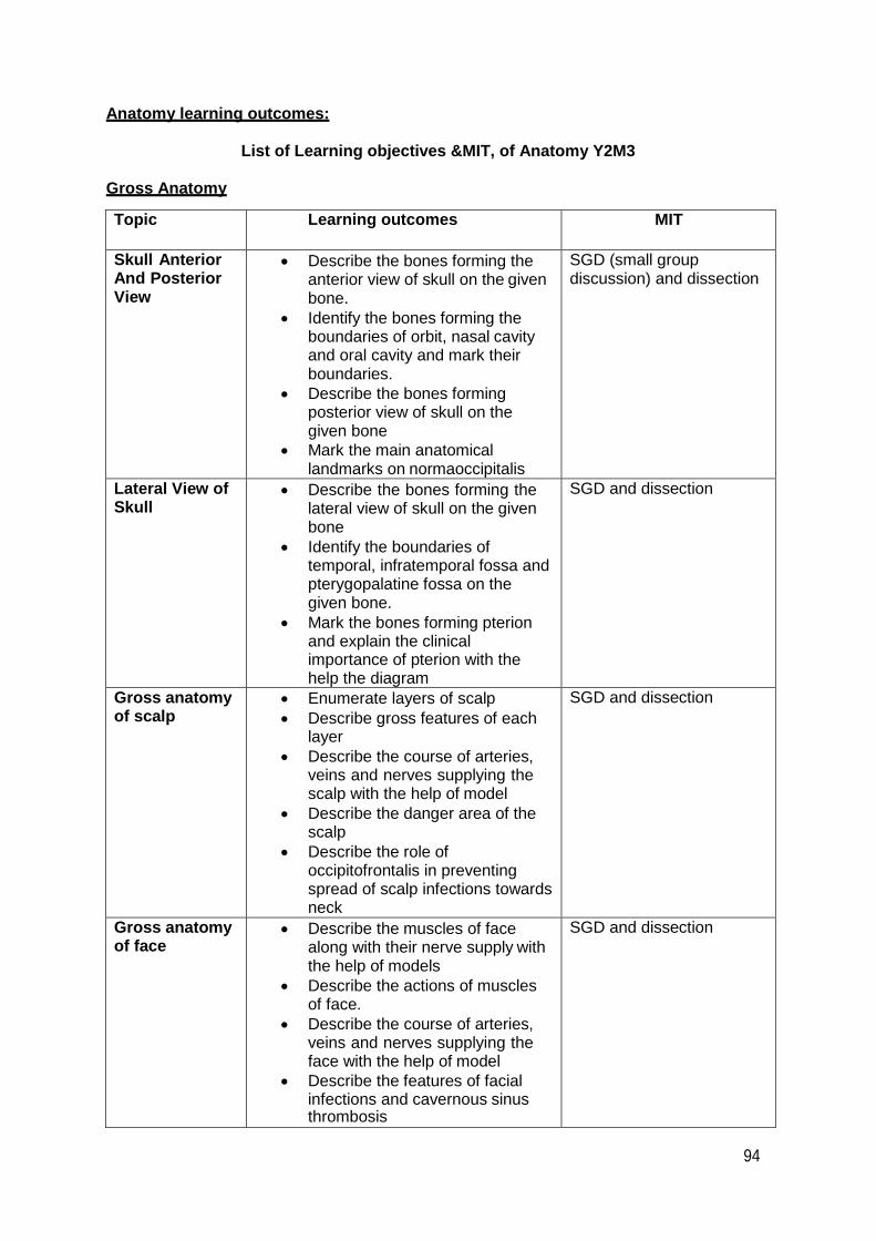
94
Anatomy learning outcomes:
List of Learning objectives &MIT, of Anatomy Y2M3
Gross Anatomy
Topic Learning outcomes MIT
Skull Anterior And Posterior View
Describe the bones forming the anterior view of skull on the given bone.
Identify the bones forming the boundaries of orbit, nasal cavity and oral cavity and mark their boundaries.
Describe the bones forming posterior view of skull on the given bone
Mark the main anatomical landmarks on normaoccipitalis
SGD (small group discussion) and dissection
Lateral View of Skull
Describe the bones forming the lateral view of skull on the given bone
Identify the boundaries of temporal, infratemporal fossa and pterygopalatine fossa on the given bone.
Mark the bones forming pterion and explain the clinical importance of pterion with the help the diagram
SGD and dissection
Gross anatomy of scalp
Enumerate layers of scalp
Describe gross features of each layer
Describe the course of arteries, veins and nerves supplying the scalp with the help of model
Describe the danger area of the scalp
Describe the role of occipitofrontalis in preventing spread of scalp infections towards neck
SGD and dissection
Gross anatomy of face
Describe the muscles of face along with their nerve supply with the help of models
Describe the actions of muscles of face.
Describe the course of arteries, veins and nerves supplying the face with the help of model
Describe the features of facial infections and cavernous sinus thrombosis
SGD and dissection

95
Gross anatomy of trigeminal nerve
Trace the pathway of trigeminal nerve from nucleus to target organs
Enumerate the divisions of trigeminal nerve
Describe the features of trigeminal neuralgia
SGD and dissection
Branches of trigeminal, nerve, mandibular, maxillary
Describe the pathway of mandibular nerve from nucleus to target organs
Describe the pathway of maxillary nerve from nucleus to target organs
Describe the lesions of nerves with special reference to infections of molar teeth
SGD and dissection
Gross anatomy of facial nerve
Describe the course of facial nerve in face
Enumerate its branches
Discuss the involvement of nuclei of facial nerve in bell palsy
Differentiate between upper and lower motor neuron lesions
SGD and dissection
Salivary glands Enumerate salivary glands
Describe the locations of major salivary glands
Trace the secretomotor nerve supply of major salivary glands
Describe the structures involved in parotid infections
SGD and dissection
Temporomandi bular joint
Identify the type of TMJ.
Identify the articular surfaces of TMJ on a given model or dry bones.
Name the ligaments of TMJ.
Describe the movements of jaw at TMJ with special reference to axis and muscles producing them.
Describe the clinical signs of anterior dislocation of TMJ and explain the steps of its reduction.
SGD and dissection
Infratemporal region
Identify the location of infratemporal fossa on a given model and skull.
Enlist the structures forming various boundaries of infratemporal fossa.
Enlist the communications of infratemporal fossa and the structures traversing each.
Enumerate the contents of infratemporal fossa.
SGD and dissection
96
Discuss the relationships of various contents of infratemporal fossa.
Discuss the attachments, actions and nerve supply of muscles of mastication.
Deep cervical fascia – I
Enumerate the layers of deep cervical fascia
Describe the attachments of investing, pretracheal, and prevertebral layers of fascia
Describe the modification of prevertebral layer into axillary sheath
Describe the formation of carotid sheath and its contents
SGD and dissection
Deep cervical fascia – II
Describe the spaces within fascia
Describe the clinical significance of retropharyngeal space
Describe the relation of layers of fascia and spread of infection
Describe the significance of merging of carotid sheath with pretracheal layer of fascia to prevent spread of infections
SGD and dissection
Muscles of neck
Describe the muscles of neck along with their nerve supply with the help of models
Describe the features of torticollis
SGD and dissection
Triangles of neck
Enumerate triangles of neck
Describe the muscles forming the boundaries of triangles
Describe the contents of triangles and their importance
Describe the lesions of the spinal accessory nerve in posterior triangle
SGD and dissection
Vessels of neck Enumerate the main vessels in neck
Describe the course and branches of:
Common carotid artery External carotid artery Internal carotid artery subclavian artery External jugular vein Internal jugular vein
Describe the importance of monitoring jugular venous pulse in heart diseases
Enumerate causes of prominence of external jugular vein
SGD and dissection

97
Discuss subclavian vein thrombosis
Oral Cavity Identify structures forming the boundaries of oral cavity
Identify structures in the floor of oral cavity with the help of models
Identify the structures forming the boundaries of oral vestibule
Enumerate the vessels and verves supplying the oral cavity
Discuss clinical correlations of oral cavity
SGD and dissection
Gross anatomy of palate
Identify the main features of hard palate and soft palate.
Enumerate muscles of soft palate on the model
Enumerate blood supply and nerve supply of soft palate
Identify the main muscles forming the palatoglossal and palatopharyngeal arches
SGD and dissection
Tongue Describe the gross features of parts of tongue
Describe the blood supply, nerve supply, lymphatic drainage of tongue
Describe the movements of tongue
SGD and dissection
Pharynx Describe the following parts of pharynx on the given model
Oropharynx
Nasopharynx
Laryngopharynx
Describe muscles of pharynx
Describe lymphoid tissue in the pharynx
Describe the importance of structures passing through the spaces between muscles of pharynx while performing tonsillectomy
Describe spread of infections from nasopharynx to middle ear
SGD and dissection
Viscera of neck Describe the relations of trachea and esophagus in neck region with the help of dissection
Describe the structures involved in cricothyroidotomy and tracheostomy with the help of dissection
SGD and dissection
98
Demonstrate the gross features of thyroid and parathyroid glands on models
Describe blood supply and nerve supply of thyroid and parathyroid gland through models
Describe the relations of vessels and nerves supplying the thyroid gland and their significance while performing thyroidectomy
Joints of neck
Name the typical and atypical intervertebral joints of neck.
Identify the types of atlanto- occipital and atlanto-axial joints.
Describe the movements of these joints with muscles producing them.
SGD and dissection
Prevertebral region and root of neck
Name the prevertebral muscles. Give their origin, insertion, action and nerve supply of prevertebral muscles
Describe the relations of key muscle of root of neck (scalenus anterior)
Describe the parts and branches of subclavian artery.
SGD and dissection
Back of neck
Name the muscles of back of neck.
Identify the boundaries and contents of suboccipital triangle.
Describe the course and relations of 3rd and 4th parts of vertebral arteries.
SGD and dissection
Submandibular region
Describe the muscles present in the submandibular region and sublingual region with the help of model and prosection.
Enumerate the nerves vessels and ganglion in submandibular and sublingual region
SGD and dissection
Nerves of neck Enumerate the main nerves in neck
Trace the course of glossopharyngeal nerve, vagus nerve, accessory nerve and hypoglossal nerve on the given model,from nucleus to target organs.
Enumerate branches of each of the above nerve
SGD and dissection
Lymphatic drainage of head and neck
Enumerate the groups of lymph of nodes draining the neck
SGD and dissection
99
Describe their location and areas of drainage
Describe the formation of jugular lymph trunk
Describe the clinical importance of lymphatic drainage of head and neck
Gross anatomy of larynx
Explain the gross features of inlet of larynx, piriform fossa, laryngeal folds, cavity of larynx
Correlate the laryngeal anatomy to foreign bodies aspiration
Explain the gross features of intrinsic muscles of larynx, extrinsic muscles of larynx, movements of vocal folds
Describe the cartilage involvement in fractures of the laryngeal skeleton
SGD and dissection
Gross anatomy of larynx
Identify the gross features of cartilages of larynx membranes of larynx
Trace the course of following nerves of larynx
Internal laryngeal nerve External laryngeal nerve Inferior laryngeal nerve
Discuss clinical correlations of larynx including recurrent laryngeal nerve and external laryngeal nerve damage
SGD and dissection
Gross Anatomy External Ear
Describe the gross anatomical features of external ear Auricle External auditory meatus
Describe the blood supply, nerve supply and lymphatic drainage of external ear.
Correlate the significance of straightening the auditory canal during clinical examination with the anatomical structure of canal.
SGD and dissection
Functions of external and middle ear
Describe the following three functions of the external ear:
Transmission of sound to tympanic membrane
Amplification of sound Prevention of dust and dirt
from reaching ear drum
Describe the function of ossicles of the middle ear in conduction of sound from tympanic membrance to cochlea
SGD and dissection

100
Functions of external and middle ear
Describe the phenomenon of impedance matching provided by the tympanic membrane and Ossicular system of the ear
Describe attenuation reflex along with its two functions of:
Protecting cochlea Masking low frequency
sound waves
SGD and dissection
Gross anatomy middle ear-I
Describe the gross anatomical features of middle ear
Describe the structures forming the walls of middle ear cavity on the given model
Describe the contents of middle ear cavity
SGD and dissection
Gross anatomy middle ear-II
Identify the parts of ear ossicles on the given model
Describe the muscles present in middle ear cavity
Describe the gross features of auditory tube
Describe the nerve supply of auditory tube
Describe the anatomical structures involved in paralysis of the stapedius
Blockage of pharyngotympanic tube
SGD and dissection
Conduction of sound
Describe the role of the following in conduction of sound vibrations:
Scala vestibule Scala media Scala tympani
SGD and dissection
Gross anatomy inner ear-I
Identify the parts of bony labyrinth on the given model
Identify the parts of membranous labyrinth on the given model
Identify the parts of cochlea of semi-circular canal on the given model.
SGD and dissection
Gross anatomy inner ear-II
Describe the gross features of bony labyrinth
Describe the gross features of membranous labyrinth
Describe the orientation of semicircular canals and ducts within the inner ear
Describe the gross features of internal acoustic meatus
Describe anatomical structures involved in perforation of tympanic membrane
SGD and dissection
101
Describe mastoiditis
Discuss the role of membranous labyrinth in motion sickness
Discuss the role of spiral organ of corti in high-tone deafness
Define otic barotraumas
Vestibulococchl ear nerve
Trace the course of vestibulocochlear nerve in the inner ear on the given model
Identify the area of supply of vestibular nerve on the given model
Identify the area of supply of cochlear nerve
Identify the gross features of vestibulocochlear ganglion on model
Discuss the consequences of damage to vestibulocochlear nerve
SGD and dissection
Gross Anatomy of Orbital Region
Describe the bony orbit
Enlist the structures present in the orbit
Describe gross features of eye lids
Describe the attachment of muscles of eyelid
Describe the attachment of orbital septum
Describe the distribution of Blood Vessels and Lymph Vessels of the Orbit
Describe the anatomical
structures involved Inflammation of the Palpebral Glands
SGD and dissection
Extraocular muscles
Describe the extraocular muscles of eye
Describe the movements of eyeball
Correlate the anatomical lesions in nuclei of nerve supplying the extraocular muscles with the loss of function in muscles.
SGD and dissection
Occulomotor Nerve
Trace the pathway of Occulomotor never from nucleus to target organs
Correlate the anatomical lesions in nuclei of occulomotor never with clinical conditions like
External Strabismus Ptosis and Diplopia
SGD and dissection
102
Trochlear Nerve Trace the pathway of Trochlear nerve from nucleus to target organs.
Discuss the effect of lesion of trochlear nerve
SGD and dissection
Gross anatomy of lacrimal apparatus
Enumerate the structures forming lacrimal apparatus
Describe the gross features of each part of lacrimal apparatus
Describe the nerve supply of lacrimal apparatus
Co relate the anatomical structures of lacrimal apparatus with the features of blocked Lacrimal duct
SGD and dissection
Gross anatomy of eyeball
Describe the coats and parts of eye ball on a given model.
Fibrous coat Vascular pigmented coat Nervous coat
Describe the blood supply and verve supply of eyeball
Describe the actions of muscles of pupil
Describe the appearance of optic disc and macula lutea on ophthalmoscope
SGD and dissection
Optic Nerve Trace the pathway of optic nerve from nucleus to target organs
Describe the formation of olfactory bulb and optic tract.
Correlate the anatomical lesions in visual pathway with clinical conditions like hemianopia, homonymous hemianopia
Bitemporal hemianopia.
SGD and dissection
Gross anatomy nose
Describe the structure of external nose and nasal cavity
Describe the concha and meatus in the lateral wall
Enumerate the sinuses opening in them
Discuss anatomical structures involved in nasal fractures
Correlate the anatomical structure of nasal mucosa with clinical manifestations of rhinitis
SGD and dissection
Olfactory Nerve Trace the pathway of Olfactory nerve form nucleus to target organs on a model
Describe the formation of olfactory bulb and olfactory tract.
SGD and dissection
103
Correlate the effects of lesion of olfactory nerve with special reference to clinical conditions causing anosmia
Paranasal Sinuses
Describe the gross features of paranasal sinuses
Describe infections of sinuses
Describe the Drainage of mucus in relation to sinusitis
Describe the Function of Paranasal Sinuses
Discuss the anatomical structures involved in sinusitis with special reference to clinical consequences of infections of the ethmoidal cells of the ethmoidal sinuses
SGD and dissection
Cranial nerves Discuss the intracranial and extra cranial course of all cranial nerves
Discuss clinical correlations and examination of all cranial nerves.
Imaging of Head
Identify the bones forming skeleton of head on radiograph
Identify boundaries of orbit ¶nasal sinuses on radiograph
SGD and dissection
Imaging of neck Describe the appearance of structures of neck and face in:
Radiograph CT scan MRI
SGD and dissection
Surface marking of Head & neck
Mark the main vessels of head & neck on the given subject
Superficial temporal artery Supra orbital and supra
trochlear Facial Artery External Carotid artery External Jugular vein Subclavian artery
Palpate the following muscles Trapezuis Sternocleidomastoid
SGD and dissection

104
Embryology
Integumentary System (LGIS)
Describe the development of skin, hair, glands, nail, mammary glands and tooth
Enlist common anomalies
Development of Pharyngeal Arches (LGIS)
Describe the development of pharyngeal arches
Describe the components of pharyngeal arches
Enumerate the components developing from all three layers of each arch
Describe the anomalies associated with them.
Development of pharyngeal pouches (LGIS)
Describe the development of pharyngeal pouches and membranes
Describe the components of pharyngeal pouches and membranes
Describe the features of anomalies associated with the development of pouches and membranes
Development of face (LGIS)
Describe the role of frontonasal prominences, maxillary prominences and mandibular prominences in development of face
Describe the formation of of oblique facial clefts Describe the features of Congenital microstomia
Development of palate (LGIS(
Describe the stages of development of primary palate
Describe the stages of development of secondary palate
Describe the process of development of Cleft Lip and Cleft palate
Tongue (LGIS) Describe the development of tongue
Describe the anomalies associated with development
Development of nose (LGIS)
Describe the development of nose
Describe the development of paranasal sinuses & nasal conchae
Development of Thyroid and parathyroid glands (LGIS)
Enumerate the arches from which thyroid and parathyroid glands develop.
Describe the mechanism of descent of thyroid and parathyroid glands during development.
Describe the features of congenital hypothyroidism
Enumerate congenital anomalies of thyroid gland
Describe thyroglossal cyst. Describe clinical features of thyroglossal cyst
Development of adrenal glands (LGIS)
Describe the development of adrenal glands
Describe the process of differentiation of fetal cortex into adult cortex
Enlist congenital anomalies of adrenal gland
Development of Eye-I (LGIS)
Describe the development of optic cup
Relate the differentiation of wall of optic cup with the formation of sclera and cornea
Enlist developmental anomalies of sclera and cornea
Development of Eye-II (LGIS)
Describe the development of ciliary body, ciliary muscles and retina
Describe the differentiation of mesenchyme into chambers of eye.
Describe the transformation of optic stalk into optic nerve
105
Enlist related common anomalies
Describe the anatomical structures involved in congenital retinal detachment
Development of external ear (LGIS)
Describe the embryological development of external & middle ear
Describe the associated developmental anomalies
Development of inner ear (LGIS)
Describe the embryological development of inner ear.
Describe the differentiation of otic vesicle into different parts of inner ear
Describe the anomalies related to the development of inner ear
Histology
Skin (LGIS, SGD/tutorial, Practical)
Describe components of skin,nail,hair.
Explain histological difference between thick and thin skin
Describe the various appendages of skin Draw labeled diagram of thick and thin skins on sketch book
Lip &Tongue
(LGIS, SGD/tutorial, Practical)
Describe the histological features of lip & tongue with the help of microscopic images.
Identify the microscopic features on given slides Lip Tongue
Draw labeled diagrams of identified tissue
Histology salivary glands
(LGIS, SGD/tutorial, Practical)
Describe components of salivary glands
Identify the following on given slides: Parotid gland Submandibular gland Sublingual glands
Draw labeled diagrams of identified tissue
Histology of pituitary gland
(LGIS, SGD/tutorial, Practical)
Describe the various parts of pituitary gland.
Identify the gland under the microscope and write two points of identification for the gland
Identify the various hormonal disorders of pituitary gland
Thyroid Gland & Parathyroid Gland
(LGIS, SGD/tutorial, Practical)
Describe histological features of thyroid and parathyroid gland
Describe the role of thyroid follicular cells in thyroid disorders
Identify the microscopic features of thyroid and parathyroid gland under microscope on the given slide.
Draw labeled diagram of the identified tissue on the histology note book
Adrenal Gland
(LGIS, SGD/tutorial, Practical)
Describe the histological features of adrenal gland
Describe the cell types present in zones of adrenal gland and enumerate the hormones produced by each zone
Identify the histological features of adrenal gland under microscope.
Draw labeled diagrams of the identified tissues on the histology notebooks
Pancreas (LGIS, SGD/tutorial, Practical)
Describe the histological features of islets of Langerhan’s
Describe the cell types present in islets of Langerhan’s
Draw labeled diagrams of the identified tissues on the histology notebooks

106
Larynx (LGIS, SGD/tutorial, Practical)
Different layers of larynx
Histological characteristics of each layer of larynx
Histological classification of laryngeal cartilage
Nose (LGIS, SGD/tutorial, Practical)
Describe the different types of mucosa of the different parts of the nose
Histology of nasal cartilage
List of Practical work:
1. Thyroid gland
2. Pituitary gland
3. Pancreas (islets) and adrenal gland
4. Lip
5. Tongue
6. Salivary glands
7. Nose and larynx
8. Skin and appendages

107
CBL 1: A case of facial trauma LEARNING OBJECTIVES:
1. Describe the topographic anatomy of facial skeleton.
2. Describe muscles responsible for various facial expressions.
3. Describe and identify nerve supply, blood supply and lymphatic drainage of face and scalp.
4. Identify muscles of facial expressions on anatomical specimens.
5. Facial nerve examination.
6. Surface marking of facial artery.
7. Demonstrate facial planes in the head & neck on cadaver.
8. Demonstrate ABC of facial trauma (Air way, Breathing and Circulation) on manikins. Identify
nerve supply, arterial supply and venous drainage of face and scalp.
CBL 2: A case of infection of facial spaces of neck LEARNING OBJECTIVES 1. Describe deep cervical fascia and division of neck into various compartments, muscles of
neck.
2. Describe the contents of these compartments. Correlate facial spaces with spread of
infection from neck to mediastinum.
3. Examine Accessory nerve and mark its surface anatomy Examine lymph nodes of neck
CBL-3 A case of ludwig’s angina
LEARNING OBJECTIVES: 1. What nerve(s) would need to be anesthetized in order to prevent pain during a lower third
molar extraction?
2. Some patients occasionally experience temporary paralysis of the muscles of facial
expression or of mastication following a dental procedure. Why do you think this might
happen?
3. When dental work is performed on the lower teeth, anesthetic injected at a single point is
often adequate to produce complete local anesthetization of all the teeth on a given side.
The same is not true for the upper teeth. Why not? How do they have to be anesthetized?
4. What are the anatomical structures that prevent joint dislocation?
5. Identify the masticatory muscles and give their functions.
6. Define the boundaries and contents of the infratemporal fossa.
7. Identify the branches of the trigeminal nerve and their functions related to mastication and
sensation from the face.
8. Identify the chorda tympani nerve and give its function.
9. Describe the structure and function of the temporomandibular joint
108
10. Identify the muscles bordering the submandibular and paralingual spaces.
11. List and identify the major nerves and vessels of these spaces.
12. Describe the submandibular and sublingual salivary glands and give their innervations.
13. List the muscles of the tongue and describe their origins.
14. Describe the oral cavity, its oral vestibule and dental arches (including temporary and
permanent dentitions), and the hard and soft palate.
CBL-4 A case of otitis media LEARNING OBJECTIVES:
1. A major factor in the pathogenesis of otitis media is dysfunction of the auditory (Eustachian)
tubes. What is the function of the auditory tubes?
2. Which muscle opens the auditory (Eustachian) tube?
3. What surgical procedures may be used to treat the fluid build-up in the middle ear?
4. What are possible complications to otitis media?
5. What would an infection of the outer ear be called? What anatomical features/structures
protect the ear from injury?
6. Define the three parts of the ear and the function of each part.
7. Describe each of the four walls of the middle ear cavity and identify deeper structures
responsible for certain of their features.
8. Describe the structure and actions of the tympanic membrane, the auditory ossicles, and
the muscles of the middle ear.
CBL 5: A case of injury to orbit
. LEARNING OBJECTIVES: 1. How would you explain the right ptosis and pupillary constriction in this case?
2. Identify the prominent bony features of the orbit with included foramina and fissures.
3. Describe the components of the eyelids with associated muscles, tarsal glands, connective
tissue fascia and conjunctiva.
4. Identify the extra ocular muscles, their function and innervation.
5. Identify all sensory, motor and autonomic nerves of the orbit and trace their routes to and
within the orbit.
6. Identify branches of ophthalmic arteries and veins

109
BLOCK-III
Section-II
Physiology
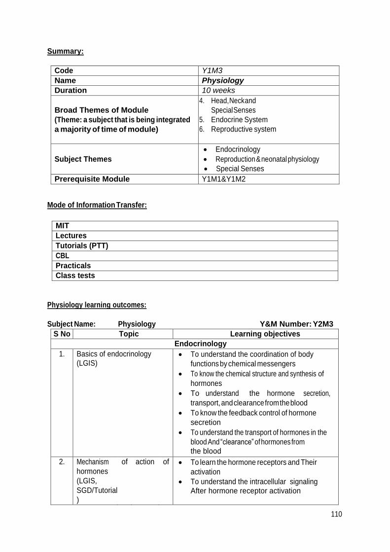
110
Summary:
Code Y1M3
Name Physiology
Duration 10 weeks
Broad Themes of Module
(Theme: a subject that is being integrated
a majority of time of module)
4. Head, Neck and
Special Senses
5. Endocrine System
6. Reproductive system
Subject Themes
Endocrinology
Reproduction & neonatal physiology
Special Senses
Prerequisite Module Y1M1&Y1M2
Mode of Information Transfer:
MIT
Lectures
Tutorials (PTT)
CBL
Practicals
Class tests
Physiology learning outcomes:
Subject Name: Physiology Y&M Number: Y2M3
S No Topic Learning objectives
Endocrinology
1. Basics of endocrinology (LGIS)
To understand the coordination of body
functions by chemical messengers
To know the chemical structure and synthesis of
hormones
To understand the hormone secretion,
transport, and clearance from the blood
To know the feedback control of hormone
secretion
To understand the transport of hormones in the
blood And “clearance” of hormones from the blood
2. Mechanism
hormones
(LGIS,
SGD/Tutorial
)
of action of To learn the hormone receptors and Their
activation
To understand the intracellular signaling After hormone receptor activation
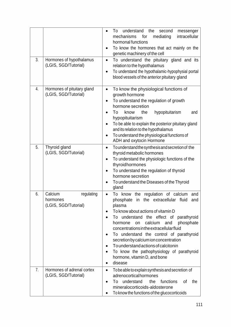
111
To understand the second messenger
mechanisms for mediating intracellular
hormonal functions
To know the hormones that act mainly on the
genetic machinery of the cell
3. Hormones of hypothalamus (LGIS, SGD/Tutorial)
To understand the pituitary gland and its
relation to the hypothalamus
To understand the hypothalamic-hypophysial portal
blood vessels of the anterior pituitary gland
4. Hormones of pituitary gland (LGIS, SGD/Tutorial)
To know the physiological functions of
growth hormone
To understand the regulation of growth
hormone secretion
To know the hypopituitarism and
hypopituitarism
To be able to explain the posterior pituitary gland
and its relation to the hypothalamus
To understand the physiological functions of ADH and oxytocin Hormone
5. Thyroid gland (LGIS, SGD/Tutorial)
To understand the synthesis and secretion of the
thyroid metabolic hormones
To understand the physiologic functions of the
thyroid hormones
To understand the regulation of thyroid
hormone secretion
To understand the Diseases of the Thyroid gland
6. Calcium regulating
hormones
(LGIS, SGD/Tutorial)
To know the regulation of calcium and
phosphate in the extracellular fluid and
plasma
To know about actions of vitamin D
To understand the effect of parathyroid
hormone on calcium and phosphate
concentrations in the extracellular fluid
To understand the control of parathyroid
secretion by calcium ion concentration
To understand actions of calcitonin
To know the pathophysiology of parathyroid
hormone, vitamin D, and bone
disease
7. Hormones of adrenal cortex (LGIS, SGD/Tutorial)
To be able to explain synthesis and secretion of
adrenocortical hormones
To understand the functions of the
mineralocorticoids-aldosterone
To know the functions of the glucocorticoids
112
To know the abnormalities of adrenocortical
secretion
8. Hormones of pancreas (LGIS, SGD/Tutorial)
To understand metabolic effects of insulin
To understand the mechanisms of insulin
secretion
To understand the control of insulin secretion
To understand glucagon and its functions
To understand regulation of glucagon
secretion
To know the pathophysiology of diabetes
mellitus
Reproductive system
1. Male reproductive
physiology
(LGIS, SGD/Tutorial)
To understand the physiologic anatomy of the
male sexual organs
To understand the process of
spermatogenesis
Function of the seminal vesicles and prostate
gland
To understand the abnormal
spermatogenesis and male fertility
To understand the testosterone and other male
sex hormones
2. Female reproductive system (LGIS, SGD/Tutorial)
To know the physiologic anatomy of the
female sexual organs
To understand the monthly ovarian cycle;
function of the gonadotropic hormones
To know the gonadotropic hormones and their
effects on ovarian follicle growth—the “follicular”
phase of the ovarian cycle corpus luteum—
“luteal” phase of the
To understand ovarian cycle
To know about functions of the ovarian
hormones—estradiol and progesterone
3. Menstrual cycle (LGIS, SGD/Tutorial)
To know the regulation of the female monthly
rhythm—interplay between the ovarian and
hypothalamic-pituitary hormones and
feedback oscillation of the hypothalamic-
To understand the pituitary-ovarian system
To be able to explain puberty and menarche and
menopause
4. Pregnancy (LGIS, SGD/Tutorial, Practical)
To be able to describe maturation and
fertilization of the ovum
Transport of the fertilized ovum in the
fallopian tube
To understand the implantation of the blastocyst in the uterus and early nutrition of
113
the embryo
To know about the response of the mother’s body
to pregnancy
To be able to explain changes in the maternal
circulatory system during pregnancy
5. Placenta (LGIS, SGD/Tutorial) To know about hormonal factors in pregnancy
To understand human chorionic gonadotropin
and its effect to cause persistence of the
corpus luteum and to prevent menstruation
To know the placental hormones
6. Parturition (LGIS, SGD/Tutorial)
To know about the mechanism of parturition and
onset of labor—a positive feedback
To know about the mechanics of parturition
Neonatal physiology
1. Neonatal physiology (LGIS) To understand growth and functional
development of the fetus development of the organ
systems
To understand adjustments of the infant to
extrauterine life
To understand the circulatory readjustments at
birth
To know the special problems of prematurity
List of practical
Module 3
1. Testing the Visual Acuity
2. Determination of the Field of Vision
3. Testing the Color vision
4. Demonstration of Sensation of Taste (Gustation)
5. Determination of sense of olfaction
6. Demonstration / Performance of Hearing Tests
7. Demonstration of pure Tone Audiometry
8. Pregnancy Test
9. Any other practical relevant to that Module
114
List of Case Based Learning Scenarios:
Physiology CBLs Year 2ndModule 3rd Endocrinology
CBL-1
29 years old Farzana presented to gynaecological OPD with difficulty to feed her first born baby.
On detailed history she revealed failure of milk ejection, difficulty in conceiving her first baby and
prolonged labour during birth process. Detailed gynaecological screening could not identify any
gynaecological abnormality and she was referred to Medical OPD as a diagnostic dilemma. Detailed
imaging revealed a benign tumor of hypothalamus involving paraventricular nucleus and failed
secretion of oxytocin hormone. She was maintained on synthetic oxytocin for a trial period with relief
of symptoms.
Learning objectives:
1. To define, classify and understand mechanism of action of hormones.
2. To learn sites of secretion of various hormones of body.
3. To understand storage, release and feedback control of various hormones.
4. To understand the mechanism of hormone action.
CBL-2
Bakhtawar is a 20 year old college student. Lately she has noticed increased facial hair
growth and roughness of facial skin. There are also stain marks on her limbs and she complains
of chronic back ache. She has tried sun block creams with little relief from her complaints. On seeking
medical advice, she was referred to an Endocrinologist who told her that she might have a hormonal
imbalance and various hormone assays may be required.
Learning objectives:
1. To learn definition, classification and functions of hormones.
2. To learn feedback control to hormonal secretion.
3. To understand the concept of “clearance” of hormones from blood.
4. To learn the mechanisms of action of hormones.
5. To elaborate the second messenger system of hormone action.
6. To understand concept of radio immune assay and ELISA.

115
CBL-3
A 44 years old man presented in OPD with complaints of gradual deterioration of
vision, progressive increase in body weight and increased frequency of micturition. He also told the
doctor that his shoe size had increased and he had facing problems in finding extra large shoes for the
last 2 years. His physical examination revealed Height = 1.73m (5 ft 8 inches), Weight = 114 kg (250
lbs), B.P = 140/80 mm Hg, thickened fingers and a large jaw and tongue. Biochemical tests revealed
IGF-1 = 1144 ng/ml (normal: 109 – 284 ng/ml)
Basal GH = 24 ng /ml (normal: 5 ng/ml)
TSH = 1.46 µIU/ml (normal: 0.49 – 4.67 µIU/ml)
Random blood glucose = 280 ml/dl (normal: 70 – 140 mg/dl)
Chest radiograph showed gross cardiomegaly. A pituitary MRI revealed a 4.5 cm
macroadenoma which was compressing the optic chiasma significantly. A diagnosis of acromegaly
was made.
Learning objectives:
1. Relate the symptoms and lab tests to the affected endocrine gland and the hormones.
2. Identify the manifestations of acromegaly which are normal or abnormal in this patient.
3. Understand the neuronal and vascular connections between hypothalamus and pituitary
gland.
4. Elucidate the action of growth hormone and role of somatomedins in bone growth.
5. Comprehend feedback control of growth hormone secretion.
6. Differentiate between Gigantism, acromegaly and dwarfism.
7. Hormones secreted by pituitary gland, their mechanism of action and then regulation.
CBL-4
A 28 years old woman presented in medical OPD of Military Hospital with
complaints of restlessness, palpitation, disturbed sleep and difficulty in concentrating on her job for
the past one month. During history taking, she revealed that she had lost 5 kg body weight during
last one month, despite good eating habits.
On general physical examination she was looking nervous and restless. Her body
temperature was 37.5 0C, pulse rate; 101 / min, respiratory rate; 18 / min and blood pressure
was 145 / 80 mm Hg. She also had fine tremors and her knee jerk was brisk.

116
Learning objectives:
Students should know:
1. The possible underlying derangement in this patient and its pathophysiology.
2. Factors and hormones affecting body metabolism.
3. Effect of thyroid hormones on metabolic functions of body.
4. Effect of thyroid hormone on CVS.
5. Mechanism of action of thyroid hormone.
6. Effect of decreased and increased secretion of thyroid hormone.
7. Causes of hypothyroidism and hyperthyroidism.
8. Physiological basis of various signs and symptoms in the patient.
CBL-5
A 30 year old female referred by psychiatrist presents with tremor, weight loss,
emotional lability, frequent bowel movements, swollen neck, bulging eyes, double vision, heat
intolerance, fatigue, scanty periods, filthy nails. On examination enlarged thyroid gland (4 times normal)
was found. Thyroid scan also showed greatly enlarged thyroid compared with normal. Further testing
confirmed hyperthyroidism. The patient was treated with propylthiouracil for 6 months. Symptoms
decreased over time and patient was maintained on follow up therapy.
Learning objectives:
1. To clinically correlate signs and symptoms of thyroid disease.
2. To learn synthesis, storage and release of thyroid hormones.
3. To understand modes of transport and clearance of thyroid hormones.
4. To understand the mechanism of action of thyroid hormones.
5. To learn regulation of thyroid hormone secretion.
6. To describe pathophysiology of thyroid hormone secretion abnormalities and interpret
the diagnosis of thyroid disease.
7. To differentiate between Grave’s disease, goiter and cretinism.
8. To know pathophysiology of thyrotoxicosis and consequent laboratory findings.

117
CBL-6
A 35-year-old woman comes to her physician’s office with the complaint of recent
rapid weight gain and excessive sweating. What initiated her visit was a recent panic attack that
frightened her. Her face looks swollen compared with the rest of her body. She complains of recent
weakness, backaches, and headaches, and her periods have lately been irregular. Over the past month,
she has noticed frequent bruising with slow healing. She is not on any birth control or using any
medication except for acetaminophen for the headaches.
PHYSICAL EXAMINATION
Vital Signs: Temp: 37°C, Pulse 68/min, Resp rate 14/min, BP 130/86 mm Hg, BMI 33 Physical
Examination: The patient’s face is round and her trunk is swollen, but her arms and legs are thin. She
sounds depressed. She has supraclavicular fat pads.
LABORATORY STUDIES
Pregnancy test (HCG): Negative
Glucose tolerance: Abnormal, consistent with insulin resistance.
Plasma cortisol levels: 4 pm: 25 μg/dL (normal: 3-15μg/dL). Dexamethasone is given orally at 11 pm. At
around 8 am the next morning, cortisol levels are 35 μg/dL (normal: < 5 μg/dL).
24-Hour urine collection for free cortisol: Abnormally high Plasma
ACTH: 7 pg/mL (normal: > 20 pg/mL)
MRI of the pituitary: Normal
CT of abdomen and chest: Adrenal tumor
DIAGNOSIS: Cushing’s syndrome (primary hypercortisolism from adrenal tumor)
Learning objectives:
1. After discussion you should be able to:
2. Describe hormones and their contribution to whole body homeostatic mechanisms.
3. Understand the chemical nature of different classes of hormones and how this
determines their mechanism of action on target cells.
4. Define how hormones are synthesized and secreted by cells of endocrine glands, including
how peptide hormones are cleaved from longer precursors.
5. Explain the relevance of protein carriers in the blood for hydrophobic hormones, and the
mechanisms that determine the level of free circulating hormones.

110
6. Understand the principles of feedback control for hormone release and its relevance for
homeostasis.
7. Understand the principles governing disease states that result from over- or under- production
of key hormones.
CBL-7
A 10 yrs old boy presented in pediatric OPD. He had complaints of significant weight
loss in last six months despite having voracious appetite. He was constantly thirsty and was urinating
every 30-40 min. He also had episodes of bed wetting. On physical examination his height was 5 ft 1 inch.
His weight was 95 lb which was 100 lb 2 months back. BP was found to be 90/55 mm Hg in supine and
75/45 mm Hg in standing position. Laboratory findings revealed the following
Fasting plasma glucose-------------320mg/dl (70-110) Plasma
ketones------------------------1+ (none)
Urinary glucose------------------------4+ (none)
Urinary ketones------------------------2+ (none)
On the basis of these findings diagnosis of type 1 diabetes was made. He was immediately started
injectable insulin and counseled on how to monitor blood glucose with finger stick. He was advised
regular check up for monitoring of renal function.
Learning objectives:
1. To know about hormones of pancreas and their functions.
2. Effects of insulin on carbohydrate, protein and fat metabolism.
3. Mechanism of action of insulin.
4. Role of insulin in controlling blood sugar level in postprandial and fasting state.
5. Discuss appearance of ketones in blood and urine.
6. To know the importance of monitoring blood sugar level.
7. To know the importance of monitoring renal function.
111
CBL-1
Reproductive Physiology CBLs
A couple presents to clinic for work up of infertility after 6 years of unprotected
intercourse. The wife declines any medical problems and notes regular menstrual cycles. The husband
states that he has had chronic sinusitis and lower respiratory tract infections. Physical examination
of the woman is unremarkable. Physical examination of the man is remarkable for dextrocardia.
Further testing of the man revealed immotile sperms on semen analysis. The man was labeled infertile due
to autosomal recessive Kartagner’s syndrome and counseled accordingly.
Learning objectives:
1. To know the physiologic anatomy of male reproductive organs.
2. To elaborate steps of spermatogenesis.
3. To elucidate hormonal influences on spermatogenesis.
4. To understand the function of prostate gland and seminal vesicle.
5. To learn the concept of “acrosome reaction”.
6. To understand regulation, effects and pathophysiology of testosterone.
7. To understand the abnormalities of male sexual physiology.
CBL-2
A 26 yrs old female presented in gynae OPD with complaint of amenorrhea for the last
6 months. On inquiring it was revealed that she had normal vaginal delivery 7 months back and currently
she is breast feeding the baby. There was no history of use of contraceptives. On examination vitals
were found to be normal. The gynecologist advised urine pregnancy test which was found to be
negative. A diagnosis of lactational amenorrhea was made and the patient was counseled.
Learning objectives:
1. What are the phases of normal menstrual cycle?
2. What are the phases of normal ovarian cycle?
3. To know about the levels of FSH, LH, estrogen and progesterone throughout the cycle.
4. Discuss prolactin, its site of secretion, mechanism of action and its functions.
5. Discuss different types of amenorrhea.
6. To know about oxytocin and its functions.
112
CBL-3
A 50 yrs old woman presented to gynae OPD with complaints of hyperpigmentation,
hot flashes, night sweats, fatigue, irritability, decreased vaginal secretions and history of
osteoporosis. Her investigations revealed decreased estrogen & decreased progesterone levels. The
gynecologist diagnosed her as a case of menopause.
Learning objectives:
1. To know about the monthly ovarian cycle.
2. To understand the functions of female hormones.
3. Explain the phases of menstrual cycle.
4. To know the regulation of female monthly rhythm.

113
BLOCK-III
Section-III
Biochemistry
114
Summary:
Code Y1M3
Name Biochemistry
Duration 10 weeks
Broad Themes of Module
(Theme: a subject that is being integrated
a majority of time of module)
Head, Neck and Special Senses
Endocrine System
Reproductive system
Subject Themes
Metabolism of Lipids
Integration of metabolism
Bioenergetics
Biochemistry of Endocrine System
Prerequisite Module Y1M1&Y1M2
Mode of Information Transfer:
MIT
Lectures
Tutorials (PTT)
CBL
Practicals
Class tests
Biochemistry learning outcomes and MITs:
Subject Name: Biochemistry
SNo Topic Learning objectives
Endocrinology – I
(Receptors)
(LGIS,
SGD/Tutorial)
Cell surface receptors
G protein coupled receptors
Intracellular second messenger signaling
cascade
Intracellular ligand receptors
1 Endocrinology (LGIS, SGD/Tutorial)
Chemistry, Secretion, Mechanism of action,
regulation and effect on Carbohydrates,
Lipids, Proteins, Mineral and water
metabolism and disorders of various
endocrine glands
Mechanism of action of hormones
Functions of growth hormone
Functions of thyroid hormone
Functions of adrenal hormone

115
Functions of pancreatic hormone
Functions of parathyroid hormone
2 Reproduction (LGIS, SGD/Tutorial)
Androgens synthesis and functions
Estrogens synthesis and functions
3 Metabolism of
Lipids
(LGIS,
SGD/Tutorial)
Mobilization and transport of fatty acids,
tricylglycerol, and sterols
Oxidation of fatty acids - Activation and
transport of fatty acid in the mitochondria - B-
oxidation, fate of Acetyl CoA, regulation of B-
oxidation - Other types of oxidation, i.e. alpha-
oxidation, w-oxidation, peroxisome oxidation,
oxidaton of odd number carbon containing fatty
acids and Unsaturated fatty acids etc.
Ketogenesis - Mechanism and utilization of
Ketone bodies and significance - Ketosis and its
mechanism
Biosynthesis of fatty acids
Eicosanoids, synthesis from Arahidonic acid, their
mechanism and biochemical functions
Triacylgycerol synthesis and regulation
Synthesis and degradation of phospholipids and
their metabolic disorders
Cholesterol synthesis, regulation, functions, fate
of intermediates of Cholesterol synthesis,
Hypercholesterolemia, Atherosclerosis
Plasma Lipoproteins, VLDL, LDL, HDL, and
Chylomicrons, their transport, functions and
importance in health and disease
Glycolipid metabolism and abnormalities
4 Bioenergetics (LGIS,
SGD/Tutorial)
For the current 2nd Year MBBS (inducted in
2016 and have appeared in 1st Prof) Topic
of “Nutrition “ to be taught in place of Bioenergetics
5 Integration and regulation of
Metabolic Pathways in
Different Tissues
(LGIS, SGD/Tutorial)
Chemistry and Metabolic effects of insulin and
glucagon on metabolism
Feed fast cycle
Liver, brain, skeletal muscle and kidney in
fasting state
Liver, brain, skeletal muscle and kidney in fed
state
Adipose tissue in fed and fasting state
Diabetes mellitus
Obesity
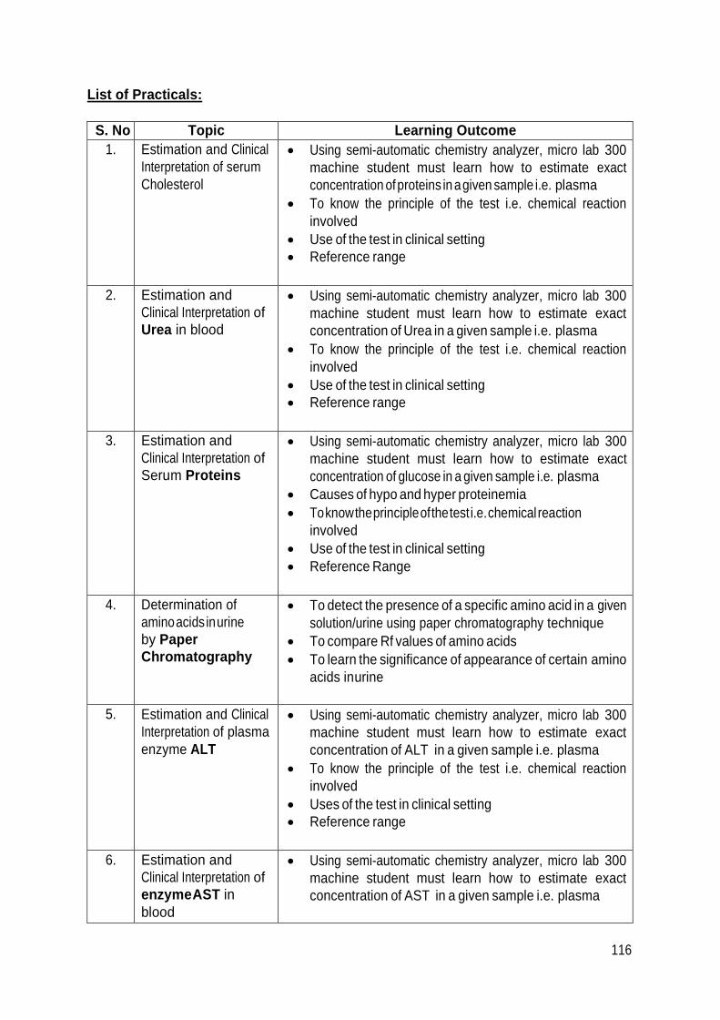
116
List of Practicals:
S. No Topic Learning Outcome
1. Estimation and Clinical
Interpretation of serum
Cholesterol
Using semi-automatic chemistry analyzer, micro lab 300
machine student must learn how to estimate exact
concentration of proteins in a given sample i.e. plasma
To know the principle of the test i.e. chemical reaction
involved
Use of the test in clinical setting
Reference range
2. Estimation and
Clinical Interpretation of
Urea in blood
Using semi-automatic chemistry analyzer, micro lab 300
machine student must learn how to estimate exact
concentration of Urea in a given sample i.e. plasma
To know the principle of the test i.e. chemical reaction
involved
Use of the test in clinical setting
Reference range
3. Estimation and
Clinical Interpretation of
Serum Proteins
Using semi-automatic chemistry analyzer, micro lab 300
machine student must learn how to estimate exact
concentration of glucose in a given sample i.e. plasma
Causes of hypo and hyper proteinemia
To know the principle of the test i.e. chemical reaction
involved
Use of the test in clinical setting
Reference Range
4. Determination of
amino acids in urine
by Paper
Chromatography
To detect the presence of a specific amino acid in a given
solution/urine using paper chromatography technique
To compare Rf values of amino acids
To learn the significance of appearance of certain amino
acids in urine
5. Estimation and Clinical
Interpretation of plasma
enzyme ALT
Using semi-automatic chemistry analyzer, micro lab 300
machine student must learn how to estimate exact
concentration of ALT in a given sample i.e. plasma
To know the principle of the test i.e. chemical reaction
involved
Uses of the test in clinical setting
Reference range
6. Estimation and
Clinical Interpretation of
enzyme AST in
blood
Using semi-automatic chemistry analyzer, micro lab 300
machine student must learn how to estimate exact
concentration of AST in a given sample i.e. plasma

117
To know the principle of the test i.e. chemical reaction
involved
Use of the test in clinical setting
Reference range
List of Case Based Learning Scenerios:
List of Biochemistry CBLs Year 2nd Module 3rd
CBL-1
Ethanol Intoxication (Water, electrolyte and acid base balance)
A 28 year old man was brought to emergency when found unresponsive by his family. Due to social
stigma (worldwide), the family members were hesitant to tell that he consumed almost half a liter of ethanol in
the last evening on marriage ceremony of his cousin. His peers reported that he was a known drinker for last
five years. His vitals were normal but gag reflex and response to pain were absent. Pupils were miotic and
pinpoint but reactive to light. His respiration was inadequate (oxygen saturation 70 percent) and he was
intubated. There was no history of use of sleeping pills (barbiturates etc) or painkillers (NSAIDS).
RELATED LAB INVESTIGATIONS:
Test Name Result Normal Values
Serum ethanol levels 410mg/dL Nil
GGT 270 U/L 30 U/L in males
Na+ 133mEq/L 142mEq/L
pH 7.34 7.35 to 7.45
HCO3 18 (26 at some other
time)*
24 mEq/L
Cl- 103 103 mEq/L
pO2 70 80-100mmHg
pCO2 58 40 mmHg
Ketone bodies in urine Present Absent (present in fasting)
Orally ingested alcohol is absorbed quickly without chemical change from GIT, reaching maximum
plasma concentration in almost an hour. It crosses capillary membrane through simple diffusion and affects
almost every organ of the body. Though pathophysiology of alcohol related metabolic derangements is not
completely understood, its main effects are through its metabolic products, acetaldehyde and acetate.
Malnutrition is common in chronic alcoholics and it further worsens the scenario. Acetaldehyde is the main
injurious metabolic product of alcohol produced in

118
cytosolic as well as microsomal ethanol metabolism. Compared to non-alcoholic subjects, chronic alcoholic
patients have lower serum concentrations of potassium, magnesium, bicarbonate, calcium and
phosphate as well as a lower arterial pH value.
LEARNING OBJECTIVES:
1. How to diagnose acid base disorders
2. Alcohol metabolism in human body
3. Effects of alcohol on electrolyte and acid base balance
REFERENCE BOOKS:
1. Harper’s text book of Biochemistry
2. Davidson’s Practice of Medicine
3. Internet sources
ALCOHOL METABOLISM

119
CBL-2
Protein Metabolism (Urea Cycle- Cirrhosis of Liver)
A 47 year old truck driver was brought in hospital with hematemesis, confusion, delirium and
inappropriate behaviour. He was a diagnosed case of Hep B virus infection for last ten years and was treated
with entecavir (nucleoside analog) 0.5mg orally daily for one year, but response was not good. He was not
vaccinated for Hep B previously. He has been visiting medical OPD off and on for general weakness,
tiredness, itchy and yellow skin, swelling on lower legs and abdominal distension. He was
advised operation for esophageal varices few months back which he could not plan. He was having fever
for last three days and was taking Panadol two tablets when required. He was admitted and transfused with
blood and saline. His hands and feet had to be tied on bed to prevent self- harm and misbehavior in
stuporous state. He was advised endoscopic variceal ligation, restriction in protein intake,
neomycin and laxatives.
RELATED LAB INVESTIGATIONS:
Test Name Result Normal Values
Serum albumin 28 g/L 35-50 g/L
ALT 74 U/L Upto 42 U/L in males
USG Abdomen Shrunken, nodular , coarse liver &
splenomegaly
Normal study
Clotting time 8 mins 2-6 mins
Hb 11 g/dL 14-16 g/dL in males
HBsAg Repeatedly Positive Negative

120
Hepatitis B is a viral infection of liver caused by hepatitis B virus and transmitted through blood or body
secretions. A three dose vaccine in recommended for general population. Only 5-30 percent infections can
become chronic and can cause severe liver damage. As it causes continuous slow damage to hepatocytes, ALT
is mildly or moderately increased. Over the span of 5-8 years a significant harm to liver is done and liver
appears shrunken, fibrosed and nodular. When intrahepatic blood pressure increases the portal
flow is shunted to systemic circulation at lower end of esophagus and in superior and middle rectal veins
of rectum. Here the blood vessels dilate and can be torn with physical trauma (friction by bollus in esophagus
and fecal matter in rectum) leading to frank bleeding. Ammonia made by breakdown of urea by
microorganisms in the gut is reabsorbed into the portal circulation (being lipid soluble gas) which is again
detoxified by liver in normal individuals. But as portal blood is shunted to systemic circulation, ammonia
can cause toxicity to CNS and results in altered consciousness and behavior called hepatic
encephalopathy. This hyperammonemia is secondary to liver disease and is much more common as
compared to primary hyperammonemia. Decrease in albumin production by liver will cause fluid to escape to
peritoneum causing ascities.
LEARNING OBJECTIVES:
1. Disposal of amino groups of amino acids and Urea Cycle
2. The role of liver and portal circulation in entero-hepatic circulation of free ammonia
3. Hyperammonemia and fate of ammonia
REFERENCE BOOKS:
1. Harper’s text book of Biochemistry
2. Davidson’s Practice of Medicine
3. Internet sources
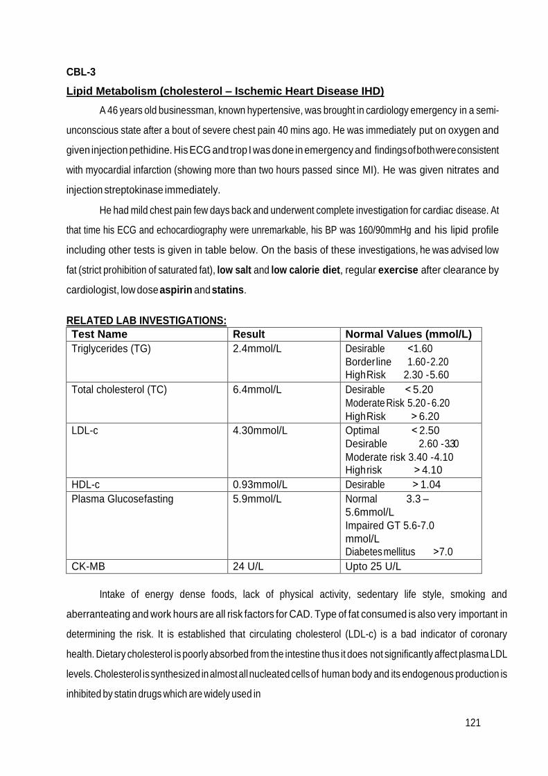
121
CBL-3
Lipid Metabolism (cholesterol – Ischemic Heart Disease IHD)
A 46 years old businessman, known hypertensive, was brought in cardiology emergency in a semi-
unconscious state after a bout of severe chest pain 40 mins ago. He was immediately put on oxygen and
given injection pethidine. His ECG and trop I was done in emergency and findings of both were consistent
with myocardial infarction (showing more than two hours passed since MI). He was given nitrates and
injection streptokinase immediately.
He had mild chest pain few days back and underwent complete investigation for cardiac disease. At
that time his ECG and echocardiography were unremarkable, his BP was 160/90mmHg and his lipid profile
including other tests is given in table below. On the basis of these investigations, he was advised low
fat (strict prohibition of saturated fat), low salt and low calorie diet, regular exercise after clearance by
cardiologist, low dose aspirin and statins.
RELATED LAB INVESTIGATIONS:
Test Name Result Normal Values (mmol/L)
Triglycerides (TG) 2.4mmol/L Desirable ˂1.60
Border line 1.60 - 2.20 High Risk 2.30 - 5.60
Total cholesterol (TC) 6.4mmol/L Desirable ˂ 5.20
Moderate Risk 5.20 - 6.20
High Risk ˃ 6.20
LDL-c 4.30mmol/L Optimal ˂ 2.50
Desirable 2.60 - 3.30
Moderate risk 3.40 - 4.10 High risk ˃ 4.10
HDL-c 0.93mmol/L Desirable ˃ 1.04
Plasma Glucose fasting 5.9mmol/L Normal 3.3 –
5.6mmol/L
Impaired GT 5.6-7.0
mmol/L
Diabetes mellitus ˃7.0
CK-MB 24 U/L Upto 25 U/L
Intake of energy dense foods, lack of physical activity, sedentary life style, smoking and
aberranteating and work hours are all risk factors for CAD. Type of fat consumed is also very important in
determining the risk. It is established that circulating cholesterol (LDL-c) is a bad indicator of coronary
health. Dietary cholesterol is poorly absorbed from the intestine thus it does not significantly affect plasma LDL
levels. Cholesterol is synthesized in almost all nucleated cells of human body and its endogenous production is
inhibited by statin drugs which are widely used in

122
clinics. Limitation on dietary cholesterol has been lifted recently and it is now allowed for healthy individuals. On
the other hand taking saturated fat in diet significantly increases plasma cholesterol (LDL-c) levels and make one
prone to atherosclerosis and cardiac event. Plasma glucose surge after intake of refined sugars has also been
found to be associated with cardiac morbidity.
LEARNING OBJECTIVES:
1. Metabolism of lipoproteins
2. Hyperlipidemias especially hypercholesterolemia type II
3. Cholesterol synthesis and statin drugs
REFERENCE BOOKS:
1. Lippincott’s illustrated reviews and Harper’s text book of Biochemistry
2. Davidson’s Practice of Medicine
3. Internet sources
CBL-4
Lipid Metabolism (FA oxidation – MCAD Deficiency)
A 10 month old infant was admitted in pediatric ward for pneumonia. He was being given appropriate
antibiotics but suddenly went into syncope after an episode of vomiting. He was found to have severe
hypoglycemia while his BP was normal. Ketonemia and ketonuria were absent. On examination there
was hepatomegaly (examination was done after patient was stable). He was normal at birth and no
newborn screening was done for inborn errors of metabolism. The measurement of MCAD enzyme
activity in leukocytes confirmed the diagnosis of MCAD deficiency. Mother was advised to never let the
baby empty stomach and ensure feeding at regular small intervals especially of carbohydrate rich
meals. In infants 2 g/kg of uncooked cornstarch at bedtime ensures sufficient glucose overnight.
RELATED LAB INVESTIGATIONS:
Test Name Result Normal Values
Plasma Glucose random 2.0 mmol/L 3.3-11.1 mmol/L
Plasma ketones 0.04 mmol/L 0.05-3.0 mmol/L
Urinary ketone bodies Present Absent (may be present
during fasting)
Plasma acylcarnitines tandem mass spectrometry
Increased -

123
In mitochondria, there are four fatty acyl CoA dehydrogenase species, each with a specificity for either
short-, medium-, long-, or very-long-chain fatty acids. MCAD deficiency, an autosomal recessive disorder, is
one of the most common inborn errors of metabolism, and the most common inborn error of fatty acid
oxidation, being found in 1:14,000 births worldwide, with a higher incidence in Northern Europeans. It
results in decreased ability to oxidize fatty acids with six to ten carbons (these accumulate and can be measured
in urine), and severe hypoglycemia (because the tissues must increase their reliance on glucose). Treatment
includes avoidance of fasting. MCAD deficiency has been identified as the cause of some cases originally
reported as sudden infant death syndrome (SIDS) or Reye syndrome.
LEARNING OBJECTIVES:
1. Fatty Acid synthesis
2. Beta oxidation
3. Ketogenesis, ketonemia and ketonuria
REFERENCE BOOKS:
1. Lippincott’s illustrated reviews and
2. Harper’s text book of Biochemistry
3. Internet sources
CBL-5
Protein Metabolism (PKU)
A-20-month-old boy born to consanguineous parents was evaluated for marked delay in
achievement of milestones, blond hair and irritability. Large amount of phenyl-ketones in urine, and raised
plasma phenylalanine led to the diagnosis of phenylketonuria. He was placed on a low phenylalanine diet
formulated from locally available foods and the clinical and biochemical response was monitored with the
aid of urinary ferric chloride test. On this diet he showed a mild clinical and biochemical improvement.
RELATED LAB INVESTIGATIONS:
Test Name Result Normal Values
Plasma Phenylalanine 24mg/dL 1-2 mg/dL
Urinary Phenyl lactate Present Absent
Urinary Phenyl acetate Present Absent
Urinary Phenyl pyruvate Present Absent
124
Phenylketonuria (PKU) is a rare genetic disorder with an incidence of 1 in 15,000 births. It is
characterized by a deficiency of a hepatic enzyme (phenylalanine hydroxylase, or PAH) responsible for the
conversion of the amino acid phenylalanine (Phe) into tyrosine. An absence of (or deficiency in) PAH results in
increased blood concentrations of Phe and toxic accumulation in the brain. If PKU is left untreated, it may lead
to deficit in cognitive functions, intellectual impairment, seizures, various behavioral problems, and
psychiatric symptoms.
Most individuals with phenylketonuria (PKU) appear normal at birth. If newborn screening fails,
progressive developmental delay is the most common presentation. Other findings in untreated children
in later infancy and childhood may include vomiting, mousy odor, eczema, seizures, self-mutilation, and
severe behavioral disorders.
LEARNING OBJECTIVES:
1. Metabolism of amino acids and role of folic acid
2. Inborn errors of metabolism
3. Phenylketonuria
REFERENCE BOOKS:
1. Lippincott’s illustrated reviews and
2. Harper’s text book of Biochemistry
3. Internet sources
CBL-6
Obesity (obesity)
A 40 years old house wife presented with chronic pain in both knees and back. Her weight was 93 kg
and height was 5’3”. Her waist to hip ratio was measured to be 1.1. She also complained of inguinal hernia. She
was diagnosed as a case of android obesity with BMI almost 36. Her doctor advised her to change dietary and
life style habits and gave some symptomatic relief medicines after seeing the report of tests ordered
empty stomach.
RELATED LAB INVESTIGATIONS:
Test Name Result Normal Values
TC 6.2 mmol/L Less than 5.6 mmol/L
TGs 1.8 mmol/L Less than 1.6 mmol/L
LDL 3.6 mmol/L Less than 2.6 mmol/L
HDL 90 mmol/L More than 1.04 mmol/L
BSF 120 mg/dL Less than 110 mg/dL

125
Obesity is a disorder of body weight regulatory systems characterized by an accumulation of excess
body fat. The BMI (weight in kg) (height in meters) 2 provides a measure of relative weight, adjusted for
height. This allows comparisons both within and between populations. The healthy range for the BMI is
between 18.5 and 24.9. Individuals with a BMI between 25 and 29.9 are considered overweight, those with a
BMI equal to or greater than 30 are defined as obese, and a BMI over 40 is considered extremely obese.
Anyone more than 100 pounds overweight is considered severely obese. These cutoffs are based on the
studies examining the relationship of BMI to premature death, and are similar in men and women.
LEARNING OBJECTIVES:
1. Obesity and how to measure
2. Body weight regulation
3. Weight reduction
REFERENCE BOOKS:
1. Lippincott’s illustrated reviews and Harper’s text book of Biochemistry
2. Davidson’s Practice of Medicine
3. Internet sources
CBL-7
Endocrinology (Hyperthyroidism)
A 50 years old anxious & over-active lady presented with weight loss and occasional diarrhea
for few months. Her appetite was moderately increased and she thought she had become more heat
intolerant. She was also experiencing palpitations, tremors and irritability for few months. After physical
examination and lab investigations, physician diagnosed her a case of hyperthyroidism and advised her
Carbimazole 20mg daily and propranolol 40mg thrice daily for four weeks.
RELATED LAB INVESTIGATIONS:
Test Name Result
T3 Raised
T4 Raised
TSH Low
IgG anti thyroid Ab Present
126
There are two major systems in human body for inter organ coordination. One is wired and called
Nervous system while other is wireless type and called endocrine system. The hormones of endocrine
gland are produced by specialized cells/ tissues located distant to the site of action. They produce minute
quantities of hormones which always travel in blood, get access to almost all tissues but produce transient
effects only on the cells which have receptors for specific hormone. Thyroid hormones control metabolic
rate or in other words if we consider human body as an automobile and its engine represents the
metabolism the thyroid is the accelerator.
LEARNING OBJECTIVES:
1. Endocrine hormones – introduction and classification
2. Pituitary Hormones
3. Thyroid & parathyroid hormones
REFERENCE BOOKS:
1. Guyton text book of physiology
2. Davidson’s Practice of Medicine
3. Internet sources
CBL-8
Endocrinology (Cushing Syndrome)
A 40 year old lady presented with weight gain, lethargy, depression, general weakness and irregularity
in menses. On examination she was found to have BP 145/90 mmHg and her face appeared moon like while
buffalo neck hump and supraclavicular fat distribution were evident. She was a known patient of rheumatoid
arthritis and has been on steroids for last two years. Doctor diagnosed her as a case of Iatrogenic Cushing
syndrome.
RELATED LAB INVESTIGATIONS:
Test Name Result Reference
Plasma Glucose levels random 12mmol/L Upto 11.1mmol/L
Cortisol levels Raised
ACTH Normal
Adrenal cortex produces 1) glucocorticoids e.g. cortisol which affect carbohydrate, protein and fat
metabolism 2) mineralocorticoids i.e. aldosterone which regulates Na and water balance 3) androgens which are
weak until converted to potent compounds in the periphery. A chronic excess of glucocorticoid with loss of
feedback mechanism of hypothalamo-pituitary adrenal axis and loss

127
of circadian rhythm of cortisol secretion is called Cushing syndrome and its chief causes are chronic exogenous
steroid use and pituitary adenoma.
LEARNING OBJECTIVES:
1. Hormones of adrenal cortex
2. Mechanism of action and effects of adrenal cortical hormones
3. Biochemical Basis of sign and symptoms of Cushing syndrome and disease
4. Biochemical Diagnosis of Cushing syndrome
REFERENCE BOOKS:
1. Harpers text book of Biochemistry
2. Guyton text book of physiology
3. Internet sources
CBL-9
Endocrinology (DM)
A 47 years old lady presented with polyuria, polydipsia and weight loss for few months. She also
complained for recurrent burning micturition/ genital thrush for some time. Her plasma glucose levels
were checked and she was diagnosed as a case of DM.
RELATED LAB INVESTIGATIONS:
Test Name Result Reference
Plasma Glucose levels random 15mmol/L Upto 11.1mmol/L
Plasma glucose levels fasting (next day) (DM if more than 7mmol/L)
9 mmol/L 5.6 mmol/L
HbA1c 60mmol/L Upto 48mmol/L (6.5%)
Diabetes mellitus is a serious pandemic metabolic derangement syndrome in which decreased
endogenous insulin or its decreased effectiveness is the hallmark. Insulin and glucagon are major regulatory
hormones of metabolism. Insulin is the hormone of fed state and it’s the hormone of storage (causes
deposition of carbohydrate and fat stores). Decreased synthesis or release of insulin from beta cells of
pancreas leads to hyperglycemia in addition to other metabolic derangements and the condition is called type 1
DM. On the other hand, despite normal release of insulin from pancreas there may be resistance to its action in
peripheral tissue (adipose and Skeletal muscle) mostly secondary to obesity and this condition is called type
2 DM.
128
LEARNING OBJECTIVES:
1. Hormones of endocrine pancreas
2. Mechanism of action and effects of Insulin and Glucagon
3. Biochemical Basis of sign and symptoms of DM
4. Biochemical Diagnosis of DM
REFERENCE BOOKS:
1. Harpers text book of Biochemistry
2. Guyton text book of physiology
3. Internet sources

129
Second Professional MBBS Examination ANATOMY
Table of Specifications for Annual Second Professional Examination: Theory
Time Allowed =03 hrs (Including MCQs)
Marks of theory paper =90
Internal assessment =10
Total marks =100
Pass Marks =50
25 x MCQs (on separate sheet) (25 Marks) Time =30 min
Q. No. 1,2,3,4,5,6,7,8,9
(7x SAQs/SEQs (C1 & C2) = 07 marks each
2 x SAQs/SEQs (C3) = 08 marks each) (65 Marks) Time = 2 hours 30 minutes
S.No
Topic
NUMBER OF MCQs (25)
(C1=10, C2=10, C3=5)
1 mark each
NUMBER OF SAQs/SEQs
(09)
7x SAQs/SEQs (C1 & C2)
= 07 marks each
2 x SAQs/SEQs (C3)
= 08 marks each
1. Special Embryology 05 01
2. Special Histology 05 01
3. Abdomen Pelvis & Perineum
05 03
4. Head and neck 05 02
5. Brain & Neuro Anatomy 05 02
Total 25 (25 Marks) 09 (65 Marks)

130
Theory: Internal Assessment (IA) Calculation
A B C D
Roll No. Name All Modules/
Pre annual
Exams or any other exam
Total Marks of internal
assessment
Out 0f 10
Total Marks Sum of Marks obtained x10/
sum of total marks in all exams
Table of Specifications for Annual Professional Exam: Practical
VIVA
50 marks
Practical 40 marks
Total
Non Observed OSPE Observed station
OSPE
Histology
Manual
Internal
Examiner
External
Examiner
Gross
embryology,
& X – rays
Histology Long Slides 05
marks.
Surface marking
02 marks.
25 Marks 25 Marks 20* Marks 10 Marks 07 Marks 03 marks 90 marks
10 stations (2 Abdomen and Pelvis, 2 Head & Neck, 2 Brain, 2 embryology, 2 radiology)
Practical: Internal Assessment Calculation
A B C D
Roll No. Name OSPE /Practical Class tests
throughout the year
/Pre annual Practical
Exams or any other exam
Total Marks of internal
assessment
Out 0f 10
Total Marks Sum of Marks obtained x10/
sum of total marks in all exams
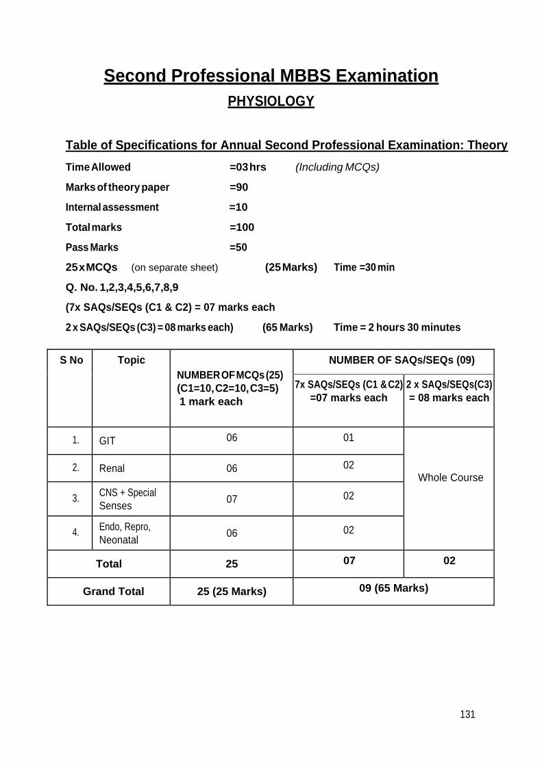
131
Second Professional MBBS Examination
PHYSIOLOGY
Table of Specifications for Annual Second Professional Examination: Theory
Time Allowed =03 hrs (Including MCQs)
Marks of theory paper =90
Internal assessment =10
Total marks =100
Pass Marks =50
25 x MCQs (on separate sheet) (25 Marks) Time =30 min
Q. No. 1,2,3,4,5,6,7,8,9
(7x SAQs/SEQs (C1 & C2) = 07 marks each
2 x SAQs/SEQs (C3) = 08 marks each) (65 Marks) Time = 2 hours 30 minutes
S No Topic NUMBER OF MCQs (25)
(C1=10, C2=10, C3=5)
1 mark each
NUMBER OF SAQs/SEQs (09)
7x SAQs/SEQs (C1 & C2)
=07 marks each
2 x SAQs/SEQs(C3)
= 08 marks each
1. GIT 06 01
Whole Course
2. Renal 06 02
3. CNS + Special
Senses
07 02
4. Endo, Repro,
Neonatal
06 02
Total 25 07 02
Grand Total 25 (25 Marks) 09 (65 Marks)
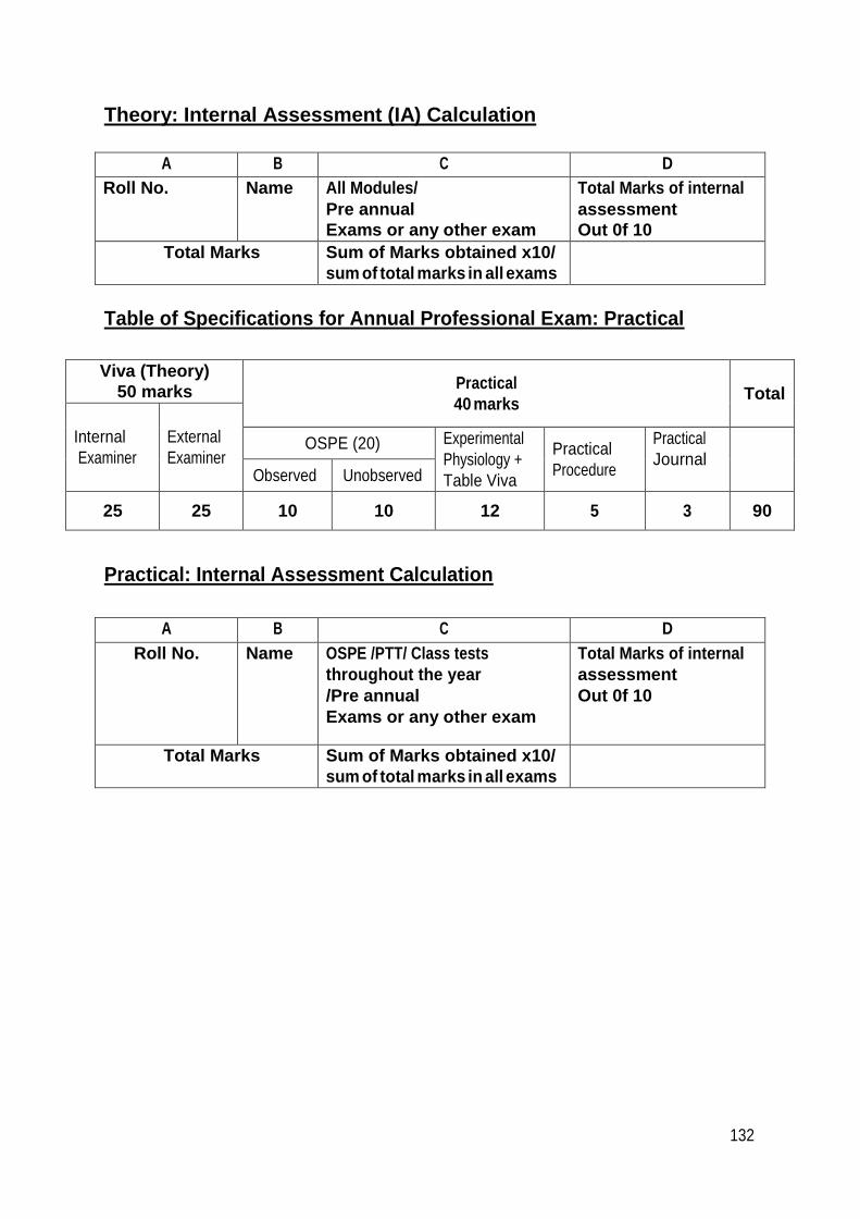
132
Theory: Internal Assessment (IA) Calculation
A B C D
Roll No. Name All Modules/
Pre annual
Exams or any other exam
Total Marks of internal
assessment
Out 0f 10
Total Marks Sum of Marks obtained x10/
sum of total marks in all exams
Table of Specifications for Annual Professional Exam: Practical
Viva (Theory)
50 marks Practical
40 marks
Total
Internal
Examiner
External
Examiner OSPE (20) Experimental
Physiology +
Table Viva
Practical
Procedure
Practical
Journal
Observed Unobserved
25 25 10 10 12 5 3 90
Practical: Internal Assessment Calculation
A B C D
Roll No. Name OSPE /PTT/ Class tests
throughout the year
/Pre annual
Exams or any other exam
Total Marks of internal
assessment
Out 0f 10
Total Marks Sum of Marks obtained x10/
sum of total marks in all exams
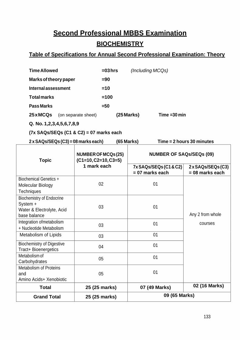
133
Second Professional MBBS Examination
BIOCHEMISTRY
Table of Specifications for Annual Second Professional Examination: Theory
Time Allowed =03 hrs (Including MCQs)
Marks of theory paper =90
Internal assessment =10
Total marks =100
Pass Marks =50
25 x MCQs (on separate sheet) (25 Marks) Time =30 min
Q. No. 1,2,3,4,5,6,7,8,9
(7x SAQs/SEQs (C1 & C2) = 07 marks each
2 x SAQs/SEQs (C3) = 08 marks each) (65 Marks) Time = 2 hours 30 minutes
Topic
NUMBER OF MCQs (25)
(C1=10, C2=10, C3=5)
1 mark each
NUMBER OF SAQs/SEQs (09)
7x SAQs/SEQs (C1 & C2)
= 07 marks each
2 x SAQs/SEQs (C3)
= 08 marks each
Biochemical Genetics +
Molecular Biology
Techniques
02
01
Any 2 from whole
courses
Biochemistry of Endocrine
System +
Water & Electrolyte, Acid
base balance
03
01
Integration of metabolism
+ Nucleotide Metabolism 03 01
Metabolism of Lipids 03 01
Biochemistry of Digestive Tract+ Bioenergetics
04 01
Metabolism of
Carbohydrates 05 01
Metabolism of Proteins
and Amino Acids+ Xenobiotic
05
01
Total 25 (25 marks) 07 (49 Marks) 02 (16 Marks)
Grand Total 25 (25 marks) 09 (65 Marks)
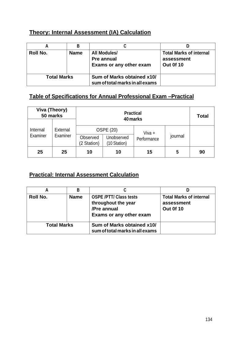
134
Theory: Internal Assessment (IA) Calculation
A B C D
Roll No. Name All Modules/
Pre annual
Exams or any other exam
Total Marks of internal
assessment
Out 0f 10
Total Marks Sum of Marks obtained x10/
sum of total marks in all exams
Table of Specifications for Annual Professional Exam –Practical
Viva (Theory)
50 marks Practical
40 marks
Total
Internal
Examiner
External
Examiner
OSPE (20)
Viva +
Performance
journal
Observed (2 Station)
Unobserved (10 Station)
25 25 10 10 15 5 90
Practical: Internal Assessment Calculation
A B C D
Roll No. Name OSPE /PTT/ Class tests
throughout the year
/Pre annual
Exams or any other exam
Total Marks of internal
assessment
Out 0f 10
Total Marks Sum of Marks obtained x10/
sum of total marks in all exams