studies pathogenesis of angioimmunoblastic...
TRANSCRIPT
Studies of the Pathogenesis of Angioimmunoblastic LymphadenopathyMasaaki Honda, Howard R. Smith, and Alfred D. SteinbergCellular Immunology Section, Arthritis and Rheumatism Branch, National Institute of Arthritis, Diabetes,and Digestive and Kidney Diseases, National Institutes of Health, Bethesda, Maryland 20205
Abstract
Westudied the immune functions of two patients with angioim-munoblastic lymphadenopathy (AILD) in an attempt to deter-mine whether the B cells were primarily hyperactive or, rather,if T cell abnormalities might underlie the B cell hyperactivityobserved in these patients. We found that the B cells of theAILD patients did not proliferate spontaneously, nor were theyinduced to proliferate excessively by fresh normal T cells. Incontrast, AILD T cells induced both autologous and allogeneicB cells to proliferate and to differentiate into Ig secreting cells.Spontaneous culture supernates of T cells obtained from eachpatient induced substantial proliferation of B cells (B cell-activating activity) as well as proliferation in a standard co-stimulatory assay (B cell growth factor activity). The culturesupernate of a T cell line, which was established from onepatient, showed both activities. The T cell line supernate alsoinduced Ig production by staphylococcal A Cowan-activated Bcells. None of these properties of AILD T cells was foundamong 10 normal controls.
The addition of AILD T cells to autologous or allogeneicB cells in the presence of pokeweed mitogen (PWM) led tomarked suppression of both proliferation and Ig production.This was true even in the presence of fresh normal T cells.Pretreatment studies showed that suppressor cells were inducedby the interaction of AILD T cells with PWM-activatedB cells.
The present study suggests that the B cell hyperactivityobserved in AILD patients might in part be due to excessiveT cell effects on B cells. In addition, our results may helpclarify the paradoxical impaired responsiveness to in vitrostimulation with PWMby active B cells from patients withautoimmune diseases.
Introduction
Angioimmunoblastic lymphadenopathy (AILD)' is a disorderthat has been described in the last 30 yr (1), but which has
Address correspondence to Dr. Steinberg, Building 10, Room9N-218,National Institutes of Health, Bethesda, MD20205.
Received for publication 4 December 1984 and in revised form IMarch 1985.
1. Abbreviations used in this paper: AILD, angioimmunoblastic lymph-adenopathy; anti-IA, F(ab)2 fragment of goat anti-human IgM antibody;BCDF, B cell differentiation factor(s); BCGF, B cell growth factor(s);[3H]TdR, tritiated thymidine; IL-1, IL-2, interleukins I and 2; MNC,peripheral blood mononuclear cells; MNC-CS, culture supernate ob-tained from MNC, PHA, phytohemagglutinin; PHA-CS, culture su-pernate obtained from PHA; PWM,pokeweed mitogen; SLE, systemiclupus erythematosus.
The Journal of Clinical Investigation, Inc.Volume 76, July 1985, 332-340
only been accepted as a clinical and pathologic entity worldwidefor the past decade (2-5). This disorder is characterized by aclinical picture of fever, chills, sweats, malaise, anorexia, rash,hepatosplenomegaly, and marked lymphadenopathy. Labora-tory abnormalities usually include hypergammaglobulinemiaand anemia, which is often Coombs positive. The diagnosismust be confirmed histologically. The lymph nodes show apleomorphic infiltration that includes immunoblasts and im-munocytes and can obliterate the normal lymph node archi-tecture. An important diagnostic criterion is the marked ar-borization of small vessels in the lymph node. In addition, anamorphous acidophilic interstitial material is often prominentlyfound. Despite the impressive clinical and histologic abnor-malities, the adenopathy consists of polyclonally activatedimmunoblasts, often B cells, without obvious malignancy.Nevertheless, the typical patient has a life expectancy of - Iyr. Most patients with AILD die of infection. A minority ofpatients go on to develop a frank lymphoreticular malignancy;the potential to develop such a malignancy may possibly bepredicted on the basis of hyperdiploidy of lymphoid cells earlyin the course of the illness (6). Although the clinical featureshave led to the belief that AILD is a single illness, differenceshave been stressed in some reports (7), and it is possible thatAILD represents a syndrome with more than one clinicopath-ologic syndrome and/or more than one etiology.
Although the clinical and pathologic features of patientswith AILD are becoming better understood, there is consid-erable confusion regarding the cellular abnormalities thatunderlie this disorder (7-13). Most patients have an abnormalcomposition of their peripheral blood leukocytes. It has beensuggested that suppressor T cell functions may be deficient inAILD, thereby leading to B cell hyperactivity (9). AutonomousB cell hyperactivity has been proposed by other investigatorsas the major problem in the disorder (7). However, patientswith AILD may vary over time so that cellular functions maybe different at different times; moreover, the exact mechanismsof B cell hyperactivity are not well defined by the previousstudies. Because anti-erythrocyte autoantibodies are found inmany patients (2), and there is an association with otherautoimmune syndromes (14-16), understanding the immunebasis of AILD might shed light upon other diseases as well.For these reasons, as well as the rapidly progressive nature ofthe disorder, we undertook a study of the cellular basis ofdisease in two patients with AILD. We were aided in ourhuman studies by observations in two mouse strains, MRL-lpr/lpr and C3H-gld/gld. These two strains also develop massivelymphadenopathy, hypergammaglobulinemia, and autoanti-bodies (17-20). Their T cells produce factors that can activateB cells and drive them to proliferate and become immuno-globulin secreting cells (17). Therefore, our studies attemptedto determine whether AILD B cells were spontaneously hyper-active or whether an excess of helper T cell function mightunderlie the syndrome. The studies suggest that AILD T cellsactivate B cells and drive them to proliferate and to produce
332 M. Honda, H. R. Smith, and A. D. Steinberg

immunoglobulin. Wealso conducted experiments to explainthe paradoxical impaired responsiveness to in vitro stimulationwith pokeweed mitogen by B cells which are active in vivo(21-25).
Methods
Patients and source of lymphocytes and plasma. Patients with AILDand systemic lupus erythematosus (SLE) were hospitalized at theClinical Center, National Institutes of Health, Bethesda, Maryland; thepatients agreed in writing to being studied according to an approvedinformed consent form. The patients with AILD had classic lymphnode histology and clinical symptomatology (2-5). They were studiedearly in the course of their illness. Lymphocytes and plasma from thepatients with AILD used in in vitro studies were obtained before anytreatment. Plasma of patients with AILD used in this study had beenfresh frozen and had not been previously thawed. All plasma werecentrifuged at 13,000g for 15 min at 40C to remove aggregatedmaterials before use.
Isolation of T cells, B cells, and monocytes. Peripheral bloodmononuclear cells (MNC) were isolated by Ficoll-Hypaque gradientcentrifugation. T cells, and non-T cells were separated from MNCbyrosetting with neuraminidase-treated sheep erythrocytes (26, 27). Doublypurified nonrosetting cells were twice depleted of monocytes by removalof cells adhering to plastic petri dishes. The percentage of T cellcontamination in the nonadherent, nonrosetting B cell population was<1%. For some experiments, B cells were further treated with OKT3monoclonal antibody plus complement to obtain B cells greatlydepleted of T cells. After this additional purification, T cell contami-nation in the B cell enriched population was <0.5%. Monocytecontamination in the B cell-enriched population was 10-20% asdetermined by esterase staining.
Development of a T cell line from an AILD patient. MNC,2 X 106, were cultured in RPMI 1640 with 10% fetal calf serum, 5X lo-5 M2-mercaptoethanol, 1% L-glutamine, penicillin (100 U/ml),streptomycin (100 Ag/ml), and 20 ng/ml phorbol myristate acetate forI wk. After harvesting the MNCculture supernate (MNC-CS), theseprecultured MNCwere separated into a T cell-enriched population bythe E-rosette method. These separated T cells were cultured withirradiated autologous non-T cells or an irradiated Epstein-Barr virus-transformed B cell line for one week alternating with phytohemagglutinin(PHA) culture supernate (PHA-CS) for I wk. After - I mo, a T cellline was established. To obtain the T cell culture supernate, theseparated T cells were washed well and then 1 X 10⁶/ml of T cellswere cultured in the same medium mentioned above for 1 wk, andthe culture supernate was harvested for in vitro studies. T cell linesfrom patients with SLE were also established by the same method.
Flow cytometry. 106 cells/0.1 ml of sorter buffer (phosphate-buffered saline containing 0.05% bovine serum albumin and 0.1%sodium azide) were incubated with each monoclonal antibody for 30min on ice, washed twice with sorter buffer, and then stained with theF(ab)2 fragment of fluoresceinated goat anti-mouse IgG for 30 min onice. After an additional three washes, these cells were analyzed on afluorescence-activated cell sorter (FACS IV, Becton-Dickinson & Co.,Mountain View, CA). For some experiments, fluorescein-conjugatedmonoclonal antibodies were used.
Monoclonal antibodies. OKT 3, OKT 4, OKT 8, OKT 9, andOKT10 monoclonal antibodies were purchased from Ortho DiagnosticSystems Inc., Raritan, NJ. Leu-10 and anti-HLA-DR were purchasedfrom the Becton-Dickinson Monoclonal Center, Inc., Mountain View,CA. Anti-Tac was kindly provided by Dr. T. A. Waldmann, NationalCancer Institute.
Study of culture supernate, plasma, or T cell line of AILD patientson B cell activation, proliferation, and differentiation. To assay B cellactivating activity, highly purified B cells, l0', suspended in RPMI1640 containing 10% fetal calf serum, 5 X 10⁻¹ M2-mercaptoethanol,1% L-glutamine, penicillin (100 U/mI), and streptomycin (100 ug/ml),
were cultured in 96-well flat-bottomed microtiter plates (Costar, DataPacking, Cambridge, MA) with culture supernate or plasma, or irradiatedT cell line (101) in the presence or absence of the F(ab)2 fragment ofgoat antihuman IgM antibody (anti-A) (final concentration: 25 Ag/ml;Cappel Laboratories, Inc., Cochranville, PA) in a 95% air/5% CO2humid atmosphere at 370C for 72 h, and pulsed with I ACi of[3H]thymidine (New England Nuclear, Boston, MA). For a B cellgrowth factor (BCGF) standard, we used a PHA culture supernateobtained from Electronucleonics, Inc., Silver Spring, MD. This PHAculture supernate (PHA-CS) did not contain PHAor immune interferonand was used as a BCGFsource after absorbing it with concanavalinA-activated T cells to remove irrelevant factors capable of binding toactivated T cells. Incorporation of [3H]thymidine was measured in aliquid scintillation counter. For some experiments, highly purified Bcells (10) were cultured with irradiated (2,000 rad) fresh T cells or anirradiated (3,000 rads) T cell line (101 T cells) in the presence ofpokeweed mitogen (PWM; 1:100 dilution; Gibco Laboratories, GrandIsland, NY). In other experiments, irradiated fresh T cells or a T cellline (l0 T cells), or culture supernate were added to the cultureconsisting of highly purified B cells (l0) plus anti-A (25 Ag/ml) andBCGFto study whether or not the T cell line or culture supernate ofAILD patients has additive or synergistic effects on B cell proliferation.
The ability of a T cell line to induce Ig synthesis was examined.Briefly, 2 X 101 B cells were cultured with various numbers of irradiated(3,000 rad) T cells in the presence or absence of PWM(1:100 dilution)for 10 d. To study the ability of a culture supernate to induce B cellsto produce immunoglobulin, B cells, 2 X 10⁶/ml, were cultured withformalin-fixed staphylococcal A cells (final concentration, 0.001%;Bethesda Research Laboratories, Inc., Gaithersburg, MD) for 3 d. Afterwashing, preactivated B cells, 2 X l0, were cultured with variousamounts of culture supernate for an additional 5 d. Culture supernateswere harvested, and polyclonal Ig synthesis was measured by enzyme-linked as immunosorbent assay described previously (19). For a B celldifferentiation factor (BCDF) standard, PHA and immune interferonfree PHA-CS was used. For some experiments, fresh B cells or a T cellline (106 cells) were precultured with PWM(1:100 dilution) for 30 h.After washing thoroughly, cell mixing studies were carried out todetermine the effect of T cells on Ig synthesis of B cells.
Interleukin-1 (IL-I) assay. The IL-i assay was performed asdescribed (28). Briefly, a single cell suspension of thymocytes (1.5X 10⁷/ml) from C3H/HeJ mice was prepared and suspended in RPMI1640 with 5% fetal calf serum, 2.5 X l0on M 2-mercaptoethanol 2mML-glutamine, and antibiotics. Thymocytes were cultured for 72 hat 1.5 X 10⁶ cells/well in 96-well flat-bottomed plates in the presenceof I Ag/ml PHA (Wellcome Reagents Division, Research TrianglePark, NC) and serial dilutions of the test samples. Cultures were pulsedfor the final 5 h with 0.5 ACi [3HJthymidine.
Interleukin-2 (IL-2) assay. Plasma or supernates from NMC, Tcells, or a T cell line were tested for their ability to maintain thegrowth of the IL-2-dependent HT-2 T cell clone (29). Briefly, triplicatewells containing 4 X 10³ HT-2 cells were suspended in 100A ofmedium and incubated with an equal volume of various concentrationsof the test samples for 24 h. The cultures were then pulsed with I ACiof [3H]thymidine 5 h before harvest.
Production of a monoclonal antibody (33.2.1) against human Bcells. A monoclonal antibody against human B cells (33.2.1) wasestablished as described previously (30), and characterized in ourprevious paper (31). Briefly, this antibody recognizes an Ia-like deter-minant on non-T cells (a 28,000/32,000-mol wt heterodimer) andinterferes with B cell activation more effectively than conventionalanti-HLA-DR and anti-HLA-DS/DC reagents.
Results
Studies of AILD T cells. Because AILD is characterized bymarked numbers of immunoblasts and production of largeamounts of antibody, we wished to study whether the T cells
Studies of Angioimmunoblastic Lymphadenopathy 333

might be important in the induction of B cell activation,proliferation, and/or differentiation. Either the AILD T cellsare important in such induction or the AILD B cells arespontaneously hyperactive or non-T cell stimuli (e.g., exogenousagents) might be responsible for the B cell hyperactivity.
We obtained fresh MNCfrom two patients with AILDand studied their ability to induce B cell proliferation anddifferentiation. Wefound that spontaneous culture supernateof T cells obtained from each patient induced substantialproliferation of B cells (B cell activation activity) as well asproliferation in a standard costimulatory assay (BCGF activity)(Table I). Neither of these properties of AILD T cells wasfound among 10 normal controls studied at the same timeand in an identical manner. In addition, there was no detectableIL-I or IL-2 activity in the AILD T cell supernates. The wholemononuclear cell supernate (MNC-CS) did not have B cellactivating or BCGFactivities. In that this is the same populationfrom which the T cells were obtained for the T cell supernate,we presume that either other cells are capable of inhibitingproduction of the factor(s), or, more likely, that they canabsorb out or conceivably destroy the factors. In Table I wehave listed the results of the assay for B cell activating factor
Table I. B Cell Activating Activity andBCGFActivity of Spontaneous Culture Supernateor Plasma of Patients with AILD*
B cellPlasma activating activity BCGF IL-I IL-2
AILD no. I 0 0 ND NDMNC-CS 0 0 0 0T-CS + + 0 0
AILD no. 2 0 0 ND NDMNC-CS 0 0 0 0T-CS + + 0 0TCL-CS + + 0 0
Normal (n = 10) 0 0 ND NDMNC-CS 0 0 0 0T-CS 0 0 0 0
MNC-CS, mononuclear cell culture supernate; ND, not determined;T-CS, T cell culture supernate; TCL-CS, T cell line culture super-nate.Criteria for positive B cell activating activity or BCGFactivity.(a) Criteria for positive B cell activating activity:
actual counts per minute - counts per minuteby B cell alone
Positive: >5=counts per minute by B cell alone
Normal T-CS is <3; PHA-CS absorbed by Con A-T cells is <5.(b) Criteria for positive BCGFactivity:
actual counts per minute - counts per minute
Positive: >3 = by B cells stimulated with anti-gscounts per minute by B cells stimulated with anti-M
Activity of B cell activating factor(s) and BCGFwas examined by us-ing at least two different normal individuals as a source of B cells.* Each activity was assayed by the methods described in Methods.Experiments were repeated three times with similar results. The pro-liferation induced by the AILD T-CS was significantly greater thanthat induced by normal T-CS, P < 0.0001.
(assayed by addition of supernate to B cells without anti-M) asseparate from the assay for BCGF(assayed on B cells plusanti-it, the standard co-stimulatory assay). Whether or notthese are different factors remains to be determined. Of interest,the plasma of the patients did not contain measurable activity.(This, however, may be a false negative result due to any ofseveral possibilities including binding to or inhibition by serumproteins).
Patients with AILD are often quite ill and require vigoroustherapy. Wedid not want to depend on fresh T cells from ourpatients, whom we knew would receive therapies that couldalter their cellular functions. To study systematically theproperties of the AILD T cells and to obtain enough supernateto do many studies, we tried to establish a T cell line fromboth patients. Wewere successful with one. The T cell linewas 100% OKT3+ OKT4+, and also expressed activated cellmarkers: OKT9 (transferin receptor) 27%, OKT 10 (activatedT cell marker) 39%, Leu-10 (HLA-DS/DC/MB) 10%, HLA-DR28%, and Tac (IL-2 receptor) 29%.
Ability of the AILD T cell line to induce proliferation ofautologous and allogeneic B cells. The AILD T cell line wascompared with fresh normal T cells and T cell lines from twopatients with SLE for the ability to induce B cells to proliferate.All T cells were irradiated before culture so that their contri-bution to the proliferation would be eliminated (this wasconfirmed; data not shown). We found that the fresh normalT cells failed to induce dramatic proliferation of B cells. Thiswas true whether allogeneic or autologous B cells were used(Table II). Moreover, the AILD B cells did not proliferatespontaneously (data not shown), nor were they induced toproliferate by fresh normal T cells (Table II). In contrast, theAILD T cell line induced both allogeneic and autologous Bcells to proliferate dramatically (Table II). The SLE T celllines also induced significant proliferation; however, this wasnot as great as that induced by the AILD T cell lines.
Ability of the AILD T cell line to induce B cells to produceimmunoglobulin. We next asked whether, in addition to in-ducing proliferation of B cells, the AILD T cell line couldinduce Ig secretion. The data in Table III show that the AILDT cell line can, in fact, induce Ig production by unstimulated
Table II. Ability of the AILD T Cell Line, but NotSLE T Cell Lines or Fresh T Cells, to InduceProliferation of Autologous or Allogeneic B Cells*
Irradiated T cells B cells Expt. I Expt. 2 Expt. 3
Acpmt cpm* Acpmt
Fresh + autologous 68 119 364Fresh + AILD 101 115 98AILD line + autologous ND 9,008 12,148AILD line + allogeneic 8,681 12,477 48,640SLE line no. I + autologous 1,320 1,115 NDSLE line no. I + allogeneic 1,854 2,520 5,528SLE line no. 2 + autologous 613 519 NDSLE line no. 2 + allogeneic 1,126 1,420 3,841
* B cells (l0) were cultured with irradiated T cells (l0) for 3 d.[3H]TdR was added for the last 16 h of culture.f Results were expressed as Acpm (mean counts per minute by Bcells plus irradiated T cells - mean counts per minute by B cellsalone). Standard errors of the means were <15% of the means.
334 M. Honda, H. R. Smith, and A. D. Steinberg

Table III. Ability of the AILD T Cell Line to Induce Autologousor Allogeneic B Cells to Produce Immunoglobulin*
Ig production§
Cell combinationt Expt. I Expt. 2t Expt. 3t Expt. 4t
ng/ml ng/ml ng/ml ng/ml
B 8 8 13 10B+PWM 4 5 25 10B + Fresh autologous T 8 8 10 22B + Fresh autologous T
+ PWM 551 604 6,935 6,472B + AILD T cell line 604 653 317 224
* B cells (2 X 10) and irradiated T cells (2 X 10) were cultured for10 d with or without PWMand the supernates were harvested andanalyzed for Ig production by the enzyme-linked immunosorbentassay.t For exps. I and 2, all cells were obtained from the AILD patient.For exps. 3 and 4, fresh T and B cells were obtained from a singlenormal donor.§ Total Ig (IgM + IgG + IgA) was measured. Standard errors were<20% of the means.
B cells. The Ig production induced was as great as that inducedby the combination of fresh T cells plus PWMin the autologousassay (Table III, exps. 1 and 2). When B cells were obtainedfrom normal donors, the combination of fresh T cells plusPWMinduced much more Ig production than that inducedby the AILD T cell line, but the AILD T cell line was stillable to induce normal B cells to produce Ig (Table III, exps. 3and 4).
Additional studies with the T cell line were carried outwith regard to both proliferation and differentiation (these arepresented later); however, we next wished to ask whether asoluble factor or factors from AILD T cells or the T cell linemight be responsible for the activation and differentiation ofB cells. As a result, we obtained culture supernates from theAILD T cell line and performed the following studies.
Ability of the supernate from the AILD T cell line toactivate B cells to proliferate. B cells from normal donors werestimulated with one of the following: anti-M, BCGF, or theAILD supernate. Anti-,u or BCGFseparately induced modestproliferation with an increase in tritiated thymidine ([3H]TdR)incorporation of about three- to fourfold (Table IV). Thegreatest degree of proliferation was induced by the supernateof the AILD T cell line (-20-fold) (Table IV).
The addition of anti-A to the cultures containing B cellsplus the AILD T cell line supernate led to an additionalincrease of - 1.5-2-fold in proliferation (Table IV). The ad-dition of the AILD supernate to the combination of anti-su+ BCGFled to another increase in [3H]TdR incorporation(Table IV). This result suggested that the AILD supernate hada factor or factors that could stimulate a population of B cellsto proliferate. Because the population of B cells used was notdepleted of B cells partially activated in vivo, it is possible thatthe proliferation induced by anti-A alone or by BCGFalonerepresented addition to a signal already received in vivo beforethe cell separation and culture.
A dose-response curve for the AILD T cell line supernatewas performed in three different experiments. With increasing
Table IV. Effects of Culture Supernate of T Cell Line Obtainedfrom AILD Patient on Activation of B Cells to Proliferate*
B cells cultured with Experiment I Experiment 2
mean cpmt mean cpmt
0 322 851Anti-it 1,234 2,225BCGF 1,561 2,588Culture supernate of AILD T cell line 6,633 15,346Anti-i + BCGF 5,450 38,219Anti-1s + culture supernate 8,750 36,540Anti-A + BCGF+ culture supernate 10,000 75,373
* Highly purified B cells (l0) were cultured with anti-ji (25 tig/ml),BCGF, culture supernate, or the combination of them for 3 d.[3H]TdR was added for the last 16 h of culture.t Results were expressed as mean counts per minute of triplicate cul-tures. Standard errors of the means were <15% of the means.
amounts of supernate, there was increasing proliferation ofallogeneic normal B cells (data not shown). The magnitude ofthe increase reached 50 times control proliferation. In contrast,the ability of the AILD T cell supernate to augment theproliferation of a stimulated (by 25 gg/ml anti-is) populationof B cells was marginal (less than twofold).
Ability of the AILD T cell line supernate to induce immu-noglobulin production: Normal B cells were activated withformalin-fixed Staphylococcal A Cowan I (Sac) organisms for3 d. The B cells were washed and then either PHA culturesupernate or the AILD T cell line supernate was added andthe cells were cultured for an additional 5 d. The AILD T cellculture supernate, like PHA-CS, also was able to induce Igproduction, but not quite to the same magnitude (Table V).Weconclude from this experiment that the AILD supernatehad BCDFactivity as well as the prior demonstrated BCGFactivity. During these experiments we found that unactivatedB cells were not induced to produce Ig by PHA-CS. Finally,unactivated B cells were not induced to produce Ig by 25 ,ug/ml anti-is or the AILD T cell supernate. This last result maynot be a true reflection of the capacity of the AILD supernate,
Table V. Effects of Culture Supernate of the AILD T Cell Lineon Ig Production by Fresh B Cells or Sac-activated B Cells*
B cells (2 X 10⁵) Source of BCDF Ig production
ng/ml
Fresh B PHA-CS 25% <1Fresh B AILD-CS 25% <1Fresh B Anti-M 25 ig/ml 2Sac-activated B 0 1Sac-activated B PHA-CS 25% 830Sac-activated B AILD-CS 25% 295t
* Fresh B cells or Sac-activated B cells (2 X l0) were cultured withPHA-CS or culture supernate obtained from the AILD T cell line, orwith anti-iu. The supernates were harvested as described in Methods,and analyzed for immunoglobulin production. Standard errors were
<20% of the means. This experiment was repeated twice.t Significantly greater than with fresh B cells, P < 0.001, but signifi-cantly less than PHA-CS with Sac-activated B cells, P < 0.05.
Studies of Angioimmunoblastic Lymphadenopathy 335

but rather our inability to sequentially provide the properfactors in the proper concentrations from the AILD supernate.
Effect of the AILD T cell line on B cell proliferation in thepresence of PWM.A series of experiments was initially set upto determine whether there might be an additive or synergisticeffect of AILD T cell stimulation on B cells plus PWMstimulation. In these experiments, the AILD T cells werecompared with fresh T cells autologous to the normal B cellsunder study. The fresh irradiated T cells failed to stimulate Bcell proliferation very much. In contrast, the AILD T cell linewas a very effective stimulus for B cell proliferation (TableVI). Whereas the fresh T cells caused only a twofold increaseor less, the AILD T cell line caused a 19-30-fold increase.However, the combination of fresh T cells plus PWMcausedas good proliferation as did the AILD T cells. Thus, the AILDT cells provided an extremely strong proliferative signal. WhenPWMand the AILD T cell line were combined, the result wasnot a further increase, but rather, a decrease of 60% (TableVI). Wethen asked whether this was a result of suprastimu-lation. No evidence for this could be found in that furtheraddition of anti-a and/or PHA-CS to the culture of the AILDT cell line plus B cells (without PWM) led to even moreproliferation (data not shown). Thus, there appeared to besomething special about the combination of stimulation withPWMplus the AILD T cell line which led to suppression ofB cell proliferation relative to that observed with the AILD Tcell line alone. Wepursued this observation in the hope thatit might shed some light upon the previously reported defectin the SLE MNCresponse to PWMas well as on the AILD Tcells.
This suppression in association with PWMwas furtherstudied in a mixing experiment in which both the AILD Tcell line and fresh T cells were added to cultures of B cells,and total Ig was assayed (Fig. 1). The fresh T cells plus PWMled to Ig production as did the AILD T cell line withoutPWM. However, the AILD T cell line plus PWM, with orwithout fresh T cells, led to marked suppression. This resultsuggested that PWMmight be inducing the AILD T cell line
Table VI. Although the AILD T Cell Line HasMarked Stimulatory Effects on B Cells, in thePresence of PWMSuppression Is Observed*
Regulatory T cellst PWM Experiment I Experiment 2 Experiment 3
mean cpm§ mean cpm§ mean cpm§
Fresh T cells 0 584 963 565AILD T cell line 0 9,197 14,967 16,387Fresh T cells + ND 13,686 9,424AILD T cell line + ND 5,6921 5,1801
Proliferation of unstimulated B cells was: experiment 1, 516 cpm; ex-periment 2, 456 cpm; experiment 3, 469 cpm.* Highly purified B cells (10') were cultured with irradiated regula-tory T cells (IO'), with or without PWM(1:100 dilution).t Fresh T cells (10'), and B cells (105) were obtained from the samenormal donor. Fresh T cells and the T cell line were irradiated with2,000 and 3,000 rad, respectively.§ Results were expressed as mean counts per minute of triplicate cul-tures. Standard errors of the means were < 15%. This experiment wasrepeated three times with similar results."Significant decrease, P < 0.01, compared to both AILD T cell line+ PWMor fresh T cells + PWM.
Irradiated AILDFresh T cells T Cell Line
0 0
2x10⁵
2x1 05
4x1 05
4x10⁵
0
0
2x1 05
2x1 05
0
0
0
0
2x1 05
2x1 05
2x10⁵
2x1 05
PWM
0
0
0
0
0 I]
0 200 400 600 800 1,00
Ig Ing/ml)
Figure 1. Effects of T cell line on polyclonal Ig synthesis. Fresh Bcells (2 X 10⁵) were cultured for 10 d with irradiated fresh T cellsand/or the irradiated T cell line in the presence or absence of PWM.The supernates were harvested and analyzed for immunoglobulin(IgG + IgM + IgA) production. Standard errors of the means were<20% of the means.
to suppress B cell Ig production, a suppression which couldnot be overcome by fresh T cells.
Suppression in the presence of PWMmaps to the B cell.To determine whether the PWMeffect was on B cells or onthe AILD T cell line, B cells and T cells were separatelypreactivated by PWM. Preactivated T cells were effectivehelpers for fresh B cells in the absence of PWM(Table VII,line 3). In contrast, preactivation of the B cells preventedinduction of Ig production by the AILD T cells (Table VII,line 8). This was true even when fresh T cells were added.These results suggest that the AILD T cell line fails to stimulatepreactivated B cells to secrete Ig, perhaps because the PWMsignal in some way preempts the AILD T cell line signal. Inaddition, however, the AILD T cells suppressed the stimulatorycapacity of fresh T cells on preactivated B cells (Table VII,lines 7 and 9).
Finally, we examined whether during culture in the presenceof PWMcytotoxic T cells against B cells might be induced.For this purpose fresh B cells were cultured with the AILD Tcell line in the presence or absence of PWMfor 3 d, then theviability of the B cells was studied. The B cells' viability withPWMand without PWMwas similar. Thus the suppressionby the AILD T cell line observed in the presence of PWMappears not to be due to the induction of cytotoxic T cellsby PWM.
Effect of a monoclonal antibody that inhibits early eventsin B cell activation. Wehave recently developed a monoclonalantibody which recognizes a new Ia-like determinant on Bcells (31). Wehave taken advantage of that antibody to testwhether or not it might interfere with the ability of AILD Tcells to activate B cells, and thereby provide informationregarding the activation of B cells by the AILD T cells. Thisstudy, shown in Table VIII, demonstrates that the antibodyinhibited the proliferation of B cells in response to stimulationby the irradiated AILD T cells alone, in the presence of anti-ji, or in the presence of anti-is plus BCGF. Although theinhibition was significant and substantial, it was not complete;moreover, there was little inhibition of the response of B cellsto PWM+ AILD T cells. In that the AILD T cells might havemore than one effect on B cells, the supernate of the AILD T
336 M. Honda, H. R. Smith, and A. D. Steinberg
i

Table VII. Effect of PWMPreactivation of B Cellsor AILD T Cells on Induction of Suppression
Freshirradiated Irradiated AILD
B cells* T cells* T cells* PWM Ig
ng/ml
Fresh 0 + 0 450Fresh 0 + + 21fFresh 0 PWM-preactivated 0 304§Fresh 0 PWM-preactivated + 25tPWM-activated 0 0 + 3PWM-activated + 0 0 44PWM-activated + 0 + 657PWM-activated 0 + 0 2"1PWM-activated + + + 361
A number of controls not shown were as follows: fresh B cells alone(<I ng/ml) or with PWM(< 1); PWM-activated B cells alone (12);PWM-activated B cells + AILD T cells + PWM(2); PWM-activatedB cells + fresh T cells + AILD T cell line without PWM(16).* Fresh B cells or PWM-preactivated B cells (2 X 10⁵) were culturedwith fresh irradiated T cells (2 X l05) or cells (4 X 10⁵) from theAILD T cell line which were either preactivated with PWMor notand then irradiated. Cultures were with or without PWMfor 10 dafter which supernatants were harvested and analyzed for Ig produc-tion. Fresh B and T cells were from the same patient. Standard errorswere <20% of the means. This experiment was repeated twice withsimilar results.t Significant suppression compared with cultures without PWM,P < 0.01.§ Not significantly different from the result with nonpreactivatedAILD T cells."l Significantly less than the same culture containing fresh B cells,P < 0.01.T Significantly less than the same culture without AILD T cell line(657 ng/ml), P < 0.01.
cell line was also studied (Table IX). In this study, the antibody33.2.1 was able to completely overcome the stimulatory effectsof the AILD T cell supernate. Control monoclonal antibodieshad no effect.
Discussion
In this study, we found that the T cells of two patients withAILD and a T cell line from one produce a factor or factors
that activate autologous and allogeneic B cells and inducethem to proliferate and secrete immunoglobulin. Whereas theAILD T cells had abnormally increased inducer and helperactivity for B cells, the AILD B cells did not manifest excessiveresponsiveness to T cell signals nor were they spontaneouslyhyperactive. Thus, AILD patients examined in our studyappeared to suffer from excessive T cell effects on B cells. Avery similar mechanism for B cell hyperactivity has beendescribed recently for Kawasaki disease (32). In addition, ourstudies are reminiscent of those of Clement et al. (33), whichdemonstrated that mitogen activated T cells could induceproliferation of small, resting B cells. These results suggest thepossibility that an endogenous mitogen might be activating Tcells in AILD and that the activated T cells, in turn, drive theB cells.
The ability of the AILD T cell supernates to induce B cellproliferation was assessed in two assay systems. The firstmeasured B cell activating function in that B cell proliferationwas induced without addition of anti-/i or other B cell mitogens.The second assay was a standard co-stimulatory assay; highlypurified B cells were cultured with the AILD or control T cellsupernates in the presence of a suboptimal dose of anti-s,. Inthese assays, T cells were carefully removed from B cellpopulations, a depletion that was confirmed both by cell sorteranalysis and failure to respond to T cell mitogens. However,because the B cell population may not have been depleted ofpartially activated B cells, it is possible that the proliferationinduced by anti-s alone or by BCGFalone represented additionto a signal already received in vivo before cell separation.Moreover, we do not know in this study whether the twoassays measuring B cell activation or co-stimulation are mea-suring the same or different factors, nor whether any of theactivities involve multiple factors. Wedo know that the culturesupernates do not contain measurable amounts of IL- 1 or IL-2; therefore, there was not a mere excessive production of allcytokines. Further study will be required for a full analysis ofthe factor or factors produced by the AILD T cells.
Previous studies have demonstrated spontaneous Ig pro-duction by MNCfrom AILD patients (8). Our findings areconsistent with those reports in that AILD T cells plus AILDB cells led to proliferation and Ig production (7, 10, 11).However, in a previous study, AILD T plus AILD B plusPWMdid not lead to proliferation (7) or to Ig production(10); unfortunately, those studies did not examine AILD Tplus AILD B without PWMas we have. As a result, they may
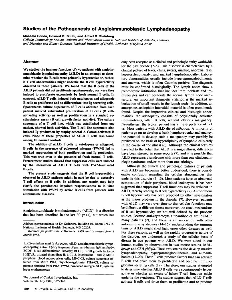
Table VIII. Monoclonal Antibody (33.2.1) Inhibits B Cell Activation and Proliferation by the AILD T Cell Line*
Regulatory T cells and/ormonoclonal antibody B B + PWM B + anti-s B + anti-s + BCGF
mean cpmt mean cpmt mean cpmt mean cpmt
0 469 623 507 9,978Normal T cells 565 9,424 936 14,984T cell line 16,387 5,180 33,088 50,725T cell line + anti-DNA 15,942 4,873 28,341 52,386T cell line + 33.2.1 6,190 [-62.0]§ 4,480 [-13.5]§ 7,968 [-75.9]§ 23,204 [-54.3]§
* Highly purified B cells (10') were cultured with one of the following or with a combination of them in the presence or absence of monoclonalantibody (33.2.1; 5 ug/ml) or a control anti-DNA monoclonal antibody, 5 ug/ml; PWM(1:100 dilution); anti-is (25 Ug/ml); BCGF(20% vol/vol); irradiated regulatory T cells (105). Cultures were incubated for 3 d. [3H]TdR was added the last 16 h of culture. t Standard errors were< 15%. § [ I indicate %suppression by monoclonal antibody.
Studies of Angioimmunoblastic Lymphadenopathy 337
Table IX. Monoclonal Antibody (33.2.1) Inhibits B Cell Activation and Proliferation by the AILD T Cell Line Culture Supernate*
Culture supernate and/ormonoclonal antibody B B + anti-cu B + anti-A + BCGF
mean cpm§ mean cpm§ mean cpm§
0 257 2,225 23,864AILD-CS 7,67311 25,54911 37,6871AILD-CS + 33.2.lt 616 [-92.0]11* 2,325 [-90.9]"** 1,740 [-95.4]¶**
* Cultures were performed as in Table 8 except that the culture supernate obtained from the AILD T cell line (AILD-CS) was used instead ofthe T cells themselves. t A control anti-DNA monoclonal antibody did not suppress (data not shown). § Results were expressed as meanCPMof triplicate cultures. Standard errors of the means were <15%. An additional two experiments were performed and similar results wereobtained. 1l Final concentration of culture supernate is 40%. 1 Final concentration of culture supernate is 20%. ** [ ] indicate %suppres-sion by monoclonal antibody.
have come to an incomplete conclusion based upon theirfinding of proliferation when AILD B cells were cultured withnormal T cells plus PWM. This result led to the conclusionthat the AILD B cells were hyperactive. Our studies of theeffects of PWMclarify the issue. In the presence of PWM,theAILD T cells led to marked suppression of Ig production. Thisis consistent with the findings of the incomplete studies citedabove (7, 10, 11). Moreover, such suppression was observedeven in the presence of normal T cells. Preactivation experi-ments implicated PWM-activated B cells in the induction ofthe suppression. This last result confirms previous reports ofactivated B cells inducing suppression (34, 35).
In the present study, we used the AILD T cells, theirsupernates, and a T cell line established from one of thepatients. The T cell line was used in an attempt to carry outstudies despite treatment of the patients. Although all of thecells in the T cell line were OKT 4+, we recognize that theline might have been heterogeneous. The PWMsuppressionstudies suggest that the T cell line contained, in addition tohelper cells, cells that participated in suppression. Alternatively,a single OKT 4+ cell might have been able, under differentconditions, to provide either helper or suppressor functions.Previous studies have demonstrated the ability of OKT 4+cells to suppress as well as serve as inducers and helpers (36,37). Moreover, we have recently cloned the AILD T cell lineand have been able to separate a clone which is able to mediatehelper functions but not suppressor functions. Thus, we believethat the different functions observed with the AILD T cell linemight well derive from the functional heterogeneity within theline. Although the line does not mirror the T cell populationin vivo, we believe that the T cell line represents cells whichare critical to the AILD disease process.
The PWMsuppression study may shed some light onprevious studies of patients with SLE. Such patients manifestB cell hyperactivity in vivo and yet often have very poorresponses to stimulation with PWMin vitro (21-25). It ispossible that PWMinduces suppression in those circumstancessimilar to the way that it induced suppression with the AILDcells.
Other studies of immune abnormalities in patients withAILD have implicated Epstein-Barr virus (38), impaired sup-pressor T cell function (9), and autonomous B cell hyperactivity(7). Moreover, occasional patients with AILD have been foundto manifest impaired Ig production (10). Two explanations arepossible for such diversity. The first is that patients may havedifferent immune abnormalities at different stages of their
disease. It is clear that some patients have hypergammaglob-ulinemia at one stage and either a severe infection or alymphoreticular malignancy at another. Whereas excessive Tcell driven B cell function might occur at the first stage,impaired immune function might be caused by or result inthe latter complications. In fact, excessive monocyte inhibitionof B cell function has been implicated in the late hypogam-maglobulinemia (10). Our patients have been studied relativelyearly in the course of their illness; we believe that the presentobservations may account for the B lymphoblasts and hyper-gammaglobulinemia. Just as autoimmune MRL-lpr/lpr miceeventually fail to respond well to immune stimulation afterthe lymphoproliferative process is well underway (39), so AILDpatients may fail to respond to exogenous immune stimulationand thereby be susceptible to exogenous infections.
A second explanation for different findings in differentpatients with AILD is that the syndrome may have differentcauses. Previous authors have stressed differences among pa-tients with AILD (2-4, 6, 40-42). Thus, one group could bean extreme example of autoimmune lymphoproliferation andanother group of AILD patients an early form of a lymphoidmalignancy. However, it is not always easy to distinguish thosetwo possibilities as many years of study of autoimmunelym-phoproliferative mice indicate. Those mice often, but notinvariably, carry retroviruses (43). The recent discovery ofhuman retrovirus diseases (44-50) certainly raises the possibilitythat retroviruses may play a role in AILD. Just as infectiousmononucleosis and Burkitt's lymphoma differ in being benignand malignant lymphoproliferation resulting from Epstein-Barr infection, so AILD may represent benign or malignantlymphoproliferation resulting from infection with one or moreviruses. Inasmuch as studies of patients with Epstein-Barrinfections have been quite informative, further study of AILDwill undoubtedly shed light on various aspects of immunedysregulation in humans.
Our finding of T cell driven B cell hyperactivity couldconceivably be exploited therapeutically. Current therapy ofAILD is marred by the desire to control the lymphoproliferationand yet not further predispose the patient to the infectiouscomplications that claim the lives of the majority of AILDpatients. It might be possible to interfere selectively withcertain T cell functions while leaving others intact. Alternatively,a direct effect on the B cells might be attempted. In thisregard, our in vitro inhibition of T cell driven B cell activationwith an anti-Ia-like antibody suggests the possibility that onemight be able to modulate early events in B cell activation so
338 M. Honda, H. R. Smith, and A. D. Steinberg
as to reduce but not eliminate B cell functions. Such a titrationcould, conceivably, allow the patient to experience a clinicalbenefit without the requirement for toxic drugs. Clearly muchfuture research is necessary before such ideas leave the realmof speculation.
Acknowledgments
Wethank Ms. Cheryl Yarboro and Ms. Carole Berkebile for performingleukaphereses, Mr. Pete Smith for assistance with flow cytometry, andMs. Betty Irene Roupe for the skillful typing of this manuscript.
References
1. Forster, G., and S. Moeschlin. 1954. Extramedullares, leukem-isches Plasmocytom mit Dysproteinamie underworbener hamolytischerAnamie. Schweiz. Med. Wochenschr. 84:1106-1110.
2. Frizzera, G., E. M. Moran, and H. Rappaport. 1974. Angioim-munoblastic lymphadenopathy with dysproteinaemia. Lancet. 1:1070-1073.
3. Frizzera, G., E. M. Moran, and H. Rappaport. 1975. Angioim-munoblastic lymphoadenopathy. Diagnosis and clinical course. Am. J.Med. 59:803-818.
4. Luhes, R. J., and B. H. Tindle. 1975. Immunoblastic lymphad-enopathy. A hyperimmune entity resembling Hodgkins disease. N.Engl. J. Med. 292:1-8.
5. Cullen, M. H., A. G. Stansfeld, R. T. D. Oliver, T. A. Lister,and J. S. Malpas. 1979. Angioimmunoblastic lymphadenopathy: reportof ten cases and review of literature. Q. J. Med. 48:151-176.
6. Kaneko, Y., R. A. Larson, D. Variakojis, J. M. Haren, andJ. D. Rowley. 1982. Nonrandom chromosome abnormalities in an-gioimmunoblastic lymphadenopathy. Blood. 60:877-887.
7. Ershler, W. B., A. L. Moore, S. L. Bums, and B. H. Tindle.1983. Immunoblastic lymphadenopathy: failure of, rather than lackof, immunoregulation. J. Med. (Westbury). 14:81-94.
8. Palutke, M., P. Khilanani, and R. Weise. 1976. Immunologicand electronmicroscopic characteristics of a case of immunoblasticlymphadenopathy. Am. J. Clin. Pathol. 65:929-941.
9. Bluming, A. Z., H. G. Cohen, and A. Saxon. 1979. Angioim-munoblastic lymphadenopathy with dysproteinemia. Am. J. Med. 67:421-427.
10. Rice, L., S. L. Abramson, A. H. Laughter, T. M. Wheeler, andJ. J. Twomey. 1982. Angioimmunoblastic lymphadenopathy withhypogammaglobulinemaia. Am. J. Med. 72:998-1004.
11. Rubinstein, A., and L. G. Dauber. 1983. Lymphoma ofcytotoxic/suppressor T cell phenotype (T8) following angioimmuno-blastic lymphadenopathy. Oncology. 40:195-199.
12. Ligler, F. S., M. Patel, D. Strayer, I. Brodsky, H. Bonner, andT. Juhanilinna. 1983. Extremely high levels of natural killer cells inangioimmunoblastic lymphadenopathy. J. Clin. Immunol. 3:375-381.
13. Stensvold, K., P. Brandtzaeg, S. Kvalog, M. Seip, and S. 0.Lie. 1984. Immunoblastic lymphadenopathy with early onset in twoboys: immunohistochemical study and indication of decreased propor-tion of circulating T-helper cells. Br. J. Haematol. 56:417-430.
14. Banhurst, A. D., and R. C. Williams. 1976. Cellular origins ofautoantibody-A perplexing question. Am. J. Med. 61:303-307.
15. Popa, G., V. Nastase, and E. Hanganu. 1982. Angioimmuno-blastic lymphadenopathy associated with systemic lupus erythematosus.Folia Haematol. (Leipz.). 109:430-434.
16. Starke, I. D., K. B. Elkon, C. L. Harmer, G. R. V. Hughes,and E. Wiltshaw. 1983. Pulmonary involvement in angioimmunoblasticlymphadenopathy following autoimmune disease. Respiration. 44:136-142.
17. Prud'homme, G. J., C. L. Park, T. M. Fieser, R. Kofler, F. J.Dixon, and A. N. Theofilopoulos. 1983. Identification of a B celldifferentiation factor(s) spontaneously produced by proliferating T cells
in murine lupus strains of the lpr/lpr genotype. J. Exp. Med. 157:730-742.
18. Roths, J. B., E. D. Murphy, and E. M. Eicher. 1984. A newmutation, gld that produces lymphoproliferation and autoimmunityin C3H/HeJ mice. J. Exp. Med. 159:1-20.
19. Steinberg, E. B., T. J. Santoro, T. M. Chused, P. A. Smathers,and A. D. Steinberg. 1983. Studies of congenic MRL-lpr/lpr-xid mice.J. Immunol. 131:2789-2795.
20. Mountz, J. D., A. D. Steinberg, D. M. Klinman, H. R. Smithand J. F. Mushinski. 1984. Autoimmunity and increased c-mybtranscription. Science Wash. DC. 225:1087-1089.
21. Jasin, H. E., and M. Ziff. 1975. Immunoglobulin synthesis byperipheral blood cells in systemic lupus erythematosus. Arthritis Rheum.18:219-228.
22. Ginsburg, W. W., F. D. Finkelman, and P. E. Lipsky. 1979.Circulating and pokeweed mitogen-induced immunoglobulin-secretingcells in systemic lupus erythematosus. Clin. Exp. Immunol. 35:76-88.
23. Blaese, R. M., J. Grayson, and A. D. Steinberg. 1980. Increasedimmunoglobulin-secreting cells in the blood of patients with activesystemic lupus erythematosus. Am. J. Med. 69:345-350.
24. Budman, D. R., E. B. Merchant, A. D. Steinberg, B. Doft,M. E. Gershwin, E. Lizzio, and J. P. Reeves. 1977. Increased sponta-neous activity of antibody-forming cells in the peripheral blood ofpatients with active SLE. Arthritis Rheum. 20:829-833.
25. Cohen, P. L., D. A. Lituin, and J. B. Winfield. 1982. Associationbetween endogenously activated T cells and immunoglobulin secretingB cells in patients with active systemic lupus erythematosus. ArthritisRheum. 25:168-173.
26. Honda, M., T. Sakane, A. D. Steinberg, H. Kotani, T. Tsune-matsu, K. Moriyama, and M. Fukase. 1982. Studies of immunefunctions of patients with systemic lupus erythematosus. Antibody todesialized, rather than intact, T cells preferentially bind to and eliminatesuppressor effector T cells. J. Clin. Invest. 69:940-949.
27. Honda, M., and A. D. Steinberg. 1984. Effect of prostaglandinE2 on responses of T cell subsets to mitogen and autologous non-Tcell stimulation. Clin. Immunol. Immunopathol. 33:111-121.
28. Rosenwasser, L. J., and C. A. Dinarello. 1981. Ability ofhuman leukocytic pyrogen to enhance phytohemagglutinin-inducedmurine thymocyte proliferation. Cell. Immunol. 63:134-142.
29. Watson, J., S. Gillis, J. Marbrook, D. Mochizuki, and K. A.Smith. 1979. Biochemical and biological characterization of lymphocyteregulatory molecules. I. Purification of a class of murine lymphokines.J Exp. Med. 150:849-855.
30. Kohler, G., and C. Milstein. 1975. Continuous cultures offused cells secreting antibody of predefined specificity. Nature (Lond.).256:495-497.
31. Honda, M., and A. D. Steinberg. 1985. Production andcharacterization of a unique monoclonal antibody against human Bcells. Cell. Immunol. 93:105-123.
32. Leung, D. Y. M., E. T. Chu, N. Wood, S. Grady, R. Meade,and R. S. Geha. 1983. Immunoregulatory T cell abnormalities inMucocutaneous lymph node syndrome. J. Immunol. 130:2002-2004.
33. Clement, L. T., M. K. Dagg, and G. L. Gartland. 1984. Small,resting B cells can be induced to proliferate by direct signals fromactivated helper T cells. J. Immunol. 132:740-744.
34. Tosato, G., I. Magrath, I. Koski, N. Dooley, and R. M. Blaese.1979. Activation of suppressor T-cells during Epstein-Barr virus inducedinfectious mononucleosis. N. Engl. J. Med. 301:1133-1137.
35. Calkins, C. E. 1982. Interactions between primed and unprimedcells in the regulation of in vitro antibody responses. I. Role of "plasmacells" as inducers of suppression. Eur. J. Immunol. 12:70-75.
36. Thomas, Y., L. Rogozinski, 0. H. Irigoyen, S. M. Friedman,P. C. Kung, G. Goldstein, and L. Chess. 1981. Functional analysis ofhuman T cell subsets defined by monoclonal antibodies. IV. Inductionof suppressor cells within the OKT 4+ population. J. Exp. Med. 154:459-467.
37. Kotani, H., S. Takada, Y. Ueda, Y. Muradawa, N. Suzuki,and T. Sakane. 1984. Activation of immune regulatory circuits among
Studies of Angioimmunoblastic Lymphadenopathy 339

OKT4+ cells by autologous mixed lymphocytes reactions. Clin. Exp.Immunol. 56:390-398.
38. Robinson, J. E., N. Brown, W. Andiman, K. Halliday, V.Francke, M. F. Robert, M. Anderson-Anunet, D. Horstman, and G.Miller. 1980. Diffuse polyclonal B-cell lymphoma during primaryinfection with Epstein-Barr virus. N. Engl. J Med. 303:1293.
39. Wilson, D. A., and H. Braley-Mullen. 1982. Immunoregulationin MRL-lpr/lpr mice. Evidence for decreased helper T cell and increasedsuppressor T cell function with age. Cell. Immunol. 74:72-80.
40. Hossfeld, D. K., K. Hofiken, C. G. Schmidt, and H. Diedrichs.1976. Chromosome abnormalities in angioimmunoblastic lymphade-nopathy. Lancet. 1: 198.
41. Castroldi, G., G. Scapoli, G. D. Grunsovin, M. Gualandi, R.Spanedda, L. Cavazzini, and D. Anzanel. 1976. Chromosomal abnor-malities in angioimmunoblastic lymphadenopathy. Ric. Clin. Lab. 6:121-129.
42. Goh, K., and R. F. Bakemeier. 1970. Is angioimmunoblasticlymphadenopathy with dysproteinemia a malignant disease? J. Am.WomenAssoc. 33:38.
43. Datta, S. D., F. L. Owen, J. E. Womack, and R. J. Riblet.1982. Analysis of recombinant inbred lines derived from "autoimmune"(NZB) and "high leukemia" (C58) strains: independent multigenicsystems control B cell hyperactivity, retrovirus expression, and autoim-munity. J. Immunol. 129:1539-1544.
44. Poiesz, B. J., F. W. Ruscetti, M. S. Reitz, U. S. Kalyanataman,and R. C. Gallo. 1981. Isolation of a new type C retrovirus (HTLV)in primary uncultured cells of a patient with Sezary T-cell leukemia.Nature (Lond.). 294:268-271.
45. Robert-Gurott, M., Y. Nakano, Y. Ito, A. Slishi, and R. C.Gallo. 1982. Natural antibodies to human retrovirus HTLV in acluster of Japanese patients with adult T cell leukemia. Science (Wash.DC). 215:975-978.
46. Sarin, P. S., T. Aoki, A. Sibata, Y. Ohnishi, Y. Aoyagi, H.Miyakoshi, I. Emura, U. S. Kalyanaraman, P. C. Nowell, and R. C.Gallo. 1983. High incidence of human type-C retrovirus (HTLV) infamily members of a HTLV-positive Japanese T-cell leukemia patient.Proc. Natl. Acad. Sci. USA 80:2370-2374.
47. Gelman, E. P., M. Popovic, D. Blayney, H. Masur, G. Sidhu,R. E. Stahl, and R. C. Gallo. 1983. Proviral DNA of a retrovirus,human T-cell leukemia virus, in two patients with AIDS. Science(Wash. DC). 220:862-865.
48. Gallo, R. C., P. S. Sarin, E. P. Gelmann, M. Robert-Guroff, E.Richardson, V. S. Kalyanaraman, D. Mann, G. Sidhu, R. E. Stahl, S.Zolla-Pazner, J. Leibovitch, and M. Popovic. 1983. Isolation of humanT-cell leukemia virus in acquired immune deficiency syndrome (AIDS).Science (Wash. DC). 220:865-867.
49. Barre-Sinoussi, F., J. C. Chermann, F. Rey, M. T. Nugeyre, S.Chamaret, J. Gruest, C. Danguet, C. Axler-Blin, F. Vezinet-Brun, C.Rouzioux, W. Rozenbaum, and L. Montagnier. 1983. Isolation of aT-lymphotropic retrovirus from a patient at risk for acquired immunedeficiency syndrome (AIDS). Science (Wash. DC). 220:868-871.
50. Blattner, W. A., V. S. Kalyanaraman, M. Robert-Gurott, T.Andrew-Lister, D. A. G. Galton, P. S. Sarin, M. H. Crawford, D.Catovsky, M. Greaves, and R. C. Gallo. 1982. The human type-Cretrovirus, HTLV, in blacks from the Caribbean region, and relationshipto adult T-cell leukemia/lymphoma. Int. J. Cancer. 30:257-264.
340 M. Honda, H. R. Smith, and A. D. Steinberg