studies on the incidence and inheritability of spontaneous...
TRANSCRIPT
PRIMARY SPONTANEOUS TUMORS OF THE OVARY IN MICE
STUDIES ON THE INCIDENCE AND INHERITABILITY OF SPONTANEOUS TUMORS IN MICE
FOURTEENTH COMMUNICATION MAUD SLYE, HARRIET F. HOLMES AND H. GIDEON WELLS
From the Otho S. A . Sprague Memorial Znstitute and the Department of Pathology of the University of Chicago
Received for publication, September 2, 1919
.
Throughout the animal kingdom the solid ovarian tumors seem to be infrequent, as they also are, relatively, among human tumors. Cystic tumors are described occasionally, but appar- ently less frequently in the lower animals than in man. Even among dogs, with their high incidence of tumor growth, ovarian tumors are rare according to the evidence furnished by literature dealing with canine neoplasms.1 In Sticker’s (2) compilation of tumors in domestic animals, of 766 tumors in dogs but 3 were in the ovary. As to other animals of Sticker’s series, in 509 cases of tumors in horses, 4 were in the ovary; of 110 in cattle, 6 were ovarian; there was 1 ovarian tumor among 23 tumors in cats, and none at all among sheep, goats, and swine. Kimura (3) has reported 142 cases of tumors in horses, among which were no ovarian tumors, although there were 49 in the testicle. Other evidence supports the figures of Sticker in indicating that cows have ovarian tumors more often than other species. Trotter (4) reported 305 bovine tumors observed in the Glasgow slaughter house, of which 5 were in the ovary (4 carcinomas and
1 Goodpasture (1) notes the occurrence of small hyperplastic areas in the senile ovaries of old dogs, but even these do not seem to be of very frequent occur- rence in proportion to the high incidence of proliferative changes noted by him in other tissues.
205
TEE JOURNAL OF CANCER RESEARCH, VOL. V, NO, 3

206 MAUD BLYE, H. F. HOLNES AND H. G. WELLS
1 sarcoma). These were all large lobulated solid tumors, and no metastases were observed in any. Leo Loeb (5) has de- scribed a tumor arising in the ovary of a six-months-old calf, composed chiefly of cells resembling luteum tissue.
Among the numerous instances of tumors in wild rats reported by McCoy (6), Woolley and Wherry (7), and Beatti (8) there is no case of ovarian growth; nor have we found reports of any cases occurring in domesticated rats.2
Wolff (9) mentions 2 cases of ovarian tumors in cats: One a primary carcinoma in the ovary of a thirteen-year old cat with metastasis in the liver, reported by Kitt; the other a sarcoma of the ovary and pelvis reported by Stroud.
Wild animals are also unlikely to have ovarian tumors. Fox (lo), in his extensive studies of tumors in wild animals, has described no cases whatever of ovarian tumors among the mammalia.
Only in birds do ovarian tumors seem to be relatively frequent. Burger (11) found, among 852 fowls autopsied at the Leipzig veterinary institute, 12 tumors, of which 7 were in the ovary, 4 being sarcomas and 3 carcinomas; 2 of the sarcomas and 2 of the carcinomas had produced metastases. In their review of the literature on tumors in birds, Joest and Ernesti (12) found 112 cases reported and added 50 more. Of these 162 cases, 21 were primary carcinoma of the ovary, commonly with extensive peritoneal and visceral metastasis. There was also one case of ovarian sarcoma.
An ovarian tumor has been described in awild turkey (Meleapis gallapavo) by Fox (lo), as follows: The growth was “about the size of 3 English walnuts placed in triangular posi- tion.” Tt consisted of 3 subdivisions, covered with papilloma- tous prominences like a hydatid mole. Microscopically it was a papillary cystadenoma.
*While this article was in press there appeared under the title “Carcino- .sarcoma de rata blanca,” an article by Dr. A. H. Roffo (Revista del Instituto Bacteriologico, Buenos Aires, 1919, ii, 2831, reporting the finding of a large tumor in the ovary of a white rat. This seems, from the illustration and de- scription, to have been a papillary oystadenoma with areas of more compaat cell growth, some of which are interpreted as carcinoma and some as sarcoma.

TUMORS O F THE OVARY IN MICE 207
Cold-blooded animals also have furnished occasional cases of ovarian tumor. Bland-Sutton (13) has reported a case of tumor of the ovaries of a python, with growths also in the lungs, liver, and peritoneum; he believed the ovarian growth to have been primary but without conclusive evidence. Plehn (14) has described tumors arising in the ovary of an old grass frog (Rana esculenta) through growth of primitive ova cells without differ- entiation. There were numerous nodules, from millet seed to cherry size, apparently benign in character although resembling a malignant tumor in histological structure. A cystic tumor of the ovary has been described in a fish, the ling (Molua molua) by Johnstone (15).
In mice, also, ovarian tumors are not common, but we have found mention of 8 cases in the literature. The first 2 cases were described by Jobling (16). One was bilateral, each ovary being eight times the normal size. Both ovaries showed the same structure, which is described as follows :
A great increase in epithelial cells, which formed solid masses, somewhat compressed and elongated into spindle cells and cysts of different sizes, usually small and more or less occupied by the papillary outgrowths from their walls. These outgrowths developed from narrow or somewhat thickened pedicles and spread out into a fanlike structure. . . . . The more solid portions were at one time cystic but the cysts became occluded by the ingrowth of papillae. Acini pos- sessing a distinctly granular [sic., glandular?] form and arrangement also occurred. Mitosis was rare, direct division more frequent. Hemorrhage had taken place into some of the cysts. Mallory stain showed a delicate connective-tissue basement membrane surrounding the small cysts and the more solid areas, the latter being filled with epithelial cells of a granular quality resembling somewhat the lutein cells.
In the other case the growth was unilateral, there being numerous large cysts, separated by smaller ones and by the tubules of the ovary, lined by high columnar epithelium without cilia. Between the cysts the tissue was composed largely of smooth muscle fibers, resembling in places a leiomyoma.

208 MAUD SLYE, H. F. HOLMES AND H. G . WELLS
Tyzzer (17) has reported 4 cases. 1. This tumor was bilateral, the ovaries being replaced by an irregularly glandular epithelial growth, in places with flattened epithelial cells intimately mingled with the connective tissue; in others the glands con- tained thick papillary processes of epithelium without central connective tissue core protruding into the lumen, the epithelium being in places high columnar, in others merely a fused mass. There were no mitotic figures, but the presence of masses of epithelium in the lymphatics suggested malignancy. 2. In a mouse with a renal tumor resembling a hypernephroma, one ovary was replaced by an irregular gland-like structure similar to the above. 3. A mouse of a series bred for heredity studies, which died from a lung tumor, had the right ovary replaced by a mass the size of a large pea, composed of irregular epithelium having in places a partially glandular structure, without ten- dency to form spaces. 4. A mouse from the same family as (3), had both ovaries replaced by masses of translucent whitish tissue, 5 by 3 by 3 mm. This also had the structure of a papillary adenoma. Tyzzer notes that these growths differ from the ordinary types of ovarian tumors seen in human beings, in that the epithelium is more or less glandular with but little tendency to form cysts. The epithelium resembles the peritoneal meso- thelium found at the attachment of the ovary.
Haaland (18) found 2 ovarian tumors among 353 primary tumors observed in 288 mice, of which 325 were mammary gland growths. 1. A mouse that had a sarcoma arising in a scar also had a tumor the size of a hazel nut in the left ovary. The ovarian tumor was composed of large alveoli with a peri- pheral layer of low columnar epithelial cells, the lumen filled with round cells and sometimes exhibiting spaces filled with serous fluid. This tissue was transplanted into 120 normal mice, but no growths resulted. 2. A mouse that had been operated for mammary carcinoma with recurrence. A tumor
as large as a pea” replaced the right ovary, and in structure was a papillary adenocarcinoma, probably primary.
Of these 8 recorded cases of ovarian tumors in mice there were 3 cases of bilateral tumors, although in no case was there metas-
t (
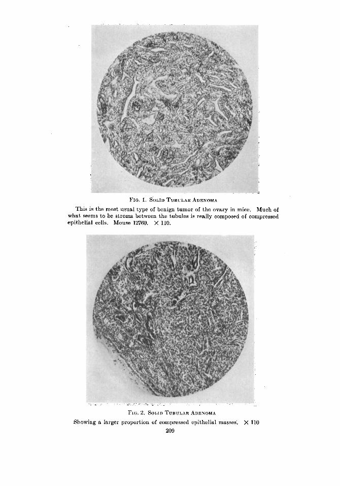
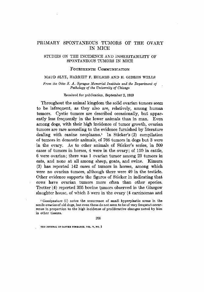
FIG. 1. SOLID TUBULAR ADENOMA This is the most usual type of benign tumor of the ovary in mice. Much of
what seems to be stroma between the tubules is really composed of compressed epithelial cells. Mouse 12760. X 110.
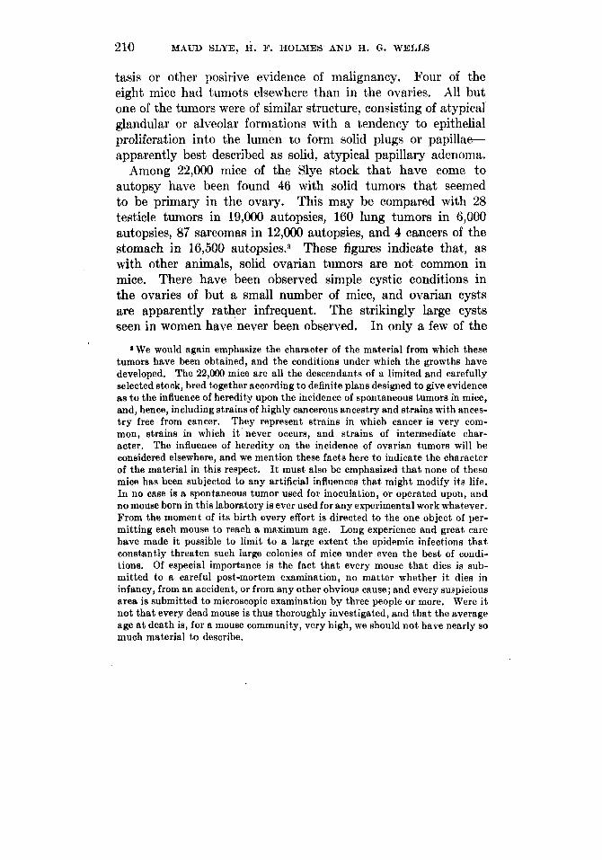
FIG. 2. SOLID TUBULAR ADENOMA
Showing a larger proportion of compressed epithelial masses. X 110 209
210 MAUD SLYE, H. F. HOLMES AND H. G . WELLS
tasis or other posirive evidence of malignancy. Four of the eight mice had tumots elsewhere than in the ovaries. All but one of the tumors were of similar structure, consisting of atypical glandular or alveolar formations with a tendency to epithelial proliferation into the lumen to form solid plugs or papillae- apparently best described as solid, atypical papillary adenoma.
Among 22,000 mice of the Slye stock that have come to autopsy have been found 46 with solid tumors that seemed to be primary in the ovary. This may be compared with 28 testicle tumors in 19,000 autopsies, 160 lung tumors in 6,000 autopsies, 87 sarcomas in 12,000 autopsies, and 4 cancers of the stomach in 16,500 aut~psies.~ These figures indicate that, as with other animals, solid ovarian tumors are not common in mice, There have been observed simple cystic conditions in the ovaries of but a small number of mice, and ovarian cysts are apparently rather infrequent. The strikingly large cysts seen in women have never been observed. In only a few of the
*We would again emphasize the character of the material from which these tumors have been obtained, and the conditions under which the growths have developed. The 22,000 mice are all the descendants of a limited and carefully selected stock, bred together according t o definite plans designed t o give evidence as t o the influence of heredity upon the incidence of spontaneous tumors in mice, and, hence, including strains of highly cancerous ancestry and strains with ances- t ry free from cancer. They represent strains in which cancer is very com- mon, strains in which i t never occurs, and strains of intermediate char- acter. The influence of heredity on the incidence of ovarian tumors will be considered elsewhere, and we mention these facts here to indicate the character of the material in this respect. It must also be emphasized tha t none of these mice has been subjected to any artificial influences that might modify its life. In no case is a spontaneous tumor used for inoculation, or operated upon, and no mouse born in this laboratory is ever used for any experimental work whatever. From the moment of its birth every effort is directed to the one object of per- mitting each mouse to reach a maximum age. Long experience and great care have made it possible to limit t o a large extent the epidemic infections tha t constantly threaten such large colonies of mice under even the best of condi- tions. Of especial importance is the fact that every mouse t h a t dies is sub- mitted to a careful post-mortem examination, no matter whether i t dies in infancy, from an accident, or from any other obvioua cause; and every suspicious area is submitted to microscopic examination by three people or more. Were i t not that every dead mouse is thus thoroughly investigated, and that the average age at death is, for a mouse community, very high, we should not have nearly so much material t o describe.

FIG. 3. SOLID TUBULAR ADENOMA In this growth there is an unusually large proportion of ovarian stroma type.
X 76.
FIG. 4. PAPILLARY PORTION OF A GROWTH THAT ELSEWHERE IS A SOLID
TUBULAR ADENOMA, AS I N 1 AND 2
Small areas thus disclosing an’existing tendency to papillary structure are not. infrequently found in the ordinary solid ovarian tumors of mice. Mouse 2205. x 110.
31 1
212 MAUD SLYE, H. F. HOLMES AND H. G. WELLS
ovarian cysts that we have examined have small cystic papiIla- matous areas been found. Therefore, mouse ovarian tumors present a marked preponderance of solid adenomatous growth, as compared with the proportion of cystic ovarian enlargements in women. We have eliminated from our consideration the simple cysts, as probably not examples of neoplastic growth, with the exception of those cysts that result from secretion by a cyst- adenoma,
Most of our tumors seem to be benign in character, although we have found a few examples of undoubted malignant tumors primary in the ovary. In all, 38 mice have exhibited tumors that may be classified as solid benign ovarian tumors, and there was one typical papillomatous cystoma. As 19 of these had bilateral tumors there are 58 ovarian tumors occurring in a stock of mice that have yielded over three thousand primary spon- taneous tumors of other tissues, chiefly the mammary gland.
BENIGN OVARIAN TUMORS
I n structure these tumors vary considerably, although most of them correspond closely to the descriptions given by Jobling, Tyzzer, and Haaland, and may be most appropriately designated as solid alveolar adenoma. Tendency to cyst formation is exceptional, in contrast to the adenomas of the human ovary, and the same is true of papillary types of growth, which are rarely exhibited. In general these solid adenomas of the mouse ovary exhibit a growth apparently under great pressure, with crowding of the cells so that it is usually difficult to differentiate readily between stroma cells and flattened epithelial cells (see figs. 1 and 2). Probably this crowding of the growth accounts for the absence of papillary tendency, for often a small area may be found where there is less pressure or where part of the tissue has been destroyed by hemorrhage or necrosis, in which a distinct tendency to papillary structure is seen (see fig. 4). We have only one case (12111) in which the structure corresponds a t .all closely with the human papillary cystoma (fig. 7).
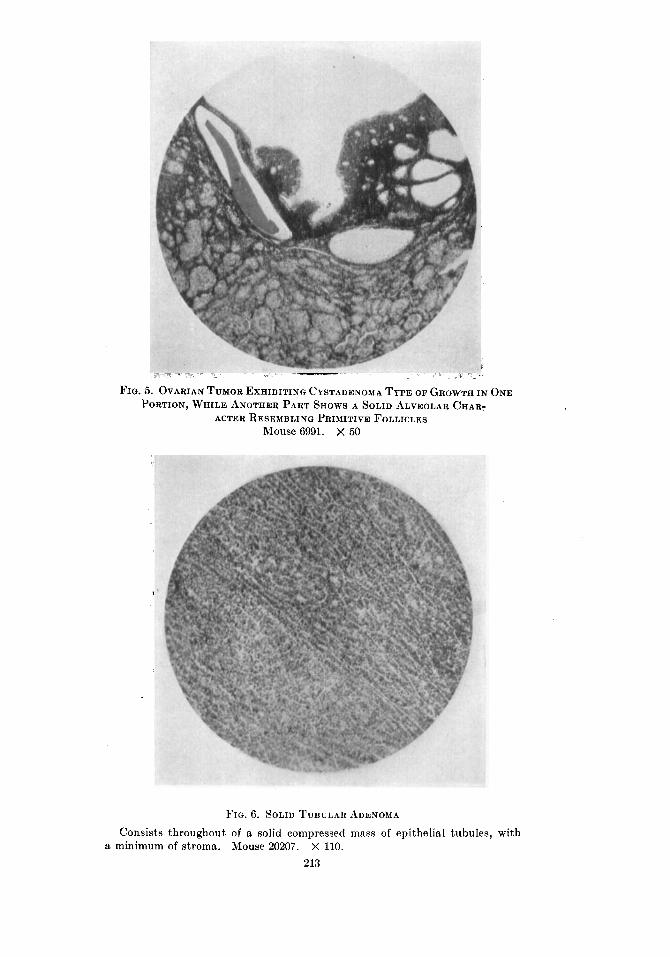
FIG. 5. OVARIAN TUMOR EXHIBITING CYSTADENOMA TYPE OF GROWTH IN ONE PORTION, WHILE ANOTHER PART SHOWS A SOLID ALVEOLAR CHAR:
Mouse 6991. X 50 ACTER RESEMBLING PRIMITIVE FOLLICLES
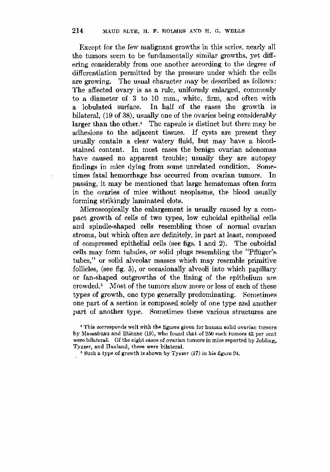
FIG. 6. SoLrn TUBULAR ADENOMA
Consists throughout of a solid compressed mass of epithelial tubules, with a minimum of stroma. Mouse 20207. X 110.
213
214 MAUD SLYE, H. F. HOLMES AND H. G. WELLS
Except for the few malignant growths in this series, nearly all the tumors seem to be fundamentally similar growths, yet diff- ering considerably from one another according to the degree of differentiation permitted by the pressure under which the cells are growing. The usual character may be described as follows: The affected ovary is as a rule, uniformly enlarged, commonly to a diameter of 3 to 10 mm., white, firm, and often with a lobulated surface. In half of the cases the growth is bilateral, (19 of 38), usually one of the ovaries being considerably larger than the 0ther.4 The capsule is distinct but there may be adhesions to the adjacent tissues. If cysts are present they usually contain a clear watery fluid, but may have a blood- stained content. In most cases the benign ovarian adenomas have caused no apparent trouble; usually they are autopsy findings in mice dying from some unrelated condition. Some- times fatal hemorrhage has occurred from ovarian tumors. In passing, it may be mentioned that large hematomas often form in the ovaries of mice without neoplasms, the blood usually forming strikingly laminated clots.
Microscopically the enlargement is usually caused by a com- pact growth of cells of two types, low cuboidal epithelial cells and spindle-shaped cells resembling those of normal ovarian stroma, but which often are definitely, in part at least, composed of compressed epithelial cells (see figs. 1 and 2). The cuboidal cells may form tubules, or solid plugs resembling the “Pfliiger’s tubes,” or solid alveolar masses which may resemble primitive follicles, (see fig. 5 ) , or occasionally alveoli into which papillary or fan-shaped outgrowths of the lining of the epithelium are c r~wded .~ Most of the tumors show more or less of each of these types of growth, one type generally predominating. Sometimes one part of a section is composed solely of one type and another part of another type. Sometimes these various structures are
4 This corresponds well with the figures given for human solid ovarian tumors by Massabuau and Etienne (19), who found that of 250 such tumors 43 per cent were bilateral. Of the eight cases of ovarian tumors in mice reported by Jobling, Tyezer, and Haaland, three were bilateral.
Such a type of growth is shown by Tyeaer (17) in his figure 24.

FIQ. 7. PAPILLARY CYSTADENOMA The only specimen in our series typically reproducing this type common in
the human ovary. Mouse 12111. X 45.
FIG. 8. SOLID TERATOMA
Shows in this field islands of cartilage, epithelial plugs with hornified epithe- lium, cysts lined with squamous and with columnar epithelium, and varying types of stroma elements. Mouse 9278. X 60.
21.5
'216 MAUD SLYE, H. I?. HOLMES AND H. G. WELLS
limited by a distinct basement membrane of true stroma cells, but often there seems to be a false stroma of crowded, spindle- shaped epithelial cells without sharp demarcation from the true stroma cells that may be present. Usually the total amount of stroma is small, most of the growth being composed of epithelial cells, and this stroma is of the cellular ovarian type and not ordinary fibrous tissue. Not infrequently, however, the stroma elements form the bulk of the growth (fig. 3). Generally the capsule is formed by compressed ovarian tissue, in which ova or follicles are rarely seen. Blood vessels are usually scanty; but, nevertheless, necrosis or other retrogressive change is seldom found. Mitosis is very rarely observed, nor are forms sugges- tive of amitotic division common. The epithelium resembles the germinal epithelium and occasionally the surface of the tumor is covered with this cuboidal epithelium which dips down at intervals to divide the growth deeply into lobules.
As variations from the structure described above we have in a few instances the formation of several small cysts with lining of flattened epithelium. In a few also the structure suggests a papilloma with all spaces obliterated by pressure or from lack of secretion by the surface epithelium. If the growth is of tubular character the pressure usually causes the lumens to resemble narrow slits (figs. 2 and 6). In some instances the alveolar structure is filled with flattened cells consisting chiefly of deeply staining nuclei, producing a growth resembling the adenomatous growths often found in the human vermiform appendix. In only one instance have we a true fibroadenoma (12922) in which a large part of the tumor is composed of definite fibrous stroma with abundance of collagenous material, rather than the cellular ovarian type of stroma. We have not identi- fied any of our tumors as of lutein cell structure, nor have we observed fibromas or myofibromas in the ovary.
MALIGNANT OVARIAN TUMORS
We have found the following tumors that seem to be unques- tionably primary malignant tumors arising in the ovary.
6487. The left ovary was 10 mm. in diameter, nearly spher- ical, partly white and fleshy, partly cystic. There were no

FIQ. 9. MALIQNANT EPITHELIAL TUMOR OF OVARY
Showing both tubular and alveolar types of growth. Mouse 6487. X 100
FIG. 10. MALIGNAKT EPITHELIAL TUMOR OF OVARY Showing solid type of growth of large cells without any well defined arrange-
Similar structure is also shown by tumors of testicle and adrenal. Mouse
217
ment. 12552. X 110.

218 MAUD SLYE, H. F. HOLMES AND H. G. WELLS
adhesions and no evidence of metastasis. The mammary gland exhibited a large hemorrhagic carcinoma and also a minute early carcinomatous nodule, both of these growths being typically and unquestionably primary carcinomas of the mammary gland; structurally they were entirely distinct from the ovarian growth. About one half of the latter was composed of a cavity filled with a protein-rich fluid, and with walls formed by an irregular border of tumor tissue. Apparently this cyst was formed by degeneration of tumor tissue, as it had no proper cyst wall, although on the side towards the tumor there are
. some papillary outgrowths into the cavity. The solid part of the tumor consists of epithelial cells arranged mostly in large irregular alveoli, often exhibiting central necrosis, and occasion- ally atypical tubules are seen (fig. 9). The stroma is scanty and of ordinary connective tissue type for the most part, although these are areas with abundant elongated cells. The cells are large with a clear cytoplasm, well defined borders, and oval nuclei which are partly solid and partly slightly vesicular. Occa- sional mitotic figures are seen. Although there are no metas- tases, the histological structure of this tumor is such that it must be considered to be malignant, an alveolar carcinoma of the ovary.
12552. The left ovary measured 25 by 18 by 18 mm., nodular, with many distended blood-vessels coursing over it. It was not adherent to adjacent tissue and no metastatic growths were found. In the mammary gland were two hemorrhagic car- cinomas, entirely different in structure from the ovarian tumor. Microscopically the ovarian tumor is composed of large irregular alveoli filled with large cells with a moderate amount of cyto- plasm and a large pale nucleus, with very little stroma (fig. 10). Several mitotic figures were found. The original ovarian cap- sule surrounds the growth, but no recognizable ovarian tissue remains. Areas of necrosis and hemorrhage are numerous. This tumor closely resembles 6487 but exhibits a greater degree of malignancy.
6801. The left ovary was converted into a hemorrhagic nodular mass, 18 by 15 by 10 mm. The right ovary was normal.
TUMORS OF THE OVARY IN MICE 219
A mass of tumor tissue lay anterior to the left kidney, about the size of the kidney itself. There was a large carcinoma of the mammary gland arising in the left flank, and producing metastases in the lung, as shown by microscopic examination.
The large ovarian tumor consists of about equal parts of extravasations of blood or large dilated blood channels and of tumor tissue. Nothing of the original tissue remains, except that in the capsule appear a few cells suggestive of ovarian stroma elements. The tumor has no well defined structure, consisting of large solid masses of cells which occasionally are formed into trabeculae by blood channels, but otherwise show no tendency toward grouping of any kind. The cells are char- acterized by a considerable amount of cytoplasm which is usu- ally homogeneous and often with well defined cell borders. The nuclei are round, sometimes solid and sometimes vesicular. Where the cells are largest-they resemble slightly liver cell types. There is practically no stroma besides the blood vessels, which are scanty except for the large channels in the tumor without vessel walls. The retroperitoneal mass presents quite the same structure. Mitotic figures are not seen. A few multinucleated cells are found. The general character of the growth resembles that of other malignant tumors found in the adrenal and testicle.
14099. The left ovary is about three times the normal size, and of a red color. The uterus is distended with a milky fluid. In the upper lobe of the right lung is a firm nodule, 2 111111. in diameter, which proved to be a simple papillary adenoma, appar- lently benign but with a projection growing into a blood-vessel. There were no changes of significance in the other organs. Mi- croscopically the ovary is found to be almost entirely replaced by a mass of large cells. with no particular arrangement, which invade the remaining recognizable ovarian tissue, and in which appear epithelial structures that resemble overgrown Pfluger’s tubules. The nuclei are deeply stained and irregular in size. In the perirenal tissue is a small growth of similar character.
It is interesting to observe that the malignant types of tumors arising from the ovary, testicle, and adrenal that we have studied exhibit such a similar histological picture, and one quite dis-

220 MAUD SLYE, H. F. HOLMES AND H. G . WELLS
similar from tumors arising in other tissues. There can be little doubt that they all represent reversions to the primitive em- bryonal tissues of the urogenital anlage, and are probably best designated by Adami’s term, mesothelioma. This similarity of structure makes it difficult to determine the origin of a tumor involving both the ovary and the adrenal, as in the following case.
12307. The abdominal cavity shows several nodules whose exact origin js difficult to determine as the mate has partly de- voured the body. The right ovary is, however, easily distin- guished. It measures 18 by 12 by 12 mm. What seems to be the left ovary is 10 by 8 by 6 mm. There are 8 other similar nodules in the abdominal cavity, one being in the position of the left adrenal, measuring 10 by 8 by 8 mm. The other nodules are apparently in the mesentery. One lobe of the liver is con- verted into a tumor nodule 14 by 10 by 18 mm., irregular and lumpy in outline, pink in color.
The tumor tissue shows everywhere the same structure, con- sisting of irregular alveoli composed of large cells with abundant cytoplasm with well defined borders and deeply staining nuclei. Mitotic figures are numerous. The character is that usual to mesothelial growths. The ovary cannot be positively identified, but one mass exhibited in the capsule structure suggests com- pressed ovarian tissue with degenerated ovum. In all respects this tumor is identical with the malignant ovarian tumors just described.
It seems probable that this tumor arose in the ovary which exhibited the largest growth, but it is not possible to exclude the adrenal as the primary site.
Still more difficult to locate is the primary growth in the following case.
12876. The left kidney contains a mass of pink, fleshy tissue, 18 by 14 by 14 mm. The right kidney, which is slightly en- larged, contains no tumor mass. The right ovary consists of a pinkish tissue resembling that in the kidney, and measures 12 by 8 by 8 mm. In the mesentery is a similar, slightly paler mass, 16 by 8 by 8 mm. The retroperitoneal and subcutaneous glands are not enlarged and no nodules are found in the lungs.

TUMORS OF THE OVARY IN MICE 221
Microscopically the tumor tissue is alike in all three places, consisting of a diffuse infiltrating growth of large round cells, which also invade the connective tissues about the kidney and ovary. It does not at all resemble the typical ovarian tumors, being apparently a round-cell sarcoma. We have no way of telling which of the three tumors was primary. The next case presents similar difficulties.
26. This mouse had a tumor mass about 8 by 10 111111. in the upper portion of the liver, with other smaller nodules near it. A similar small nodule was found in the right kidney. The right ovary was enlarged to two-thirds the size of a kidney, and was solid. Microscopically all these growths are composed of round cells, apparently a round-cell sarcoma. It is impossible to say which growth was primary.
We have seen few instances of secondary tumors occurring in the ovary. The Krukenberg tumor is not found because mice do not have gastric or other abdominal carcinomas, except most rarely (20).
In leukemia and pseudoleukemia, infiltration of the ovary with lymphoid elements is common, and often very striking. In our collection there has been no case observed of secondary carcinoma of the ovary, which is not surprising in view of the relatively slight tendency of mouse carcinomas to produce metas- tasis elsewhere than in the lungs. The following examples of secondary sarcoma have been noted :
12058. A bilateral sarcoma of the uterus, round-cell in type, with metastasis in the right kidney. The left ovary was 10 mm. in diameter and showed complete replacement by sarcoma tissue. As near as we can determine the growth in this case arose in the uterus, and invaded the ovary by infiltration.
A spindle-cell sarcoma growth infiltrated the retro- peritoneal tissues extensively, including the pancreas, also with metastasis in the liver. Apparently primary in either the retroperitoneal tissues or in the mesentery. The uterus and both ovaries were infiltrated by the same tissue.
To summarize, we have found four malignant growths that seem to be certainly primary in the ovary. Each of these
19061,
TEE JOURNAL OF CANCER RESEARCH, VOL. V, NO. 3 I

222 MAUD SLYE, H. F. HOLMES AND H. ff. WELLS
exhibited the structure common to malignant tumors arising in the sex glands and adrenal. One exhibited retroperitoneal metastasis near the kidney. Another tumor of similar histo- logical type also involved the adrenal, and produced numerous metastases; in this case it was not possible to determine whether the growth was primary in the ovaries or in the adrenal.
There were two round cell sarcomas involving the ovary as well as other organs, which might have been primary in the ovary, but the evidence did not permit of deciding this. There were two other cases in which the sarcomatous invasion of the ovary seemed to be unquestionably secondary. No secondary carcinomas were observed in the ovary.
TERATOMA OF THE OVARY
From the accounts of animal tumors in the literature it would seem that teratomas are extremely infrequent in the lower mammals. In reviewing the literature on the occurrence of tumors of the ovary and testicle, in the lower animals the chief sites of teratomatous growths, we have found mention of but one case. That was described by Winokuroff (21) as a teratoma of the testicle in a chicken, the growth exhibiting cartilage, bone, striated muscle, squamous epithelium, and cysts. Our own experience supports the view that the lower animals rarely exhibit teratomas, for in the 22,000 mice here considered contain- ing over three thousand spontaneous primary tumors, we have observed but one teratoma. This arose in the ovary, and is a typical example of solid teratoma as shown by the following description :
9278. Death resulted from pulmonary infection. There were no other changes of importance except in the left ovary, which measured 20 by 18 mm., and yielded an exudate from the cut surface. Microscopically the growth has a delicate but distinct capsule in one portion of which there still remains a trace of the original ovarian tissue with one follicle containing an ovum. Except for this the entire section shows a mass of tissues of all sorts thrown together in an entirely disordered

TUMORS OF THE OVARY IN MICE 223
manner (fig. 8). There are numerous small, cavities, some of which are lined with squamous epithelium containing chiefly desquamated cells and some polynuclear leukocytes. Occasional solid plugs of squamous epithelium also occur. In places these are branching and occasionally a basal-cell type of growth is seen. No true hair follicles are recognized although occasionally the epithelial downgrowth suggests these structures. No seba- ceous glands are found. Most of the squamous-cell structures are grouped together in localized mas. There are also spaces lined with columnar or flattened epithelium, sometimes with a content resembling a diluted mucin. Spaces are found in which part of the lumen is lined with stratified epithelium and part by cuboidal or columnar epithelium. Sometimes these tubules have a well defined coat of non-striated muscle fiber. Small islands of cartilage are abundant, usually having no definite relation to other structures. No bone is seen. Some of the cavities contain old extravasations of blood in varying stages of disintegration. In one place there is a mass of heavy brown pigment near which are collections of small round cells; the whole appearance suggests that possibly this area represents undeveloped retinal tissue. The stroma in general consists of a loose fibrous tissue with abundant cells. There are also many cells with much more cytoplasm than ordinary connective tissue cells, this cytoplasm having a slight basophilic tendency. No striated muscle is found or definite organ tissues but there are many accumulations of cells which are not stroma cells and which presumably are undifferentiated cells of special tissues.
Since this analysis of 22,000 autopsies was made, another instance of teratorna of the ovary has been observed, so that we now have found two teratomas among 25,000 autopsied mice. The chief features of this second case are as follows:
24172. Mouse, eleven months old, died apparently from acute pulmonic infection following delivery, but when found post-mortem changes were too far advanced to be certain of the diagnosis. The puerperal uterus showed no gross evidence of infection. There were no tumors except that involving the right ovary, which measures 30 x 15 x 15 mm., and is nodular,

224 MAUD SLYE, H. F. HOLMES AND H. ff. WELLS
encapsulated, but not adherent. The mass is soft in consistency and heterogeneous in appearance. There is no exudate or cyst formation on the cut surface.
Microsopically the growth is found to be composed of many different sorts of tissue elements, but unfortunately a large part has undergone so much necrosis and post-mortem change that the structures cannot be well studied. In the portions that do stain the elements are extremely varied and without any par- ticular relation to one another. The greater part of the tissue elements cannot be identified as to their origin or character, being merely masses of cells with small nuclei and considerable cytoplasm. Conspicuous are the plugs of squamous epithelium, with masses of hornified material in the center, but recogniz- able skin, hair follicles, or sebaceous glands are not found. Tubules lined with columnar epithelium are also occasionally found. There is unusually little tendency to form epithelial- lined cavities or cysts. Numerous areas of mucoid degeneration are present, but goblet cells are not present in these areas, nor can the cellular origin of the mucin be determined. There are a few small islands of bone tissue, but only a few minute groups of cartilage cells. Nonstriated muscle fibers are abundant, usually in distinct bands, but striated muscle fibers are not found. A very little fatty areolar tissue is present. Except for fibrous tissue and blood vessels these are all the tissue elements that can be identified, although some areas resemble liver cells, and others simulate developing nervous tissues. Nothing suggestive of malignancy is found. The diagnosis is clearly solid teratoma of the ovary.
COEXISTENCE OF OVARIAN TUMORS WITH OTHER TUMORS
As emphasized in previous papers of this series, mice with tumors in one tissue exhibit tumors in other organs with a fre- quency greater than would correspond to the average incidence of tumors. It may be recalled that of the 8 cases of ovarian tumors recorded in the literature, in 4 there were tumors else- where in the body.
TUMORS OF THE OVARY IN MICE 225 ,
Among our 40 mice with benign ovarian tumors, 22 had tumors elsewhere, and frequently these were multiple tumors. Of our 4 mice with primary malignant ovarian tumors, all had tumors elsewhere; two mice having each two carcinomas of the mammary gland, one having a single carcinoma of the mam- mary gland, and one a papillary adenoma of the lung which had invaded a blood-vessel, being therefore presumably malignant. The additional tumors in the 26 mice with both ovarian and other tumors, were located as follows:
Sixteen had one or more primary carcinomas of the mammary gland.
Four had papillary adenoma of the lung. Two had papillary adenoma of the lung and carcinoma of the
One had adenoma of the mammary gland. One had a carcinoma and a sarcoma of the mammary gland. One had a subcutaneous sarcoma. One had a subcutaneous sarcoma and an osteosarcoma.
mammary gland. .
SUMMARY
Among 22,000 mice of the Slye stock dying natural deaths at all ages were 44 mice with spontaneous primary ovarian tumors not including simple ovarian cysts. Of these, 38 had simple benign solid papillary adenomas, only occasionally with slight cyst formation. One showed a typical papillary cystoma. One had a typical solid teratoma containing a great diversity of tissue elements.6 Of the 38 cases of solid papillary adenomas, 19, or 50 per cent, were bilateral, so that there were 57 tumors of this class. There were 4 unquestionably primary malignant tumors of the ovary, all showing the “mesothelioma” type of growth characteristic of malignant tumors derived from the sex glands; one of these produced perirenal metastases. There was one other tumor of the same type primary in either the ovary or the adrenal. Two round-cell sarcomas were found that arose either from the ovary or some other organ, while two other sar-
LI A second case of teratoma has been observed since this analysis was made.
226 MAUD SLYE, H. F. HOLMES AND H. Q. WELL8
comas had produced secondary growths in the ovary. Of the 44 mice with primary ovarian tumors, 26 had tumors in other parts of the body.
In the literature were found reports of 8 other cases of tumors arising in the ovaries of mice, which exhibited quite the same characteristics as the tumors described in this paper.
REFERENCES
(1) GOODPASTURE: Jour. Med. Research, 1918, xxxviii, 159. (2) STICKER: Arch. klin, Chir., 1902, lxv, 616 and 1023. (3) KIMURA, TETSUJI: Gann (Japanese Jour. Cancer Res.), 1917, xi, 38. (4) TROTTER: Jour. Comp. Path. and Therap., 1911, xxiv, 1. (5) LOEB, LEO: Virchows Archiv., 1901, olxvi, 168. (6) McCoy: Jour. Med. Research, 1909, xxi, 286. (7) WOOLLEY AND WHERRY: Jour. Med. Researoh, 1911, xxv, 205. (8) BEAFTI: Semana mdd., Buenos Aires, 1917, xxiv, 643. (9) WobrF: Die Lehre v. d. Krebskrankh., Teil 111, 1913, 254. (10) Pox: Jour. Path. and Bscteriol, 1912, xvii, 217. (11) BURQER: Ztschr. f. Krebsforsch., 1914, xiv, 526. (12) JOEST AND ERNESTI: Ztschr. f. Krebsforsch., 1915, xv, 1. (13) BLAND-SUTTON: Jour. Anat. and Physiol., 1885, xix, 464. (14) PLEHN: Wien. klin. Wchnschr., 1912, xxv, 691. (15) JOHNSTONE: Trans. Liverpool Biol. Soo., 1915, xxix, 80. (16) JoBbINQ: Monographs Rockefeller Inst., No. 1, 1910, p. 113. (17) TYZZER: Jour. Med. Research, 1909, xxi, 602. (18) HAALAND: Fourth Sci. Report, Imp. Cancer Research Fund, London,
(19) MASSABUAU AND ETIENNE: Rev. de. gyn., Paris, 1913, xx, 225. (20) SLYE, HOLMES, AND WELLS: Jour. Cancer Research, 1917, ii, 401. (21) WINOKUROFF: Inaug. Dissert., Bonn, 1908.
1911, p. 1.