studies on the exostome of brachytheciaceae...
TRANSCRIPT

Arctoa (1998) 7: 153-188
STUDIES ON THE EXOSTOME OF BRACHYTHECIACEAE (MUSCI)
ÈÇÓ×ÅÍÈÅ ÝÊÇÎÑÒÎÌÀ BRACHYTHECIACEAE (MUSCI)
MICHAEL S. IGNATOV1, HAROLD ROBINSON2, ELENA A. IGNATOVA3
Ì. Ñ. ÈÃÍÀÒÎÂ1, Ã. ÐÎÁÈÍÑÎÍ2, Å. À. ÈÃÍÀÒÎÂÀ3
Abstract
The exostome structure of Brachytheciaceae was studied with the scanning electronmicroscope. 63 species of 17 genera were involved: Brachythecium (21 species),Bryoandersonia (1), Camptothecium (2), Cirri phyllum (1), Eurhynchiella (1),Eurhynchium (9), Homalotheciella (1), Homalothecium (5), Myuroclada (1),Palamocladium (2), Platyhypnidium (4), Rhynchostegiella (6), Rhynchostegium (5),Scleropodium (2), Scorpiurium (1), Steerecleus (1). Rhynchostegiella, Homalotheciumand Homalotheciella have many advanced characters, mostly clearly adaptive to xericenvironments. Exostome patterns provide evidences for the segregation of Camptotheciumfrom Homalothecium, Platyhypnidium from both Rhynchostegium and Eurhynchium,Steerecleus from Rhynchostegium, Eurhynchiella from Rhynchostegiella.
Ðåçþìå
Ñ ïîìîùüþ ñêàíèðóþùåãî ýëåêòðîííîãî ìèêðîñêîïà èçó÷åíî ñòðîåíèå ýêçîñòîìàñåìåéñòâà Brachytheciaceae. Áûëè èññëåäîâàíû 63 âèäà èç 17 ðîäîâ: Brachythecium (21species), Bryoandersonia (1), Camptothecium (2), Cirriphyllum (1), Eurhynchiella (1),Eurhynchium (9), Homalotheciella (1), Homalothecium (5), Myuroclada (1),Palamocladium (2), Platyhypnidium (4), Rhynchostegiella (6), Rhynchostegium (5),Scleropodium (2), Scorpiurium (1), Steerecleus (1). Rhynchostegiella, Homalothecium èHomalotheciella èìåþò ìíîãî ïîäâèíóòûõ ïðèçíàêîâ, â áîëüøèíñòâå ñâîåì àäàïòèâíûõê êñåðîôèòíûì óñëîâèÿì îáèòàíèÿ. Îñîáåííîñòè ñòðîåíèÿ ýêçîñòîìà ñâèäåòåëüñòâóþòâ ïîëüçó âûäåëåíèÿ Camptothecium èç Homalothecium, Platyhypnidium èçRhynchostegium èëè Eurhynchium, Eurhynchiella èç Rhynchostegiella.
INTRODUCTIONBrachytheciaceae is a family which is well-
delimited from most of other pleurocarpousmosses. At the same time, limits between gen-era within Brachytheciaceae are not all as dis-tinct. The problems of placement of some spe-cies exist for the generic pairs of Brachy-thecium—Eurhynchium, Rhynchostegium—Eu-rhynchium, Platyhypnidium—Rhynchostegium,Cirri phyllum—Eurhynchium, Homalothe-cium—Brachythecium, etc. This is because thediagnostic characters of the genera are confus-ingly combined in some species. Obviously, forthe better circumscri ption of the genera weneed new characters. For this reason we havestudied the exostomes of some species ofBrachytheciaceae with the scanning electronmicroscope (SEM).
SEM studies of peristomes of pleurocarpousmosses were applied successfully for finding acorrect familial placement for many genera (Buck,1980), for delimiting Entodontaceae and Hylo-comiaceae from Hypnaceae (Buck, 1980; Rohrer,1985a,b), for demonstrating that there is no closerelationship between Pylaisiella and Platy-gyrium (Ignatov & al., 1996), for an improvedclassification of Hookeriaceae (Tan & Robinson,1990), etc. However, Brachytheciaceae were nev-er explored extensively by this method.
MATERIAL AND METHOD
For the SEM observations capsules from her-barium collections were used (a list of speci-mens is given in appendix). We studied 84 spec-imens of 63 species of the following genera:Brachythecium (21 species), Bryoandersonia (1),
1 – Main Botanical Garden of Russian Academy of Sciences, Botanicheskaya 4, Moscow 127276 Russia – Ðîññèÿ127276 Ìîñêâà, Áîòàíè÷åñêàÿ 4, Ãëàâíûé áîòàíè÷åñêèé ñàä ÐÀÍ
2 – Department of Botany, Museum of Natural History, Smithsonian Institution, Washington, D.C. 20560, U. S. A.3 – Department of Geobotany, Biological Faculty, Moscow State University, Moscow 119899 Russia – Ðîññèÿ
119899, Ìîñêâà, Ìîñêîâñêèé óíèâåðñèòåò, Áèîëîãè÷åñêèé ôàêóëüòåò, êàô. ãåîáîòàíèêè

154 MICHAEL S. IGNATOV, HAROLD ROBINSON, ELENA A. IGNATOVA
Camptothecium (2), Cirri phyllum (1), Eu-rhynchiella (1), Eurhynchium (9), Homalotheciel-la (1), Homalothecium (5), Myuroclada (1),Palamocladium (2), Platyhypnidium (4), Rhyn-chostegiella (6), Rhynchostegium (5), Scleropo-dium (2), Scorpiurium (1), Steerecleus (1). Alist of specimens is given in the appendix. Peris-tomes were studied either from deoperculate cap-sules or from mature capsules after careful oper-culum removal. Capsules were glued on the stub,covered by gold or platinum and studied at 10kv with Hitachi S-420 in Museum of NaturalHistory of Smithsonian Institution or at 15 kvwith Hitachi S-405 in Moscow State University.In most cases capsules were oriented to makevisible the outer surface of exostome and also theinner surface of distal parts of 2-3 teeth. In sev-eral species with strongly curved teeth we re-moved some teeth and glued them separately.
For several species 2-3 speciemens were studied,showing that the main pattern of exostome orna-mentation is relatively stable within a species. Thus,for most species we studied only one capsule.
Most species were studied also for the hygro-scopic movements of the peristome. For this pur-pose we mostly used herbarium specimens. A com-parisons with the movement of newly openedcapsules of living mosses were made for a fewspecies and confirm the principal identity of themovement pattern, though fresh material displayslonger “pulsing” movements after the peristomedrying. All observations on peristome movementwere made under ordinary laboratory condition.
GENERAL COMMENTS ON STRUCTURE OFPERISTOME AND TERMINOLOGY
In Brachytheciaceae there are 16 exostome teethjoined with each other in the 2-5 lowermostplates. In species with large teeth (Eurhynchiumangustirete, Homalothecium philippeanum, etc.)they are joined in more plates, in Palamocladi-um euchloron up to 13 plates. The color below islight to dark-brown, sometimes reddish (in wetcondition the color becomes paler). In the upperpart the teeth are paler to whitish.
The exostomes of most species of Brachyth-eciaceae are xerocastique (involute when wet,straight or straight-curving when dry), but inHomalotheciella, some species of Homalotheci-um s. l., and Rhynchostegiella s. l. they are hy-grocastique (involute when dry, straight whenwet). Hygrocastique behavior of exostome de-pends, as far as we can see, on the heavier cellmaterial deposition on the outer than on theinner surface of teeth. In Homalotheciella andsome Homalothecium species the dorsal trabec-ulae1 are especially high, and they are probablyresponsible for the outward curving of the teethafter wetting (simulating ventral trabeculae inxerocastique peristomes of most Brachythe-ciaceae). However, some hygrocastique species(Rhynchostegiella tenella, Homalothecium ae-neum) have quite low dorsal trabeculae, andtheir curved to straight position after wettingis a result of other features of the outer orna-mentation (which is probably thicker than theinner one).
1 – The terms lamella and trabecula were both applied to the projecting transversal walls on either surface of theexostome tooth. We will name them dorsal and ventral trabeculae, because the term “lamella” is sometimes used forthe anticlinal cell wall (Mueller & Neumann, 1988), which we here call “ plate”.
Scheme 1. Hygroscopic movements of exostome tooth in Brachytheciaceae (based on observation in Brachytheciumoedipodium (Mitt.) Jaeg. A – open dry capsule; B – immediately after wetting; C – after complete wetting; D –beginning of reflexing mowement during drying; E – maximal reflexing during drying; F – median stage of inflexingafter stage E; G – return to the starting position. F, G and H show the range of reflexing during changing of air moisture.
�
�
� �
� � �
��
A B C D
E F G H

155Studies on the exostome of Brachytheciaceae (Musci)
Xerocastique teeth have hygroscopic move-ments generally similar to those in many otherpleurocarps (Hypnaceae, Amblystegiaceae, Thuid-iaceae, Homalia, etc.). This main pattern was nicelydescribed by Steinbrinck (1897). Teeth in opencapsules, as well as separated teeth, are arch-shaped to question-mark-shaped in side view.After wetting they firstly curve strongly neartheir bases. Then, after a short time teeth straightenand are only a little curved near their bases (sothat the complete peristome forms a cone at thisstage), and remain in this position until drying.After drying the teeth rather suddenly widelyreflex, and then slowly come to the starting po-sition (Scheme 1).
The mechanism of spore dissemination by themovements described above is probably as fol-lows. (A) In dry condition the endostome seg-ments form a more or less complete cone withrather narrow spaces between the teeth, provid-ing little opportunity for spores to escape. Theupper parts of the teeth are usually inserted be-tween the endostome segments and force the cil-iae move to a more inclined orientation than thesegments (sometimes to horizontal), so an addi-tional net is formed against spore release. (B)After wetting the teeth first move fastly wellinside the urn. (C) Then after a relatively longtime of wetting, straighten and rest upon thecone formed by segments as long as the capsuleremains wet; this stage provides virtually no
way for spore release. (D-E) After drying theteeth fastly reflex and then slowly inflex (F)and finally reach the starting position (G). Typ-ically the reflexing movement starts from thebases of the teeth or otherwise from the transi-tion zone (so the upper tooth suddenly raise up-ward from the segments without jerking of thelatter). After reflexing, the inner surface of theexostome bears some spores between the ventraltrabeculae (cf. Fig. 57) and other spores looselystick to papillose surfaces of the teeth. Most ofthese spores are carried away before the teethreturn into the urn next time. Thus, during thesemovements, the exostome works as an excavator,the scoop being series of ventral trabeculae.
However, this “excavator-mechanism” is prob-ably not the most important and most effective.Its limited role can be assumed already from thefact that only a few spores are released duringeach drying-wetting cycle. At the same time, theexostome teeth have at least one other type ofmovement. It can be observed without putting adrop of water on the capsule, but by raising theair humidity (by bringing to tooth a needle witha drop of water into close proximity, or by put-ting the capsule in dry place in Petri dish withwater). Under such condition, teeth from capsuleseven from old herbarium specimens usually ex-hibit some movements. After the raising of hu-midity, the upper parts of the teeth penetrate intothe urn somewhat more than in the “starting posi-
21
Figs. 1-2. 1. Brachythecium reflexum (Starke) B. S. G. (Russia, Altai, Ignatov 0/442): base of tooth from outside,1750x; 2. B. populeum (Hedw.) B. S. G.. (Russia, Altai, Ignatov 1/16): base of teeth from inside, 635x.
156 MICHAEL S. IGNATOV, HAROLD ROBINSON, ELENA A. IGNATOVA
tion” (Scheme 1, H). A weak wind (blowings)on the teeth under such a conditions results in areflexing movement of the teeth (to position Fon Scheme 1). During these reflexing movements,teeth pull and jerk the endostome structures (bothmembrane, segments, and ciliae). The rigid andspringy endostome suddenly release tension, re-sulting in a flight of group of spores from theurn. The peristome movements with the sameamplitude (F–G–H) were observed in fresh cap-sules, soon after their dehiscence, when they re-lease spores especially actively. In this period, theteeth are more flexible and have a “pulsing”movements which continue at least several hours.Slight wind reflexes teeth (to position H), butin a few secunds they inflex again (to positionF or G), probably receiving moisture from thespore mass and other structures of a newly openedcapsule. Each reflexing cycle results in jerkingof endostome and the releasing of spores. Simi-lar “pulsing” movements are known in manymosses with “perfect” double peristome.
There are several structures on the teeth whichhelp in the jerking: (1) ventral trabeculae; (2) aserrate margin (projecting upper angles of dorsalplates); (3) relatively large papillae on the incras-sate margin1 of the upper teeth; (4) a specialdouble teeth on the plate joints in several species(Fig. 101).
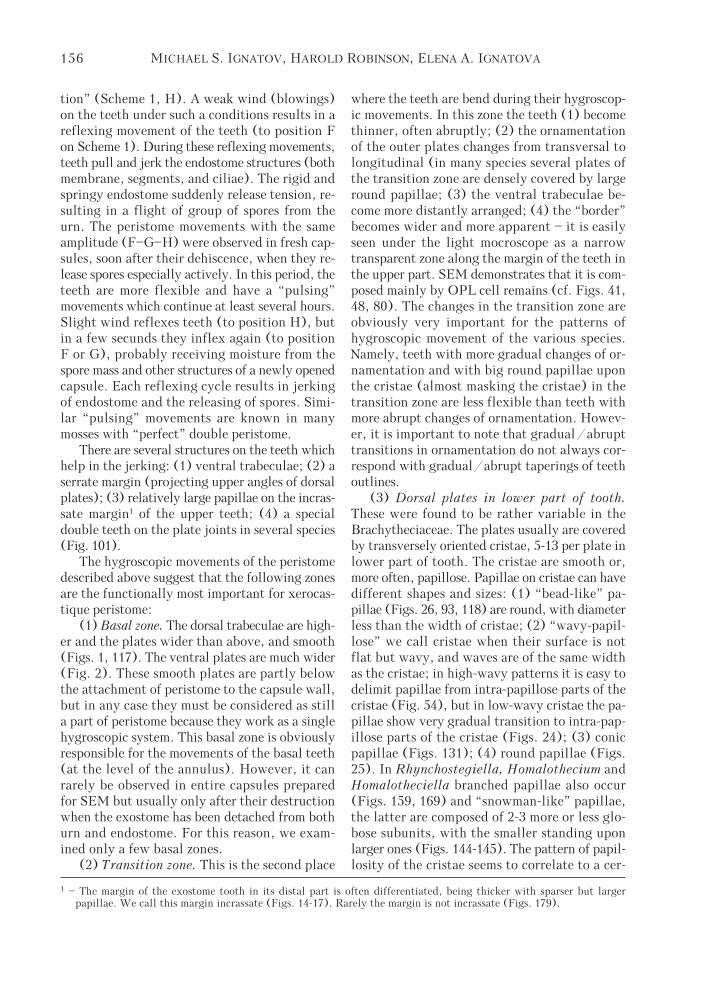
The hygroscopic movements of the peristomedescribed above suggest that the following zonesare the functionally most important for xerocas-tique peristome:
(1) Basal zone. The dorsal trabeculae are high-er and the plates wider than above, and smooth(Figs. 1, 117). The ventral plates are much wider(Fig. 2). These smooth plates are partly belowthe attachment of peristome to the capsule wall,but in any case they must be considered as stilla part of peristome because they work as a singlehygroscopic system. This basal zone is obviouslyresponsible for the movements of the basal teeth(at the level of the annulus). However, it canrarely be observed in entire capsules preparedfor SEM but usually only after their destructionwhen the exostome has been detached from bothurn and endostome. For this reason, we exam-ined only a few basal zones.
(2) Transition zone. This is the second place
where the teeth are bend during their hygroscop-ic movements. In this zone the teeth (1) becomethinner, often abruptly; (2) the ornamentationof the outer plates changes from transversal tolongitudinal (in many species several plates ofthe transition zone are densely covered by largeround papillae; (3) the ventral trabeculae be-come more distantly arranged; (4) the “border”becomes wider and more apparent – it is easilyseen under the light mocroscope as a narrowtransparent zone along the margin of the teeth inthe upper part. SEM demonstrates that it is com-posed mainly by OPL cell remains (cf. Figs. 41,48, 80). The changes in the transition zone areobviously very important for the patterns ofhygroscopic movement of the various species.Namely, teeth with more gradual changes of or-namentation and with big round papillae uponthe cristae (almost masking the cristae) in thetransition zone are less flexible than teeth withmore abrupt changes of ornamentation. Howev-er, it is important to note that gradual/abrupttransitions in ornamentation do not always cor-respond with gradual/abrupt taperings of teethoutlines.
(3) Dorsal plates in lower part of tooth.These were found to be rather variable in theBrachytheciaceae. The plates usually are coveredby transversely oriented cristae, 5-13 per plate inlower part of tooth. The cristae are smooth or,more often, papillose. Papillae on cristae can havedifferent shapes and sizes: (1) “bead-like” pa-pillae (Figs. 26, 93, 118) are round, with diameterless than the width of cristae; (2) “wavy-papil-lose” we call cristae when their surface is notflat but wavy, and waves are of the same widthas the cristae; in high-wavy patterns it is easy todelimit papillae from intra-papillose parts of thecristae (Fig. 54), but in low-wavy cristae the pa-pillae show very gradual transition to intra-pap-illose parts of the cristae (Figs. 24); (3) conicpapillae (Figs. 131); (4) round papillae (Figs.25). In Rhynchostegiella, Homalothecium andHomalotheciella branched papillae also occur(Figs. 159, 169) and “snowman-like” papillae,the latter are composed of 2-3 more or less glo-bose subunits, with the smaller standing uponlarger ones (Figs. 144-145). The pattern of papil-losity of the cristae seems to correlate to a cer-
1 – The margin of the exostome tooth in its distal part is often differentiated, being thicker with sparser but largerpapillae. We call this margin incrassate (Figs. 14-17). Rarely the margin is not incrassate (Figs. 179).

157Studies on the exostome of Brachytheciaceae (Musci)
tain extent with the environmental preferencesof individual species (and also corresponds some-what to their generic position). For example,branched papillae are known mainly in speciesgrowing in xeric areas; bead-like papillae on lowerdosral plates are found also in species with tem-perate to subtropical distribution, etc.
(4) Ventral trabeculae in the upper part oftooth. These are important structures for releas-ing of spores. They are especially diverse in shapeand ornamentation, and some of their charactersare more or less correlated with the systematicposition of the taxa. Ventral trabeculae typicallyend at 4-7 plates below teeth apices. However,in some Rhynchostegium and Eurhynchium spe-cies, and more rare in Brachythecium ones, theexostome teeth lack ventral trabeculae on 10-15distal plates. In the lower part of teeth, ventraltrabeculae are usually smooth (among xerocas-tique peristomes exceptions found in Brachyth-ecium rivulare and Eurhynchium asperisetum).In the transition zone some papillae appear on thetrabeculae and somewhat above the transitionzone the papillae are densest and highest. The de-scription of the ventral trabeculae in the upperteeth always refers to this area of strongest pap-illosity (usually at about 3/4–4/5 of the toothlength). Above this, the ventral trabeculae becomelower and usually less papillose (an exception isfound in some Rhynchostegium species, wherethe uppermost low trabeculae are more highly pap-illose than those below).
The three latter characters (2-4) were a mainfocus of our SEM studies.
Brachythecium (Figs. 3-48 & 70, 72, 74)Species studied: B. acuminatum (Hedw.) Aust.,
B. albicans (Hedw.) B. S. G., B. auriculatum Jaeg.,B. cirrosum (Schwaegr.) Schimp., B. complanatumBroth., B. erythrorrhizon B. S. G., B. falcatulum(Broth.) Par., B. lamprocarpum (C. Muell.) Jaeg.,B. mildeanum (Schimp.) Schimp. ex Milde, B. oe-dipodium (Brid.) Mitt., B. laetum (Brid.) B. S. G.,B. plumosum (Hedw.) B. S. G., B. populeum(Hedw.) B. S. G., B. reflexum (Starke) B. S. G., B.rivulare B. S. G., B. roteanum De Not., B. rutabulum(Hedw.) B. S. G., B. salebrosum (Web. et Mohr) B.S. G., B. starkei (Brid.) B. S. G., B. trachypodium(Brid.) B. S. G., B. velutinum (Hedw.) B. S. G.
Twenty-one species (i. e. 20-25% of those ingenus) were studied. The main structures of peris-tome are rather uniform.
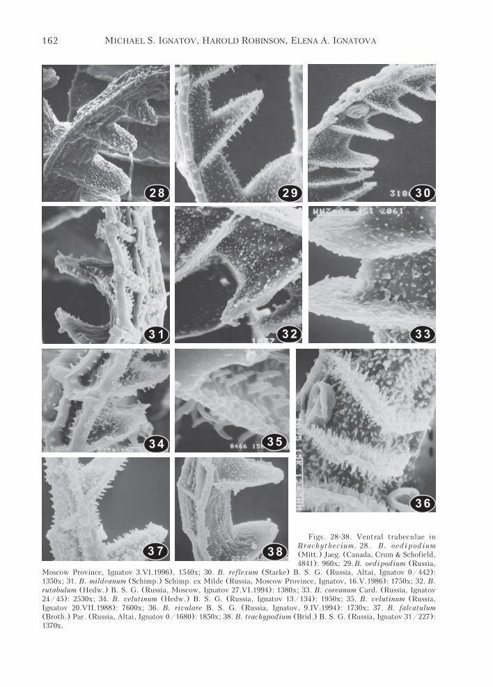
The teeth taper gradually in the transitionzone and form a wide arch (Figs. 3-6) or rarertaper rather abruptly, without an obvious arch(Figs. 10-12). In the upper part teeth are serru-late (rarely subentire). The median zig-zag line inthe lower half of teeth is not immersed. Thedorsal trabeculae in the proximal part of teethare smooth, somewhat exserted above the cristaelevel, rarely moderately exserted (B. oedi-podium), or partly sinking among cristae (B.plumosum, B. populeum, B. rivulare). The cristaeare dense and straight to slightly flexuose, smoothto relatively lowly wavy-papillose (typical vari-ant is illustrated in Fig. 24; exceptions are seenin B. complanatum, with loose and strongly flex-uose cristae; in B. falcatulum, with small rare“bead-like” papillae on the few lowermost plates,though most of the lower cristae are smooth; inB. rivulare, with cristae papillose almost to thebase). Towards the transition zone the papillae arealways become more prominent. The papillositypatterns are gradually changing from low-wavypapillose bolow to high-wavy papillose and then,in most of species dense round papillae totallymask the cristae on 1-2 plates in the transitionzone (cf. Figs. 18, 19, 21), rarely dense papillaecover more plates below the transition zone (inB. falcatulum, B. salebrosum, B. laetum, B. rivu-lare, B. mildeanum – cf. Figs. 22, 25). In somespecies the plates with transversal cristae havinghigh-wavy papillae are immediately changing toplates with longitudinal cristae, which are typi-cal for the upper part (cf. Figs. 20). In the upperpart the dorsal trabeculae are low and lowly pap-illose. The incrassate margins of teeth are usuallywell defined and less densely papillose than theplates, but often its papillae are larger, especiallyon the lateral surface. On the plates in upperteeth papillae are rare and arranged in longitudi-nal rows, or more rarely papillae are denser, mask-ing the longitudinal rows. The ventral trabeculaeare lowly semiorbicular, triangular to high-ligu-late, relatively thin to incrassate, with a smoothto rugose surface, and densely to spersely papil-lose, or smooth. The papillae are typically restrict-ed to several ventral trabeculae, and are rarelyfound below the transition zone (B. rivulare).
The ventral trabeculae are densely and evenlyhigh-papillose in B. velutinum and in the relatedB. falcatulum (Figs. 34-35, 37); B. rivulare (Fig.

158 MICHAEL S. IGNATOV, HAROLD ROBINSON, ELENA A. IGNATOVA
87
65
43
Figs. 3-8. Peristomes of Brachythecium: 3. B. populeum (Hedw.) B. S. G.. (Russia, Altai, Ignatov 1/16): 130x; 4.B. salebrosum (Web. et Mohr) B. S. G. (Sweden, Lule Lappmark, Hj. Moller): 120x; 5. B. rutabulum (Hedw.) B. S.G. (Russia, Moscow, Ignatov 27.VI.1994): 150x; 6. B. mildeanum (Schimp.) Schimp. ex Milde (Russia, MoscowProvince, Ignatov 16.V.1986): 120x; 7. B. velutinum (Hedw.) B. S. G. (Russia, Moscow Provice, Ignatov 20.VII.1988):115x; 8. B. plumosum (Hedw.) B. S. G. (Russia, Altai, Ignatov 16/29): 150.
36); B. acuminatum. In these species the papillaeare high, cylindric, about 1.5 x 0.3 μm, very similarto papillae commonly occurring in many Eurhynch-ium-species (cf. Figs. 57, 59, 60, 62 etc.).
However, 16 of 21 species studied have onlylow papillae on the ventral surface, or otherwisehave rows of high papillae only along the mar-
gin of the ventral plates and trabeculae (Figs.29, 45, 48).
Species without dense papillae on the ven-tral trabeculae can be classified into severalgroups, which more or less correspond to thesections of the genus that are accepted in mostof treatments of Brachythecium (cf. Broth-

159Studies on the exostome of Brachytheciaceae (Musci)
1 09
1 1 1 2
1 3
Figs. 9-13. Peristomes of Brachythecium: 9. B. lampro-carpum (C. Muell.) Jaeg. (Papua New Guinea, Koponen,34493): 100x; 10. B. acuminatum (Hedw.) Aust.(Macoun: Canadian Musci n° 282), 120x; 11. B. rivulareB. S. G. (U.S.A., Michigan, Robinson & Sharp, 20.VI.1995),85x; 12. B. oedipodium (Mitt.) Jaeg. (Russia, MoscowProvince, Ignatov 3.VI.1996): 115x; 13. B. coreanumCard. (Russia, Ignatov 24/45): 120x.
erus, 1925, Robinson, 1962, Takaki, 1955, etc.).Species of sect. Reflexa (B. oedipodium, B.reflexum, B. starkei – Figs. 28-30) have lingu-late to highly lingulate, rather thin ventral tra-beculae, with an almost smooth surface andsparse, small, low papillae, and sometimes withhigher thin papillae concentrated along the mar-gin of the inner plates and ventral trabeculae.Species of sect. Salebrosa (B. salebrosum, B.laetum, B. albicans, B. rotaeanum, B. erythror-rhizon) have low and semiorbicular relativelyincrassate ventral trabeculae, with ±rugulosesurfaces,which are moderately to sparsely low-ly papillose with papillae that are often unevenin shape (Figs 39-42, 44, 47). The group of spe-cies around B. rutabulum (B. rutabulum, B.
complanatum, B. mildeanum – Figs. 31-33) haveintermediately incrassate ventral trabeculae andsparse low papillae (more resembling those insect. Reflexa). The species of sect. Cirriphyl-lopsis (B. populeum, B. plumosum) have incras-sate semiorbicular ventral trabeculae, whichsometimes bear higher papillae, especially nearthe margin (Figs. 45-46, 48).
Especially deviating exostomes in Brachy-thecium were found in B. complanatum and B.rivulare. In B. complanatum the teeth are espe-

160 MICHAEL S. IGNATOV, HAROLD ROBINSON, ELENA A. IGNATOVA
1 4 1 5 1 6
1 7 1 8 1 9
20 2 1
Figs. 14-21. Upper (14-17) and middle (18-21) parts of teeth from outside. 14. B. rutabulum (Hedw.) B. S. G.(Russia, Moscow, Ignatov 27.VI.1994): 1400x; 15. B. populeum (Hedw.) B. S. G. (Russia, Altai, Ignatov 1/16):2000x; 16. B. erythrorrhizon B. S. G. (Russia, Ignatov & Bezgodov, 133): 1850x. 17. B. rivulare B. S. G. (Russia,Moscow Province, Ignatov, 4.5.1985): 2100x; 18. B. trachypodium (Brid.) B. S. G. (Russia, Ignatov 31/227): 1220x.19. B. populeum (l. c.): 1350x; 20. B. erythrorrhizon B. S. G. (Russia, Ignatov & Bezgodov, 133): 2300x. 21. B.falcatulum (Broth.) Par. (Russia, Altai, Ignatov 0/1680): 1400x.
Figs. 22-27. Lower parts of teeth from outside. 22. Brachythecium laetum (Brid.) B. S. G. (Crum & Anderson,Mosses of North America, n° 871): just below transition zone, 1300x; 23. B. populeum (Hedw.) B. S. G. (Russia,Altai, Ignatov 1/16): cristae demonstrating transition from clear striolate to papllose pattern, 10000x; 24. B.oedipodium (Mitt.) Jaeg. (Russia, Moscow Province, Ignatov 3.VI.1996): cristae in lower part – pattern typicalfor many other species, 3800x; 25. B. rivulare B. S. G. (Russia, Moscow Province, Ignatov, 4.5.1985): papillose

161Studies on the exostome of Brachytheciaceae (Musci)
22 23
24 25
26 2 7
cristae in lower part of tooth, 5400x; 26. B. falcatulum (Broth.) Par. (Russia, Altai, Ignatov 0/1680): “bead-likepapillae” on lowermost cristae, 3900x; 27. B. coreanum Card. (Russia, Altai, Ignatov 24/45): anomalous rare cristaein lower part of tooth, 3000x.
162 MICHAEL S. IGNATOV, HAROLD ROBINSON, ELENA A. IGNATOVA
2928 30
333 1 32
34 35
3 7
36
38
Figs. 28-38. Ventral trabeculae inBrachythecium. 28. B. oedi podium(Mitt.) Jaeg. (Canada, Crum & Schofield,4841): 960x; 29. B. oedipodium (Russia,
Moscow Province, Ignatov 3.VI.1996), 1540x; 30. B. reflexum (Starke) B. S. G. (Russia, Altai, Ignatov 0/442):1350x; 31. B. mildeanum (Schimp.) Schimp. ex Milde (Russia, Moscow Province, Ignatov, 16.V.1986): 1750x; 32. B.rutabulum (Hedw.) B. S. G. (Russia, Moscow, Ignatov 27.VI.1994): 1380x; 33. B. coreanum Card. (Russia, Ignatov24/45): 2530x; 34. B. velutinum (Hedw.) B. S. G. (Russia, Ignatov 13/134): 1950x; 35. B. velutinum (Russia,Ignatov 20.VII.1988): 7600x; 36. B. rivulare B. S. G. (Russia, Ignatov, 9.IV.1994): 1730x; 37. B. falcatulum(Broth.) Par. (Russia, Altai, Ignatov 0/1680): 1850x; 38. B. trachypodium (Brid.) B. S. G. (Russia, Ignatov 31/227):1370x.

163Studies on the exostome of Brachytheciaceae (Musci)
4 1
42 43
45
39 40
Figs. 39-48. Ventral trabeculae in Brachythecium. 39. B. salebrosum (Web.et Mohr) B. S. G. (Sweden, Hj. Moller 10.VII.1919): 1520x; 40. B. salebrosum(Russia, Altai, Ignatov 0/97): 2000x; 41. B. laetum (Brid.) B. S. G. (Crum &Anderson, Mosses of North America, n° 871): 2100x; 42. B. roteanum DeNot.: Russia, Altai, Ignatov, 9/159 (MHA): 1250x; 43. B. lamprocarpum (C.Muell.) Jaeg. (Papua New Guinea, Koponen 34493): 2000x; 44. B. albicans(Hedw.) B. S. G. (Sweden, Ystad, Ignatov): 2000x; 45. B. plumosum (Hedw.)B. S. G.: Taiwan, T. Koponen 16918): 1730x; 46. B. plumosum (Russia, Altai,Ignatov 16/29): 2800x; 47. B. erythrorrhizon B. S. G. (Russia, Ignatov &Bezgodov, 133): 2000x. 48. B. populeum (Hedw.) B. S. G. (Russia, Altai,Ignatov 1/16): 2000x.
46 4 7
48
44

164 MICHAEL S. IGNATOV, HAROLD ROBINSON, ELENA A. IGNATOVA
Figs. 49-56. Peristomes (49-53), outer surface of tooth in lower part (54), and outer surface of teeth in upper part(55-56) of Eurhynchium. 49. E. angustirete (Broth.) Kop. (Russia, Moscow Prov., Ignatov 29.IV.1985): 115x; 50. E.striatum (Hedw.) Schimp. (Russia, Krasnodar Territory, L. Vasil'eva, 16.VIII.1935): 115x; 51. E. pulchellum (Hedw.)Jenn. (Russia, Altai, Ignatov 0/1226): 115x; 52. E. hians (Hedw.) Sande Lac. (Russia, Pskov Province, Ignatov &Zolotov 20.X.1996): 80x; 53. E. praelongum (Hedw.) B. S. G. (Aslatia, Schimper): 220x; 54. E. striatum (l. c.):2300x; 55. E. praelongum (l. c.): 1500x; 56. E. angustirete (l. c.): 650x.
5 1
52
49 50
54
55 5653

165Studies on the exostome of Brachytheciaceae (Musci)
cially massive, slightly curved, with rather stoutdorsal trabeculae (Fig. 13), and cristae in lowerpart flexuose and very loose, exposing a rugosesurface (Fig. 27) [similar loose cristae we haveotherwise seen among other studied taxa onlyin Homalia trichomanoides (Hedw.) B.S.G.].
In B. rivulare the teeth are less hygroscopicand more papillose than in other species of thegenus. Round papillae are found on cristae fromthe transition zone far into the lower part, in onespecimen nearly to the base. In the upper part thedorsal plates are densely papillose, totally mask-ing the longitudinal cristae. The ventral trabecu-lae are densely papillose not only above, but alsoin the lower part of the teeth (Figs. 17, 25).
It is rather interesting, that the general charac-ters of the exostome remain the same in some trop-ical species, growing in quite different environ-ment, for example in B. lamprocarpum (Figs. 9).
Eurhynchium (Figs. 49-65)Species studied: E. angustirete (Broth.) Kop.,
E. asperisetum (C. Müll.) Bartr., E. hians (Hedw.)Sande Lac., E. praelongum (Hedw.) B. S. G., E. pul-chellum (Hedw.) Jenn., E. savatieri Schimp. ex Be-sch., E. schleicheri (Hedw. f.) Jur., E. striatum(Spruce) B. S. G., E. vagans (Jaeg.) Bartr.
The teeth are abruptly tapered in the transi-tion zone (cf. Figs. 49, 50, 52, 53), more rarelygradually tapered (E. pulchellum, Fig. 51). Inthe lower part the dorsal trabeculae are exserted(Fig. 54), more rarely not exserted above cristae(E. praelongum, E. pulchellum, E. savatieri).The cristae below are smooth or almost so (E.pulchellum, E. savatieri, E. schleicheri, E. va-gans), wavy-papillose (E. angustirete, E. praelon-gum, E. striatum), or more or less papillose (inE. asperisetum, E. hians). In the upper part theplates are papillose, without clear longitudinalrows of papillae (Figs. 54, 55), more rarely withpapillae in more or less distinct rows (E. schle-icheri, E. vagans). The margin is incrassate andweakly papillose. The ventral trabeculae are rel-atively high, lingulate to triangular, rarely low-ly semiorbicular (E. vagans), relatively thin inmost species, more rarely somewhat incrassateand rugulose (E. schleicheri). The papillae onthe ventral trabeculae show several patterns:(1) papillae moderately dense, high cylindric, to2.0 μm high, 0.3 μm in diameter, found in E.angustirete, E. striatum, E. praelongum, E. pul-chellum, E. savatieri; (2) papillae absent or
nearly so, found in E. hians and E. schleicheri;(3) papillae dense and low, seen in E. vagans;(4) upper ventral trabeculae densely unevenlypapillose, papillae spinulose to shortly conic, inlower part papillae on ventral trabeculae high-spinulose, found in E. asperisetum.
Thus, all the characters of the exostome ofEurhynchium are similar to those in Brachytheci-um, but in the former (1) the teeth are more com-monly abruptly tapered in the transition zone;(2) the dorsal plates in the upper teeth are morepapillose and usually without distinct longitudi-nal rows; (3) the ventral trabeculae are almostalways higher and thinner and often bear densehigh cylindric papillae (rare in Brachythecium).
Some infrageneric units are correlated withthe papillosity of ventral trabeculae. Species ofEurhynchium s. str. (E. striatum, E. angustirete,E. pulchellum, E. savatieri) have very dense andhigh papillae, whereas E. hians, classified oftenin the subgenus or genus Oxyrrhynchium (Bruch& al., 1851-55; Warnstorf, 1906; Brotherus, 1925)has smooth ventral trabeculae. However, charac-ter state in other species disagrees with the rec-ognition of Oxyrrhynchium as a distinct unit:E. asperisetum (close to E. hians) has papilloseventral trabeculae, whereas E. schleicheri (be-longing to Eurhynchium s. str.) has smooth ones.
Eurhynchium praelongum is often segre-gated in Kindbergia by many authors, butIgnatov (1998) failed to delimit it from Eu-rhynchium. The exostome of E. praelongumis similar to other species of this genus, espe-cially in their dense high papillae on the ven-tral trabeculae and in densely papillose dorsalplates in the upper tooth. The only differencefrom other Eurhynchium species includes therather limited flexibility of the transition zoneand the restricted hygroscopic movement(though this might be an artifact, because westudied the peristome of a 150 years old cap-sule, the only we could obtain for this study).Also, the character of flexibility may be of alimited taxonomic importance (cf. Brachyth-ecium rivulare).
Bryhnia (Figs. 66-68)Species studied: B. novae-angliae (Sull. et
Lesq.) Grout.The teeth are abruptly tapered in the transi-
tion zone. In the lower part the stout dorsaltrabeculae are somewhat exserted above the cris-

166 MICHAEL S. IGNATOV, HAROLD ROBINSON, ELENA A. IGNATOVA
64
5 7 58
59 60 6 1
62 63
Figs. 57-65. Ventral trabecu-lae of upper part of tooth (57-60, 62-65), ventral trabeculae oflower part of tooth (61) ofEurhynchium. 57. Eurhynchiumangustirete (Broth.) Kop. (Rus-sia, Moscow Prov., Ignatov29.IV.1985): 2900x; 58&61.E. asperisetum (C. Müll.) Bartr.(Papua New Guinea, T. Koponen30850): 2000x&1630x; 59. E.
65
striatum (Hedw.) Schimp. (Russia, Krasnodar Territory, L. Vasil'eva,16.VIII.1935): 5900x; 60. E. pulchellum (Hedw.) Jenn. (Russia, Altai,Ignatov 0/1226): 1830x; 62. E. praelonga (Hedw.) Ochyra (Aslatia,Schimper): 2400x; ; 63. E. savatieri Schimp. ex Besch. (Japan, Mockiziki,1143): 2950x; 64. E. hians (Hedw.) Sande Lac. (Russia, Pskov Province,Ignatov & Zolotov 20.X.1996): 1900x; 65. E. schleicheri (Hedw. f.)Jur. (C. Jensen, Musci Danici, 5.11.1899): 1500x.

167Studies on the exostome of Brachytheciaceae (Musci)
tae, the cristae are straight, smooth to lowly wavy-papillose. In the transition zone a few plates aredensely papillose, in the upper part the plates arelowly papillose, with papillae in longitudinal rows.The dorsal trabeculae are exserted, and the mar-gin is incrassate, lowly papillose, but relativelyhighly papillose in the uppermost non-trabecu-late zone. The ventral trabeculae are smooth tosparsely and lowly papillose.
The exostome character states are within thevariation of Brachythecium.
Cirriphyllum (Figs. 69, 71, 73)Species studied: C. piliferum (Hedw.) GroutThe teeth are abruptly tapered in the transi-
tion zone, and serrulate above. In the lower partthe dorsal trabeculae are lowly exserted abovethe cristae. The cristae are dense, smooth to lowlywavy-papillose. In the transition zone 4-5 platesare densely covered by large round papillae, andthe dorsal trabeculae here are relatively higher,and somewhat flexuose. In the upper part ofteeth the dorsal trabeculae are low, and the papil-lae are arranged in longitudinal rows. The mar-ginn is incrassate and sparsely lowly papillose.The ventral trabeculae are highly ligulate, thin,with sparse and low papillae.
The exostome character states of Cirri ph-yllum are within the variation of Brachytheci-um. However there are a number of differencesbetween C. piliferum and Brachythecum cirro-sum, a species often treated within Cirriphyl-lum. However according to Robinson (1962)and Ignatov (1998), the latter species should
not be placed in Cirriphyllum. The present studyadds two more differences between these spe-cies. In B. cirrosum the teeth are abruptly ta-pered in the transition zone, and the ventraltrabeculae have relatively high papillae (like inEurhynchium spp., Brachythecium velutinum,etc.). Cirriphyllum piliferum has gradually ta-pered teeth with 4-5 plates covered by round-papillae, and the ventral trabeculae are coveredby sparse and low papillae (cf. Figs. 69-74).
Myuroclada (Figs. 75-78)Species studied: M. maximowiczii (Borszcz.)
Steere et Schof.The teeth are abruptly tapered, and are serrulate
above. In the lower part the dorsal trabeculae arelowly exserted. In the transition zone 2-4 plates havedense, round papillae, masking the cristae. In theupper part the plates are papillose, without clearlongitudinal rows. The dorsal trabeculae are low andlowly papillose, and the margin is moderately in-crassate and papillose. The ventral trabeculae are semi-orbicular, incrassate, and densely conic-papillose.
The exostome character states are within thevariation of Brachythecium and are especiallysimilar to those in some Eurhynchium species.
Bryoandersonia (Figs. 79-81)Species studied: B. illecebra (Hedw.) Robinson.The teeth are gradually tapered in the transi-
tion zone and are serrulate above. In the lowerpart the dorsal trabeculae are lowly exserted abovethe cristae. In the transition zone 4-5 plates aredensely covered by large round papillae. The dorsaltrabeculae in the transition zone are relatively
66 6 7 68
Fig. 66-68. Bryhnia novae-angliae (Sull. et Lesq.) Grout (China, T. Koponen). 66. Peristome, 105x; 64. Outersurface of middle part of tooth, 1400x; 65. Inner surface in upper part of tooth, 1000x.

168 MICHAEL S. IGNATOV, HAROLD ROBINSON, ELENA A. IGNATOVA
Figs. 69-74. 69, 71, 73 – Cirriphyllum piliferum (Hedw.) Grout (Finland, Roivainen, 13.V.1961) and 70, 72, 74– Brachythecium cirrosum (Schwaegr.) Schimp. (China, Sichuan, Si He, 30410). 69&70 – peristome, 73&92x;71&72 – side view of upper tooth, showing inner trabeculae, 2950x&2570x; 73&74 – outer surface of tooth in middlepart, 850x&2700x.
69
7 0
7 1 7 2
7 3 7 4

169Studies on the exostome of Brachytheciaceae (Musci)
Figs. 75-78. Myurocladamaximowiczii (Borszcz.)Steere et Schof. (Verdoorn, Exs.n° 282): 75. Peristome, 127x;2. Outer surface of tooth in themiddle, 960x; 3 – outer sur-face of tooth in upper part,1600x; 4 – side view of tooth,showing inner trabeculae,1600x.
Fig. 79-81. Bryoandersoniaillecebra (Hedw.) Robinson(USA, Evans, X.1890): 79.Peristome, 85x; 80. Ventral sur-face and trabeculae, 2050x;81. Outer surface of tooth inthe middle, 640x.
7 5 7 6
7 7 7 8
7 9 80 8 1

170 MICHAEL S. IGNATOV, HAROLD ROBINSON, ELENA A. IGNATOVA
high, in the upper part the dorsal trabeculae areprominent, with round papillae as well sa thedistinctly incrassate margin. The plates are longi-tudinally striolate, and the cristae covered bysmall scattered papillae. The ventral trabeculaeare semiorbicular, somewhat incrassate, rugulose,with relatively high papillae near the margin ofthe inner plates and ventral trabeculae, and withlower papillae in their central (median) part.
The exostome character states are within thevariation of Brachythecium.
Scleropodium (Figs. 82-87)Species studied: S. caespitans (C. Müll.) L. Koch,
S. touretii (Brid.) L. Koch.In two species studied exostomes were found
different in many characters:
S. caespitans S. tourettii
teeth shape gradually tapered abruptly taperedmargins above entire serrulate
cristae in loose densetransition with sparse small with high-wavyzone papillae upon cristae papillose cristae
ventral with sparse very densely, highlytrabeculae low papillae conic-papillose
The diversity of exostome character states inScleropodium is within that of Brachythecium,but the former is a small genus and such heter-ogeneity could indicate a polyphyletic origin ofspecies presently included in Scleropodium. Fur-ther studies are needed to understand if thisgenus is monophyletic or not.
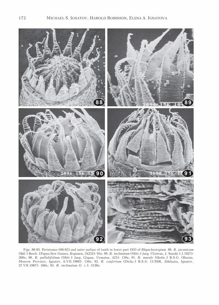
Rhynchostegium (Figs. 88-99 &101) andSteerecleus (Figs. 100)
Species studied: R. confertum (Dicks.) B.S.G.,R. herbaceum (Mitt.) Jaeg., R. inclinatum (Mitt.)Jaeg., R. javanicum (Bel.) Besch., R. murale(Hedw.) B.S.G., R. pallidifolium (Mitt.) Jaeg.,Steerecleus serrulatum (Hedw.) Robinson.
The teeth in Rhynchostegium are abruptlytapered in the transitional zone and are slightlyserrulate to subentire above. The lower part isrelatively massive, with a somewhat immersedmedian line. The ventral trabeculae end shortlyabove the transition zone, so that the uppermost,non-trabeculate part is relatively long (10-15plates). In the lower part the dorsal trabeculaeare shortly exserted above the cristae. The cris-tae below are dense, straight, wavy-papillose orwith bead-like papillae (R. pallidifolium, R. in-
clinatum, R. herbaceum – Fig. 93), further above,closer to the transition zone the cristae are pap-illose. In the upper part the dorsal trabeculae arerelatively low and papillose (the uppermost non-trabeculate part is more densely papillose onboth surfaces, Fig. 94). The margin is incrassateand papillose, the papillae on the plates are ar-ranged in longitudinal rows, or rarely not inrows. The ventral trabeculae are semiorbicular,slightly incrassate, evenly densely lowly papil-lose in most species (high-papillose in R. mu-rale), but the ventral plates are sometimes morestrongly papillose than the trabeculae. The up-permost low trabeculae are often more denselypapillose, with high, cylindric papillae.
Two species are peculiar in their exostomecharacters:
R. inclinatum. In the upper part of teeth theouter plate angles bear high papillae, which arepaired at the joint of the plates, forming “swal-low-tail”-shaped double papillae (Figs. 89, 101).This character is clearly seen under the com-pound miscrscope and can be used for the iden-tification of this species.
R. murale. The teeth are relatively graduallytapering upward in the transition zone, have anextensive area in the transition zone covered bybig round papillae (like in Brachythecium laetum,Fig. 22), and the ventral trabeculae in the upperpart have relatively dense high papillae (Fig. 99)[though another specimen studied had lower andsparser papillae on the ventral trabeculae, and higherpapillae were restricted to the uppermost non-trabeculate part].
As a whole, Rhynchostegium resembles Brachy-thecium, but many species are peculiar in theirvery abruptly tapering teeth, with a relativelyincrassate lower part (so median line somewhatimmersed), long uppermost non-trabeculate part,and relatively uniform, thin semiorbicular ven-tral trabeculae with evenly arranged low papil-lae. Rhynchostegium differs from Platyhypnid-ium by teeth that are massive only below and inthe long, narrow and delicateupper part.
Steerecleus serrulatus was traditionally placesin Rhynchostegium, but Robinson (1987) segre-gated it and some other neotropical species in anew genus Steerecleus. The present study supportthis idea, since S. serrulatus would be anomalousin Rhynchostegium with its strongly adaxiallypapillose teeth, more resembling Eurhynchium.

171Studies on the exostome of Brachytheciaceae (Musci)
Fig. 82-87. Scleropodium. 82, 84, 86 – S. caespitans (C. Muell.) L. Koch (Canada, Schofield 20410); 83, 85, 87– S. tourettii (Brid.) L. Koch (USA, California). 82&83. Peristomes, 176x&108x; 84&85. Side view of teeth, showinginner trabeculae, 2400x&1600x; 86&87. Outer surface of teeth in transition zone, 1040x&1600x.
82 83
84 85
86 8 7
172 MICHAEL S. IGNATOV, HAROLD ROBINSON, ELENA A. IGNATOVA
88 89
90 9 1
92 93
Figs. 88-93. Peristomes (88-92) and outer surface of tooth in lower part (93) of Rhynchostegium. 88. R. javanicum(Bél.) Besch. (Papua New Guinea, Koponen, 34252): 95x; 89. R. inclinatum (Mitt.) Jaeg. (Taiwan, J. Suzuki 1.I.1927):260x; 90. R. pallidifolium (Mitt.) Jaeg. (Japan, Uematsu, 425): 150x; 91. R. murale (Hedw.) B.S.G. (Russia,Moscow Province, Ignatov, 6.VII.1988): 150x; 92. R. confertum (Dicks.) B.S.G. (USSR, Abkhazia, Ignatov,27.VII.1987): 160x; 93. R. inclinatum (l. c.): 3130x.

173Studies on the exostome of Brachytheciaceae (Musci)
Platyhypnidium (Figs. 102-113)Species studied: P. aquaticum (Hampe) Fleisch.,
P. austrinum (Hook. f. et Wils.) Fleisch., P. muelleri(Sande Lac.) Fleisch., P. riparioides (Hedw.) Dix.
The important character state of all of spe-cies studied is the strongly incrassate teeth, dueto additional deposits of cell-wall material ofboth OPL and PPL. Most material is deposit-ed on the central part of the OPL cell wall, sothe columns of the OPL' cell remains appearsomewhat convex whereas the median line ap-pears somewhat immersed. In the lower tooththe latter is slightly zig-zag-shaped, or in P.
austrinum straight. Hygroscopic movements inPlatyhypnidium are more limited and unusual-ly slow comparatively with Rhynchostegiumor Eurhynchium. In addition, the following arecharacteristic for all species of Platyhypnidi-um. In the lower teeth the cristae are smooth. Inthe transition zone, several plates have dense largepapillae. In the upper tooth, longitudinal rows ofpapillae on the outer plates are indistinct tolacking. Finally, the ventral trabeculae are rela-tively densely and lowly papillose.
Platyhypnidium austrinum is the most pe-
94
95
96
9 7 98
99 100 101
Figs. 94-101. Upper parts of teeth of Rhynchostegium and Steerecleus (94 – uppermost non-trabeculate part; 95,101 – outer surface; 96-100 – side views showing ventral trabeculae). 94. R. javanicum (Bél.) Besch. (Papua NewGuinea, Koponen, 34252): 865x; 95. R. confertum (Dicks.) B.S.G. (USSR, Abkhazia, Ignatov, 27.VII.1987): 1650x;96. R. pallidifolium (Mitt.) Jaeg. (Japan, Uematsu, 425): 1350x; 95. R. murale (Hedw.) B.S.G. (Russia, MoscowProvince, Ignatov, 6.VII.1988): 150x; 97. R. javanicum (l. c.): 1450x; 98. R. confertum (l. c.): 1650x; 99. R. murale(l. c.): 1700x; 100. Steerecleus serrulatum (Hedw.) Robinson (U.S.A., Norris 11456): 2180x; 101. R. inclinatum(Mitt.) Jaeg. (Taiwan, J. Suzuki 1.I.1927): 1900x.

174 MICHAEL S. IGNATOV, HAROLD ROBINSON, ELENA A. IGNATOVA
109
110
102 103
104
105 106
107
108
Figs. 102-110. Platygypnidium. 102. – Platyhypnidium aquaticum(Hampe) Fleisch. (Bolivia, R.S.Williams, 2020); 103, 106, 108-109 –P. austrinum (Hook. f. et Wils.) Fleisch. (New Zealand, Beckett 12/84,484); 104-105, 107, 110 – P. muelleri (Sande Lac.) Fleisch. (PapuaNew Guinea, Koponen, 30296). 102&103. Perostomes, 95x&140x. 104.Outer surface of tooth in upper part, 1700x; 105&106. Ventral surfaceand trabeculae, 2000x&1700x; 107. Outer surface of tooth in the middle,1500x; 108. Side view of tooth in lower part, showing small papillae oncristae, 2450x; 109. Outer surface of tooth in lower part showing im-mersed median line, 1750x; 110. Outer surface of tooth in lower part,1700x.

175Studies on the exostome of Brachytheciaceae (Musci)
culiar species in its clearly straight medianline and relatively slightly curved teeth (Figs.103, 109), and in its more strongly papilloseventral trabeculae (Fig. 106). This species hasalso the most special gametophytic charactersin the genus.
Differences between the species include char-acters of the dorsal trabeculae in the lower partof the teeth (exserted above cristae in P. ripar-ioides and P. aquaticum, not-exserted in P.austrinum and P. muelleri) and the dorsal up-per trabeculae (much higher in P. riparioidesthan in the other species).
Palamocladium (Figs. 114-123)Species studied: P. euchloron (C. Muell.) Wijk
et Marg., P. leskeoides (Hook.) Britt.Both species studied have unique character
states not observed in the other Brachytheciace-ae. Relatively high and solid longitudinal cristaeon the outer surface of the upper tooth (moresimilar to the cristae of the lower teeth, than tothe longitudinal ridges of the upper teeth ofBrachythecium). Both species have also relative-ly massive teeth that gradually taper in the tran-sition zone and are joined by 5-13 basal plates.The dorsal trabeculae in the lower teeth are high,reaching upwards above the middle. In the lowerpart the cristae have small bead-like papillae, inthe upper part the longitudinal cristae are distinctand high, smooth or covered by sparse, smallpapillae. In the uppermost, non-trabeculate part of
Fig. 111-113. Platyhypnidium ri parioides (Hedw.)Dix. (USA, Reed, 64277). 111 – peristome, 130x; 112 –outer surface of tooth in upper part, 2000x; 113 – sideview of tooth, showing ventral trabeculae, 1650x.
111 112
113
the teeth the papillae are denser. The dorsal trabe-culae are low, with relatively high conic papillae,and the margin is moderately incrassate and highlyconic papillose. The ventral trabeculae are trian-gular, incrassate, unevenly papillose, with spinu-lose to short-conic papillae.
In P. leskeoides the marginal zone of teethin the lower and middle parts is exceptionallythin and wide, forming “wings”. This featurewas not observed in P. euchloron and wasnot reported for P. leskeoides by Hofmann(1997), who illustrated American specimen ofthis species.
The exostomes of Palamocladium euchloronand P. leskeoides differ in the extensive transi-tion zone of the former, with many plates havinghigh and dense papillae that mask the cristae,whereas in P. leskeoides the transition from trans-versal to longitudinal cristae is very abrupt (Figs.118, 122).

176 MICHAEL S. IGNATOV, HAROLD ROBINSON, ELENA A. IGNATOVA
Fig. 114-123. Pala-mocladium. 114-120 –P. euchloron (C.Muell.) Wijk et Marg.(USSR, Adzharia, On-i pchenko, 2.II.1979);121-123. P. leskeoides(Hook.) Britt. (China, T.Koponen, 46384). 114.Peristome, 105x; 115&116. Outer surface ofupper parts of tooth,2100x; 117. Outer sur-face of lowermost partof tooth, showingsmooth basal plates,2660x; 118. Outer sur-face of lower part of
120
121
123
114 115 116
117 118 119
122
tooth, 2660x; 119. Side view of ventral trabecula, 3370x; 120. Outer surface of lower part of tooth, 980x; 121.Peristome, 210x; 122. Outer surface of tooth in upper part, 1330x; 123. Basal parts of teeth, showing wing-like thinedges of teeth, 1140x.

177Studies on the exostome of Brachytheciaceae (Musci)
Figs. 124-127. Camptothecium lutescens (Hedw.) B. S. G.(British Mosses, J. G. Baker): 124 – peristome, 96x; 125 –outer surface of middle tooth, 960x; 126 – side view ofupper tooth, showing inner trabeculae, 1600x; 127 – papil-lae of inner trabeculae, 4100x.
Fig. 128-132. Camptotheciumpinnatifidum (Sull. et Lesq.) Sull.(U.S.A., Norris 17.I.1982). 128. Peris-tome, 125x; 129. Side view of uppertooth, showing inner trabeculae, 2400x;130. Outer surface of upper tooth,1870x; 131. Outer surface of lowertooth, 1890x; 132. Outer surface oftooth above transition zone, 1890x.
124
125
126
127
128 130
131 132
129

178 MICHAEL S. IGNATOV, HAROLD ROBINSON, ELENA A. IGNATOVA
Fig. 133-136. Homalothecium laevisetum (Hedw.) B. S. G.(Japan, H. Mayr, 3). 133. Ventral surface and trabeculae,3110x; 134. Outer surface of tooth in transition zone, 1260x;135. Outer surface of tooth above transition zone, 3150x;136. Outer surface of lower tooth, 2300x.
Homalothecium and Camptothecium (Figs.124-155)
Species studied: Camptothecium lutescens (Hedw.)B. S. G., C. pinnatifidum (Sull. et Lesq.) Sull., Homa-lothecium aeneum (Mitt.) Lawt., H. laevisetum SandeLac., H. nuttallii (Wils.) Jaeg., H. philippeanum(Spruce) B. S. G., H. sericeum (Hedw.) B. S. G.
We studied five species of Homalotheciums. str. and to of Camptothecium (all sometimesconsidered within Homalothecium s. lat.). Peris-tomes were found to differ considerably betweenand within this genera. Homalothecium serice-um, H. philippeanum, H. laevisetum, H. nuttalii,H. aeneum have hygrocastique peristomes (withteeth straight when wet), whereas Camptotheci-um lutescens (Homalothecium lutescens) and C.pinnatifidum (H. pinnatifidum) have xerocas-tique peristomes (teeth covering the urn whenwet). The two latter species are similar to eachanother in their curved capsules, well-developedendostome and many structural characters of theexostome: (1) in the lower part the dorsal trabe-culae are moderately high, thin, smooth; (2) in thelower part the cristae are dense and wavy; (3) inthe transition zone the plates are densely papil-lose, with large round papillae; (4) in the upperpart the papillae are arranged in longitudinal rows;(5) the incrassate margin is differentiated; (6)the ventral trabeculae are semiorbicular-triangu-lar, moderately incrassate, and have high cylin-dric papillae (Figs. 124-132).
Contrary to this, the five other studied spe-cies of Homalothecium have straight capsules,
strongly reduces endostomes, and the states ofthe exostome characters mentioned above arenearly totally different. However these 5 spe-cies themselves exhibit strong diversity, whichcan be understood as a series of reductions asfollows:
I. Homalothecium laevisetum (Figs. 133-136)has the most derived exostome with: (1) veryhigh and stout dorsal trabeculae, up to 2/3 ofteeth length or more; (2) smooth lower dorsalplates and trabeculae, becoming papillose onlyabove the transition zone; (3) high and branchedpapillae in upper tooth, not arranged in longitu-dinal rows; (4) low-semiorbicular, much incras-sate, rugose and densely papillose ventral trabec-ulae, with irregular-shaped large papillae.
II. Homalothecium philippeanum (Figs. 137-141) has also high dorsal trabeculae, but the platesin the lower teeth are distinctly striolate withcristae covered by small bead-like papillae. In theupper part the papillae are arranged in longitudi-nal rows.
III. Homalothecium sericeum (Figs. 142-146)is similar to two above species in its high dorsaltrabeculae in most parts of the tooth, but differs
133 134 135
136

179Studies on the exostome of Brachytheciaceae (Musci)
137
138
140 141
142 143
144
145 146
139
Fig. 142-146. Homalo-thecium sericeum (Hedw.)B. S. G. (Brotherus, Bryo-theca Fennica). 142. Partof peristome, 350x; 143.Side view of tooth, 720x;144. Ventral trabeculae,2420x; 145. Outer surfaceof upper tooth, 2300x;146. Outer surface of up-per tooth, 2900x;
Fig. 137-141. Homalotheciumphilippeanum (Spruce) B. S. G.:USSR (Crimea, Belyanina,3.V.1985). 137. Outer surface ofupper tooth, 105x; 138. Ventraltrabecula, 5320x; 139. Part ofperistome, 300x; 140. Outer sur-face of middle tooth, 1400x; 141.Outer surface of lower tooth,1450x.

180 MICHAEL S. IGNATOV, HAROLD ROBINSON, ELENA A. IGNATOVA
Fig. 147-150. Homalothecium nuttallii (Wils.) Jaeg.(Crum & Anderson, Mosses of North America, n° 807):147. Peristome, 120x; 148. Outer surface of middle tooth,1060x; 149. Side view of tooth, showing inner trabeculae,1340x; 150. Outer surface of upper teeth, 830x.
in having densely papillose dorsal trabeculae. Itis similar to H. philippeanum in having cristaeon plates below, but differs in having high andbranched papillae – they are high and branched(bead-like in H. philippeanum).
IV. Homalothecium nuttalii (Figs. 147-150)has high dorsal trabeculae only in the lowerpart of tooth. The dorsal plates are densely pap-illose with high papillae below (very rarelybranched). The upper part of the tooth has in-conspicuous dorsal trabeculae not exserctedabove dense papillae, and the ventral trabeculaeare semiorbicular above, relatively thin, and havesmall low papillae.
V. Homalothecium aeneum (Figs. 151-155)has very low dorsal trabeculae (not exsertedabove the cristae or papillae level) in all parts ofthe tooth. The cristae are straigth below, not verydense, slightly flexuose, and wavy to nearlysmooth. In the upper part of the tooth low papil-
lae are arranged in longitudinal rows. The ventraltrabeculae are semiorbicular, slightly incrassate,and sparsely papillose, and the uppermost non-trabeculate part is very lowly papillose or rugu-lose on both surfaces.
Taxa with rather complete xerocastique peris-tomes and curved capsules are probably bettersegregate in Camptothecium, as did Bruch & al.(1851-55). The diversity of taxa with stronglyderived peristomes need further studies. It isprobable that several lines of peristome reduc-tions will be found, so that the genus may needfurther splitting into smaller and more homoge-neous entities.
Homalotheciella (Figs. 156-159)Species studied: H. subcapillata (Hedw.) Broth.Exostome ornamentation of H. subcapil-
lata is rather similar to that of Homaloth-ecium sericeum. Both have very gradually ta-pered teeth, the dorsal trabeculae are highlyexserted in at least 2/3 of the tooth length.The cristae and trabeculae are densely cov-ered by branched papillae, in the upper partalso densely papillose with branched papillaethat are not arranged in longitudinal rows.The margin is not incrassate and the ventraltrabeculae are lowly semiorbicular, incrassate,and papillose. Differences between Homal-otheciella and Homalothecium sericeum in-clude more dense papillae on outer plates inthe former, making cristae hardly discernible,
147 149
150
148

181Studies on the exostome of Brachytheciaceae (Musci)
Figs. 156-159. Homalotheciella subcapillata (Hedw.)Broth. (USA, Holzinger, I.1892). 156. Peristome, 236x; 157.Side view of upper tooth, showing inner trabeculae, 2400x;158. Outer surface of upper tooth, 2000x; 159. Branchedpapillae on the outer surface of middle tooth, 15300x.
151 152 153
154 155
156
157
158 159
Fig. 151-155. Homalotheciumaeneum (Mitt.) Lawt. (USA,Ignatov & Norris, 12.VIII.1989).151. Peristome, 170x; 152. Ven-tral trabeculae, 2100x; 153.Outer surface of tooth in up-permost part, 2660x; 154. Outersurface of lower tooth, 3500x;155. Outer surface of toothabove transition zone, 2660x;

182 MICHAEL S. IGNATOV, HAROLD ROBINSON, ELENA A. IGNATOVA
and also less papillose ventral trabeculae. Bythe presence of branched papillae Homaloth-eciella is also resembling some tropical andsubtropical Rhynchostegiella species (R. brac-hypodia, R. papuensis, R. leptoneura).
Rhynchostegiella and Eurhynchiella (Figs.160-190)
Species studied: Eurhynchiella zeycheri (C.Muell.) Fleisch., Rhynchostegiella brachypodia
Fleisch., R. leptoneura Card., R. menadensis (Lac.)Bartr., R. papuensis Bartr., R. teesdalei (B. S. G.)Limpr., R. tenella (Dicks.) Limpr. [and Scorpiuri-um cucullatum (Mitt.) Hedenäs (= E. cucullatum(Mitt.) Steere et G.A.M.Scott)].
These species is worth discussing togetherfor the following reason. Fleischer (1923) out-lined the strong heterogeneity of Rhynchoste-giella and suggested the segregate Eurhynchiel-
160
161
162 163
164 165
Fig. 160-165. 160, 162-164 – Rhynchostegiella brachypodia Fleisch. (Papua New Guinea, Norris, 61083); 161, 165 –R. papuensis Bartr. (Nederlands Nieuw-Guinea, van Zanten, 571). 160 & 161. Peristomes, 165x for both; 162. Outersurface of upper tooth, 2800x; 163 – side view of upper tooth, showing inner trabeculae, 2870x; 164 & 165 – outersurface of lower tooth, 3220x & 4630x.

183Studies on the exostome of Brachytheciaceae (Musci)
Figs. 166-169. Rhynchostegiella leptoneura Card. (China, Yunnan, Magill & al., 7830). 166. Peristome, 120x; 167.Outer surface of middle tooth, 1450x; 168. Side view of upper tooth, 1200x; 169. Ornamentation of outer surface ofmiddle tooth, 5600x.
la for small rather rigid plants, with short thick-walled laminal cells, and a costa ending in aspine (retaining in Rhynchostegiella plants ofmore delicate stature, with long, thin-walled lam-inal cells, and a costa that does not not end in aspine). According to this concept, Rhynchoste-giella papuensis and Scorpiurium (Eurhynchi-um) cucullatum should be placed in Eurhynchiel-la. But if so, the diversity of peristomes withinEurhynchiella will be enormously great. More-over, the advanced characters in Eurhynchiellazeycheri, the type of the genus (Ignatov & al.,1998), are quite different from the advanced char-acters in R. papuensis. Obviously, at least thesetwo taxa represent two different lines of evolu-
tion and their correct generic classification needfurther studies.
Species of this group represent a series ofperistome reduction, in some respects similar tothose in Homalothecium. The perisomes are hy-grocastique and xerocastique, some species havedense branched papillae on dorsal plates and tra-beculae in lower tooth, other ones have bead-likeor nearly smooth cristae.
Interestingly, the relation of papillosity-pat-tern to xero-/hygrocastique movements is almostopposite to that in the Homalothecium-Camp-tothecium group. Species with especially strong-ly papillose cristae and dorsal trabeculae exhibitxerocastique movements, whereas hygrocastique
166
167
169168

184 MICHAEL S. IGNATOV, HAROLD ROBINSON, ELENA A. IGNATOVA
movement is observed only in one species,Rhynchostegiella tenella, which has a rela-tively “hypnoid” peristome. In its peristomestructure the latter species is closest to R.teesdalei, which has a xerocastique peristome.Certainly the structural basis for movementsof peristomes need much more studies.
The exostome structure of the species canbe classified in four types as follows:
1. Rhynchostegiella brachypodia, R. lep-toneura, R. papuensis (Figs. 160-169)
The teeth are abruptly tapered in the tran-sition zone and serrulate above. In the drycondition they are somewhat twisted and
“hooked” around the endostome segments (cf. Figs.160, 161, 166). In the lower part the dorsal trabec-ulae are highly exserted above the cristae, withhigh, branched and “snowman”-shaped papillae.Above the transition zone the papillae are sparserand lower, conic to “snowman”-shaped, and at someplaces longitudinal or vermicular cristae are seen. Inthe upper part the dorsal trabeculae are low andpapillose. The margin is incrassate (R. papuensis)or not incrassate (R. brachypodia, R. leptoneura).The ventral trabeculae are lowly semiorbicular, mod-erately to strongly incrassate, and are densely tomoderately densely, highly papillose.
2. Rhynchostegiella tenella (Figs. 170-176) andR. teesdalei (Figs. 177-181)
The teeth are ±abruptly narrowed in the transi-tion zone and serrulate above. The median line iszig-zag-shaped below, somewhat immersed. In thelower part the dorsal trabeculae are lowly exsertedabove the cristae level (R. tenella) or not exserted
170 171 172
173 174 175
176
Figs. 170-176. Rhynchostegiella tenella (Dicks.) Limpr.(Europe, Rabenhorst): 170. Peristome, 135x; 171. Side view oftooth, showing inner trabeculae, 1500x; 172 – outer surfaceof upper tooth, 1720x; 173. Outer surface of lower tooth,2660x; 174. Lower part of transition zone, 1400x; 175. Outersurface of upper part of transition zone, 1740x; 176. Cristae inlower tooth, 5250x.

185Studies on the exostome of Brachytheciaceae (Musci)
(R. teesdalei). The cristae have scattered small,bead-like papillae near the base, further upslightly wavy to nearly smooth and towardsthe transition zone with large conic-wavy pa-pillae on the cristae. The cristae are oblique in1-2 plates in the transition zone. In the upperpart the plates are lowly papillose with the papil-lae arranged in indistinct rows, or in the upper-most, non-trabeculate part with sparse papillae onthe smooth surfaces. The dorsal cristae are lowand lowly papillose in upper part. The margin ismoderately incrassate. The ventral trabeculae arehigh, ligulate-triangular, incrassate, and densely(R. tenella) or moderately densely (R. teesda-lei) covered by irregular papillae (thinly spinu-lose, conic, rarely branched).
3. Rhynchostegiella menadensis, Scorpiuri-um cucullatum (Figs. 182-186)
The teeth are abruptly tapered in the transitionzone and serrulate above. In the dry condition theyare slightly to moderately twisted and form a“hooks” around the endostome segments. In thelower part the dorsal trabeculae are exserted abovethe cristae and low-papillose (R. menadensis), ornot exserted (S. cucullatum). The cristae arestraight and covered with small papillae. In the
upper part the dorsal trabeculae are low and papil-lose, and the plates have low papillae, that are ar-ranged in indistinct longitudinal rows. The marginis incrassate and lowly -papillose (in R. menaden-sis the papillae near the joints of plates are higherand sometimes form “double”-teeth). The ventraltrabeculae are semiorbicular and moderately tostrongly incrassate, rarely lowly papillose.
4. Eurhynchiella zeycheri (Figs. 187-190)The teeth are massive, slightly curved when
dry, abruptly tapered in transition zone andserrulate above. In the lower part the teeth areincrassate, with a somewhat immersed medianline that is zig-zag-shaped to almost straight.In the lower part the dorsal trabeculae arestout, exserted above the cristae and distinct-ly projecting from the sides of the teeth. Thecristae are very dense, straight, and smooth toslightly wavy. In the transition zone the cristaeare densely papillose. In the upper part the dor-sal trabeculae are exserted, with high-conic pa-pillae. The plates have high papillae arranged inindistinct longitudinal rows. The margin is dis-tinctly incrassate, with relatively sparse highpapillae (papillae near the joint of plates higherand sometimes forming “double-tooth”). The
177 178
180 181
179
Figs. 177-181. Rhyncho-stegiella teesdalei (Kindb.)Limpr. (Fleischer & Warn-storf, Bryotheca Europ. me-ridion. 94): 177. Peristome,205x; 178. Side view of tooth,showing ventral trabeculae,1960x; 179. Outer surface ofupper tooth, 1970x; 180.Outer surface of lower tooth,1750x; 181. Outer surface oftooth at transition zone,1550x.

186 MICHAEL S. IGNATOV, HAROLD ROBINSON, ELENA A. IGNATOVA
182 183
184 185 186
187
189
188
190
Figs. 182-190. 182, 184-185 – R. mena-densis (Lac.) Bartr. (Papua New Guinea,Streimann & Naomi, 15014). 183 & 186– Scorpiurium cucullatum (Mitt.)Hedenäs (Australia, Watts, 8443); 187-190 – Eurhynchiella zeycheri (C. Müll.)Fleisch. (Natal, Rehmann). 182, 183 & 187.Peristomes, 205x, 195x & 195x; 184 &189. Outer surface of lower tooth, 2600x& 1800x; 185, 186 & 190. Side views ofupper tooth, showing inner trabeculae,1400x, 1750x & 1300x; 188. Outer sur-face of upper tooth, 3480x.

187Studies on the exostome of Brachytheciaceae (Musci)
ventral trabeculae are semiorbiculate and dense-ly covered by thin papillae.
The great diversity of exostome structurefound in the few species studied of Rhynchost-egiella s. l. do not allow us to draw conclusionsother than that this genus is very heterogeneousand needs more studies.
CONCLUSIONS
1. Most genera of the Brachytheciaceae havea relatively uniform structure of their exos-tomes, hardly exceeding the variability foundwithin Brachythecium: (Bryoandersonia, Bryh-nia, Camptothecium, Cirriphyllum, Eurhynch-ium, Kindbergia, Myuroclada, Pseudocirri-phyllum, Rhynchostegium, Scleropodium). Atthe same time there are some relatively stableminor characters states (especially in the struc-ture of the ventral trabeculae in the upper partof teeth), which are more or less stable withinsome small natural groups of large genera (e. g.in sections of Brachythecium ).
2. The exostomes are especially advanced andheterogeneous in Homalothecium and Rhyncho-stegiella. Even after the segregation of Camp-tothecium as a genus with the peristome almostof the Brachythecium-type, Homalothecium rep-resents several contrasting pattern. Rhynchoste-giella also exhibits several distinct types of exos-
tomes. The systematics of these genera should notbe based on the gametophytic characters only. Adescription of the peristomes simply as “reduced”often include clearly different lines of evolution.
3. Scleropodium caespitans and S. tourettiishow many differences in their exostome struc-tures and may not belong to one genus.
4. Platyhypnidium species have several fea-tures which provide additional evidence for itssegregation from both Rhynchostegium and Eu-rhynchium.
5. Xero/hygrocastique movements of the ex-ostome are not clearly correlated with the pat-terns of ornamentation. The more or less “hyp-noid” peristome of Rhynchostegiella tenella ishygrocastique, whereas the much “reduced” ex-ostomes with branched papillae (e. g. in R. brac-hypodia and R. papuensis) are xerocastique.
ACKNOWLEDGMENTS
We are grateful for the curator of H for thepermission of use some collection for SEM stud-ies and to Lars Hedenäs for critical readingand suggestions on the manuscript. The studyof Ignatov has been supported by a Smithso-nian Institution Short-Term Visitor appoint-ment and the study of Ignatov and Ignatovaby the Russian Foundation of Scientific Re-searches, 96-04-48033.
LITERATURE CITED
BROTHERUS, V. F. 1925. Musci. – In: Engler, A. &Pranl, K (eds.), Die Natürlichen Pflanzenfamilien, ed.2, 11: 1-522. W. Engelmann, Leipzig.
BRUCH, PH., W. PH. SCHIMPER & TH. HUEMBEL1851-55. Bryologia Europaea seu genera muscorum Eu-ropaeorum. Stuttgartiae.
BUCK, W. R. 1980. A generic revision of the Entodonta-ceae. – J. Hattori Bot. Lab. 48: 71-159.
FLEISCHER, M. 1923. Die Musci der Flora von Buiten-zorg 4: 1105-1729. E. J. Brill., Leiden.
HOFMANN, H. 1997. A monograph of the genus Palam-ocladium (Brachytheciaceae, Musci). – Lindbergia22: 3-20.
IGNATOV, M. S. 1998. Bryphyte flora of Altai Moun-tains. IIII. Brachytheciaceae. – Arctoa 7:
IGNATOV, M. S., H. ANDO & E. A. IGNATOVA 1996.Bryophytes of Altai Mountains. VII. Hypnaceae andrelated pleurocarps with bi- and ecostate leaves. – Arc-toa 6: 21-112.
IGNATOV, M. S., KOPONEN, T. & NORRIS, D. H. 1998.Bryophyte folra of Huon Peninsula, Papua New Guinea.000. Brachytheciaceae. – Acta Bot. Fennici (in press).
MUELLER, D. M. J. & A. J. NEUMANN 1988. Peristome
structure and the regulation of spore release in arthrod-ontous mosses. – Advances Bryol. 3: 135-158.
ROBINSON, H. 1962. Generic revisions of North Ameri-can Brachytheciaceae. – Bryologist 65: 73-146.
ROBINSON, H. 1987. Nots on generic concepts in theBrachytheciaceae and the new genus Steerecleus. –Mem. New York Bot. Gard. 45: 678-681.
ROHRER, J. R. 1985a. A phenetic and phylogenetic anal-ysis of the Hylocomiaceae and Rhytidiaceae. – J. Hat-tori Bot. Lab. 59: 185-240.
ROHRER, J. R. 1985b. A generic revision of the Hylocomi-aceae. – J. Hattori Bot. Lab. 59: 241-278.
STEINBRINCK, O. 1897. Der hygroscopische Mechanis-mum des Laubmoosperistoms. – Flora 84: 131-158.
TAN, B. C. & H. ROBINSON 1990. A revision of Philip-pine Hookeriaceous taxa (Musci). – Smithsonian Con-tr. Bot. 75: 1-41.
TAKAKI, N. 1955. Researches on the Brachytheciaceae ofJapan and its adjacent areas. I. – J. Hattori Bot. Lab.14: 1-28.
WARNSTORF, C. 1906. Kryptogamenflora der Mark Bran-denburg und angrenzender Gebiete, 2. Leipzig, Verlagvon Gebrueder Borntraeger, 1160.
Brachythecium acuminatum (Hedw.) Aust.: Macoun, Cana-dian Musci n° 282: Canada, Ontario, (US).
B. albicans (Hedw.) B. S. G.: U.S.A., Idaho, Hermann 20226 (US).B. auriculatum Jaeg.: China, Sichuan, Tan 95-100 (MHA ex FH).B. complanatum Broth.: Russia, Altai, Ignatov 24/45 (MHA).B. erythrorrhizon B. S. G.: Russia, Ural, Reserve “Basegi”,
Ignatov & Bezgodov, 133 (MW ex MHA).B. falcatulum (Broth.) Par.: Russia, Altai, Ignatov 0/1680 (MHA).B. laetum (Brid.) B. S. G.: Crum & Anderson, Mosses of North
America, n° 871: U.S.A., Tennessee, Schofield 10548 (US).B. lamprocarpum (C. Muell.) Jaeg.: Papua New Guinea, 2070-
2200 m, Koponen 34493 (MHA ex H).B. mildeanum (Schimp.) Schimp. ex Milde: Moscow Prov-
ince, Belye Kolodezi, 16.V.1986 (MHA).B. oedipodium (Mitt.) Jaeg.: Canada, British Columbia, Crum
& Schofield, 4841 (US); Russia, Moscow Province, Igna-tov 3.VI.1996 (MHA).
B. plumosum (Hedw.) B. S. G.: Russia, Altai, Ignatov 16/29(MHA); Ural, Reserve “Basegi”, Igantov & Bezgodov, 171(MW ex MHA).
B. plumosum (Hedw.) B. S. G. (“B. oedistegium-phenotype”):Taiwan, T. Koponen 16918 (MHA ex H).
B. populeum (Hedw.) B. S. G.: Russia, Altai, Ignatov 1/16(MHA); same 0/45 (MHA).
B. reflexum (Starke) B. S. G.: Russia, Altai, Ignatov 0/442(MHA); same 0/1637 (MHA); Russia, Ural, Reserve “Base-gi”, Ignatov & Bezgodov, 644 (MW ex MHA).
B. rivulare B. S. G.: U.S.A., Michigan, Robinson & Sharp,20.VI.1995 (US); Russia, Moscow, Ignatov, 9.IV.1984(MHA); Moscow Province, Ignatov, 4.5.1985 (MHA).
B. roteanum De Not.: Russia, Altai, Ignatov, 9/159 (MHA).B. rutabulum (Hedw.) B. S. G.: Russia, Altai, Igantov 0/1672
(MHA); Russia, Moscow, Ignatov 27.VI.1994 (MHA).B. salebrosum (Web. et Mohr) B. S. G.: Sweden, Lule Lapp-
mark, Hj. Moller 10.VII.1919 (US ex UPS); Russia, Altai,Ignatov 0/97 (MHA).
B. starkei (Brid.) B. S. G.: Russia, Altai, Ignatov 0/1675(MHA).
B. trachypodium (Brid.) B. S. G.: Russia, Altai, Ignatov 31/227 (MHA).
B. velutinum (Hedw.) B. S. G.: Russia, Altai, Ignatov 13/134(MHA); Russia, Moscow Provice, Ignatov 20.VII.1988(MHA).
Bryhnia novae-angliae (Sull. et Lesq.) Grout: U.S.A., New-York, Howe, 1867 (US ex NY); China, Jilin, T. Koponen37290 (H).
Bryoandersonia illecebra (Hedw.) Robinson: U.S.A., Or-ange (CI), Evans, X.1890 (US).
Camptothecium lutescens (Hedw.) B. S. G.: British Mosses, J.G. Baker (US).
C. pinnatifidum (Sull. et Lesq.) Sull.: U.S.A., California,Norris 17.I.1982 (MHA ex HSU).
Cirriphyllum cirrosum (Schwaegr.) Grout: China, Sichuan,Si He, 30410 (MO).
C. piliferum (Hedw.) Grout: Finland, Roivainen, 13.V.1961 (US).Eurhynchiella zeycheri (C. Muell.) Fleisch. (sub Rhynchoste-
gium austro-strigosum C. Müll.): Natal, Rehmann (H-BR).Eurhynchium angustirete (Broth.) Kop.: Russia, Moscow Prov.,
Bogoslovskaya, 28.VI.1961 (MW); Russia, Moscow Prov.,Lytkino, 29.IV. 1985 Ignatov (MHA).
E. asperisetum (C. Muell.) Bartr.: Papua New Guinea, T.Koponen 30850 (MHA ex H).
E. hians (Hedw.) Sande Lac. (from Russia, Pskov Province,Ignatov & Zolotov 20.X.1996 (MHA); Russia, Altai, Igna-tov, 35/56 (MHA).
E. praelongum (Hedw.) B. S. G.: [Germany], Aslatia,Schimper (US).
E. pulchellum (Hedw.) Jenn.: Russia, Moscow Province, Ig-
natov 29.IV.1985 (MHA); Russia, Altai, Ignatov 0/648(MHA); same, 0/1226 (MHA).
E. savatieri Schimp. ex Besch.: Japan, Mockiziki, 1143 (H, Rel.Broth. 5649).
E. schleicheri (Hedw. f.) Jur.: C. Jensen, Musci Danici, Sjael-land, 5.11.1899 (H).
E. striatum : Russia, Krasnodar Territory, Kavkazsky Reserve,L. Vasil'eva, 16.VIII.1935 (MW).
E. vagans (Jaeg.) Bartr.: Papua New Guinea, Koponen 33209(MHA ex H).
Homalotheciella subcapillata (Hedw.) Broth.: U.S.A., D.C.,Holzinger, I.1892 (US).
Homalothecium aeneum (Mitt.) Lawt.: USA, California, 1400m, Ignatov & Norris, 12.VIII.1989 (MHA)
H. laevisetum Sande Lac.: Japan Centr., 3000-5000', 20-25.X.1890, H. Mayr, 3 (H-BR).
H. nuttallii (Wils.) Jaeg.: Crum & Anderson, Mosses of NorthAmerica, n° 807: U.S.A., California, Schofield 11164 (US).
H. philippeanum (Spruce) B. S. G.: USSR [Ukraine], Crimea,Belyanina, 3.V.1985 (MHA).
H. sericeum (Hedw.) B. S. G.: Suecia, Stockholm, [?] 5.VI.1918(H); Brotherus, Bryotheca Fennica: Nylandia, Lojo,11.X.1909, Buch (MW).
Myuroclada maximowiczii (Borszcz.) Steere et Schof.: Rus-sia, Primorkiy Terrotory, L. Vasil'eva, 8.V.1946 (MW);Verdoorn, Exs. n° 282 (1939): Russia, Primorkiy Terrotory,Lazarenko, X.1934 (US).
Palamocladium euchloron (C. Muell.) Wijk et Marg.: USSR,[Georgia], Adzharia, Onipchenko, 2.II.1979 (MHA).
P. leskeoides (Hook.) Britt. (sub P. nilgheriense (Mont.) C.Muell.): China, Sichuan, T. Koponen, 46384 (MHA ex H).
Platyhypnidium aquaticum (Hampe) Fleisch.: Bolivia, 2400,R.S.Williams, 2020 (H-BR ex NY).
P. austrinum (Hook. f. et Wils.) Fleisch.: New Zealand, Beck-ett 12/84, 484 (H).
P. muelleri (Sande Lac.) Fleisch.: Papua New Guinea, Ko-ponen, 30296 (MHA ex H).
P. riparioides (Hedw.) Dix.: U.S.A., Maryland, Reed, 64277 (US).Rhynchostegiella brachypodia Fleisch.: Papua New Guinea,
Norris, 61083 (MHA ex H).R. leptoneura Card.: China, Yunnan, Magill & al., 7830 (MO).R. menadensis (Lac.) Bartr.: Papua New Guinea, Streimann &
Naomi, 15014 (MHA ex H).R. papuensis Bartr.: Nederlands Nieuw-Guinea, van Zanten,
571 (US ex Leiden).R. teesdalei (Kindb.) Limpr.: Fleischer & Warnstorf, Bryoth-
eca Europ. meridion. 94: Corsica (H).R. tenella (Dicks.) Limpr.: Europe, Rabenhorst [unclear hand-
writing], (MW).R. confertum (Dicks.) B.S.G.: USSR [Georgia], Abkhazia,
Ignatov, 27.VII.1987 (MHA).R. inclinatum (Mitt.) Jaeg.: Taiwan, J. Suzuki 1.I.1927 (H-BR).R. javanicum (Bel.) Besch.: Papua New Guinea, Koponen,
34252 (MHA ex H).R. murale (Hedw.) B.S.G.: Russia, Moscow Province, Grig-
orchikovo, Ignatov, 6.VII.1988 (MHA); Russia, LipetzkProvince, Samsel, 8.IX.1962, n 61, (MW).
R. pallidifolium (Mitt.) Jaeg.: Japan, Sendai, Uematsu,425 (H-BR).
Scleropodium caespitans (C. Muell.) L. Koch: Canada, BritishColumbia, Schofield 20410 (US).
S. touretii (Brid.) L. Koch: U.S.A., California, Santa ClaraCo., Schofield & Thomas 11200 (US).
Scorpiurium cucullatum (Mitt.) Hedenäs: (type of Rhyn-chostegium subconvolutifolium Broth. et Watts, Australia,Watts, 8443 (H-BR).
Steerecleus serrulatum (Hedw.) Robinson: U.S.A., Ala-bama, Norris 11456 (MHA ex HSU).
APPENDIX: SPECIMENS STUDIED116