studies on the chemistry of blood coagulation iii. the
TRANSCRIPT

STUDIES ON THE CHEMISTRY OF BLOOD COAGULATION
III. THE CHEMICAL CONSTITUENTS OF BLOOD PLATELETS AND THEIR ROLE IN BLOOD CLOTTING, WITH REMARKS ON
THE ACTIVATION OF CLOTTING BY LIPIDS*
BY ERWIN CHARGAFF, FREDERIC W. BANCROFT, AND
MARGARET STANLEY-BROWN
(From the Departments of Biological Chemistry and Surgery, College OJ Physicians and Surgeons, Columbia University, New York)
(Received for publication, July 7, 1936)
The blood platelets, or thrombocytes, are the third formed ele- ment of mammal blood. Since their discovery by Hayem in 1877 (1) and by Bizzozero in 1882 (2), considerable work has been done concerning their origin, morphology, and function (3, 4). While most of these questions still are controversial, it has been well established that the platelets play an important part in the extravascular and intravascular clotting of blood (4). They are considered to be the sole source, or, with the leucocytes possibly playing a minor r81e, the main source of the substance which acti- vates prothrombin to thrombin and which has variously been called thromboplastin, thrombokinase, thrombozyme, cytozyme, etc.
The difficulty of obtaining these fragile corpuscles in sufficient amount has prevented any thorough chemical investigation. Apart from microscopical findings and work with emulsions of platelets (see e.g. (5)), the importance for blood clotting which has been assigned to these cells in various theories has been corrob- orated by very little experimental evidence.
From a chemical study of blood platelets not only a deeper un- derstanding of the mechanism of blood clotting can be expected, but also an important contribution to our knowledge concerning the formation of thromboses where the so called platelet thrombi
* Study of the mechanism of thrombosis and embolism supported by the Carnegie Corporation of New York.
237
by guest on February 10, 2018http://w
ww
.jbc.org/D
ownloaded from

238 Chemistry of Blood Coagulation. III
play an important r81e (6). The only chemical data on platelets to be found in the literature are contained in a paper by Abder- halden and Deetjen (7) on the proteases of platelets and in a short note by Haurowitz and Slfidek (8) according to whom horse throm- bocytes contain 71 per cent of proteins, 12 per cent of lipids, 1.7 per cent of cholesterol (determined by calorimetry), and 5.5 per cent of ash.
In the present paper a study is presented of the various chemical constituents, primarily the lipids, of platelets from horse blood, together with experiments on the importance of these fractions for the clotting of blood. The main difficulty encountered in this investigation was the extreme scarcity of material, which made it necessary to control almost every step in the isolation and purifica- tion of the various fractions by conducting model experiments with mixtures of similar substances from other sources.
A number of phosphatides of plant and bacterial origin also were examined as to their effect on blood clotting in order to com- pare their activity with that of the platelet preparations.
EXPERIMENTAL
Composition of Blood Platelets
Preparation of Material-The starting material consisted of the upper layer of sedimented blood cells obtained from large amounts of oxalated horse blood. The operations had to be repeated sev- eral times until enough material was available. We describe here one experiment only. The cells were suspended in a chilled solu- tion containing 0.8 per cent of sodium chloride and 0.6 per cent of sodium citrate. A solution of similar composition which is far superior to the ones containing higher citrate concentrations has been used by Hamburger (9) in his fundamental work on leuco- cytes. The cell suspension is placed in high glass cylinders and kept in the cold for a few hours. The suspension separates into a leucocyte layer on the bottom, a thin grayish white layer above, and a turbid supernatant liquid. Both the supernatant liquid and the narrow upper layer are drawn off separately and sub- mitted to cell counts. The leucocyte-platelet quotient is usually found between 1:60 and 1:200. These suspensions are centri- fuged in the angle centrifuge at 3000 R.P.M. for 20 minutes and the white precipitates are washed twice with the ice-cold citrate solu-
by guest on February 10, 2018http://w
ww
.jbc.org/D
ownloaded from
Charga,ff, Bancroft, and Stanley-Brown 239
tion. The sediments from all suspensions, the cell counts of which are within the range just mentioned, are united, resuspended in citrate solution, and submitted to sedimentation. (All the opera- tions are carried out in the cold.) The bottom layer consisting of leucocytes and coagulated platelets is discarded; the milky su- pernatant liquid contains one leucocyte in 350 platelets. By re- peating the operations described more platelet material is obtained from the bottom layer of the first sedimentation. The suspensions with a leucocyte-platelet quotient of about 1: 350 are again united. The centrifugation, washing, suspension, and sedimentation are repeated five to eight times, until the counts show the presence of one leucocyte in 3000 to 4000 platelets. The actual contamina- tion of the material with leucocytes is even smaller, as the plate- lets have the tendency to clump together and thereby escape the counting. The final suspensions are centrifuged, washed once with physiological saline, and suspended in a mixture of equal amounts of absolute alcohol and peroxide-free ether. The prep- aration of the material lasts about 72 hours.
In this manner two batches of blood platelets were secured, which had a calculated total dry weight of 472 mg. and 493 mg. respectively. There is no doubt that only a small fraction of the platelets present in our starting material could be obtained at this degree of purity.
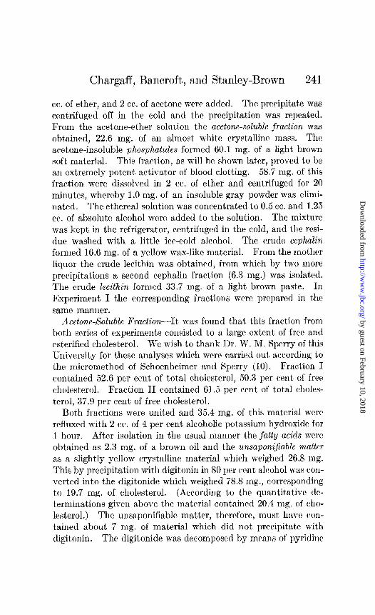
Isolation of Lipids-The platelets were extracted with alcohol- ether in the dark at room temperature for 21 days. (All the operations were carried out, as far as possible, in Nz atmosphere.) They were then filtered off and extracted with chloroform for the same period of time. Two individual batches of platelets were worked up, as mentioned above, and the distribution of the various fractions is presented in Table I. Although the fractions isolated in both series of experiments were practically identical, it will be seen that the amounts in which the substances were isolated from the material varied considerably. This certainly is partly due to a difference in the manner of isolation.
In Experiment I (Table I) the alcohol-ether extract was con- centrated in vacua, diluted with water, and repeatedly extracted with ether. An extremely tenacious emulsion formed, which could only partly be broken. By a comparatively large volume of ether 8.5 mg. only of lipid material were extracted. A larger
by guest on February 10, 2018http://w
ww
.jbc.org/D
ownloaded from
240 Chemistry of Blood Coagulation. III
fraction (25.8 mg.) was given off when the aqueous layer was acidified with dilute HzS04. But even then the aqueous emulsion obviously retained part of the lipids. As the main difference be- tween the results of Experiments I and II lies in the phosphatide fraction, it appears probable that at least part of this fraction en- tered the aqueous phase in a complex water-soluble form. The combined lipids weighed 34.3 mg. and formed a light brown soft mass.
In Experiment II (Table I) the alcohol-ether extract was con- centrated to dryness in uacuo and the residue treated with ether. An ether-insoluble fraction, 7.6 mg. of a white powder, was elimi-
Fractim No.
Fractions Platelets
Experiment I / Experiment II
mg.
1 Lipids (soluble in alcohol-ether) 34.3 2 IL
( IL I‘ chloroform) 7.1
3 Phosphatides (insoluble in acetone) 17.5
4 Lecithin 10 4 5 Cephalin 5.5 6 Acetone-soluble fraction 16.6
7 Cholesterol 8.7
8 Defatted platelets 431.1
TABLE I
Composition of Blood Platelets
____ 3er cent
dv ml. naterial
7.3 82.6 1.5 1.2 3.7 59.1 2.2 34.9
1.2 23.3 3.5 22.6 1.8 13.9
91.2 408.7
-
1
oer cent dv
?llZlWid
16.8 0.2
12.0 7.1
4.7 4.6 2.8
83.0
nated by centrifugation. This fraction was inactive in blood clotting. It proved to be the emulsifying agent which had made extraction so difficult in Experiment I. The ethereal solution, when washed with water, formed almost no emulsions. From it 83.6 mg. of lipids were obtained.
The chloroform-soluble lipids were yellow oils which were entirely inactive in blood clotting and were not further examined.
The defatted platelets formed an almost white, extremely light powder. The question of the activity of aqueous extracts of this material will be discussed later in this paper.
Separation of Lipids-In order to save space, only Experiment II will be described. The lipids (83.6 mg.) were dissolved in 1
by guest on February 10, 2018http://w
ww
.jbc.org/D
ownloaded from
Chargaff, Bancroft, and Stanley-Brown 241
cc. of ether, and 2 cc. of acetone were added. The precipitate was centrifuged off in the cold and the precipitation was repeated. From the acetone-ether solution the acetone-soluble fraction was obtained, 22.6 mg. of an almost white crystalline mass. The acetone-insoluble phosphatides formed 60.1 mg. of a light brown soft material. This fraction, as will be shown later, proved to be an extremely potent activator of blood clotting. 58.7 mg. of this fraction were dissolved in 2 cc. of ether and centrifuged for 20 minutes, whereby 1.0 mg. of an insoluble gray powder was elimi- nated. The ethereal solution was concentrated to 0.5 cc. and 1.25 cc. of absolute alcohol were added to the solution. The mixture was kept in the refrigerator, centrifuged in the cold, and the rrsi- due washed with a little ice-cold alcohol. The crude cephalin formed 16.6 mg. of a yellow wax-like material. From the mother liquor the crude lecithin was obtained, from which by two more precipitations a second cephalin fraction (6.3 mg.) was isolated. The crude lecithin formed 33.7 mg. of a light brown paste. In Experiment I the corresponding fractions were prepared in the same manner.
Acetone-Soluble Fraction-It was found that this fraction from both series of experiments consisted to a large extent of free and esterified cholesterol. We wish to thank Dr. W. M. Sperry of this University for these analyses which were carried out according to the micromethod of Schoenheimer and Sperry (10). Fraction I contained 52.6 per cent of total cholest.erol, 50.3 per cent of free cholesterol. Fraction II contained 61.5 per cent of total cholcs- terol, 37.9 per cent of free cholesterol.
Both fractions were united and 35.4 mg. of this material werr refluxed with 2 cc. of 4 per cent alcoholic potassium hydroxide for 1 hour. After isolation in the usual manner the fatty acids were obtained as 2.3 mg. of a brown oil and the unsaponifiablc matter as a slightly yellow crystalline material which weighed 26.8 mg. This by precipitation with digitonin in 80 per cent alcohol was con- verted into the digitonide which weighed 78.8 mg., corresponding to 19.7 mg. of cholesterol. (A ccording to the quantitative de- terminations given above the material contained 20.4 mg. of cho- lesterol.) The unsaponifiable matter, thercfore, must have con- tained about 7 mg. of material which did not precipitate with digitonin. The digitonide was decomposed by means of pyridine
by guest on February 10, 2018http://w
ww
.jbc.org/D
ownloaded from

242 Chemistry of Blood Coagulation. III
and ether (11) and 18.1 mg. of the sterol were isolated. After four crystallizations from small amounts of 90 per cent alcohol 10.4 mg. of large plates were obtained, which were identified as cholesterol. The substance melted at 146” and showed no depres- sion of the melting point on admixture of pure cholesterol. It gave the usual cholesterol color reactions. A 0.25 per cent solu- tion in absolute alcohol showed no selective absorption in the ul- traviolet region. The amount of ergosterol or other absorbing sterols present, therefore, must have been very small. We are indebted to Mr. F. Rosebury of this Department for these meas- urements.
An~Zy.sis-C~~H~~0. Calculated. C 83.8, H 12.0 386.4 Found. “ 83.8, “ 12.3
The aqueous phase from the saponification was neutralized with dilute barium hydroxide, centrifuged, and evaporated to complete dryness in vacua. From the residue by extraction with absolute alcohol 1.8 mg. of a yellow oil were obtained. This substance, when oxidized with bromine, gave a weak reaction for dihydroxy- acetone with m-hydroxybenzoic acid, according to the sensitive microtest for glycerol recently described by Eegriwe (12). A small amount of glycerol, therefore, seems to have been present. The reactions for carbohydrates were negative.
Cephalin-The united crude cephalin fractions (27.7 mg.) were dissolved in ether. A small amount of insoluble material (2.3 mg.) was removed by centrifugation, the ethereal solution was concen- trated to 0.4 cc., and the cephalin was precipitated by slowly adding 1.5 cc. of absolute alcohol. The precipitation was twice repeated, the ethereal solution of the cephalin finally being poured into chilled acetone. The cephalin was obtained as an almost white wax-like substance which weighed 15.0 mg.
Analysis-Found. C 55.0, H 8.8, N 1.5, P 2.9, ash 14.7
There was not enough material available for a determination of amino N, but the substance gave a strong ninhydrin reaction (13). The mother liquors from the precipitations yielded 9.0 mg. of a light brown substance.
Lecithin-The united crude lecithin fractions (44.0 mg.) were dissolved in 0.4 cc. of absolute alcohol, and 1 cc. of a saturated
by guest on February 10, 2018http://w
ww
.jbc.org/D
ownloaded from

Chargaff, Bancroft, and Stanley-Brown 243
solution of cadmium chloride in methyl alcohol was added. The treatment of the precipitate with ether and chloroform, the re- moval of cadmium by means of a 20 per cent solution of dry am- monia in absolute methyl alcohol, and the reprecipitation with acetone were carried out according to Levene and Rolf (14). The lecithin finally obtained formed a slightly yellow plastic mass and weighed 16.9 mg.
Analysis-Found. C 64.2, H 11.2, N 1.9, P 3.7, ash 3.5
The ninhydrin reaction was negative. The residue from the com- bined mother liquors was freed of cadmium. A less pure material was obtained which weighed 26.8 mg.
Plant and Bacterial Phosphatides
Phosphatides from Soy Beans-A preparation of the mixed crude phosphatides supplied by the American Lecithin Corpora- tion was used as starting material. The crude cephakn was purified by repeated precipitation from its ether solution with alcohol and from an emulsion in 10 per cent acetic acid with acc- tone. The final product formed a light brown, somewhat sticky powder.
Analysis-Found. C 59.7, H 10.1, N 1.4, amino N 1.5, P 3.4, ash 13.4
The preparation of the lecithin was carried out according to Levene and Rolf (14) with the modification that the cadmium com- pound before decomposition with ammonia was recrystallized twice from a mixture of 2 parts of ethyl acetate and 1 part of 80 per cent alcohol (15). The lecithin was light yellow and contained C 58.6, H 9.0, N 2.1, P 3.9, ash 4.0.
Cephalin from Cotton Seeds-The dark colored crude phospha- tides were treated with norit. The cephalin prepared in the usual manner formed a brown powder.
Analysis-Found. C 54.8, H 9.2, N 1.2, amino N 1.3, P 3.4, ash 14.2
Bacterial Phosphatides-The cephalin and lecithin preparations from yeast (13) were kindly given us by Dr. L. F. Salisbury of Connecticut State College. The phosphatides from the bacillus Calmette-Guerin (16) and the diphtheria bacillus (17), which likewise were examined, have been previously described by one of us.
by guest on February 10, 2018http://w
ww
.jbc.org/D
ownloaded from

244 Chemistry of Blood Coagulation. III
Experiments on Activation of Blood Clotting
Method-The experiments on the activation of blood clotting by means of various lipids were carried out with chicken plasma as substrate. The technique was essentially the same as that described in a recent communication from this laboratory (18). The measurements were carried out by adding 0.03 cc. of the ac- tivator at various dilutions in physiological saline to 0.1 cc. of plasma. The reaction temperature was 30”. The readings were repeated every 3 to 5 minutes until clotting was indicated by the immobility of the glass bead that was contained in every glass tube. All measurements were made at least in duplicate.
110
IOO- O
90 -
&I- (’ 0
70 - 0
Ml-
10 - MICROGR4MS
O 0 IO 30 4.0 50 fi0 70
FIG. 1. Activation of plasma clotting by platelet phosphatide
The preparation of the lipid emulsions that were tested was made in the following manner: A weighed amount of the sub- stance in a small agate mortar was dissolved by the addition of a little peroxide-free ether. The solvent was driven off by a stream of nitrogen and the evenly dist,ributed, very thin lipid film was dried in vacua for a few minutes. On treatment with physiologi- cal saline a quite stable and uniform emulsion could thus be ob- tamed. The preparations from breast muscle which were ex- amined could be dissolved directly in saline.
Activity of Fractions from Blood Platelets-The acetone-soluble and the chloroform-soluble fractions from platelets (Fractions 6 and 2, Table I) were entirely inactive. The mixed phosphatide
by guest on February 10, 2018http://w
ww
.jbc.org/D
ownloaded from
Chargaff, Bancroft, and Stanley-Brown 245
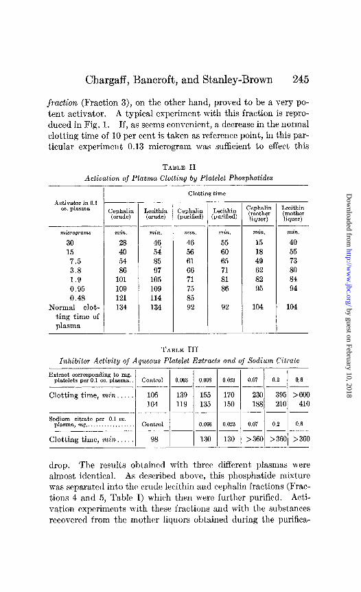
fraction (Fraction 3), on the other hand, proved to be a very po- tent activator. A typical experiment with this fraction is repro- duced in Fig. 1. If, as seems convenient, a decrease in the normal clotting time of 10 per cent is taken as reference point, in this par- ticular experiment 0.13 microgram was sufficient to effect this
TABLE II
Activation of Plasma Clotting by Platelet Phosphatides
-r Activator in 0.1
cc. plasma
micrograms
30 15
7.5 3.8 1.9 0.95
0.48 Normal clot-
ting time of
plasma
.-
-
min.
28 40
54 86
101
109 121 134
-
-
hciec&l
min.
46. 54
85 97
105 109
114 134
Clotting time
Cephalin (purified)
min.
46 56 61
66 71 75
85 92
- I( Lecithin
:purified)
min. min. min. 55 15 40 60 18 55
65 49 73
71 62 80 81 82 84
86 95 94
92 104 104
Zephalin Lecithin (mother (mother liquor) liquor)
TABLE III
Inhibitor Activity of Aqueous Platelet Extracts and of Sodium Citrate
Extract corresponding to mg. platelets per 0.1 cc. plasma. Control
Clotting time, min. 106 104
Sodium citrate per 0.1 cc. plasma, mg.. . Control
Clotting time, min. 98
139 119
T
0.008 0.023 ~~
155 170 135 150
__~
0.008 0.023 --
130 130 -
0.07 0.2 0.6 ---
230 395 >600 188 210 410
0.07 0.2 0.6 ---
>360 >360 >360
drop. The results obtained with three different plasmas were almost identical. As described above, this phosphatide mixture was separated into the crude lecithin and cephalin fractions (Frac- tions 4 and 5, Table I) which then were further purified. Acti- vation experiments with these fractions and with the substances recovered from the mother liquors obtained during the purifica-
by guest on February 10, 2018http://w
ww
.jbc.org/D
ownloaded from

246 Chemistry of Blood Coagulation. III
tion are reproduced in Table II. These results will be discussed later in this paper.
Aqueous extracts of the defatted platelets were examined in an endeavor to determine whether the activator had completely been removed by the organic solvents. It was very interesting to find that, contrary to expectation, these extracts possessed a marked inhibiting activity, as shown in Table III. 1 part of platelets was suspended in 50 parts of physiological saline for 5 minutes, the mixture was then centrifuged, and the clear extract was tested in various dilutions, 0.1 cc. of plasma and 0.03 cc. of the solution being used. As sodium citrate had been used ex-
FIG. 2. Activation of plasma clotting by plant cephalins. Curve 1
represents yeast cephalin; Curve 2, soy bean cephalin; Curve 3, cotton seed
cephalin.
tensively during the collection of the platelets, there was a possi- bility that the inhibiting action on clotting was due to sodium citrate absorbed in the cells. This does not seem to be the case. As shown in Table III, sodium citrate, even in comparatively high concentrations, affects the clotting of plasma in a different way. Sodium citrate inhibits the clotting not only of ordinary chicken plasma but also of plasma which has been activated by the addition of muscle extract (18). When tested in this man- ner, it is found to contain about 5 inhibitor units per mg. The aqueous platelet extract, however, is unable to prevent the clot- ting of plasma which contains muscle extract. It seems that this inhibitor which is extremely strong in non-activated plasma is
by guest on February 10, 2018http://w
ww
.jbc.org/D
ownloaded from

Chargaff, Bancroft, and Stanley-Brown 247
different from heparin. It should be mentioned that among five plasmas examined one was found which did not respond to the aqueous platelet extracts.
Activation Experiments with Other Phosphatides-The following phosphatides were found to be entirely inactive: yeast lecithin (13), soy bean lecithin, phosphatides from BCG (16) and diph- theria bacillus (17). A commercial synthetic cephalin prepared according to Griin and Limpacher (19) was likewise found inac- tive. A phosphatide from Bacterium tumefaciens (20) showed a
01
02 03
0 zo-
10 - . MICROGRAMS
0 “““““““““““I0 0 1 2 3 4 5 6 7 8 9 10 11 12 13 14 15 16 17 16 19 20 21 22
FIG. 3. Activation of plasma clotting by extract from chicken breast muscle. Experiments 1, 2, and 3 were carried out with plasma from three different chickens. The drawn out curve corresponds to Experiment 3.
slight activity. Yeast cephalin (13) and soy bean cephalin showed a very marked activating effect on plasma clotting. The cephalin preparation from cotton seeds was not quite as active. The activation of blood clotting by these three cephalins is shown in Fig. 2.
Activation Experiments with Muscle Extract-The extract from chicken breast muscle was prepared according to Fischer (21). In a large number of activation experiments very uniform results were obtained with different plasmas. The activation of plasma clotting by muscle extract is shown in Fig. 3.
by guest on February 10, 2018http://w
ww
.jbc.org/D
ownloaded from
248 Chemistry of Blood Coagulation. III
DISCUSSION
Regarding the chemical composition of blood platelets, three points are particularly notable: the high amount of phosphatides and cholesterol extracted, and the low, almost negligible amount of glycerides. (We take Experiment II in Table I as the basis of our discussion.) Whether there is any essential difference in the composition of the various blood cells cannot yet be said, as our knowledge concerning the chemical constituents of erythrocytes and the various forms of white cells (with the exception of pig- ments and enzymes) is still very limited (compare the reviews in (22) and Boyd (23)). The comparison of results obtained by different methods cannot be of great value, as the blood cells are comparatively very unstable and may undergo permeability changes during their preparation. It furthermore should be pointed out that in most cases where the presence of substances like phosphatides or sterols has been reported in blood cells, this was only done on the basis of the phosphorus content of the cx- tracts, or, in the case of sterols, by means of calorimetric estimation.
The experiments on the importance of platelets in blood clotting showed that there is contained in them a very potent activator of clotting which is associated with the phosphatide fraction. The activation curves (Fig. 1) obtained with this fraction are very characteristic, showing a steep fall at low concentrations and a gradual flattening out at high concentrations of the activator.
The active phosphatide fraction was separated into the cephalin and lecithin fractions and t.hese wrre further purified. From the results of the activation experiments which are contained in Table II it will be seen that while all the fractions retained a certain amount of activity, the activating effect was most marked with the more soluble cephalin fraction isolated from the mother liquor. As regards the activity of the lecithin fraction, it should be emphasized that in a model experiment with a mixture of similar amounts of cephalin and lecithin from another source, and by exactly duplicating the separation procedure followed with the platelet phosphatides, an entirely inactive lecithin and a highly active cephalin could be obtained.
It is a well established fact that the activating influence of lipids on blood clotting is associated with the cephalin fraction
by guest on February 10, 2018http://w
ww
.jbc.org/D
ownloaded from

Chargaff, Bancroft, and Stanley-Brown 249
(4), i.e. with a group of substances built up of fatty acids, glycero- phosphoric acid, and aminoethyl alcohol. Whether there is one particular representative of this group which is active or whether the activity is a common property of the cephalin group cannot be said. Our present methods for the separation of phosphatides are still much too crude to permit the isolation of compounds in a state even approaching purity. It may very well be that a cepha- lin containing highly unsaturated fatty acids, and, therefore, more soluble in ethyl alcohol, is the real activator of blood clotting.
The fact that synthetic distearyl cephalin (19) was found en- tirely inactive is in harmony with the &dings of McLean (24) according to whom the activity of the cephalin preparation was parallel to its degree of unsaturation.
A number of phosphatides prepared from plants and micro- organisms were also tested for activity. The bacterial phospha- tides were inactive. The cephalin preparations from yeast, soy beans, and cotton seeds were active, as shown in Fig. 2. The lecithin preparations from yeast and soy bean were devoid of any activity. The fact that cephalins of vegetable origin were found active tends to show that the activity is not associated with a clotting factor contained in animal tissue with which the lipids are contaminated.
The activation of clotting by muscle extract, as reproduced in Fig. 3, shows the same general picture. The activity of this sub- stance is still more pronounced than that of the cephalin group. It is uncertain whether the mechanism of action of these two activators is the same. It must be noted that, contrary to the lipids, the muscle extract is unstable toward heating and that in some cases plasmas are found which, while responding to muscle extract, cannot be activated by cephalin.
The finding in plate&s of a substance acting as a strong in- hibitor of blood clott,ing, if borne out by repeated experiments with other platelet preparations, would prove of the greatest in- terest. It could serve as an explanation for the occurrence of certain blood diseases (e.g. hemophilia) which often have been assumed to be connected with an abnormal behavior of the blood cells. A disturbance in the permeability of the platelets for the activator or the inhibitor of clotting or the absence of either the one or the other in certain pathological cases would make for an increased bleeding or clotting tendency.
by guest on February 10, 2018http://w
ww
.jbc.org/D
ownloaded from

250 Chemistry of Blood Coagulation. III
We are highly indebted to the Lederle Laboratories, Inc., Pearl River, New York, for the supply of blood cells. We wish to thank Mrs. Charlotte Breitung for assistance in some of the experiments, and Mr. W. Saschek for numerous microanalyses.
SUMMARY
1. The preparation of blood platelets from horse blood is described.
2. The platelet lipids were isolated and examined. The cepha- lin, lecithin, and sterol fractions are described.
3. The various platelet fractions were examined for their ac- tivating influence on plasma clotting. The phosphatide fraction was found to contain a potent activator.
4. From defatted blood platelets a substance can be extracted with water which acts as inhibitor of blood clotting. The im- portance of this finding is discussed.
5. The preparation of phosphatides from soy beans and cotton seeds is described. The activity in blood clotting of these sub- stances as well as of other phosphatides of plant and bacterial origin is discussed and compared with that of tissue extracts. The cephalin fractions from soy beans, cotton seeds, and yeast were found active. This shows that the activation of clotting is the property of certain phosphatides, regardless of their origin.
BIBLIOGRAPHY
1. Hayem, G., L’hematoblaste, Paris (1923). 2. Bizzozero, J., Virchow’s Arch. path. Amt., 90, 261 (1882).
3. Btirker, K., Handbuch der normalen und pathologischen Physiologie, Berlin, 6, pt. 1, 67 (1928). Mouzon, J., Les plaquettes du sang humain, Paris (1921).
4. Wohlisch, E., Ergebn. Physiol., 26, 443 (1929). Zunz, E., Trait6 de
physiologie normale et pathologique, Paris7 2nd edition, 7, 189 (1934). Howell, W. H., Physiol. Rev., 16, 435 (1935).
5. Eagle, H., J. Gen. Physiol., 18, 531 (1935).
6. Benda, C., Handbuch der speziellen pathologischen Anatomie und His- tologie, Berlin, 2, 810 (1924).
7. Abderhalden, E., and Deetjen, H., 2. physiol. Chem., 63, 280 (1907).
8. Haurowitz, F., and Sladek, J., 2. physiol. Chem., 173, 233 (1928). 9. Hamburger, H. J., Physikalisch-chemische Untersuchungen iiber
Phagozyten, Wiesbaden, 4 (1912).
10. Schoenheimer, R., and Sperry, W. M., J. BioZ. Chem., 106, 745 (1934). 11. Schoenheimer, R., and Dam, H., 2. physiol. Chem., 216, 59 (1933).
by guest on February 10, 2018http://w
ww
.jbc.org/D
ownloaded from

Chargaff, Bancroft, and Stanley-Brown 251
12. Eegriwe, E., 2. anal. Chem., 100, 31 (1935).
13. Salisbury, L. F., and Anderson, R. J., J. Biol. Chem., 112,541 (1935-36). 14. Levene, P. A., and Rolf, I. P., J. Biol. Chem.., 72, 587 (1927). 15. Willstatter, R., and Ltidcckc, K., Ber. them. GES., 37, 3753 (1904).
16. Chargaff, E., 2. physiol. Chem., 217, 115 (1933). Chargaff, E., and Schaefer, W., dnn. Inst. Pasteur, 64, 708 (1935).
17. Chargaff, E., 2. physiol. Chem., 201, 191 (1931); 218, 223 (1933). 18. Chargaff, E., Bancroft, F. W., and Stanley-Brown, &I., J. Biol. Chem.,
116, 149 (1936). 19. Griin, A., and Limpacher, R., Ber. them. Ges., 60, 151 (1927). 20. Chargaff, E., and Levine, M., Proc. Sot. Exp. Biol. andMe& 34, 675
(1936). 21. Fischer, A., Biochem. Z., 240, 357 (1931). 22. Kanitz, A., in Oppenheimer, C., Handbuch der Riochemie des Mcnschen
und der Tiere, Jena, 2nd edition, 2, 560 (1925). Edlbacher, S., ibid., Erganzungswerk, Jena, 1, 769 (1933).
23. Boyd, E. M., J. Biol. Chem., 101, 623 (1933). 24. McLean, J., Am. J. Physiol., 43, 586 (1917).
by guest on February 10, 2018http://w
ww
.jbc.org/D
ownloaded from

Margaret Stanley-BrownErwin Chargaff, Frederic W. Bancroft and
CLOTTING BY LIPIDSREMARKS ON THE ACTIVATION OF
IN BLOOD CLOTTING, WITHBLOOD PLATELETS AND THEIR RÔLE
CHEMICAL CONSTITUENTS OFBLOOD COAGULATION: III. THE
STUDIES ON THE CHEMISTRY OF
1936, 116:237-251.J. Biol. Chem.
http://www.jbc.org/content/116/1/237.citation
Access the most updated version of this article at
Alerts:
When a correction for this article is posted•
When this article is cited•
alerts to choose from all of JBC's e-mailClick here
tml#ref-list-1
http://www.jbc.org/content/116/1/237.citation.full.haccessed free atThis article cites 0 references, 0 of which can be
by guest on February 10, 2018http://w
ww
.jbc.org/D
ownloaded from