studies on the bacteriophage of d'herelle
TRANSCRIPT

STUDIES ON THE BACTERIOPHAGE OF D'HERELLE.
VIII. T~x MECHANISM O1~ LYSlS OF DEAD BACTERIA IN TttE PRESENCE OF BACTERIOPHAGE.
BY J. BRONFENBRENNER, PH.D., Am) R. MUCKENFUSS, M.D.
~From the Laboratories of The Rockefeller Institute for Medical Rzsearch.)
(Received for publication, January 15, 1927.)
With but few exceptions (1, 2), those engaged in the study of bac- teriophage agree that one of the essential features of transmissible lysis of bacteria is the fact that the lytic principle undergoes an in- crease in activity exclusively in the presence of living and actively multiplying bacteria. Without active growth of susceptible bacteria, there is no reproduction of lytic agent and no observed bacterial lysis. Although dead susceptible bacteria readily adsorb the lytic agent, they do not dissolve (3-7). However, Gratia and Rhodes (8) observed that dead staphylococcus may be slowly lysed by the bacteriophage , and that the concentration of the latter in solution probably increases dur- ing this process. If live staphylococci are present simultaneously, the lysis of the dead bacteria is more rapid. The lyric effect of live on dead staphylococcus was observed by them also in the absence of bacteriophage 1 and was assumed by them to be due to the utilization of dead bacteria by the live in the process of nutrition (10). The relation of this phenomenon to the one described earlier by these authors (8) was not made clear. A year later Twort (11) independ- ently observed the lysis of dead staphylococcus in the presence of bacteriophage and live bacteria, and suggested that this was made possible through the activation of the bacteriophage by some auxiliary substance contributed by the live bacteria. Because of the impor- tance of these observations on the question of the mechanism of trans- missible lysis, we undertook to study it.
l A similar observation has also been made recently by Duran-Reynals (9). 887
888 BACTERIOPHAGE OF D'HERELLE. VIII
The L y s i s of Dead Bacteria.
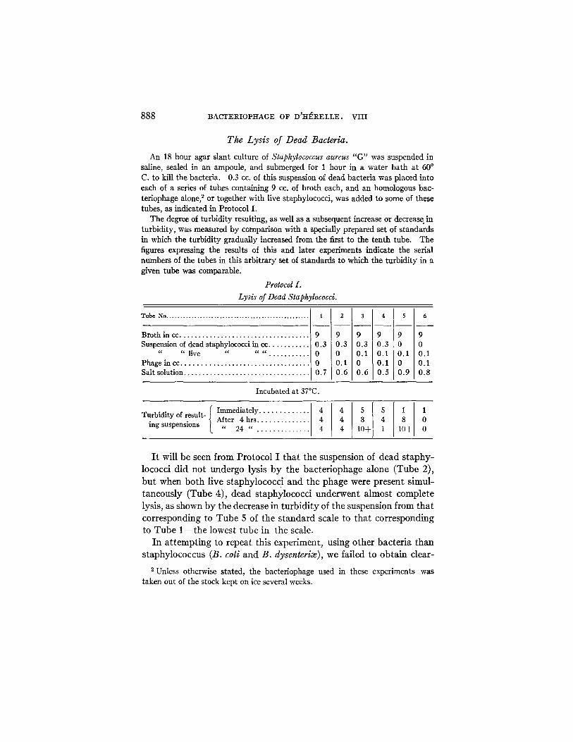
An 18 hour agar slant culture of Staphylococcus aureus "G" was suspended in saline, sealed in an ampoule, and submerged for 1 hour in a water bath at 60 ° C. to kill the bacteria. 0.3 cc. of this suspension of dead bacteria was placed into each of a series of tubes containing 9 cc. of broth each, and an homologous bac- teriophage alone, ~ or together with live staphylococci, was added to some of these tubes, as indicated in Protocol I.
The degree of turbidity resulting, as well as a subsequent increase or decrease in turbidity, was measured by comparison with a specially prepared set of standards in which the turbidity gradually increased from the first to the tenth tube. The figures expressing the results of this and later experiments indicate the serial numbers of the tubes in this arbitrary set of standards to which the turbidity in a given tube was comparable.
Protocol I.
Lysis of Dead Staphylococci.
T u b e N o . . . . . . . . . . . . . . . . . . . . . . . . . . . . . . . . . . . . . . . . . . . . . . . . . . .
Broth in ec . . . . . . . . . . . . . . . . . . . . . . . . . . . . . . . . . . . Suspension of dead staphylococci in cc . . . . . . . . . . .
" " live " " " . . . . . . . . . . . Phage in cc . . . . . . . . . . . . . . . . . . . . . . . . . . . . . . . . . Salt solution . . . . . . . . . . . . . . . . . . . . . . . . . . . . . . . . .
1
9 0.3 0 0 0.7
9 9 0.3 0.3 0 0.1 0.1 0 0.6 0.6
4
9 9 0.3 0 0.1 0.1 0.1 0 0.5 0.9
9 0 0.1 0.1 0.8
Incubated at 37°C.
Ilmme atl ........... Turbidity of result- After 4 hrs . . . . . . . . . . . . . 4 4 4 0
ing suspensions " 24 " . . . . . . . . . . . . . 4 4 10+ 1 10+ 0
I t wil l b e seen f rom P r o t o c o l I t h a t t he suspens ion of d e a d s t a p h y -
lococc i d i d n o t u n d e r g o lys i s b y the b a c t e r i o p h a g e a l o n e ( T u b e 2),
b u t when b o t h l ive s t a p h y l o c o c c i a n d the p h a g e were p r e s e n t s i m u l -
t a n e o u s l y ( T u b e 4), d e a d s t a p h y l o c o c c i u n d e r w e n t a l m o s t c o m p l e t e
lys is , as shown b y t h e d e c r e a s e in t u r b i d i t y of t he su spens ion f rom t h a t
c o r r e s p o n d i n g to T u b e 5 of t h e s t a n d a r d sca le to t h a t c o r r e s p o n d i n g
to T u b e 1 - - t h e l owes t t u b e in t he scale.
I n a t t e m p t i n g to r e p e a t th is e x p e r i m e n t , us ing o t h e r b a c t e r i a t h a n
s t a p h y l o c o c c u s (B. coli a n d B. dysenteriae), we fa i l ed to o b t a i n c lea r -
2 Unless otherwise stated, the bacteriophage used in these experiments was taken out of the stock kept on ice several weeks.

J . B R O N F E N B R E N N E R AND R. M U C ~ E N F U S S 889
cut results. This explains the negative results obtained by Doerr and Grtininger (7), who a t t empted to produce dissolution of dead colon bacillus in the presence of corresponding bacter iophage and of live bacteria. Moreover , we found tha t even with staphylococcus, re- producible results can be obta ined only if the relative bulk of dead bacter ia used is t aken into account, as will be shown in the following experiment.
Effect of Variations in Relative Concentration of _Phage and of Dead Bacteria.
Two series of eight tubes each received equal amounts of broth (5 cc.). Each of the tubes of the first series (A) received 0.1 cc. of antistaphylococcus phage diluted in broth 1:1000; and the tubes of the second series (B) each received 0.1 cc. of the same phage diluted 1:100,000. Following this, the first four tubes of each series received 0.1 cc. each of bacterial suspension containing 80 million of live staphylococci per cc., and gradually decreasing amounts of a suspension of dead staphylococci containing 220 million bacteria per cc.--the first tube of each series receiving 5 cc. of this suspension, the second 0.5 cc., the third 0.05 cc. respectively. The fourth tube served as control and received no dead bacteria. The fifth, sixth, and se~,enth tubes of each of the series received respectively 5 cc., 0.5 cc., and 0.05 cc., of the suspension of dead bacteria only and no live bacteria, and the last tube (No. 8) of each series received no bacteria at all. The volume of liquid in all the tubes was brought to 10.2 cc. with physiological salt solution, and the whole was kept for 24 hours at 37°C. Changes in turbidity due to lysis of bacteria were recorded, as indicated in Protocol II, and after 24 hours' incuba- tion all the tubes were placed for 30 minutes into a water bath kept at 56°C., and the phage titer of each mixture was determined by the method of Appelmans.
This experiment indicates tha t bo th in the series where the initial t i ter of the phage was 1 >( 10°cc. (Protocol II, B, Tube 8), and where i t was 1 )< 10 -1 "cc. (Protocol II, A, Tube 8), dead bacteria alone adsorbed the entire phage (Tubes 5, 6, and 7 of each series), the ra te of adsorption apparent ly depending on the number of dead bacteria present. When the concentrat ion of dead bacter ia was compara- t ively low, the adsorpt ion of phage was so slow tha t when live bacter ia which had been added began to multiply, there was sufficient phage left free in solution to produce its usual effect on the live bacter ia and to regenerate. In the case where the initial concentrat ion of phage was lower (Series B), the regeneration of phage took place only in T u b e 3, containing the lowest number of dead bacteria. In Series A,

8 9 0 B A C T E R I O P H A G E O F D ~ I - I ~ R E L L E . V I I I
"r. ~ . ~
o ~o-d
• 0
h
.~§~
o
8
o
g
?,
d g.
I
.+
+ ÷ ,,..t ,,-t v-t
I
+ ~
• °
T hi)
7
O
T
o
~ o
• -~ g =
• = . ~ . ~
,.c

J. BRONFENBRENN'ER AND R. MUCKENFUSS 891
where the initial concentration of phage was ten times greater, the regeneration of the phage took place both in Tube 3 and in Tube 2. I t is evident that the absence of regeneration of phage in Tube 1 of Series A, and in Tubes 1 and 2 of Series B was due to the fact that the phage present in the mixtures was so completely taken up by the dead bacteria that by the time live bacteria had begun to multiply actively (which is essential for the regeneration of phage) after a period of initial lag, there was no free phage left in the mixture. As a result of this, the bacteria in these tubes remained intact, as is evidenced by the fact that the original turbidity of the contents increased slightly, due to multiplication of the live bacteria. On the c6ntrary, in such tubes of each series as exhibited regeneration of phage in the presence of dead bacteria (Tubes 2 and 3 of Series A, and Tube 3 of Series B), the initial turbidity decreased, due to dissolution 6f dead bacteria.
Specificity of Lysis of Dead Bacteria.
I t was seen in the preceding experiments that while phage alone does not cause lysis of dead bacteria , the latter are lysed if phage and live bacteria are present simultaneously and if the concentration of dead bacteria is kept sufficiently low not to interfere with the process of regeneration of the phage. Another condition essential for the lysis of dead bacteria is that the dead and live bacteria be of the same species. This requirement has already been indicated by Twort (11), and we have been able to confirm it. Dead colon bacilli or dead dysentery bacilli were not lysed in the presence of live staphylococcus and staphylococcus phage.
Time Relation of the Lysis of Live and of Dead Bacteria.
The preceding experiments show that live bacteria contribute some specific active principle necessary for the production of lysis of dead bacteria. Twort (11) has suggested that bacteria supply some sort of a complementary substance which activates the phage and disap- pears as the phage ages. In order to see whether such a substance is present, and to determine more accurately at what stage of lysis of live bacteria it first appears and how long its activity continues, the following experiment was performed.

892 BACTERIOPHAGE OF D~HJ~RELLE. VIII
A series of six tubes containing 10 cc. of broth each received 0.1 cc. of a sus- pension of live staphylococcus and 0.1 cc. of phage, and were placed in the incuba- tor at 37 °. The first tube of the series received at the same time 0.2 cc. of a sus- pension of heat-killed staphylococcus. At intervals of 2, 4, 6, etc., hours after
Protoco l I I L
D i s s o l u t i o n o f D e a d B a c t e r i a A d d e d d u r i n g a n d a f ter the C o m p l e t i o n o f the L y s i s o f
L i v e B a c t e r i a i n the Presence o f Bac t e r iophage .
T u ~ N o . . . . . . . . . . . . . . . . . . . . . . . . . . . . . .
Broth in cc . . . . . . . . . . . . . . . . . Suspension of live staphylococci
in cc . . . . . . . . . . . . . . . . . . . . . . Phage (filtrate) in cc . . . . . . . . . .
T e s t p r o p e r
.0 I0 10 10 10 10
0.11 0.11 0.1 / o.1 / 0.11 0.1 o.11 o.1 / o.11 o.1 t o.1 / o.1
Controls
±1:± '°I 1_, ,ol,o ,ol,o o.1 / o.11 o.11 o Io o /o .11o io.11o
Incubated at 37°C.
Interval before addition of the suspension of dead bacteria in hrs . . . . . . . . . . . . . . . . . . . . .
Suspension of dead bacteria in C C . . . . . . . . . . . . . . . . . . . . . . . .
Turbidity
Immediately . . . . . . 2 hrs. later . . . . . .
6 " " . . . . . 24 " " . . . . .
30 " " . . . . .
48 " " . . . . .
72 " " . . . . .
0 2
.2 0.2 0,2
4 5 3
2 + 3 1+
6
4 6
0.2
4 2 3 6 1 6 7 7
i
2 4 48 i
0.: 0.2 0
1 1 5
10 13 lO+
2- 10+ 3* 7* 10+ 6* 6* 10+
0 0.2 0.2
1 4 3 1+ 4 3 1 6 3 0 10 3 0 10+ 3 1 10+ 3 4 10+ 3 6 10+ 3
~).2
* Final results in these tubes are somewhat obscured, due to increase in turbidity caused by the multiplication of the resistant bacteria, but the lysis of dead bac- teria has taken place even in these tubes, as suggested by the temporary diminu- tion in turbidity in Tubes 5 and 6.
the beginning of the experiment, other tubes of the series each received in turn 0.2 cc. of the same suspension of dead bacteria. The progress of lysis of live, as well as of dead bacteria, was followed and is recorded in Protocol I r I in terms of tur- bidity, by comparison with a standard scale, in which the higher numbers indicate greater turbidity. A fall in turbidity below that of the control tube (No. 11), containing dead bacteria alone, indicates the lysis of dead bacteria. I t will be

3. BRONFENBRENNER AND R. MUCKENFUSS 893
seen that the initial fall in turbidity in all the tubes of the series (Nos. 1 to 6) is followed by a subsequent increase. This is due to the ~owth of resistants fol- lowing the initial lysis of the susceptible live bacteria, so that the results in Tubes 5 and 6 are difficult to interpret on the basis of turbidity.
The results show tha t dead bacteria are dissolved when added to the live bacter ia undergoing lysis, any t ime within 24 hours af ter the beginning of the experiment, and probably even later, irrespective of the stage at which the lysis of live bacter ia may happen to ~e. Since the lysis of live bacter ia under the conditions of the experiment was completed in the first 6 hours (see Tube 8), and since dead bacteria were dissolved when added even much later, it is evident tha t the actual lysis of live, is not essential for the dissolution of dead bacteria, and tha t the products of such lysis alone are capable of causing dis-
solution of dead bacteria. However, we have observed tha t filtrates of lysed cultures which
should thus contain these products are inactive against dead bacteria (Protocol I). I t was suspected, therefore, tha t during filtration they might have been kept back by the filter, while phage was able to pass freely. I f such were the case, then it would appear tha t the agent responsible for the dissolution of dead bacter ia is dist inct from the phage proper and can be separated from the la t ter because of the dif- ference in their respective diffusibility.
In order to determine if such were the case, an a t t empt was made to interpose a semipermeable membrane between the live and dead bacter ia during the lysis of the former to see whether this procedure would prevent the dissolution of the dead bacter ia (12).
Separation o/ Lysed Cultures into Two Fractions by Means of a Semipermeable Membrane.
A series of cylindrical collodion membranes 15 ram. in diameter and 50 ram. long were prepared under carefully controlled conditions. After hardening in water, these membranes were tested for their relative permeability by measuring the time required for 0.1 cc. of water, under pressure of 10 cm. of mercury, to be forced through the membrane suspended in air. It was found that membranes allowing this amount of water to pass through in from 20 to 30 seconds were suitable for the experiment. A number of such selected collodion bags were filled with and sus- pended in water, and sterilized in the autoclave for 10 minutes, at 20 pounds pressure. While the autoclaving renders the membrane somewhat less permeable

894 BACTERIOPHAGE OF D'H~RELLE. VIII
X X
X ~
X X
X X
7
X
° ~
° ~
~Z
c~
c~
8
.~=
O
h 0

J. BRONFENBRENNER AND R. MUCKENFUSS 895
to water, we have found by experience that bags selected and tested as stated above are uniformly permeable to bacteriophage. At the time of the experiment the dialyzing thimbles were removed from the water in which they had been sterilized, filled with measured amounts of sterile broth, and placed into suitable containers, with aseptic precautions. In each experiment four dialyzing thimbles were set up as follows:
I. In the first dialyzing unit a measured amount of antistaphylococcus bac- teriophage was added to the broth inside of the dialyzing thimble, and a small portion of the resulting dilution of phage was immediately taken out for titration by the method of Appelmans. At the same time a small portion of the broth out- side of the bag was likewise removed for titration, and the whole unit was placed in the incubator at 37°C. After an interval of 5 and 24 hours respectively, the titration of the fluid inside and outside the thimble was carried out, and it was found that the lytic agent dialyzed freely under these conditions ((Protocol IV, A).
II . In the second dialyzing unit 5 cc. of broth were placed inside and 150 cc. of broth outside the thimble as before, following which 0.1 cc. of antistaphylococcus bacteriophage was added to the broth inside the thimble, and 0.1 cc. of an 18 hour old culture of susceptible staphylococcus to the broth outside. The whole unit was placed in the incubator and observed at intervals. I t was found that live bacteria placed outside the thimble underwent lysis (Protocol IV, B), just as they do when placed in direct contact with phage without the interposition of the membrane.
I lL The third dialyzing unit was set up exactly as the preceding one, except that both live bacteria and the phage were placed outside, and on the inside only 0.1 cc. of a suspension of dead (heat-killed) staphylococcus. As the experiment proceeded in the incubator at 37°C., lysis of live bacteria took place outside the thimble, and the phage dialyzed into the thimble, where it was demonstrated by titration. However, the actual count of dead bacteria placed into the thimble re- mained unaffected throughout the experiment (Protocol IV, C).
IV. In the fourth dialyzing unit the phage was placed outside the dialyzing thimble and allowed to dialyze for 6 hours into the sterile broth inside. At this time 0.1 cc. of a suspension of live staphylococcus was added to the fluid inside the thimble, and a sample was removed for immediate count, which was found to be 1,000,000 bacteria per cc. Immediately following the removal of the sample, 0.1 cc. of a suspension of dead staphylococcus was added, and again a sample was removed. The bacterial count in this sample indicated the presence of 267,500,000 bacteria per cc. Thus, the initial mixture inside the thimble was composed of 1,000,000 live, and 266,500,000 dead bacteria per cc. From then on the bacterial count was repeated at intervals, and it was found that at the end of 5 hours it had fallen to 29,000,000 per cc., and at the end of 24 hours it had risen to 96,000,000 per cc., due to overgrowth of resistant bacteria. As the initial mixture contained 266,500,000 dead bacteria per cc., it is evident that at the end of 5 hours, practically 90 per cent of the dead bacteria had been dissolved.

896 BACTERIOPHAGE OF D'HERELLE. V I I I
These tests indicate that the phage responsible for transmissible lysis of live bacteria is easily diffusible, but that the agent liberated during the lysis of live bacteria and causing dissolution of dead bac- teria does not pass through the membrane, so that dead bacteria remain unaffected if the active lysis of live bacteria takes place on the other side of the membrane, even though the bacteriophage can be demonstrated in abundance in the dialysate in which dead bacteria are suspended.
Protocol V.
Effect of Adsorption with Live Bacteria on the Phage Titer and Power to Dissolve Dead Bacteria of Lysed Staphylococcus Cultures.
Original lysed culture in cc . . . . . . . . . Supematant fluid after the first ad-
sorption in ec . . . . . . . . . . . . . . . . . . . Supernatant fluid after the second ad-
sorption in cc . . . . . . . . . . . . . . . . . . . Broth in ce . . . . . . . . . . . . . . . . . . . . . . . Suspension of dead bacteria in cc . . .
Dissolution of dead staphylococcus
5 0.2
5
0
0 5 0.2 0.2
5
0 0 5 0.2 0.2
5 0 0.2
Titer of phage
1 × 10 -e
1 X 10 .3
1 × 10 -1
Covered with toluene and incubated at 37°C.
Immediately . . . . . . . . . . 3 3 3 3 3 3 Turbidity After 6hrs . . . . . . . . . . . 3 3-- 3 3-- 3 3--
" 24 " . . . . . . . . . . 3 1 3 2-- 3 1
Separation, by Adsorption, of the Phage from the Agent Dissolving Dead Bacteria.
I f f r e sh ly l y s e d cu l tu re s of s t a p h y l o c o c c i c o n t a i n two a c t i v e agen t s ,
as s u g g e s t e d b y the e x p e r i m e n t j u s t desc r ibed , i t shou ld be pos s ib l e to
s e p a r a t e t h e m f rom each o t h e r a lso, b y r e m o v i n g t h e p h a g e f rom the
so lu t i on t h r o u g h a d s o r p t i o n on l ive s u s c e p t i b l e b a c t e r i a , a n d l e a v i n g
in t he so lu t i on o n l y t he a g e n t d i s so lv ing d e a d b a c t e r i a .
Accordh~gly, cultures of Staphylococcus "G" were subjected to lysis by the appropriate bacteriophage, at 37°C. At the same time mass cultures of staphy- lococcus were grown on the surface of agar in Blake bottles. The next day bacteria

J . B R O N F E N B R E N N E R AND R. M U C K E N F U S S 897
collected from the surface of three Blake bottles were washed by centrifugation, and to the solid mass of bacteria at the bottom of the centrifuge tube were added 20 cc. of lysed staphylococcus culture. At this time bacteria were suspended in the fluid above, by vigorous shaking, and the whole mixture was placed on ice for 2 hours, to allow for adsorption of the phage on bacteria. At the completion of a 2 hour period, the bacteria were thrown down by centrifugation at a high speed, for 1 hour. The centrifuge used for this purpose was supplied with a cooling device, So that 1ysis of bacteria was prevented during the experiment. A portion of the supernatant fluid was removed for examination of its bacteriophage content, as well as of its ability to dissolve dead bacteria, and to the remainder of the fluid was added another lot of bacteria collected from three Blake bottles. The mixture was shaken, placed on ice/or 1 hour, and again bacteria were separated by cen- trifugation. The supernatant fluid was again tested as before. The results of these titrations are recorded in Protocol V.
As the results of this experiment indicate, it is possible to remove most of the bacteriophage proper by adsorption on bacteria, wi thout affecting the power of the lysate to dissolve dead bacteria.
Dissolution of Dead Bacteria by Filtrates of L ysed Cultures in the Absence of Live Bacteria.
The last two experiments show that lysis of dead bacteria depends upon the presence in the freshly lysed cultures, in addit ion to the phage proper, of another active agent which does not go through the semipermeable membrane of collodion. This agent m a y conceivably be held back by the porcelain during filtration, and thus the fact tha t lyric filtrates, as usually prepared, do not cause the dissolution of dead bacteria becomes explainable. However, we found tha t the retention of this agent by the filter was complete and constant , while Grat ia and Rhodes (8), report tha t the filtrates exhibit a certain amount of act iv i ty against old as well as against dead staphylococcus cultures.
Since the failure of lyric filtrates (bacteriophage) to dissolve old or dead bacteria constitutes, in our opinion, a characteristic which as- sumes fundamental importance in an a t t emp t to unders tand the mechanism of transmissible lysis, we felt tha t it was necessary to determine beyond any doubt whether in the experiments of Gratia the active filtrates contained only the phage, or whether, under the con- ditions of his experiments, a certain amount of the second agent also had passed into the filtrate. Apar t from the possibility tha t the

898 BACTERIOPHAGE OF D'I-~RELLE. VIII
efficiency of the filters used b y Grat ia and ourselves migh t have been
different, it seemed likely, tha t if large amounts of lysed cultures are filtered through a given filter, its efficiency m a y gradual ly decrease, and the substance, which a t first is held back, m a y appear in the
filtrate af ter continued filtration, thus possibly explaining the ac t iv i ty
of his filtrates (26).
In order to test this possibility, a flask containing 1 liter of broth received a suitable amount of the suspension of the 18 hour agar growth of staphylococcus and of bacteriophage. The resulting mixture was distributed equally into three smaller flasks and incubated at 37°C. for 2, 4, and 6 hours respectively. At the end of 2 hours' incubation, one of the flasks was taken out and immediately sub- jected to fractional filtration through a new Berkefeld V candle, under pressure of 60 ram. of mercury as follows: At first 50 cc. of liquid were removed from the flask and filtered. The filtrate was collected into a sterile receptacle. Then a second 50 cc. portion of the contents of the flask was filtered through the same candle and the filtrate collected into a second receptacle. Then followed a third fraction of 50 cc. and so on--five fractions in all being employed. At this point the filter candle was discarded and the five fractions of the filtrate were immediately subjected to examination for sterility, 3 phage content, and for their power to cause lysis of dead staphylococcus, as indicated in Protocol VI, Section I A. At the proper intervals the contents of the other two flasks were similarly filtered, each through its own new filter candle, and the fractional filtrates thus obtained were examined, as indicated in Sections I B and I C of Protocol VI.
As another possibil i ty which could explain discrepancies in the
results i t seemed to us of interest to inquire also into the ra te of
deter iorat ion suffered b y the agent responsible for the lysis of dead bacteria , under the influence of hea t and preservat ion. In order to
elicit the r61e of these factors, all the fractional filtrates ( immedia te ly af ter the remova l of a small f ract ion of each for var ious tests, as shown
in Sections I A, B, and C of the protocol) were divided into two por-
tions each.
One portion of each filtrate was subjected to heating in sealed ampoules and submerged under water at 56°C. for 30 minutes. At the end of this time the tubes were removed from the water bath, cooled quickly in cold water, and the contents were tested for phage content and for their ability to cause the lysis of dead staphylococcus, as indicated in Sections I I A, B, and C of Protocol VI.
s This control is essential, for if the filtrate contains live bacteria, the subsequent lysis of dead bacteria, if it occurs, cannot be attributed directly to passage of the active agent, but may be due to the lysis of live bacteria.

r~
! e~
g~
o
~ ~.~
I. ¢~_._..._~_ ~
_ ~ I °-:~-~ ~
899
o o ° ~
0 ~
~°~ x

900 BACTERIOPHAGE OF D'HI~.RELLE. VIII
The remaining portion of fractional filtrates was allowed to stand in the room at i25°C, for 8 days. After this period they were subjected to the test outlined in Sections IH, A, B, and C of Protocol VI.
This experiment explains the discrepancy. I t appears tha t when the filter is new and only a small amount of lysed cultures is filtered, only the phage appears in the filtrate, but as more and more of the solution is forced through the same candle, it becomes less selective and allows the passage of the second agent which is capable of causing dissolution of dead bacteria. 4 This is illustrated in the protocol by a diminution of the turbidi ty of bacterial suspensions in the tubes con- taining the filtrate, as compared with the turbidi ty of the controls. In the case in which the lysis in the original mixture was allowed to proceed only 2 hours before filtration (Protocol VI, Section I A), this agent appeared la te - -on ly after the passage of the fourth f rac t ion-- but as its concentration in the solution increased with the progress of lysis of live bacteria, it appeared in the filtrate sooner and in greater concentration, so tha t in the last section of the experiment (C) traces of the agent were already present in the second fractional filtrate, and in the fourth and fifth its concentration was sufficient to destroy al- most all dead bacteria, as shown by the decrease of turbidi ty from 4 to 1. Comparison of the results in Sections I, II , and I I I , on the other hand, indicates that the agent lyric for dead bacteria is present only in freshly prepared filtrates of lysed cultures. I f these filtrates are allowed to s tand or are subjected to heating, this agent undergoes destruction, while the phage is still present in the solution and is unaffected. 5
4 Occasionally, on continued fractional filtration, not only the second active agent but also bacteria appeared in the filtrate, as was indicated by the sterility controls. Such experiments were discarded and repeated, until sterile filtrates were obtained. These findings are of especial interest in connection with the statements in the literature, in which the appearance of growth in the filtrates of lysed cultures is attributed to the existence of a filterable stage in the life cycle of bacteria (13, 14, 26).
5 While in the experiment recorded in Protocol VI the agent causing the dis- solution of dead bacteria seems to be completely destroyed on standing or on exposure to heat at 56°C. for 30 minutes, this is not always the case. Repeating these experiments and employing suspensions of dead bacteria of varying density, we found that this destruction is usually very marked but not complete--with

I . BRONFENBRENNER AND R. MUCKEN-FUSS 901
Failure of Filtrates of a Live Resistant Variant, Grown in the Presence of Bacteriophage, to Induce Lysis of Dead Staphylococcus.
We have shown in the preceding experiments that bacteriophage has no direct lytic action on dead bacteria. Apparently by causing the lysis of susceptible live staphylococcus, it merely sets free a lyric agent preexisting in the bacteria themselves.
If this inference is correct, the incubation of bacteriophage with a live resistant variant instead of with a susceptible staphylococcus should yield a filtrate that will not lyse dead bacteria. Such an experi- ment was performed with a resistant variant isolated from the sus- ceptible strain of Staphylococcus " G " used previously. I t was found that when the experiment was carried out in a manner identical with that shown in Protocol VI, no dissolution of the dead bacteria occurred. On the other hand, dead resistant bacteria were as susceptible to the lyric action of the agent produced by the live susceptible bacteria as were the dead susceptible organisms themselves.
Evidently the failure of the phage to cause the lysis of resistant staphylococcus precluded the liberation of the lyric agent from the latter. However, when susceptible live bacteria were used, the lytic agent was set free and was able to dissolve either the susceptible or resistant dead staphylococci.
In general, the results of the preceding experiment confirm our earlier observations, namely, that the agent responsible for the trans- missible lysis of live bacteria is different and independent from that which causes the lysis of dead bacteria. Moreover, the fact that the latter ferment-like agent appears comparatively late during the lysis of live bacteria, that it is thermolabile, and is inactivated on standing,, that it is specific in its action, that it does not go through the collodion membrane, that it passes the filters only with difficulty, and causes dissolution of dead bacteria, suggests the possibility that it may be identical with the autolytic endoenzyme which is set free during the lysis of live bacteria.
very light suspensions of bacteria some residual activity can still be detected, when heavier suspensions of bacteria show no apparent presence of the lytic agent, as will be shown later (Protocol IX).
6 This inactivation is probably due to its combination with the products of its own activity.
902 B ACT E RIOPHAGE O]~ D 'HI~RELLE. VI I I
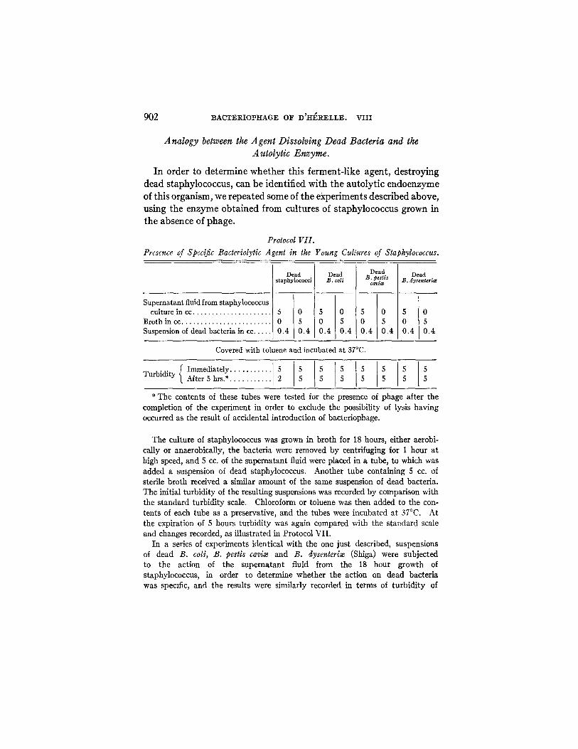
Analogy between the Agent Dissolving Dead Bacteria and the A utolytic Enzyme.
I n o r d e r to d e t e r m i n e w h e t h e r th i s f e r m e n t - l i k e a g e n t , d e s t r o y i n g
d e a d s t a p h y l o c o c c u s , can be i den t i f i ed w i t h t h e a u t o l y t i c e n d o e n z y m e
of th i s o r g a n i s m , we r e p e a t e d some of t h e e x p e r i m e n t s d e s c r i b e d a b o v e ,
us ing t h e e n z y m e o b t a i n e d f rom cu l tu r e s of s t a p h y l o c o c c u s g r o w n in
t h e a b s e n c e of p h a g e .
Protocol VII. Prescnce of Specific Bacteriolytic Agent in the Young Cullures of Staphylococcus.
Dead Dead Dead Dead staphylococci B. coli B. pestls B. dysenteria~ cavia~
Supernatant fluid from staphylococcus culture in cc . . . . . . . . . . . . . . . . . . . . 5 0 5 0 5 0 5 0
Broth in cc . . . . . . . . . . . . . . . . . . . . . . . 0 5 0 5 0 5 0 5 Suspension of dead bacteria in cc . . . . 0.4 0.4 0.4 0.4 0.4 0.4 0.4 0.4
Covered with toluene and incubated at 37°C.
(Immediately . . . . . . . . . . 1 5 1 5 1 5 1 5 1 5 1 5 5 5 Turbidity After 5 hrs.* . . . . . . . . . . 2 5 5 5 5 5 5 5
* The contents of these tubes were tested for the presence of phage after the completion of the experiment in order to exclude the possibility of lysis having occurred as the result of accidental introduction of bacteriophage.
The culture of staphylococcus was grown in broth for 18 hours, either aerobi- cally or anaerobically, the bacteria were removed by centrifuging for 1 hour at high speed, and 5 cc. of the supernatant fluid were placed in a tube, to which was added a suspension of dead staphylococcus. Another tube containing 5 cc. of sterile broth received a similar amount of the same suspension of dead bacteria. The initial turbidity of the resulting suspensions was recorded by comparison with the standard turbidity scale. Chloroform or toluene was then added to the con- tents of each tube as a preservative, and the tubes were incubated at 37°C. At the expiration of 5 hours turbidity was again compared with the standard scale and changes recorded, as illustrated in Protocol VII.
In a series of experiments identical with the one just described, suspensions of dead B. coli, B. pestis cavi~ and B. clysenteri~e (Shiga) were subjected to the action of the supernatant fluid from the 18 hour growth of staphylococcus, in order to determine whether the action on dead bacteria was specific, and the results were similarly recorded in terms of turbidity of

J'. BRONFENBRENNER AND R. MUCKENFUSS 903
the suspensions, immediately after the addition of dead bacteria, and after 5 hours of incubation at 37°C. (Protocol VII).
Thus, in the 18 hour culture of staphylococcus grown in broth in the absence of bacteriophage there was present a specific bacteriolytic agent (presumably enzyme) active against dead staphylococcus. In order to identify more closely this bacteriolytic agent with that which was found to be present in cultures of staphylococcus undergoing lysis by the bacteriophage, we repeated the fractional filtration experiment (see Protocol VI), employing the supernatant fluid of a broth culture of staphylococcus instead of the fluid resulting from the progressive lysis of staphylococcus by the bacteriophage (Protocol VIII). The experiment was carried out exactly as before (Protocol VI) and a detailed description of the procedure has therefore been omitted.
As will be seen, this experiment shows that even after 6 hours of growth the cultures of staphylococcus do not contain enough enzyme in solution for it to be demonstrable by the method used. However, after 18 hours of growth there is a measurable amount of the enzyme present. If these findings are compared with corresponding results in the preceding experiment, it will be observed that in the presence of bacteriophage the enzyme-like substance appears earlier and is present in considerably greater concentration (Protocol VI, Section I), since even the filtrate of 2 hours' growth contains the active agent. This difference might have been expected, since in the presence of phage the rate of growth is more rapid, and also because a number of bacteria are undergoing early lysis, thus setting free the enzyme, whereas in the absence of the phage the enzyme appears in solution, coincident with the late autolysis of bacteria. Moreover, in both cases the first fractional filtrates of the cultures presumably containing the enzyme do not show any activity, and it is only on repeated frac- tional filtration that the activity becomes demonstrable (compare Protocol VI, Section I A with Protocol VIII, D). Similarly, it was found that exposing to heat the filtrates containing the enzyme, or keeping them for days in the laboratory results in a gradual destruc- tion of the active agent in a manner entirely analogous to that observed in the case of the filtrates of lysed cultures of staphylococcus (Protocol VI, Sections II and III). Here again deterioration of the enzyme

904 BACTERIOPHAGE OF D ~ R E L L E . VIII
P.
I I
I
• • • o

~. BRON-FENBRENNER AND R. MUCKENFUSS 905
appeared complete when its act iv i ty was tested with heavy suspensions of bacteria, bu t when lighter suspensions were used, destruction of the enzyme under the conditions of the experiment was found to be incomplete as will be seen in Protocol IX.
So far as we have inquired into the behavior of the unknown agent appearing in cultures of staphylococcus during lysis under the influence of the bacteriophage, and responsible for the dissolution of dead bacteria, it appears in all respects analogous to the autolyt ic enzyme which can be obtained from older cultures of staphylococcus grown without the phage.
Protocol IX.
Tke Effect of Density of Bacterial Suspension on the Outcome of the Test of Activity of the Enzyme.
Filtrate of lysed culture kept for 8 days at room temperature in cc . . . . . . . . . . . . . . . . . . . . . .
Filtrate of 18 hr. old culture of staphylococcus kept for 8 days at room temperature in C C . . . . . . . . . . . . . . . . . . . . . . . .
Sterile broth in cc . . . . . . . . . . . . Suspension of dead staphy-
lococcus in cc . . . . . . . . . . . . . .
5
5
0.05 0.05
5
5 5
0.05 0.1 0.1
5
5
0.1 0.15
5
0.15
5
015
Incubated at 37°C.
I Immediately . . . . . . 2 Mter 4 hrs . . . . . . . [ 2
Turbidity " 24 " . . . . . . I 1--
" 48 " . . . . . . [ 1--
2 2 2 2 2 - 2 1 2
4 4 4 4 4 4 3 4
6 6 6 6 6 6 6 - 6
The Effect o f the Lys i s o f Dead Bacteria on the Ti ter of the Phage.
Our experiments show, we believe, conclusively tha t the enzyme- like agent is distinct from phage, and its early appearance in the cul- ture is merely incidental to the lysis of living bacteria and in no way connected with the act ivi ty of the bacteriophage itself. However, in view of the fact that Gratia and Rhodes (8) report probable regen- eration of phage during the lysis of dead bacteria, we investigated this point.

906 BACTERIOPHAGE OF D'H~RELLE. VIII
For this purpose, live staphylococd and a corresponding phage were introduced into a flask containing 200 cc. of sterile broth, and incubated at 37°C. for 4 hours• Up to that time the lysis had taken place only partially, and the titer of phage had reached 1 × 10 -6 cc., as illustrated in Protocol X. The bulk of the contents of the flask, with the exception of 10 cc., was filtered through a sterile candle in order to thoroughly saturate it, and the filtrate was discarded. The last 10 cc. of the cul- ture were then filtered through the same candle and to this filtrate---presumably containing both the phage and the agent active for dead bacteria--was added a suspension of dead bacteria. The turbidity of the resulting mixture was estimated, a sample was taken out for the immediate titration of the phage content, and the tube was placed in the incubator. After 24 hours of incubation the turbidity and phage content were reestimated.
Protocol X. Effect of Dead Bacteria on the Phage Titer.
Filtrate in cc . . . . . . . . . . . . . . . . . . . . . . . . . 10 0 10 Broth in cc . . . . . . . . . . . . . . . . . . . . . . . . . 0 10 0 Suspension of dead bacteda in cc . . . . . . 0.2 0.2 0
Incubated at 37°C.
! • • Phage [ i it Phage Phage Turbldlty titer Turb d y titer titer
. . . . . . . . ]
I Immediately . . . . . . . . . . . . . . . . . . . . . . . . 4 I0 -e 4 0 10 .6 After24hrs . . . . . . . . . . . . . . . . . . . . . . . . I 3-- ] 10-i I 4 1 0 [ 10 -6
T h e exper imen t shows t h a t dur ing the par t ia l lysis of dead bac te r ia
there was no increase in the phage . On the con t r a ry , the bu lk of the p h a g e d i sappeared f rom the solut ion dur ing the incuba t ion , p r o b a b l y
h a v i n g been adso rbed on d e a d bac te r i a .
DISCUSSION.
Accord ing to the or iginal concep t ion of d 'H6re l le (15), the clearing
of bac te r ia l suspensions in the presence of bac t e r iophage is the resul t
of g radua l swelling and even tua l bu r s t ing of bac ter ia , due to the accu- m u l a t i o n wi th in t h e m of mu l t ip ly ing paras i tes (Bacteriophagum intes- tinale). Fol lowing this burs t ing , the y o u n g paras i tes are set free in
increased n u m b e r s to i nvade o the r bac ter ia , a nd the d6bris of r u p t u r e d
bac te r ia l cells is d issolved b y the ac t ion of lysin secreted b y the
pa ras i t e (16).

J. BRONFENBRENNER AND R. MUCKENFUSS 907
Hence, the increase of the concentration of bacteriophage in solution is preceded by the bursting of bacteria, and the destruction of the lat- ter is considered essential for the regeneration of the phage. However, it has been shown repeatedly that the phage titer of a culture shows an increase before the onset of actual lysis of bacteria, and indeed, under certain conditions, it may reach very high concentration without any lysis of susceptible bacteria (17-21). Thus, while lysis of bacteria is the most striking feature of the d'H6relle phenomenon, it evidently plays no part in the production or regeneration of the bacteriophage, and when it occurs it is secondary to more essential, though obscure changes in bacterial cells which are accompanied by an increase of phage titer. Just what the nature of the process is by which bacteria undergo complete dissolution in the phenomenon of d'H~relle has not been definitely established. We have shown (22) that there is no valid evidence of the existence of "lysin," as postulated by d'H6relle, as a secretion of the ultraparasite (16). Moreover, if bacterial d~bris is dissolved by an enzyme-like lysin secreted by the Bacteriophagum intestinale, one would certainly expect that such a lysin would also dissolve dead bacteria, whereas all the experimental data presented thus far indicate that only live and actively growing bacteria are subject to lysis by the phage.
The findings of Twort seemed to us to have offered a means of solving this problem. We have been able to confirm his observations that dead bacteria remain unaffected by the bacteriophage alone, but that they undergo lysis if, in addition to the bacteriophage, homolo- gous live bacteria are present. We have shown in the experiments reported in this paper that bacteriophage itself takes no part in the dissolution of dead bacteria, but acts merely as an incitant for certain changes occurring in live bacteria and leading to their eventual lysis. The dissolution of the dead bacteria takes place at the expense of a lytic enzyme, set free as the result of lysis of the live bacteria.
These findings, taken with our observations on the viscosity of bacterial suspensions in the presence of bacteriophage (23), lead us to infer that the determining factor in the failure of bacteriophage to bring about dissolution of resistant or old bacteria is to be looked for in the failure of these bacteria to swell under the influence of the phage. Apparently the swelling itself (by dilution of intracellular
908 BACTERIOPHAGE OF D'H~RELLE. VIII
contents?), or the bursting of live bacteria as the result of the intake of water, is followed by the dissociation of the intracellular enzyme- antienzyme complex (24), with consequent activation of the autolytic enzyme, which attacks not only the d~bris of ruptured young bacteria, but if present at the same time, also the dead bacteria.
SUMM'ARY AND CONCLUSIONS.
We have been able to confirm the observations of Twort as well as of Gratia, that dead staphylococcus may undergo lysis if, in addition to a suitable bacteriophage, there is also present live staphylococcus. Moreover, we have endeavored to ascertain the mechanism of this phenomenon and have found that in order to elicit it it is necessary to control the numbers of live and dead bacteria in the mixture. An excess of dead bacteria interferes with lysis by adsorbing the bacterio- phage before it has the opportunity to initiate necessary changes in the live bacteria, so that all lysis is prevented. The phenomenon is specific, that is, the lysis of live bacteria is accompanied by lysis of dead bacteria of the same species only. Lysis of dead bacteria occurs best with staphylococcus, an organism which easily undergoes spon- taneous autolysis under appropriate conditions. In the case of B. coli or B. dysenterix the lysis of the dead bacteria is uncertain. Dead bacteria need not be present in the mixture at the beginning of the experiment; they will be dissolved if added any time before, during, or after the completion of lysis of live bacteria.
If the test is performed so that a suitable semipermeable membrane is interposed between the dead and live bacteria, the dead bacteria are not dissolved, in spite of the lysis of live bacteria on the other side of the membrane. The agent determining the lysis of dead bacteria is not diffusible, while the principle initiating the lysis of live bacteria diffuses freely and is demonstrably present on both sides of the mem- brane. The complete independence of the agent causing dissolution of dead bacteria from bacteriophage can also be shown by separating the two agents by means of filtration, or by adsorption on bacteria.
The ferment-like substance responsible for the lysis of dead bacteria is different from the bacteriophage. I t is not diffusible through col- lodion, it is easily adsorbed on clay filters, it is heat-labile, and is inactivated on standing.
j . BRONFENBRENNER AND R. MUCKENFUSS 909
An agent possessing identical properties was found in cultures of staphylococcus undergoing spontaneous autolysis in the absence of bacteriophage, bu t in this instance the agent appeared in the filtrates considerably later than it did when phage was present.
BIBLIOGRAPHY.
1. J6tten, K. W., Klin. Woch., 1922, i, 2181. 2. Wollman, E., Compt. rend. Soc. biol., 1921, lxxxiv, 3. 3. d'H~relle, F., The bacteriophage. Its r61e in immunity, translation by Smith,
G. H., Baltimore, 1922. 4. Zinsser, H., Parker, J. T., and Kuttner, A., Proc. Soc. Exp. Biol. and Med.,
1920, xviii, 49; Kuttner, A., Proc. Soc. Exp. Biol. and ]fled., 1921, xviii, 158, 222.
5. Seiffert, W., Z. Immuni~tsforsch, Orig., 1923, xxxviii, 292. 6. Otto, R., and Munter, H., Z. Ityg. u. Infectionskrankh., 1923, c, 402. 7. Doerr, R., and Grfininger, W., Z. Hyg. u. Infectionskrankh., 1923, xcvii, 209. 8. Gratia, A., and Rhodes, B., Compt. rend. Soc. biol., 1923, lxxxix, 1171. 9. Duran-Reynals, F., Compt. rend. Soc. biol., 1926, xciv, 242.
10. Gratia, A., and Rhodes, B., Compt. rend. Soc. biol., 1924, xc, 640. 11. Twort, F. W., Lancet, 1925, ccix, 642. 12. Bronfenbrenner, J., and Mnckenfuss, R. S., Proc. Soc. Exp. Biol. and Med.,
1926, xxiii, 633. 13. Hauduroy, P., Compt. rend. Soc. biol., 1926, xciv, 661. 14. Fejgin, B., Compt. rend. Soc. biol., 1925, xcii, 1528. 15. d'H~relle, F., The bacteriophage. Its r61e in immunity, translation by
Smith, G. H., Baltimore, 1922, 63. 16. d'H~relle, F., The bacteriophage. Its r61e in immunity, translation by
Smith, G. H., Baltimore, 1922, 123. 17. Doerr, R., and Berger, W., Z. ttyg. u. Infectionskrankh., 1923, xcvii, 422. 18. Otto, R., and Munter, H., Z. ttyg. u. Infectionskrankh., 1923, c, 402. 19. Brutsaert, Arch. mgd. Belges, 1924, lxxvii, 839. 20. da Costa Cruz, J., Compt. rend. Soc. biol., 1924, xci, 840. 21. Matsumoto, T., Centr. Bakt., 1. Abt., Orig., 1924, xci, 413. 22. Bronfenbrenner, J., and Korb, C., J. Exp. Med., 1925, xlii, 419. 23. Bronfenbrenner, J., Proc. Soc. Exp. Biol. and Med., 1926, xxiii, 635. 24. Northrop, J. H., J. Gen. Physiol., 1922, iv, 245; Hussey, R. G., and Northrop,
J. H., J. Gen. Physiol., 1923, v, 335. 25. Bronfenbrenner, J., and Korb, C., J. Exp. Med., 1925, xlii, 483. 26. Bronfenbrenner, J., and Muckenfuss, R., Proc. Soc. Exp. Biol. and Med.,
1927, xxiv, 371.