studies on regulation. vi. the relation between the central nervous system and regulation in...
TRANSCRIPT

STUDIES ON REGULATION. VI. T H E RELATION BETWEEN T H E CENTRAL NERV-
OUS SYSTEM A N D REGULATION I N LEPTO- PT,ANA: ANTERIOR A N D LATERAL
REGENERATION. BY
C . M. CHILD, Uniwrs i tg of Chicago.
WITH 64 FIGURES.
A. ANTERIOR REGENERATION.
Anterior regeneration in Leptoplana differs widely from pos- terior regeneration (Child, ’o4b) as regards its apparent relation to the central nervous system. I n the absence of the cephalic ganglia it is very incomplete both qualitatively and quantitatively : regeneration of the “head” never occurs when the ganglia are absent.
T h e work of Schultz (’02) upon polyclads has afforded similar results, but the interpretation given by Schultz differs widely from that offered below. Schultz denies the existence of a rela- tion of any kind between the central nervous system and anterior regeneration and believes that the almost complete absence of anterior regeneration from levels posterior to the ganglia is due to the fact that the margins of the cut surface unite and so pre- sent a mechanical obstacle to regeneration. As will appear below this is certainly not the case in Leptoplana treniellarls and probably not in L. atornata, the species employed by Schultz for his experiments. Schultz’s experiments were apparently con- fined wholly to the region posterior to the ganglia, where a head never regenerates: if he had extended his examination to the region of the ganglia and that anterior to them there is little doubt that he would have reached very different conclusions.
T h e relation between the cephalic ganglia and motor activity

5 I 4 c. M . Child.
was described in a preceding paper (Child, '04a). It was found that removal of more than half of the ganglionic tissue is followed by a marked reduction in the power of coordination and motor activity in general, though pieces without ganglia are capable of slow progression and are able to right themselves when turned over. A relation between the cephalic ganglia and the amount of posterior regeneration was found to exist, but all the organs removed are regenerated in the absence of the ganglia though in smaller size or in some cases of less complexity than when the ganglia are present. The various conditions to which the parts are subjected in consequence of motor activity and other func- tional activities are undoubtedly formative factors, as has been demonstrated by experiment for certain cases (Child, '02, '03, 'oqa), and since the motor activity is dependent in large degree upon the presence of the cephalic ganglia the relation between the ganglia and the amount of posterior regeneration is to be regarded as indirect rather than direct, i. e., the ganglia them- selves do not give rise to special formative stimuli but determine and regulate the functional conditions and so exert an influence upon regeneration.
It now remains to consider whether the conditions of anterior regeneration are similar to those of posterior regeneration or whether additional factors are concerned. T h e differences in anterior regeneration from different levels in relation to motor activity will serve as a basis for this consideration.
T h e figures are drawn in the same manner and on the same scale as in preceding papers.
I. Anterior Regeneration f rom Levels Anterior to the Cephal ic Gangl ia .
Removal of any part of the head anterior to the cephalic ganglia is followed in all cases by rapid regeneration. It makes little difference whether a small part only or the whole region anterior to the ganglia is removed. T h e larger the part removed the longer the time required for complete regeneration, although the rapidity of regeneration increases with the size of the part

Studies on Regulation. 5 ' 5
removed, so that regeneration of a large part of the head requires only a slightly longer time than regeneration of a small part.
Figs. 2-5 illustrate the course of anterior regeneration after removal of the anterior portion of the head by a cut at the level of the transverse line in Fig. I . I n this series (Series 16, August 16, '02) ten specimens were subjected to the operation and the results obtained were essentially similar in all. After section the cut surface contracts and becomes concave (Fig. 2). Six days after section (Fig. 3) a mass of new tissue with convex margin is visible on the cut surface. Fourteen days after section (Fig. 4) this inass has increased in size and is approaching in form the normal head. Eighteen day: after section (Fig. 5)
I
2
4
3
5
regeneration is practically completed. Some slight increase in the new tissue may occur after this time but it is not sufficient to alter the measurements to any marked extent. T h e distribu- tion of the intestinal branches in the new tissue is only slightly less complete than in the normal animal (not shown in the figures). Comparison of the figures shows that the old cut surface has become less concave during regeneration until in Fig. 5 it is nearly a plane surface again. Numerous other specimens sub- jected to similar operations afforded similar results.
T h e behavior of these pieces during regeneration is, as might be expected, similar in most respects to that of normal animals. There is, however, a characteristic difference in the use of the mutilated head. T h e continual searching movements of the

5 16 C. M . Child.
margins of the head during creeping are familiar to all who have observed turbellarian movements. I n the absence of the anterior part of the head, which is the chief organ for these movements, the other parts are apparently used to a greater extent than usual and the new tissue becomes functional early in the course of regeneration, being also extremely active. Discussion of the question as to the relation between the motor activity and regener- ation is postponed until the results of section at other levels have been considered.
2. Anterior Regeneration after Section Through the Ganglia.
T h e results of section through the cephalic ganglia differ accord- ing to the amount of ganglionic substance which remains intact. I n cases where the plane of section passes through the anterior half of the ganglia the course of regeneration is essentially similar to that after section anterior to the ganglia, but if the greater part of the ganglionic substance is removed the regeneration is less rapid and is usually very incomplete.
The history of a single series will serve to illustrate these points. I n Series 17 (August 16, '02) ten specimens were cut transversely as nearly as possible through the middle of the ganglia. There was some variation in the position of the cut in the different speci- mens : T h e three transverse lines in Fig. 6 will show approximately the levels of the cuts: in seven cases the cut was near the level of the anterior line, in one near the level of the middle line, and in two, one of which was lost, it was near the level of the posterior line. I n the first eight pieces in which the cut passed through the middle or anterior part of the ganglia anterior regeneration was complete; in the ninth in which only the posterior portion of the ganglia remained it was very incomplete.
T h e posterior regeneration of the anterior pieces of this series was described in the preceding paper (Child, '04b). It will be remembered that seven of the anterior pieces (Group C) in which the cut was anterior to the middle of the ganglia showed only slight posterior regeneration, one piece (B) in which the cut passed somewhere near the middle region of the ganglia regener- ated somewhat more completely, and two pieces (Group A) in

Studies on Regulation. 5 I 7
which the cut was near the posterior region of the ganglia showed complete regeneration.
The eight posterior pieces of this series corresponding to Groups B and C of the anterior pieces all showed complete regeneration; the ninth posterior piece corresponding to one of the two anterior pieces comprising Group A regenerated only very slightly, the posterior piece corresponding to the other piece of Group A was lost. This comparison of the anterior and posterior pieces shows very clearly that regeneration, whether posterior or anterior, is most complete in these pieces which contain the largest amount of ganglionic tissue. In the piece B described in the preceding
paper posterior regeneration was qualitatively complete though much less in amount than in Group A; the posterior piece corre- sponding to B showed complete anterior regeneration. Thus in this case the ganglionic tissue was divided so evenly that both pieces retained some considerable portion of it intact. If it were not for the crushing and displacing effect of the cut upon the soft tissues it would doubtless be possible to cut through the ganglia transversely in such manner that both posterior and anterior pieces would regenerate completely. This case is, however, the nearest approach to success which I have obtained in numerous experiments of this kind.
The history of the first eight pieces is illustrated by Figs. 7-10.
5 18 C. A4. Child.
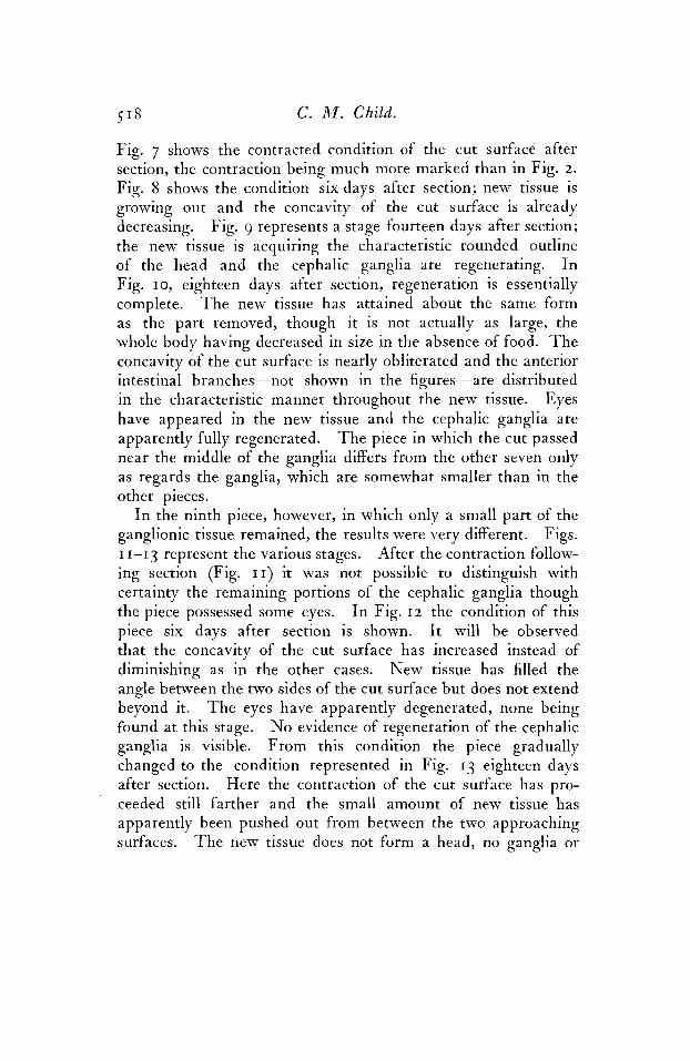
Fig. 7 shows the contracted condition of the cut surface after section, the contraction being much more marked than in Fig. 2. Fig. 8 shows the condition six days after section; new tissue is growing out and the concavity of the cut surface is already decreasing. Fig. 9 represents a stage fourteen days after section; the new tissue is acquiring the characteristic rounded outline of the head and the cephalic ganglia are regenerating. I n Fig. 10, eighteen days after section, regeneration is essentially complete. T h e new tissue has attained about the same form as the part removed, though it is not actually as large, the whole body having decreased in size in the absence of food. T h e concavity of the cut surface is nearly obliterated and the anterior intestinal branches-not shown in the figures-are distributed in the characteristic manner throughout the new tissue. Eyes have appeared in the new tissue and the cephalic ganglia are apparently fully regenerated. T h e piece in which the cut passed near the middle of the ganglia differs from the other seven only as regards the ganglia, which are somewhat smaller than in the other pieces.
I n the ninth piece, however, in which only a small part of the ganglionic tissue remained, the results were very different. Figs. I 1-13 represent the various stages. After the contraction follow- ing section (Fig. 1 1 ) it was not possible to distinguish with certainty the remaining portions of the cephalic ganglia though the piece possessed some eyes. I n Fig. 12 the condition of this piece six days after section is shown. It will be observed that the concavity of the cut surface has increased instead of diminishing as in the other cases. New tissue has filled the angle between the two sides of the cut surface but does not extend beyond it. T h e eyes have apparently degenerated, none being found at this stage. No evidence of regeneration of the cephalic ganglia is visible. From this condition the piece gradually changed to the condition represented in Fig. 13 eighteen days after section. Here the contraction of the cut surface has pro- ceeded still farther and the small amount of new tissue has apparently been pushed out from between the two approaching surfaces. T h e new tissue does not form a head, no ganglia or

Studies on Regulation. 5 I9
eyes are present, and the intestinal branches scarcely enter it. T h e margin of the new part is similar to the margins of the ad- joining old portions but no special differentiation of any kind can be observed. It is probable that the small portions of the cephalic ganglia remaining afxer section have undergone degeneration. No further advance in regeneration occurred even after months.
No less striking than the difference in regenerative power be- tween the eight pieces containing half or more of the ganglionic substance and the one piece containing only a small part is the difference in their behavior. T h e eight pieces behaved through- out essentially like normal animals. I thought I could distinguish a slight difference between them and uninjured specimens as regards rapidity and precision in locomotion, which might be expected from the absence of the chief tactile organ, the margin of the head, but the remaining parts of the head and the new tissue as soon as it became functional were even more active than these parts in uninjured animals; in the absence of the usual tactile and other stimuli from the anterior regions of the head the parts present were used all the more. T h e ninth piece, however, resembled in behavior a piece without cephalic ganglia. It was able to advance only slowly, did not adhere closely to the sub- stratum, the muscular movements were irregular and ineffective, and when turned upon its back the piece regained the normal position only after some time and many ineffective muscular contractions of various parts. T h e continual muscular plav of the lateral margins of the head was almost wholly absent in this piece. I n all probability the piece was practically without ganglia in consequence of the degeneration of the small portions of the ganglia remaining after section.
There can be little doubt that in this piece the region of the cut surface is subjected to conditions differing widely from those present in the other eight. I n the first place all conditions con- nected with the normal rapid progression are absent; the contacts with the substratum are much less close; owing to the lack of coordination and reactive power the conditions resulting from mus- cular movements of the head region are to a large extent absent; peristaltic and other muscular contractions of the whole body

5 2 0 C. M . Child.
or of parts are much less powerful, and consequently the pressure exerted upon the anterior region by such contractions through the intestinal contents or other fluids in the body are greatly reduced. I n the other eight pieces the head region functions all the more actively because parts are missing and the stimuli re- ceived from them are absent or received in a new manner. I n this piece, however, the movements of the head region are very slight. I n short, all or nearly all the functional conditions characteristic of the head region are absent or greatly reduced. Here again as in connection with posterior regeneration (Child, '04b) we find a close parallelism between functional activity and power of regeneration. T h e significance of the facts will become still more evident in the light of further data given below.
Description of other series would only multiply details without adding anything essential. All specimens cut through the middle o r anterior half of the ganglia behave much like normal animals and regenerate rapidly and completely while those in which only a small part of the ganglia remains behave much like specimens without ganglia, never regenerate a head, and apparently lose the small portion of ganglionic tissue by degeneration. T h e amount of new tissue regenerated is sometimes more and some- times less, but, as will appear below, various conditions may determine such differences.
I n the preceding section (p. 514) the statement was made that the greater the part of the head removed anterior to the ganglia the greater the rapidity of regeneration. This fact is well illus- trated by a comparison of the series described in that section (Series 16) with Series 17 described above. Both of these series were begun on the same day and both were examined at the same intervals so the results are strictly comparable. Figs. 2, 3 ,4 and 5 represent stages of Series 16 corresponding, respectively, to the stages of the eight pieces of Series 17, shown in Figs. 7, 8, 9 and 10. Fourteen days after section the pieces of Series 17 (Fig. 9) have regenerated about twice as much tissue as those of Series 16 (Fig. 4). Eighteen days after section regeneration in both is about complete, though the pieces of Series 17 (Fig. 10) have had about twice as much material to replace as those of Series 16

Studies on Regulation. 521
(Fig. 5). I am inclined to believe that functional conditions may account in large part for this difference. T h e larger the part which the new tissue represents the greater and more varied is its activity and if the various conditions connected with this activity affect growth in any way a more or less exact proportionality between the rapidity of regeneration and the size of the part removed may be expected.
3 . Anterior RPgeneration jrom Levels Immediately Porterior to the Cephalic Ganglia.
Anterior regeneration from levels only a short distance poste- rior to the cephalic ganglia differs in certain respects from that occurring from levels farther removed from the ganglia. Al- though nothing like a head is ever regenerated the amount of regeneration is usually somewhat greater than at other levels posterior to the ganglia and the new tissue possesses a somewhat different form. Individual differences which occur are doubtless due in part to slight differences in level of the plane of section, though, as will appear below, some cases indicate that internal factors differ in different individuals. T h e history of a series will serve to illustrate these points.
T h e head and ganglia were removed from a number of large specimens by a cut just posterior to the ganglia as in Fig. 14. I n all cases this cut was made as near as possible to the eyes, but without including any part of them in the posterior piece. All pieces were examined after section and those in which eyes were present in the posterior piece or in which the plane of section lay too far posteriorly were discarded. Five posterior pieces were finally obtained for the experiment.
A few hours after section all pieces appeared much like Fig. 15; contraction of the cut surface had occurred and as is usual in pieces without ganglia considerable longitudinal contraction had occurred so that the width of the body was greater than before section.
T h e pieces were examined every few days and the following figures show the condition of the various pieces six, seventeen and thirty-two days after section. Three types were recognizable as
Series 71.

5 2 2 C. M . Child.
regards the amount and form of the new tissue; these are desig- nated as A, B and C.
A. (Figs. 16-19.) One piece only regenerated in this manner. I t will be observed.from the figures that in this case the contraction of the cut surface was greater than in pieces containing ganglia (see Figs. 2-5), and that the new tissue grew out from the wound
as a rounded mass (Fig. I 7), which later became somewhat pointed (Fig. IS). Moreover, two groups of pigment spots, undoubtedly eye-spots, appeared in the new tissue just anterior to the cut surface. Regeneration never proceeded beyond the condition represented in Fig. 1 8 . No trace of regenerated cephalic ganglia could be observed at any time. Fig. 19 represents the contracted condition of the same stage; the form shown in Fig. IS appeared

Studies on Regulation. 5 2 3
when the piece was creeping; the other, Fig. 19, when the piece was at rest.
B. (Figs. 20-23.) Three pieces regenerated in this manner. Here the contraction of the cut surface was somewhat greater than in A and continued to increase thoughout the experiment. T h e new tissue grew out from the wound at first in rounded form (Fig. 21), but later acquired the tapering form shown in Fig. 22. No traces of ganglia or eyes appeared at any time. T h e differ- ences between the three pieces were slight. Figs. 22 and 23 represent, respectively, the extended form and the form during quiescence of these pieces. T h e new tissue was more pointed during the former condition.
C. (Figs. 24-26.) One piece regenerated in this manner. T h e course of regeneration in this case was much like that from more posterior levels; the contraction of the cut surface was greater than in the other cases and continued to increase. T h e new tissue filled the space between the sides of the wound, but never extended beyond the rounded margins of the old tissue. I n this piece the differences in form during movement and rest were very slight.
All of the pieces showed a somewhat greater degree of motor activity and power of coordination than pieces from which the anterior third or more of the body had been removed. After stimulation progression continued for a considerable time and the pieces changed their positions in the dishes from time to time without perceptible external stimulation. Differences in motor activity among the different pieces were not great but it seemed to me that the pieces A and B were somewhat more active than C, though in all cases of this kind differences are not strongly marked. I n A and to a less extent in B the new tissue occasionally showed movements resembling the searching movements of the head in normal animals and some slight irregular play of the margins of this region was observed. I n C nothing of this kind occurred. T h e changes in form of the anterior ends in A (Figs. 18 and 19) and B (Figs. 22 and 23) are of interest; when the pieces extended and advanced the new tissue became somewhat more slender and pointed as if it were pushed forward by internal
5 24 C. M . Child.
pressure, while during rest it assumed a more rounded form. This change in form is doubtless due primarily to muscular con- tractions; the decrease in transverse diameter in consequence of muscular contraction during progression must produce pressure in the direction of the longitudinal axis at the anterior end-and at the posterior end also if this is a cut surface: this pressure must bring about elongation of the new parts; hence the change of form in these regions. There can be little doubt that this pres- sure constitutes a factor in the outgrowth of new tissue from a cut surface and the form which it acquires. If this is the case the greater outgrowth in A and B may be due in part to the fact that such pressure has been more frequent or perhaps greater in amount than in C. T h e different degrees of contraction of the cut surface in the different pieces are also very probably due to this or other similar factors connected with motor activity.
T h e appearance of eyes in A is probably due to the outgrowth of nerves from the cut end of the cords and their union with the epithelium.
These five pieces, which were cut as nearly as possible at the same level, afford a good illustration of the difficulty of complete control of experiments of this kind. It is very probable that the differences are due to slight differences in level of the cut or in the extent of injury to the tissues posterior to the cut surface, but it is impossible to determine with certainty whether this is the case. T h e points of chief importance are, however, the occur- rence of a considerable amount of anterior regeneration (in A and B) in the absence of the ganglia and the somewhat greater degree of motor activity in all of these pieces, and especially in A and B, as compared with regions further posterior. I n this connec- tion it is also of interest to note that regeneration was more rapid in A than in B and in B than in C as may be seen by comparing the three sets of figures. Thirty-two days after section A (Figs. 18 and 19) had regenerated more than twice as much new tissue as B (Figs. 22 and 23) and several times as much as C (Fig. 26). But even in A regeneration is much less rapid and the total amount is less than in pieces containing the ganglia. Comparison of Figs. 2-5 and 8-10 with Figs. 15-18 will illustrate this fact. In
Studies on Regulation. 5 2 5
these pieces differences of this kind cannot be correlated with differences in the size of the part removed, since this was approxi- mately the same in all cases. They are rather to be compared with the cases described in the preceding paper (Child '04b) in which the rapidity and amount of posterior regeneration from a given level was much greater in the presence of the ganglia than in their absence, and I believe that they are to be explained in the same manner, viz: as due chiefly to differences in motor activity.
In no case did a characteristic head regenerate in the absence of the ganglia, but regeneration from a level immediately posterior to the ganglia was in almost every case more rapid and greater in amount than from more posterior levels. Moreover, whenever differences in motor activity could be distinguished the most active pieces showed the greatest amount of regeneration.
Numerous similar experiments afforded similar results.
4. Anter ior Regeneration from Other Levels Posterior to the Cephal ic Gangl ia .
When both the ganglia and the region immediately posterior to them are removed, anterior regeneration is usually even less in amount than in the cases above described; occasional exceptions to this rule occur, however, some of which will be described below. But in general the amount of anterior regeneration from these levels is inversely proportional to the size of the part removed, or in other words, the farther posterior the level from which it occurs, the less the regeneration. This relation is exactly the reverse of that observed in connection with posterior regeneration (Child, 'o4b) ; there the amount of regeneration is directly proportional to the size of the part removed.
My experiments along these lines include nearly two hundred specimens but with only one marked exception as regards the amount of new tissue and three cases in which a few eye-spots appeared. Experiments in three different regions are selected for description. The levels are indicated by the three unbroken transverse lines in Fig. 27. The results obtained from section at the most anterior of these three levels are indicated in Figs. 28-35. Figs. 28-3 I represent the history of a piece in which the cut surface

C. M . Child.
34 m 36
m remained widely open and Figs. 32-35 that of a piece with mar- gins of the cut approximated. These two pieces represent the extreme forms obtained after section at this level. T h e stages represented are as follows: Figs. 28 and 32 , a few hours after section; Figs. 29 and 33, six days after section; Figs. 30 and 34,
Studies on Regulation . 5 27
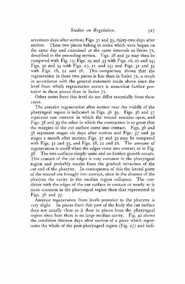
seventeen days after section; Figs. 31 and 35, thirty-two days after section. These two pieces belong to series which were begun on the same day and examined at the same intervals as Series 71, described in the preceding section. Figs. 28 and 32 may then be compared with Fig. 15; Figs. 29 and 33 with Figs. 1 6 ~ 2 0 and 24; Figs. 30 and 34 with Figs. 17, 21 and 25; and Figs. 31 and 35 with Figs. 18, 22 and 26. This comparison shows that the regeneration in these two pieces is less than in Series 71, a result in accordance with the general statement made above since the level from which regeneration occurs is somewhat further pos- terior in these pieces than in Series 7 1 .
Other series from this level do not differ essentially from these cases.
T h e anterior regeneration after section near the middle of the pharyngeal region is indicated in Figs. 36-39. Figs. 36 and 37 represent one extreme in which the wound remains open, and Figs. 38 and 39 the other in which the contraction is so great that the margins of the cut surface come into contact. Figs. 36 and 38 represent stages six days after section and Figs. 37 and 39 stages a month after section; Figs. 37 and 39 may be compared with Figs. 31 and 35, and Figs. 18, 22 and 26. T h e amount of regeneration is small when the edges come into contact as in Fig. 38. T h e two surfaces simply unite and no further growth occurs. This contact of the cut edges is very common in the pharyngeal region and probably results from the gradual retraction of the cut end of the pharynx. I n consequence of this the lateral parts of the wound are brought into contact, since in the absence of the pharynx the cavity in the median region collapses. T h e con- dition with the edges of the cut surface in contact or nearly so is more common in the pharyngeal region than that represented in Figs. 36 and 37.
Anterior regeneration from levels posterior to the pharynx is very slight. I n pieces from this part of the body the cut surface does not usually close as it does in pieces from the pharyngeal region since here there is no large median cavity. Fig. 40 shows the condition thirteen days after section of a piece which repre- sents the whole of the post-pharyngeal region (Fig. 27) and indi-

5 2 8 C. M . Child.
cates the maximum amount of regeneration obtained in cases of this kind. I n most cases such pieces die within ten days after section and none were kept alive more than twenty-eight days. Shorter pieces cut further posteriorly do not live as long and show even less regeneration than these.
These three examples are sufficient to indicate the decrease in the power of anterior regeneration with approach toward the posterior end of the body. T h e results obtained in other series were in general similar.
As regards motor activity the differences at different levels are parallel to the differences in regenerative power. T h e further posterior the level of section the less the motor activity. Pieces from widely different levels like those considered above (Fig. 27) show marked differences in motor activity. T h e pieces of the first series, which had lost only the ganglia and a short portion of the cords, were almost as active as Series 71 considered in the preceding section, which had lost only the ganglia; progression occurred, though of course slowly, and the pieces were able to adhere to the substratum in some degree and to right themselves when turned over. T h e special activity of the anterior end, present in some degree in Series 71, was not observed in these pieces. T h e pieces with anterior ends near the middle of the pharyngeal region were distinctly less active. When progression occurred it was less rapid than in the first set, direct comparison between pieces of the two kinds being frequently made; the pieces righted themselves less readily and sometimes did not succeed at all; adhesion to the substratum was very slight; and finally all movements were less frequent and powerful. But the pieces from the region posterior to the pharynx showed even less motor activity. Very slow progression sometimes occurred in conse- quence of ciliary movements, but in such cases the piece simply slid along without holding to the substratum and frequently on the dorsal surface. T h e pieces were usually incapable of righting themselves. Reactions to stimuli were slight and the pieces showed few traces of movement of any kind when left undisturbed. I n the normal animal this region of the body is the chief organ of attachment during locomotion, but separated from other parts it

Studies on Regulatioiz. 5 29
is incapable of more than a very slight degree of attachment. I n the absence of the mechanical tension to which the tissues are normally subjected, the apparent reduction in size of these pieces is considerable (compare the posterior part of Fig. 27 with Fig. 40. See also Child, '02). This apparent reduction is due in part to altered physical conditions, for the decrease in the longitudinal and transverse diameters is accompanied by a relative increase in the dorso-ventral diameter. I n general the same parallelism between motor activity and regenerative power is found in these pieces from different regions as in those already described.
Brief mention must be made of a few special cases of anterior regeneration which differ in certain respects from the average. Groups of eye-spots appear occasionally in the new tissue of posterior pieces not only when the plane of section is immediately posterior to the ganglia as in piece A, Series 71 (p. 522) but even at levels as far back as the anterior end of the pharynx. Except where the plane of section is very near the ganglia the eye-spots appear only after two or three months. I n Fig. 41 a case of this sort is shown; here the cut was made at the level of the anterior end of the pharynx and the eyes were first observed two and one- half months after section. Other cases do not differ essentially; sometimes only one or two eye-spots appear and sometimes two or three groups of them are visible. It is probable that outgrowth of nerves from the cut ends of the nerve cords is responsible for the formation of these structures. If this is the case it is an inter- esting fact that the eye-spots are developed in such cases in con- nection with a part of the nervous system different from that with which these organs are usually connected. This is another bit of evidence in favor of the view that the differences between different regions of the central nervous system are differences of degree rather than of kind. Regions of the body posterior to the anterior end of the pharynx have never been seen to produce eye- spots; on the other hand they are most common in pieces like A, Series 71, from which only the ganglia have been removed. T h e frequency of regeneration of these organs decreases poste- riorly like the power of regeneration and of motor activity. Prob- ably these organs are the result of a direct influence of the nervous

5 30 C. M . Child.
system. Another exceptional case was that of a piece cut obliquely at the level represented by the dotted line in Fig. 27. Twelve pieces composed the series of which this was one. After about five weeks it was noticed that eye-spots were present in this one piece, and also that the amount of new tissue was greater than in any other pieces of the series. T e n days later (forty-eight days after section) the piece had attained the condition represented in Fig. 42. T h e regenerative power of this piece was as great as that of the best pieces cut immediately behind the ganglia (A, Series 71, Fig. IS). As regards the motor activity of this piece during early stages, I can say nothing, since the pieces of the series were not examined individually and compared, but after the piece had reached the stage shown in Fig. 42 its motor activity was distinctly more complete than that of the other pieces of the series. It was capa- ble of more rapid progression and the margins of the new tissue were used tQ some extent like those of a head. Fifty-eight days after section the piece had attained the condition of Fig. 43; the new tissue had increased still further in amount and the motor activity of the piece, especially that of the new tissue, was greater than before. Beneath the large group of eyes a small light area, probably regenerating ganglia, was observed. It is very evident that in this case there is an approach to regeneration of a head. T h e new tissue is used as a head though in less degree than in the normal animal. It is impossible to assign a definite reason for the occurrence of this single case. I n all my experiments nothing of the sort has ever been observed in other pieces at this level. Moreover, this piece shows a degree of regeneration as great as the best cases from levels immediately posterior to the ganglia (A, Series 71, Fig. IS). Whether complete regeneration of the head would have occurred could not be determined. Seventy- eight days after section the piece died without having advanced perceptibly beyond the stage of Fig. 43. Possibly if the piece could have been fed complete regeneration might have taken place. At all events this case is of great interest, since it indicates that under certain conditions the amount of regeneration at a given level may be much greater than the usual amount. Whether
No other similar case has ever been observed.

Studies G n R egiila tion. 53 1
the result in this case was due to some difference in the distribu- tion or arrangement of nervous structures, to exceptional vigor, or to an exceptional amount of reserve energy it is impossible to determine. T h e parallelism between motor activity and regenera- tive power is also well shown in this piece.
5 . Anter ior Regeneration A f t e r Repented Section.
I n his work upon Leptoplana atornata Schultz (’02) reached certain conclusions widely different from my own with respect to anterior regeneration, yet the results of experiments described by him are essentially similar to my own as far as they go. As will appear, Schultz’s conclusions are probably due to the fact that his experiments were confined to a particular region of the body. He was never able to obtain anterior regeneration “selbst bei solchen Exemplaren nicht, denen nur ein geringer vorderer Kor- perabschnitt, weit vor dem Pharynx, abgeschnitten wurde.” T h e position of the cut with respect to the cephalic ganglia in such cases is not stated, but there can be little doubt that it was pos- terior to the ganglia. I n fact, it is probable that Schultz never observed anterior regeneration in the presence of the cephalic ganglia. I have no doubt that if he had done so he would have obtained results similar to my own and would have reached entirely different conclusions regarding the absence of anterior regeneration. His views on this subject are briefly stated as follows : the margins of the cut come together in such manner that union occurs and thus the region where regeneration would begin (“Regenerationspunkt”) is separated from the periphery by old tissue and, moreover, the union of the muscular layers over it prevents further growth.
This attempt at a simple explanation of the absence of anterior regeneration involves, I believe, a complete inversion of the actual course of events. ’The cut edges unite because there is not suffi- cient new tissue formed to prevent this union, which is probably due to mechanical conditions in the tissues. It is very probable, however, that after their union the outgrowth of new tissue from the end cannot occur. T h e multiplication of parenchyma cells which Schultz describes as occurring within the muscular layer

5 3 2 C. M . Child.
would probably, if exposed, not produce anything like complete anterior regeneration except in the presence of the cephalic ganglia. It is of course possible that differences between L. atomata and L. tremellaris may exist; for example, in the former the produc- tion of new tissue at the anterior end in the absence of the cephalic ganglia may be so slow that union of the sides of the cut occurs before its amount is appreciable, while in the latter sufficient new tissue is formed to prevent complete union of the old cut edges. It is extremely improbable, however, that regeneration of the region anterior to the cephalic ganglia does not occur. Even in the rhabdocda, most of which have little power of regeneration, the preganglionic region is regenerated rapidly and completely. T h e experiments already described are, I think, sufficient to show that no such simple explanation as that of Schultz’s will suffice for L. tremellaris. Certain other experiments which I performed with this particular purpose in view mav be described since they afford some facts of interest.
I n these experiments posterior pieces from various levels were employed, but chiefly those from the pharyngeal region, since contraction of the cut surface is most marked here (see Figs. 38 and 39). After section such pieces were allowed to remain un- disturbed during several days in order that the cut surface might be contracted as much as possible. Then they were subjected to a second operation, in which the lateral regions which had been drawn toward the median plane by the contraction were removed.
Two series of this kind from different levels are described; these were begun at the same time and examined at the same intervals and so are available for comparison as to the amount of regenera- tion at different levels after a second operation.
Series 75. Five posterior pieces were obtained by cuts just anterior to the anterior end of the pharynx.
Eight days after section all were either like Fig. 44 or somewhat more widely open, the space being filled with new tissue. All were cut in the manner indicated in Fig. 44. Contraction after this second operation was comparatively slight and the anterior cut surface remained widely open in every case. T h e condition

Studies on Regulation. 533
of the pieces ten days later is indicated in Figs. 45, 46 and 47. I n spite of the fact that there was no trace of any physical obstacle to growth, such as Schultz believes the closure of the cut surface constitutes, little regeneration has occurred. T h e condition of the pieces a month after the second operation is indicated in Figs. 48, 49 and 50. T h e new tissue has increased slightly in amount but although there is no union of the old muscular layers on the
44
m m 53
67 5.5
cut surface no head has been formed. After another month no further change except reduction in size had occurred.
Five posterior pieces were obtained by a transverse cut through the middle of the pharyngeal region. Eight days after section all resembled Fig. 51, the contraction of the cut sur- face being in some cases somewhat greater, in others less. At this time the anterior part of the contracted lateral regions was removed in the manner indicated by the lines in Fig. 51.
Figs. 52, 53 and 54 represent the condition of the pieces ten days after the second operation. T h e double outgrowth in Fig.
Series 76.

534 C. M . Child.
54 is of interest; growth was probably more rapid in the lateral regions in consequence of the presence of nerve cords and perhaps also because of the presence of the pharyngeal pouch and the almost complete absence of parenchyma in the median line. A similar duplication was observed in a number of cases of this kind, but was in all cases only temporary, disappearing completely in later stages.
A month after the second operation all five of the pieces pre- sented about the condition shown in Fig. 55, the differences being slight. No further regeneration occurred.
Here, as in Series 75, there is no physical obstacle to growth, such as Schultz believes to exist, yet regeneration of the head does not occur.
These two series show clearly that the explanation of the absence of anterior regeneration given by Schultz for L. atomata certainly does not apply to L. tremellarts. There is little doubt that the conditions in L. atomata are similar. Apparently the cut surface in L. atomata contracts to a greater extent after section than that of L. tremellarts but repeated section in the manner described would undoubtedly leave open cut surfaces sufficient for the occurrence of regeneration.
These two series are also of interest in connection with the question discussed in the preceding section, viz : the relation between the level from which regeneration occurs and the amount of regeneration. T h e level of the anterior ends in Series 75 is just anterior to the pharynx while in Series 76 it is the middle of the pharyngeal region, i. e., much further posterior. At the time of the second operation, eight days after the first, the amount of regeneration is greater in Series 75 (Fig. 44) than in Series 76 (Fig. 51). Ten days after the second operation a similar differ- ence between the two series exists (compare Figs. 45-47 with Figs. 52-54) and the same is true of the final stages a month later (compare Figs. 48-50 with Fig. 55). As regards motor activity a distinct difference between the two series was noted, Series 75 being the more active and capable of somewhat more coordinated movement. T h e differences in activity between the two series were noticeable, chiefly in the later stages. These facts agree with

Studies on Regulation. 535
the others presented in the earlier sections and show that the degree of contraction of the cut surface is not the determining factor as regards the amount of regeneration, but that both the contraction and the regeneration are determined by other factors certain of which differ qualitatively according to the level.
6. Re'sume' of the Experiments o n Anterior Regeneration.
T h e preceding sections have established several facts of impor- tance regarding anterior regeneration. It has been found that a t all levels anterior to the middle of the cephalic ganglia anterior regeneration is complete and its rapidity is in general proportional to the size of the part removed. At all levels posterior to the middle of the cephalic ganglia anterior regeneration is incom- plete and the rapidity and amount of regeneration are in general inversely proportional to the size of the part removed, i. c., the further posterior the level from which regeneration occurs the less the rapidity and amount of regeneration. Complete regen- eration of the cephalic ganglia is possible when no more than half the ganglionic material is removed, but never occurs after the removal of a larger part or all of the ganglionic material. When the ganglia are absent, or if they fail to regenerate completely, nothing that can properly be called a head is regenerated. A close parallelism between the degree of motor activity and the power of regeneration exists in all cases. T h e relation between the cephalic ganglia and anterior regeneration is very different from that between the ganglia and posterior regeneration (Child, '04b). Posterior regeneration is qualitatively complete, though some- what reduced in amount in the absence of the ganglia while under similar conditions anterior regeneration is both qualitatively and quantitatively incomplete in high degree. T h e correlation be- tween the ganglia and posterior regeneration, which is merely quantitative, was interpreted in the preceding paper as essentially functional, the motor activity of the parts being much greater though not widely different qualitatively when the ganglia are present. As regards anterior regeneration the case is different; the motor activity of this region of the body is not only less in degree in the absence of the ganglia but it is different in kind.

536 C. M . Child.
Since the head, like other parts of Leptoplana, is in large degree a complex organ of locomotion, we find that motor activity is an important factor, perhaps the most important in the formation of a characteristic “head.” T h e anterior end does not show the characteristic activity of a “head” in the absence of the ganglia and no head is regenerated. O n the other hand, in all cases where a sufficient amount of the ganglionic tissue remains intact t o permit the continuance of the characteristic functional activity the regeneration is both qualitatively and quantitatively complete and forms a “head.” I n short a “head” is regenerated at the anterior end when this part of the body functions in the manner characteristic of a head. T h e relation between the indeterminate anterior regeneration in the absence of the ganglia and motor activity is indicated by the parallelism between regeneration and motor activity at different levels and from this results the inverse proportion between the amount of material removed and the amount of anterior regeneration from levels posterior to the gan- glia.
We may conclude then that in the presence of cephalic ganglia a characteristic head must develop since the ganglia determine the functional relations of the various parts and so a characteristic structure results. But why do not the ganglia themselves regen- erate after they are completely removed? Briefly stated the answer is this: because the other parts of the nervous system do not give rise to the functional conditions necessary for formation of the ganglia.
T h e first stage in anterior regeneration is, as in the case of posterior regeneration, the appearance of new tissue at the cut surface (see Child ’o4b). T h e appearance of this tissue is doubt- less the result of the altered conditions in this region and is in part a rearrangement of old material in consequence of altered mechanical conditions and in part a proliferation. This material is without doubt capable of forming any region of the body, for if it arose from a posterior cut surface at the same level it would regenerate into a posterior end. Its fate is a function of its position, if we interpret position as not simply space-relation but functional correlation with other parts. In other words the new

Studies on Regulation. 537
tissue develops according to the manner in which it is used by the animal or piece. I n the presence of the cephalic ganglia or a sufficiently large portion of the ganglionic tissue the anterior end of the piece is used in a characteristic manner and this functioning must subject the new tissue to a characteristic complex of con- ditions. It is not my purpose to attempt a description or enu- meration of all the possible factors concerned in the differentiation of the parts; such an attempt would be at present in large part a series of surmises. Among these factors, however, are functional nerve stimuli, chemical and physical conditions in the tissues resulting from them, all the complex of metabolic factors as in- fluenced by the particular conditions, and the conditions, mechan- ical and otherwise, connected with and resulting from motor activity or attempts at such activity.
T h e relative importance of different factors differs widely in different cases. I n the present case the motor activity determined by the presence or absence of the ganglia is of great importance. It is only on this basis that we can explain the fact that anterior to the ganglia the amount and rapidity of regeneration are directly proportional to the amount of material removed. We have, I think, no adequate conception of the complex interactions leading to the formation of such a structure as the head of Leptoplana, but that motor activity is an important factor, mechanically and perhaps otherwise, cannot be denied. T h e constantly varying physical conditions resulting from motor activity may seem incapable of givino rise to any definite or characteristic form, but the important fact is that, notwithstanding their constant changes, they COQ-
stitute a characteristic series frequently repeated. From the time when the first traces of motor activity appear in the new tissue up to complete development the animal is using or attempting to use this part in a definite characteristic manner. T h e arrange- ment of muscles and the nerve connections are of course directly responsible for this characteristic motor activity, but the charac- teristic arrangement of these structures can be accounted for only by other characteristic functional activities and their correlations, and so on. We are led finally to a protoplasm possessing certain elementary and characteristic activities, but that any substances
b

538 C. M . Child.
representing in themselves a “head” or other morphological features are present it is difficult to believe. T h e head of Lepto- plana, like other parts of the body, represents the effect of a series of characteristic activities upon a given complex of substances in a given environment.
R . LATERAL REGENERATION.
Only a brief consideration of the phenomena of lateral regener- ation is necessary since the relations are not essentially different from those already described for anterior and posterior regenera- tion. I n general the amount and rapidity of lateral regeneration in the presence of the cephalic ganglia are directly proportional to the amount of tissue removed. I n the absence of the ganglia the amount and rapidity of regeneration differ in different cases; if the size of the part removed is not great the amount and rapidity of regeneration are directly proportional to it; if on the other hand the portions removed constitute the greater part of the body the amount and rapidity of regeneration are inversely proportional to the size of the part removed Qualitatively, however, lateral regeneration resembles in most respects posterior regeneration, in that absence of the cephalic ganglia does not alter the structural character of the regenerated tissue except in the extreme anterior region, though it does reduce its amount.
As in the case of anterior and posterior regeneration it was found that when about half or more of the ganglionic tissue was present complete regeneration occurred, but when less than this amount was present the results approached those obtained from pieces without ganglia. Specimens separated into halves along the median line, each half containing one-half of the ganglionic mass, were capable of complete regeneration (Child, ’o4a). If, how- ever, the plane of section did not coincide with the median plane complete regeneration occurred only in the piece containing the larger part of the ganglionic mass.
As might be expected the relation between the cephalic ganglia and motor activity is the same in pieces cut longitudinally as in other pieces. When half or more of the ganglionic substance is present the pieces behave essentially like normal animals, but
Studies on Regulation. 539
when less than half the ganglionic tissue remains their behavior approaches that of pieces without ganglia. Moreover, if after removal of the ganglia by a transverse cut the body be split longi- tudinally or near the median plane the two halves show similar activity which is not very different from that before their separa- tion; if, however, the plane of section lies far to one side of the median plane the smaller piece shows scarcely any motor activity, while the longer retains the characteristic activity of a piece with- out ganglia. These differences are of course, due to the nervous system. When the plane of section lies near the median plane each half contains the nervous structures of that half of the body, but as the plane of section approaches the lateral margin the smaller pieces contain less and less of the nervous system until in the extreme lateral region nothing remains in them but some of the peripheral nerves. Probably closer observation would show rapid changes in motor activity according as the planeof section passed on one side or the other of one of the nerve cords, but I I have not paid especial attention to this point.
Comparison of this description of the motor activity of longi- tudinally cut specimens with the statements regarding lateral regeneration renders it clear at once that the same parallelism between motor activity and regeneration exists as in the cases already discussed, so that nothing is to be gained by going over the whole series of experiments upon lateral regeneration. I shall describe only a few cases by way of illustration, calling attention to certain points of especial interest.
Cases of the removal of less than half the body by longitudinal section from end to end need not detain us since in all cases the amount and rapidity of regeneration vary with the size of the part removed, 1'. e . , the larger the part removed the more rapid the regeneration, so that regeneration of a large part does not require much more time than regeneration of a small part. These relations are similar to those described for posteribr regeneration from levels posterior to the ganglia and for anterior regeneration in the presence of the ganglia, and are to be interpreted in the same manner as due to the r6le which the new tissue is required to play in functional-doubtless chiefly motor activity.

540 C. ill. Child.
Regeneration after longitudinal section near the median plane and regeneration of small lateral pieces present some features of interest; a description of cases of each kind is accordingly given.
Series 64. A large specimen was sectioned longitudinally slightly to the left of the median plane (Fig. 56). T h e plane of section passed through the left cephalic ganglion, leaving only a small portion of the ganglion together with most of the eyes in the left piece. After section both pieces underwent considerable contraction, but the left much more than the right. T h e two pieces differed widely as regards behavior; the right piece, con- taining nearly all the ganglionic tissue, behaved like a normal animal, advancing rapidly and using the margin of the head in the characteristic manner. T h e left piece, on the other hand, behaved essentially like a piece without ganglia. It never became extended, did not adhere closely to the substratum, all movements were slow and the head region showed none of the characteristic motor activity of normal animals. T h e pieces were examined at intervals of from seven to ten days.
T h e most conspicuous difference between the two pieces is the difference in form (compare Figs. 57 and 58). T h e right piece is much contracted and bent toward the left by the reduction of the cut surface, but the left piece is contracted into almost circular form with the cut surface greatly reduced. Both pieces move in circles in consequence of the form but the radius of the circles described by the right piece is much greater than in the left piece, which simply revolves in a space scarcely greater than its own diameter.
T h e difference in form and consequently the difference in direction of movement is of course the direct result of the widely different degree of contraction in the two pieces; and this difference in contraction is undoubtedly due in large part if not wholly to the differences in motor activity in the two pieces; and finally the differences in motor activity depend essentially upon the distribu- tion of the tissue of the cephalic ganglia between the two pieces. T h e right piece creeps about like a normal animal holding to the substratum by means of the margin and posterior end; thus the body of this piece is subjected to the characteristic longitudinal

Studies on Regulation. 5 4 1
tension resulting from progressive movement (Child, 'o4a) which antagonizes the conditions at the cut surface. Thus, as the animal advances, the cut surface is frequently stretched to a considerable extent. T h e position represented in Fig. 57 is between the two extremes and represents the usual form during undisturbed pro- gression. In the rapid progressive movements following strong stimulation the body often becomes almost straight and on the other hand when at rest or moving very slowly is more curved than in the figure.
In the left piece, however, rapid progression does not occur and the body adheres only very slightly; consequently the body is not subjected to longitudinal tension and the cut surface is not stretched but continues to contract indefinitely, like the anterior cut surface in pieces without ganglia. These cases only confirm the opinion already expressed that ordinary muscular contraction has little or nothing to do with the contraction of cut surfaces; it may be that muscles which have been cut and are thus free at one end gradually shorten, but this process is by no means the same as muscular contraction in the ordinary sense. Even after long periods the animals are unable voluntarily to straighten their bodies; nothing but mechanical tension in the longitudinal direc- tion will accomplish this end-and this fact is one of the most striking proofs of the influence of mechanical tension upon form in these animals. As has been suggested elsewhere the contrac- tion is probably due to surface tension, capillarity, elasticity and other mechanical factors, which are visibly effective in this way only in the absence of the mechanical conditions which under normal circumstances antagonize them. Doubtless also the re- duction in functional activity of the parts is accompanied by more or less reduction in size.
Comparing now the regeneration of the two pieces it is evident that it is much greater in amount in the right piece (Fig. 57) than in the left (Fig. 58). T h e thickness of the new tissue is about the same in both, but in the right piece its width and length are both greater than in the left piece. Intestinal branches are growing, out into the new tissue anterior to the pharynx in the right piece, but none are visible in the left piece. T h e new tissue, especially

542 C. M . Child.
its anterior portion, was used by the right piece in movement, while in the left no such use was observed.
Forty-six D a y s after Section. (Figs. 59 and 60.) T h e two pieces present the same differences in form and activity as before. I n the right piece (Fig. 59) the amount of new tissue is much greater than before, especially anterior to the pharynx, and the intestinal branches have grown well out into the new tissue in the anterior region and to a less extent in the region posterior to the pharynx. I n the left piece, on the other hand, the changes are only slight. T h e amount of new tissue is scarcely greater than before, and the intestinal branches extend only a very short dis- tance into the anterior region and are not visible elsewhere.
O n e H u n d r e d aizd Six D a y s after Section. (Figs. 61 and 62.) I n the right piece (Fig. 61) the relative amount of new tissue has continued to increase and regeneration of the left ganglion and the left side of the pharynx is complete. T h e width of the new tissue is greatest anterior to the pharynx undoubtedly in conse- quence of the greater motor activity of this part and in this region the intestinal branches extend out to the margin and at the anterior end of the pharynx some branches extend posteriorly. Posterior to the pharynx intestinal branches are also present though they do not fill the new tissue so completely, and some of them extend anteriorly at the posterior end of the pharynx. No intestinal branches are present on the left side of the pharyngeal region except those that extend into it from around the two ends. T h e posterior end of this piece is now almost straight (compare Fig. 57); it has been subjected more frequently and in greater degree than the other parts to longitudinal tension, since it is the chief organ of attachment.
is possible that some degree of regeneration of the cephalic ganglia has occurred, though new ganglia are not clearly visible; at any rate more or less extension often occurs and results in bringing the piece into the form shown in Fig. 62. T h e physical conditions of the old tissues near the cut surface have gradually changed during contraction and the new tissue, having arisen while the piece was contracted, is capable of much less active extension than the old
T h e left piece, on the other hand, has not changed greatly.

Studies on Regulation. 5 43
parts, for the latter were originally much more extended and have not lost that power completely. When the piece extends these new parts cannot stretch as far as the old and consequently the piece assumes the spiral form shown in Fig. 62. This is the nearest approach to straightening that is possible in this piece. I n this condition the piece simply creeps over itself in the direction of the arrow. As the piece becomes quiet it gradually assumes the form of Fig. 60, though it is of course smaller than at that stage. Re- generation in this piece has not advanced perceptibly during the two months since the stage of Fig. 60.
When we compare the two pieces it is evident that although the amount of material removed was almost exactly the same in both yet the results are very different. Here, as in other cases, the only satisfactory interpretation of the difference in results is that which regards them as the consequence of the differences in functional activity of the two pieces.
T h e results of longitudinal section near the median plane were examined in numerous other series. I n every case where one piece contained most of the ganglionic tissue and the other only a small portion the results were similar to those just described. I n cases where the plane of section was nearer the median plane the difference between the two pieces was not as great and the piece containing the smaller part of the ganglia regenerated the missing parts more or less completely, and in all such cases the motor activity and general regenerative power approached that of the other piece as the part of the ganglia remaining became larger.
I n a few cases I succeeded in making the section so near to the median plane that both pieces contained approximately equal parts of the cephalic ganglia. Both behaved essentially like normal animals and regenerated completely. Other cases of this sort are described in another connection in an earlier paper (Child, '04a).
I n all pieces capable of complete regeneration, like the right piece in Series 64, certain features of interest were noted. First, the new tissue in the region anterior to the pharynx regenerated more rapidly than that in other regions and finally became wider than at any other point (Figs. 57,59,61); second, in this region the

544 C. M . Child.
intestinal branches grew into the new tissue more rapidly than elsewhere and finally filled it almost as completely as in a normal animal; some branches also extended from this region posteriorly into the pharyngeal region; third, intestinal branches also ap- peared in the new tissue posterior to the pharyngeal region but somewhat later than those in the head region; these never attained so great an egtent as those in the anterior region but some ex- tended anteriorly into the pharyngeal region.
These three features were, as stated, characteristic of all pieces of this kind and must therefore possess a certain significance. T h e first two, viz: the more rapid regeneration of the lateral regions of the head and the more rapid growth of the intestinal branches in this region are connected. In my opinion both are due to the fact that this region shows the greatest motor activity of any part of the body. Its characteristic activities have already been de- scribed (Child, 'o4a). Characteristic conditions of tension and pressure result from these characteristic movements and these are undoubtedly factors in the arrangement of the material and so in determining the form and may themselves constitute stimuli to growth. As regards the intestinal branches the internal pres- sure due to intestinal contents must undergo change with the movements as the fluid is forced into or out of the branches as contraction or extension occurs. I n the head-region the internal pressure in the peripheral branches of the intestine is greater than in other parts, i. e., the contents are forced into these branches more frequently and probably also with greater pressure than elsewhere. Brief observation of a specimen with well-filled in- testine is sufficient to demonstrate these facts very clearly. There can be no doubt that these conditions of internal pressure play a part in the development of the intestine; some experimental evidence upon this point has been obtained from study of another species; this I hope to present at another time. Admitting that the internal pressure is a factor in the development of the intestine it is easy to see that the same conditions, viz: greater functional motor activity, which bring about the more rapid regeneration in the anterior region also bring.about the more rapid growth of the intestinal branches in this region.

Studies on Regulation. 5 4 5
T h e region posterior to the pharynx does not show as great a degree of motor activity as the anterior region and we find corre- spondingly that the development of the intestinal branches is less rapid here than there.
T h e growth of intestinal branches in a postero-lateral direction from the pharyngeal region and in an antero-lateral direction from the region posterior to the pharynx is also of much interest. I n none of these pieces did lateral intestinal branches regenerate from the pharyngeal region.
We may regard the branched form of the polyclad intestine as primarily the result of distension by internal pressure of a sac sur- rounded by parenchymatous tissue. T h e distension occurs along paths of least resistance and greatest internal pressure. I ts direction is the result of several factors, viz: the direction of inter- nal pressure, the position of paths of least resistance, and the presence or absence of antagonistic external pressure, however produced.
Since the internal pressure in each branch is transferred in some degree to the tissues about it this pressure must constitute for an adjoining branch external pressure antagonizing the internal pressure in the second branch and so preventing its extension toward the first. In short, intestinal branches cannot enter a region already occupied by such branches unless the internal pressure in the former is much greater than in the latter.
I n the case under consideration at present, viz: the growth of intestinal branches in “abnormal” directions from the regions anterior and posterior to the pharynx into the pharyngeal regions, I believe that the absence of intestinal branches in this region is the determining condition. The intestinal contents, being forced into the branches anterior and posterior to the pharynx, exert pressure upon the walls of these and growth occurs along the lines of least resistance. Under ordinary conditions the presence of other similar branches would in consequence of the pressure upon the tissues prevent the growth of branches postero-laterally in the one case and antero-laterally in the other (Fig. 61). Here, how- ever, such branches are absent and consequently the mechanical resistance to extension of the branches in this direction is not

C. M . Child.
58 60

Studies on Regulation. 5 47
greater than elsewhere; hence the “abnormal” or as it might be called “regulative” distribution of these branches.
But the absence of intestinal branches in the pharyngeal region in these pieces is itself a fact of importance. Though I was un- able to determine with certainty the particular conditions to which this absence of regeneration is due certain points which appeared to me suggestive may be mentioned here. First of all it must be remembered that in the absence of food the intestinal contents decrease in amount during the course of these experiments and consequently internal pressure diminishes; the movements and muscular contractions force the contents into one part or another of the intestine thus producing distension now in one region, now in another; secondly, the regions of greatest muscular activity are the regions anterior and posterior to the pharynx, especially the former; and it is in these regions that the movements of the intes- tinal fluid are visibly most frequent and powerful. T h e lateral margin of the body in the middle region shows much less motor activity than the te.rminal regions and in pieces in which the longi- tudinal axis is bent by contraction of one side this difference is especially marked on the concave side. And finally, when con- traction of the longitudinal muscles occurs and the intestinal contents are forced back toward the pharyngeal region the concave side of the body becomes still more concave and the new tissue of this side is pressed together and often thrown into folds, a con- dition which must retard or prevent the growth of intestinal branches into this region. In short, I am inclined to believe that the form of the piece and the distribution by means of muscular activity of internal pressure due to intestinal contents are the chief factors in preventing regeneration of the intestinal branches in the pharyngeal region.
In all cases where the plane of section is far to one side of the median plane the amount of regeneration is even less than in the left piece of Series 64. Moreover, as the distance between the median plane and the plane of section increases the rapidity and amount of regeneration decreases and the life of the pieces be- comes shorter; narrow strips from the lateral regions of the body living only a few days and showing scarcely any regeneration.
548 C. M . Child.
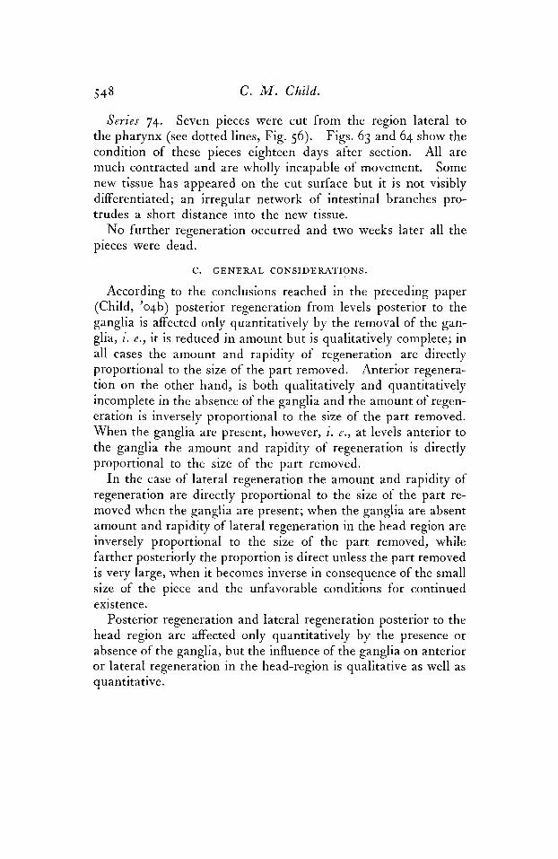
Series 74. Seven pieces were cut from the region lateral to the pharynx (see dotted lines, Fig. 56). Figs. 63 and 64 show the condition of these pieces eighteen days after section. All are much contracted and are wholly incapable of movement. Some new tissue has appeared on the cut surface but it is not visibly differentiated; an irregular network of intestinal branches pro- trudes a short distance into the new tissue.
No further regeneration occurred and two weeks later all the pieces were dead.
C. GENERAL CON SID E RATIONS . According to the conclusions reached in the preceding paper
(Child, 'o4b) posterior regeneration from levels posterior to the ganglia is affected only quantitatively by the removal of the gan- glia, i. e. , it is reduced in amount but is qualitatively complete; in all cases the amount and rapidity of regeneration are directly proportional to the size of the part removed. Anterior regenera- tion on the other hand, is both qualitatively and quantitatively incomplete in the absence of the ganglia and the amount of regen- eration is inversely proportional to the size of the part removed. When the ganglia are present, however, i. e., a t levels anterior to the ganglia the amount and rapidity of regeneration is directly proportional to the size of the part removed.
I n the case of lateral regeneration the amount and rapidity of regeneration are directly proportional to the size of the part re- moved when the ganglia are present; when the ganglia are absent amount and rapidity of lateral regeneration in the head region are inversely proportional to the size of the part removed, while farther posteriorly the proportion is direct unless the part removed is very large, when it becomes inverse in consequence of the small size of the piece and the unfavorable conditions for continued existence.
Posterior regeneration and lateral regeneration posterior to the head region are affected only quantitatively by the presence or absence of the ganglia, but the influence of the ganglia on anterior or lateral regeneration in the head-region is qualitative as well as quantitative.

Studies on Regulation. 5 49
Pieces from the region anterior to the ganglia, those from the extreme lateral margins of the body and those from the extreme posterior end are almost incapable of regeneration and live but a short time.
I n cases like that of LeptopZana where regeneration takes place by the differentiation in a particular manner of new tissue formed upon a cut surface it is very evident that the fate of the new tissue is determined primarily by its relation to the old. T h e old part is already definitely organized and functions in a particular man- ner, thus determining the conditions to which the new tissue is subjected. Attention has been called repeatedly to this relation in the description and discussion of the experiments, and a close parallelism between the amount and kind of motor activity and the amount and rapidity of regeneration and the morphological character of the regenerated part has been shown to exist. This relation between motor activity and regeneration is undoubtedly complex in character and requires some consideration.
I n the preceding paper (Child, ’o4b) the question of the relation between the nervous system and morphooenesis was briefly dis- cussed and attention was called to the possibility that an apparent relation may exist in cases where the nervous stimuli themselves are not the formative factors but merely determine the functional conditions. Now that the description of the experiments is com- pleted it is desirable to return to this question and consider it in the light of the experimental data. Three possibilities must be considered: First among these is the question as to whether nervous stimuli themselves exercise a “trophic” or a formative influence. T h e experiments leave no room for doubt that some considerable portion of the central nervous system must be present in pieces from adult specimens of Leptoplana in order that life may continue. Small anterior, lateral, and posterior pieces con- taining only peripheral branches from the ganglia or cords die within a few days. These facts may be regarded as indicating that the nervous system is necessary for continued existence. But since neither the ganglia nor the other parts which constitute a head regenerate after removal of the ganglia we must conclude either that the influence of the nervous system is not sufficient in
P

5 5 0 C. M . Child.
itself for complete regeneration or else that different influences are localized in different parts of the nervous system. T h e fact that posterior regeneration is qualitatively complete in the absence of the ganglia while anterior regeneration is not might seem to favor the second alternative. Even if we admit that nervous stimuli themselves exert a formative influence there seems to be no good reason for supposing that this influence is due to stimuli of a particular kind differing from other nervous stimuli. Indeed the general consensus of opinion is that in all probability a given nerve cannot transmit qualitatively different stimuli. T h e ques- tion of the direct formative influence of nervous stimuli has been much debated; in all cases, however, where there is a possibility of the existence of such an influence distinction between it and the formative effect of the functional conditions cannot be made experimentally.
T h e second possibility to be considered is the formative effect of functional conditions which plays so important a part in the views of Roux ('95) and others. According to these views the condi- tions connected with functional activity of a part exert an influence which may produce growth or further development of the part. T h e functional conditions may be various in kind-either physical or chemical. I n the organs of movement and support their sup- posed formative influence has been most clearly recognized. If functional conditions exert a formative effect it is clear, as Roux has pointed out, that in those organs whose function is determined by nervous stimuli a formative influence of nervous stimuli will seem to exist since the functional conditions are determined by the existence of a connection between the organs in question and the nervous system. I n reality, however, this relation between the nervous system and formative influence is indirect (see Child, 'oqb, p. j07), since not the nervous stimulus itself but the change brought about in the organ and its environment by function are the essential factors.
I n a paper which has recently appeared Goldstein ('04), after a consideration of the influence of the nervous system upon em- bryonic development and regeneration, reaches the conclusion that the functional stimuli and not the nervous stimuli themselves

Studies on Regulation. 55 1
are the important factors in cases where any relation exists. His statement of the case is as follows: "Diejenigen Organe, deren Funktion wesentlich durch den anatomischen Zusammenhang mit dem Zentralnervensysteme (durch Nerven) vermittelt wird-und um ein solches handelt es sich bei der Muskulatur-bediirfen zu ihrer Bildung sowohl als zu ihrer Regeneration in schon relativ friiher Periode der Entwicklung am notwendigsten der Verbin- dung mit dem Zentralorgan, wie auch ihre Erhaltung und Regen- eration in postembryonaler Zeit unbedingt an diese Verbindung gebunden ist. Dagegen sind diejenigen Organsysteme, deren Funktion wesentlich durch den Einfluss der unmittelbaren Umge- bung bedingt ist, wie der Knochen, unabhangig vom Zentralner- vensystem sowohl in ihrer Entwicklung (resp. Erhaltung) als ihrer Regeneration, soweit durch eine eventuelle Unterbrechung der nervosen Verbindung die normale Tatigkeit der Umgebung nicht gleichzeitig beeintrachtigt wird (z. B. Storung der Funktion des Knochens durch Aufhebung des Tonus der Muskulatur durch die Nervendurchschneidung) und das betreffende Organ, also zum Beispiel der Knochen, auch sonst in seinen normalen Bezie- hungen zur Umgebung erhalten wird."
This paper which is, I think, one of the most satisfactory con- tributions to the subject which have appeared includes much literature of which various other recent writers on this subject, notably Herbst ('01) Wolff ( '02) and Moszkowski ('03) appear to be ignorant. T h e criticism of the remarkable conclusions of Herbst is of much interest and must be regarded as effective. ,The work of Harrison ('01, '03), Bardeen ('oo), and other authors which demonstrates clearly enough that the voluntary muscles arise before nervous connections are established is given full con- sideration; for a bibliography and critical examination of these and other points the reader is referred to Goldstein's paper.
T h a t regeneration and embryonic development may differ as regards relative importance of the nervous system is also admitted by Goldstein, but, I think, the importance of the relation in regen- eration between the new part and the old completely differentiated part with its characteristic functional activity is not clearly recog- nized. I n very many cases of regeneration motor activity of a

5.52 C. M . Child.
part appears long before the characteristic form is established. I n Leptoplana, for example, when the head is cut off just anterior to the ganglia, or in Planaria at any level, the new tissue growing out from the cut surface begins to function in the manner charac- teristic of the head long before it has acquired the form of a head. All the facts indicate that the definitive form is the result of this activity. Here the indirect influence of the nervous system is a necessary factor in the result. Only the earliest outgrowth of tissue before motor activity appears is probably due in large part or wholly to the stimulus of the wound and altered local conditions.
If we attempt to interpret the close parallelism between motor activity and regeneration in Leptoplann in accordance with the hypothesis proposed above we may suppose that the motor activity exercises a formative effect upon the regenerating part and so determines its fate. But in order that coordinated motor activity may occur in a given part some degree of differentiation into con- tractile and conducting structures must exist. We may regard the regeneration of particular parts of the nervous system in the new tissue as dependent primarily upon functional stimuli pro- ceeding from the old, already organized part of the nervous system. In this relation we have a case where the nervous stimulus and the functional stimulus are identical.
T h e muscles in the new part may develop under the influence, direct or indirect, of stimuli proceeding from the nervous system, though it must be borne in mind that visible morphological con- nection between these parts and the nervous system may not be present. I n certain other cases, however, as in that of the legs of Triton described by Wolff ('02) and discussed by Goldstein, the definitive form is apparently independent of the characteristic motor activity, though in these cases the physical conditions resulting from differences in the rapidity of growth of different parts may play a rSle. T h e hypothesis of the physiological formative influence of function is undoubtedly important but still another possibility remains to be considered, viz: the direct mechanical effect of mechanical conditions. T h a t the arrange- ment and distribution of material is in many cases the direct effect of mechanical conditions cannot be doubted.

Studies on Regulation. 553
T h e possibility of a mechanical interpretation of many of the phenomena of cell life has been shown by Rhumbler in various papers, and even if his application of mechanical principles should prove to be too general, the importance of mechanical factors in the cell must be recognized. If we admit the importance of these factors in the cell we cannot avoid the conclusion that their importance is certainly not less and probably much greater in multicellular and complex structures. T h e rapid advances of physiological chemistry have turned the attention of many workers away from the physical aspects of the problem of form to such an extent that in some quarters attempts at mechanical interpretation of organic phenomena are accepted with more or less reluctance. There can be no doubt that chemistry is almost daily throwing new light upon organic phenomena but, I think, it is no less certain that any essentially chemical theory of form must be incomplete. One reason for the somewhat helpless attitude of certain authors before the problem of form is, I am convinced, due to their failure to recognize the importance of physical as opposed to chemical factors. We may recognize the importance of these factors without making of them a universal principle of interpretation. All the present and future resources of both chemistry and physics are necessary for the interpretation of biological phenomena, but especially in connection with the problem of form in which masses and mass-relations are an important element are mechanical principles of interpretation of value.
Roux has pointed out the importance of mechanical conditions as functional stimuli, and hence, according to his view, as forma- tive factors in the case of various organs whose function is wholly or in part mechanical. But we must admit that mechanical con- ditions as stimuli exert in many cases not only a more or less com- plex physiologica! influence but produce a direct mechanical effect and that this effect under typical conditions is typical, T h e change of form in pieces of Stenostorna (Child, '02, '03) in conse- quence of mechanical tension and the asymmetrical form of pieces of Leptoplana (Child, 'o4a) resulting from changes in the direction of locomotion are cases in point. No one would be more ready

5 5 4 C. M . Child.
than myself to admit that mechanical factors are not the only fac- tors in these cases, though their importance in determining the result is evident.
I n analysis it is necessary, though in practice often difficult, to distinguish sharply between mechanical conditions as functional stimuli producing a " trophic" or formative effect and mechanical conditions as direct mechanical factors determining the space relations of masses. I n the cases cited there is no doubt that tension and growth are connected in one way or another. Even in regard to this point, however, there is room for the question as to whether the effect is mechanical or not.
It is probable that in many cases the effect of mechanical con- ditions as stimuli is widely different from the mechanical effect. T h e reaction to the stimulus may be an increased power of resistance to the particular mechanical conditions involved, as in the case of tendon, bone and various other structures of animals and plants. The direct mechanical effect consists in the arrange- ment of material in accordance with the mechanical conditions of tension and pressure. I n Leptoplana not only the form and the direction of growth of the regenerated posterior end but the amount of regeneration and the direction of growth seem to de- pend to a considerable extent upon, and to correspond in direction with the mechanical tension to which the part is subjected (Child, 'oqa, 'o4b). It is difficult to determine how far this result is directly mechanical and how far it is due to mechanical stimuli to growth. I think, however, that the presence of both factors must be admitted. But in most cases the elongation of the new tissue in the direction of tension does not continue indefinitely; it gradually decreases and after a time ceases even though the mechanical conditions continue as before. Undoubtedly in many cases the differentiation of the regenerated tissues and their in- creased power of resistance to mechanical tension determine when the elongation shall cease. This may be regarded as somewhat similar to the effect of mechanical conditions on tendon, bone, etc. But in some cases it is possible that the definitive form is merely the condition of mechanical equilibrium. I n either case the part retains approximately the same form, provided that the

Studies on Regulation. 5 5 5
mechanical conditions do not undergo marked change. If any considerable permanent change in the conditions does occur the form is gradually altered until a new equilibrium of one kind or another is established.
To sum up: the close parallelism between the amount and rapidity of regeneration and the form of the regenerated part in Leptoplana and the characteristic motor activity of the part may be accounted for in three different ways: nervous stimuli them- selves may be regarded as formative factors; the functional con- ditions including mechanical conditions in particular parts which are determined by functional relation between the new and old parts may be regarded as stimuli to growth and therefore as formative factors; and lastly, mechanical conditions connected with the characteristic motor activity and other functional con- ditions may determine directly in greater or less degree the arrangement of material and so the form.
As has been indicated in the preceding paragraphs I believe that all of these factors are concerned to a greater or less extent. T h e facts regarding regeneration of the ganglia indicate that con- ditions obtaining in the parts of the nervous system from which regeneration occurs are the essential formative factors for the ganglia. Some suggestions regarding the probable rGle of and mechanical and non-mechanical effects of non-nervous functional conditions in determining form have been made in the preceding paragraphs. My successful attempts at experimental control of form-regulation (Child, '02, '03, 'o4a) have convinced me that mechanical conditions are of great importance, a t least in the cases considered. While I think it probable that they will be found to be equally important in many other cases, though per- haps in widely different manner, generalization would be prema- ture. What has been said in the present section is sufficient, I trust, to make it clear that I do not exaggerate the importance of mechanical factors in morphogenesis.
T h e mechanical aspects of morphogenesis have been much neglected; application of mechanical principles will undoubtedly assist us in future in the consideration of organic form. But no single principle of interpretation will lead us far in biological

556 C. M . Child.
science. It is necessary to recognize the interrelations of numerous and widely different factors in all organic phenomena. I believe that we have as yet no adequate conception of the complexity of these phenomena. T h e universal desire to generalize results in misleading schematization. T h e processes of organic nature are even more varied than we suspect.
SUMMARY.
I. Anterior regeneration in regions anterior to the middle of the ganglia is complete, and amount and rapidity of regeneration are directly proportional to the size of the part removed. From levels posterior to the middle of the ganglia regeneration is both qualitatively and quantitatively incomplete, no head being re- generated. In these cases the amount and rapidity of regenera- tion are inversely proportional to the size of the part removed.
Lateral regeneraion in the presence of the ganglia is com- plete; in the absence of the ganglia it is complete except in the lateral head-region and the amount and rapidity of regeneration are directly proportional to the size of the part removed if the piece is large. As the size of the piece is reduced the proportion becomes inverse.
As in the case of posterior regeneration, there is a close parallelism between the rapidity, amount and completeness of anterior and lateral regeneration and the characteristic motor activity of the part concerned.
T h e relation between the nervous system and regeneration in Leptoplana may conceivably be either direct, in that nervous stimuli themselves constitute formative factors, or indirect, in that functional conditions resulting from use of the part in a particular manner determined by its relation with the nervous system, are the formative factors properly speaking. I n the latter case the formative factors may be either stimuli to growth and other changes or they may be directly mechanical. T h e facts seem to indicate that the relation is actually indirect except perhaps as regards the regeneration of parts of the nervous system itself; the conditions are both mechanical and non-mechanical but the former are especially important in certain respects.
2.
3.
4.

Studies on Regulation. 5 5 7
5. I n cases of regeneration like the present the f'unctional relation of the regenerating part to the old is an important factor in determining the fate of the new tissue.
Hull Zoological Laboratory, University of Chicago.
April, 1904.
BIBLIOGRAPHY.
BARDEEN, C. R., '00.-The Development of the Musculature of the Body Wall in the Pig, etc.
CHILD, C. M., '02.-Studies on Regulation. I. Fission and Regulation in Stenostoma. Archiv. E. Entwickelungsmech., Bd. XV, H. 2 and 3, 1902.
'03.-Studies on Regulation. 11. Experimental Control of Form Regula- tion in Zooids and Pieces of Stenostoma. Archiv. f . Entwicke- lungsmech., Bd., XV, H. 4, 1903.
'04a.-Studies on Regulation. IV. Some Experimental Modifications of Form Regulation in Leptoplana, Journ. of Exp. Zool., Vol. I, No. 1,1904.
The Relation Between the Central Nerv- ous System and Regeneration in Leptoplana: Posterior Regeneration. Journ. of Exp. Zool., Vol. I, No. 3. 1904.
J. H. U. Hospital Reports, Vol. IX, 1900.
'04b.-Studies on Regulation. V.
DRIESCH, H., '01.-Die Organischen Regulationen. GOLDSTEIN, K.. '04.-Kritische und experimentelle Beitrage zur Frage nach dem
Einfluss des Zentralnervensystems auf die embryonale Entwicklung und die Regeneration. Archiv. f Entwickelungsmech., Bd. XVIII, H. 1,1904.
HARRISON, R. G., '01 --Ueber die Histogenese des peripheren Nervensystems bei Salmo solar. Archiv. f . mikr. Anat., Bd. LVII, 1901.
'03.-On the Differentiation of the Muscular Tissue when Removed from the Influence of the Nervous System. Amer. Journ. of Anat., Vol. 11, No. 2, 1903.
Licpzig, 1901.
HERBST, C., '01.-Formative Reize in der tierischen Ontogenese. LILLIE, F. R., '01,-Notes on Regeneration and Regulation in Planarinns. Amer.
Journ. of Physiol., Vol. VI, 1901. MOSZKOWSKI, M., '03.-Hans Driesch's Organische Regulationen. Eine kritische
Studie. Roux, W., '95.-Gesammelte Abhandlungen uber Entwickelungsmechanik. Leip-
zig, 1895. SCHULTZ, E. '02.-Aus dem Gebiete der Regeneration. 11. Ueber die Regeneration
bei Turbellarien. WOLFF, G., 'O2.-Die physiologischen Grundlagen der Lehre von den Degenera-
tionszeichen.
1901.
Biol. Centralbl., Bd. XXIII , Nos. 11 and 12, 1903.
Zeitschr. f. wiss. Zool., Bd. LXXII, H. 1, 1902.
Virchow's Archiv., Bd. CLXIV, 1902.