studies on experimental hypertension: ix. the effect
TRANSCRIPT

STUDIES ON EXPERIMENTAL HYPERTENSION
IX. THE EFFECT ON BLOOD PRESSURE Ol ~ CONSTRICTION OF THE
ABDOMINAL AORTA ABOVE AND BELOW THE SITE OF
ORIGIN OF Bo~rrr MAIN RENAL ARTERIES*t
BY HARRY GOLDBLATT, M.D., JOSEPH R. KAHN, M.D., AND RAMON F. HANZAL, P~.D.
(From the Institute o/Pathology, Western Reserve University, Cleveland)
(Received for publication, January 19, 1939)
A method has been described for the production of hypertension in dogs by the constriction of the main renal artery of one or both kidneys (2, 3). When the main renal artery of only one kidney is constricted, the blood pressure rises, but after a variable period returns to the original level. When both main renal arteries are adequately constricted, or if one is constricted and the other kidney is removed, the blood pressure usually remains elevated. This has been accom- plished successfully in dogs (3) and in monkeys (4), and the method, or some modification of it, has now been used by ourselves (2-11) and others (12-93) in the production of hypertension by renal ischemia and in the investigation of the pathogenesis and treatment of this type of hypertension, as well as the application of the results to the problem of human hypertension.
In small laboratory animals, like monkeys, cats, rabbits and rats, it is rather di~cult to constrict adequately without frequently occlud- ing the main renal artery. I t occurred to us that if moderate con- striction of the aorta just above the origin of both main renal arteries would result in hypertension, at least above the site of the damp, and if constriction immediately below the origin of the renal arteries would have no effect on the blood pressure, this would indicate that
* A preliminary report (1) was presented before the Central Society for Clinical Research on November 6, 1937, in Chicago.
t This study was supported by the Beaumont-Richman-Kohn Fund and The Josiah Macy, Jr., Foundation. It was aided by grants from the Cleveland Foundation, Mr. Nathan Dauby and Mr. Alex Wintner and associates, Cleveland.
649
650 HYPERTENSION DUE TO CONSTRICTION OF AORTA
the mechanical obstruction of the aorta is not the cause of such hypertension. Moreover, if renal excretory insufficiency, as well as hypertension, would result from very great constriction of the aorta above the origin of both main renal arteries, as it does from very great constriction of the main renal arteries (3, 7, 9), this type of hypertension, with or without renal insufficiency, could also be considered of renal origin. This method could then be used instead of constriction of the main renal arteries for the production of hyper- tension of renal origin, at least in the upper part of the body of small animals. Incidentally, this method might afford an opportuni ty to s tudy in the same animal the possible effects on aorta, arteries and
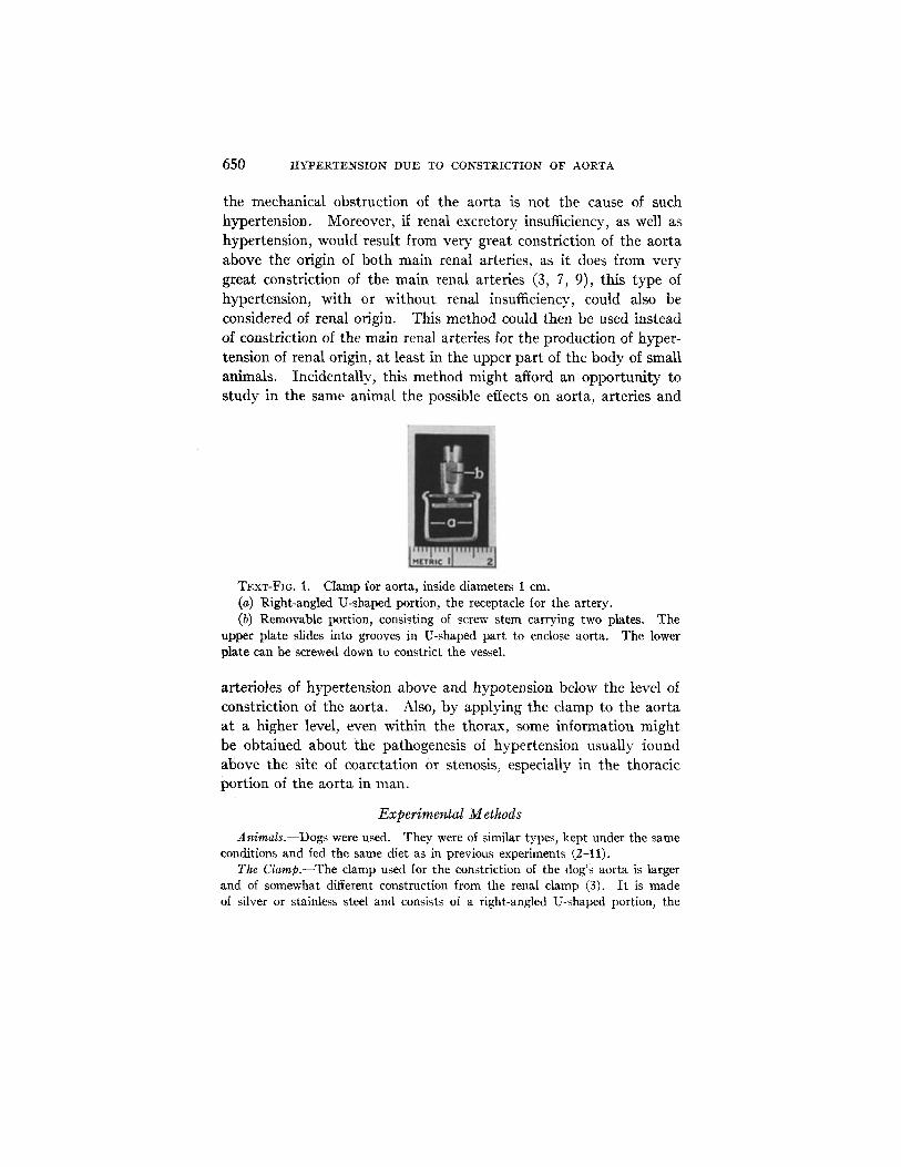
TExT-FIO. I. Clamp for aorta, inside diameters 1 cm. (a) Right-angled U-shaped portion, the receptacle for the artery. (b) Removable portion, consisting of screw stem carrying two plates. The
upper plate slides into grooves in U-shaped part to enclose aorta. The lower plate can be screwed down to constrict the vessel.
arterioles of hypertension above and hypotension below the level of constriction of the aorta. Also, by applying the clamp to the aorta at a higher level, even within the thorax, some information might be obtained about the pathogenesis of hypertension usually found above the site of coarctation or stenosis~ especially in the thoracic portion of the aorta in man.
Experimerbtal Methods
Animals.--Dogs were used. They were of similar types, kept under the same conditions and fed the same diet as in previous experiments (2-11).
The Clamp.--The clamp used for the constriction of the dog's aorta is larger and of somewhat different construction from the renal clamp (3). It is made of silver or stainless steel and consists of a right-angled U-shaped portion, the
H. GOLDBLATT~ J. 1t. KAHN~ AND R. ~'. HANZAL 651
open receptacle for the artery (Text-fig. I a), and a removable portion consisting of a screw stem carrying two plates (Text-fig. 1 b). The upper plate slides into a groove on the inner sides of the U-shaped part and serves to enclose the aorta. The lower, movable plate is attached to a double-acting screw and can be screwed down to constrict the aorta to any desired degree. A special instrument devised for the purpose of holding the U-shaped part facilitates the application of this part of the clamp on the aorta and also aids the insertion of the screw stem with the double plate into the U-shaped portion, to enclose the aorta within the clamp. This instrument is not necessary, but it simplifies the procedure.
Surgical Procedure for the Constriction of the Aorta.--All the operations but one were performed under ether anesthesia, with a previous hypodermic injection of morphine and atropine. On one dog (No. 4-13), the operation was performed under local anesthesia with procaine, after a hypodermic injection of morphine and atropine.
For the constriction of the aorta above the origin of both main renal arteries, an oblique incision about 6 cm. in length is made in the skin about 1.5 era. below and parallel to the right inferior costal margin, beginning at the costolumbar angle. This approach is the same as is used for the application of a clamp on the right main renal artery (3), or for right nephrectomy or suprarenalectomy (7). A gridiron incision in the internal oblique and transversalis muscles, lateral and posterior to the right border of the external oblique muscle, reveals peritoneum, which is then pushed away manually from its loose attachment to the under- lying tissues of the lateral wall of the abdomen. A self-retaining retractor with long thick wire blades is then inserted to retract the wound and keep it open. The peritoneal covering of the aorta is incised on the right side, just above the origin of the right main renal artery, and the aorta immediately above the origin of this vessel is then exposed and dissected away for a distance of about 1 era. from its posterior attachment to the tissues in front of the spine. In some eases, when they are situated close to the right main renal artery, it is necessary to ligate and sever two lumbar arteries which originate from the aorta posteriorly between the levels of the superior mesenteric and right main renal artery. This must be done in order to make room for the clamp which is applied to the aorta just proximal to the origin of the right main renal artery.
As soon as the aorta is enclosed in the clamp, a 20 gauge needle connected with a mercury manometer is inserted in the femoral artery to measure direct or so called mean blood pressure. The degree of constriction of the aorta is deter- mined by the immediate effect on the femoral mean pressure and it can be varied at will in different animals. In these experiments the constriction was usually made adequate to reduce the femoral mean pressure to 50 per cent, or less, of the normal for the animal. Closure of the incision is effected by means of continuous catgut sutures for the muscle and subcutaneous tissue and continuous black silk sutures for the skin.
For the application of a clamp on the aorta just below both main renal arteries, the incision is made on the left side, and the clamp is placed just below the site of

652 HYPERTENSION DUE TO CONSTRICTION OF AORTA
origin of the left main renal artery. This portion of the aorta is free of any other arterial branches, so that it is not necessary to sever any vessels to make room for the clamp. Otherwise the operation is similar to that described for the application of the damp above the origin of both main renal arteries. The incision on the left side is made a little lower than on the right (about 2 cm. below and parallel to the costal margin). It is sometimes necessary also to split the external oblique muscle in making the gridiron incision on the left side.
Measurement of Blood Pressure.--In all the animals blood pressure was deter- mined by two methods. Carotid systolic pressure was measured by the van Leersum carotid loop method (94), and direct or mean pressure was measured by inserting into the carotid or femoral artery a needle connected by means of a liquid system (2 per cent sodium citrate) with a mercury manometer. During the operation for constriction of the aorta a 20 gauge needle was used, but for other determinations of femoral mean pressure, the needle was 21 gauge. Normal blood pressure was determined frequently by both methods for at least 1 month before the aorta was constricted.
The Effect of Constriction of the Aorta Just above the Site of Origin of Both Main Renal Arteries
Constriction of the a o r t a just above the origin of bo th main renal arteries, or above the functioning main renal a r te ry in a unilateral ly nephrectomized animal, invariably resulted in lowering of the femoral mean pressure and elevation of carotid systolic or carot id mean pressure. The effect on the blood pressure in the carotid loop was not immediate, bu t developed in about 24 to 48 hours, tha t is, in about the same t ime as following constriction of one or bo th main renal arteries (3). The reduction of femoral mean pressure was imme- diate and varied with the degree of constriction of the aorta. In many instances the femoral mean pressure rose later, coincidentally with the rise of pressure in the carotid loop, and in about 1 or 2 weeks reached its maximum level, usually the previous normal. In some instances, especially if the aor ta below the origin of bo th renal arteries was also constricted, even the femoral mean pressure finally reached levels higher than the normal.
A. The Development of Hypertension without Renal Excretory Insu~ciency (the Benign Phase)
In four animals (Nos. 3-21, 3-49, 3-51 and 3-60) the carotid systolic pressure became greatly elevated as a result of constriction of the aor ta just above the origin of bo th main renal arteries, and the
H. GOLDBLATT, ~. P.. KAHN, AND R. F. HANZAL 653
hyper tens ion persis ted unti l death, wi thout accompanying renal excretory insufficiency. Three of these animals died of in t ra-ab-
dominal hemorrhage due to erosion of the wall of the aor ta b y the clamp. The longest survival was 25 days. I n one animal, (No.
3-60), t ha t survived 8 days wi th e levated blood pressure, a f te r con-
str ict ion of the aor ta above the origin of b o t h renal arteries, the
cause of dea th was not de te rmined a t autopsy. To save space, the exper imenta l h is tory of only the m o s t i m p o r t a n t
animals will be given in full detail, the others will be abbrevia ted .
No. 3-21: Mongrel, short haired, female, young, 14.2 kg. Mar. 16 to July 28, 1937. During this pre-operative period the systolic blood
pressure in the carotid loop (carotid systolic pressure) varied between 140 and 160 nun. Hg, and the mean pressure in the femoral artery (femoral mean pressure) varied between 120 and 140 ram. Hg.
July 28. The aorta was moderately constricted by a clamp applied just above the origin of both main renal arteries. July 29 to Aug. 5. During this period the carotid systolic pressure varied between 220 and 280 rum. Hg. On July 29, 1937, 24 hours after the constriction of the aorta, the carotid systolic pressure was 276 rum. Hg. The femoral mean pressure, which was 80 rum, Hg
immediately after the constriction of the aorta, rose to 90 mm. Hg on Aug. 3. Aug. 6. At 9 a.m. the carotid systolic pressure had dropped to 135 into. Hg. The animal appeared ill. Blood urea nitrogen (B.U.N.) 20.3 mg., non-protein nitrogen (N.P.N.) 35.4 mg., creatinine (Cr.) 1.3 rag. and CO2 combining power (CO2) 24.2 volumes per 100 cc. of plasma. At 3 p.m. the animal was found dead. At autopsy, the cause of death was found to have been erosion of a lumbar artery by one edge of the clamp which impinged on this vessel.
No. 3-49: Hound, female, old, 14.0 kg. July 6 to Sept. 16, 1937. During this pre-opemtive period the carotid systolic
pressure varied between 134 and 152 mm. Hg, and the femoral mean pressure varied between 128 and 144 mm. Hg. Sept. 16. The aorta was greatly con- stricted by a clamp applied just above the origin of both main renal arteries. Sept. 17 to Oct. 10. During this period the carotid systolic pressure varied between 240 and 284 mm. Hg. On Oct. 9, 2 days before death, it was still 260 rum, Hg. The femoral mean pressure was 75 mm. Hg immediately after constriction of the aorta and rose to a maximum of 130 ram, Hg on Sept. 27. Oct. 11. The animal was found dead. At autopsy, the cause of death was found to have been due to erosion of the aorta by the clamp.
No. 3-51: German shepherd, male, middle age, 22.0 kg. July 9 to Sept. 21, 1937. During this pre-operative period the carotid systolic
654 HYPERTENSIOlq DUE TO CONSTRICTION OF AORTA
pressure varied between 144 and 166 ram. Hg, and the femoral mean pressure varied between 115 and 135 ram. Hg.
Sept. 21. The aorta was greatly constricted by a clamp applied just above the origin of both main renal arteries. Sept. 22 to Sept. 30. During this period the carotid systolic pressure varied from 210 to 260 ram. Hg. On Sept. 30, it was still 230 ram. Hg. Oct. 1. The animal was found dead. At autopsy, the cause of death was found to have been intra-abdominal hemorrhage caused by erosion of the aorta by the clamp.
No. 3-60: Black and white short haired mongrel, female~ young, 11.8 kg. In this animal, the carotid systolic pressure rose from the normal of 150 to
170 ram. Hg (Sept. 16 to Oct. 27, 1937) to a maximum of 256 mm. Hg, on Nov. 3, 1 week after the constriction of the aorta above the origin of both main renal arteries. Femoral mean pressure fell from the normal of 130 to 65 ram. Hg, immediately after the clamp was applied, but rose to 105 ram. Hg on Nov. 3. The animal was found dead on Nov. 5. 2 days after the operation there was no chemical evidence of renal excretory insufficiency, but during the last 7 days of life chemical examination of the blood was not made. The cause of death was not determined. There were no lesions suggestive of renal insufficiency com- plicating the hypertension.
B. The Development of Hypertension, Renal Excretory Insuficiency, and Fatal Convulsive Uremia with Degenerative, Necrotizing
and Inflammatory Arteriolar Disease in Many Organs (the A cute Malignant Phase)
Three animals (Nos. 3-37, 3-59, 3-63) with the aor ta great ly con- str icted just above the origin of bo th main renal arteries, died in uremia, a variable period af ter application of the clamp. The uremia was obviously due to renal excretory insufficiency caused by marked renal ischemia, the result of sudden excessive constriction or actual occlusion of the aorta. In all the animals the carotid systolic pressure rose progressively to high levels as the degree of uremia also increased. At autopsy, in the gross, some of the uremic animals had petechiae in various organs and, microscopically, showed arteriolar degenera- tion and necrosis, as well as acute arteriolitis in these organs, princi- pal ly the a l imentary t rac t (except rectum), gall bladder, pancreas, myocard ium and brain. This was similar to the effect of excessive constrict ion or occlusion of the main renal arteries (the acute malig- nan t phase) previously described (9).
No. 3-37: Collie, female, young, 14.4 kg. Apr. 20 to Oct. 27, 1937. During this pre-operative period the carotid systolic

1~. GOLDBLATT, J. R. KAHN, AND R. F. HANZAL 655
pressure varied between 132 and 158 nnn. Hg, and the femoral mean pressure varied between 112 and 125 mm. Hg. On Oct. 27, B.U.N. 15.7 rag., Cr. 1.3 mg., CO2 49.4 volumes per 100 cc. of plasma.
Oct. 27. The aorta was greatly constricted by a clamp applied just above the origin of both main renal arteries. The femoral mean pressure was reduced immediately from 115 to 55 ram. Hg, and it remained at this level until death. During the 3 days after the constriction of the aorta, the carotid systolic pressure varied between 198 mm. Hg, 24 hours after the operation, and 245 mm. Hg on Oct. 29, the day before death. During this period the animal became progres- sively more uremic. Oct. 29. B.U.N. 128 mg., Cr. 7.6 nag., CO2 28.6 volumes per 100 cc. of plasma.
Oct. 30. The animal was found dead. At autopsy, in the gross, there were petechiae in the stomach, small and large intestine and pancreas. Microscopically, these petechiae were associated with arteriolar degeneration and necrosis, but many were obviously of capillary origin.
No. 3-59: Mongrel collie, female, 10.4 kg. Sept. 7 to Oct. 20, 1937. During this pre-operative period the carotid systolic
pressure varied between 143 and 156 ram. Hg, and the femoral mean pressure varied between 115 and 135 ram. Hg.
Oct. 20. The aorta was greatly constricted by a clamp applied just above the origin of the main renal arteries. The femoral mean pressure was reduced im- mediately from 115 to 50 ram. Hg. Oct. 21 to Oct. 24. During this time the carotid systolic pressure rose progressively from 188 ram. Hg, 24 hours after the operation, to 282 rnm. Hg on Oct. 23. On Oct. 24, B.U.N. 104 rag., Cr. 6.8 rag.
Oct. 25. The animal was found dead. At autopsy, in the gross, there were petechiae, some confluent, in the mucosa and serosa of stomach, small and large intestines and gall bladder, and also in the pancreas and epicardium. The hemolymph nodes were intensely hyperemic. Microscopically, in the organs that showed gross petechiae and in some that did not, many arterioles were the seat of hyalinization, fibrinoid degeneration or necrosis. In many instances the extravasation of blood appeared to be of capillary origin.
No. 3-63: Chow, female, middle age, 19.6 kg. In this animal also the carotid systolic pressure rose from the normal of 151
to 168 ram. Hg (Sept. 22 to Dec. 22, 1937), to 272 ram. Hg on Dec. 24, 2 days after the constriction of the aorta. Femoral mean pressure fell from the normal of 130 to 155 mm. Hg, to 75 mm. Hg, immediately after the constriction of the aorta, and it remained at this level. The nitrogenous products of the blood rose, and CO2 combining power fell, progressively, until on Dec. 26, they were, B.U.N. 188 rag., Cr. 6.0 mg. and CO2 30.3 volumes per 100 cc. of plasma. On Dec. 27, the animal was found dead. In the gross, and microscopically, the typical lesions of the malignant phase were found in the small and large intestine and pancreas. In the liver, hyalinized arterioles without accompanying petechiae were found.
656 HYPERTENSION DUE TO CONSTRICTION O~' AORTA
The Effect of Constriction of the Aorta above the Origin of Both Main Renal Arteries Followed by Constriction of the Aorta below the
Renal A rteries and Finally Constriction of the Main Renal Arteries
In those animals that survived several weeks or months the con- striction of the abdominal aorta above the origin of both main renal arteries, there was a tendency for the elevated carotid systolic or carotid mean pressure to return to a lower or even to the normal level. Increased constriction, and finally occlusion, of this part of the aorta was followed by re-elevation of the carotid systolic or carotid mean pressure, which, after a variable time, again tended to fall. In such an animal (No. 3-50), subsequent constriction, and then occlusion of the aorta just below the origin of both renal arteries caused a temporary re-elevation of the carotid systolic pressure. Even femoral mean pressure reached a level above normal. Constriction of one main renal artery of such an animal again re-elevated the pressure, bu t it was necessary to constrict, or even gradually to occlude, both main renal arteries, in order to effect marked renal ischemia and persistent elevation of carotid and femoral blood pressure at a high level.
No. 3-50: Collie, mongrel, female, middle age, 14.1 kg. July 16 to Sept. 14, 1937. During this pre-operative period the carotid systolic
pressure varied between 134 and 165 ram. Hg, and the femoral mean pressure varied between 110 and 135 ram. Hg.
Sept. 14. The aorta was moderately constricted just above the site of origin of both main renal arteries. The femoral mean pressure fell immediately from 125 mm. Hg to 90 mm. Hg. Sept. 15. Carotid systolic pressure rose to 204 mm. Hg. Sept. 16 to Oct. 18. Carotid systolic pressure rose to a maximum of 246 mm. Hg, on Sept. 27, and then gradually fell to 187 ram. Hg, on Oct. 18. The femoral mean pressure gradually rose to 145 mm. Hg.
Oct. 18, 1937, to Nov. 22, 1938. During this period, the aorta above both main renal arteries was occluded, on Oct. 18, and the aorta below both main renal arteries was first constricted, on Dec. 15, 1937, and then occluded, on Mar. 3, 1938. During the period after each operation, the carotid systolic pressure rose and then gradually fell again.
Then in succession the renal arteries were first constricted and finally occluded. The left main renal artery was constricted, June 9, 1938, and occluded Oct. 27. The right main renal artery was constricted on June 30, and occluded on Nov. 22.
Nov. 23 to Dec. 24. The carotid systolic pressure rose to 304 mm. Hg on Nov. 25, and it has remained greatly elevated. Even femoral mean pressure rose to 200 ram. Hg on Nov. 11, despite the occlusion of the aorta above and
H. GOLDBLATT, J. R. KAJ=IN, AND R. F. HANZAL 657
below the main renal arteries. Chemical examination of the blood showed B.U.N. 15.7 nag., Cr. 1.3 nag., COs 47.0 volumes per 100 cc. of plasma. The urea clearance was 24.1 which was about 60 per cent of the normal for this dog, and indicated moderate impairment of renal excretory function, although it was not obvious from the determinations of urea and creatinine in the blood. On Dec. 24, femoral mean pressure was still 190 ram. Hg, while the carotid systolic pressure was 285 nun. Hg. On this date, B.U.N. 18.8 rag., Cr. 1.7 nag., CO~ 48.3 vol- umes per 100 cc. of plasma. In a concentration test the specific gravity of the urine was 1.025. The animal is still living.
The Effect of Releasing the Clamp on the Aorta Constricted above the Origin of the Main Renal Arteries
In one animal (No. 3-71), the release of the clamp on the aor ta just
above the origin of both main renal arteries was followed within 24
hours by a fall of the elevated carotid systolic pressure to the original
level. The femoral mean pressure returned to normal immediately
after the release of the constriction.
No. 3-71: Doberman pinscher, male, old, 22.8 kg. Oct. 30 to Dec. 28, 1937. During the pre-operative period the carotid systolic
pressure varied between 154 and 172 tom. Hg. The femoral mean pressure varied between 140 and 155 ram. Hg.
Dec. 28. The aorta was greatly constricted just above the origin of both main renal arteries. The femoral mean pressure was reduced immediately from 140 to 70 nun. Hg.
Dec. 29. 24 hours after the operation the carotid systolic pressure was 248 ram. Hg. The dog's hind limbs were paralyzed, the anus was incontinent, and some bloody fluid material kept passing out from the intestine. Under local anesthesia, the aortic clamp was removed. The carotid systolic pressure re- mained elevated at 240 nam. Hg immediately after removal of the clamp. Dec. 30, 1937, to Jan. 9, 1938. 24 hours after the operation the carotid systolic pressure was 156 tuna. Hg, which was well within the normal range for this animal, and it remained down for the next 9 days. 2 days before death the carotid systolic pressure was 148 mm. Hg, and the femoral mean pressure was 130 nam. Hg. Jan. 10. The animal was found dead. The cause of death was not determined. There were no lesions suggestive of uremia.
The Effect of Constriction of the Abdominal Aorta Just below the Origin of Both Renal Arteries Followed Later by Constriction of the Aorta
above the Origin of Both Main Renal Arteries
In order to constrict the aorta just below the origin of bo th ma in renal arteries, it was necessary to apply the clamp just below the

658 HYPERT]ENSION DUE TO CONSTRICTION OF AORTA
origin of the left main renal artery. Full details about the method of application of the clamp have already been given.
Constriction of the aorta just below the origin of both main renal arteries had no significant effect on the carotid systolic blood pressure of animals with two kidneys or of unilaterally nephrectomized animals. When, however, constriction of the aorta below the origin of the main renal arteries was followed by constriction of the abdominal aorta just above the origin of both main renal arteries, a significant elevation of blood pressure always occurred. The hypertension was usually higher and lasted longer than when the aorta was constricted only above the site of origin of the main renal arteries. However, even the constriction of the aorta below and above the main renal arteries was followed, after a variable period, by a lowering of the elevated blood pressure, or a return to the original level. Increased constric- tion or occlusion of the abdominal aorta above and below the origin of the main renal arteries caused significant but temporary re-elevation of the blood pressure. In some of these animals, even the femoral mean pressure reached a level significantly higher than the normal. Three of these animals (Nos. 3-44, 3-91, 3-92) finally died in uremia due to renal excretory insufficiency caused by sudden, excessive con- striction of the aorta above the origin of the main renal arteries. These animals developed the necrofizing and inflammatory arteriolar disease previously described (9) in many of the organs in which the arterial pressure was elevated. This result was similar to that of some of the animals with the aorta greatly constricted only above the origin of the main renal arteries.
One animal (No. 3-57) with the aorta constricted first below and then above the main renal arteries, died of intra-abdominal hemor- rhage, due to erosion of the aorta above the origin of both main renal arteries. This animal lived 9 weeks after the constriction of the aorta below the main renal arteries, and 19 days following the con- striction of the aorta above the main renal arteries. During the latter period the carotid systolic pressure remained elevated considerably above the normal. Even the femoral mean pressure finally became elevated above the original level.
In a unilaterally nephrectomized animal (No. 3-89) the application of the clamp on the aorta below the main renal artery of the remain-
I=I. GOLDBLATT~ 3. R. KAHN~ AND R. F. /:IANZAL 659
i ng k i d n e y was n o t fo l lowed b y e l e v a t i o n of t h e c a r o t i d sys to l i c
p res su re .
No. 3-44: White spitz, female, young, 14.0 kg. J~tne 22 to Sept. 13, 1937. During this pre-operative period the carotid systolic
pressure reached 172 ram. Hg, but during the last 2 weeks of this period, it varied between 141 and 152 ram. Hg. The femoral mean pressure varied between 115 and 145 ram. Hg.
On Sept. 13, the aorta was greatly constricted just below the origin of both main renal arteries. The femoral mean pressure was reduced immediately from 126 to 70 ram. Hg. There was no immediate effect on the carotid systolic pres- sure, and it remained within the limits of the normal range, until Oct. 14, when the aorta just above the origin of both main renal arteries was moderately con- stricted by another clamp. The femoral mean pressure was immediately reduced slightly from 95 to 80 ram. Hg. There was no immediate effect on the carotid systolic pressure, but in several days it rose to 217 ram. Hg. During the next 7 weeks it fell gradually to normal. On Dec. 7, the carotid systolic pressure was 152 ram. Hg, and the femoral mean pressure was 120 ram. Hg.
Occlusion of the aorta by tightening the clamp above the renal arteries, on Dec. 7, 1937, and below the renal arteries, on Mar. 3, 1938, caused a re-elevation of carotid systolic pressure, but after each damping it fell gradually to normal.
Apr. 11. Right nephrectomy was also followed by re-elevation of carotid systolic pressure, but again it fell gradually to normal. There was still no in- dication of renal excretory insufficiency. B.U.N 18.8 nag., Cr. 1.1 rag., CO~ 44.8 volumes per 100 cc. of plasma.
May 4. The left main renal artery was greatly constricted. May 5. Carotid systolic pressure rose to 263 mm. Hg, B.U.N. 69.0 rag., Cr. 3.6 rag., CO2 36.6 volumes per 100 cc. of plasma.
May 7. At 9 a.m. the carotid systolic pressure was 280 mm. Hg, B.U.N. 99.9 nag., Cr. 5.7 mg., CO2 40.0 volumes per 100 cc. of plasma. The dog was in coma, had convulsions and passed bloody material from the rectum and bladder. During one convulsion the carotid systolic pressure was 330 ram. Hg. Removal of the clamp on the renal artery was followed in about 6 hours by a fall of carotid systolic pressure to 140 ram. Hg, but the animal was in convulsive uremia, B.U.N. 131.3 nag., Cr. 5.1 nag., and died 10 hours after the removal of the clamp.
At autopsy, in the gross, and microscopically, the typical lesions of the malig- nant phase of this type of hypertension were present in stomach, small and large intestine (except rectum), brain, pituitary, myocardium and epicardium.
No. 3-91: Tan and white hound, female, middle age, 17.0 kg. Feb. 10 to Mar. 16, 1938. During this pre-operative period the carotid systolic
pressure varied between 152 and 172 ram. Hg. Mar. 16 to Apr. lg. Constric- tion of the aorta below the origin of both main renal arteries, on Marl 16, had no significant effect on the carotid systolic pressure which varied between 150 and
660 HYPERTENSION DUE TO CONSTRICTION OF AORTA
176 mm. Hg, during the normal period. Femoral mean pressure fell from 130 to 60 ram. Hg, immediately after the clamping, but on Apr. 12, it had returned to 110 nun. Hg.
After constriction of the aorta above the origin of both main renal arteries, on Apr. 12, carotid systolic pressure rose progressively to a maximum of 280 turn. Hg, on Apr. 18. By June 9, it had fallen to 192 ram. Hg. Occlusion of the aorta above (June 9) and below (June 27) the main renal arteries was followed by temporary re-elevation of carotid systolic pressure, but, on Aug. 29, it was again 190 ram. Hg. On this day the left main renal artery was moderately constricted. Following this, carotid systolic pressure rose to a maximum of 306 mm. Hg. On Sept. 14, when it was still 292 ram. Hg, the right main renal artery was greatly constricted.
Sept. 15. The dog appeared obviously ill. The pulse was very rapid and feeble, yet the blood pressure was still high. Carotid systolic pressure was 284 ram. Hg at 9 a.m. B.U.N. 29.3 rag., Cr. 3.0 mg., CO2 36.2 volumes per 100 cc. of plasma. At 3 p.m. the animal died, immediately after an electrocardiogram was taken.
At autopsy, in the gross, no obvious cause of death was found. The heart weighed 194 gm. and its chambers were greatly dilated. Although the blood creatinine was 3.0 rag. about 6 hours before death, yet the gross anatomical signs of the malignant phase of hypertension, found in other animals, were absent. Microscopically, however, some of the arterioles of the intestine showed hyaliniza- tion of the wall. Acute cardiac failure in an animal with early renal excretory insufficiency was considered the most probable cause of death.
No. 3-92: Brown hound, female, middle age, 17.0 kg. Feb. 10 to Mar. 16. During this pre-operative period the carotid systolic
pressure varied between 159 and 172 ram. Hg. Mar. 16. Moderate constriction of the aorta below the origin of both main
renal arteries reduced femoral mean pressure immediately from 120 to 70 mm. Hg, but from Mar. 17 to Apr. 12 the carotid systolic pressure remained unchanged and varied between 156 and 176 ram. Hg. On Apr. 12, it was 156 ram. Hg, and the femoral mean pressure was 90 ram. Hg.
Apr. 12. The aorta was very greatly constricted by a clamp applied just above the origin of both main renal arteries. The femoral mean pressure was reduced immediately to 50 mm. Hg. Apr. 13 to June 9. The carotid systolic pressure rose to a maximum of 318 mm. Hg, on Apr. 20, then it fell very gradually to 176 nun. Hg, the upper limit of normal, on June 9.
June 9. The clamp above the origin of both main renal arteries was tightened so as to occlude the aorta. Jun~ 11 to June 27. The carotid systolic pressure became re-elevated to a maximum of 246 ram. Hg, on June 17, then fell grad- ually, but was still 220 mm. Hg on June 27.
June 27. The aorta below both main renal arteries was also occluded by tightening of the clamp at this site. June 28 to Aug. 29. The carotid systolic

H. GOLDBLATT, ]. R. KAHN, AND R. F. HANZAL 661
pressure rose again to a maximum of 248 ram. Hg, and then fell gradually to 182 ram. Hg, on Aug. 29. B.U.N 11.4 rag., Cr. 1.6 mg., COs 55.0 volumes per 100 cc. of plasma.
Aug. 29. The left main renal artery was moderately constricted. Aug. 30 to Sept. 14. The carotid systolic pressure rose to a maximum of 296 ram. Hg on Sept. 8. I t remained greatJy elevated.
Sept. 14. The right main renal artery was greatly constricted. Sept. 15 to Sept. 24. The carotid systolic pressure was 280 ram. Hg before the operation on Sept. 14, and it remained elevated at about this level during the entire period following the operation, but the animal progressively developed profound uremia and acidosis until, on Sept. 28, the day of death, B.U.N. 225.0 nag., Cr. 11.2 rag. and COs 24.1 volumes per 100 cc. of plasma.
At autopsy, in the gross, and microscopically, petechiae and the typical arteri- olar lesions of the malignant phase were present in the mucoss and serosa of the stomach, small intestine, large intestine, pancreas, urinary bladder, mesenteric and periaortic lymph nodes, epicardium, brain and diaphragmatic muscle.
No. 3-57: Collie, male, young, 10.0 kg. Aug. 31 to Oct. 20, 1937. During the pre-operative period the carotid systolic
pressure varied between 120 and 135 ram. Hg. Oct. 20. The aorta below the origin of both main renal arteries was very
greatly constricted. The femoral mean pressure was reduced immediately from 120 to 48 ram. Hg. Oct. 21 to Dec. 7. During this period the carotid systolic pressure varied between 144 and 150 ram. Hg, which was well within normal limits for this animal. The femoral mean pressure gradually rose to 120 ram. Hg. On Dec. 7, the carotid systolic pressure was 146 mm. Hg.
Dec. 7. The aorta above the origin of both main renal arteries was greatly constricted by means of another clamp. This reduced the femoral mean pressure only slightly, to 100 mm_ Hg, although the plate was screwed down almost as far as it would go. Dec. 8 to Dec. 24. The carotid systolic pressure rose in a few days to a maximum of 240 ram. Hg. At the same time even the femoral mean pressure rose above normal to 175 ram. Hg. On Dec. 24, the carotid systolic pressure was 236 ram. Hg, the carotid mean pressure was 195 ram. Hg, and the femoral mean pressure was 160 ram. Hg. Dec. 26. The animal was found dead. At autopsy, the cause of death was found to have been intra-abdominal hemorrhage caused by erosion of the aorta by the clamp above the origin of both main renal arteries.
No. 3-89: Black and white, short haired mongrel, male, middh age, 9.0 kg. Jan. 29 lo Mar. 1, 1938. During the pre-operative period the carotid systolic
pressure varied between 134 and 160 ram. Hg, and the femoral mean pressure varied between 120 and 145 ram. Hg.
Mar. 2. Right nephrectomy. Mar. 3 to May 5. Carotid systolic pressure remained within normal limits.
662 H~/PERTENSION DEE TO CONSTRICTIO!~ OF AORTA
May 6. The aorta below the origin of both main renal arteries was greatly constricted. The femoral mean pressure was immediately reduced from 120 to 60 ram. Hg. During the next 3 days, there was no elevation of the systolic blood pressure, which was 152, 146 and 154 ram. Hg on successive days. May 10. The animal was found dead. The cause of death was not determined.
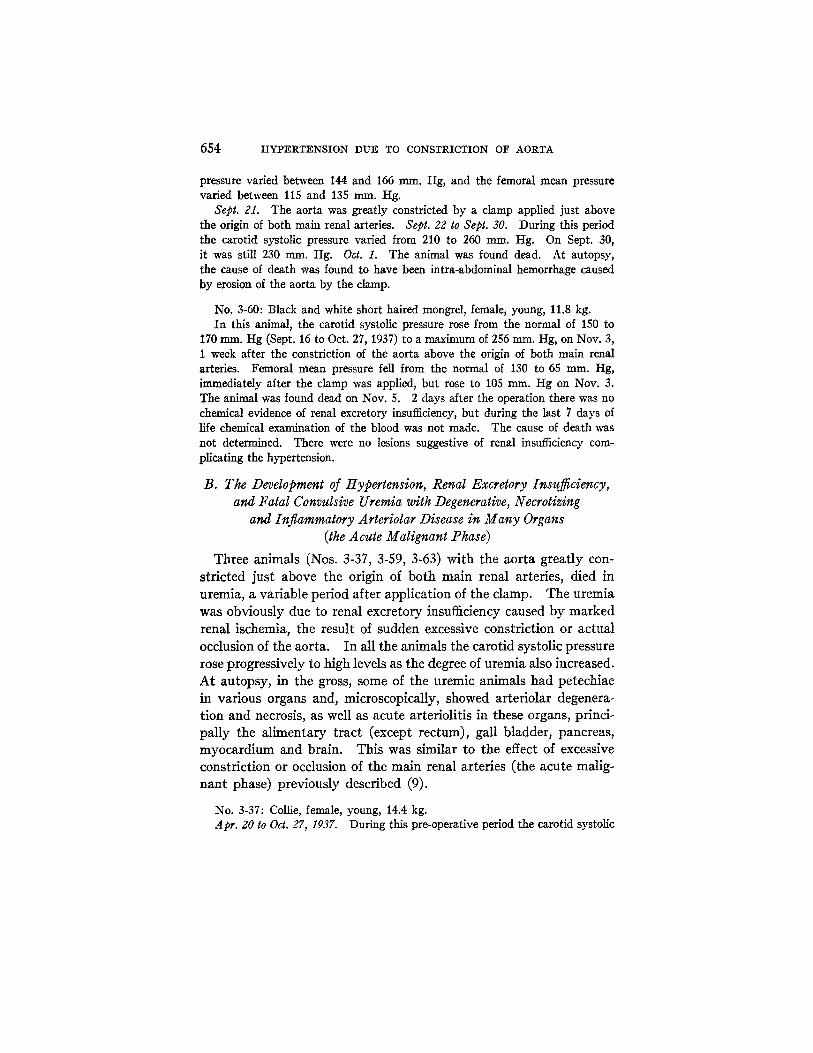
,°° I 280 ;~60 Z.40 ~,Z.O
::~?..00 18o
160 " " ' " - '
~. 140 ~ °
lzo
~lO0 8o ,,.
,o
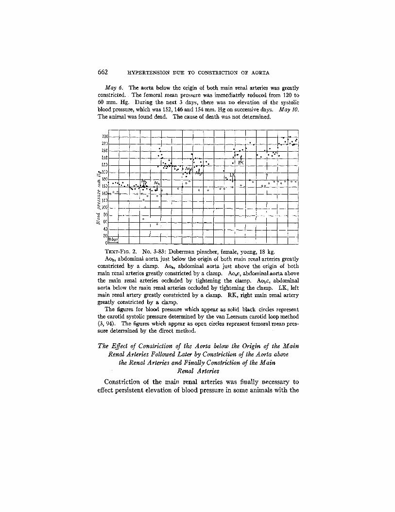
T~x¢-FIG. 2.
• ] • • I o , ,
% o t
#-7"'^!' "
- I t Ao. _ o - , . o O > o O O O o • • , ~ t . - ~ . l a o o . o o o o
' ~ . " - . - . . - , t o o , o o
O
O
No. 3-83: Doberman pinscher, female, young, 18 kg. A O b , abdominal aorta just below the origin of both main renal arteries greatly
constricted by a clamp. Aoa, abdominal aorta just above the origin of both main renal arteries greatly constricted by a clamp. Aoac, abdominal aorta above the main renal arteries occluded by tightening the clamp. Aobc, abdominal aorta below the main renal arteries occluded by tightening the clamp. LK, left main renal artery greatly constricted by a clamp. RK, right main renal artery greatly constricted by a clamp.
The figures for blood pressure which appear as solid black circles represent the carotid systolic pressure determined by the van Leersum carotid loop method (3, 94). The figures which appear as open circles represent femoral mean pres- sure determined by the direct method.
The Effect of Constriction of the Aorta below the Origin of the Main Renal Arteries Followed Later by Constriction of the Aorta above
the Renal Arteries and Finally Constriction of the Main Renal Arteries
Constriction of the main renal arteries was finally necessary to
effect persistent elevation of blood pressure in some animals with the
H. GOLDBLATT, 3. R. KAHN, AND R. ~F. HANZAL 663
aorta constricted or even occluded below, and later above, the origin of the main renal arteries. On dog 3-83 (Text-fig. 2), the constriction of the aorta below the origin of the main renal arteries had no signifi- cant effect on the carotid systolic pressure. When this was followed by constriction of the aorta above the site of origin of both main renal arteries, there was a prompt and significant rise of carotid systolic as well as mean pressure. Carotid systolic pressure then fell gradually over a period of about 2 months, but did not quite return to the normal level. Occlusion of the aorta first above and, about 3 weeks later, also below the origin of the main renal arteries, resulted in a re-elevation of carotid blood pressure which again did not persist. In about 3 months the carotid systolic pressure had gradually returned to slightly above the normal level for this dog. Moderate constric- tion of first the left and, a few days later, the right renal artery, re- sulted in great elevation of carotid systolic and carotid mean pressure, and even the femoral mean pressure became and has remained sig- nificantly elevated above the normal, despite the occlusion of the abdominal aorta at two sites: The animal is still alive about 4 months after the constriction of the renal arteries, the blood pressure in the carotid loop is still greatly elevated (Text-fig. 2), and there is no chemical evidence of renal excretory insufficiency.
The Effect of Constriction of the Abdominal Aorta Just above the Origin of the Main Renal Arteries on the Blood Pressure of Animals with
the Main Renal Arteries Previously Constricted
In a few animals in which both main renal arteries had been con- stricted, but in which, after a variable period of hypertension, the blood pressure had returned to a lower level, constriction of the abdominal aorta above the origin of both main renal arteries resulted in a re-elevation of blood pressure. Two animals (Nos. 2-48, 3-03) died of hemorrhage due to erosion of the aorta. One of these survived 8 days, the other 21 days. During the period of survival both animals showed a persistent elevation of carotid systolic pressure.
One animal (No. 2-31), with renal arteries previously constricted, died of uremia after the abdominal aorta just above the renal arteries was constricted. In this dog the blood pressure had returned to a lower level, but was still elevated, and r~nal excretory function was
664 HYPERTENSION DUE TO CONSTRICTION OF AORTA
normal. Dur ing the period of survival following constr ict ion of the
aor ta above the main renal arteries, the carot id systolic pressure became grea t ly elevated.
In one an imal (No. 1-40) wi th the aor ta constr ic ted above the
origin of the ma in renal arteries, a f te r previous constr ict ion of bo th main renal ar ter ies had failed to keep the blood pressure a t a high level,
the exact cause of dea th was not determined. A f t e r constr ict ion of
the aor ta , the period of survival was short , bu t the carot id systolic blood pressure rose to a ve ry high level.
No. 2-48: Shepherd, female, middle age, 18.0 kg. Jan. 15, 1936, to Mar. 3, 1937. During the normal period of more than 1 year,
the carotid systolic pressure varied between 120 and 156 ram. Hg. During the last 6 months of this period, it never was higher than 138 ram. Hg, and most frequently it varied between 120 and 130 nun. Hg. On Mar. 3, 1937, the carotid systolic pressure was 128 rum. Hg.
Mar. 3. The right main renal artery was moderately constricted. Mar. 4 to Mar. 31. During this period the carotid systolic pressure slowly reached a maximum of 196 mm. Hg, and then fell rather abruptly to 166 mm. Hg.
Mar. 31. The left main renal artery was moderately constricted by a clamp. Apr. 1 to Sept. 22. During this time the carotid systolic pressure rose in a few ~lays to a maximum of 210 mm. Hg, and then fell gradually to 168 ram. Hg.
Sept. 22. B.U.N 17.9 mg., Cr. 1.5 rag., CO~ 48.2 volumes per 100 cc. of plasma. The aorta was very greatly constricted by a clamp applied just above the origin of the main renal arteries. The femoral mean pressure was reduced immediately from 140 to 50 ram. Hg. Sept. 23 to SepL 30. During this period the carotid systolic pressure rose to 292 mm. Hg, on Sept. 25, and remained above 240 ram. Hg until death. Oct. 1. The animal was found dead. At autopsy, death was found to have been due to erosion of the aorta by the clamp.
No. 3-03: Hound, black and white, middle age, female, 15.0 kg. Dec. 1, 1936, to .Tan. 12, 1937. During the normal period the carotid systolic
pressure varied between 160 and 170 mm. Hg, and the femoral mean pressure varied between 115 and 145 ram. Hg. During the last weeks of this period the femoral mean pressure was never higher than 130 mm. Hg. Jan. 12, 1937, to Sept. 8, 1938. Laminectomy and section of anterior nerve roots from sixth dorsal to second lumbar, inclusive, were performed on Jan. 12, 1937. This had no significant effect on blood pressure. On Jan. 26, 1938, and Feb. 3, the left and right main renal arteries, respectively, were moderately constricted. On Apr. 22 and June 30, the left and right main renal arteries, respectively, were occluded. During the intervals between the operations the carotid systolic pressure first rose to high levels (maximum 290 ruru~ Hg), but always fell again. On Sept. 8,
H. GOLDBLATT~ J. R. KAHN~ AND 1~. F. HANZAL 665
1938, left nephreetomy was performed. From Sept. 9 to Oct. 26, despite the occluded right main renal artery, and absence of left kidney, no chemical evidence of renal insufficiency developed. During this period carotid systolic pressure was well over 200 ram. Hg for about 3 weeks, and then fell to 190 ram. Hg.
Oct. 27. The aorta was moderately constricted by a clamp applied just above the origin of both main renal arteries. The femoral mean pressure was reduced immediately from 160 to 80 ram. Hg. Oct. g7 to Nov. 17. The carotid systolic pressure rose to 240 man. Hg, and remained around this levd until Nov. i5, on which day the last determination was made. The femoral mean pressure, despite the clamped aorta, gradually rose to 180 ram. Hg, which was well above the normal range when the aorta was wide open. The animal was seen at 9 a.m. on Nov. 11, and seemed perfectly well. At 10 a.m. it was found dead. At autopsy, the cause of death was found to have been massive intra-abdominal hemorrhage due to erosion of the aorta by the clamp.
No. 2-31: Black mongrel terrier, female, young, 20.4 kg. Nov. 20 to Dec. 13, 1935. During the normal period the carotid systolic
pressure varied between 142 and 160 ram. Hg. Dec. 13. The fight main renal artery was moderately constricted by a clamp.
Dec. 14, 1935, to Jan. 7, 1936. The carotid systolic pressure rose to a maximum of 232 rnrn. Hg, on Dec. 20, 1935, and then fell gradually to 166 rnrn. Hg on Jan. 7, 1936.
.Tan. 7, 1936. The left main renal artery was moderately constricted. Jan. 8, 1936, to Jan. 18, 1938. During more than 2 years the carotid systolic pressure varied between 190 and 230 mm. Hg. On Jan. 15, 1938, it was 202 ram. Hg.
Jan. 18, 1938. The aorta was very greatly constricted by a clamp applied just above the origin of both main renal arteries. The femoral mean pressure was reduced immediately from 160 to 70 mm. Hg. B.U.N. 24.0 rag., Cr. 1.3 nag., CO~ 48.2 per 100 cc. of plasma. .ran. gO. B.U.N. 64.0 rag., Cr. 4.9 mg. per 100 cc. of plasma. The carotid systolic pressure was 255 nun. Hg. Jan. 21. The animal was found dead. At autopsy, petechiae were found in the mucosa of small and large intestine and of urinary bladder. Microscopically, the arterioles were hyalinized and some were necrotic. Much of the hemorrhage was of capil- lary origin.
No. 1-40: Black and white, short haired mongrel, female, 12.8 kg. Jan. 5, 1934, to Mar. 28, 1935. During this normal period of more than 1
year the carotid systolic pressure varied between 134 and 162 nun. Hg. Mar. 29, 1935, to Sept. 23, 1937. During this period of more than 2 years,
first one and then the other main renal artery was constricted and finally occluded. After each operation the carotid systolic pressure rose and at times reached a maximum of 290 ram. Hg. However, on Sept. 24, 1937, carotid systolic pressure had returned to 170 nun. Hg, and there was no chemical evidence of renal excretory
666 HYPERTENSION DUE TO CONSTRICTION OF AORTA
insufficiency. B.U.N. 10.0 rag., Cr. 1.2 nag., COs 49.0 volumes per 100 cc. of plasma.
Sept. 24. The aorta above the origin of both main renal arteries was greatly constricted. The femoral mean pressure was reduced immediately from 150 to 60 ram. Hg. Sept. 25. The carotid systolic pressure was 236 mrn~ Hg, B.U.N. 26.0 nag., Cr. 1.3 nag., COs 34.8 volumes per 100 cc. of plasma. Sept. 26. The carotid systolic pressure was 270 ram. Hg. The animal died suddenly soon after the blood pressure was determined. The blood was not examined chemically on this day, so that the presence of uremia cannot be excluded, but there were no anatomical signs of uremia associated with hypertension. The heart chambers were greatly dilated. The lungs were edematous and hyperemic. Acute cardiac failure was regarded as the most probable cause of death.
The Effect of Constriction of the Abdominal Aorta in a Bilaterally Nephrectomized Animal
I n one an ima l (No. 4-13) the a o r t a was cons t r i c t ed a t the same t ime
t h a t the second k i d n e y was removed . T h e an imal s u r v i v e d 2 days .
D u r i n g this t ime the ca ro t id systol ic pressure d id n o t become elevated,
a l t h o u g h azo t emia of a h igh degree, w i t h o u t convuls ions , deve loped
before dea th .
No. 4-13: Black and white, short haired, mongrel, female, 13.6 kg. May 12 to Oct. 26; 1938. During the pre-operative period the carotid systolic
pressure varied between 142 and 162 ram. Hg. The femoral mean pressure varied between 120 and 14.5 m m Hg.
Oct. 26. Right nephrectomy. Oct. 27 to Nov. 8. During this period the blood pressure did not become elevated. Carotid systolic pressure varied between 136 and 160 ram. Hg, and the femoral mean pressure varied between 125 and 140 ram. Hg. Nov. 7. B.U.N. 12.0 mg., Cr. 1.7 rag., CO2 50.1 volumes per 100 cc. of plasma.
Nov. 8. Left nephrectomy. A clamp was also applied to the aorta just above the site of the left main renal artery. The aorta was greatly constricted. The femoral mean pressure was reduced immediately from 140 to 55 mm. Hg. The operations were performed under local anesthesia (procaine) with a previous hypodermic injection of morphine and atropine. Immediately after the operation the animal appeared perfectly normal and was able to eat and drink as if nothing had been done to it. For the next 2 days it continued to appear well. I t drank water, but refused food. There was no immediate effect on the carotid systolic or carotid mean pressure which remained unchanged at 146 mm. Hg immediately after the clamping. Several determinations of blood pressure were made during the day of the operation and during the survival period of 2 days. At no time was the pressure higher than 156 ram. Hg, which was within the normal limits,
H. GOLDBLATT, J. R. KAHN, AND R. F. HANZAL 667
although the animal developed marked azotemia. Nov. 9. Carotid systolic pressure 142 mm. Hg at 9 a.m. and 114 ram. Hg at 4 p.m. Nov. 10. B.U.N. 123.0 mg., Cr. 8.4 rag., CO~ 60.4 volumes per 100 cc. of plasma. The carotid systolic pressure was 74 mm. Hg at 9 a.m. The animal appeared moribund. I t was found dead at 9.15 a.m.
This animal developed marked azotemia, without convulsions or hypertension. At autopsy, there were no gross petechiae or microscopic lesions in the arterioles such as have been found commonly in our animals with convulsive uremia com- plicating hypertension (9). There was also one other striking difference. In this animal, even terminally, the C09. capacity was higher than normal, whereas in animals with uremia complicating hypertension, even terminally, it is usually lower than normal.
The Effect of Constriction of the Thoracic Aorta
Several attempts to constrict the aorta at various levels within the thorax were without conclusive results, due to technical difficulties. Acute dilatation of the heart, intrathoracic hemorrhage due to erosion of the aorta by the clamp, pneumonia with empyema, or hydrothorax withpulmonary atelectasis caused the early death of these animals. None of the animals lived long enough without complications to permit any conclusion about the effects on blood pressure of persistent intrathoracic constriction of the aorta. This part of the study is being continued.
DISCUSSION AND SU'M~AR¥
Constriction of the aorta just above the origin of both main renal arteries invariably resulted in elevation of the carotid systolic and carotid mean pressure. The hypertension was not immediate, but developed in about the same time as after constriction of the main renal arteries (3). Constriction of the aorta just below the origin of both main renal arteries had no significant effect on the carotid systolic or carotid mean pressure. Since these results were first reported (1), Rytand (88, 89) has shown by an indirect method, namely, the demonstration of the development of cardiac hypertrophy, that hypertension in the upper part of the body can be produced in the rat by constriction of the aorta just above the origin of both main renal arteries.
The immediate effect of constriction of the aorta either below or above the main renal arteries is a fall of blood pressure (femoral mean
668 HYPERTENSION DUE TO CONSTRICTION OF AORTA
pressure) below the site of the damp, the extent of the fall being di- rectly dependent upon the degree of constriction of the aorta. Of particular interest is the eventual elevation of the femoral mean pressure above the normal in some animals with the aorta constricted or even occluded above the origin of the main renal arteries. This was most pronounced and persistent in those animals in which, in addition, the aorta below the origin of the renal arteries, and, in some animals, the main renal arteries, also were constricted. The most important factors which determined this elevation of blood pressure in the lower part of the body were probably increased flow of blood into the vascular bed below the clamp and peripheral vasoconstriction of renal and humoral origin, as in the case of the hypertension pro- duced by constriction of the main renal arteries alone (2-86). Al- though elevation of the carotid systolic or carotid mean pressure occurred invariably within 24 to 48 hours after the constriction of the aorta above the site of origin of both main renal arteries, yet there was a tendency, after a variable period, for the elevated blood pressure to become lower or even to drop to the original level. Increased con- striction, and finally occlusion of the aorta, above the origin of the main renal arteries, and even constriction or occlusion of the aorta below the renal arteries, in addition, failed to induce hypertension that persisted for a long time at a high level. In order to produce this effect, it was necessary to constrict the main renal arteries as well.
The possible explanation of the failure of the hypertension to persist for a long time after constriction of the aorta alone, is that the initial ischemia of the kidneys disappeared due to the improvement of the blood flow through the kidneys as a result of (a) the increase of the natural accessory circulation to the kidneys; (b) the increased blood pressure above the site of the clamp and consequent increased flow of blood into the part of the aorta below the clamp; (c) increased pressure below the site of the clamp due, in great part, to peripheral vasoconstriction, and in part to the increased inflow of blood into the lower part of the body through the aorta and collateral channels.
For the dog, this method is not necessary for the production of persistent hypertension. Constriction of the main renal arteries is easUy performed and is effective for the production of generalized hypertension (2-11). However, constriction of the aorta in addition
H. GOLDBLATT, J. R. KAHN, AND R. F. HANZAL 669
to constriction of the renal arteries results in greatly elevated per- sistent hypertension. Constriction of the aorta alone above the origin of the main renal arteries would be useful in the dog only for the production of relatively short periods of hypertension in the upper part of the body. For small animals it may be a more effective and useful method. In the dog, the only technical di~culty encountered was the erosion of the wall of the aorta by the damp. This may not occur in small animals. In previous studies (2-11) that have dealt with the constriction of the main renal arteries, this accident rarely occurred.
When the constriction of the aorta above the origin of the main renal arteries was of moderate degree, or was gradually made very great, the resultant hypertension was not accompanied by impair- ment of renal excretory function, as determined by urea clearance or by the quantity of urea, creatinine or non-protein nitrogen in the blood, the benign phase of hypertension (3). When the constriction of the aorta was suddenly made very great, impairment of the renal excretory function usually followed, and the animal developed fatal convulsive uremia and characteristic vascular lesions, the malignant phase of hypertension (9). These facts are all indicative of the renal origin of the hypertension which results from the constriction of the aorta just above the origin of both main renal arteries.
Hypertension did not persist for a su~ciently long time to permit any conclusive comparison between the effect of the high and low pressures on the structure of the vascular system, above and below the site of the clamp, respectively. During the period of survival of these animals, no significant differences were observed between the appearance of the vascular system of the upper part of the body and that of the lower part of the body, and significant cardiac hypertrophy did not develop. In the aorta and large arteries, intimal arterio- sclerosis was not observed. In the aorta of one old animal several small plaques of calcification were found in the media, but these were present in the portion of the aorta below, as well as above the clamp, and they were no larger or more abundant than were observed in some old dogs with normal blood pressure. Dogs 3-50 and 3-83, that are still alive, with very high blood pressure above the site of the aortic clamps, and relatively low pressure (though greater than
670 HYPERTENSION DUE TO CONSTRICTION OF AORTA
normal) below the site of the aortic damps, will be valuable for the determination of possible differences between the effects of the two levels of blood pressure in the large and small blood vessels. In these dogs also, it will be possible to determine the effect of the persistently high blood pressure on the myocardium.
The possible application of the results of this study to the problem of the pathogenesis of human edampsia is mentioned here for con- sideration. Since this condition occurs in pregnancy only at a time when the uterus is greatly enlarged, it is at least possible that the mass may press on the aorta or both main renal arteries sufficiently to produce renal ischemia. The suddenness with which the uremic convulsive phase of eclampsia develops is in keeping with this idea. In the dog, an aggravating effect of pregnancy on an already estab- lished hypertension has not been noted. As a matter of fact, most of the hypertensive dogs that have become pregnant, have. shown a slight or moderate fall, rather than an increased rise of pressure. Since the dog stands with the body in a horizontal position, and does not lie on its back, pressure of the pregnant uterus on the aorta and blood vessels is less than in human beings who stand erect and fre- quently lie on their backs. The soundness of this suggestion could be tested by placing pregnant women, in the early stage of edampsia, in a position which could relieve possible pressure on the aorta and main renal arteries.
A possible explanation of the fall of pressure in the pregnant hyper- tensive dogs is the compensatory effect of the normal kidneys of the pups, as in the case of an animal with one main renal artery constricted and the other kidney normal. As has been shown (3, 31, 72), the pre- sence of one normal kidney in an animal hypertensive due to constric- tion of the other main renal artery, results, after a variable period, in a return of the blood pressure to normal. How the normal kidney acts to produce this effect is not known.
CONCLUSIONS
Constriction of the abdominal aorta just above the site of origin of both main renal arteries has little or no immediate effect on the blood pressure above the site of the clamp, (carotid systolic or mean pres- sure), but in about 24 hours, hypertension develops. Below the site
H. GOLDBLATT, J. R. KAHN, AND R. ~F. HANZAL 671
of the damp, the immediate effect is a lowering of the femoral mean pressure. As the carotid systolic pressure becomes elevated, the femoral mean pressure also begins to rise and, in some animals, eventu- ally reaches a level higher than normal, despite great constriction or even occlusion of the abdominal aorta.
Constriction of the aorta just below the origin of both main renal arteries has no significant effect on the blood pressure (carotid systolic or mean pressure) above the site of the clamp. Below the site of t h e clamp, the blood pressure falls and tends to remain down, or at most returns to the pre-operative level.
The uremic, convulsive (malignant or eclamptic) phase of hyperten- sion, with renal excretory insufficiency and degenerative, necrotizing and inflammatory arteriolar lesions in many organs has been produced by suddenly constricting to a great degree the abdominal aorta just above the origin of both main renal arteries. The presence of renal excretory insufficiency in the animals that develop hypertension is directly dependent upon the degree of constriction of the abdominal aorta, and especially the rapidity with which it is produced.
Hypertension following the constriction of the abdominal aorta just above the origin of both main renal arteries, whether or not accompanied by renal excretory insufficiency, is of renal origin.
BIBLIOGRAPHY
1. Goldblatt, H., and Kahn, J. R., J. Am. Med. Assn., 1938, 110, 686. 2. Goldblatt, H., Lynch, J., Hanzal, R. F., and Summerville, W. W., Bull.
Acad. Med., Cleveland, 1932, 16, 6. 3. Goldblatt, H., Lynch, J., I-Ianzal, R. F., and SummerviUe, W. W., J. Exp.
Med., 1934, 59, 347. 4. Goldblatt, H., Gross, J., and I-Ianzal, R. F., J. Exp. Med., 1937, 65~ 233. 5. Goldblatt, H., J. Exp. Med., 1937, 65, 671. 6. Goldblatt, H., and Keyes, J. E. L., Arch. Ophth., New York, 1937, 17, 1040. 7. Goldblatt, H., Ann. Int. Med., 1937, 11~ 69. 8. Goldblatt, H., and Wartman, W. B., J. Exp. Med., 1937, 66, 527. 9. Goldblatt, H., J. Exp. Med., 1938, 67, 809.
10. Keyes, J. E. L., and Goldblatt, H., Arch. Ophth., New York, 1938, 20, 812. 11. Goldblatt, H., Harvey Lectures, 1937-38, 33, 237. 12. Elaut, L., Compt. rend. Soc. biol., 1935, 119, 318. 13. Page, I. H., Am. f . Physiol., 1935, 112, 166. 14. Bouckaert, J. J., Elaut, L., and Heymans, C., J. Physiol., 1936, 89, 3. 15. Collins, D. A., Am. J. Physiol., 1936, 116, 616.
672 HYPERTENSION DUE TO CONSTRICTION OF AORTA
16. Dicker, E., Compt. rend. Soc. biol., 1936, 122, 476. 17. Elaut, L., Compt. rend. Soc. biol., 1936, 123, 1244. 18. El~ut, L., Compt. rend. Soc. biol., 1936, 122, 126. 19. Govaerts, P., and Dicker, E., Compt. rend. Soc. biol., 1936, 129., 807. 20. Govaerts, P., and Dicker, E., Compt. rend. Soc. biol., 1936, 122, 809. 21. Govaerts, P., and Dicker, E., Bull. Acad. roy. m~d. Belgique, 1936, 1, 141. 22. Grimson, K. S., Proc. Soc. Exp. Biol. and Med., 1936, 34, 235. 23. Harrison, T. R., Blalock, A., and Mason, M. F., Proc. Soc. Exp. Biol. and
Med., 1936, 35, 38. 24. Harrison, T. R., Mason, M. F., Resnik, H., and Rainey, J., Tr. Assn. Am.
Physn., 1936, 51, 280. 25. Page, I. H., Proc. Soc. Exp. Biol. and Med., 1936, 35, 112. 26. Page, I. H., and Sweet, J. E., Proc. Soc. Exp. Biol. and Med., 1936, 34, 260. 27. Prinzmetal, M., and Friedman, B., Proc. Soc. Exp. Biol. and Med., 1936,
35, 122. 28. Prinzmetal, M., Friedman, B., and Rosenthal, N., Proc. Soc. Exp. Biol. and
Med., 1936, 84, 545. 29. Wood, J. E., and Cash, J. R., J. Clin. Inv., 1936, 15, 543. 30. Alpert, L. K., Airing, A. S., and Grimson, K. S., Proc. Soc. Exp. Biol. and Meal.,
1937, 37, 1. 31. Blalock, A., and Levy, S. E., Ann. Surg., 1937, 106, 826. 32. Child, C. G., and Glenn, F., Proc. Soc. Exp. Biol. and Med., 1937, 37, 217. 33. Collins, D. A., and Hoiibauer, F. W., Proc. Soc. Exp. Biol. and Med., 1937,
35, 539. 34. Dicker, E., Acta Med. Stand., 1937, 93, 265. 35. Dicker, E., Compt. rend. Soc. biol., 1937, 125, 1046. 36. Dicker, E., Compt. rend. Soc. biol., 1937, 126, 88. 37. Dicker, E., Compt. rend. Soc. biol., 1937, 19.6, 912. 38. Dicker, E., Acta reed. Stand., 1937, 93, 265. 39. Freeman, N. E., and Page, I. H., Surgery, 1937, 2, 487. 40. Glenn, F., Child, C. G., and Heuer, G. J., Ann. Surg., 1937, 106, 848. 41. Govaerts, P., Bull. Acad. roy. ra~d. Be~gique, 1937, 2, 34. 42. Harrison, T. R., Blalock, A., Mason, M. F., and Williams, J. R., Arch. Int.
Med., 1937, 60, 1058. 43. Heymans, C., Bouckaert, J. J., Elaut, L., Bayless, F., and Samaan, A.,
Compt. rend. Soc. biol., 1937, 19.6, 434. 44. Houssay, B. A., and Fasciolo, J. C., Bol. Acad. nac. reed. Buenos Aires, 1937,
342. 45. Houssay, B. A., and Fasciolo, J. C., Rev. Soc. argent, biol., 1937, 13, 284. 46. Mason, M. F., and Blalock, A., Proc. Soc. Exp. Biol. and Med., 1937, 36, 819. 47. Page, I. H., and Sweet, J. E., Am. J. Physiol., 1937, 19.0, 238. 48. Cash, J. R., and Wood, J. E., South. Med. J., 1938, 31, 270. 49. Child, C. G., Y. Exp. Med., 1938, 6/, 521. 50. Child, C. G., Y. Clin. Inv., 1938, 17~ 301.

H. GOLDBLATT~ J. R. KAHN~ AND R. i ~. HANZAL 673
51. Child, C. G., and Glenn, F., Arch. Surg., 1938, 36, 376. 52. Collins, D. A., and Wood, E. H., Am. J. Physiol., 1938, 123, 224. 53. Dicker, E., Arch. internat, m~d. exp., 1937, 13, 27. 54. Dill, L. V., and Erickson, C. C., Proc. Soc. Exp. Biol. and Med., 1938, 89, 362. 55. Drury, D. R., J. Exp. Med., 1938, 68, 693. 56. Enger, R., and Gerstner, H., Z. ges. exp. Me.d, 1938, 102, 413. 57. Enger, R., Linder, F., and Sarre, H., Z. ges. exp. Med., 1938, 104, 1. 58. Enger, R., Linder, F., and Sarre, H., Z. ges. exp. ivied., 1938, 104, 10. 59. Fasciolo, J. C., Rev. Soc. argent, biol., 1938, 14, 15. 60. Fasciolo, J. C., Rev. Soc. argent, biol., 1938, 14, 25. 61. Fasciolo, J. C., Houssay, B. A., and Taquini, A. C., Y. Physiol., 1938, 94, 281. 62. Friedman, M., and Katz, L. N., J. Exp. Med., 1938, 68, 485. 63. Gerstner, H., and Enger, R., Z. ges. exp. Med., 1938, 102, 413. 64. Glenn, F., and Child, C. G., Arch. Surg., 1938, 36, 373. 65. Glenn, F., Child, C. G., and Hener, G. J., Ann. Surg., 1938, 107, 618. 66. Glenn, F., Child, C. G., and Page, I., Am. J. Physiol., 1938, 122, 506. 67. Glenn, F., and Lasher, E. P., Proc. Soc. Exp. Biol. and Med., 1938, 38, 158. 68. Grossman, E. B., Proc. Soc. Exp. Biol. and Med., 1938, 39, 40. 69. Helmer, O. M., and Page, I. H., Proc. Soc. Exp. Biol. and Meal., 1938, 37, 680. 70. Hessel, G., VerhandL deutsch. Ges. inn. Med., 1938, 50, 272. 71. Hessel, G., Klin. Woch., 1938, 17, 1. 72. Hessel, G., Arch. exp. Path. u. PharmakoL, 1938, 190, 180. 73. Heymans, C., and Bouckaert, J. J., Proc. Soc. Exp. Biol. and Med., 1938,
39, 94. 74. Holman, D. V., and Page, I. H., Am. Heart Y., 1938, 16, 321. 75. Houssay, B. A., and Fasciolo, J. C., Compt. rend. Soc. biol., 1938, 127, 147. 76. Houssay, B. A., and Taquini, A. C., Rev. Soc. argent, biol., 1938, 14, 5. 77. Houssay, B. A., and Taquini, A. C., R~. Sac. argent, biol., 1938, 14, 86. 78. Introzzi, A. S., and Canonico, A. N., Seraana reed., Buenos Aires, 1938,
45, 841. 79. Katz, L. N., Mendlowitz, M., and Friedman, M., Proc. Soc. Exp. Biol. and
Med., 1938, 37, 722. 80. Levy, S. E., and Blalock, A., Surgery, 1938, 3, 899. 81. Levy, S. E., Light, 17,. A., and Blalock, A., Am. J. Physiol., 1938, 122, 38. 82. Levy, S. E., Robinson, C. S., and Blalock, A., Am. J. Physiol., 1938, 123,
383. 83. Page, I. H., Am. J. Physiol., 1938, 122, 352. 84. Picketing; G. W., and Prinzmetal, M., Clin. Sc. Inc. Heart, 1938, 3, 211. 85. Pickering, G. W., and Prinzmetal, M., Clin. S¢. Inc. Heart, 1938, 3, 357. 86. Prinzmetal, M., Friedman, B., and Oppenheimer, E. T., Proc. Soc. Exp.
Biol. and Med., 1938, 38, 493. 87. Rogoff, J. M., Marcus, E., and Wasserman, P., Proc. Soc. Exp. Biol. and Med.,
1938, 38, 199. 88. Rytand, D. A., Proc. Soc. Exp. Biol. and Med., 1938, 38, 10.
674 HYPERTENSION DUE TO CONSTRICTION OF AORTA
89. Rytand, D. A., J. CIin. Inv., 1938, 17, 391. 90. Vallery-Radot, P., Blondin, S., Israel, R., and Cachin, C., Presse m~d.,
1938, 46, 969. 91. Verney, E. B., and Vogt, M., Quart. J. Exp. Physiol., 1938, 28~ 253. 92. Wilson, C., and Picketing, G. W., Clin. So. Inc. Heart, 1938, 3, 343. 93. Wilson, C., and Byrom, F. B., Lancet, 1939, 1, 136. 94. van Leersum, E. C., Arch. ges. Physiol., 1911, 142, 377.