studies on early cellular responses during epstein …
TRANSCRIPT

From the Department of Cell and Molecular Biology Karolinska Institutet, Stockholm, Sweden
STUDIES ON EARLY CELLULAR RESPONSES DURING EPSTEIN-BARR
VIRUS INFECTION
Xinsong Chen
Stockholm 2015

All previously published papers were reproduced with permission from the publisher. Published by Karolinska Institutet. Printed by Eprint AB 2015 © Xinsong Chen, 2015 ISBN 978-91-7676-158-8

“Seeing is Believing”


Studies on early cellular responses during Epstein-Barr virus infection
THESIS FOR DOCTORAL DEGREE (Ph.D.)
By
Xinsong Chen
Principal Supervisor: Professor Maria G. Masucci Karolinska Institutet Department of Cell and Molecular Biology Co-supervisor(s): Assoc. Professor Teresa Frisan Karolinska Institutet Department of Cell and Molecular Biology
Opponent: Professor Thomas F. Schulz Hannover Medical School Institute of Virology Examination Board: Professor Galina Selivanova Karolinska Institutet Department of Microbiology, Tumour and Cell Biology Assoc. Professor Lena Ström Karolinska Institutet Department of Cell and Molecular Biology Professor Ann-Kristin Östlund Farrants Stockholm University Department of Molecular Biosciences


ABSTRACT The human gamma-herpesvirus Epstein-Barr virus (EBV) has been implicated in the pathogenesis of a broad spectrum of lymphoid and epithelial cell malignancies. A characteristic property of the virus is the capacity to establish a non-productive growth-promoting infection in B-lymphocytes. Although the induction of cell proliferation is a key feature in oncogenesis, it is not sufficient for full malignancy. In the work presented in this thesis my colleagues and I have asked whether the virus might contribute to oncogenesis by triggering additional events that are required for tumor progression. Replicative immortality is dependent on the activation of mechanisms that maintain the integrity of telomeres. Malignant cells achieve this by activating telomerase or a recombination-dependent pathway known as alternative lengthening of telomeres (ALT). We observed multiple signs of telomere dysfunction consistent with the activation of ALT in newly EBV infected B-lymphocytes. These include accumulation of telomere-associated promyelocytic leukemia nuclear bodies (APBs), telomeric-sister chromatid exchange (T-SCE), and low expression of telomere associated proteins such as TRF1, TRF2, POT1, and ATRX, pointing to telomere de-protection as possible cause of telomere damage. The early phase of EBV induced B-cell immortalization is characterized by the accumulation of DNA damage and activation of a DNA damage response (DDR) that limits the efficiency of growth transformation. By comparing the response of B-lymphocytes infected with EBV or stimulated with a potent B-cell mitogen, we found that significant higher levels of damage occur in EBV infected blasts due to stronger and sustained accumulation of reactive oxygen species (ROS). Quenching of ROS did not affect the kinetics and magnitude of viral gene expression but dramatically decreased the efficiency of B-cell transformation, which correlated with selective downregulation of the viral LMP1 and the phosphorylated form of the cellular transcription factor STAT3. Analysis of the mechanism by which high levels of ROS support LMP1 expression revealed selective inhibition of viral microRNAs that target the LMP1 transcript. Viral products that are delivered to the infected cells by the incoming virions are likely to play important roles in regulating the cellular response to infection. One of such products, the large tegument protein BPLF1, is a cysteine protease with potent ubiquitin and NEDD8-specific deconjugase activities. We found that targeting of the deneddylase activity of BPLF1 to nucleus of productively infected cells requires processing of the catalytic N-terminus by caspase-1. Inhibition of caspase-1 severely impairs viral DNA synthesis and the release of infectious viruses. Collectively, the findings summarized in this thesis provide new insights on the capacity of EBV to contribute to tumor initiation and progression by triggering events, such as oxidative stress and ALT, that favor the acquisition of both genomic instability and replicative immortality. Regulation of viral functions by the cellular response to danger signals delivered by incoming virions may further contribute to the remodeling of the host cell environment allowing successful infection.

LIST OF SCIENTIFIC PUBLICATIONS
This thesis is based on the following papers that will be referred in the text by their roman numerals:
I. Siamak A. Kamranvar*, Xinsong Chen* and Maria G. Masucci. Telomere dysfunction and activation of alternative lengthening of telomeres in B-lymphocytes infected by Epstein–Barr virus. Oncogene. 2013 Dec 5;32(49):5522-30. *The authors contributed equally to the work.
II. Xinsong Chen, Siamak A. Kamranvar and Maria G. Masucci. Oxidative stress enables Epstein–Barr virus-induced B-cell transformation by posttranscriptional regulation of viral and cellular growth-promoting factors. Oncogene. 2015 EPub ahead of print. doi:10.1038/onc.2015.450
III. Stefano Gastaldello, Xinsong Chen, Simone Callegari and Maria G. Masucci. Caspase-1 promotes Epstein-Barr virus replication by targeting the large tegument protein deneddylase to the nucleus of productively infected cells. PLoS Pathog. 2013;9(10):e1003664.
OTHER RELATED PUBLICATION
Xinsong Chen, Siamak A. Kamranvar and Maria G. Masucci. Tumor viruses and replicative immortality—avoiding the telomere hurdle. Semin Cancer Biol. 2014 Jun;26:43-51. Review.

CONTENTS 1 Foreword .......................................................................................................................... 1 2 Introduction ..................................................................................................................... 3
2.1 Epstein-Barr virus ................................................................................................. 3 2.1.1 EBV pathogenesis .................................................................................... 3 2.1.2 The EBV life cycles ................................................................................. 4
2.2 DNA damage, DNA damage response and DNA damage repair ........................ 9 2.2.1 DNA damage response ........................................................................... 10 2.2.2 Cell cycle and checkpoints ..................................................................... 10 2.2.3 DNA damage repair ............................................................................... 11 2.2.4 Tumor viruses manipulate the DNA damage response ......................... 13
2.3 Telomeres and telomere homeostasis ................................................................. 14 2.3.1 Telomere maintenance mechanisms ...................................................... 15 2.3.2 Tumor viruses and replicative immortality ............................................ 17
2.4 Oxidative stress ................................................................................................... 19 2.4.1 Reactive oxygen species ........................................................................ 19 2.4.2 Oxidative stress and tumorigenesis ........................................................ 19 2.4.3 Tumor viruses and oxidative stress ........................................................ 21
2.5 Signaling by post-translational modifications .................................................... 21 2.5.1 The Ub/UbL modification cascade ........................................................ 22 2.5.2 Viruses interfere with the Ub/UbL pathway .......................................... 24
3 Aims of this thesis ......................................................................................................... 25 4 Methodology .................................................................................................................. 27
4.1 Telomere analysis by FISH based techniques .................................................... 27 4.2 Detection of telomerase activity by TRAP assay ............................................... 28 4.3 Measurement of reactive oxygen species ........................................................... 28 4.4 Measurement of cell divisions ............................................................................ 29
5 Results and Discussions ................................................................................................ 30 5.1 EBV-induced B-cell proliferation is accompanied by telomere dysfunction
and activation of ALT (paper i) .......................................................................... 30 5.2 EBV infection induces oxidative stress that is required for B-cell
immortalization (paper ii) ................................................................................... 32 5.3 Cleavage by caspase-1 targets the EBV deconjugase to the nucleus of the
infected cells (paper iii) ...................................................................................... 35 6 Conclusions and Future Prospectives ........................................................................... 37 7 Acknowledgements ....................................................................................................... 39 8 References ..................................................................................................................... 43

LIST OF ABBREVIATIONS ALT Alternative lengthening of telomeres
APBs ALT associated promyelocytic leukemia nuclear bodies
ATM Ataxia telangiectasia mutated
ATR Ataxia telangiectasia and Rad3 related
BART BamHI A rightward transcript
BL Burkitt’s lymphoma
BER Base excision repair
CAND1
CRL
CTAR
Cullin-associated NEDD8-dissociated protein 1
Cullin RING ligase
C-terminal activation region
CIN Chromosome instability
DDR DNA damage response
DNA-PK DNA dependent protein kinase
DSB Double strand break
DUB
EBV
Deubiquitinating enzymes
Epstein-Barr virus
EBNA EBV nuclear antigen
ECT Extra chromosomal telomere
EBER EBV encoded RNA
TERT Telomerase reverse transcriptase
HR Homologous recombination
HD Hodgkin’s disease
HJ Holliday Junction
IM Infectious mononucleosis
KSHV
LCL
Kaposi’s sarcoma associated herpesvirus
Lymphoblastoid cell line
LMP Latent membrane protein
MRN
NPC
Mre11-Rad50-Nbs1
Nasopharyngeal carcinoma
NEDD8
NER
Neural precursor cell expressed, developmentally down-regulated 8
Nucleotide excision repair
NHEJ Non-homologous end joining

NF-kB Nuclear factor-kappa B
OriP Origin of replication
ORC Origin recognition complex
PTLD Post-transplant lymphoproliferative disorder
PML-NB Promyelocytic leukemia nuclear body
RBP-Jk Recombinant binding protein J-kappa
RING
ROS
Really interesting new gene
Reactive oxygen species
SSB Single strand break
TRF Telomere repeat binding factor
TERC Telomerase RNA component
T-SCE Telomere sister chromatid exchange
TNFR Tumor necrosis factor receptor
TIF Telomere dysfunction induced foci
TERRA Telomeric repeat containing RNA
UbL Ubiquitin like modifier
UPS Ubiquitin proteasome system
USP Ubiquitin specific protease


1
1 FOREWORD
According to conservative estimates approximately 20% of all human cancers arise as the consequence of infections. Some infections are necessary and sufficient for the establishment and persistence of the malignant phenotype, while others are probably co-factors of cancer development. In addition, infection may also act indirectly by suppressing the host immune response and cancers arising under these conditions are frequently linked to the reactivation of latent tumor viruses. Understanding the biology of tumor-associated infections has significantly improved the outcomes of some cancers, as illustrated by the prevention of liver cancers by hepatitis B virus (HBV) vaccination of newborn children in Taiwan 1 and the reduction of cervical carcinoma precursor lesions in girls vaccinated against HPV16 and HPV18 2. Highly active anti-retrovirus therapy (HAART) has substantially reduced the incidence of Kaposi’s sarcomas and Epstein-Barr virus-associated B-cell lymphomas in human immunodeficiency virus (HIV)-infected patients 3, and new drugs against persistent hepatitis C virus (HCV) infections will probably reduce the incidence of HCV-associated liver cancers 4. Tumor viruses cause the majority of infection-associated cancers. Seven families of human tumor viruses are currently known. These include five DNA viruses: Epstein-Barr virus (EBV, HHV4) and Kaposi’s sarcoma associated herpesvirus (KSHV, HHV8), the hepadnavirus HBV, several members of the alpha- and beta- families of oncogenic human papilloma viruses (HPVs) and a newly discovered polyomavirus that causes Merkel cell carcinoma (MCV); and two RNA viruses: the flavivirus HCV and the retrovirus that causes human T-cell leukemia (HTLV1). A common property of these viruses is their capacity to establish persistent infections in the majority of individuals worldwide or within populations where virus is endemic and the associated diseases occur at higher incidence. In the majority of individuals the infection is either asymptomatic or accompanied by benign proliferations that often appear in concomitance with disturbances of the host immune responses and tend to regress spontaneously once full immunocompetence is restored. Thus, viral infection acts as an “initiating event” while tumor progression is mediated by multiple genetic or epigenetic changes that enhance cell proliferation and provide the means to avoid immune control. A corollary of this scenario is the expression of viral gene products that drive virus replication by regulating the proliferation, apoptosis and immunogenicity of virus infected cells and halting immune responses. Studies of viral oncogenesis have mainly dealt with the capacity of viral oncoproteins to interfere with critical cellular functions such as cell division, apoptosis, differentiation and interaction with the environment. However, the long delay between primary infection and the development of malignancies, together with the monoclonality of the tumors suggest that tumor progression requires multiple genetic and epigenetic changes. Critical properties acquired during tumor progression include the capacity to escape differentiation programs

2
and senescence, independence from growth regulatory factors, inactivation of apoptotic responses, the ability to induce the formation of blood vessels and the capacity to leave the original tissue environment and establish distant metastasis. In addition to the activity of viral oncoproteins, each of these properties may be acquired through the genetic or epigenetic alteration of cellular genes, a phenomenon collectively known as genomic instability 5. Telomere dysfunction associated with alterations of the machinery that maintains telomere homeostasis plays a major role in the induction of genomic instability. Different types of genetic alterations have been identified in virus-associated malignancies and there is evidence for a causative role of viral proteins in their occurrence. An important mechanism by which viruses may promote genomic instability is their capacity to interfere with the host DNA damage response (DDR). DNA tumor viruses manipulate the DDR in multiple ways. This often involves the expression of viral proteins that may either activate a growth-suppressive DDR in response to proliferation-induced replicative stress, or mitigate the DDR either downstream, by modulating apoptosis, or upstream by attenuating the strength of the oncogenic signal. A key aspect of viral oncogenesis is the capacity of the virus to reprogram the host cell environment in order to counteract the cell intrinsic and innate immune defense. The early events following virus entry are likely to play a pivotal role in determining the fate of the infection at the cellular level. Thus, virion-associated viral products may serve an important function in oncogenesis by allowing the establishment of a stable virus-host cell interaction that could progress to malignancy. In the work described in this thesis, my colleagues and I have focused our attention on the early consequences of EBV infection in B-lymphocytes with the aim to understand whether and through which mechanisms the virus is capable of initiating the cascade of events that leads to malignant transformation.

3
2 INTRODUCTION
2.1 EPSTEIN-BARR VIRUS
Epstein-Barr virus (EBV, or human herpesvirus-4 HHV4) is a ubiquitous human γ-herpesvirus that infects the majority of adults globally. It was first detected in 1964 in a cell line derived from an African Burkitt’s lymphoma, and it is the first recognized human tumor virus 6. Asymptomatic primary infection normally occurs during childhood and is accompanied with the establishment of life-long protective immunity 7. However, delayed primary infection may cause a self-limiting lymphoproliferative disease known as infectious mononucleosis (IM), while in immunosuppressed patients EBV might cause aggressive B-cell lymphomas 8 9.
The prototype EBV derived from the B95.8 cell line has a 184 kbp double-stranded DNA genome that encodes for more than 85 open reading frames (ORFs) 10. The viral DNA contains two to five 0.5 kbp tandem terminal repeats (TRs), and six to twelve 3 kbp internal repeats (IRs) that divide the genome into short and long unique domains. Fusion of the TRs during the early phase of infection leads to the formation of viral episomes that remain anchored to the cellular chromatin and replicate together with the cellular DNA in latently infected cells.
2.1.1 EBV pathogenesis
EBV is implicated in the pathogenesis of a wide spectrum of lymphoid and epithelial malignancies, including Burkitt’s lymphoma, a subset of Hodgkin’s lymphoma, post-transplant lymphoproliferative disorders (PTLD), a subset of T and NK cell lymphomas, nearly all nasopharyngeal carcinomas (NPC) and approximately 10% of gastric carcinomas 11.
Burkitt’s lymphoma (BL) is a B-cell malignancy. Almost all the endemic BLs are EBV positive while only 10% of the sporadic BLs carry the virus 12. All Burkitt’s lymphomas carry chromosomal translocations that place the c-Myc close to the enhancer region of either immunoglobulin heavy or light chains, resulting in constitutive activation of this oncogene 13. The aberrant activation of c-Myc is the key factor in the pathogenesis of BL 14 15. Agents like malaria or HIV act as co-factors in the pathogenesis of BLs, possibly through their capacity to provide a chronic stimulus for B-cell proliferation, which may promote the occurrence of c-Myc chromosomal translocations.
Hodgkin’s lymphoma (HL) is an unusual tumor since the malignant Hodgkin Reed Sternberg (HRS) cells are only a minor component of the tumor mass. Histologically here are three different subtypes of HL: the nodular sclerosis (NS), the mixed cellularity (MC) and the lymphocyte-depleted (LD). Of those only the mixed cellularity subtype is consistently associated with EBV. The role of EBV in the pathogenesis of the tumor is still unclear but the

4
expression of virus encoded membrane proteins such as LMP1 and LMP2A/2B suggests that the activation of signal transduction pathways may be a key event in the pathogenesis 16 17.
Post-transplant lymphoproliferative disorder (PTLD) is a B-cell lymphoma that occurs when T-cell immunity is strongly suppressed, such as in organ and bone marrow transplant recipients or AIDS patients 18. Most of the PTLDs are EBV positive and may regress upon reestablishment of specific immunity following cessation of immunosuppressive therapy or adoptive transfer of EBV specific T lymphocytes. These evidences indicate that PTLDs are the effect of a direct outgrowth of EBV transformed B-cells in the absence of T-cell mediated immunity 19.
Nasopharyngeal Carcinoma (NPC) is an epithelial cell tumor that most commonly occurs in South East Asia and Northern Africa, which indicates the importance of genetic predisposition and environmental cofactors such as dietary habits. Almost all the NPC cases are associated with EBV infection 20 and approximately 50% of the tumors express the oncoprotein LMP1 at high levels, suggesting a possible role of the viral protein in pathogenesis 20 21.
EBV is found in approximately 10% of gastric carcinoma (GC) 22 23 24. EBV positive GCs are genetically and phenotypically different from the EBV negative GCs 25. As for NPC, the exact role of EBV in the pathogenesis of GC is still unclear, although some evidence suggests that infection may be a late event in pathogenesis since EBV negative pre-neoplastic gastric lesions are also observed 26.
2.1.2 The EBV life cycles
Similar to other herpesviruses, EBV infects different cell types where it establishes prevalently latent or productive infections. Latent infection usually occurs in B-lymphocytes where a limited number of “latency associated” viral gene products promote cell proliferation, immune evasion, viral episome maintenance and modification of cellular environment. Productive infection is predominant in epithelial cells where it is accompanied by the expression of most of the viral genes and production of infectious viral particles 27. In latently infected cells, the replication of viral genomes is dependent on the cellular DNA replication machinery and occurs only once during the S-phase starting from the viral origin of latent replication, OriP 28. In contrast, during productive infection replication of the viral genome mediated by the viral DNA polymerase is initiated from the origin of lytic replication, OriLyt, and through a rolling circle method, generates more than one genome copies from each template.
EBV infection of B-lymphocytes is initiated by binding of the virus to the CD21 and the human leukocyte antigen (HLA) class II receptors. Binding of the viral glycoprotein gp350/220 to CD21 brings the virus close to the B-cell surface, whereas a stable complex of gp42 with gHgL and gB binds to HLA class II and mediates membrane fusion and virion entry 29. Binding of gp350/220 to CD35 may promote infection of CD21 negative but HLA class II positive B cells 30. In vitro, EBV infects CD21 negative and HLA class II negative

5
epithelial cells with much lower efficiency. Direct contact of apical cell membranes with EBV-infected lymphocytes, entry of cell-free virions through basolateral membranes mediated by interaction of the BMRF2 glycoprotein with β1 integrin, and cell-to-cell transmission of virus across lateral membranes were proposed as potential mechanisms 31.
After entering into the cytoplasm of B-lymphocytes, the virions are disassembled at the nuclear pore and the viral DNA is translocated to the nucleus where viral gene expression is initiated. Growth-transformation of the infected cells is induced by the expression of nine latent proteins. EBV infected blasts that migrate to lymph nodes are rescued from germinal center selection by the expression of viral proteins that inhibit apoptosis. The surviving cells may differentiate into memory B-cells with restricted viral gene expression, or occasionally into plasma cells that are permissive for virus replication. EBV carrying memory B cells with either no or very restricted expressions of viral genes are found in the circulation of all healthy EBV carriers. When these cells circulate within the lymphoepithelial tissues of the tonsils and nasopharynx, reactivation of the productive cycle may occur, resulting in productive infection of epithelial cells and release of large amount of virus in the saliva. Thus, a persistent infection state is established, which is characterized by latent infection of B cells as well as infrequent virus reactivation in B cells and epithelial cells 32 (Figure 1).
Figure 1. EBV life cycle (modified from Murray and Young, 2001). EBV infection of B cells is mostly latent and induces growth-transformation, whereas infection of epithelial cells is productive and induces virus replication. After primary infection of B cells, the outgrowth of transformed cells is controlled by EBV specific cytotoxic T cells that are re-activated from virus specific memory T-cell pool. The infected cells that survive successfully become EBV carrying memory B cells. Some latently infected B cells could become permissive for virus reactivation from time to time. Infectious virions released from these cells could initiate a new round of infection of both epithelial cells and B cells. Thus a persistent EBV infection is established, which is featured by latent infection in circulating B cells and occasional virus reactivation in B cells and epithelial cells.
Epithelial cell
Epithelial cell
Memory T-cell
EBV-specific Cytotoxic T-cell
Infected memory B-cell B-cells
Lytic infection
Priming of immune responses Latent infection
New round of infection
Growth transformation
B-cell EBV replicates
in B-cell
Shedding of progeny virions
Pe
rsis
ten
t in
fec
tio
n

6
2.1.2.1 Latent infection and latency products
Following entry of the virus into B-lymphocytes and circularization by fusion of the TRs, the EBV episomes are anchored to the host cell DNA. Due to extensive methylation of the viral genome only a small number of latency genes is expressed 33. Four latency programs (latency 0, I, II and III) characterized by a distinct pattern of viral gene expression have been extensively characterized in EBV infected cells 11. In addition, a pre-latency concept has been proposed recently to describe the period immediately following EBV infection and before cell division.
Within two hours of infection, multiple EBV transcripts are detected by sensitive PCR. These include the mRNAs for the latency genes EBNA2, EBNA-LP and non-coding RNAs EBER1 and EBER2, as well as the lytic immediate early genes BZLF1 and BRLF1, the viral immune evasins BCRF1, BGLF5 and BNLF2a; and the virus encoded apoptosis antagonists BHRF1 and BALF1 34. This transient expression of lytic genes during the pre-latent phase is the consequence of virion associated RNAs entry during infection, and they do play important roles for establishing early infection 35. Due to the incapability to initiate transcription, the mRNA levels of lytic genes rapidly decline, while the transcripts starting from the Wp promoter that prevalently encode for EBNA2 and EBNA-LP accumulate and activate the stronger latency promoter Cp 36. This will lead to the establishment of latency III where EBNA3 proteins, EBNA1 and the LMPs are expressed 36 37. Post-transplant lymphoproliferative disorder (PTLD) and establishment of immortalized lymphoblastoid cell lines (LCLs) represent the latency III type of EBV infection in vivo and in vitro respectively.
While EBV infected B cells are maintained at latency III (growth transformation) in vitro, a more complicated scenario describes the establishment of latent infection in vivo. Since the EBV specific cytotoxic T-lymphocytes could recognize many epitopes derived from the latency proteins, the latency III infected B cells are either eliminated or pressured to migrate into germinal centers where the latency II program is expressed. Latency II is characterized by expression of EBNA1 from the Qp promoter, together with the three latent membrane proteins LMP1, -2A and -2B from the bidirectional LMP promoter. This type of latency can be detected also in a subset of Hodgkin’s lymphomas (HD), and it is considered that cells expressing latency II phenotype are the potential precursors for the HRS cells 38. It is yet not clear what kind of factors drive the transition from latency III to latency II but it was shown that T cell secreted cytokines may diminish the Cp promoter activity, which may down-regulate the expression of the highly immunogenic EBNAs 39.
The EBV infected B cells exit the germinal center as resting memory B cells. These cells do not express the latency genes (latency 0) or express EBNA1 from the Qp promoter (latency I). EBV positive Burkitt’s lymphoma (BL) is the typical representative of latency I associated disease 40. In addition, EBV expresses two non-coding RNAs, EBER1 and 2, and several microRNAs in all type of latency programs 41.

7
EBNA1 is the only viral protein that is ubiquitously expressed in all EBV-positive malignancies 42. EBNA1 encoded by the prototype B95.8 derived EBV is a 641 amino acids long protein, which consists of an N-terminal domain that contains two Gly-Arg rich regions (GR) spaced by a long Gly-Ala repeat (GAr), and a C-terminal domain that contains a viral DNA binding and dimerization domain (DBD) and a nuclear localization signal (NLS). EBNA1 forms stable homodimers and binds to the OriP through its DBD domain 43, while the GR domains tether the viral episomes to cellular DNA 44 and recruit the cellular origin recognition complex (ORC) and the replication protein A (RPA) to initiate replication 45 46 47. The GAr stabilizes EBNA1 by inhibiting ubiquitin proteasome-dependent degradation, and thereby prevents the presentation of EBNA1 antigens on MHC class I molecules 48 49. Thus, EBNA1 plays an essential role in replication, partitioning and maintenance of the viral episomes during cell cycle in all types of latency. EBNA1 also acts as both an activator and a repressor in viral gene transcription. It binds to OriP and enhances the transcription of other latent genes from the Cp and LMP1 promoters 50 51 52 and negatively regulates its own expression via interaction with the Qp promoter 53. In addition, EBNA1 has been shown to alter the cellular environment by regulating the expression of host cell genes that are involved in proliferation, survival and tumor progression. For instance, it was reported that EBNA1 binds to sequence motifs close to the transcription initiation sites of various cellular genes such as HDAC3, CDC7 and MAP3K1, that are important for sustaining cell proliferation signals 54. Expression of EBNA1 was also shown to induce oxidative stress, genomic instability and telomere dysfunction in B-cell lymphoma cell lines through activation of the NADPH oxidase NOX2 55 56. Moreover, EBNA1 competes with p53 for binding to a pocket in the cellular ubiquitin specific protease USP7, which results in destabilization of p53 and inhibition of apoptosis in EBV infected cells 57. More recently, EBNA1 was shown to affect the chromatin organization by promoting de-compaction similarly to the high mobility group-A (HMGA) remodelers. Most notably, this effect on the chromatin structure does not require the recruitment of ATP-dependent chromatin remodelers, histone acetylases and acetylated histone binding proteins. This function of EBNA1 is mediated by the two Gly-Arg rich domains (GR), which resemble the AT-hook of HMGAs 58. However, the relationship between this chromatin remodeling effect and the transcriptional activity of EBNA1 still needs to be better studied.
EBNA2 is the first viral protein expressed after infection of B-lymphocytes. It is essential for EBV induced transformation through its activity as a transcription activator of both viral and cellular genes including CD23, c-Myc, CD21 and the EBV latent membrane proteins 59 60. EBNA2 does not directly bind the DNA but exerts its transcriptional activity by binding to sequence specific DNA binding proteins, such as the recombinant binding protein (RBP)-Jk and PU.1 61 62. By activating RBP-Jk mediated transcription, EBNA2 mimics a constitutively activated Notch receptor signaling that maintains the cell proliferation signals 63.
EBNA-LP (EBNA-leader protein or EBNA5) is also expressed early after infection of B-lymphocytes. EBNA-LP serves as co-activator of EBNA2 on specific promoters, and is required for efficient establishment of LCLs though its role is not fully understood 64. The

8
protein interacts with BCL-2 or the EBV homologue BHRF1, to regulate cell death by apoptosis 65. Interaction with both the tumor suppressor pRb and p53/MDM2 may enable cell cycle progression of infected B cells 66 67.
The EBNA3 proteins, including EBNA3A, 3B and 3C (also called EBNA3, -4 and -6) are transcription regulators encoded from three adjacent ORFs. EBNA3A and EBNA3C are essential for EBV induced B-cell transformation, while EBNA3B is dispensable 68. EBNA3 proteins compete with EBNA2 for binding to RBP-Jk and may thereby repress EBNA2 mediated gene transactivation 69. EBNA3C can up-regulate CD21 and up- or downregulate LMP1 70 71, and inhibit the Cp promoter by recruiting histone deacetylase-1 (HDAC1) 72. Moreover, EBNA3C interacts with pRb and cyclin D1 to promote cell cycle progression and bypass of the G1 checkpoint 73 74.
The latent membrane proteins (LMPs) are proteins with several transmembrane domains that mimic cellular receptors. LMP1 is required for B-cell transformation and it is the only acknowledged EBV oncoprotein 41 75. LMP1 is a functional homolog of human CD40, and acts as a constitutively active tumor necrosis factor (TNF) receptor 76 77 78. The carboxy-terminal of LMP1 consists two C-terminal activating regions (CTAR1 and 2), which mediate signaling by direct interaction with the TNF receptor associated factors (TRAFs) or the TNF receptor associated death domain (TRADD) 77. Through the CTAR domains LMP1 activates a variety of signaling pathways including NF-kB, MAPK kinase, PI3K kinase, extra-cellular signal-regulated kinase (ERK), c-Jun N-terminal kinase (JNK) and Janus kinase/signal transducer and activator of transcription (JAK/STAT) to provide cell proliferation and anti-apoptosis signals (BCL-2, A20), as well as regulates cytokine production (IL-10) and cell surface marker expression (CD21, CD23, CD40, HLA-II etc.) 79 80 81 82 83 84 85 86 87. LMP2A and 2B share the 12 transmembrane domains and C-termini, while LMP2A has an extra 119 amino acid cytoplasmic N-terminal domain that is involved in mimicking the B-cell receptor (BCR) function 88 89 90. LMP2A governs virus reactivation through its capacity to promote the ubiquitin-dependent proteasomal degradation of the tyrosine kinase Syk and Lyn, and is also involved in the transcriptional down regulation of hTERT 90 91 92 93. The function of LMP2B is less studied, though it seems to modulate LMP2A activity 94.
EBER1 and EBER2 are small non-coding RNAs transcribed by the cellular RNA polymerase III. They are expressed abundantly in all types of latency. They induce expression of various interleukins (IL) in EBV infected malignant and non-malignant cells, including IL-10 in Burkitt’s lymphoma cells, insulin-like growth factor (IGF)-1 in nasopharyngeal carcinoma and gastric carcinoma cells, IL-9 in T-cell lymphoma cells and IL-6 in transformed B cells 95 96 97 98 99. Moreover, EBERs can modulate the interferon-dependent antiviral immune response by inhibiting the RNA-activated protein kinase (PKR), leading to resistance of PKR induced apoptosis 100.
EBV also encodes microRNAs that regulate gene expression by controlling the stability of target mRNAs. There are two clusters of EBV miRNAs in the EBV genome. The BHRF1 transcript encodes 3 precursors with 4 mature miRNAs, while the BART region encodes 22

9
precursors with 40 mature miRNAs 101 102 103. The EBV miRNAs have distinct expression patterns depending on cell type and latency program. For example, BHRF1 miRNAs are mostly expressed in latency III and productively infected cells but are hardly detected in latency I BL cells and latency II NPC cells 102 104 105 106 107. In contrast, BART miRNAs are expressed in all types of infection but especially abundant in epithelial cells 108. These discrepancies are due to transcription of BHRF1 and BART miRNAs utilizing different viral promoters 108. Infection with miRNA mutants or ectopic expression of single miRNA has contributed to elucidate some of the functions of these molecules. The BHRF1 miRNAs were shown to promote B cell proliferation, modulate the cell cycle and inhibit apoptosis during the early phases of EBV infection 109. A subset of BART miRNAs can suppress the expression of LMP1 and regulate the NF-kB pathway in NPC cells 110. Moreover, BART miRNAs are also shown to prevent apoptosis by repressing the translation of caspase-3, contributing thereby to the proliferation of newly infected B cells 111.
2.1.2.2 Productive infection
The physiological signals that trigger the reactivation of latent infection to produce new infectious viruses are poorly understood. Spontaneous reactivation is rare in EBV carrying B cell lines but can be triggered by cross-linking of surface immunoglobulin (Ig), or treatment with tumor promoters such as TPA and sodium butyrate 112 113 114. B-cell receptor cross-linking is believed to be the stimulus that triggers reactivation of the productive cycle in vivo 91 115. Following reactivation, the first detected lytic proteins are the immediate early protein BZLF1 and BRLF1, which activate the viral promoters that drive expression of early and late products 27. Many of the early genes are factors required for viral DNA replication, such as the viral DNA polymerase BALF1, the DNA polymerase processivity factor BMRF1, the single-stranded DNA binding protein BALF2, the helicase BBLF4, the primase BSLF1 and the primase associated protein BBLF2/3. Efficient viral genome replication requires an S-phase-like cellular environment. This is achieved via manipulation of the DNA damage response and cell cycle checkpoints by viral products such as BZLF1 and the large tegument protein BPLF1 116 117 118. After viral replication, the late gene products are expressed. These encode mostly structural viral proteins including the nucleocapsid proteins for virion particle packaging. Some late gene products contribute to efficient virus production by inhibiting apoptosis and counteracting the host immune defenses. For example, BPLF1 suppresses NF-kB signaling and its downstream pro-inflammatory cytokine in virion producing B cells 119 120. This tegument protein is incorporated into the viral particles and may also interferes with the immune response during a new round of infection by diminishing TLR signaling via its ubiquitin deconjugase activity 119.
2.2 DNA DAMAGE, DNA DAMAGE RESPONSE AND DNA DAMAGE REPAIR
DNA damage is a common event in the life of cells. According to some estimates DNA lesions are produced at a rate of 1,000 to 1,000,000 events per cell per day throughout the

10
whole genome 121. There are endogenous sources of DNA damage such as collapsed replication forks or oxidative damage induced by reactive oxygen species produced during metabolism, and exogenous sources such as exposure to UV light, mutagenic chemicals, bacteria toxins and virus infection. The damage can be categorized into three main classes: DNA base damages including reduced, oxidized or fragmented bases; backbone damages including single and double strand DNA breaks; and DNA inter-strand cross-links or DNA-protein cross-links.
2.2.1 DNA damage response
Cells have evolved several mechanisms to face the threat of damaged DNA and these responses are collectively known as DNA damage response (DDR). The DDR enables the cells to either repair the damage, or undergo cell cycle arrest and cell death 122. If the rate of repair is capable to mend the damaged DNA, the cells can still proliferate. However, if the amount of DNA damage exceeds the capacity of the cellular repair machineries, senescence and apoptosis that serve as tumorigenesis barrier will be elicited to destroy the cells. In some occasions, damaged cells might escape from this self-destructing fate and survive. This process may promote further cell proliferation, accumulation of genomic instability, and lead to tumor initiation and/or progression 123.
The cellular DDR is activated in multiple steps. Various sensor proteins first recognize the DNA lesions, then the primary protein kinases are activated. These kinases phosphorylate target mediator proteins, which are important for initiation of repair process and transmission of the DNA damage signals to downstream transducers. The transducers will further amplify these signals and phosphorylate effector proteins that determine the cellular response to the damage 124 125. Depending on when the damage occurs, several DNA damage checkpoints might be activated to halt the cell cycle progression. Besides, various DNA damage repair machineries may be elicited to repair different type of DNA lesions. These cellular reactions may function independently, though the same DDR signaling components might participate in activation of both checkpoints and repair pathways.
2.2.2 Cell cycle and checkpoints
The cell cycle can be divided into four phases. The replication of the DNA takes place during the synthesis (S) phase, and the equal segregation of doubled DNA copies occurs during mitosis (M) phase. The S and M phases are separated by two gap (G) phases named G1 and G2. The G1 phase comprises the period between the ending of mitosis to the beginning of next round DNA synthesis. During G1, the cell grows and increases its organelles like mitochondria and ribosomes, which are important for the coming S phase replications. The gap from the end of S phase until the beginning of mitosis is defined as G2, where cells keep growing and prepare to divide 126. In mammalian cells, proper progression of the cell cycle is regulated by several cyclins and cyclin-dependent kinases (CDKs), while multiple checkpoint responses halt this process upon induction of DNA damage to allow repair 127 126.

11
To date, several cell cycle checkpoints have been defined. The G1/S checkpoint prevents cells with DNA damage from entering S phase by inhibiting the initiation of replication. The intra S checkpoint is activated by the damage induced during S phase or in those cells that escaped from the G1/S checkpoint. The G2/M checkpoint prevents the cells from entering mitosis while carrying DNA damage in order to avoid aberrant chromosome segregation.
2.2.3 DNA damage repair
Different types of DNA damage are repaired by specific repair mechanisms including direct repair, base excision repair, nucleotide excision repair, single and double strand break repair, and cross-link repair 128. Here, I will briefly summarize several major DNA repair pathways with particular focus on DNA double strand break (DSB) repair, which is most relevant for this thesis since DSBs are most frequently induced during tumor virus infection.
The base excision repair (BER) is responsible for repairing oxidized bases, alkylated bases and base mismatches. It is initiated by DNA glycosylases that recognize and release the damaged base from DNA to form an abasic (AP) site. The sugar residue is then removed by an AP endonuclease (APE1 in mammalian) to form a gap that is filled by DNA polymerases. The single nucleotide replacement is called short-patch base excision repair while long-patch base excision repair replaces 2-10 nucleotides at the damage site 129 130.
The nucleotide excision repair (NER) is a multistep repair process that removes bulky DNA lesions 131. NER utilizes over 30 proteins that exert their function in multiple steps including damage recognition, strand dual incisions to bracket the damage lesion, release of the excised oligomer sequence, repair synthesis to refill the gap and final ligation of the strand 132 133.
Single strand DNA breaks (SSB) are normally generated by collapsed replication forks and often progress to form DSB. The replication protein A (RPA) coats the damage exposed single-strand DNA to prevent degradation or secondary structures formation, and recruits the Ataxia Telangectasia and Rad3 related (ATR) kinase and the ATR interacting protein (ATRIP) complex 122 134 135 136. The Rad9-Rad1-Hus1 (9-1-1) complex and TopBP1 are also loaded to the DNA, possibly for proper activation of the complex 128 137. ATR phosphorylates histone H2AX and BRCA1, for repair initiation, and the checkpoint kinase Chk1 that will further phosphorylate Cdc25 and p53 to activate the cell cycle checkpoints 138 139 140 141.
Double strand breaks (DSB) can be generated by various sources such as reactive oxygen species, ionizing radiation and genotoxic chemicals, as well as by collapsed replication forks. DSBs can be repaired via two distinct mechanisms: an error free mechanism called homologous recombination (HR), and an error prone one called non-homologous end joining (NHEJ) 142 (Figure 2).

12
Figure 2. DSB repair mechanisms. Two distinct mechanisms of DNA double strand break repair, the homologous recombination and the non-homologous end joining, are schematically represented.
2.2.3.1 Homologous recombination
Homologous recombination (HR) guarantees high fidelity DSBs repair by using a homologous template sequence to regenerate lost information at the damaged site. It is active mostly during the S/G2 phases where the newly synthesized sister chromatid serves as template for repair 143.
Homologous recombination starts from recognition of the damaged site by the Mre11/Rad50/Nbs1 (MRN) complex that senses the damaged DNA and processes the termini to generate 3’ single strand DNA overhangs. Rad50 is an ATPase that binds to the DNA, whereas the exo-endonucleases Mre11 and Nbs1 process the DNA strands 144. The generated 3’ overhang is then coated by RPA, while Nbs1 recruits the primary kinase ataxia telangiectasia mutated (ATM) kinase 136 145 146. ATM mediates the phosphorylation of histone H2AX. Upon phosphorylation by ATM, the Carboxy-terminal interacting protein (CtIP) recruits BRCA1 to the damage site and starts an extensive resection of the DNA 147 148. The recombinase Rad51 replaces RPA and initiates the repair by promoting strand invasion with the help of accessory proteins like BRCA2, forming a displacement loop structure 149 150. This is followed by DNA synthesis that extends both the invading and the remaining 3’ ends using an intact strand as template, while Rad52 mediates the capture and ligation of both ends that results in the formation of a Holliday Junction (HJ) 151. The resolution of HJ by Rad51 and XRCC3 generates either crossover or non-crossover products 149.
Ku70/80 binding
Ku70/80
MRN complex binding
Ku70/80
Artemis
NHEJ
MRN
MRN
ATM pH2AX pH2AX
RPA RPA
Rad51
HR
HJ formation and resolution
DSBs
MRN
DNA-PK recruitment
DN
A-P
K
DN
A-P
K
DN
A-P
K
DN
A-P
K
Artemis
Artemis recruitment
Ligases recruitment
XRCC
4% XLF$
DNA$$ligese$IV$$
BRCA1$
ATM recruitment
Rad52 BRCA2$
RPA coating
Strand invasion

13
2.2.3.2 Non-homologous end joining
Non-homologous end joining (NHEJ) repairs DSBs by direct ligation of the two DNA ends. It is prevalent in G1 phase since NHEJ does not require a DNA template 152. DNA end processing is essential for NHEJ and this could lead to loss of genetic information. The process is initiated by recruitment of the Ku70/80 heterodimers at the DNA ends. This protects the DNA from further damage and recruits processing factors such as the DNA-dependent protein kinase (DNA-PK) and the nuclease Artemis that trims the DNA ends 153 154. Finally, the DNA ends are ligated by the cooperative action of the DNA ligase IV, XRCC4-like factor (XLF) and X-rays cross-complementing 4 (XRCC4) 155.
2.2.4 Tumor viruses manipulate the DNA damage response
In the recent years it has become clear that successful virus infection is dependent on viral-mediated manipulation of the host DDR. This involves both a growth-suppressive DDR in response to proliferation-induced replicative stress and aberrant activation of the DDR to promote virus replication 156. Most tumor viruses infect quiescent cells and drive them into the cell cycle to establish an environment conducive for cell immortalization and viral genome replication. This aberrant induction of cell proliferation may lead to replicative stress and activation of the DDR, which is commonly associated with decreased proliferation. Thus, in order to avoid this detrimental fate, many viruses have developed their own machineries to regulate the DDR.
Small DNA viruses usually target the tumor suppressor protein Rb to promote E2F activation and cell proliferation. On one hand, this induces replicative stress and activates the DDR response, which promotes S phase arrest that is required for virus replication. On the other hand, these viruses manipulate DDR downstream effectors, such as p53 to ensure the survival of the infected host cells. For example, the coordinated activity of HPV E6 and E7 drives the hyper-proliferation of undifferentiated keratinocytes. This is due to the E7-mediated inhibition of Rb, which induces replicative stress and activates an ATM/ATR-dependent DDR 157 158 159 160 161. Checkpoint-mediated cell cycle arrest is prevented by the E6 protein that forms a complex with the cellular E6-AP (E6 associated protein) ubiquitin E3 ligase and promotes p53 degradation, thereby antagonizing the DDR-mediated senescence and apoptosis 162. Similarly, the SV40 large T antigen induces evasion of the G1 checkpoint by interacting with Rb, which activates the ATM pathway 163 164 165. The Large T antigen may regulate ATM activity by acting as an upstream activator via binding to the MRN complex component Nbs1, and by serving as a phosphorylation substrate 166 167. Accordingly, ATM mediated phosphorylation of large T antigen is necessary for replication of the viral genome 168 169. The virus also encodes viral products that hamper its cell growth suppressive effects. Middle T antigen activates phosphatidylinositol 3-kinase (PI3K) and AKT to prevent apoptosis in the infected cells 170.
The large DNA tumor viruses also promote cell proliferation and replicate their episomes during S phase. Their large genomes allow for the expression of multiple proteins that

14
interfere with both the activation and repression of the DDR. Inhibition of replicative stress signals is required to maintain a successful latent infection. For example, the expression of EBNA2 in newly EBV infected B-lymphocytes drives c-Myc induced hyperproliferation followed by activation of ATM signaling. Inhibition of both ATM and Chk2 remarkably elevates the transformation efficiency, suggesting an inhibitory role of the DDR during the early stage of EBV-induced B cell transformation 37. This growth inhibitory effect is attenuated by other latency proteins including EBNA3C that acts as an EBNA2 transcriptional repressor, and LMP1 that inhibits the activity of ATM 171.
2.3 TELOMERES AND TELOMERE HOMEOSTASIS
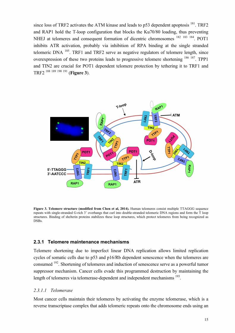
The packaging of the eukaryotic cell genome into linear chromosomes poses two problems with fundamental biological importance. First, the chromosomes ends must be distinguished from double strand breaks (DSBs) to avoid improper DNA damage repair, which could result in chromosome end fusions and chromosome breakage during mitosis. Second, the linear chromosomes ends cannot be completely replicated by the DNA replication machinery, thus DNA sequences are lost during every cell division 172 173. These problems are overcome by specialized nucleic acid-protein complexes structures at the chromosome ends known as telomeres174. Telomeric DNA consists of G-rich DNA repeat sequences (e.g. TTAGGG in human) that buffer DNA erosion and assure the integrity of the coding regions. The length varies greatly between species, from approximately 300 bases in yeast to many kilo-bases in human 175. The actual terminus of a telomere is not blunt-ended but consists a single-stranded 3’ tail, known as the G overhang, which is evolutionarily conserved and essential for telomeres. Studies have shown large loop structures called telomere loops (T-loops), where the single-stranded telomere overhangs curl around in long circles and are stabilized by telomere-binding proteins 176. The T-loop structure protects the telomere regions from being recognized as DNA double strand breaks. At the very end of the T-loop, the single-stranded telomeric DNA invades into a region of double-stranded DNA. The resulting triple-stranded structure is called a displacement loop or D-loop 177. Six interdependent telomere-binding proteins form the shelterin complex, which consist of the telomeric-repeat-binding factor 1 and 2 (TRF1 and TRF2), the TRF1-interacting protein 2 (TIN2), the transcriptional repressor/activator protein 1 (RAP1), the protection of telomeres 1 (POT1), and the POT1 and the TIN2 organizing protein TPP1 178. Removal of individual members causes instability of the whole structure, which leads to uncapping and de-protection of the telomeres. The telomere complex also comprises a non-coding telomeric repeat-containing RNA referred as TERRA 179, which is transcribed from subtelomeric regions and interacts with several telomere associated proteins such as TRF1, TRF2, the heterochromatin protein 1 (HP1) and histone H3 trimethyl K9 (H3K9me3) 180. TERRA plays a key role in maintaining the telomere structure and heterochromatin formation along with the shelterin proteins 180.
Numerous roles of the shelterin proteins in maintaining telomere integrity have been reported. For example, TRF2 has been shown to protect human telomeres against DDR activation,
15
since loss of TRF2 activates the ATM kinase and leads to p53 dependent apoptosis 181. TRF2 and RAP1 hold the T-loop configuration that blocks the Ku70/80 loading, thus preventing NHEJ at telomeres and consequent formation of dicentric chromosomes 182 183 184. POT1 inhibits ATR activation, probably via inhibition of RPA binding at the single stranded telomeric DNA 185. TRF1 and TRF2 serve as negative regulators of telomere length, since overexpression of these two proteins leads to progressive telomere shortening 186 187. TPP1 and TIN2 are crucial for POT1 dependent telomere protection by tethering it to TRF1 and TRF2 188 189 190 191 (Figure 3).
Figure 3. Telomere structure (modified from Chen et al, 2014). Human telomeres consist multiple TTAGGG sequence repeats with single-stranded G-rich 3’ overhangs that curl into double-stranded telomeric DNA regions and form the T loop structures. Binding of shelterin proteins stabilizes these loop structures, which protect telomeres from being recognized as DSBs.
2.3.1 Telomere maintenance mechanisms
Telomere shortening due to imperfect linear DNA replication allows limited replication cycles of somatic cells due to p53 and p16/Rb dependent senescence when the telomeres are consumed 192. Shortening of telomeres and induction of senescence serve as a powerful tumor suppressor mechanism. Cancer cells evade this programmed destruction by maintaining the length of telomeres via telomerase-dependent and independent mechanisms 193.
2.3.1.1 Telomerase
Most cancer cells maintain their telomeres by activating the enzyme telomerase, which is a reverse transcriptase complex that adds telomeric repeats onto the chromosome ends using an
RA
P1
TIN2
TRF1
TRF2
RA
P1
TTP1
TIN2
TRF2
RAP1
TRF1
5’-TTAGGG 3’-AATCCC
ATM
ATR
POT1
TTP1
POT1
TIN2
TRF2
RAP1
TRF1
POT1
16
RNA template 194 195 . Three major components were purified and identified from human telomerase, a reverse transcriptase (TERT), a folded RNA containing a telomere repeat recognizing sequence and a template (TER), and the small nucleolar ribonucleoproteins (snoRNPs) family member dyskerin (DKC1) 196. This enzyme is normally active only in stem cells, germ line cells, embryonic tissues and a subset of somatic cells such as activated lymphocytes 197, while it is either not expressed or kept at very low levels in most of the somatic cells 198.
The template region of human TER (hTER) is complementary to the telomeric repeat. The telomerase promotes binding of the first few nucleotides of the template to the last telomere sequence on the chromosome ends, adds a new telomere repeat sequence, sets free the telomerase complex, complements the new synthesized 3' ends of telomeres by DNA polymerase, and repeats the process. Thus, the conventional replication machinery and telomerase are closely coordinated during new telomere synthesis 199.
The mechanism for telomerase recruitment to telomeres is not completely clear, but at least two shelterin proteins, POT1 and TPP1, may provide a physical link between telomerase and the shelterin complex 200. TRF1 is proposed to have a negative effect on telomerase dependent telomere homeostasis by binding to telomeres and providing a negative feedback signal to telomerase 186.
2.3.1.2 Alternative lengthening of telomeres
A telomerase-independent mechanism for telomere maintenance, known as alternative lengthening of telomeres (ALT), is based on homologous recombination of telomere sequences. Evidences for ALT activation in human were first provided by the observation that telomere length was maintained after many divisions in cell lines lacking telomerase activity 201. The first indication of ongoing recombination was the observation of sharp changes in telomere length in telomerase-negative cells 202. ALT cells are characterized by the presence of telomere dysfunction induced foci (TIFs), extra-chromosomal telomeres, and highly heterogeneous telomere lengths 201 203. Additionally, ALT associated promyelocytic (PML) bodies (APBs) that contain PML and telomere DNA are frequently observed 204. Although it is generally accepted that telomere elongation in ALT cells requires a DNA recombination step, the exact mechanism leading to heterogeneous telomere length is uncertain. Two possible not mutually exclusive models have been proposed.
The first model is based on the finding that telomere sister chromatid exchange (T-SCE) occurs much more frequently in ALT cells than in telomerase-positive cell lines or normal somatic cells 205 206. The molecular mechanism of T-SCE activation is still unknown, although there is evidence that in ALT cells telomeric DNA contains nicks and gaps that may serve as a structural barrier to DNA replication and therefore cause T-SCE 207. In this model sister chromatid exchanges of unequal size occur during replication, resulting in one daughter cell with a lengthened telomere and the other carrying a shortened telomere 208. According to the second model, ALT may be achieved using telomeric sequences from close-by

17
chromosomes as templates 209 210, which may result in a net increase of telomeric DNA length. In this model, the telomere may also copy its own sequence through T-loop formation, or use the telomere sequence of its sister chromatid as template 211. In addition, it has been proposed that linear and circular extrachromosomal telomeric DNA could also act as a template for HR mediated ALT 210 212 213.
Several proteins have been shown to be necessary for telomere maintenance in ALT cells. The MRN complex (Mre11, Rad50 and Nbs1) promotes ALT activity by recruiting ATM to telomeres, which initiates recombination, and by processing the chromosome end to form an extended telomeric overhang, which could serve as HR template after invasion of adjacent telomere sequences 214 215 216 217 218. The SMC5-SMC6 complex seems to be important for the recruitment of telomeres to PML bodies through sumoylation of shelterin proteins, and APB is believed to be the platform for ongoing telomeric DNA repair 219 220. RecQ-like helicase WRN or BLM are responsible for removing replicating intermediates like G-quadruplexes at telomeres, thus loss of them may result in telomere shortening 221. Besides, FEN1, MUS81, FANCD2 and FANCA play essential roles in the recombination repair of stalled or broken replication forks, which is important for telomere sister chromatid exchange 222 223 224. Nevertheless, mutations are often observed in genes that suppress HR in ALT cells, such as telomeric heterochromatin remodeler complex ATRX/DAXX and Histone variant 3.3 225 226 227.
2.3.2 Tumor viruses and replicative immortality
Tumor viruses often reprogram the host cells environment to promote proliferation, which creates a favorable environment for viral genome replication and enlarges the pool of infected cells. A common consequence of tumor virus infection at last is malignant transformation, which enables the cells to proliferate indefinitely. However, the telomere erosion during replication will finally induce cell senescence, and this machinery serves as an important tumorigenesis barrier. Thus, tumor viruses infected cells must evolve a mechanism to bypass this blockade. One strategy to achieve it would be to (re)-activate the telomerase activity in the infected host cells 228. This scenario is actually supported by studies from various viruses.
The HPV E6 and E7 proteins together promote the cooperative binding of Sp1 and c-Myc to the promoter region of hTERT and transcriptionally enhance its activity. The interaction between E6 and cellular E3-ligase E6AP is also required for this process 229 230. The EBV encoded latent membrane protein-1 (LMP1) is a functional homologue of tumor necrosis factor receptor (TNFR), which could constitutively stimulate hTERT transcription via activation of NF-kB and JAK/STAT pathways 231 232.
Conversely, some viral proteins have been suggested to have a suppressor effect on telomerase as well. For instance, the HPV E2 protein down regulates hTERT transcription by interfering Sp1 activity 233. Additionally, the EBV LMP2A inhibits telomerase in a mechanism that is not fully understood yet, though its N-terminus immunoreceptor tyrosine-based activation motif (ITAM) function seems to be required 93.

18
Interestingly, infection with tumor viruses not only prevent telomere erosion, but it is also associated with frequent occurrence of telomere dysfunction, which could further induce genomic instability 234. These abnormalities could be generated through distinct mechanisms after infection, such as: i) rapid cell proliferation without simultaneously activating telomere maintaining mechanisms; ii) expressing viral proteins that could disturb telomeric protein binding directly; iii) virus promoted reactive oxygen species (ROS) accumulation that could induce oxidative damage at telomere region; and iv) integration of viral genome into host telomeric DNA. For example, it has been shown earlier that EBV infected primary B-cells and EBNA1 expressing B-lymphoma cells are suffering from high levels of telomere dysfunction and genomic instability. Formation of TIFs and displacement of TRF2 from telomeres are frequently observed as well 235 236. A possible explanation for these could be the EBNA1-mediated binding of TRF2, RAP1 and tankyrase (telomere-associated poly-ADP ribose polymerase) at OriP, which further regulate the viral episome maintenance and replication, though it is not determined yet how these events could affect the shelterin complex function 237 238 239. Nevertheless, we have reported that EBNA1 induces the accumulation of intracellular ROS via transcriptional activation of the catalytic subunit of the NADPH oxidase NOX2. This is accompanied with high levels of DNA damage, telomere abnormalities and chromosomal instability 55.
The frequent occurrence of telomere dysfunction seems to be an early event during infection relative to the increase in telomerase activity, indicating that an alternative mechanism could explain this phenomenon. Indeed, the work presented in this thesis indicates that ALT might be activated during early stage of tumor virus induced cell transformation. Thus, this hypothesis is consistent with improper protection of telomere, extrachromosome telomeres and formation of telomere associated DNA damage foci observed in the early stages of infection. Emerging signs for ALT activation were actually observed in the context of EBV freshly infected B cells and EBNA1 expressing B-lymphoma cells 236, KSHV vGPCR immortalized human umbilical vein endothelial cells, and HPV E6 or E7 immortalized human embryonic fibroblasts 240 241. And sometimes various ALT markers were shown to co-exist with elevated telomerase activity as well 242.
Thus, tumor viruses utilize both telomerase activity elevation and ALT activation strategies to evade replicative senescence. We propose a scenario where ALT activation during the early transformation period promotes high levels of telomere dysfunction and chromosome instability. Cells that would survive these events might acquire genetic alterations that could favor viral transformation. Survival and proliferation of these selected cells can further benefit from the increased telomerase activity, observed in later stages post-infection, which could further contribute to tumor progression by attenuating level of genomic instability.

19
2.4 OXIDATIVE STRESS
Oxidative stress is an important biomarker for several diseases, including neurodegenerative diseases, cardiovascular diseases, aging-related development of cancer, and infectious diseases. While redox homeostasis is essential for the optimal function of cellular processes, malignant cells are often characterized by chronic oxidative stress, which is caused by imbalance between the generation and elimination of reactive oxygen species (ROS).
2.4.1 Reactive oxygen species
ROS are chemically reactive oxygen-containing molecules due to the presence of unpaired electrons. These include highly reactive radicals like superoxide anion (O2•–) and hydroxyl radical (HO•), as well as non-radical molecules such as hydrogen peroxide (H2O2) 243 244 245. These molecules are continuously produced during oxygen consumption in metabolic reactions that mainly occur in mitochondria 246 247, peroxisomes 248 and endoplasmic reticulum 249, as well as through enzymatic reactions that involve NADPH oxidase, xanthine oxidase, lipoxygenase and cylooxgenase 250 251 252 253. Superoxide anions are considered to be the primary ROS product from both mitochondrial electron transport chain complexes and oxidases reactions. It is estimated that about 2% of the total oxygen consumed in mitochondria is converted to form superoxide anions, therefore mitochondria are considered to be a major source of ROS 246 247. After releasing into the cytoplasm, superoxide dismutases (SODs) are responsible for converting them into hydrogen peroxide 254 255, which is further converted to water by catalase and glutathione peroxidase 245 256. Hydrogen peroxide may be further converted to highly reactive product hydroxyl radicals through the Fenton reaction, where election transition metal ions like Fe2+ serve as catalyzers 257 258 259.
Since different levels of ROS can induce distinct biological responses, tight regulation of both ROS promoting and scavenging pathways is required 260 261. Under physiology condition, ROS act as messenger molecules that sustain essential cell signaling, and activate the cellular responses to stress. For example, intracellular ROS were shown to promote cell proliferation and migration, and induce pro-inflammatory cytokine secretion 262 263 264. However, excess levels of ROS could damage cell structures like lipids, proteins and DNA, activate oncogenic signaling pathways, trigger cell senescence, or cause mitochondria failure that results in release of cytochrome c and apoptosis 265 266. The effects of excessive ROS production are normally balanced by enzymatic and non-enzymatic cellular antioxidants. The most efficient enzymatic antioxidants are superoxide dismutase, catalase and glutathione peroxidase 267 256, while the non-enzymatic antioxidants include vitamin C, vitamin E, glutathione and thioredoxin (TRX) 268 269 270. These molecules cooperate in reducing the overproduced ROS to prevent irreparable cellular damage.
2.4.2 Oxidative stress and tumorigenesis
Tumor cells are commonly under oxidative stress, probably due to the aberrant metabolism induced by oncogene activation. Although the contribution of oxidative stress to malignant

20
transformation is still controversial, it is implicated during all stages of tumorigenesis including initiation, promotion and progression 271.
The initiation stage involves non-lethal but irreversible mutations of DNA, where ROS-induced oxidative DNA damage is one of the common contributors. The majority of the cells will be arrested by the DNA damage response, but some cells may keep dividing in spite of mutations, thus representing the initiated cells. The promotion stage is experimentally defined as clonal expansion of the initiated cells, where cell proliferation is enhanced and apoptosis is inhibited. This stage is a reversible process during which ROS contribute to altered gene expression and therefore modified signal transduction. The outcome of the promotion phase is the formation of focal lesions where the expanded cells reach a pre-neoplastic state. During the final progression stage, accumulation of more irreversible genomic alterations is achieved, leading to transition of expanded pre-neoplastic cells to more aggressive malignant cells. Genomic instability, loss of chromosome integrity and functionally inhibited tumor suppressors are the features of this process, where oxidative stress could further enhance these aberrations 272.
Cancer cells are characterized by abnormal metabolism and protein synthesis, which results in chronic oxidative stress. However, cellular antioxidant responses are commonly increased in malignant cells through mutations and activated oncogenes, which allows escape from cell death. Thus, under condition of moderate ROS levels, cancer cells may acquire additional mutations that further drive tumorigenesis. Hence, the antioxidant capacity of tumor cells could be considered as a potential therapeutic target 273 (Figure 4).
Figure 4. Interplay between ROS level and cancer (modified from Cairns et al, 2011). Low levels of ROS are beneficial for cell proliferation and survival, while high levels of ROS trigger senescence and cell death. In cancer cells, aberrant metabolism and protein synthesis promote high levels of ROS. While adapted mutations and altered gene expressions enable the cancer cells to exert cellular antioxidants for reducing ROS levels toward moderate levels. On one hand, this tight control of redox allows the cancer cells to avoid the detrimental fate of high levels of ROS. On the other hand, it also increases the chance of these cells to acquire more ROS-mediated mutations.
Proliferation Cell survive
Altered Gene expression Mutagenesis
Senescence Cell death
ROS level
- Metabolism - Protein synthesis
- Antioxidants
Cancer cell

21
2.4.3 Tumor viruses and oxidative stress
Oxidative stress is often observed during infection by various tumor viruses, and recent studies have started to elucidate its role in the context of virus-induced transformation. Elevated ROS production has been reported following binding and entry of EBV, KSHV, and HSV1 274 275 276 277 278. For example, within two hours of infection, EBV induces oxidative stress with decreased levels of different cellular antioxidants including SODs and catalase 274. ROS are also produced early after KSHV infection where they promote efficient entry of the virus into endothelial cells by regulating macro-pinocytosis 275. The early ROS production was initially explained by the activation of phagocytes, which represents a non-specific immune response to eliminate the pathogens 279. However, binding of the virus appears to be required to trigger ROS production, since pretreatment of KSHV with heparin abolishes it 275.
Multiple viral products contribute to the establishment of oxidative stress during both latent and productive infection. The EBV latent protein EBNA1 promotes ROS production in BL cells via transcriptional activation of the catalytic subunit of the NADPH oxidase NOX2, which is associated with DNA damage and genomic instability 55 56. Additionally, the EBV latent product LMP1 was shown to promote the accumulation of ROS in NPC by up-regulating the NADPH oxidase subunit p22phox via activation of JNK signaling, which may result in increased oxygen consumption, hypoxia and enhanced glycolysis 280. Likewise, the KSHV glycoprotein K1 induces elevation of intracellular ROS through upregulation of Rho-like small GTPase Rac1, which activates the NADPH oxidase. High levels of ROS enhance vascular permeability, which alters the tumor microenvironment 281. The early protein of KSHV vGPCR also induces ROS via the same mechanism, while quenching of ROS by treatment with NAC leads to impaired tumor angiogenesis and proliferation 282. Furthermore, the cellular redox master regulator NRF2 is activated during KSHV infection of endothelial cells, which suggests that a precise modulation of the oxidative status is crucial for viral oncogenesis 283 284.
2.5 SIGNALING BY POST-TRANSLATIONAL MODIFICATIONS
Due to limited genome size, tumor viruses need to rely on the host cell machinery for almost every single step of their life cycle, including virus entry, proliferation initiation, viral genome replication, virion packaging and egress. In addition, viruses also need to overcome the host immune responses in order to survive in the infected cells. One strategy by which viruses may use their limited genetic information to achieve a broad remodeling of the cellular environment is by interfering with protein post-translational modifications (PTMs). PTMs involve the addition of chemical residues, such as phosphate, glycan, methyl and acetyl groups, or small polypeptides, which changes the conformation, stability, interaction properties, subcellular localization and ultimately the function of the substrate. Ubiquitin (Ub) and ubiquitin-like (UbL) proteins, such as the small ubiquitin-related modifier (SUMO), the multiple neural precursor cell-expressed developmentally down-regulated 8 (NEDD8), and

22
the interferon-stimulated gene 15 (ISG15), are small polypeptides modifiers that share structural similarities 285 286. Cellular signaling pathways and functions that are regulated by this type of post-translational modification include transcription, protein trafficking and degradation, signal transduction, DNA replication, DNA damage repair and apoptosis 286 287.
2.5.1 The Ub/UbL modification cascade
The conjugation of Ub and UbLs to protein substrates is a multistep process where three enzymes are sequentially involved 288. First, the modifiers are activated by an activating enzyme (E1) through formation of a thiolester bond between their C-terminal glycine and a cysteine residue in the E1, where ATP hydrolysis is also required. Then, the activated modifiers are transferred to the catalytic cysteine residue of a conjugating enzyme (E2). Finally, a ligase (E3) catalyzes the transfer of the modifier from the E2 to a lysine residue of the substrate 289. Once the first Ub or UbL is attached to the target, this process can be repeated by adding a new modifier to the previous one, resulting in the formation of a poly-modifier chain. Depending on the modifier, one or two E1, a limited number of E2 and a great number of E3 have been identified 290. The number of E3s guarantees the specificity of substrate recognition and the types of conjugation.
The E3 ligases can be divided into three major groups depending on their structure and ubiquitination mechanisms: the HECT (Homologue of E6-AP C-terminus) domain containing ligases, the RING (Really Interesting New Gene) domain containing ligases and the U-box ligases 291 292 293. The RING domain containing E3s constitute the largest ligase family that regulates various cellular events including DNA replication, cell proliferation, cell cycle progression and apoptosis 294. The major family of the RING domain-containing ligases is the Cullin-RING ligases (CRLs), where a multi-protein complex is assembled around the NEDD8 conjugated cullins. This complex serves as a scaffold that binds to E2 via its RING domain and to substrate with an adaptor protein, which facilitates the transfer of ubiquitin from the E2 to the substrate 295 296.
The Ub and UbLs are synthesized as inactive precursors and specific proteases are required to cleave their C-termini in order to expose the terminal glycine. This process is managed by specific deconjugating enzymes called deubiquitinating enzymes (DUBs) and UbL-specific proteases (ULPs). DUBs and ULPs also can hydrolyze the covalent bond that link the modifiers to their substrates, which reverse the signal and maintain a constant intracellular pool of free Ub and UbLs 297. Approximately 100 DUBs are encoded by the human genome. They can be categorized into five families: the ubiquitin specific proteases (USPs), the ubiquitin C-terminal hydrolases (UCHLs), the ovarian tumor related proteases (OTUs), the Machado-Joseph disease proteases (MJDs) and the Jab1/MPN/Mov34 domain containing metalloenzymes (JAMM) 297. The human ULPs are represented by the sentrin specific peptidases (SENPs). All DUBs and ULPs are cysteine proteases except for the JAMMs that cleave the modifiers from substrate in a Zinc ion and ATP dependent manner 297 298.

23
Different types of ubiquitin modification are known. These modifications include monoubiquitination, multiubiquitination where monoubiquitination occurs at multiple substrate sites, and polyubiquitination where Ubs are sequentially added onto each other on the substrate. Monoubiquitination can regulate DNA damage repair, histone function, gene expression and receptor endocytosis 299 300 301 302 303, while multiubiquitination mainly modulates receptor endocytosis 304. Additionally, poly-ubiquitin chains can be formed on each of seven Lysine (K) residues of ubiquitin molecule, including K6, K11, K27, K29, K33, K48 and K63 305. The length and linkage structure of the chain will further determine the fate of substrates and their interaction with different cellular function proteins 306. For example, K48 linked Ub chains are the most abundant type in human, which mediate the proteasome degradation of various substrates 305. Additionally, the K63 Ub chains are also well characterized modifications involved in non-proteolytic cellular functions such as intracellular trafficking, autophagy, DNA damage response and cell signaling transduction 306 307 (Figure 5). On the other hand, UbLs also contain multiple lysines but the types and the fates of different UbLs chains are still largely unknown. For example, while mono- or poly-SUMO chains have been identified the only defined NEDD8 modification is the mononeddylation of the CRLs.
Figure 5. The Ub/UbL system. Inactive precursors of Ub and UbLs need to be processed by DUBs or ULPs to expose their terminal glycines for conjugation. Throughout the enzymatic cascade that involves activating enzyme E1, conjugating enzyme E2s and substrate ligase E3s, Ub and UbL modifiers are evantually linked to specific substrates. These modifications result in both non-proteolytic and proteolytic cellular functions. Afterwards, substrates linked modifiers are deconjugated by DUBs or ULPs for recycling.
Precursor Active
DUB
ULP
Ubiquitin SUMO NEDD8
DUB
E1 E2
E2
E3 Substrate
Substrate
E2
E3
Substrate
Non-proteolytic regulation on: - Kinase activation - Gene expression - Intracellular trafficking - DNA repair
Proteolytic regulation on: - Protein homeostasis - Replication and apoptosis - Antigen processing

24
2.5.2 Viruses interfere with the Ub/UbL pathway
Due to the versatility of Ub and UbL signaling in regulating protein functions, it is not surprising that viruses have evolved mechanisms to mimic, inhibit or redirect the activities of the Ub and UbL signaling cascade 290 308. Two general strategies are used by viruses to interfere with this system. Viruses may either encode adaptor proteins that hijack the cellular enzymes to target their own favored substrates, or they may encode homologues of the cellular enzymes with distinct structure and substrate specificity.
Virus-encoded E3 ligases are normally found in large DNA viruses. For example, the KSHV encoded protein K3 and K5 regulate the expression of the major histocompatibility complex (MHC) class I molecules by acting as ubiquitin E3 ligases. The N-terminal domain of K3 and K5 add a K63-linked ubiquitin chain to MHC I molecules, therefore promote endosomal sorting and further lysosome degradation 309 310 311. In this way, KSHV decreases antigen presentation and manages to escape from cytotoxic T-lymphocyte recognition.
Viruses encoded adaptor proteins recruit or redirect cellular E3 ligases. A classic example of this strategy is the HPV E6 protein that reprograms the cellular E3 ligase to promote proteasome-dependent degradation of p53 162. The EBV membrane protein LMP2A also acts as an E3 adaptor that redirects the cellular NEDD4 ligase for proteasome-dependent degradation of tyrosine kinases Lyn and Syk, which inhibits triggering of the productive virus cycle by preventing the activation of BZLF1 upon engagement of the B-cell receptor 312 313.
Viruses also encode functional homologs of Ub/UbL deconjugases. A particularly interesting example is the family of deconjugases encoded by the large tegument proteins of herpesviruses. The EBV encoded member of this family, BPLF1, was shown to promote the production of infectious virions by deubiquitinating the proliferating cell nuclear antigen (PCNA), the E3 ligase Rad18 and the viral ribonucleotide reductase 314 315 316. BPLF1 was also shown to deubiquitinate TRAF6, which my favor virus production by inhibiting NF-kB signaling and β-interferon production 120 119. Moreover, BPLF1 and other member for this enzyme family exhibit a NEDD8-specific deconjugase activity, which regulates the activity of cellular Cullin-RING ubiquitin ligases (CRLs) and promotes efficient viral DNA replication 118 317.

25
3 AIMS OF THIS THESIS
The overall aim of this thesis was to uncover the complexity of virus and host interactions
during EBV infection. In particular we wished to assess how EBV could adapt to and exploit
cellular responses to establish an environment conducive to B-cell immortalization and
productive infection, respectively. To this end, my colleagues and I have addressed the
following specific aims:
1. Investigate the mechanisms that rescue EBV infected B cells from replicative senescence
during early phases of transformation.
2. Identify the key factors that limit the efficiency of EBV induced B-cell transformation
during early stage of infection.
3. Elucidate the machineries by which EBV remodels cellular environment to maintain
successful productive infection.

26

27
4 METHODOLOGY
A complete description of the experiment procedures used in this thesis is available in the appended articles. In this section I will briefly discuss the features and principles of chosen experimental methodologies.
4.1 TELOMERE ANALYSIS BY FISH BASED TECHNIQUES
Q-FISH: Quantitative fluorescence in situ hybridization (Q-FISH) was used as a method of choice to examine the size and status of individual telomeres. A more traditional way for measuring telomere size is southern blot that uses restriction endonuclease for cleavage of genomic DNA at sub-telomeric sites to create terminal restriction fragments (TRFs) 318. The fragmented DNA is then gel electrophoresed, transferred to membrane, hybridized with labeled probes recognizing telomere sequences and visualized by chemiluminescence. Southern blot provides an estimation of the telomere size of entire cell population without specific information on individual telomeres. In contrast, labeling of individual telomeres by Q-FISH allows direct assessment of the size of individual telomeres with similar or even better sensitivity of southern blot 319. The peptide nucleic acid (PNA) is a synthetic DNA probe that hybridizes to target telomere sequences with specificity, high affinity, stability and low background 320 321. In addition, Q-FISH offers the additional advantage of allowing direct analysis of a small subpopulation of proliferating cells, avoids possible artifacts due to the presence of infrequent numbers of non-proliferating or dead cells upon EBV infection. By Q-FISH we could also visualize telomere abnormalities such as absence of telomere signals, telomere fusion and extra-chromosome telomere.
CO-FISH: Telomere-sister chromatid exchange (T-SCE), which is the outcome of HR dependent repair at telomeres, can be detected with a FISH based technique known as chromosome orientation FISH (CO-FISH). The cells were first cultured in the medium containing Bromodeoxyuridine (BrdU) and Bromodeoxycytidine (BrdC), which are synthetic nucleotides analogue of thymidine and cytidine, for a single cell cycle. Then the complementary telomeric DNA was removed by UV exposure and exonuclease III digestion, followed by sequential hybridization with Cy3-TelG and FITC-TelC specific probes that target lagging G-rich and leading C-rich telomere strand respectively. After microscopy analysis, sister chromatin exchange was scored by the presence of chromosomes with both TelG and TelC probe signals at telomeres of both sister chromatids.
IF-FISH: Accumulation of PML nuclear bodies (PNBs) containing telomeric DNA, known as ALT-associated PNBs (APBs), is a characteristic feature of ongoing homologous recombination repair at telomeres. TRF2 is an important shelterin complex component involved in telomere protection, where displacement of TRF2 at telomeres could lead to aberrant activation of HR. Combination of specific immunofluorescence with in situ
28
hybridization (IF-FISH) provides a direct and specific method for detection of proteins that are associated with telomere sequences, such as PML and TRF2. Immunofluorescence (IF) is performed prior to fluorescence in situ hybridization (FISH) in this method. The presence of APBs was scored in images captured by confocal microscope based on colocalization of PNBs and telomere signals on a single focal plane. For quantification of telomere associated TRF2, images were acquired and analyzed with a ImageXpress Micro device. Two thousand telomere signals were analyzed from different images for each condition and the intensity of TRF2 and telomeres fluorescence within each telomere signal was quantified.
4.2 DETECTION OF TELOMERASE ACTIVITY BY TRAP ASSAY
Telomerase is a ribonucleoprotein that adds telomeric repeats to the 3’ end of telomeres using its RNA template. The telomeric repeat amplification protocol (TRAP) is a sensitive and efficient PCR based assay for detection of telomerase activity. This assay is an improved version of original method described by Kim et al. 322, which reduced the amplification artifacts and magnified its detection sensitivity 323. Moreover, large scales of surveys on telomerase activity in various human cells and tissues have been carried out with this method 324 197 325 326 327. The experimental procedure started from extraction of lysates containing telomerase, followed by extension of kit supplemented substrate oligonucleotide with telomerase, PCR amplification of extended products, separation of individual product by gel electrophoresis, gel staining and imaging, and analysis of the images with calculated total product generated (TPG) units. Cell lysates with confirmed telomerase activity were provided in the kit as standard control. Heat inactivated samples and lysate from the telomerase negative cell line U2OS were used as negative control.
4.3 MEASUREMENT OF REACTIVE OXYGEN SPECIES
All cells constantly produce ROS during aerobic metabolism, and oxidative stress occurs when the ROS generation overwhelms the cellular antioxidant capacity. The most straightforward technique for measuring cellular ROS levels uses cell permeable fluorescent and chemiluminescent probes, followed by flow cytometry analysis. The colorless 2,7-dichlorodihydrofluorescein diacetate (H2DCF-DA), which is the non-fluorescent precursor of H2DCF, is one of the most widely used probes for direct measurement of ROS 328 329. Intracellular esterase cleaves H2DCF-DA at its two-ester bond producing the relatively impermeable form H2DCF. Upon oxidation, a highly fluorescent product DCF is yielded from its reduced form. The increased DCF fluorescence is then detected by flow cytometry, which reflects the redox status of samples. Dihydroethidium (DHE) may also be used as probe for r ROS detection. However, DHE is predominantly oxidized by superoxide anion, while H2DCF-DA indicates oxidation by hydrogen peroxide, peroxynitrite, hydroxyl radical and also superoxide anion though at a lesser degree. Thus, we used H2DCF-DA in our experiments for broader detection of intracellular ROS.

29
4.4 MEASUREMENT OF CELL DIVISIONS
Carboxyfluorescein diacetate, succinimidyl ester (CFSE) serves as a cell-retained tracing reagent. CFSE can diffuse into the cells and it is colorless and non-fluorescent until the acetate groups are cleaved by intracellular esterase. The carboxyfluorescein succinimidyl ester becomes highly fluorescent and its succinimidyl ester reacts with intracellular amines, forming conjugates that are very well retained in cells. Unconjugated CFSE diffuses back to the extracellular medium can be washed away. Combined with flow cytometry analysis, CFSE incorporation provides a convenient method for monitoring cell division. CFSE was incorporated into freshly isolated B cells prior to both mitogen stimulation and EBV infection, and cell division was traced until 10 days of culture. Individual cell division peaks were then identified by 50% fluorescence decrease, and the mean division number of total cell population was calculated as (sum of % of cells in each peak x number of cell divisions)/100.

30
5 RESULTS AND DISCUSSIONS
At the time the work described in this thesis was initiated the accepted scenario of EBV pathogenesis suggested a clear distinction between the capacity of the virus to promote the autonomous proliferation of latently infected but virtually normal B-lymphocytes, which could give rise to life-threatening lymphoproliferations when the host immune defenses are severely compromised, and its involvement as a promoting factor in “true” malignancies that are driven by distinct virus-independent genetic alterations. This view was challenged by several observations suggesting a more direct role of the virus as an initiating factor of malignancy. In particular, cytogenetic studies performed in EBV negative and positive B-lymphoma lines suggested that virus carriage is associated with the establishment of an oxidative environment and with the induction of chromosomal instability, DNA damage, activation of the DNA damage response, telomere dysfunction and activation of ALT 55 330 171 56. Chromosomal aberrations were also observed in freshly infected normal B-cells already within the first weeks after growth transformation followed by the establishment of monoclonal cultures 235, suggesting that virus carriage may promote the occurrence of random genetic alterations that provide a selective growth advantage. Furthermore, the early proliferative response of EBV infected normal B lymphocytes was shown to be accompanied by the occurrence of extensive DNA damage 37, suggesting that viral products may be directly involved in the establishment of a mutator phenotype that could promote malignancy. The work described in this thesis aimed to critically assess these possibilities by performing a detailed analysis of the early cellular response to EBV infection (Papers I and II).
The EBV transforming capacity can be exerted only if the virus can establish a successful infection, and this could be achieved by counteracting or exploiting the cell intrinsic and innate immune defense. Thus in Paper III, we have assessed how viral products that are delivered to the infected cells together with the incoming virion, modulate these cellular functions.
5.1 EBV-INDUCED B-CELL PROLIFERATION IS ACCOMPANIED BY TELOMERE DYSFUNCTION AND ACTIVATION OF ALT (PAPER I)
Telomere dysfunction and chromosomal instability are hallmarks of cancer 331. EBV-positive BL cell-lines exhibit significantly more chromosomal instability compared to their EBV negative counterparts and this is associated with multiple signs of telomere dysfunction and activation of ALT 56. Since the observation was made in established long term cultured cell lines, it may be argued that the effect is dependent on inherent properties of the malignant cells and facilitated by the long-term culture conditions. Thus, the primary goal of this work was to assess whether this EBV-driven phenotype is also observed in freshly infected normal B cells.

31
To address this possibility we first compared the occurrence of telomere abnormalities in metaphase chromosomes from EBV-infected B-lymphocytes. As a control we used B- or T-cell blasts where proliferation was driven by mitogenic stimulation rather than EBV immortalization. In spite of comparable levels of cell proliferation, the EBV infected cells showed a high prevalence of chromosomes with abnormal telomeres, usually loss or duplication of telomere signals and telomere fusion. By day 7 between 50-60% of the metaphases of EBV infected cells contained one or more chromosomes with abnormal telomeres. Extra-chromosomal telomere signals were also frequently detected, indicating double strand breaks of telomeric DNA. Proliferation of the EBV-infected B lymphocytes was associated with a significant increase of the intensity of the telomere signals, as assessed by FISH analysis of metaphase plates already after 2 weeks of culture while the signal intensity remained constant or was decreased in the mitogen blasts. Most importantly, the infected cells exhibited highly heterogeneous signal intensity, with a strongest:weakest signal ratio exceeding 50 fold. These data indicate that EBV infection induces telomere elongation. However, this effect was not due to an increased telomerase activity, since TRAP assays revealed a very low activity in both EBV- and mitogen-induced blasts, suggesting that EBV infection promotes an alternative mechanism to ensure telomere elongation.
As summarized in the introduction, the accumulation of extra-chromosomal telomeres and increased telomere length in the absence of telomerase activity are indicative for the activation of ALT 201. Recognized markers of ALT are the accumulation of PML nuclear bodies containing telomeric DNA known as ALT-associated PNBs (APBs) 204. In addition to PML, APBs contain a variety of proteins involved in the DNA damage response (DDR), and polymerases that use sister chromatids or extra-chromosomal telomeric sequences as templates for telomere extension 205 206. We found that APBs are regularly detected in EBV infected cells already after 3 days of infection while they are virtually absent in mitogen-induced blasts. This was accompanied by the occurrence of telomere sister chromatid exchange (T-SCE) measured by chromosome orientation (CO)-FISH. This series of experiments confirmed that the early phases of B-cell immortalization is accompanied by telomeric DNA damage, and the activation of recombination-based mechanisms of repair may assure the maintenance of telomere homeostasis in rapidly proliferating cells with low or absent telomerase activity.
Telomere de-protection is a common cause of DNA damage at telomeres 332. In order to assess whether uncapping might explain the telomere dysfunction phenotype induced by EBV infection, the functionality of the shelterin complex was investigated by monitoring the co-localization of telomeres with the DNA binding subunit TRF2 in IF-FISH assays. We found that telomeres lacking co-localized TRF2 signals are frequent in the interphase nuclei of EBV-infected cells. To assess whether this may correlate with defects in the expression of TRF2 or other proteins involved in the maintenance of telomere structure, the expression of the shelterin subunits TRF1, TRF2 and POT1, and the ATRX subunit of the ATRX/DAXX chromatin remodeler were compared in western blots. We found that the proteins are expressed at comparable levels in mitogens and EBV induced blasts suggesting that the

32
telomere de-protection observed in infected cells is not primarily due to selective loss of the shelterin or ATRX/DAXX complexes.
The evasion of proliferative senescence is a key event in viral oncogenesis. Many tumor viruses, most notably KSHV, HPV, HCV and HTLV1, achieve this goal by regulating the activity of telomerase, which maintains the length of telomeres and ensures proliferative immortality 333. EBV infected cells may adopt an alternative strategy for maintenance of telomere homeostasis based on the activation of homologous recombination.
An interesting question is why EBV would use a different strategy to maintain telomere homeostasis. Our laboratory has previously shown that EBNA1 promotes genomic instability and telomere dysfunction in BL cells by transcriptional activation of the catalytic subunit of the NADPH oxidase, NOX2, resulting in increased intracellular levels of ROS 55. It is possible that EBNA1 exerts a similar effect also in primary infected B-lymphocytes. The oxidative environment may directly affect the function of the shelterin and ATRX/DAXX complexes since oxidation disrupts the recognition of telomeric DNA by TRF1 and TRF2 334. This effect, possibly associated with the inability of EBV to up-regulate TRF1, TRF2, POT1 and ATRX in newly infected cells, may explain why a significant proportion of the telomeres has little or no associated TRF2 in spite of unchanged levels of TRF2 detected in western blot.
The enhanced recombination rate observed at telomeres of EBV infected cells may also depend on the high sensitivity to oxidative-mediated DNA damage of the telomeric G-triplet. This effect increases the frequency of S1 nuclease sensitive sites at telomeres and could promote the activation of recombination-based repair 335. Therefore, the oxidative damage-induced telomere deprotection may result in activation of DNA repair pathways and ALT-mediated telomere elongation and/or telomere abnormalities, in absence of increased telomerase activity, allowing EBV-infected cells to overcome replicative senescence during the early stages of infection.
This scenario differs from the situation observed in long-term-established LCLs, which are characterized by increased telomerase activity and enhanced levels of the shelterin proteins TRF1, TRF2, POT1 and ATRX. Most likely this mechanism is associated with lower levels of genomic instability that would ensure a higher rate of cells survival.
5.2 EBV INFECTION INDUCES OXIDATIVE STRESS THAT IS REQUIRED FOR B-CELL IMMORTALIZATION (PAPER II)
To address whether early stages of infection are associated with induction of an oxidative environment as suggested in Paper I, we compared the levels of ROS over time by H2DCF-DA fluorescence in freshly infected normal B-lymphocytes and mitogen stimulated cells. A peak of fluorescence was observed in both mitogen stimulated and EBV infected cells after culture for 24 hr. However, while this was followed by a rapid return to baseline levels in

33
mitogen-stimulated cells, a steady increase was observed in EBV-infected cells followed by stabilization at levels comparable to those detected in LCL cells. As predicted from the discussion of Paper I, this oxidative environment promoted induction of single and double stand DNA breaks, detected by γ-H2AX staining. EBV infection caused a striking increase of γ-H2AX foci in the nuclei of EBV-infected cells whereas significantly fewer mitogen-stimulated cells were γ-H2AX positive and the nuclei exhibited fewer and smaller foci. The accumulation of ROS correlated with up-regulation of NOX2, while treatment with ROS scavengers reduced the percentage of γ-H2AX positive cells and fluorescence signals to the levels observed in mitogen-induced blasts.
Induction of DNA damage in infected cells was previously reported. It has been proposed that the activation of c-Myc by EBNA2 may generate hyperproliferation-associated replicative stress and give rise to large numbers of DNA double-strand breaks that trigger the cellular DDR and halt cell growth until a balance favoring long-term proliferation is achieved through the activity of viral products that slow-down cell proliferation and inhibit the DDR 37. Our data proposes a different scenario, where oxidative stress appears to be the main cause of DNA damage during the early phase of EBV induced B-cell growth transformation.
While the induction of ROS provides a satisfactory explanation for the capacity of EBV infection to induce DNA damage and might also explain the relative inefficiency of transformation, the purpose of this effect is unclear. To address this question, freshly infected cells were cultured in medium supplemented with ROS scavengers. Surprisingly, while the addition of NACA had no appreciable effect on the proliferation of mitogen-stimulated cells, it virtually abolished cell proliferation of EBV-infected cells. This was neither due to decreased virus load, nor to significant differences in the levels of viral transcripts that are expressed during the initial phase of infection. However, monitoring of protein expression by immunofluorescence and western blot revealed a significant decrease of LMP1. Thus, the majority of EBNA2 positive cells in the NACA treated cultures expressed low or undetectable levels of LMP1 and the intensity of the LMP1 specific band detected in western blots was significantly decreased.
Quenching the intracellular levels of ROS by NACA treatment also promoted a significantly decrease of the cellular transcription factor STAT3, and almost complete disappearance of its phosphorylated form. Similar results were obtained by treatment with the STAT3 specific small molecule inhibitor Stattic that prevents STAT3 phosphorylation.336. This indicates that the viral-induced oxidative stress promotes STAT3 activation, which was previously shown to play a key role in EBV-induced transformation 337,338. Stattic treatment inhibited EBV-induced cell proliferation but did not affect LMP1 expression, suggesting that, although both proteins are ROS regulated and are essential for immortalization, they act independently.
The effect of LMP1 can mimic survival signals that are normally delivered to antigen-stimulated B cells by the T helper lymphocytes 339. Indeed, LMP1 regulates the activity of cellular signaling pathways, such as the NF-κB and MAPK pathways that control B-cell

34
proliferation, up-regulates the expression of anti-apoptotic proteins, such as the cellular A20, and promotes downregulation of the ATR kinase, which hampers the DDR 80 79 81 83 87 85. Conversely, the ROS-dependent STAT3 pathway may mirror the proliferative and survival effect provided by the T helper secreted cytokines 340. The effect of ROS on this transcription factor is likely to be mediated by oxidative inactivation of specific phosphatases 341.
Interestingly, while treatment with ROS scavengers drastically decrease the efficiency of transformation, quenching of ROS affected only marginally the proliferation of established LCLs and did not alter the expression of LMP1 though the phosphorylation of STAT3 was still inhibited. Thus, the effect of ROS on these signaling pathways, and their contribution to cell growth appear to be different in different phases of the infection.
Since LMP1 mRNA levels and protein turnover were not affected, we surmised that ROS might regulate LMP1 mRNA translation, since several cellular and viral miRNAs are known to target this transcript 110,342,343, and oxidative stress-dependent alterations of miR profiles have been reported in a variety of different tissues and pathologic conditions 344. Cellular miRs of the miR-17/20/106 family and several EBV BART miRs were shown to target the LMP1 3’UTR 110,342,343,345. We found that quenching of ROS is accompanied by a reproducible increase of miR-BART1-5p and miR-BART3-5p.
Although the effect on each miR was relatively small, a physiologically relevant level of inhibition is likely to be achieved by the combined action of several miRs targeting the same mRNA. The ROS-dependent inhibition of BART miRs during the early phase of infection has interesting implications for the dynamics of the transformation process. These miRNAs are expressed in LCLs and in EBV-associated malignancies where targeting of LMP1 promotes cell survival by dampening the toxic effects of LMP1 overexpression 346. However, during the early stage of infection when the transcripts are less abundant, high levels of the miRs could keep the amount of protein below the threshold required for activation of the signaling cascades that promote efficient cell proliferation and survival.
The results presented in Paper II highlight a previously unrecognized role of oxidative stress in EBV-induced B-cell transformation via regulation of viral and cellular proteins that control signal transduction and cell proliferation. Although the association between EBV infection and oxidative stress was observed before, the contribution of ROS to the establishment of EBV latency was not explored. We found that, in spite of causing significant DNA damage, the initial accumulation of high levels of ROS is essential for B-cell immortalization by modulating two key molecules: the viral protein LMP1 and the cellular transcription factor STAT3, possibly mirroring the normal pathway of antigen-induced B cell activation.

35
5.3 CLEAVAGE BY CASPASE-1 TARGETS THE EBV DECONJUGASE TO THE NUCLEUS OF THE INFECTED CELLS (PAPER III)
Viral-induced modulation of the cellular microenvironment is essential for viral DNA synthesis and for the regulation of acute, chronic and latent infections 347. Post-translation modification of proteins by covalent linkage of ubiquitin UbLs, such as SUMO, NEDD8, ISG15, regulates diverse cellular processes, including the cell cycle, DNA repair, transcription, signal transduction and immune responses 348,349. Thus, one efficient viral strategy for interference with this regulatory system, leading to a wide remodeling of the cellular environment, is the expression of homologs of cellular ligases and deconjugases 290. These viral enzymes are often multifunctional proteins that share little homology with their cellular counterparts and are therefore attractive targets for selective inhibition. The EBV encoded deconjugase, BPLF1, has very potent ubiquitin deconjugase activity in various experimental models. Ectopic expression of BPLF1 N-terminus promotes the dislocation of ubiquitinated ERAD substrates 350, and it is also associated with deubiquitination of the viral ribonucleotide reductase (RR) 315 and the cellular DNA polymerase processivity factor PCNA 314, resulting in downregulation of the viral RR activity and attenuation of Polη at DNA damage sites. Furthermore, expression of the catalytically active BPLF1 was shown to correlate with deubiquitination of TRAF6 and inhibition of NF-kB signaling during productive infection 352. The enzyme is also a potent deneddylase that hydrolyzes NEDD8 conjugates in vitro and stabilizes several CRL substrates in transfected cells. Expression of BPLF1 alone or in the context of the productive virus cycle induced the accumulation of the licensing factor CDT1 and arrest of the cells in S-phase. It is noteworthy that this tegument protein is incorporated in the virus particle and is delivered as a preformed viral product to the cytoplasm of newly infected cells. Thus, owing to its dual substrate specificity and expression during different phases of the infection, BPLF1 may target a broad variety of substrates and regulate different cellular functions, including the early anti-viral response. In this paper we have investigated the effect of BPLF1 on virus replication and revealed an unexpected contribution of cellular factors in the regulation of its activity.
The Akata-Bx1 cell line was used to study the abundance of Ub and NEDD8 conjugates during the productive infection triggered by surface IgG cross-linking. While the levels of conjugated and free Ub remained virtually unchanged over time, the NEDD8 conjugates progressively decreased in parallel with the increase of free NEDD8. The effect was abrogated in cells expressing a BPLF1 specific shRNA, supporting the conclusion this endogenous enzyme acts as a deneddylase during virus replication. Induction of the productive virus cycle was accompanied by a gradual decrease of the Cul1, Cul3, Cul4A and Cul5 specific bands while Cul2 and the CRL subunit RBX1 were not affected. The nuclear and cytoplasmic abundance of Cul1, Cul2, Cul3, Cul4A and Cul5 was then monitored in the induced cells. Surprisingly, while nuclear Cul1, Cul3, Cul4A and Cul5 decreased, the amount of proteins detected in the cytoplasm remained unchanged. Cul2, which has an exclusively cytoplasmic localization, was not affected. These data indicate that only nuclear cullins are affected by the deneddylase activity of BLPF1. In line with this observation, induction of the

36
productive cycle was accompanied by the accumulation of several nuclear substrates of CRL1 and CRL4A, whereas two cytosolic substrates of CRL2, the Rho GTP exchange factor VAV 353, and the hypoxia induced factor HIF1α 354, were not affected. In addition, IkBα, a cytosolic substrate of CRL1-βTRCP, was degraded, confirming that the ligase is inactivated only in the nucleus.
The large tegument proteins of herpesviruses are predominantly localized in the cytoplasm of the infected cells 355. However, the preferential effect on nuclear cullins and their substrates implies that enzymatically active BPLF1 is found in the nucleus. To address this issue, we investigated the subcellular localization of BPLF1 in induced Akata-Bx1 cells. Cell lysates were labeled with the HA-Ub-VS and FLAG-NEDD8-VS functional probes and immunoprecipitated with anti-HA and anti-FLAG specific antibodies. Western blots were probed with antibodies to HA, FLAG, and with a rabbit polyclonal serum specific for the catalytic N-terminus of BPLF1 (amino acids 1-325). Two de-novo expressed enzymatic activities were identified: the full-length BPLF1 and shorter species representing the N-terminal catalytic domain cross-linked to the probe. Bioinformatics predictions suggested that the short fragment might be generated by cleavage at a caspase-1 cleavage site in position Asp222. Indeed, the shorter species was not detected when the productive cycle was induced in the presence of caspase inhibitors. Furthermore, treatment with the inhibitors abrogated nuclear fluorescence suggesting that accumulation of the catalytic N-terminus of BPLF1 in the nucleus is dependent on cleavage of the cytosolic protein by caspase-1. Most importantly, in line with the nuclear localization of the BPLF1 substrates involved in the regulation of productive infection, treatment with caspase-1 inhibitors abrogated the degradation of Cul1 and Cul4A and consequent stabilization of CDT1 and Cdc25A, which correlated with a significant decreased yield of viral DNA. The same effect was observed in the EBV producer B95.8 cells line where treatment with caspase-1 inhibitors resulted in a dose-dependent inhibition of the release of infectious virus.
These findings illustrate two important issues: firstly they provide a clear example of how, under physiologic conditions of expression, subcellular localization may determine the substrate specific function of a viral enzyme; secondly they highlight the contribution of the cell to the regulation of viral function that could play important roles in different phases of the infection. Caspase-1 is well known as the converging target of danger signals such as physical stress, extracellular ATP, bacterial and viral products, that via a sensing molecule and adaptors, promotes the assembly of a multisubunit complex known as the inflammasome 356. The inflammasome triggers the self-activation of caspase-1, which in turn mediates the maturation of pro-inflammatory cytokines like interleukin (IL)-1β and IL-18, and executes a program of cell death known as pyroptosis 357. Many viruses are known to inhibit the inflammasome or block the activity of caspase-1 to counteract antiviral responses 358. Our findings highlight a previously unrecognized role of the cellular response to danger signals triggered by EBV reactivation in promoting rather than inhibiting virus replication.

37
6 CONCLUSIONS AND FUTURE PROSPECTIVES
EBV is the first human virus to be associated with malignancies but its contribution to the pathogenesis of the different tumors that carry the virus has been lively debated. The strongest argument in favor of EBV as a “tumor virus” is provided by the clonality of the malignant infected cells, which indicates their origin from EBV positive precursor. This scenario is supported by the capacity of EBV to immortalize human B-lymphocytes in vitro. However, the EBV carrying immunoblastic lymphomas rising in immunosuppressed individuals are often regarded as “immunological accidents” rather than “true malignancies” while other EBV associated malignancies, most notably BL and HD lymphomas, occur in EBV positive and EBV negative variants suggesting that virus infection is not a rate limiting step in pathogenesis. Furthermore, the malignant cells are phenotypically different from EBV immortalized cell line cells and do not express several of the viral proteins required for growth transformation. The work presented in this thesis has contributed to this debate by providing new evidences for the capacity of EBV to contribute to different aspects of tumor initiation and progression. In Paper I we have shown that, in addition to its capacity to induce cell proliferation, the virus can also promote replicative immortality through activation of a telomerase-independent pathway of telomere elongation known as ALT. The inherently imprecise recombination mechanisms that characterize ALT may give rise to inappropriate repair and chromosomal aberrations. Thus, EBV infection may directly induce several key phenotypic properties of malignant transformation, including autonomous growth, genomic instability and the escape from replicative senescence through activation of recombination-based mechanisms for telomere homeostasis. While the results of Paper I identify telomere deprotection as one possible mechanism for ALT activation in freshly infected B-lymphocytes, several important issues remain unresolved, in particular with regard to the molecular events involved in shelterin inactivation. A detailed analysis of the components of the DNA damage response and repair machineries that are activated in the early phase of the infection may identify specific features of the EBV-induced cellular response and suggest new ways for interfering with infection.
In Paper II we have shown that ROS are specifically induced during the early phase of EBV infection in order to establish a cellular environment conducive to B-cell immortalization. The particular susceptibility of telomere DNA to ROS induced damage suggests a possible triggering mechanism for telomere dysfunction that, together with the activation of different DNA repair pathways, could select for mutations that provide growth advantages both in vitro and in vivo. It remains to be seen how the infected cells overcome the challenge posed by the oxidative environment that appears to be required for efficient expression of growth promoting viral and cellular factors such as LMP1 and phosphorylated STAT3. Conceivably, the activation of cellular antioxidant pathways could contribute to quench the burst of ROS

38
observed during the initial phases for the infection. Interestingly, established LCLs appear to be less dependent on sustained levels of ROS for LMP1 expression and are also relatively insensitive to the inhibition of STA3 phosphorylation induced by Stattic, which emphasizes the dynamic nature of the infection process and strengthens the notion of a distinct type of virus-host cell interaction occurring in early EBV-infected proliferating B-lymphocytes. A particularly interesting observation is that the oxidative environment induced by the virus affects the expression of viral microRNAs. Very little is known on how these viral products contribute to regulate different phases of the infection and a more detailed analysis of their effects on viral and cellular gene expression could provide interesting new insights. Most importantly, the finding that ROS quenching during the very early phases of EBV infection dramatically decreases the efficiency of B-cell immortalization suggests a new strategy for interfering with the infection that may be directly applicable to the clinic, particularly in immunosuppressed patients where uncontrolled reactivation of the productive virus cycle may promote de novo infection of B lymphocytes.
In Paper II we could confirm the previously reported observation that detectable levels of preformed viral RNAs are delivered to the infected cells by the incoming virions. This finding highlights an aspect of EBV infection that has received very little attention, namely the capacity of virion components to contribute to the remodeling of the cellular environment during the very early phases of the infection.
In Paper III we have performed a detailed analysis of the activity of one of these virion-associated components, the large tegument protein BPLF1. We have previously shown that BPLF1 is expressed as an early viral protein during the productive cycle of EBV and contributes to EBV replication by inactivating cullin ligases through its deneddylase activity. Surprisingly this activity appears to be selectively exerted in the nucleus of the infected cells. BPLF1 is also a potent deubiquitinase and several candidate substrates for this activity have been identified. Conceivably, while acting as a deneddylase in the nucleus, BPLF1, or perhaps the unprocessed form of the enzyme, may act as an ubiquitin-specific deconjugase in the cytoplasm of the infected cells. It remains to be seen whether different sets of cellular and viral substrates may be affected during these phases of the infection. The identification of additional BPLF1 substrates, possibly based on co-immunoprecipitation and mass spectrometry analysis, will be required to answer these questions. We have found that localization of the deneddylase activity to the nucleus is dependent on cleavage of the catalytic N-terminus by caspase-1. Cleavage may also activate the enzymatic activity as suggested in Paper III by the more efficient labeling of the caspase-1 cleavage product compared to the full-length protein. The importance of this event in the context of the productive cycle is confirmed by the observation that treatment with caspase inhibitors inhibited virus replication and prevented the release of infectious virus. It will be interesting to test whether cleavage of the virion-associated BPLF1 is also required for its activity. If so, treatment with caspase-1 inhibitors may provide an additional tool for interfering with the initial events of EBV infection.

39
7 ACKNOWLEDGEMENTS
I would like to express my sincere gratitude to everyone who has supported and helped me during my PhD study and life in Sweden. Especially to:
My supervisor Maria Masucci: Your passion and precise attitude on research greatly influenced me during all these years, and they have become my major motivations at work. I also feel very grateful for your motherly care and warmhearted advices on my life. Thank you for being not only my scientific supervisor but also a great mentor. You made me a better person.
My co-supervisor Teresa Frisan: Thank you for being supportive and optimistic when I have hard times. I will always remember your kindness. You probably can be a millionaire by charging me ten kronor for each question I have ever asked you.
Siamak: It was a great pleasure for me to work with you in the beginning of my study. As my “executive supervisor”, you really taught me a lot about how to work and think precisely. Very best wishes to you and your family. Those midnight experiments we have done together are really unforgettable.
Javier: Thank you for all the guidance I got from you on FACS and B-cell separation, as well as your company at all the pubs we went together.
Linda (T): I am really thankful for your kind help since the very beginning of my arrival. Matti: Thank you for your support, trust and encouragement from all aspects. Florian: Thank you for all your help with microscopy that time.
Current and previous friends and colleagues from the group:
Simone, thank you for all the time and fun we shared. You are a reliable true friend and a brother. I wish you, Helena and Hector all the best in the future.
Bin, special thanks to you for introducing me the lab and a lot of friends from the very beginning. We had really nice talks and you are a great company at work.
Nouman, the “best” English speaker from the lab and the best (actually only) Pakistani I know. Please stay fluffy and silky over there. Take care buddy.
Valeria, my dear Gina, wish you all the best in Italy. Sergej, hope you are enjoying your family time now and good luck for the future. Manuela, it was nice to meet you, best wishes for life and career. Pino, or Dr. Coppotelli, you finally went back to US, I miss your fun jokes all the time. Stefano, you have a very lovely daughter, all the best to your family. Omid, thank you for giving me advice from time to time. Riccardo, I am sure you are going to shine in London. Laura, keep traveling, good luck to you and Daniel. Helena, you were a great lab manager for us and saved me from chemical shortage crisis several times. Roberta,

40
good luck for everything in Uppsala. Rikard, thank you for being my mentor voluntarily when I started.
Jinlin, as a father to be soon, I wish all the best to you and Jiaxin. Please try to find a balance between work and family now. Soham, good luck with your project and good luck for family. Robert, I am sure you will make everything work at last. Francesca, it is great to have you here, please keep on taking care of the other guys and girls. Lisa, please have fun in Canada soon and just follow your heart when feel puzzled. Boris and Miguel, new blood of the group, hope you will enjoy the stay here and achieve a lot.
Iris, thank you for working with me during the last period of my PhD. Your daily sunshine smile brought me new energy, and we both learnt a lot from each other. Good luck with the future! Sebastian, you are the best as I always say. Rock your PhD study at EMBL, and give my best wishes to Annika. Sandra, you are the best too, good luck with your “monkey”! Hicham, you are a great guy, looking forward to see you again. Sri Harsha, it was a fun time when all of you were here, I wish you a successful PhD in Göteborg. Kelly, I am glad that you are enjoying your life in Norway. We should meet up more often. Naida, good luck with your study at CCK now. Kerstin, thank you for bringing the office a lot of fun during your stay.
And thanks to all the other previous members of the group: Rama, Steffan, Kristina, Victoria, Mia, Haiyin, Ximena, Natalia and Eugenie.
Other friends from CMB:
Jay, Nevin, Xiaobing, Boon, Federica, Makoto, Elif and other members of Chein’s group, it is great to have you around every day as neighbors and friends.
Heng, Shaobo, Qiaoling, Qian, Yao, Jianguo Liu, Shahul, Davide, Martin, Tiago and many more I could not list all here, thank you for the pleasant time we shared.
Special thanks to Lina, Margaret, Linda (L), Zdravko and all the other CMB administration and technical support staffs, for creating such a nice working environment.
Friends outside of work:
Hao and Dan, I am very glad that we have been so supportive for each other during all these years. It has been a great time and no word could express my appreciation enough. Good luck with the coming baby, I know you are going to be great parents. Kun and Yunle, it is always pleasant for us to spend some time together. We had nice picnic, great dinner, game nights, cat talks and many more. It is a pity for me that you are moving away. But I wish you all the best with new lives in Atlanta. Take care of Albin and Erya. Lidi and Peng, looking forward to seeing your coming baby. Thank you for all the help over these years. Especially for taking care of Rustan when we were away. Qinzi and Shuo, thank you for all the nice times we had, I learnt lots of “wisdoms” from you.

41
Yaoyao, we really had some nice trips together, you are the best photographer ever. Good luck to you, Qian and the young one. Mao and Nina, good luck for your new adventures in Cambridge. Xiaonan and Fan, good luck with your coming newborn and hope you could settle down together soon. Junwei and Daohua, good luck to your coming newborn and two naughty cats. Xin, hope everything is going well with your new job in China now. Bing, really felt great to have you as one of my earliest friends in Sweden. I feel really happy for you and Donald. Shanshan, it is always enjoyable to talk to you. Xuan, you are always welcome to visit us. Ning, you are one of the most energetic and optimistic person I have known, please carry on with it. Lisa (V), thank you for all your help and suggestions, they are very useful. Best wishes to you, Jens and lovely Wilma. Miguel and Edel, we had lots of fun. Thank you again for inviting us to your amazing wedding during this summer and all the best for future. Johanna, Xianli, Tim, Clare and Vincent, Carina, Sara and Arash, thank you for being great companions during our Ireland trip, I enjoyed it a lot. Mathilde, I really appreciate that you could help us taking care of Rustan, as well as your great sense of humor. Karthik and Swetha, good luck for your study and work. Pedro, Sophia, Aravindh, Alex, Yuanyuan, Qiang, Qiongzi, Zhuochun, Yuanjun, Yabin, Na and Kelin, Limin, Hongya and Xinyan, Linjing and Zhangsen, Pan, Tong, Jiaqi, Xun, Tianle, Qin, Mei, Xinghai, Sheng, Moshi, Ying and Ge, Rui and Yu, Yixin, Jianren and Na, Bo and Wanjiao, Xintong, Xinming, Zhili. It is my great pleasure to have known all of you. Thank you for being supportive and kind to me. Good luck with everything!
Family members:
Ran’s family: Thank you for embracing me as a member. I am really grateful for all your trust and support. You make me feel at home all the time. 非常感激与感谢您们家人般的关心与祝福。 My family: It has been ten years since I left home. Without your continuous encouragement and endless love, it cannot be manageable for me. Thank you for all your inspirations, Dad. Thank you for always being tolerant to me, Mom. 母爱似水,父爱如山,感谢你们给予了我这般幸福的人生。我爱你们。Dear uncle, thank you for sharing with me your life experiences. They are my great treasures. 亲爱的舅舅,感谢你与我分享生活的感悟,对我益处良多。 At last, may you rest in peace my beloved grandparents. I will always remember you. 我深爱的祖父母们,愿你们安息,我将会永远把和你们的点点滴滴记在心底。 Ran: My appreciation to you overtopped the language. You are gifted in making me calm, relaxing and happy by simply being yourself. I feel so blessed to have you sharing all the pieces of my life. As both my best friend and life companion, your unconditional support and trust made me a better myself. Thank you for all what you have done for me. I love you. 我爱你。 Rustan: You are the best cat ever. Thank you for sleeping peacefully beside me when I wrote down all of these words above.

42

43
8 REFERENCES 1 Chang, M. H. et al. Decreased incidence of hepatocellular carcinoma in hepatitis B vaccinees:
a 20-year follow-up study. J Natl Cancer Inst 101, 1348-1355, doi:10.1093/jnci/djp288 (2009). 2 Paavonen, J. et al. Efficacy of human papillomavirus (HPV)-16/18 AS04-adjuvanted vaccine
against cervical infection and precancer caused by oncogenic HPV types (PATRICIA): final analysis of a double-blind, randomised study in young women. Lancet 374, 301-314, doi:10.1016/S0140-6736(09)61248-4 (2009).
3 Simard, E. P., Pfeiffer, R. M. & Engels, E. A. Cumulative incidence of cancer among individuals with acquired immunodeficiency syndrome in the United States. Cancer 117, 1089-1096, doi:10.1002/cncr.25547 (2011).
4 Lu, T., Seto, W. K., Zhu, R. X., Lai, C. L. & Yuen, M. F. Prevention of hepatocellular carcinoma in chronic viral hepatitis B and C infection. World J Gastroenterol 19, 8887-8894, doi:10.3748/wjg.v19.i47.8887 (2013).
5 Charames, G. S. & Bapat, B. Genomic instability and cancer. Curr Mol Med 3, 589-596 (2003).
6 Epstein, M. A., Achong, B. G. & Barr, Y. M. Virus Particles in Cultured Lymphoblasts from Burkitt's Lymphoma. Lancet 1, 702-703 (1964).
7 Rickinson, A. Epstein-Barr virus. Virus research 82, 109-113 (2002). 8 Diehl, V., Henle, G., Henle, W. & Kohn, G. Demonstration of a herpes group virus in cultures
of peripheral leukocytes from patients with infectious mononucleosis. Journal of virology 2, 663-669 (1968).
9 Nalesnik, M. A. et al. The pathology of posttransplant lymphoproliferative disorders occurring in the setting of cyclosporine A-prednisone immunosuppression. The American journal of pathology 133, 173-192 (1988).
10 Young, L. S., Arrand, J. R. & Murray, P. G. in Human Herpesviruses: Biology, Therapy, and Immunoprophylaxis (eds A. Arvin et al.) (2007).
11 Dolcetti, R. & Masucci, M. G. Epstein-Barr virus: induction and control of cell transformation. Journal of cellular physiology 196, 207-218, doi:10.1002/jcp.10263 (2003).
12 Thorley-Lawson, D. A. & Allday, M. J. The curious case of the tumour virus: 50 years of Burkitt's lymphoma. Nature reviews. Microbiology 6, 913-924, doi:10.1038/nrmicro2015 (2008).
13 Zimonjic, D. B., Keck-Waggoner, C. & Popescu, N. C. Novel genomic imbalances and chromosome translocations involving c-myc gene in Burkitt's lymphoma. Leukemia 15, 1582-1588 (2001).
14 Polack, A. et al. c-myc activation renders proliferation of Epstein-Barr virus (EBV)-transformed cells independent of EBV nuclear antigen 2 and latent membrane protein 1. Proceedings of the National Academy of Sciences of the United States of America 93, 10411-10416 (1996).
15 Li, Z. et al. A global transcriptional regulatory role for c-Myc in Burkitt's lymphoma cells. Proceedings of the National Academy of Sciences of the United States of America 100, 8164-8169, doi:10.1073/pnas.1332764100 (2003).
16 Deacon, E. M. et al. Epstein-Barr virus and Hodgkin's disease: transcriptional analysis of virus latency in the malignant cells. The Journal of experimental medicine 177, 339-349 (1993).
17 Chapman, A. L. & Rickinson, A. B. Epstein-Barr virus in Hodgkin's disease. Annals of oncology : official journal of the European Society for Medical Oncology / ESMO 9 Suppl 5, S5-16 (1998).
18 Raphael, M. M. et al. Immunophenotypic and genotypic analysis of acquired immunodeficiency syndrome-related non-Hodgkin's lymphomas. Correlation with histologic features in 36 cases. French Study Group of Pathology for HIV-Associated Tumors. American journal of clinical pathology 101, 773-782 (1994).
19 Young, L. et al. Expression of Epstein-Barr virus transformation-associated genes in tissues of patients with EBV lymphoproliferative disease. The New England journal of medicine 321, 1080-1085, doi:10.1056/NEJM198910193211604 (1989).
20 Raab-Traub, N. Epstein-Barr virus in the pathogenesis of NPC. Seminars in cancer biology 12, 431-441 (2002).

44
21 Pathmanathan, R., Prasad, U., Sadler, R., Flynn, K. & Raab-Traub, N. Clonal proliferations of cells infected with Epstein-Barr virus in preinvasive lesions related to nasopharyngeal carcinoma. The New England journal of medicine 333, 693-698, doi:10.1056/NEJM199509143331103 (1995).
22 Shibata, D. & Weiss, L. M. Epstein-Barr virus-associated gastric adenocarcinoma. The American journal of pathology 140, 769-774 (1992).
23 Tokunaga, M. et al. Epstein-Barr virus in gastric carcinoma. The American journal of pathology 143, 1250-1254 (1993).
24 Imai, S. et al. Gastric carcinoma: monoclonal epithelial malignant cells expressing Epstein-Barr virus latent infection protein. Proceedings of the National Academy of Sciences of the United States of America 91, 9131-9135 (1994).
25 Schneider, B. G. et al. Loss of p16/CDKN2A tumor suppressor protein in gastric adenocarcinoma is associated with Epstein-Barr virus and anatomic location in the body of the stomach. Human pathology 31, 45-50 (2000).
26 Zur Hausen, A. et al. Epstein-Barr virus in gastric carcinomas and gastric stump carcinomas: a late event in gastric carcinogenesis. Journal of clinical pathology 57, 487-491 (2004).
27 Tsurumi, T., Fujita, M. & Kudoh, A. Latent and lytic Epstein-Barr virus replication strategies. Reviews in medical virology 15, 3-15, doi:10.1002/rmv.441 (2005).
28 Yates, J., Warren, N., Reisman, D. & Sugden, B. A cis-acting element from the Epstein-Barr viral genome that permits stable replication of recombinant plasmids in latently infected cells. Proceedings of the National Academy of Sciences of the United States of America 81, 3806-3810 (1984).
29 Hutt-Fletcher, L. M. Epstein-Barr virus entry. Journal of virology 81, 7825-7832, doi:10.1128/JVI.00445-07 (2007).
30 Ogembo, J. G. et al. Human complement receptor type 1/CD35 is an Epstein-Barr Virus receptor. Cell reports 3, 371-385, doi:10.1016/j.celrep.2013.01.023 (2013).
31 Tugizov, S. M., Berline, J. W. & Palefsky, J. M. Epstein-Barr virus infection of polarized tongue and nasopharyngeal epithelial cells. Nature medicine 9, 307-314, doi:10.1038/nm830 (2003).
32 Young, L. S. & Rickinson, A. B. Epstein-Barr virus: 40 years on. Nature reviews. Cancer 4, 757-768, doi:10.1038/nrc1452 (2004).
33 Bergbauer, M. et al. CpG-methylation regulates a class of Epstein-Barr virus promoters. PLoS pathogens 6, e1001114, doi:10.1371/journal.ppat.1001114 (2010).
34 Price, A. M. & Luftig, M. A. Dynamic Epstein-Barr virus gene expression on the path to B-cell transformation. Advances in virus research 88, 279-313, doi:10.1016/B978-0-12-800098-4.00006-4 (2014).
35 Jochum, S., Ruiss, R., Moosmann, A., Hammerschmidt, W. & Zeidler, R. RNAs in Epstein-Barr virions control early steps of infection. Proceedings of the National Academy of Sciences of the United States of America 109, E1396-1404, doi:10.1073/pnas.1115906109 (2012).
36 Alfieri, C., Birkenbach, M. & Kieff, E. Early events in Epstein-Barr virus infection of human B lymphocytes. Virology 181, 595-608 (1991).
37 Nikitin, P. A. et al. An ATM/Chk2-mediated DNA damage-responsive signaling pathway suppresses Epstein-Barr virus transformation of primary human B cells. Cell host & microbe 8, 510-522, doi:10.1016/j.chom.2010.11.004 (2010).
38 Thorley-Lawson, D. A. & Gross, A. Persistence of the Epstein-Barr virus and the origins of associated lymphomas. The New England journal of medicine 350, 1328-1337, doi:10.1056/NEJMra032015 (2004).
39 Kis, L. L. et al. IL-21 imposes a type II EBV gene expression on type III and type I B cells by the repression of C- and activation of LMP-1-promoter. Proceedings of the National Academy of Sciences of the United States of America 107, 872-877, doi:10.1073/pnas.0912920107 (2010).
40 Babcock, G. J., Decker, L. L., Volk, M. & Thorley-Lawson, D. A. EBV persistence in memory B cells in vivo. Immunity 9, 395-404 (1998).
41 Elliott D. Kieff, A. B. R. in Epstein-Barr Virus Vol. 2 (ed Peter M. Howley David M. Knipe) 2603-2700 (Lippincott, Williams, and Wilkins, Philadelphia, 2007).
42 Thompson, M. P. & Kurzrock, R. Epstein-Barr virus and cancer. Clinical cancer research : an official journal of the American Association for Cancer Research 10, 803-821 (2004).

45
43 Bochkarev, A. et al. Crystal structure of the DNA-binding domain of the Epstein-Barr virus origin-binding protein, EBNA1, bound to DNA. Cell 84, 791-800 (1996).
44 Nayyar, V. K., Shire, K. & Frappier, L. Mitotic chromosome interactions of Epstein-Barr nuclear antigen 1 (EBNA1) and human EBNA1-binding protein 2 (EBP2). Journal of cell science 122, 4341-4350, doi:10.1242/jcs.060913 (2009).
45 Zhang, D., Frappier, L., Gibbs, E., Hurwitz, J. & O'Donnell, M. Human RPA (hSSB) interacts with EBNA1, the latent origin binding protein of Epstein-Barr virus. Nucleic acids research 26, 631-637 (1998).
46 Sears, J. et al. The amino terminus of Epstein-Barr Virus (EBV) nuclear antigen 1 contains AT hooks that facilitate the replication and partitioning of latent EBV genomes by tethering them to cellular chromosomes. Journal of virology 78, 11487-11505, doi:10.1128/JVI.78.21.11487-11505.2004 (2004).
47 Lieberman, P. M. Keeping it quiet: chromatin control of gammaherpesvirus latency. Nature reviews. Microbiology 11, 863-875, doi:10.1038/nrmicro3135 (2013).
48 Levitskaya, J. et al. Inhibition of antigen processing by the internal repeat region of the Epstein-Barr virus nuclear antigen-1. Nature 375, 685-688, doi:10.1038/375685a0 (1995).
49 Levitskaya, J., Sharipo, A., Leonchiks, A., Ciechanover, A. & Masucci, M. G. Inhibition of ubiquitin/proteasome-dependent protein degradation by the Gly-Ala repeat domain of the Epstein-Barr virus nuclear antigen 1. Proceedings of the National Academy of Sciences of the United States of America 94, 12616-12621 (1997).
50 Gahn, T. A. & Sugden, B. An EBNA-1-dependent enhancer acts from a distance of 10 kilobase pairs to increase expression of the Epstein-Barr virus LMP gene. Journal of virology 69, 2633-2636 (1995).
51 Mackey, D., Middleton, T. & Sugden, B. Multiple regions within EBNA1 can link DNAs. Journal of virology 69, 6199-6208 (1995).
52 Mackey, D. & Sugden, B. Studies on the mechanism of DNA linking by Epstein-Barr virus nuclear antigen 1. The Journal of biological chemistry 272, 29873-29879 (1997).
53 Nonkwelo, C., Skinner, J., Bell, A., Rickinson, A. & Sample, J. Transcription start sites downstream of the Epstein-Barr virus (EBV) Fp promoter in early-passage Burkitt lymphoma cells define a fourth promoter for expression of the EBV EBNA-1 protein. Journal of virology 70, 623-627 (1996).
54 Lu, F. et al. Genome-wide analysis of host-chromosome binding sites for Epstein-Barr Virus Nuclear Antigen 1 (EBNA1). Virol J 7, 262, doi:1743-422X-7-262 (2010).
55 Gruhne, B. et al. The Epstein-Barr virus nuclear antigen-1 promotes genomic instability via induction of reactive oxygen species. Proceedings of the National Academy of Sciences of the United States of America 106, 2313-2318, doi:0810619106 (2009).
56 Kamranvar, S. A. & Masucci, M. G. The Epstein-Barr virus nuclear antigen-1 promotes telomere dysfunction via induction of oxidative stress. Leukemia 25, 1017-1025, doi:leu201135 (2011).
57 Saridakis, V. et al. Structure of the p53 binding domain of HAUSP/USP7 bound to Epstein-Barr nuclear antigen 1 implications for EBV-mediated immortalization. Molecular cell 18, 25-36, doi:10.1016/j.molcel.2005.02.029 (2005).
58 Coppotelli, G. et al. The Epstein-Barr virus nuclear antigen-1 reprograms transcription by mimicry of high mobility group A proteins. Nucleic acids research 41, 2950-2962, doi:10.1093/nar/gkt032 (2013).
59 Kempkes, B. et al. B-cell proliferation and induction of early G1-regulating proteins by Epstein-Barr virus mutants conditional for EBNA2. The EMBO journal 14, 88-96 (1995).
60 Zimber-Strobl, U. & Strobl, L. J. EBNA2 and Notch signalling in Epstein-Barr virus mediated immortalization of B lymphocytes. Seminars in cancer biology 11, 423-434, doi:10.1006/scbi.2001.0409 (2001).
61 Henkel, T., Ling, P. D., Hayward, S. D. & Peterson, M. G. Mediation of Epstein-Barr virus EBNA2 transactivation by recombination signal-binding protein J kappa. Science 265, 92-95 (1994).
62 Laux, G., Adam, B., Strobl, L. J. & Moreau-Gachelin, F. The Spi-1/PU.1 and Spi-B ets family transcription factors and the recombination signal binding protein RBP-J kappa interact with an Epstein-Barr virus nuclear antigen 2 responsive cis-element. The EMBO journal 13, 5624-5632 (1994).

46
63 Grossman, S. R., Johannsen, E., Tong, X., Yalamanchili, R. & Kieff, E. The Epstein-Barr virus nuclear antigen 2 transactivator is directed to response elements by the J kappa recombination signal binding protein. Proceedings of the National Academy of Sciences of the United States of America 91, 7568-7572 (1994).
64 Allan, G. J., Inman, G. J., Parker, B. D., Rowe, D. T. & Farrell, P. J. Cell growth effects of Epstein-Barr virus leader protein. The Journal of general virology 73 ( Pt 6), 1547-1551 (1992).
65 Matsuda, G., Nakajima, K., Kawaguchi, Y., Yamanashi, Y. & Hirai, K. Epstein-Barr virus (EBV) nuclear antigen leader protein (EBNA-LP) forms complexes with a cellular anti-apoptosis protein Bcl-2 or its EBV counterpart BHRF1 through HS1-associated protein X-1. Microbiology and immunology 47, 91-99 (2003).
66 Szekely, L., Selivanova, G., Magnusson, K. P., Klein, G. & Wiman, K. G. EBNA-5, an Epstein-Barr virus-encoded nuclear antigen, binds to the retinoblastoma and p53 proteins. Proceedings of the National Academy of Sciences of the United States of America 90, 5455-5459 (1993).
67 Jiang, W. Q. et al. Co-localization of the retinoblastoma protein and the Epstein-Barr virus-encoded nuclear antigen EBNA-5. Experimental cell research 197, 314-318 (1991).
68 Tomkinson, B., Robertson, E. & Kieff, E. Epstein-Barr virus nuclear proteins EBNA-3A and EBNA-3C are essential for B-lymphocyte growth transformation. Journal of virology 67, 2014-2025 (1993).
69 Cooper, A. et al. EBNA3A association with RBP-Jkappa down-regulates c-myc and Epstein-Barr virus-transformed lymphoblast growth. Journal of virology 77, 999-1010 (2003).
70 Allday, M. J. & Farrell, P. J. Epstein-Barr virus nuclear antigen EBNA3C/6 expression maintains the level of latent membrane protein 1 in G1-arrested cells. Journal of virology 68, 3491-3498 (1994).
71 Lin, J., Johannsen, E., Robertson, E. & Kieff, E. Epstein-Barr virus nuclear antigen 3C putative repression domain mediates coactivation of the LMP1 promoter with EBNA-2. Journal of virology 76, 232-242 (2002).
72 Radkov, S. A. et al. Epstein-Barr virus EBNA3C represses Cp, the major promoter for EBNA expression, but has no effect on the promoter of the cell gene CD21. Journal of virology 71, 8552-8562 (1997).
73 Parker, G. A. et al. Epstein-Barr virus nuclear antigen (EBNA)3C is an immortalizing oncoprotein with similar properties to adenovirus E1A and papillomavirus E7. Oncogene 13, 2541-2549 (1996).
74 Saha, A. et al. Epstein-Barr virus nuclear antigen 3C facilitates G1-S transition by stabilizing and enhancing the function of cyclin D1. PLoS pathogens 7, e1001275, doi:10.1371/journal.ppat.1001275 (2011).
75 Wang, D., Liebowitz, D. & Kieff, E. An EBV membrane protein expressed in immortalized lymphocytes transforms established rodent cells. Cell 43, 831-840 (1985).
76 Kaye, K. M., Izumi, K. M. & Kieff, E. Epstein-Barr virus latent membrane protein 1 is essential for B-lymphocyte growth transformation. Proceedings of the National Academy of Sciences of the United States of America 90, 9150-9154 (1993).
77 Mosialos, G. et al. The Epstein-Barr virus transforming protein LMP1 engages signaling proteins for the tumor necrosis factor receptor family. Cell 80, 389-399 (1995).
78 Uchida, J. et al. Mimicry of CD40 signals by Epstein-Barr virus LMP1 in B lymphocyte responses. Science 286, 300-303 (1999).
79 Eliopoulos, A. G., Gallagher, N. J., Blake, S. M., Dawson, C. W. & Young, L. S. Activation of the p38 mitogen-activated protein kinase pathway by Epstein-Barr virus-encoded latent membrane protein 1 coregulates interleukin-6 and interleukin-8 production. The Journal of biological chemistry 274, 16085-16096 (1999).
80 Dawson, C. W., Tramountanis, G., Eliopoulos, A. G. & Young, L. S. Epstein-Barr virus latent membrane protein 1 (LMP1) activates the phosphatidylinositol 3-kinase/Akt pathway to promote cell survival and induce actin filament remodeling. The Journal of biological chemistry 278, 3694-3704, doi:10.1074/jbc.M209840200 (2003).
81 Eliopoulos, A. G. & Young, L. S. Activation of the cJun N-terminal kinase (JNK) pathway by the Epstein-Barr virus-encoded latent membrane protein 1 (LMP1). Oncogene 16, 1731-1742, doi:10.1038/sj.onc.1201694 (1998).

47
82 Huen, D. S., Henderson, S. A., Croom-Carter, D. & Rowe, M. The Epstein-Barr virus latent membrane protein-1 (LMP1) mediates activation of NF-kappa B and cell surface phenotype via two effector regions in its carboxy-terminal cytoplasmic domain. Oncogene 10, 549-560 (1995).
83 Gires, O. et al. Latent membrane protein 1 of Epstein-Barr virus interacts with JAK3 and activates STAT proteins. The EMBO journal 18, 3064-3073, doi:10.1093/emboj/18.11.3064 (1999).
84 Kieser, A. et al. Epstein-Barr virus latent membrane protein-1 triggers AP-1 activity via the c-Jun N-terminal kinase cascade. The EMBO journal 16, 6478-6485, doi:10.1093/emboj/16.21.6478 (1997).
85 Roberts, M. L. & Cooper, N. R. Activation of a ras-MAPK-dependent pathway by Epstein-Barr virus latent membrane protein 1 is essential for cellular transformation. Virology 240, 93-99, doi:10.1006/viro.1997.8901 (1998).
86 Devergne, O. et al. Association of TRAF1, TRAF2, and TRAF3 with an Epstein-Barr virus LMP1 domain important for B-lymphocyte transformation: role in NF-kappaB activation. Molecular and cellular biology 16, 7098-7108 (1996).
87 Mainou, B. A., Everly, D. N., Jr. & Raab-Traub, N. Epstein-Barr virus latent membrane protein 1 CTAR1 mediates rodent and human fibroblast transformation through activation of PI3K. Oncogene 24, 6917-6924, doi:10.1038/sj.onc.1208846 (2005).
88 Longnecker, R. & Kieff, E. A second Epstein-Barr virus membrane protein (LMP2) is expressed in latent infection and colocalizes with LMP1. Journal of virology 64, 2319-2326 (1990).
89 Longnecker, R. Epstein-Barr virus latency: LMP2, a regulator or means for Epstein-Barr virus persistence? Advances in cancer research 79, 175-200 (2000).
90 Caldwell, R. G., Wilson, J. B., Anderson, S. J. & Longnecker, R. Epstein-Barr virus LMP2A drives B cell development and survival in the absence of normal B cell receptor signals. Immunity 9, 405-411 (1998).
91 Miller, C. L. et al. Integral membrane protein 2 of Epstein-Barr virus regulates reactivation from latency through dominant negative effects on protein-tyrosine kinases. Immunity 2, 155-166 (1995).
92 Ikeda, M., Ikeda, A., Longan, L. C. & Longnecker, R. The Epstein-Barr virus latent membrane protein 2A PY motif recruits WW domain-containing ubiquitin-protein ligases. Virology 268, 178-191, doi:10.1006/viro.1999.0166 (2000).
93 Chen, F., Liu, C., Lindvall, C., Xu, D. & Ernberg, I. Epstein-Barr virus latent membrane 2A (LMP2A) down-regulates telomerase reverse transcriptase (hTERT) in epithelial cell lines. International journal of cancer. Journal international du cancer 113, 284-289, doi:10.1002/ijc.20594 (2005).
94 Rovedo, M. & Longnecker, R. Epstein-barr virus latent membrane protein 2B (LMP2B) modulates LMP2A activity. Journal of virology 81, 84-94, doi:10.1128/JVI.01302-06 (2007).
95 Samanta, M., Iwakiri, D. & Takada, K. Epstein-Barr virus-encoded small RNA induces IL-10 through RIG-I-mediated IRF-3 signaling. Oncogene 27, 4150-4160, doi:10.1038/onc.2008.75 (2008).
96 Iwakiri, D., Sheen, T. S., Chen, J. Y., Huang, D. P. & Takada, K. Epstein-Barr virus-encoded small RNA induces insulin-like growth factor 1 and supports growth of nasopharyngeal carcinoma-derived cell lines. Oncogene 24, 1767-1773, doi:10.1038/sj.onc.1208357 (2005).
97 Iwakiri, D., Eizuru, Y., Tokunaga, M. & Takada, K. Autocrine growth of Epstein-Barr virus-positive gastric carcinoma cells mediated by an Epstein-Barr virus-encoded small RNA. Cancer research 63, 7062-7067 (2003).
98 Ho, J. W., Liang, R. H. & Srivastava, G. Differential cytokine expression in EBV positive peripheral T cell lymphomas. Molecular pathology : MP 52, 269-274 (1999).
99 Wu, Y., Maruo, S., Yajima, M., Kanda, T. & Takada, K. Epstein-Barr virus (EBV)-encoded RNA 2 (EBER2) but not EBER1 plays a critical role in EBV-induced B-cell growth transformation. Journal of virology 81, 11236-11245, doi:10.1128/JVI.00579-07 (2007).
100 Nanbo, A., Yoshiyama, H. & Takada, K. Epstein-Barr virus-encoded poly(A)- RNA confers resistance to apoptosis mediated through Fas by blocking the PKR pathway in human epithelial intestine 407 cells. Journal of virology 79, 12280-12285, doi:10.1128/JVI.79.19.12280-12285.2005 (2005).

48
101 Pfeffer, S. et al. Identification of virus-encoded microRNAs. Science 304, 734-736, doi:10.1126/science.1096781 (2004).
102 Cai, X. et al. Epstein-Barr virus microRNAs are evolutionarily conserved and differentially expressed. PLoS pathogens 2, e23, doi:10.1371/journal.ppat.0020023 (2006).
103 Cosmopoulos, K. et al. Comprehensive profiling of Epstein-Barr virus microRNAs in nasopharyngeal carcinoma. Journal of virology 83, 2357-2367, doi:10.1128/JVI.02104-08 (2009).
104 Yuan, J., Cahir-McFarland, E., Zhao, B. & Kieff, E. Virus and cell RNAs expressed during Epstein-Barr virus replication. Journal of virology 80, 2548-2565, doi:10.1128/JVI.80.5.2548-2565.2006 (2006).
105 Kim do, N. et al. Expression of viral microRNAs in Epstein-Barr virus-associated gastric carcinoma. Journal of virology 81, 1033-1036, doi:JVI.02271-06 (2007).
106 Motsch, N. et al. MicroRNA profiling of Epstein-Barr virus-associated NK/T-cell lymphomas by deep sequencing. PloS one 7, e42193, doi:10.1371/journal.pone.0042193 (2012).
107 Qiu, J. et al. A novel persistence associated EBV miRNA expression profile is disrupted in neoplasia. PLoS pathogens 7, e1002193, doi:10.1371/journal.ppat.1002193 (2011).
108 Klinke, O., Feederle, R. & Delecluse, H. J. Genetics of Epstein-Barr virus microRNAs. Seminars in cancer biology 26, 52-59, doi:10.1016/j.semcancer.2014.02.002 (2014).
109 Seto, E. et al. Micro RNAs of Epstein-Barr virus promote cell cycle progression and prevent apoptosis of primary human B cells. PLoS pathogens 6, e1001063, doi:10.1371/journal.ppat.1001063 (2010).
110 Lo, A. K. et al. Modulation of LMP1 protein expression by EBV-encoded microRNAs. Proceedings of the National Academy of Sciences of the United States of America 104, 16164-16169, doi:10.1073/pnas.0702896104 (2007).
111 Vereide, D. T. et al. Epstein-Barr virus maintains lymphomas via its miRNAs. Oncogene 33, 1258-1264, doi:10.1038/onc.2013.71 (2014).
112 Faggioni, A. et al. Calcium modulation activates Epstein-Barr virus genome in latently infected cells. Science 232, 1554-1556 (1986).
113 Takada, K., Shimizu, N., Sakuma, S. & Ono, Y. trans activation of the latent Epstein-Barr virus (EBV) genome after transfection of the EBV DNA fragment. Journal of virology 57, 1016-1022 (1986).
114 zur Hausen, H., O'Neill, F. J., Freese, U. K. & Hecker, E. Persisting oncogenic herpesvirus induced by the tumour promotor TPA. Nature 272, 373-375 (1978).
115 Daibata, M., Humphreys, R. E., Takada, K. & Sairenji, T. Activation of latent EBV via anti-IgG-triggered, second messenger pathways in the Burkitt's lymphoma cell line Akata. Journal of immunology 144, 4788-4793 (1990).
116 Kudoh, A. et al. Epstein-Barr virus lytic replication elicits ATM checkpoint signal transduction while providing an S-phase-like cellular environment. The Journal of biological chemistry 280, 8156-8163, doi:10.1074/jbc.M411405200 (2005).
117 Guo, Q. et al. Transactivators Zta and Rta of Epstein-Barr virus promote G0/G1 to S transition in Raji cells: a novel relationship between lytic virus and cell cycle. Molecular immunology 47, 1783-1792, doi:10.1016/j.molimm.2010.02.017 (2010).
118 Gastaldello, S. et al. A deneddylase encoded by Epstein-Barr virus promotes viral DNA replication by regulating the activity of cullin-RING ligases. Nature cell biology 12, 351-361, doi:10.1038/ncb2035 (2010).
119 van Gent, M. et al. Epstein-Barr virus large tegument protein BPLF1 contributes to innate immune evasion through interference with toll-like receptor signaling. PLoS pathogens 10, e1003960, doi:10.1371/journal.ppat.1003960 (2014).
120 Saito, S. et al. Epstein-Barr virus deubiquitinase downregulates TRAF6-mediated NF-kappaB signaling during productive replication. Journal of virology 87, 4060-4070, doi:10.1128/JVI.02020-12 (2013).
121 Giglia-Mari, G., Zotter, A. & Vermeulen, W. DNA damage response. Cold Spring Harbor perspectives in biology 3, a000745, doi:10.1101/cshperspect.a000745 (2011).
122 Zhou, B. B. & Elledge, S. J. The DNA damage response: putting checkpoints in perspective. Nature 408, 433-439, doi:10.1038/35044005 (2000).
123 Kolodner, R. D., Putnam, C. D. & Myung, K. Maintenance of genome stability in Saccharomyces cerevisiae. Science 297, 552-557, doi:10.1126/science.1075277 (2002).

49
124 Harper, J. W. & Elledge, S. J. The DNA damage response: ten years after. Molecular cell 28, 739-745, doi:10.1016/j.molcel.2007.11.015 (2007).
125 Jackson, S. P. & Bartek, J. The DNA-damage response in human biology and disease. Nature 461, 1071-1078, doi:10.1038/nature08467 (2009).
126 Hartwell, L. H. & Weinert, T. A. Checkpoints: controls that ensure the order of cell cycle events. Science 246, 629-634 (1989).
127 Nurse, P., Masui, Y. & Hartwell, L. Understanding the cell cycle. Nature medicine 4, 1103-1106, doi:10.1038/2594 (1998).
128 Sancar, A., Lindsey-Boltz, L. A., Unsal-Kacmaz, K. & Linn, S. Molecular mechanisms of mammalian DNA repair and the DNA damage checkpoints. Annual review of biochemistry 73, 39-85, doi:10.1146/annurev.biochem.73.011303.073723 (2004).
129 Liu, Y. et al. Coordination of steps in single-nucleotide base excision repair mediated by apurinic/apyrimidinic endonuclease 1 and DNA polymerase beta. The Journal of biological chemistry 282, 13532-13541, doi:10.1074/jbc.M611295200 (2007).
130 Frosina, G. et al. Two pathways for base excision repair in mammalian cells. The Journal of biological chemistry 271, 9573-9578 (1996).
131 Dixon, K. & Kopras, E. Genetic alterations and DNA repair in human carcinogenesis. Seminars in cancer biology 14, 441-448, doi:10.1016/j.semcancer.2004.06.007 (2004).
132 Aboussekhra, A. et al. Mammalian DNA nucleotide excision repair reconstituted with purified protein components. Cell 80, 859-868 (1995).
133 Mu, D. et al. Reconstitution of human DNA repair excision nuclease in a highly defined system. The Journal of biological chemistry 270, 2415-2418 (1995).
134 Abraham, R. T. Cell cycle checkpoint signaling through the ATM and ATR kinases. Genes & development 15, 2177-2196, doi:10.1101/gad.914401 (2001).
135 Cortez, D., Guntuku, S., Qin, J. & Elledge, S. J. ATR and ATRIP: partners in checkpoint signaling. Science 294, 1713-1716, doi:10.1126/science.1065521 (2001).
136 Zou, L. & Elledge, S. J. Sensing DNA damage through ATRIP recognition of RPA-ssDNA complexes. Science 300, 1542-1548, doi:10.1126/science.1083430 (2003).
137 Kumagai, A., Lee, J., Yoo, H. Y. & Dunphy, W. G. TopBP1 activates the ATR-ATRIP complex. Cell 124, 943-955, doi:10.1016/j.cell.2005.12.041 (2006).
138 Ball, H. L., Myers, J. S. & Cortez, D. ATRIP binding to replication protein A-single-stranded DNA promotes ATR-ATRIP localization but is dispensable for Chk1 phosphorylation. Molecular biology of the cell 16, 2372-2381, doi:10.1091/mbc.E04-11-1006 (2005).
139 Falck, J., Coates, J. & Jackson, S. P. Conserved modes of recruitment of ATM, ATR and DNA-PKcs to sites of DNA damage. Nature 434, 605-611, doi:10.1038/nature03442 (2005).
140 Liu, Q. et al. Chk1 is an essential kinase that is regulated by Atr and required for the G(2)/M DNA damage checkpoint. Genes & development 14, 1448-1459 (2000).
141 Shieh, S. Y., Ahn, J., Tamai, K., Taya, Y. & Prives, C. The human homologs of checkpoint kinases Chk1 and Cds1 (Chk2) phosphorylate p53 at multiple DNA damage-inducible sites. Genes & development 14, 289-300 (2000).
142 Cahill, D., Connor, B. & Carney, J. P. Mechanisms of eukaryotic DNA double strand break repair. Frontiers in bioscience : a journal and virtual library 11, 1958-1976 (2006).
143 Li, X. & Heyer, W. D. Homologous recombination in DNA repair and DNA damage tolerance. Cell research 18, 99-113, doi:10.1038/cr.2008.1 (2008).
144 Hopfner, K. P. et al. Structural biology of Rad50 ATPase: ATP-driven conformational control in DNA double-strand break repair and the ABC-ATPase superfamily. Cell 101, 789-800 (2000).
145 Lee, J. H. & Paull, T. T. ATM activation by DNA double-strand breaks through the Mre11-Rad50-Nbs1 complex. Science 308, 551-554, doi:10.1126/science.1108297 (2005).
146 Paull, T. T. & Lee, J. H. The Mre11/Rad50/Nbs1 complex and its role as a DNA double-strand break sensor for ATM. Cell cycle 4, 737-740 (2005).
147 Sartori, A. A. et al. Human CtIP promotes DNA end resection. Nature 450, 509-514, doi:10.1038/nature06337 (2007).
148 Yun, M. H. & Hiom, K. CtIP-BRCA1 modulates the choice of DNA double-strand-break repair pathway throughout the cell cycle. Nature 459, 460-463, doi:10.1038/nature07955 (2009).

50
149 Liu, Y., Masson, J. Y., Shah, R., O'Regan, P. & West, S. C. RAD51C is required for Holliday junction processing in mammalian cells. Science 303, 243-246, doi:10.1126/science.1093037 (2004).
150 Ciccia, A. & Elledge, S. J. The DNA damage response: making it safe to play with knives. Molecular cell 40, 179-204, doi:10.1016/j.molcel.2010.09.019 (2010).
151 Wu, Y., Kantake, N., Sugiyama, T. & Kowalczykowski, S. C. Rad51 protein controls Rad52-mediated DNA annealing. The Journal of biological chemistry 283, 14883-14892, doi:10.1074/jbc.M801097200 (2008).
152 Mladenov, E. & Iliakis, G. Induction and repair of DNA double strand breaks: the increasing spectrum of non-homologous end joining pathways. Mutation research 711, 61-72, doi:10.1016/j.mrfmmm.2011.02.005 (2011).
153 Spagnolo, L., Rivera-Calzada, A., Pearl, L. H. & Llorca, O. Three-dimensional structure of the human DNA-PKcs/Ku70/Ku80 complex assembled on DNA and its implications for DNA DSB repair. Molecular cell 22, 511-519, doi:10.1016/j.molcel.2006.04.013 (2006).
154 Mahaney, B. L., Meek, K. & Lees-Miller, S. P. Repair of ionizing radiation-induced DNA double-strand breaks by non-homologous end-joining. The Biochemical journal 417, 639-650, doi:10.1042/BJ20080413 (2009).
155 Lieber, M. R., Lu, H., Gu, J. & Schwarz, K. Flexibility in the order of action and in the enzymology of the nuclease, polymerases, and ligase of vertebrate non-homologous DNA end joining: relevance to cancer, aging, and the immune system. Cell research 18, 125-133, doi:10.1038/cr.2007.108 (2008).
156 McFadden, K. & Luftig, M. A. Interplay between DNA tumor viruses and the host DNA damage response. Current topics in microbiology and immunology 371, 229-257, doi:10.1007/978-3-642-37765-5_9 (2013).
157 Dyson, N., Howley, P. M., Munger, K. & Harlow, E. The human papilloma virus-16 E7 oncoprotein is able to bind to the retinoblastoma gene product. Science 243, 934-937 (1989).
158 Munger, K. et al. Complex formation of human papillomavirus E7 proteins with the retinoblastoma tumor suppressor gene product. The EMBO journal 8, 4099-4105 (1989).
159 Moody, C. A. & Laimins, L. A. Human papillomaviruses activate the ATM DNA damage pathway for viral genome amplification upon differentiation. PLoS pathogens 5, e1000605, doi:10.1371/journal.ppat.1000605 (2009).
160 Bester, A. C. et al. Nucleotide deficiency promotes genomic instability in early stages of cancer development. Cell 145, 435-446, doi:10.1016/j.cell.2011.03.044 (2011).
161 Cheng, S., Schmidt-Grimminger, D. C., Murant, T., Broker, T. R. & Chow, L. T. Differentiation-dependent up-regulation of the human papillomavirus E7 gene reactivates cellular DNA replication in suprabasal differentiated keratinocytes. Genes & development 9, 2335-2349 (1995).
162 Scheffner, M., Huibregtse, J. M., Vierstra, R. D. & Howley, P. M. The HPV-16 E6 and E6-AP complex functions as a ubiquitin-protein ligase in the ubiquitination of p53. Cell 75, 495-505 (1993).
163 Doherty, J. & Freund, R. Polyomavirus large T antigen overcomes p53 dependent growth arrest. Oncogene 14, 1923-1931, doi:10.1038/sj.onc.1201025 (1997).
164 Dey, D., Dahl, J., Cho, S. & Benjamin, T. L. Induction and bypass of p53 during productive infection by polyomavirus. Journal of virology 76, 9526-9532 (2002).
165 Boichuk, S., Hu, L., Hein, J. & Gjoerup, O. V. Multiple DNA damage signaling and repair pathways deregulated by simian virus 40 large T antigen. Journal of virology 84, 8007-8020, doi:10.1128/JVI.00334-10 (2010).
166 Zhao, X. et al. Ataxia telangiectasia-mutated damage-signaling kinase- and proteasome-dependent destruction of Mre11-Rad50-Nbs1 subunits in Simian virus 40-infected primate cells. Journal of virology 82, 5316-5328, doi:10.1128/JVI.02677-07 (2008).
167 Wu, X. et al. SV40 T antigen interacts with Nbs1 to disrupt DNA replication control. Genes & development 18, 1305-1316, doi:10.1101/gad.1182804 (2004).
168 Dahl, J., You, J. & Benjamin, T. L. Induction and utilization of an ATM signaling pathway by polyomavirus. Journal of virology 79, 13007-13017, doi:10.1128/JVI.79.20.13007-13017.2005 (2005).

51
169 Shi, Y., Dodson, G. E., Shaikh, S., Rundell, K. & Tibbetts, R. S. Ataxia-telangiectasia-mutated (ATM) is a T-antigen kinase that controls SV40 viral replication in vivo. The Journal of biological chemistry 280, 40195-40200, doi:10.1074/jbc.C500400200 (2005).
170 Dahl, J., Jurczak, A., Cheng, L. A., Baker, D. C. & Benjamin, T. L. Evidence of a role for phosphatidylinositol 3-kinase activation in the blocking of apoptosis by polyomavirus middle T antigen. Journal of virology 72, 3221-3226 (1998).
171 Gruhne, B., Sompallae, R. & Masucci, M. G. Three Epstein-Barr virus latency proteins independently promote genomic instability by inducing DNA damage, inhibiting DNA repair and inactivating cell cycle checkpoints. Oncogene 28, 3997-4008, doi:10.1038/onc.2009.258 (2009).
172 Olovnikov, A. M. A theory of marginotomy. The incomplete copying of template margin in enzymic synthesis of polynucleotides and biological significance of the phenomenon. Journal of theoretical biology 41, 181-190 (1973).
173 Hayflick, L. & Moorhead, P. S. The serial cultivation of human diploid cell strains. Experimental cell research 25, 585-621 (1961).
174 Blackburn, E. H. & Gall, J. G. A tandemly repeated sequence at the termini of the extrachromosomal ribosomal RNA genes in Tetrahymena. Journal of molecular biology 120, 33-53 (1978).
175 Shampay, J., Szostak, J. W. & Blackburn, E. H. DNA sequences of telomeres maintained in yeast. Nature 310, 154-157 (1984).
176 Griffith, J. D. et al. Mammalian telomeres end in a large duplex loop. Cell 97, 503-514 (1999). 177 Burge, S., Parkinson, G. N., Hazel, P., Todd, A. K. & Neidle, S. Quadruplex DNA: sequence,
topology and structure. Nucleic acids research 34, 5402-5415, doi:10.1093/nar/gkl655 (2006). 178 de Lange, T. Shelterin: the protein complex that shapes and safeguards human telomeres.
Genes & development 19, 2100-2110, doi:10.1101/gad.1346005 (2005). 179 Azzalin, C. M., Reichenbach, P., Khoriauli, L., Giulotto, E. & Lingner, J. Telomeric repeat
containing RNA and RNA surveillance factors at mammalian chromosome ends. Science 318, 798-801, doi:10.1126/science.1147182 (2007).
180 Deng, Z., Norseen, J., Wiedmer, A., Riethman, H. & Lieberman, P. M. TERRA RNA binding to TRF2 facilitates heterochromatin formation and ORC recruitment at telomeres. Molecular cell 35, 403-413, doi:10.1016/j.molcel.2009.06.025 (2009).
181 Karlseder, J., Broccoli, D., Dai, Y., Hardy, S. & de Lange, T. p53- and ATM-dependent apoptosis induced by telomeres lacking TRF2. Science 283, 1321-1325 (1999).
182 Bae, N. S. & Baumann, P. A RAP1/TRF2 complex inhibits nonhomologous end-joining at human telomeric DNA ends. Molecular cell 26, 323-334, doi:10.1016/j.molcel.2007.03.023 (2007).
183 Celli, G. B., Denchi, E. L. & de Lange, T. Ku70 stimulates fusion of dysfunctional telomeres yet protects chromosome ends from homologous recombination. Nature cell biology 8, 885-890, doi:10.1038/ncb1444 (2006).
184 Sarthy, J., Bae, N. S., Scrafford, J. & Baumann, P. Human RAP1 inhibits non-homologous end joining at telomeres. The EMBO journal 28, 3390-3399, doi:10.1038/emboj.2009.275 (2009).
185 Denchi, E. L. & de Lange, T. Protection of telomeres through independent control of ATM and ATR by TRF2 and POT1. Nature 448, 1068-1071, doi:10.1038/nature06065 (2007).
186 van Steensel, B. & de Lange, T. Control of telomere length by the human telomeric protein TRF1. Nature 385, 740-743, doi:10.1038/385740a0 (1997).
187 Smogorzewska, A. et al. Control of human telomere length by TRF1 and TRF2. Molecular and cellular biology 20, 1659-1668 (2000).
188 Frescas, D. & de Lange, T. Binding of TPP1 protein to TIN2 protein is required for POT1a,b protein-mediated telomere protection. The Journal of biological chemistry 289, 24180-24187, doi:10.1074/jbc.M114.592592 (2014).
189 Hockemeyer, D. et al. Telomere protection by mammalian Pot1 requires interaction with Tpp1. Nature structural & molecular biology 14, 754-761, doi:10.1038/nsmb1270 (2007).
190 Takai, K. K., Kibe, T., Donigian, J. R., Frescas, D. & de Lange, T. Telomere protection by TPP1/POT1 requires tethering to TIN2. Molecular cell 44, 647-659, doi:10.1016/j.molcel.2011.08.043 (2011).
52
191 O'Connor, M. S., Safari, A., Xin, H., Liu, D. & Songyang, Z. A critical role for TPP1 and TIN2 interaction in high-order telomeric complex assembly. Proceedings of the National Academy of Sciences of the United States of America 103, 11874-11879, doi:10.1073/pnas.0605303103 (2006).
192 Rogan, E. M. et al. Alterations in p53 and p16INK4 expression and telomere length during spontaneous immortalization of Li-Fraumeni syndrome fibroblasts. Molecular and cellular biology 15, 4745-4753 (1995).
193 Blackburn, E. H., Greider, C. W. & Szostak, J. W. Telomeres and telomerase: the path from maize, Tetrahymena and yeast to human cancer and aging. Nature medicine 12, 1133-1138, doi:10.1038/nm1006-1133 (2006).
194 Feng, J. et al. The RNA component of human telomerase. Science 269, 1236-1241 (1995). 195 Gavory, G., Farrow, M. & Balasubramanian, S. Minimum length requirement of the alignment
domain of human telomerase RNA to sustain catalytic activity in vitro. Nucleic acids research 30, 4470-4480 (2002).
196 Cohen, S. B. et al. Protein composition of catalytically active human telomerase from immortal cells. Science 315, 1850-1853, doi:10.1126/science.1138596 (2007).
197 Broccoli, D., Young, J. W. & de Lange, T. Telomerase activity in normal and malignant hematopoietic cells. Proceedings of the National Academy of Sciences of the United States of America 92, 9082-9086 (1995).
198 Masutomi, K. et al. Telomerase maintains telomere structure in normal human cells. Cell 114, 241-253 (2003).
199 Blackburn, E. H. The end of the (DNA) line. Nature structural biology 7, 847-850, doi:10.1038/79594 (2000).
200 Wang, F. et al. The POT1-TPP1 telomere complex is a telomerase processivity factor. Nature 445, 506-510, doi:10.1038/nature05454 (2007).
201 Bryan, T. M., Englezou, A., Gupta, J., Bacchetti, S. & Reddel, R. R. Telomere elongation in immortal human cells without detectable telomerase activity. The EMBO journal 14, 4240-4248 (1995).
202 Murnane, J. P., Sabatier, L., Marder, B. A. & Morgan, W. F. Telomere dynamics in an immortal human cell line. The EMBO journal 13, 4953-4962 (1994).
203 Cesare, A. J. & Griffith, J. D. Telomeric DNA in ALT cells is characterized by free telomeric circles and heterogeneous t-loops. Molecular and cellular biology 24, 9948-9957, doi:10.1128/MCB.24.22.9948-9957.2004 (2004).
204 Yeager, T. R. et al. Telomerase-negative immortalized human cells contain a novel type of promyelocytic leukemia (PML) body. Cancer research 59, 4175-4179 (1999).
205 Londono-Vallejo, J. A., Der-Sarkissian, H., Cazes, L., Bacchetti, S. & Reddel, R. R. Alternative lengthening of telomeres is characterized by high rates of telomeric exchange. Cancer research 64, 2324-2327 (2004).
206 Bechter, O. E., Zou, Y., Walker, W., Wright, W. E. & Shay, J. W. Telomeric recombination in mismatch repair deficient human colon cancer cells after telomerase inhibition. Cancer research 64, 3444-3451, doi:10.1158/0008-5472.CAN-04-0323 (2004).
207 Nabetani, A. & Ishikawa, F. Unusual telomeric DNAs in human telomerase-negative immortalized cells. Molecular and cellular biology 29, 703-713, doi:10.1128/MCB.00603-08 (2009).
208 Bailey, S. M., Brenneman, M. A. & Goodwin, E. H. Frequent recombination in telomeric DNA may extend the proliferative life of telomerase-negative cells. Nucleic acids research 32, 3743-3751, doi:10.1093/nar/gkh691 (2004).
209 Dunham, M. A., Neumann, A. A., Fasching, C. L. & Reddel, R. R. Telomere maintenance by recombination in human cells. Nature genetics 26, 447-450, doi:10.1038/82586 (2000).
210 Henson, J. D., Neumann, A. A., Yeager, T. R. & Reddel, R. R. Alternative lengthening of telomeres in mammalian cells. Oncogene 21, 598-610, doi:10.1038/sj.onc.1205058 (2002).
211 Muntoni, A., Neumann, A. A., Hills, M. & Reddel, R. R. Telomere elongation involves intra-molecular DNA replication in cells utilizing alternative lengthening of telomeres. Human molecular genetics 18, 1017-1027, doi:10.1093/hmg/ddn436 (2009).
212 Henson, J. D. et al. DNA C-circles are specific and quantifiable markers of alternative-lengthening-of-telomeres activity. Nature biotechnology 27, 1181-1185, doi:10.1038/nbt.1587 (2009).

53
213 Tomaska, L., Nosek, J., Kramara, J. & Griffith, J. D. Telomeric circles: universal players in telomere maintenance? Nature structural & molecular biology 16, 1010-1015, doi:10.1038/nsmb.1660 (2009).
214 Jiang, W. Q. et al. Suppression of alternative lengthening of telomeres by Sp100-mediated sequestration of the MRE11/RAD50/NBS1 complex. Molecular and cellular biology 25, 2708-2721, doi:10.1128/MCB.25.7.2708-2721.2005 (2005).
215 Zhong, Z. H. et al. Disruption of telomere maintenance by depletion of the MRE11/RAD50/NBS1 complex in cells that use alternative lengthening of telomeres. The Journal of biological chemistry 282, 29314-29322, doi:10.1074/jbc.M701413200 (2007).
216 Zhu, X. D., Kuster, B., Mann, M., Petrini, J. H. & de Lange, T. Cell-cycle-regulated association of RAD50/MRE11/NBS1 with TRF2 and human telomeres. Nature genetics 25, 347-352, doi:10.1038/77139 (2000).
217 Deng, Y., Guo, X., Ferguson, D. O. & Chang, S. Multiple roles for MRE11 at uncapped telomeres. Nature 460, 914-918, doi:10.1038/nature08196 (2009).
218 Dimitrova, N. & de Lange, T. Cell cycle-dependent role of MRN at dysfunctional telomeres: ATM signaling-dependent induction of nonhomologous end joining (NHEJ) in G1 and resection-mediated inhibition of NHEJ in G2. Molecular and cellular biology 29, 5552-5563, doi:10.1128/MCB.00476-09 (2009).
219 Potts, P. R. & Yu, H. The SMC5/6 complex maintains telomere length in ALT cancer cells through SUMOylation of telomere-binding proteins. Nature structural & molecular biology 14, 581-590, doi:10.1038/nsmb1259 (2007).
220 Potts, P. R. The Yin and Yang of the MMS21-SMC5/6 SUMO ligase complex in homologous recombination. DNA repair 8, 499-506, doi:10.1016/j.dnarep.2009.01.009 (2009).
221 Barefield, C. & Karlseder, J. The BLM helicase contributes to telomere maintenance through processing of late-replicating intermediate structures. Nucleic acids research 40, 7358-7367, doi:10.1093/nar/gks407 (2012).
222 Saharia, A. & Stewart, S. A. FEN1 contributes to telomere stability in ALT-positive tumor cells. Oncogene 28, 1162-1167, doi:10.1038/onc.2008.458 (2009).
223 Zeng, S. et al. Telomere recombination requires the MUS81 endonuclease. Nature cell biology 11, 616-623, doi:10.1038/ncb1867 (2009).
224 Fan, Q., Zhang, F., Barrett, B., Ren, K. & Andreassen, P. R. A role for monoubiquitinated FANCD2 at telomeres in ALT cells. Nucleic acids research 37, 1740-1754, doi:10.1093/nar/gkn995 (2009).
225 Goldberg, A. D. et al. Distinct factors control histone variant H3.3 localization at specific genomic regions. Cell 140, 678-691, doi:10.1016/j.cell.2010.01.003 (2010).
226 Wong, L. H. et al. ATRX interacts with H3.3 in maintaining telomere structural integrity in pluripotent embryonic stem cells. Genome research 20, 351-360, doi:10.1101/gr.101477.109 (2010).
227 Lewis, P. W., Elsaesser, S. J., Noh, K. M., Stadler, S. C. & Allis, C. D. Daxx is an H3.3-specific histone chaperone and cooperates with ATRX in replication-independent chromatin assembly at telomeres. Proceedings of the National Academy of Sciences of the United States of America 107, 14075-14080, doi:10.1073/pnas.1008850107 (2010).
228 Bellon, M. & Nicot, C. Regulation of telomerase and telomeres: human tumor viruses take control. Journal of the National Cancer Institute 100, 98-108, doi:10.1093/jnci/djm269 (2008).
229 Oh, S. T., Kyo, S. & Laimins, L. A. Telomerase activation by human papillomavirus type 16 E6 protein: induction of human telomerase reverse transcriptase expression through Myc and GC-rich Sp1 binding sites. Journal of virology 75, 5559-5566, doi:10.1128/JVI.75.12.5559-5566.2001 (2001).
230 Liu, X. et al. The E6AP ubiquitin ligase is required for transactivation of the hTERT promoter by the human papillomavirus E6 oncoprotein. The Journal of biological chemistry 280, 10807-10816, doi:10.1074/jbc.M410343200 (2005).
231 Ding, L. et al. Latent membrane protein 1 encoded by Epstein-Barr virus induces telomerase activity via p16INK4A/Rb/E2F1 and JNK signaling pathways. Journal of medical virology 79, 1153-1163, doi:10.1002/jmv.20896 (2007).
232 Terrin, L. et al. Latent membrane protein 1 of Epstein-Barr virus activates the hTERT promoter and enhances telomerase activity in B lymphocytes. Journal of virology 82, 10175-10187, doi:10.1128/JVI.00321-08 (2008).

54
233 Lee, D. et al. Human papillomavirus E2 down-regulates the human telomerase reverse transcriptase promoter. The Journal of biological chemistry 277, 27748-27756, doi:10.1074/jbc.M203706200 (2002).
234 Frias, C., Pampalona, J., Genesca, A. & Tusell, L. Telomere dysfunction and genome instability. Frontiers in bioscience 17, 2181-2196 (2012).
235 Lacoste, S. et al. Chromosomal rearrangements after ex vivo Epstein-Barr virus (EBV) infection of human B cells. Oncogene 29, 503-515, doi:10.1038/onc.2009.359 (2010).
236 Kamranvar, S. A. & Masucci, M. G. The Epstein-Barr virus nuclear antigen-1 promotes telomere dysfunction via induction of oxidative stress. Leukemia 25, 1017-1025, doi:10.1038/leu.2011.35 (2011).
237 Deng, Z. et al. Telomeric proteins regulate episomal maintenance of Epstein-Barr virus origin of plasmid replication. Molecular cell 9, 493-503 (2002).
238 Deng, Z., Atanasiu, C., Burg, J. S., Broccoli, D. & Lieberman, P. M. Telomere repeat binding factors TRF1, TRF2, and hRAP1 modulate replication of Epstein-Barr virus OriP. Journal of virology 77, 11992-12001 (2003).
239 Tempera, I. et al. Regulation of Epstein-Barr virus OriP replication by poly(ADP-ribose) polymerase 1. Journal of virology 84, 4988-4997, doi:10.1128/JVI.02333-09 (2010).
240 Bais, C. et al. Kaposi's sarcoma associated herpesvirus G protein-coupled receptor immortalizes human endothelial cells by activation of the VEGF receptor-2/ KDR. Cancer cell 3, 131-143 (2003).
241 Yamamoto, A., Kumakura, S., Uchida, M., Barrett, J. C. & Tsutsui, T. Immortalization of normal human embryonic fibroblasts by introduction of either the human papillomavirus type 16 E6 or E7 gene alone. International journal of cancer. Journal international du cancer 106, 301-309, doi:10.1002/ijc.11219 (2003).
242 Cerone, M. A., Londono-Vallejo, J. A. & Bacchetti, S. Telomere maintenance by telomerase and by recombination can coexist in human cells. Human molecular genetics 10, 1945-1952 (2001).
243 Loschen, G., Flohe, L. & Chance, B. Respiratory chain linked H(2)O(2) production in pigeon heart mitochondria. FEBS letters 18, 261-264 (1971).
244 Boveris, A., Oshino, N. & Chance, B. The cellular production of hydrogen peroxide. The Biochemical journal 128, 617-630 (1972).
245 Chance, B., Sies, H. & Boveris, A. Hydroperoxide metabolism in mammalian organs. Physiological reviews 59, 527-605 (1979).
246 Finkel, T. Signal transduction by mitochondrial oxidants. The Journal of biological chemistry 287, 4434-4440, doi:10.1074/jbc.R111.271999 (2012).
247 Handy, D. E. & Loscalzo, J. Redox regulation of mitochondrial function. Antioxidants & redox signaling 16, 1323-1367, doi:10.1089/ars.2011.4123 (2012).
248 Schrader, M. & Fahimi, H. D. Peroxisomes and oxidative stress. Biochimica et biophysica acta 1763, 1755-1766, doi:10.1016/j.bbamcr.2006.09.006 (2006).
249 Malhotra, J. D. & Kaufman, R. J. Endoplasmic reticulum stress and oxidative stress: a vicious cycle or a double-edged sword? Antioxidants & redox signaling 9, 2277-2293, doi:10.1089/ars.2007.1782 (2007).
250 Wientjes, F. B. & Segal, A. W. NADPH oxidase and the respiratory burst. Seminars in cell biology 6, 357-365 (1995).
251 Vignais, P. V. The superoxide-generating NADPH oxidase: structural aspects and activation mechanism. Cellular and molecular life sciences : CMLS 59, 1428-1459 (2002).
252 Brar, S. S. et al. NOX5 NAD(P)H oxidase regulates growth and apoptosis in DU 145 prostate cancer cells. American journal of physiology. Cell physiology 285, C353-369, doi:10.1152/ajpcell.00525.2002 (2003).
253 Curtin, J. F., Donovan, M. & Cotter, T. G. Regulation and measurement of oxidative stress in apoptosis. Journal of immunological methods 265, 49-72 (2002).
254 Loschen, G., Azzi, A., Richter, C. & Flohe, L. Superoxide radicals as precursors of mitochondrial hydrogen peroxide. FEBS letters 42, 68-72 (1974).
255 Boveris, A. & Cadenas, E. Mitochondrial production of superoxide anions and its relationship to the antimycin insensitive respiration. FEBS letters 54, 311-314 (1975).

55
256 Storey, K. B. Oxidative stress: animal adaptations in nature. Brazilian journal of medical and biological research = Revista brasileira de pesquisas medicas e biologicas / Sociedade Brasileira de Biofisica ... [et al.] 29, 1715-1733 (1996).
257 Stohs, S. J. & Bagchi, D. Oxidative mechanisms in the toxicity of metal ions. Free radical biology & medicine 18, 321-336 (1995).
258 Thannickal, V. J. & Fanburg, B. L. Reactive oxygen species in cell signaling. American journal of physiology. Lung cellular and molecular physiology 279, L1005-1028 (2000).
259 Leonard, S. S., Harris, G. K. & Shi, X. Metal-induced oxidative stress and signal transduction. Free radical biology & medicine 37, 1921-1942, doi:10.1016/j.freeradbiomed.2004.09.010 (2004).
260 Cairns, R. A., Harris, I. S. & Mak, T. W. Regulation of cancer cell metabolism. Nature reviews. Cancer 11, 85-95, doi:10.1038/nrc2981 (2011).
261 Sena, L. A. & Chandel, N. S. Physiological roles of mitochondrial reactive oxygen species. Molecular cell 48, 158-167, doi:10.1016/j.molcel.2012.09.025 (2012).
262 Janssen-Heininger, Y. M. et al. Redox-based regulation of signal transduction: principles, pitfalls, and promises. Free radical biology & medicine 45, 1-17, doi:10.1016/j.freeradbiomed.2008.03.011 (2008).
263 Rhee, S. G. Cell signaling. H2O2, a necessary evil for cell signaling. Science 312, 1882-1883, doi:10.1126/science.1130481 (2006).
264 Naik, E. & Dixit, V. M. Mitochondrial reactive oxygen species drive proinflammatory cytokine production. The Journal of experimental medicine 208, 417-420, doi:10.1084/jem.20110367 (2011).
265 Trachootham, D., Alexandre, J. & Huang, P. Targeting cancer cells by ROS-mediated mechanisms: a radical therapeutic approach? Nature reviews. Drug discovery 8, 579-591, doi:10.1038/nrd2803 (2009).
266 Vaughn, A. E. & Deshmukh, M. Glucose metabolism inhibits apoptosis in neurons and cancer cells by redox inactivation of cytochrome c. Nature cell biology 10, 1477-1483, doi:10.1038/ncb1807 (2008).
267 Mates, J. M., Perez-Gomez, C. & Nunez de Castro, I. Antioxidant enzymes and human diseases. Clinical biochemistry 32, 595-603 (1999).
268 Holmgren, A. Thioredoxin. Annual review of biochemistry 54, 237-271, doi:10.1146/annurev.bi.54.070185.001321 (1985).
269 Sasada, T. et al. Redox control of resistance to cis-diamminedichloroplatinum (II) (CDDP): protective effect of human thioredoxin against CDDP-induced cytotoxicity. The Journal of clinical investigation 97, 2268-2276, doi:10.1172/JCI118668 (1996).
270 McCall, M. R. & Frei, B. Can antioxidant vitamins materially reduce oxidative damage in humans? Free radical biology & medicine 26, 1034-1053 (1999).
271 Klaunig, J. E. & Kamendulis, L. M. The role of oxidative stress in carcinogenesis. Annual review of pharmacology and toxicology 44, 239-267, doi:10.1146/annurev.pharmtox.44.101802.121851 (2004).
272 Dreher, D. & Junod, A. F. Role of oxygen free radicals in cancer development. European journal of cancer 32A, 30-38 (1996).
273 Valko, M., Rhodes, C. J., Moncol, J., Izakovic, M. & Mazur, M. Free radicals, metals and antioxidants in oxidative stress-induced cancer. Chemico-biological interactions 160, 1-40, doi:10.1016/j.cbi.2005.12.009 (2006).
274 Lassoued, S. et al. Epstein-Barr virus induces an oxidative stress during the early stages of infection in B lymphocytes, epithelial, and lymphoblastoid cell lines. Molecular and cellular biochemistry 313, 179-186, doi:10.1007/s11010-008-9755-z (2008).
275 Bottero, V., Chakraborty, S. & Chandran, B. Reactive oxygen species are induced by Kaposi's sarcoma-associated herpesvirus early during primary infection of endothelial cells to promote virus entry. Journal of virology 87, 1733-1749, doi:10.1128/JVI.02958-12 (2013).
276 Hu, S., Sheng, W. S., Schachtele, S. J. & Lokensgard, J. R. Reactive oxygen species drive herpes simplex virus (HSV)-1-induced proinflammatory cytokine production by murine microglia. Journal of neuroinflammation 8, 123, doi:10.1186/1742-2094-8-123 (2011).
277 Schachtele, S. J., Hu, S., Little, M. R. & Lokensgard, J. R. Herpes simplex virus induces neural oxidative damage via microglial cell Toll-like receptor-2. Journal of neuroinflammation 7, 35, doi:10.1186/1742-2094-7-35 (2010).

56
278 Gonzalez-Dosal, R. et al. HSV infection induces production of ROS, which potentiate signaling from pattern recognition receptors: role for S-glutathionylation of TRAF3 and 6. PLoS pathogens 7, e1002250, doi:10.1371/journal.ppat.1002250 (2011).
279 Schwarz, K. B. Oxidative stress during viral infection: a review. Free radical biology & medicine 21, 641-649 (1996).
280 Sun, J. et al. LMP1 Increases Expression of NADPH Oxidase (NOX) and Its Regulatory Subunit p22 in NP69 Nasopharyngeal Cells and Makes Them Sensitive to a Treatment by a NOX Inhibitor. PloS one 10, e0134896, doi:10.1371/journal.pone.0134896 (2015).
281 Guilluy, C. et al. Latent KSHV infection increases the vascular permeability of human endothelial cells. Blood 118, 5344-5354, doi:10.1182/blood-2011-03-341552 (2011).
282 Ma, Q. et al. A role for virally induced reactive oxygen species in Kaposi's sarcoma herpesvirus tumorigenesis. Antioxidants & redox signaling 18, 80-90, doi:10.1089/ars.2012.4584 (2013).
283 Gjyshi, O. et al. Kaposi's sarcoma-associated herpesvirus induces Nrf2 during de novo infection of endothelial cells to create a microenvironment conducive to infection. PLoS pathogens 10, e1004460, doi:10.1371/journal.ppat.1004460 (2014).
284 Gjyshi, O. et al. Kaposi's sarcoma-associated herpesvirus induces Nrf2 activation in latently infected endothelial cells through SQSTM1 phosphorylation and interaction with polyubiquitinated Keap1. Journal of virology 89, 2268-2286, doi:10.1128/JVI.02742-14 (2015).
285 Hershko, A. & Ciechanover, A. The ubiquitin system. Annual review of biochemistry 67, 425-479, doi:10.1146/annurev.biochem.67.1.425 (1998).
286 Taherbhoy, A. M., Schulman, B. A. & Kaiser, S. E. Ubiquitin-like modifiers. Essays in biochemistry 52, 51-63, doi:10.1042/bse0520051 (2012).
287 Dikic, I. & Dotsch, V. Ubiquitin linkages make a difference. Nature structural & molecular biology 16, 1209-1210, doi:10.1038/nsmb1209-1209 (2009).
288 Ciechanover, A. The ubiquitin-proteasome proteolytic pathway. Cell 79, 13-21 (1994). 289 Pickart, C. M. Mechanisms underlying ubiquitination. Annual review of biochemistry 70, 503-
533, doi:10.1146/annurev.biochem.70.1.503 (2001). 290 Randow, F. & Lehner, P. J. Viral avoidance and exploitation of the ubiquitin system. Nature
cell biology 11, 527-534, doi:10.1038/ncb0509-527 (2009). 291 Berndsen, C. E. & Wolberger, C. New insights into ubiquitin E3 ligase mechanism. Nature
structural & molecular biology 21, 301-307, doi:10.1038/nsmb.2780 (2014). 292 Hatakeyama, S. & Nakayama, K. I. U-box proteins as a new family of ubiquitin ligases.
Biochemical and biophysical research communications 302, 635-645 (2003). 293 Bernassola, F., Karin, M., Ciechanover, A. & Melino, G. The HECT family of E3 ubiquitin
ligases: multiple players in cancer development. Cancer cell 14, 10-21, doi:10.1016/j.ccr.2008.06.001 (2008).
294 Shi, D. & Grossman, S. R. Ubiquitin becomes ubiquitous in cancer: emerging roles of ubiquitin ligases and deubiquitinases in tumorigenesis and as therapeutic targets. Cancer biology & therapy 10, 737-747 (2010).
295 Bosu, D. R. & Kipreos, E. T. Cullin-RING ubiquitin ligases: global regulation and activation cycles. Cell division 3, 7, doi:10.1186/1747-1028-3-7 (2008).
296 Lipkowitz, S. & Weissman, A. M. RINGs of good and evil: RING finger ubiquitin ligases at the crossroads of tumour suppression and oncogenesis. Nature reviews. Cancer 11, 629-643, doi:10.1038/nrc3120 (2011).
297 Amerik, A. Y. & Hochstrasser, M. Mechanism and function of deubiquitinating enzymes. Biochimica et biophysica acta 1695, 189-207, doi:10.1016/j.bbamcr.2004.10.003 (2004).
298 Johnson, E. S. Protein modification by SUMO. Annual review of biochemistry 73, 355-382, doi:10.1146/annurev.biochem.73.011303.074118 (2004).
299 Passmore, L. A. & Barford, D. Getting into position: the catalytic mechanisms of protein ubiquitylation. The Biochemical journal 379, 513-525, doi:10.1042/BJ20040198 (2004).
300 Haglund, K., Di Fiore, P. P. & Dikic, I. Distinct monoubiquitin signals in receptor endocytosis. Trends in biochemical sciences 28, 598-603, doi:10.1016/j.tibs.2003.09.005 (2003).
301 Hicke, L. Protein regulation by monoubiquitin. Nature reviews. Molecular cell biology 2, 195-201, doi:10.1038/35056583 (2001).
302 Bergink, S. & Jentsch, S. Principles of ubiquitin and SUMO modifications in DNA repair. Nature 458, 461-467, doi:10.1038/nature07963 (2009).

57
303 Sarcevic, B., Mawson, A., Baker, R. T. & Sutherland, R. L. Regulation of the ubiquitin-conjugating enzyme hHR6A by CDK-mediated phosphorylation. The EMBO journal 21, 2009-2018, doi:10.1093/emboj/21.8.2009 (2002).
304 Haglund, K. et al. Multiple monoubiquitination of RTKs is sufficient for their endocytosis and degradation. Nature cell biology 5, 461-466, doi:10.1038/ncb983 (2003).
305 Komander, D. The emerging complexity of protein ubiquitination. Biochemical Society transactions 37, 937-953, doi:10.1042/BST0370937 (2009).
306 Pickart, C. M. & Fushman, D. Polyubiquitin chains: polymeric protein signals. Current opinion in chemical biology 8, 610-616, doi:10.1016/j.cbpa.2004.09.009 (2004).
307 Pickart, C. M. Ubiquitin in chains. Trends in biochemical sciences 25, 544-548 (2000). 308 Calistri, A., Munegato, D., Carli, I., Parolin, C. & Palu, G. The ubiquitin-conjugating system:
multiple roles in viral replication and infection. Cells 3, 386-417, doi:10.3390/cells3020386 (2014).
309 Ishido, S., Wang, C., Lee, B. S., Cohen, G. B. & Jung, J. U. Downregulation of major histocompatibility complex class I molecules by Kaposi's sarcoma-associated herpesvirus K3 and K5 proteins. Journal of virology 74, 5300-5309 (2000).
310 Hewitt, E. W. et al. Ubiquitylation of MHC class I by the K3 viral protein signals internalization and TSG101-dependent degradation. The EMBO journal 21, 2418-2429, doi:10.1093/emboj/21.10.2418 (2002).
311 Duncan, L. M. et al. Lysine-63-linked ubiquitination is required for endolysosomal degradation of class I molecules. The EMBO journal 25, 1635-1645, doi:10.1038/sj.emboj.7601056 (2006).
312 Fruehling, S. & Longnecker, R. The immunoreceptor tyrosine-based activation motif of Epstein-Barr virus LMP2A is essential for blocking BCR-mediated signal transduction. Virology 235, 241-251, doi:10.1006/viro.1997.8690 (1997).
313 Ikeda, M. & Longnecker, R. The c-Cbl proto-oncoprotein downregulates EBV LMP2A signaling. Virology 385, 183-191, doi:10.1016/j.virol.2008.11.018 (2009).
314 Whitehurst, C. B., Vaziri, C., Shackelford, J. & Pagano, J. S. Epstein-Barr virus BPLF1 deubiquitinates PCNA and attenuates polymerase eta recruitment to DNA damage sites. Journal of virology 86, 8097-8106, doi:10.1128/JVI.00588-12 (2012).
315 Whitehurst, C. B. et al. The Epstein-Barr virus (EBV) deubiquitinating enzyme BPLF1 reduces EBV ribonucleotide reductase activity. Journal of virology 83, 4345-4353, doi:10.1128/JVI.02195-08 (2009).
316 Kumar, R., Whitehurst, C. B. & Pagano, J. S. The Rad6/18 ubiquitin complex interacts with the Epstein-Barr virus deubiquitinating enzyme, BPLF1, and contributes to virus infectivity. Journal of virology 88, 6411-6422, doi:10.1128/JVI.00536-14 (2014).
317 Gastaldello, S. et al. Herpes virus deneddylases interrupt the cullin-RING ligase neddylation cycle by inhibiting the binding of CAND1. Journal of molecular cell biology 4, 242-251, doi:10.1093/jmcb/mjs012 (2012).
318 Kimura, M. et al. Measurement of telomere length by the Southern blot analysis of terminal restriction fragment lengths. Nature protocols 5, 1596-1607, doi:10.1038/nprot.2010.124 (2010).
319 Hultdin, M. et al. Telomere analysis by fluorescence in situ hybridization and flow cytometry. Nucleic acids research 26, 3651-3656 (1998).
320 Egholm, M. et al. PNA hybridizes to complementary oligonucleotides obeying the Watson-Crick hydrogen-bonding rules. Nature 365, 566-568, doi:10.1038/365566a0 (1993).
321 de Pauw, E. S. et al. Assessment of telomere length in hematopoietic interphase cells using in situ hybridization and digital fluorescence microscopy. Cytometry 32, 163-169 (1998).
322 Kim, N. W. et al. Specific association of human telomerase activity with immortal cells and cancer. Science 266, 2011-2015 (1994).
323 Wright, W. E., Shay, J. W. & Piatyszek, M. A. Modifications of a telomeric repeat amplification protocol (TRAP) result in increased reliability, linearity and sensitivity. Nucleic acids research 23, 3794-3795 (1995).
324 Sommerfeld, H. J. et al. Telomerase activity: a prevalent marker of malignant human prostate tissue. Cancer research 56, 218-222 (1996).

58
325 Counter, C. M., Hirte, H. W., Bacchetti, S. & Harley, C. B. Telomerase activity in human ovarian carcinoma. Proceedings of the National Academy of Sciences of the United States of America 91, 2900-2904 (1994).
326 Hiyama, E. et al. Correlating telomerase activity levels with human neuroblastoma outcomes. Nature medicine 1, 249-255 (1995).
327 Hiyama, K. et al. Telomerase activity in small-cell and non-small-cell lung cancers. Journal of the National Cancer Institute 87, 895-902 (1995).
328 LeBel, C. P., Ischiropoulos, H. & Bondy, S. C. Evaluation of the probe 2',7'-dichlorofluorescin as an indicator of reactive oxygen species formation and oxidative stress. Chemical research in toxicology 5, 227-231 (1992).
329 Royall, J. A. & Ischiropoulos, H. Evaluation of 2',7'-dichlorofluorescin and dihydrorhodamine 123 as fluorescent probes for intracellular H2O2 in cultured endothelial cells. Archives of biochemistry and biophysics 302, 348-355, doi:10.1006/abbi.1993.1222 (1993).
330 Kamranvar, S. A., Gruhne, B., Szeles, A. & Masucci, M. G. Epstein-Barr virus promotes genomic instability in Burkitt's lymphoma. Oncogene 26, 5115-5123, doi:10.1038/sj.onc.1210324 (2007).
331 Hanahan, D. & Weinberg, R. A. The hallmarks of cancer. Cell 100, 57-70 (2000). 332 Martinez, P. & Blasco, M. A. Telomeric and extra-telomeric roles for telomerase and the
telomere-binding proteins. Nat Rev Cancer 11, 161-176, doi:nrc3025 (2011). 333 Chen, X., Kamranvar, S. A. & Masucci, M. G. Tumor viruses and replicative immortality--
avoiding the telomere hurdle. Seminars in cancer biology 26, 43-51, doi:10.1016/j.semcancer.2014.01.006 (2014).
334 Opresko, P. L., Fan, J., Danzy, S., Wilson, D. M., 3rd & Bohr, V. A. Oxidative damage in telomeric DNA disrupts recognition by TRF1 and TRF2. Nucleic acids research 33, 1230-1239, doi:10.1093/nar/gki273 (2005).
335 Honda, S., Hjelmeland, L. M. & Handa, J. T. Oxidative stress--induced single-strand breaks in chromosomal telomeres of human retinal pigment epithelial cells in vitro. Investigative ophthalmology & visual science 42, 2139-2144 (2001).
336 Schust, J., Sperl, B., Hollis, A., Mayer, T. U. & Berg, T. Stattic: a small-molecule inhibitor of STAT3 activation and dimerization. Chemistry & biology 13, 1235-1242, doi:10.1016/j.chembiol.2006.09.018 (2006).
337 Koganti, S. et al. STAT3 interrupts ATR-Chk1 signaling to allow oncovirus-mediated cell proliferation. Proceedings of the National Academy of Sciences of the United States of America 111, 4946-4951, doi:10.1073/pnas.1400683111 (2014).
338 Koganti, S., de la Paz, A., Freeman, A. F. & Bhaduri-McIntosh, S. B lymphocytes from patients with a hypomorphic mutation in STAT3 resist Epstein-Barr virus-driven cell proliferation. Journal of virology 88, 516-524, doi:10.1128/JVI.02601-13 (2014).
339 Rastelli, J. et al. LMP1 signaling can replace CD40 signaling in B cells in vivo and has unique features of inducing class-switch recombination to IgG1. Blood 111, 1448-1455, doi:10.1182/blood-2007-10-117655 (2008).
340 Cua, D. J. & Tato, C. M. Innate IL-17-producing cells: the sentinels of the immune system. Nature reviews. Immunology 10, 479-489, doi:10.1038/nri2800 (2010).
341 Liu, T. et al. Reactive oxygen species mediate virus-induced STAT activation: role of tyrosine phosphatases. The Journal of biological chemistry 279, 2461-2469, doi:10.1074/jbc.M307251200 (2004).
342 Skalsky, R. L. et al. The viral and cellular microRNA targetome in lymphoblastoid cell lines. PLoS pathogens 8, e1002484, doi:10.1371/journal.ppat.1002484 (2012).
343 Skalsky, R. L., Kang, D., Linnstaedt, S. D. & Cullen, B. R. Evolutionary conservation of primate lymphocryptovirus microRNA targets. Journal of virology 88, 1617-1635, doi:10.1128/JVI.02071-13 (2014).
344 Dando, I. et al. Antioxidant Mechanisms and ROS-Related MicroRNAs in Cancer Stem Cells. Oxid Med Cell Longev 2015, 425708, doi:10.1155/2015/425708 (2015).
345 Riley, K. J. et al. EBV and human microRNAs co-target oncogenic and apoptotic viral and human genes during latency. The EMBO journal 31, 2207-2221, doi:10.1038/emboj.2012.63 (2012).
346 Le Clorennec, C. et al. Molecular basis of cytotoxicity of Epstein-Barr virus (EBV) latent membrane protein 1 (LMP1) in EBV latency III B cells: LMP1 induces type II ligand-

59
independent autoactivation of CD95/Fas with caspase 8-mediated apoptosis. Journal of virology 82, 6721-6733, doi:10.1128/JVI.02250-07 (2008).
347 Gredmark-Russ, S. et al. A gammaherpesvirus ubiquitin-specific protease is involved in the establishment of murine gammaherpesvirus 68 infection. J Virol 83, 10644-10652, doi:JVI.01017-09 (2009).
348 Kirkin, V. & Dikic, I. Role of ubiquitin- and Ubl-binding proteins in cell signaling. Curr Opin Cell Biol 19, 199-205, doi:10.1016/j.ceb.2007.02.002 (2007).
349 van der Veen, A. G. & Ploegh, H. L. Ubiquitin-like proteins. Annu Rev Biochem 81, 323-357, doi:10.1146/annurev-biochem-093010-153308 (2012).
350 Ernst, R. et al. Enzymatic blockade of the ubiquitin-proteasome pathway. PLoS biology 8, e1000605, doi:10.1371/journal.pbio.1000605 (2011).
351 Bheda, A. et al. Positive reciprocal regulation of ubiquitin C-terminal hydrolase L1 and beta-catenin/TCF signaling. PloS one 4, e5955, doi:10.1371/journal.pone.0005955 (2009).
352 Saito, S. et al. Epstein-Barr Virus Deubiquitinase Down-regulates TRAF6-mediated NF-kappaB Signaling during Productive Replication. J Virol 87, 4060-4070, doi:10.1128/JVI.02020-12 (2013).
353 De Sepulveda, P., Ilangumaran, S. & Rottapel, R. Suppressor of cytokine signaling-1 inhibits VAV function through protein degradation. J Biol Chem 275, 14005-14008 (2000).
354 Ivan, M. et al. HIFalpha targeted for VHL-mediated destruction by proline hydroxylation: implications for O2 sensing. Science 292, 464-468, doi:10.1126/science.1059817 (2001).
355 Schipke, J. et al. The C terminus of the large tegument protein pUL36 contains multiple capsid binding sites that function differently during assembly and cell entry of herpes simplex virus. J Virol 86, 3682-3700, doi:10.1128/JVI.06432-11 (2012).
356 Lamkanfi, M. & Dixit, V. M. Inflammasomes and their roles in health and disease. Annu Rev Cell Dev Biol 28, 137-161, doi:10.1146/annurev-cellbio-101011-155745 (2012).
357 Vande Walle, L. & Lamkanfi, M. Inflammasomes: caspase-1-activating platforms with critical roles in host defense. Frontiers in microbiology 2, 3, doi:10.3389/fmicb.2011.00003 (2011).
358 Gram, A. M., Frenkel, J. & Ressing, M. E. Inflammasomes and viruses: cellular defence versus viral offence. J Gen Virol 93, 2063-2075, doi:10.1099/vir.0.042978-0 (2012).

60