student study guide - corinthian sailing club 2010/defibulator/student study...student study guide...
TRANSCRIPT

LIFEPAK® 500automated external defibrillator
Student Study Guide

Student Study Guide
Contents
Early Defibrillation 3
The Electrical System of the Heart 7
How to Defibrillate 13
Safety First 24
1

Student Study Guide
Introduction
The purpose of this guide is to review some important concepts related to defibrillation with the
LIFEPAK® 500 automated external defibrillator (AED). It will give you a basic understanding of automated
defibrillation; however, this guide is only part of an entire program of defibrillation training. Other
components include hands-on instruction and periodic skill reviews. Please read this guide and the
LIFEPAK 500 AED Operating Instructions before attending class.
The information in this guide is based on currently available guidelines from the American Heart
Association (AHA) and is provided for your general education and information. In 2000, AHA/ILCOR
decided that lay rescuers would not be taught to check for a patient's pulse. Instead, the lay rescuer will
check for signs of circulation: normal breathing, coughing, movement. Healthcare providers should
continue to check for a patient's pulse, along with observing for signs of circulation.
This study guide will use the generic term pulse/circulation. The student should perform the method
that they have been instructed to use for patient assessment. Guidelines evolve and some of your
organization’s policies may differ from those outlined in this study guide. In all cases you should follow
local protocols, policies and operating procedures, as well as the advice and direction of your instructor
and medical director. You are responsible for being familiar with local protocols and standing orders.
It is important that you have successfully completed a course in cardiopulmonary resuscitation (CPR)
before learning defibrillation. As an AED operator, you should be able to direct the CPR efforts of your
team. You may have to perform CPR yourself.
Scientific research conducted over the years has demonstrated that early defibrillation is a key
factor in saving someone in cardiac arrest. You will now have the potential to help save lives.
2

Student Study Guide
Early Defibrillation
In this chapter you will learn the chain of survival concept. The chain of survival represents the
sequence of four events that must occur quickly to optimize a person’s chance of surviving a
cardiac arrest.
Key Points
There are four links in the chain of survival:
• Early Access
• Early CPR
• Early Defibrillation
• Early ACLS
3
Student Study Guide
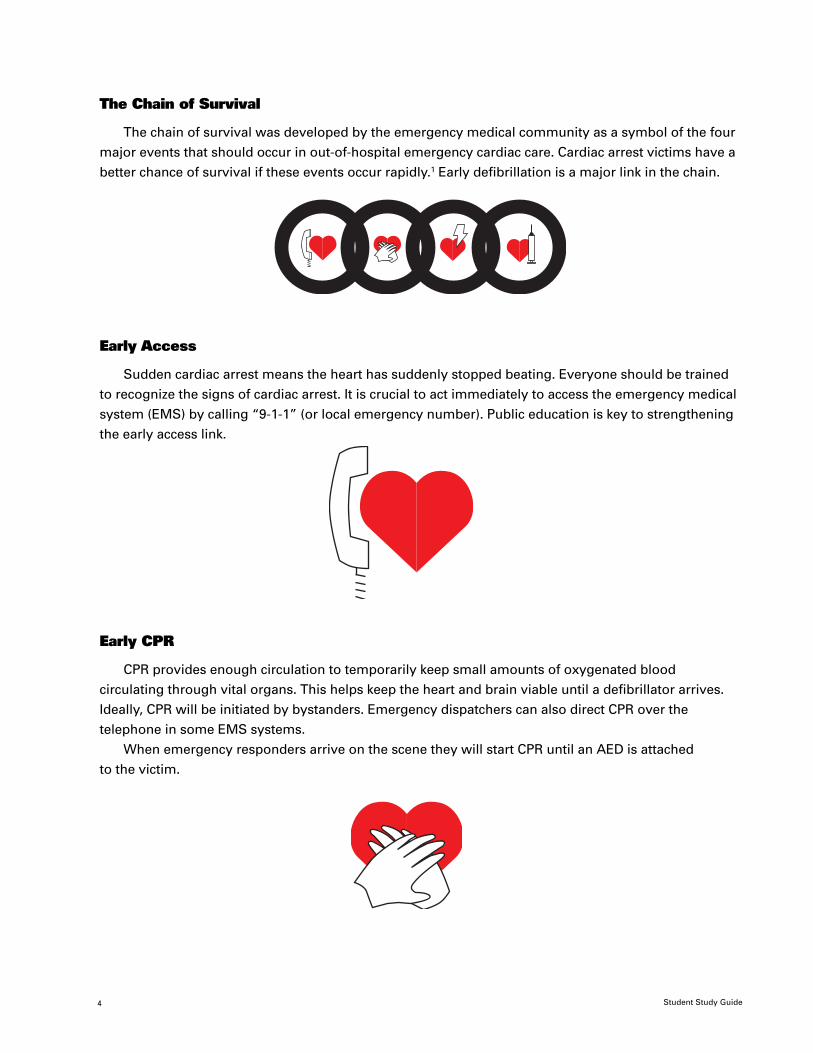
The Chain of Survival
The chain of survival was developed by the emergency medical community as a symbol of the four
major events that should occur in out-of-hospital emergency cardiac care. Cardiac arrest victims have a
better chance of survival if these events occur rapidly.1 Early defibrillation is a major link in the chain.
Early Access
Sudden cardiac arrest means the heart has suddenly stopped beating. Everyone should be trained
to recognize the signs of cardiac arrest. It is crucial to act immediately to access the emergency medical
system (EMS) by calling “9-1-1” (or local emergency number). Public education is key to strengthening
the early access link.
Early CPR
CPR provides enough circulation to temporarily keep small amounts of oxygenated blood
circulating through vital organs. This helps keep the heart and brain viable until a defibrillator arrives.
Ideally, CPR will be initiated by bystanders. Emergency dispatchers can also direct CPR over the
telephone in some EMS systems.
When emergency responders arrive on the scene they will start CPR until an AED is attached
to the victim.
4
Student Study Guide
The purpose of early defibrillation is to reestablish a normal heart rhythm. Scientific research has
shown that early defibrillation greatly increases the chances of survival for someone in cardiac arrest.2, 3, 4
The sooner you deliver a shock, the more likelythe heart will start pumping again.
A growing number of emergency responders throughout the world are performing defibrillation
because defibrillators have become more affordable, easier to use and maintain as well as more widely
available. Most people believe that defibrillation is easier to learn than CPR.5
The International Association of Fire Chiefs (IAFC), the AHA, the National Heart, Lung and Blood Institute
(NHLBI) and the European Resuscitation Council (ERC) have all endorsed the concept of early defibrillation.
In some areas, where once only highly trained physicians, nurses or paramedics provided defibrillation, basic
life support providers such as police, security guards, flight attendants and lifeguards now defibrillate. In
Rochester, MN, when rapid defibrillation by police and paramedics was made a community objective in
1990, survival to hospital discharge from sudden cardiac arrest rose to 49 percent by 19956. The strategy of
defibrillation by basic life support personnel has decreased time to defibrillation by as much as 3.5 to 7
minutes.7 Early defibrillation programs can double or triple survival rates.1
Early ACLS
Early advanced cardiac care is the treatment provided by the advanced cardiac life support (ACLS)
team which is beyond the scope of practice of basic life support providers. ACLS skills include
endotracheal intubation and intravenous administration of drugs.
5

Student Study Guide
Quick Review
1. The four links of the chain of survival are early , early ,early , and early .
2. keeps small amounts of blood circulating through vital organs .
3. The most effective treatment for VF is .
4. Early defibrillation can increase survival rates from VF by to timesthe current rates.
5. When someone’s heart stops beating, this is called .
Answers
1.early access, early CPR, early defibrillation, early ACLS2.CPR3.defibrillation4.two to three5.cardiac arrest
6

Student Study Guide
The Electrical System of the Heart
This chapter briefly reviews the electrical system of the heart, including the concepts of conductivefibers, ECG, pacemaker and dysrhythmia.
Key Points
• The heart has a network of specialized conductive fibers that conduct electrical impulses to thecardiac muscle tissues.
• A pacemaker is an area within the electrical system that generates electrical impulses. Theseimpulses travel through the heart and stimulate it to contract.
• An electrocardiogram (ECG) is a graphical record of the impulses as they travel through theheart and excite the cardiac cells.
• Ventricular fibrillation (VF) is the most common initial cardiac arrest heart rhythm.
• An electrical shock is the most effective treatment for VF.
• Defibrillation must occur early to have the best chance of success.
7

Student Study Guide
Electrical System
The human heart has a channel of specialized tissue called conductive fibers that distribute
electricity throughout the heart. This is known as the “electrical” system of the heart. This network
delivers electrical impulses directly to the cardiac muscle tissue, which is stimulated to contract and
pump blood. The pumping of the heart is the “mechanical” activity which results in a pulse. Without
an electrical signal the heart will not pump. This chapter focuses on the electrical activity of the heart.
P T
Atrioventricular (AV) node
Left bundle branches
Right bundle branch
Purkinje fibers
Sino-atrial (SA) node
QRS
P = Atrial DepolarizationQRS = Ventricular DepolarizationT = Ventricular Repolarization
Normal conduction pathway in the heart
Pacemakers
Conductive fibers have the unique ability to generate their own electrical impulses. The heart’s
primary impulse generator is the sinoatrial (SA) node located in the right atrium. It is called the primarypacemaker because it is the site that normally generates impulses.
The conductive fiber network carries an impulse generated in the SA node through the cardiac
muscle tissue of the atria. This causes the atria to contract. Next, the impulse travels through the
network to the ventricles causing them to contract. The resulting action forces blood out of the
ventricles into the connecting blood vessels.
If the SA node fails to generate an impulse, another site in the network will usually take over and
generate impulses. The atrioventricular (AV) node is an example of one site along the network that can
also be a pacemaker.
8

Student Study Guide
Electrocardiograms
The electrocardiogram (ECG) is a measurement of the electrical activity in the heart. The impulses
from the heart pass through body tissues and reach the skin. This electrical energy can be detected by
disposable electrodes placed on the skin. The LIFEPAK 500 AED analyzes the electrical impulses it
receives through the disposable electrodes.
The heart’s electrical signals detected on the skin are a very low voltage so they must be amplified
by the LIFEPAK 500 AED. AEDs also amplify any other electrical signals (called artifact) detected by the
disposable electrodes. It is important to minimize all movement and extraneous sources of electrical
signals because they could be confused with or mask the heart’s electrical activity. Artifact may be
induced by:
• Victim movement
• Muscle tremors
• Poor skin prep under the electrodes
• Use of dried out or poor quality electrodes
• Loose electrodes
• Interference from electronic devices and lighting
Normal Sinus Rhythm
Dysrhythmias
The ECG of a healthy heart shows an organized, uniform rhythm called normal sinus rhythm (NSR).The person with NSR will have a pulse you can feel at the carotid artery. This pulse is produced by the
heart’s pumping.
®
Sinoatrial Node
9

Student Study Guide
Dysrhythmias are abnormal heart rhythms that can prevent the heart from pumping properly.
There are numerous causes of dysrhythmias including:
• Narrowing of the arteries of the heart • Central nervous system damage(coronary heart disease)
• Chemical imbalances • Drugs and medications
• Trauma to the heart muscle • Electrocution
• Low blood oxygen levels (drowning, • Hypothermia (low bodysuffocation) temperature)
Coronary heart disease—narrowing and hardening of the arteries—is a major cause of cardiac
arrest. A heart attack (acute myocardial infarction or AMI) is caused by heart disease, too. However,
when someone has a heart attack, the heart does not usually stop beating. Any of the above conditions
may cause an abnormal rhythm.
Ventricular Fibrillation
Sudden cardiac arrest (SCA) means the heart has stopped beating unexpectedly. The most common
dysrhythmia associated with SCA is ventricular fibrillation (VF). VF is an unorganized rhythm in which
many sites in the heart attempt to function as the pacemaker. The chaotic electrical activity results in
uncoordinated and ineffective cardiac muscle contractions which prevents the circulation of blood.
There is no pulse/circulation or blood pressure. A heart in VF looks like a quivering bowl of jelly.
If left untreated, VF results in death. The only effective treatment is defibrillation—the delivery of anelectrical shock to a heart in VF.
Ventricular Fibrillation
The goal of defibrillation is to reorganize the chaotic electrical activity of VF and return the heart
to a normal rhythm. After a shock the SA node or another area of the heart can regain control as the
primary pacemaker.
You must deliver defibrillatory shocks within minutes of cardiac arrest in order to have the best
chance of victim survival. Success rates decline approximately 7 to 10 percent for every minute that
defibrillation is delayed.8
®
10

Student Study Guide
Other Dysrhythmias
If you arrive early on the scene of a cardiac arrest you will be more likely to find VF or pulselessventricular tachycardia (VT). Pulseless VT is a rhythm that often precedes VF. It occurs when a site in theventricular muscle fires rapidly and takes over as the dominant pacemaker. As the heart rate increases,there is less time for the ventricles to fill with blood. This reduces the amount the heart can pump andblood pressure falls. If the blood pressure drops severely, consciousness and pulse/circulation will belost. Like VF, if left untreated, pulseless VT will result in death within minutes. Both VF and VT are treatedwith electrical shocks.
There are some dysrhythmias of cardiac arrest which are not treated with electrical shocks. Asystole(“flat line” or no electrical activity of the heart) and pulseless electrical activity (“PEA”—electrical activitybut no pumping of the heart) are examples of dysrhythmias that do not respond to external shocks.
Traditionally, dysrhythmia courses have been used to teach ECG interpretation skills necessary fordefibrillation. Defibrillation technology has evolved to give us automated defibrillators which simplifydefibrillator operation and greatly reduce the training needed to use a defibrillator. ECG interpretation isdone by software internal to the defibrillator that has been tested in thousands of simulated cases in thelaboratory and clinically field tested.
The LIFEPAK 500 AED is designed to advise the operator if a “shockable” rhythm is detected (rhythmis VF or pulseless VT). If a non-shockable rhythm is detected it is designed to give the operator a “NOSHOCK ADVISED” message. Although AEDs are designed not to shock victims with non-shockable heartrhythms you should never attach an AED to someone with a pulse/circulation.
The 500 automatically charges its capacitor only when a shock is advised. This combination of safetyfeatures helps protect the victim from an inappropriate shock.
0
10
20
30
40
50% Success
Time (minutes)
60
70
80
90
100
1 2 3 4 5 6 7 8 9
Success rates decrease7–10% each minute
* Non-linear
Adapted from text: Cummins RO, Annals Emerg Med. 1989, 18:1269-1275.
Resuscitation Success vs. Time*
11

Student Study Guide
Quick Review1. Electrical impulses that cause the heart to beat are generated in the (list
an organ).
2. A graphical record of the heart’s electrical activity is called an .
3. VF is often the result of a disease process associated with .
4. The heart’s main electrical impulse generator is in the .(specific location).
5. The ECG of a healthy heart shows an organized rhythm called
.
ANSWERS
1.heart2.ECG3.heart disease4.right atrium (sinoatrial node)5.normal sinus rhythm (NSR)
12

Student Study Guide
How to Defibrillate
The LIFEPAK 500 AED is easy to use because it gives prompts for each step in the defibrillation
process. Read this chapter to understand why and how to do each step.
During the initial setup, the 500’s operating features can be defined in various ways. Be sure to
become familiar with the particular way your device has been set up. The procedure listed in this chapter
is modeled after the default (factory) settings.
Note: This guide covers only the essentials of defibrillation using the LIFEPAK 500 AED. You must
read the LIFEPAK 500 AED Operating Instructions to learn about other important information related to
defibrillation. You should also study your local automatic
defibrillation protocols.
Key Points
• Verify the victim is unconscious, breathless and pulseless/without circulation.
• Turn on the LIFEPAK 500 AED and attach disposable electrodes to the victim.
• Stop CPR and analyze the heart rhythm.
• Follow the voice prompts and screen messages.
13

Student Study Guide
The LIFEPAK 500 AED
The LIFEPAK 500 automated defibrillator analyzes the heart rhythm and advises the operator if a
shockable rhythm is detected. The operator must press the SHOCK button to deliver the shock. It is
simple to use because it interprets the heart’s ECG signal and advises the operator what to do.
1. ON/OFF button
2. ANALYZE button (optional)
3. SHOCK button
4. Liquid Crystal Display (LCD)
1
2
4
3
14

Student Study Guide
STEP 1
Verify that the victim is unconscious, breathless and pulseless/without circulation.• The victim must be in cardiac arrest, which means he or she is unresponsive, not breathing and
without pulse/circulation. First, see if the victim responds to a firm shake at the shoulders and byshouting “Can you hear me?”
• If there is no response, call loudly for help, open the victim’s airway using the head-tilt chin lift9
and check for breathing. If you do not detect any breaths after 3 to 5 seconds, deliver two initialventilations.
• Next, check for signs of circulation. If you have been trained, check the carotid pulse for 5–10seconds. If there is no pulse/circulation, prepare the victim for the AED. Place the victim on ahard, firm surface. This makes CPR more effective. Attach an AED only to someone who isunconscious, not breathing and pulseless/without circulation.
15

Student Study Guide
STEP 2
Turn on the LIFEPAK 500 AED and attach the disposable electrodes to the victim.• Press the ON/OFF button. The green LED indicator illuminates when the device is ready to go.
Speak to the device to give a verbal report if required by local protocols. Remove the disposableelectrode pads from the packaging. Make sure the electrode cable connector is plugged intothe 500.
Connecting the QUIK-COMBO™ electrode cable
• Bare the victim’s chest. Remove anything that comes between the electrode and bare skin suchas clothing, medication patches, sweat, moisture, a thick layer of chest hair. If possible, avoidplacing the electrodes directly over surgically implanted devices such as internal cardioverter/defibrillators or implantable pacemakers. Remember, the electrode should touch only bare skin.
• Remove the self-adhesive backing and place the electrodes on the victim by following thediagrams on the electrodes. Press the electrodes firmly to the skin. Do not place over the breastbone. Bone is a poor conductor of electricity.
• Depending on the setup of your AED, analysis will occur automatically or the AED will promptPUSH ANALYZE, and this message will appear on the screen.
Cable connector QUIK-COMBO cable
16
Student Study Guide
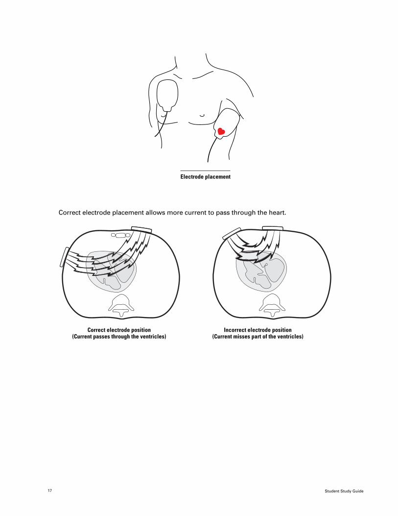
Electrode placement
Correct electrode placement allows more current to pass through the heart.
Correct electrode position Incorrect electrode position(Current passes through the ventricles) (Current misses part of the ventricles)
17

Student Study Guide
STEP 3
Clear the area and analyze the heart rhythm.• Tell everyone to stand clear of the victim while analysis is in progress. Wait about
6 to 9 seconds for the analysis to finish and the next message and voice promptto occur.
• Once the analysis of the heart rhythm begins, the STAND CLEAR, ANALYZINGNOW, STAND CLEAR message and voice prompt will occur. Stop all motion priorto analysis.
Do not touch the victim and do not cause any victim movement during analysis.
STEP 4A
SHOCK ADVISEDThe SHOCK ADVISED voice prompt and message occur when the AED determines the rhythm is
shockable. The AED will automatically begin charging.
• Make certain no one is touching the victim. Do this by saying “I’m clear, you’re clear,everybody’s clear” and scan the victim from head to toe and observe that all is clear.
• Push the SHOCK button when the AED gives the PUSH TO SHOCK prompt.
The AED will automatically analyze again after shocking to see the results of the shock.The AED will automatically analyze after shocks 1 and 2, 4 and 5, etc. (e.g., after the first
2 shocks of each set of 3 consecutive shocks). Listen to the voice prompts and stand clear during
analysis and shock.
• After a NO SHOCK ADVISED or 3 consecutive shocks the AED will prompt you to check thevictim’s pulse/circulation.
• The AED will prompt you to perform CPR for up to 1 minute if there is no pulse/circulation. It willthen prompt you to check for a pulse/circulation after one minute of CPR, and if no pulse/circulation,reanalyze.
• Your local protocols may dictate a maximum number of shocks to give.
• If the victim has a pulse/circulation, support airway and breathing. Monitor closely whileawaiting transport.
• If advanced life support units arrive, they should take control of the resuscitation effort. Briefthem with a short report covering the actions taken prior to their arrival. They may ask you tocontinue your defibrillation procedures.
18

Student Study Guide
STEP 4B
NO SHOCK ADVISED
Not all heart rhythms of cardiac arrest require a shock. When the LIFEPAK 500 AED detects one of
these rhythms it will give you a message of NO SHOCK ADVISED. The AED will prompt you to check
for a pulse/circulation. Always check the victim’s pulse/circulation when the defibrillator analysis
results in NO SHOCK ADVISED.
• If there is no pulse/circulation, the AED will prompt you to perform CPR for 1 minute. Your
local protocols will dictate how to proceed when the result you get is a repeated NO SHOCK
ADVISED message.
19

Student Study Guide
Automated External Defibrillation Procedure
• Determine if the victim is unconscious, pulseless/without circulation andnot breathing (ABCs)
• Turn on the AED and have someone perform CPR until the electrodes are attached
• Stop CPR and analyze the heart rhythm
• Follow the voice prompts and screen messages
Analyze, Charge and Shock up to 3 times (if prompted), or if NO SHOCK ADVISED
Check for pulse/circulation
Pulse/circulation present No pulse/circulation
Assess and support ABCs CPR for 1 minute
Check pulse/circulation,if no pulse/circulation:
Analyze and Shock up to 3 times, orif NO SHOCK ADVISED
Check for pulse/circulation,
if no pulse/circulation:
CPR for 1 minute
Check for pulse/circulation,
if no pulse/circulation:
Continue as above untilACLS team arrives
20
Student Study Guide
Continuous Patient Surveillance
The LIFEPAK 500 AED monitors the ECG—even when it is not analyzing. Sometimes the heart’s
rhythm will spontaneously change from a non-shockable rhythm to a shockable rhythm. If the
Continuous Patient Surveillance System (CPSS) detects a potentially shockable rhythm, the AED will
prompt PUSH ANALYZE.
In these situations, the LIFEPAK 500 AED is warning you the rhythm has changed and
it is possible the victim needs to be shocked. Stop all victim movement and check the
pulse/circulation. If there is no pulse/circulation, press ANALYZE.
NOTE: CPSS is not active in LIFEPAK 500 AEDs that do not have an ANALYZE button or when
Autoanalyze 2 is selected. In these cases, the LIFEPAK 500 AED analyzes automatically.
Troubleshooting
If the CONNECT ELECTRODES message appears there is either an inadequate connection to the AED
or the electrodes are not adhered firmly to the skin. Do a quick check of the connection to be sure the
electrode connector is completely inserted into the AED.
If the disposable electrodes do not stick to a hairy chest, quickly shave the hair with a razor. Remove
moisture with a cloth. Remove creams or ointments and medication patches that could come in contact
with the electrodes.
Remember not to touch the victim when analyzing and shocking. The LIFEPAK 500 AED will not
analyze if it detects victim movement through the electrodes.
Motion artifact is the ECG signal distortion created by movement of the victim or defibrillation
cables. It can cause incorrect interpretation of the ECG. To prevent this situation, the LIFEPAK 500 AED
has special circuitry that detects motion. The unit avoids analyzing the rhythm until all motion has
stopped. It will display the MOTION DETECTED and
STOP MOTION messages. It will not analyze the rhythm if motion is detected.
If the MOTION DETECTED, STOP MOTION message occurs, try to eliminate all sources of motion
such as breathing assistance, CPR compressions, and electrode/cable movement.
If motion stops within 20 seconds, analysis will continue. If the motion does not stop within 20
seconds, analysis will stop. Push ANALYZE (if button present) to restart analysis. In LIFEPAK 500 AEDs
that do not have an ANALYZE button, analysis will restart automatically.
Readiness Display
The LIFEPAK 500 AED with the biphasic waveform includes a Readiness Display on the device's
handle that can be seen at all times. OK displays if the automatic self-test is completed successfully. If
the self-test detects that service is required or if the device detects that the battery needs replacement,
the OK indicator disappears and a service and/or battery indicator appear(s).
To obtain more specific information, turn on the device and observe symbols (indicators) on
key panel.
21

Student Study Guide
Low Battery Detection
When the battery symbol is lit and the low battery message is displayed, the battery
is low. Lithium batteries will provide approximately eleven more shocks. If they have been properly
maintained, sealed lead-acid (SLA) batteries will provide approximately six more shocks.
When the battery symbol flashes on and off and the REPLACE BATTERY voice prompt and
message occur, the battery is very low and should be replaced immediately.
Service Indicator and Message
When the service indicator is on (but not flashing), you can still use the AED for therapy. Contact
an authorized service person to correct the problem as soon as possible.
When the AED detects a problem that requires immediate service, the service indicator
flashes and the CALL SERVICE message is displayed. Turn the AED off, then on again.
If the CALL SERVICE message is still displayed you will not be able to use the AED until the problem is
corrected. Contact authorized service personnel immediately to correct the problem.
NOTE: For LIFEPAK 500 AEDs without a readiness display: If the self-test detects a problem, an audio
alarm will be activated. Turn the device on and refer to LOW BATTERY DETECTION and SERVICE
INDICATOR AND MESSAGE information.
22

Student Study Guide
Quick Review
1. True or False: CPR is always done before defibrillation.
2. After analyzing and getting the SHOCK ADVISED message, you must always the scenebefore shocking.
3. What will the machine do immediately after the first shock is delivered?
4. Should you analyze while CPR is in progress?
5. Number the five general AED steps in the correct order.
If SHOCK ADVISED, clear the victim and push the SHOCK button when the AED saysPRESS TO SHOCK.
Turn on the LIFEPAK 500 AED and attach the defibrillator electrodes to the victim.
Press the ANALYZE button again after shocking to see the results of the shock (device may beconfigured to automatically reanalyze).
Determine if the victim is unconscious, not breathing and pulseless/without circulation.
Stop CPR and analyze the heart rhythm.
6. When the battery symbol is flashing, and REPLACE BATTERY voice prompt and message occur,the battery should be replaced .
ANSWERS
1.False, Defibrillation has priority when device is available and ready.2.clear3.analyze4.No. There must be no motion when analyzing.5.4, 2, 5, 1, 36.immediately
23
Student Study Guide
Safety First
This chapter briefly reviews some of the safety precautions you must know about defibrillation.
Always consider the safety of the rescuers and the victim.
Key Points
• Attach the defibrillator only to someone who is unconscious, not breathing and pulseless/without circulation.
• Make sure no one is touching the victim before analyzing or shocking.
• Be sure the electrodes are firmly adhered to the victim’s bare chest.
• Move oxygen well away from the rescue effort.
• Use Infant/Child Reduced Energy Defibrillation Electrodes when using an AED onyoung children.
24

Student Study Guide
Cardiac Arrest Only!
Be certain the victim is unresponsive, breathless and pulseless/without circulation. Remember:
• Shake and shout
• The ABCs:
- open the Airway
- check for Breathing
- check Circulation (pulse/circulation, coughing or movement)
The Safety Zone
Defibrillation can be dangerous if performed improperly. The good news is that AEDs are very
safe if you take several important precautions. First—never touch the victim when the device is
analyzing or shocking.
When analyzing or shocking maintain a buffer or safety zone around the victim. Imagine an invisible
shield surrounding the victim. Allow no one to penetrate this zone.
Finally, check each time before shocking by saying “I’m clear, you’re clear, everybody’s clear” and
looking to see that no one is within the safety zone.
Electrodes firmly adhered
Make sure the victim’s chest has been wiped dry, excess chest hair removed, and the electrodes are
firmly adhered to the victim’s chest. Allow no air gaps between the electrodes and skin. Good technique
when applying the defibrillation electrodes will minimize the possibility of a spark.
Defibrillation in the presence of oxygen
Use care when defibrillating near oxygen sources (such as bag-valve-mask devices). Remove
oxygen from the victim and place it well away from the rescue effort prior to delivering a shock to help
prevent a fire hazard.
Age/Weight Limit
Follow your local protocol regarding age recommendations. The LIFEPAK 500 AED may be used on
children under eight (8) years of age or 55 lbs (22kg) only if the Infant/Child Reduced Energy
Defibrillation Electrode is used and the biphasic AED has a pink connector.
25

Student Study Guide
Quick Review
1. You should attach a defibrillator only to someone who is ,
not and .
2. Excessive chest hair should be before defibrillation.
3. When oxygen is used prior to defibrillation, it should be placed
from the rescue effort.
4. A biphasic AED with Infant/Child Reduced Energy Defibrillation Electrodes may be used on
a patient under years of age.
5. What are two very important words to remember in defibrillation? (Hint: they start with
the letters “S” and “F”.)
1.unconscious, not breathing, and pulseless/without circulation2.removed3.away4.8 years old (or according to local protocol)5.Safety First!
ANSWERS
26

Student Study Guide
References1. Improving Survival From Sudden Cardiac Arrest: The “Chain of Survival Concept.” Dallas:
American Heart Association, 1991.
2. Eisenberg, M.S., Hallstrom, A.P., Copass, M.K., Bergner, L., Short, F., Pierce, J. 1984. Treatmentof ventricular fibrillation: emergency medical technician defibrillation and paramedic services.Journal of the American Medical Association 251:1723–1726.
3. Vukov, L.F., White, R.D., Bachman, J.W., O’Brien, P.C. 1988. New perspective on ruraldefibrillation. Annals of Emergency Medicine 17:318–321.
4. Bachman, J.W., McDonald, G.S., O’Brien, P.C. 1986. A study of out-of-hospital cardiac arrestsin northeastern Minnesota. Journal of the American Medical Association 256:477–483.
5. Stults, K.R., Brown, D.D. 1986. Refibrillation managed by EMT-Ds: incidence and outcomewithout paramedic backup. American Journal of Emergency Medicine 4:491–495.
6. White, R.D., Asplin, B.R., Bugliosi, T.F., et al. 1996. High discharge survival rate after out-of-hospital ventricular fibrillation with rapid defibrillation by police and paramedics.Annals of Emergency Medicine 28:480–485.
7. Eisenberg, M.S., Horwood, B.T., Cummins, R.O., Reynolds-Haertle, R., Hearne, T.R. 1990.Cardiac arrest and resuscitation: a tale of 29 cities. Annals of Emergency Medicine 19:179–186.
8. Cummins, R.O. 1989. From concept to standard-of-care? Review of the clinical experience withautomated external defibrillators. Annals of Emergency Medicine 8:1269–1275.
9. Basic Life Support for Healthcare Providers. Dallas: American Heart Association, 1994.
LIFEPAK is registered trademark of Medtronic Physio-Control Corp.QUIK-COMBO is a trademark of Medtronic Physio-Control Corp.©2003 Medtronic Physio-Control Corp.
27