structure, turnover, and heme-mediated suppression … · structure, turnover, and heme-mediated...
TRANSCRIPT
THE JOURNAL OF BIOLOGICAL CHEMI8TRY 0 1988 by The American Society for Biochemistry and Molecular Biology, Inc.
Vol . 263. No. 31. Issue of November 5, pp. 15973-15979,1988 Printed in U.S.A.
Structure, Turnover, and Heme-mediated Suppression of the Level of mRNA Encoding Rat Liver 6-Aminolevulinate Synthase*
(Received for publication, January 19, 1988)
Masayuki Yamamoto, Shigeo Kure, James Douglas Engel$, and Koichi Hiraga From the Department of Biochemistry, Toyama Medical and Pharmaceutical University School of Medicine, Toyama, 930-01, Japan and the $Department of Biochemistry, Molecular Biology, and Cell Biology, Northwestern University, Evanston, Illinois 60201
cDNA libraries were constructed with RNA DreDarations from both porphyric
hgt 1 1 pol?(A)+ chicken and rat liveis. A cDNA which encodes chicken hepatic 6-aminolevulinate synthase was cloned by screening with an anti-chicken liver 6-aminolevulinate synthase antibody. Using this cDNA as a probe, cDNAs encoding the entire protein coding sequence of rat hepatic 6-aminolevulinate synthase were then cloned. The complete nucleotide sequences of the cDNAs have been determined. The result predicts that the rat he- patic pre-6-aminolevulinate synthase comprises 642 amino acids. We measured the half-life of the hepatic 6-aminolevulinate synthase mRNA by RNA blot hy- bridization analysis using allylisopropylacetamide-in- duced porphyric rats as an experimental model and the rat cDNA as a hybridization probe. The half-life of the mRNA determined by the injection of a-amanitin is as short as 20 min. This value is significantly shorter than the estimated half-lives of most other mRNAs in the differentiated tissues of animals. The effect of hemin administration on the level of hepatic &amino- levulinate synthase mRNA was also examined. The half-disappearance time of the mRNA after the hemin administration was essentially the same as that deter- mined by a-amanitin or actinomycin D, and no additive effect was observed between a-amanitin and hemin on the half-life determination. The results provide con- vincing evidence that heme inhibits the transcription of 6-aminolevulinate synthase mRNA.
6-Aminolevulinate (ALA)l synthase (EC 2.3.1.37) is the first enzyme in the heme biosynthesis pathway in animals catalyzing the condensation of glycine and succinyl-CoA to form 6-aminolevulinic acid (1). ALA synthase is encoded by the nuclear genome, and ALA synthase mRNA is translated in the cytoplasm. The pre-ALA synthase synthesized is then transferred into the mitochondrial matrix (with coincident
*This work was supported in part by grants from the Tamura Foundation for Encouragement of Science and Technology (to M. Y . ) , the Ministry of Education, Science and Culture (Grant 62770199 to M. Y. and Grants 61219007 and 62109001 to K. H.), and the National Institutes of Health (Grant HL24415 to J . D. E.) The costs of publication of this article were defrayed in part by the payment of page charges. This article must therefore be hereby marked “adver- tisement” in accordance with 18 U.S.C. Section 1734 solely to indicate this fact.
The nucleotide sequence(s) reported in thispaper has been submitted
504044. to the GenBankTM/EMBL Data Bank with accession number(s)
’ The abbreviations used are: ALA, 6-aminolevulinate; AIA, allyli- sopropylacetamide; bp, base pair(s); kb, kilobase(s); pfu, plaque- forming unit.
processing to generate mature ALA synthase) in which this enzyme functions physiologically ( 2 , 3).
There is now ample evidence that heme biosynthesis in the liver of animals is subject to feedback regulation by heme, the end product of the pathway (1-3). Specifically, heme nega- tively regulates both the synthesis (4-6) and intracellular transfer of the enzyme (7) to cause a reduction of the ALA synthase enzyme level in liver mitochondria. This feedback regulation by heme is an attractive mechanistic control circuit in the animal system, since heme exerts its regulatory effect through multiple feedback pathways to reduce the amount of the regulatory enzyme in the organelles where the enzyme functions.
We reported previously that, when assayed using a cell-free protein synthesis system supported by liver polysomes, intra- venous administration of hemin to allylisopropylacetamide (A1A)-induced experimentally porphyric rats resulted in the reduction of the level of the polysomes which direct the synthesis of ALA synthase in the liver (5). This result has been interpreted as meaning that heme inhibits the transcrip- tion of liver type ALA synthase gene (5). Similar observations have been reported from an analogous experiment using chick embryo liver polysomes (4). The transcriptional regulation by heme of the synthesis of ALA synthase has also been sug- gested by experiments in which changes in ALA synthase activity after the addition of cordycepin (an inhibitor of transcription) to primary cultures of chick embryo liver cells were determined (8).
In experiments with the rat liver model, the half-disap- pearance time of ALA synthase mRNA associated with poly- somes (measured by the ability to direct the synthesis of ALA synthase in uitro) was about 1 h after the administration of hemin to porphyric rats (5). Half-life measurements of rat liver ALA synthase mRNA have also been estimated to be 72-74 min by determining the changes in the ALA synthase enzyme activity after the administration of actinomycin D to rats (9). These values are significantly shorter than the esti- mated half-lives of most other mRNAs in the differentiated tissues of animals (10-12). However, previous analyses were clearly indirect estimates of the amount and stability of ALA synthase mRNA, and no confirmatory results either showing a direct effect of heme on transcription of the hepatic ALA synthase gene or demonstrating the rapid turnover of the mRNA have been obtained because of the lack of cDNA probes encoding rat liver ALA synthase. These serve as mo- tives to our present study of molecular cloning of the rat liver ALA synthase cDNA.
Thus far, cDNA and genomic clones encoding ALA syn- thase in chick embryo liver have been described and their sequences determined (13-15). On the other hand, we have reported the molecular cloning of ALA synthase cDNA from
15973

15974 Rat Liver b-Aminolevulinate Synthase mRNA
erythroid cells of the chicken (16) and have found that genes encoding those two ALA synthases, namely hepatic and eryth- roid, are different from each other (16, 17). For this reason, we refer in this paper to the mRNA encoding the enzyme which is localized primarily in liver mitochondria and induced by the porphyrinogenic drugs (such as AIA) as hepatic ALA synthase mRNA, and the mRNA for the enzyme found in erythroid cells (18) as erythroid ALA synthase mRNA. The molecular cloning of ALA synthase cDNAs has also been reported from mouse liver (19) and human liver (20), and the molecular cloning of the genes has been reported from the microorganisms, Saccharomyces cerevisiae (21, 22), Brady- rhyzobium japonicum (23), and Rhyzobium meliloti (24).
In the present study, we have constructed Xgtll cDNA libraries from both porphyric chicken and rat liver. A cDNA which encodes chicken ALA synthase was cloned by screening for the epitope expressed by the recombinant Xgtll phage with an anti-ALA synthase-specific antibody. Using this cDNA as a probe, cDNAs encoding the entire protein coding sequence of the rat hepatic ALA synthase were then cloned. RNA blot hybridization analysis using this rat cDNA as a probe directly demonstrates that the half-life of the hepatic ALA synthase mRNA (determined by the injection of a- amanitin to rats) is as short as -20 min. The administration of hemin to rats also suppresses the level of ALA synthase mRNA in the liver, and the half-disappearance time of ALA synthase mRNA in this case is essentially the same as that determined by the inhibitors of transcription. These data provide convincing evidence that heme inhibits the transcrip- tion of ALA synthase mRNA.
MATERIALS AND METHODS*
Treatment of Animals and Extraction of RNAs-AIA (generous gift from Nippon Roche Co., Tokyo) was dissolved at a concentration of 20 mg/ml in physiological saline. 3,5-Dicarbethoxy-1,4-dihydrocolli- dine (Eastman Kodak Co., Rochester) was suspended in corn oil at a concentration of 20 mg/ml as described (25). White Leghorn hens of about 1 month of age were induced to porphyria with the administra- tion of 300 mg of AIA/kg of body weight and 400 mg/kg 3,5-dicar- bethoxy-l,4-dihydrocollidine subcutaneously injected at 4.5 h before death. Female Stdwister rats (Toyama Experimental Animal Farm, Toyama) weighing about 100 g were fasted for 24 h, then administered with 300 mg/kg AIA subcutaneously, and killed at the time indicated. Normally, rats were divided into a group of three rats and processed further. When poly(A)+ RNA was prepared, rats were killed 4.5 h after the injection of AIA.
Isolated fresh livers were homogenized in guanidine thiocyanate solution (26), and then RNA was banded in CsCl according to the method previously described (27). For cDNA library construction, poly(A)+ RNA was selected by two cycles of oligo(dT)-cellulose (Col- laborative Research Inc., Waltham, MA) column chromatography.
RESULTS*
Characterization of the cDNA Clones Encoding the Rat Hepatic ALA Synthase-The nucleotide sequence of the cDNA insert in pKRA2c (1972 base pairs) is shown in Fig. 2. We found that the sequence surrounding a methionine codon near the 5’ end of the cDNA matches to the consensus expectations for the initiator methionine reported by Kozak (38), and in fact, following the methionine codon, there is a long open reading frame which encodes a peptide of 642 amino acids. Therefore, primary structure of the deduced peptide was compared to the reported chick embryo liver ALA syn- thase sequence (14) (see “Materials and Methods”). As a
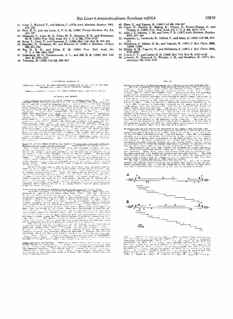
Portions of this paper (includingpart of “Materials and Methods,” part of “Results,” and Fig. 1) are presented in miniprint at the end of this paper. Miniprint is easily read with the aid of a standard magnifying glass. Full size photocopies are included in the microfilm edition of the Journal that is available from Waverly Press.
result, the primary structure of the peptide shares substantial homology with that of the chick embryo liver enzyme, and those two sequences show 82% homology when they are aligned for maximum homology. In particular, the initial 38- amino acid sequence is highly homologous to that in the chick embryo enzyme. Because of the similarity, we concluded that the cDNA fragment cloned encodes the entire coding sequence of rat liver ALA synthase. While the chick embryo hepatic enzyme is reported to have a poly(A) signal after a long 3’- noncoding region (14), the cDNA clones analyzed in the present study only have a short 3‘-noncoding region, but not any poly(A) consensus sequence. These clones may have lost their 3”flanking sequence during the molecular cloning steps (for unknown reasons).
It has been reported that the cleavage site by a processing enzyme in importing the chick embryo liver ALA synthase into mitochondria is located between the 56th and 57th glu- tamines (14). Since the homologous sequence is conserved in the predicted sequence of the rat hepatic ALA synthase (Fig. 2), we estimated that the peptide of 56 amino acids functions as a presequence for the mitochondrial transport of the en- zyme. The size of rat hepatic pre-ALA synthase is calculated to be 71,028. This is smaller than that of 77,000 which has been estimated by polyacrylamide gel electrophoresis in the presence of sodium dodecyl sulfate (6, 17). Subunit molecular weight of the mature form rat hepatic ALA synthase is cal- culated to be 64,872 assuming that the glutamine dipeptide is the site for the cleavage by a processing enzyme. This estimate is very close to our previous estimate of 66,000 which was determined electrophoretically (6, 17).
Validity of the Assignment for the Rat Liver ALA Synthase cDNAs-We have further confirmed the nature of the cDNA, taking advantage of the fact that the hepatic ALA synthase is induced, possibly at the same time as transcriptional stim- ulation of the gene, in the liver of experimentally porphyric rats. For this purpose, rats were induced to porphyria with the administration of 300 mg of AIA/kg of body weight and killed 3.5 h later. After the isolation of total RNA from the livers of the porphyric and untreated (control) rats, change in the level of mRNA encoding the hepatic ALA synthase was measured by RNA blot hybridization analysis using the pKRA2c insert as a specific probe. As a result, the level of hepatic ALA synthase mRNA (2.5 kilobases) is increased about 23-fold in the liver of rats treated with AIA for 3.5 h (Fig. 3, lane 3) as compared to the level of the mRNA in control rat liver (Fig. 3, lane 2). The poly(A)+ RNA, which was used in the preparation of the cDNA library in the present study, also gave a band of significant intensity (lane I), demonstrating that the mRNA induced in the porphyric rat liver hybridizes specifically with the cDNA insert in pKRA2c. This result is consistent with the finding of the experiment described in the earlier section with a chicken liver ALA synthase cDNA clone (pALlOa) in that the change in the amount of hepatic ALA synthase mRNA in chicken could be differentiated by RNA blot hybridization analysis using the pALlOa as a hybridization probe, and in that the size of the mRNA in rat liver and kidney which hybridizes to the pALlOa was 2.5 kilobases. These are additional indications that the cloned cDNA inserts are those encoding the rat hepatic ALA synthase.
Effect of Administration of the Inhibitors of Transcription and Hemin on the Level of ALA Synthase mRNA in Rat Liuer-Since it was demonstrated that rat hepatic ALA syn- thase cDNA was cloned and that the cDNA gave a confirm- atory result in estimating the amount of ALA synthase mRNA in various RNA preparations, next we tried to examine the

Rat Liver 6-Aminolevulinate Synthase mRNA 15975 1 0
CAGGACCCGGGACACTTTGCAGAC~GGAGACTGTCGTTCGCAGATGCCCATTCTTATCCCGAGTCCCTCAGGCCTTTCTGCAGAAGGCAGGGAAATCTC 2 0 3 0 4 0 5 0 6 0 7 0 8 0 9 0 1 0 0
1 1 0 1 2 0 1 3 0 1 4 0 1 5 0 1 6 0 1 7 0 1 8 0 1 9 0 2 0 0 TGCTGTTCTATGCTCAAAACTGCCCCAAGATGATGGAAGTCGGGGCCAAGCCGGCTCCTCGGACCGTGTCCACTTCAGCAGCACAGTGCCAGCAGGTCAA
M E T V V R R C P F L S R V P Q A F L Q K A G K S L
L F Y A Q N C P K M M E V G A K P A P R T V S T S A A Q C Q Q V K
2 1 0 2 2 0 2 3 0 2 4 0 2 5 0 2 6 0 2 7 0 2 8 0 2 9 0 3 0 0 AGAAACCCCTCCAGCCAATGAGAAAGAGAAAACTGCCAAAGCCGCAGTCCAGCAGGCTCCTGACGAGTCCCAGATGGCACAGACTCCAGACGGCACACAG
7
E T P P A N E K E K T A K A A V Q Q A P D E S Q M A Q T P D G T Q
3 1 0 3 2 0 3 3 0 3 4 0 3 5 0 3 6 0 3 7 0 3 8 0 3 9 0 4 0 0
L P P G H P S P S T S Q S S G S K C P F L A A Q L S Q T G S S V F R
4 1 0 4 2 0 4 3 0 4 4 0 4 5 0 4 6 0 4 7 0 4 8 0 4 9 0 5 0 0
CTCCCGCCTGGACACCCGTCACCCTCTACAAGCCAGAGCTCTGGGAGCAAGTGCCCTTTCCTGGCAGCACAGCTGAGCCAGACGGGCAGCAGCGTCTTCC
GCAAGGCCAGTCTGGAGCTTCAGGAGGACGTGCAGGAAATGCATGCTGTGAGGAAAGAGGTTGCTCAAAGCCCAGTGCTCCCCAGCTTGGTCAATGCAAA K A S L E L Q E D V Q E M H A V R K E V A Q S P V L P S L V N A K
AAGGGATGGAGAAGGTCCAAGCCCACTGCTGAAGAACTTCCAGGACATCATGAGAAAGCAAAGGCCAGAAAGAGTGTCTCATCTTCTTCAGGATAACTTG 5 1 0 5 2 0 5 3 0 5 4 0 5 5 0 5 6 0 5 7 0 5 8 0 5 9 0 6 0 0
R D G E G P S P L L K N F Q D I M R K Q R P E R V S H L L Q D N L
6 1 0 6 2 0 6 3 0 6 4 0 6 5 0 6 6 0 6 7 0 6 8 0 6 9 0 7 0 0 CCAAAGTCCGTTTCCACTTTTCAATATGATCATTTCTTTGAGAAGAAAATTGACGAGAAAAAAAATGACCACACCTACCGAGTTTTTAAAACTGTGAACC P K S V S T F Q Y D H F F E K K I D E K K N D I I T Y R V F K T V N R
7 1 0 GGAGAGCACAGATCTTCCCTATGGCAGATGACTACACGGACTCCCTCATCACCAAAAAGCAGGTGTCGGTCTGGTGCAGCAACGACTATCTAGGCATGAG
7 2 0 7 3 0 7 4 0 7 5 0 7 6 0 7 7 0 7 8 0 7 9 0 8 0 0
R A Q I F P M A D D Y T D S L I T K K Q V S V W C S N D Y L G M S
8 1 0 TCGACACCCACGGGTGTGTGGGGCCGTCATAGAGACTGTGAAACAGCATGGTGCCGGAGCAGGTGGAACTAGAAATATTTCTGGAACGAGCAAGTTCCAT
8 2 0 8 3 0 8 4 0 8 5 0 8 6 0 8 7 0 8 8 0 8 9 0 9 0 0
R H P R V C G A V I E T V K Q H G A G A G G T R N I S G T S K F H
9 1 0 9 2 0 9 3 0 9 4 0 9 5 0 9 6 0 9 7 0 9 8 0 9 9 0 1 0 0 0 GTGGAACTGGAGCAGGAGCTGGCTGACCTCCACGGCAAGGACGCGGCGCTCTTGT’rCTCTTCCTGCTTCGTGGCCAACGACTCCACTCTCTTCACCCTGG V E L E Q E L A D L H G K D A A L L F S S C F V A N D S T L F T L A
1 0 1 0 1 0 2 0 1 0 3 0 1 0 4 0 1 0 5 0 1 0 6 0 1 0 7 0 1 0 8 0 1 0 9 0 1 1 0 0 CTAAGATGATGCCAGGCTGTGAAATTTACTCTGATTCCGGGAACCATGCCTCCATGATCCAAGGGATTCGCAACAGTCGAGTGCCAAAGTATATCTTCCG
K M M P G C E I Y S D S G N H A S M I Q G I R N S R V P K Y I F R
1 1 1 0 1 1 2 0 1 1 3 0 1 1 4 0 1 1 50 1 1 6 0 1 1 7 0 1 1 8 0 1 1 9 0 1 2 0 0 CCACAATGATGTCAACCATCTCAGAGAACTGTTGCAGAGATCCGACCCCTCGCTCCCCAAGATCGTAGCATTCGAAACTGTCCATTCAATGGATGGAGCA H N D V N H L R E L L Q R S D P S L P K I V A F E T V H S M D G A
1 2 1 0 1 2 2 0 1 2 3 0 1 2 4 0 1 2 5 0 1 2 6 0 1 2 7 0 1 2 8 0 1 2 9 0 1 3 0 0 GTGTGCCCCCTGGAAGAGCTGTGTGATGTGGCCCATGAGTTTGGAGCGATCACGTTTGTGGACGAGGTCCATGCAGTAGGGCTCTATGGGGCTTCAGGTG V C P L E E L C D V A H E F G A I T F V D E V H A V G L Y G A S G G
1 3 1 0 1 3 2 0 1 3 3 0 1 3 4 0 1 3 5 0 1 3 6 0 1 3 7 0 1 3 8 0 1 3 9 0 1 4 0 0 GAGGGATCGGTGATCGGGATGGAGTCATGCCAAAAATGGACATCATTTCTGGAACACTCGGTAAAGCGTTCGGCTGTGTTGGAGGATACATTGCCAGCAC
G I G D R D G V M P K M D I I S G T L G K A F G C V G G Y I A S T
GAGTTTGCTGATCGACACCGTCCGGTCCTACGCTGCGGGCTTCATCTTCACCACCTCCCTGCCACCAATGCTGCTGGCTGGAGCCCTGGAGTCTGTGCGG 1 4 1 0 1 4 2 0 1 4 3 0 1 4 4 0 1 4 5 0 1 4 6 0 1 4 7 0 1 4 8 0 1 4 9 0 1 5 0 0
S L L I D T V R S Y A A G F I F T T S L P P M L L A G A L E S V R
1 5 1 0 1 5 2 0 1 5 3 0 1 5 4 0 1 5 5 0 1 5 6 0 1 5 7 0 1 5 8 0 1 5 9 0 1 6 0 0 ATCCTGAAGAGCAATGAGGGACGTGCCCTTCGCCGCCAGCACCAGCGCAATGTCAAGCTTATGAGGCAGATGCTAATGGACGCTGGCCTCCCAGTCATCC I L K S N E G R A L R R Q H Q R N V K L M R Q M L M D A G L P V I H
1 6 1 0 1 6 2 0 ACTGCCCCAGCCACATCATCCCTGTGCGGGTTGCCGATGCTGCTAAAAACACAGAAATCTGTGATGAGTTGATGACCAGGCATAATATCTACGTCCAGGC
1 6 3 0 1 6 4 0 1 6 5 0 1 6 6 0 1 6 7 0 1 6 8 0 1 6 9 0 1 7 0 0
C P S H I I P V R V A D A A K N T E I C D E L M T R H N I Y V Q A
1 7 1 0 CATTAATTACCCAACAGTGCCTCGTGGGGAGGAGCTCCTCCGGATCGCCCCCACCCCGCACCACACACCGCAGATGATGAACTTCTTCCTAGAGAAGCTG
1 7 2 0 1 7 3 0 1 7 4 0 1 7 5 0 1 7 6 0 1 7 7 0 1 7 8 0 1 7 9 0 1 8 0 0
I N Y P T V P R G E E L L R I A P T P H H T P Q M M N F F L E K L
1 8 1 0 1 8 2 0 1 8 3 0 1 8 4 0 1 8 5 0 1 8 6 0 1 8 7 0 1 8 8 0 1 8 9 0 1 9 0 0 CTGCTCACGTGGAAGCGAGTCGGGCTGGAACTGAAGCCACATTCGTCAGCTGAATGCAACTTCTGCAGGAGGCCCTTACACTTCGAAGTGATGAGCGAGA L L T W K R V G L E L K P H S S A E C N F C R R P L H F E V M S E R
1 9 1 0 1 9 2 0 1 9 3 0 1 9 4 0 1 9 5 0 1 9 6 0 1 9 7 0 GAGAGAAAGCCTATTTCTCAGGCATGAGCAAGATGGTGTCTGCCCAGGCC~CTCAGTTATTCACAAACCC
E K A Y F S G M S K M V S A Q A *
FIG. 2. Nucleotide sequence of the cDNA insert in pKRA2c and the de- duced primary structure of the pep- tide. Numbers above the sequence refer to nucleotides. The predicted initiation methionine and termination codon are underlined. An arrow after glutamine 56 indicates the probable processing site of the presequence for mitochondrial trans- port.
effect of administration of hemin on the level of mRNA in the porphyric rat liver. At 3.5 h after administration of AIA, the porphyric rats received a further 4 mg/kg hemin by intravenous injection and were killed 1 h later. This dose of hemin was previously shown to induce a maximum effect when assaying for decreases in the enzyme activity (39) or for a decrease of the activity of polysomes which direct the synthesis of the enzyme in vitro ( 5 ) . It has also been shown that when this dose of hemin is injected into the tail vein, hemin is absorbed quite rapidly into the liver cells and satu- rates tryptophan pyrrolase fully within 10 min (39). Total RNA was prepared as described (see “Materials and Meth- ods”) and analyzed by RNA blot hybridization using the nick- translated pKRA2c insert as a specific probe. As controls for this experiment, a group of three rats were killed 3.5 h after the administration of AIA and processed by the same way as for the hemin-treated rats (AIA-treated control rats). An autoradiogram of this experiment is shown in Fig. 4, together with densitometric profiles for individual lanes. As can be seen in lane 1 , the increase in the level of ALA synthase mRNA in the porphyric rat liver is experimentally repro-
duced. In contrast, the intensities of the bands in lanes 2 and 3 (on which RNA preparations from the rats treated with hemin were loaded) are significantly weaker than the control band in lune 1. The numerical values determined by densito- metric measurement of the main peak are 2,189,186 (in arbi- trary units) for the band in lune 1, and 419,125 and 522,084 for those in lanes 2 and 3, respectively. These data indicate that the level of mRNA encoding ALA synthase was decreased to about 21.5% of the AIA-treated control rats (lane 1 ) . The size of the band in the lane of hemin-treated rats is the same as that in the lane of control rats, and no other transcripts are detectable in the lane for hemin-treated rats. The half- disappearance time of the mRNA in vivo after the adminis- tration of hemin is shorter than 30 min in this experiment (assuming that the degradation of ALA synthase mRNA takes place with linear proportion to time in the presence of hemin).
Because of this rapid decrease of the level of mRNA encod- ing rat hepatic ALA synthase, it is tempting to speculate that the decrease was brought about not only by the inhibitory effect of hemin on transcription of the hepatic ALA synthase gene but also by an acceleration of the degradation of the

15976
Lanes
Top +
28S+
18S+
Rat Liver 6-Aminolevulinate Synthase mRNA
1 2 3
Bottom + FIG. 3. Differentiation of the extent of ALA synthase
mRNA expression in the liver of rats treated or untreated with AIA. Rats were administered with AIA subcutaneously 3.5 h before death. Total RNA fractions were prepared as described in the text, and each lane represents the values obtained for pooled livers of three rats. Forty micrograms of total RNA from the livers of drug- untreated control rats (lane 2) or AIA-treated rats ( l a n e 3) was electrophoresed on a 1.5% formamide gel (56), transferred to nitro- cellulose filter, and probed with the nick-translated cDNA insert of pKRA2c plasmid. Two micrograms of the poly(A)' RNA used for the cDNA library preparation was also loaded (lane I ) . The filter was washed a t 50 "C in the washing solution used in the library screening (see "Materials and Methods") for a total of 6 h. Exposure time was 4 h with an intensifying screen.
mRNA. In order to gain more insight as to whether or not hemin acts through such a dual pathway to reduce the mRNA level, we designed the following experiments to determine the half-life of the ALA synthase mRNA in uiuo in the porphyric rat liver. First, actinomycin D (250 pg/lOO g of body weight) was injected intraperitoneally into rats a t 3.5 h after the injection of AIA, and in this experiment (as shown in Table I), rats were killed 1 h after the inhibitor injection. Total RNA fractions were then prepared and analyzed by similar RNA blot hybridization methods as used in the experiment shown in Fig. 3. In Table I (and also in the subsequent experiment shown in Fig. 5), we describe the difference of the content of ALA synthase mRNA in the individual RNA preparations on the basis of a ratio of the mRNA content ( i e . the numerical value from the densitometrical analysis) in actinomycin D/AIA-treated animals (or a-amanitinlheminl AIA-treated animals) to that in the rats treated with AIA alone. The level of ALA synthase mRNA in the liver of rats treated with AIA for 3.5 h and then with saline for one additional hour was virtually identical to that in control rats treated with AIA alone for 3.5 h. This suggests that the induction of ALA synthase mRNA reaches maximum soon after the administration of AIA, and this level is maintained for several hours, while the amount of the enzyme increased with time during this period of induction as has been reported (39, 40). The level of ALA synthase mRNA decreased to
A B Lanes 1 2 3
) Top +
285 "*
18s +
1. I / ii
Bottom + FIG. 4. Decrease of the level of ALA synthase mRNA caused
by the hemin administration. A, total RNA fractions were pre- pared either from the liver of rats which were killed 3.5 h after the administration of AIA (lane I) or from the liver of rats which were treated with hemin for an additional 1 h after the 3.5-h induction of AIA (lanes 2 and 3, duplicate experiment). RNA blot hybridization analysis was done as described in the legend to Fig. 3. B, densitometric profiles of the autoradiogram. Numbers correspond to each lane in A. Peak positions are shown in arbitrary units of scanning time.
TABLE I Effect of the administration of actinomycin D on the level of ALA
synthase mRNA in rat liver Total RNA fractions were prepared either from the liver of rats
which were killed 3.5 h after the administration of AIA (AIA) or from the liver of rats which were further treated with saline (AIA + saline) or actinomycin D (AIA + actinomycin D) for 1 additional hour after the 3.5-h induction of AIA. RNA blot hybridization analysis was done as described in the legend to Fig. 3. The numerical values determined by the densitometric measurement of the bands are expressed in arbitrary units.
ALA synthase mRNA level Treatment Density Ratio relative
to control
AIA %
1,611,755
AIA + saline 1,760,546 109.2
AIA + actinomvcin D 356.604 22.1
22.1% of the AIA-treated control rats a t 1 h after the admin- istration of actinomycin D (Table I). This result demonstrates that the half-life of ALA synthase mRNA in rat liver is less than 30 min and very similar to that observed after the administration of hemin.
In a separate experiment, we have determined the half-life of the ALA synthase mRNA using a-amanitin (150 pg/kg of body weight). a-Amanitin was injected intraperitoneally into rats at 3.5 h after the injection of AIA, and rats were killed 20, 40, and 60 min after the a-amanitin injection. In some cases, hemin was injected intravenously into the rats at the same time as the a-amanitin injection to see whether or not hemin gives rise to an additional decrease in the level of ALA synthase mRNA. The dose of a-amanitin used in this study has been shown to result in a 50% reduction of the activity of RNA polymerase I1 in the liver within 5 min, followed by complete suppression at 15 min after the administration to

Rat Liver 6-Aminolevulinate Synthase mRNA 15977
10 c
0 20 40 60 80
Time (min ) FIG. 5. Half-life of rat hepatic ALA synthase mRNA deter-
mined either by the administration of a-amanitin or by the simultaneous administration of a-amanitin and hemin. Rats were administered with either a-amanitin alone (0) or a-amanitin and hemin (e) at 3.5 h after the administration of AIA and killed at the indicated time after the injection of a-amanitin or a-amanitin and hemin. As a control for this experiment, a group of three rats was also killed 3.5 h after the administration of AIA. Total RNA fractions were then prepared and analyzed by similar RNA blot hybridization methods as used in the experiment shown in Fig. 3. The difference of the content of ALA synthase mRNA in the individ- ual RNA preparations is described in the text.
rats when employed by the intraperitoneal injection (41). Therefore, it seems reasonable to observe initial lag phase in the decrease curve of the level of ALA synthase mRNA. In fact, the intracellular level of ALA synthase mRNA does not decrease appreciably during the initial 10 min after the ad- ministration of a-amanitin (Fig. 5 ) . However, following the lag time, the mRNA level decreases quite rapidly and reached 17.5% of that in the AIA-treated control rats (ie. start point) at 1 h after the administration of a-amanitin . The half-life of ALA synthase mRNA is determined to be about 20 min from the experiment as shown in Fig. 5 .
It is clear from these analyses that hemin, actinomycin D, and a-amanitin give rise to a rapid decrease in the intracel- lular level of ALA synthase mRNA. The half-disappearance time brought about by each chemical appears to be very similar. Furthermore, as can be seen in Fig. 5 , the half-life determined by the simultaneous administration of a-amanitin and hemin is essentially the same as that determined by the administration of a-amanitin alone, indicating that there is no additive effect between a-amanitin and hemin on the half- life determination. This observation excludes the possibility that the administration of hemin gives rise to the decrease of ALA synthase mRNA principally by stimulating the degra- dation of this mRNA. Therefore, it is concluded from these results that heme inhibits the transcription of the hepatic ALA synthase gene.
DISCUSSION
In the current study, we have cloned cDNAs encoding rat liver ALA synthase and determined the nucleotide sequence of these cDNAs. Comparison of the deduced amino acid sequence to that of the chick embryo liver ALA synthase suggests that rat liver ALA synthase contains a presequence consisting of 56 amino acids. There are eight basic amino acid residues and seven amino acid residues with a hydroxyl group in this presequence (see Fig. 2), which is characteristic of the structure of presequences of mitochondrial matrix proteins
(42). In addition to those residues, there are two glutamates in this presequence; one glutamate residue is located just after the initiation methionine. So far, however, several mitochon- dria transport presequences have been reported to contain one or a few acidic amino acid residues. For example, bovine adrenodoxin has a presequence which contains an acidic amino acid in the predicted mitochondria targeting domain (43). Interestingly, only nine amino-terminal residues of yeast pre-ALA synthase have been reported to be sufficient to localize /?-galactosidase to the mitochondrial matrix (44). It has been reported that the half-time of the incorporation of rat liver pre-ALA synthase into mitochondria is much longer than that of the other mitochondria matrix protein (2, 45). Also, it is well documented that the incorporation of the hepatic pre-ALA synthase into mitochondria is inhibited spe- cifically by heme, the end product of the pathway (2, 7). However, the precise mechanism of the rat pre-ALA synthase transport into mitochondria, as well as that of the inhibition by heme at this step, remains to be clarified.
The immunochemical analysis of the relationship between hepatic ALA synthase and erythroid ALA synthase in the rat has been reported using antibodies directed against rat liver ALA synthase and chicken liver ALA synthase (17). As a result, rat erythroid ALA synthase showed no cross- reactivity with anti-liver ALA synthase antibodies, but hepatic ALA synthases from rat, mouse, and chicken share substantial cross-reactivity with one another (17). This is in very good agreement with the observed high homology between the predicted amino acid sequences of the hepatic ALA synthase of the chicken and rat. The primary structure of the mouse hepatic ALA synthase which was deduced from the cDNA clone sequence (19) also shares substantial homology with the rat liver enzyme sequence. However, surprisingly, the N- terminal region of the mouse enzyme does not show similarity with that of the rat liver enzyme. The ALA synthases in animal livers also share considerable sequence homology with those ALA synthases in yeast (22) and B. japonicum (23).
The half-life of rat liver ALA synthase mRNA is estimated to be about 20 min from the present analyses. What is the mechanism which confers such instability to the rat hepatic ALA synthase mRNA? It has been proposed that a highly conserved 50-60-nucleotide AU-rich sequence found in the 3"noncoding region of the mRNAs encoding certain lympho- kines, cytokines, and proto-oncogenes, which also turn over with a half-life in the range of 30 min (46-50), may be involved in their rapid degradation (49, 50). The most striking feature shared by 21 AU-rich sequences in the mRNAs of this group is the occurrence of a single adenosine nucleotide followed by a tract of three or more uridine nucleotides (49). Although most 3"untranslated regions of the rat hepatic ALA synthase mRNA seem to be absent from the cDNA clones in the present study, we find two sets of AT4.5A sequence in the 3"untrans- lated region of chicken hepatic ALA synthase cDNA (pALlOa). It is of interest to examine whether the hepatic ALA synthase mRNA is degraded through an mRNA proc- essing pathway similar to that for lymphokines or not.
Since the level of mRNA for the regulatory enzyme is regulated through negative feedback pathways by the end product in this case, the rapid degradation of the ALA syn- thase mRNA may be an essential factor for the regulation of the heme biosynthesis in the liver. In this regard, it should be noted that the hepatic ALA synthase also turns over quite rapidly in rat liver mitochondria showing about 35 min of half-life (45).
It is clearly demonstrated in this study that the level of mRNA encoding hepatic ALA synthase is suppressed by the
15978 Rat Liver 6-Aminolevt
administration of hemin. This observation excludes most of the explanations derived from the uncertainty involved in a previous study using the cell-free protein synthesis system supported by liver polysomes ( 5 ) and demonstrates that heme somehow inhibits the transcription of the hepatic ALA syn- thase gene. One possible mechanism remaining in the present study would be to assume an additional mechanism in which heme acts to accelerate the degradation of ALA synthase mRNA. We think this assumption is not realistic because the suppression of the mRNA level by the hemin administration is essentially the same as was observed in the rats treated with the inhibitors of transcription. Furthermore, no additive effect was observed when a-amanitin and hemin were injected simultaneously. The half-disappearance time of ALA syn- thase mRNA associated with polysomes was about 1 h after the administration of hemin ( 5 ) . This is significantly longer than the half-life of ALA synthase mRNA determined in this study. An explanation for this difference is to assume ALA synthase mRNA in a different intracellular compartment from polysomes, such as in messenger ribonucleoprotein com- plex, although we have never examined this point. The mRNA in this pool could not be detected in the determination of polysome activity, but, when transcription was inhibited by heme, it might flow from that pool to polysomes. Thus, the apparent half-life of the polysome-associated ALA synthase mRNA appeared to be much longer than that determined by measuring the total amount of the mRNA. Whether this explanation is appropriate or not awaits further studies.
Ades and colleagues (51) recently reported that ALA syn- thase mRNA concentration was increased significantly by the addition of AIA to the media of primary cultures of embryonic chick hepatocytes, and, when hemin was added to the culture together with AIA, the increase of this messenger was blocked. Although their experimental conditions are appreciably dif- ferent from ours, their result may be in good agreement with our present and previous observations (5) on the porphyric rats. However, one cannot estimate the half-disappearance time of the ALA synthase mRNA in the cultured embryonic chick hepatocytes in that study, and therefore the results in Figs. 4 and 5 here considerably add to our quantitative un- derstanding of the relationships of mRNA synthesis, degra- dation, and ALA synthase enzyme activity.
Recently, heme has been implicated as an important factor for the transcriptional regulation of several genes. Pioneering work in this field has been made in studies on yeast. It has been shown that the regulation of the CYCl (which encodes iso-1-cytochrome c ) transcription by heme is at the level of initiation and mediated by sites, UASl and UAS2, that lie upstream of the transcription initiation site (52). In animal systems, hemin treatment of rat glioma cell caused an increase in heme oxygenase mRNA (53), but there is not a UAS-like sequence upstream of the rat heme oxygenase gene (54). While heme acts to stimulate gene expression in these cases, heme- mediated transcriptional inhibition is also known in yeast. ANBl gene expression in yeast is repressed by heme (55). The present study demonstrates that the transcription of rat liver ALA synthase is also repressed by heme. Further analysis of the transcriptional regulation of the rat liver ALA synthase gene may enable us to understand comprehensively the role of heme in the gene expression in higher eukaryotic cells.
Acknowledgments-We thank Dr. Norio Hayashi for providing us with the anti-chicken liver ALA synthase antibody, the Nippon Roche Company for a generous supply of allylisopropylacetamide, and Dr. Mitsuaki Aka0 for his help with the densitometrical analysis. We also thank Dr. Nobuo Ishihara for his encouragement of our study, Dr. Tomoe Haruki for her technical assistance. and Eriko Takado for
ilinate Synthase mRNA
her help in preparing the manuscript.
Note Added in Proof-Srivastava et al. recently reported nucleotide sequence of a rat hepatic ALA synthase cDNA (Srivastava, G., Borthwick, I. A., Maguire, D. J., Elferink, C. J., Bawden, M. J., Mercer, J. F. B., and May, B. K. (1988) J. Biol. Chem. 263, 5202- 5209). However, the translated sequence for the mature protein obtained in the present study differs substantially from that described by Srivastava et al. In particular, the presence of an additional guanine nucleotide (G) at base position 375 and the absence of a G between base positions 432 and 433 in our sequence endows a frame- shift that affects the sequence over a span of 19 amino acids. The sequence proposed by us has been confirmed experimentally, since the guanine nucleotide at base position 375 is located in the recogni- tion site of a restriction endonuclease pvuII (CAGCTG) and pvuII digests the cDNA insert in pKRA2c at this position as can be seen in Fig. 1 in the present study.
REFERENCES 1. Granick, S., and Beale, S. I. (1978) Adu. Enzymol. Relat. Areas Mol. Biol.
46,33-203 2. Kikuchi, G., and Hayashi, N. (1982) in Structure, Dynamics, and Biogenesis
of Biomembranes (Sato, R., and Onishi, S., eds) pp. 131-156, Japan
3. Kappas, A., Sassa, S., and Anderson, K. E. (1983) in The Metabolic Basis Scientific Societies Press, Tokyo
of Inherited Disease (Stanbury, J. B., Wyngaarden, J. B., Fredrickson, D. S., Goldstein, J. L., and Brown, M. S., eds) pp. 1302-1384, McGraw- Hill Book Co., New York
4. Whiting, M. J. (1976) Biochem. J. 158,985-990 5. Yamamoto, M., Hayashi, N., and Kikuchi, G. (1982) Biochem. Biophys.
6. Yamamoto, M., Hayashi, N., and Kikuchi, G. (1983) Biochem. Biophys.
7. Hayashi, N., Watanabe, N., and Kikuchi, G. (1983) Biochem. Bwphys. Res.
8. Srivastava, G., Brooker, J. D., May, B. K., and Elliott, W. H. (1980)
9. Marver, H. S., Collins, A., Tschudy, D. P., and Rechigl, M., Jr. (1966) J.
10. Guyette, W. A,, Matusik, R. J., and Rosen, J. M. (1979) Cell 17,1013-1023 11. Brock, M. L., and Shapiro, D. J. (1983) Cell 34,207-214 12. Raghow, R. (1987) Trends Biochem. Sci. 12,358-360 13. Borthwick, I. A,, Srivastava, G., Hobbs, A. A,, Pirola, B. A., Brooker, J. D.,
14. Borthwick, I. A,, Srivastava, G., Day, A. R., Pirola, B. A., Snoswell, M. A., May, B. K., and Elliott, W. H. (1984) Eur. J . Bwchem. 144,95-99
15. Maguire, D. J., Day, A. R., Borthwick, I. A., Srivastava, G., Wigly, P. L., May, B. K., and Elliott, W. H. (1985) Eur. J . Biochem. 150,481-484
16. Yarnamoto, M., Yew, N. S., Federspiel, J. B., Dodgson, N Hayashi N., May, B. K., and Elliott, W. H. (1986) Nucleic Acids Res. 14, 1379-1391
17. Yamamoto, M., Fujita, H., Watanabe, N., Hayashi, N., and Kikuchi, G. and Engel, J. D. (1985) Proc. Natl. Acad. Sci. U. S. A. 82I'3702-37&
18. Watanabe, N., Hayashi, N., and Kikuchi, G. (1983) Bwchem. Biophys. Res. (1986) Arch. Biochem. Biophys. 245, 76-83
19. Shoenhaut, D. S., and Curtis, P. J. (1986) Gene (Amst.) 48,55-63 Commun. 113,377-383
20. Bawden, M. J., Borthwick, I. A,, Healy, H. M., Morris, C. P., May, B. K.,
21. Arrese, M., Carvajal, E., Robison, S., Sambunaris, A., Panek, A., and
22. Urban-Grimal, D., Volland, C., Garnier, T., Dehoux, P., Labbe-Bois, R.
23. Robertson McClung, C., Somerville, J. E., Guerinot, M. L., and Chelm, B.
24. Leong, S. A., Williams, P. H., and Ditta, G. S. (1985) Nucleic Acids Res.
25. Igarashi, J., Hayashi, N., and Kikuchi, G. (1976) J. Biochem (Tokyo) 80,
Res. Commun. 105,451-459
Res. Commun. 115,225-231
Commun. 115, 700-706
Biochem. J. 188, 781-788
Biol. Chem. 241,4323-4329
and Elliott, W. H. (1987) Nucleic Acids Res. 15,8563
Mattoon, J. (1983) Curr. Genet. 7,175-183
(1986) Eur. J . Biochem. 156,511-519
K. (1987) Gene (Amst.) 54,133-139
13,5965-5976
1091-1099 26. Chirgwin, J. M., Przybyla, A. E., MacDonald, R. J., and Rutter, W. J.
27. Fryberg, E. A,, Kindle, K. L., Davidson, N., and Sodja, A. (1980) Cell 19, (1979) Biochemistry 18,5294-5299
2fi.5-87A I" I.I
28. Reinach, F. C., and Fishman, D. A. (1985) J. Mol. Biol. 181, 411-422 29. Gubler, U., and Hoffman, B. J. (1983) Gene (Amst.) 25,263-269 30. Maniatis, T., Fritsch, E. F., and Sambrook, J. (1982) Molecular Cloning, A
Laboratory Manual. Cold Spring Harbor Laboratory, Cold Spring Harbor, NY
31. Benton, W. D., and Davis, R. W. (1977) Science 196,180-182
33. Brush, D., Dodgson, J. B., Choi, 0.-R., Stevens, P. W., and Engel, J. D. 32. Henikoff, F. (1984) Gene (Amst.) 28,351-359
34. Sanger, F., Nicklen, S., and Coulson, A. R. (1977) Proc. Natl. Acad. Sci.
35. Mizusawa, S., Nishimura, S., and Seela, F. (1986) Nucleic Acids Res. 14,
(1985) Mol. Cell. Biol. 5, 1307--1317
U. S. A. 74,5463-5467
1319-1324 36.
37.
38. 39.
40.
Watanabe, N., Hayashi, N., and Kikuchi, G. (1984) Arch. Biochem. Bwphys.
Elferink, C. J., Srivastava, G., Maguire, D. J., Borthwick, I. A., May, B. K.,
Kozak, M. (1986) Cell 44,283-292 Yamamoto, M., Hayashi, N., and Kikuchi, G. (1981) Arch. Biochem. Bio-
Yamauchi, K., Hayashi, N., and Kikuchi, G. (1980) J. Biol. Chem. 255,
232,118-126
and Elliott, W. H. (1987) J. Biol. Chem. 262,3988-3992
phys. 209,451-459
1746-1751
Rat Liver 6-Aminolevulinate Synthase mRNA 15979 41.
42.
43.
44. 45.
46.
47.
48.
Voigt, J., Wieland, T., and Sekeris, C. (1978)Arch. Biochern. Biophys. 191 ,
Hurt, E. C., and van Loon, A. P. G. M. (1986) Trends Biochern. Sci. 11 , 101-109
Okamura, T., John, M. E., Zuber, M. X., Simpson, E. R., and Waterman,
Keng, T., Alani, E., and Guarente, L. (1986) Mol. Cell. Biol. 6,355-364 Hayashi, N., Terasawa, M., and Kikuchi, G. (1980) J. Btoehem. (Tokyo)
Raj, N. B. K., and Pitha, P. M. (1983) Proc. Natl. Acad. Sci.
Greenberg, M. E., Harmanowski, A. L., and Ziff, E. B. (1986) Mol. Cell.
Treisman, R. (1985) Cell 42,889-902
204-207
M. R. (1985) Proc. Natl. Acad. Sci. U. S. A. 82,5705-5709
88,921-926
U. S. A. SO, 3923-3927
Biol. 6,1050-1057
antlbady. Uslna a chlcken henatlc ALA synthase cDNA clone t h u s Isolated I L I A L l O d ,
49. 50.
51.
52.
53.
54.
55. 56.
CaDut. D.. Beul
Shibahara, S., Muller, R. M., and Taguchi, H. (1987) J. Biol. Chern. 2 6 2 ,
Muller, R. M., Taguchi, H., and Shibahara, S. (1987) J. Biol. Chern. 2 6 2 ,
Lowry, C. V., and Lieber, R. H. (1986) Mol. Cell. Biol. 6,4145-4148 Lehrach, H., Diamond, D., Wonzey, J. M., and Boedtker, H. (1977) Bio-
511
12889-12892
6795-6802
chemistry 16,4743-4751
RESULTS
~olecular clan~ng and chardcterlzation of a chicken liver ALA synthase <DNA We flrst attempted to clone =DNA encoding the chlcken llver ALA synthase
dlrecely. Uslng an antr-chicken l l v e r ALA synthase antlbody. about 5 0 p o s l - t l v e clones were lsolated from the ~ c r e e n r n g of t he chlcken l l v e r i q t l l llbrary, and a n insert DNA fragment from a i i L i 0 a vhlch gave the stronqest signal I " the lmrnunologlca l Screening was subcloned ~ n f o pGEM1 plasmld. On RNA blot hybrldlzatlon analysre. nlck-translated pALlOa ~ n s e r t hybrldlzes t o a band whlch 1 s 2.3 kb Ikllabasesi I n length. and the 2.3 kb transcrlpt 1 5 srqnlflcantly induced l n RNA Isolated from the llver of drug-lnduced p o r ~ phyrrc c h l c k e n . S l n c e the part>ally determlned nucleotlde sequence of t h c lnsert =DNA in pALlOa 1 5 completely involved ~n the sequence of p10501. a chlck embryo l r v e r ALA synthase cDNA clonell41, we concluded that PALlOa en^
codes chrcken liver ALA s y n t h a s e . pALlOa 1 s 1.802 bp ~n length, and s t a r t s
codon of the pi05811141 to 3' end. l n d l c a t l n y Chat the p A L l 0 a encodes a 220 bp downstream from the adenlne nucleotlde ~n t h e l n l f ~ a t ~ o n methlonlne
mature form of chrck embryo llver ALA synthase. T h l s result provides corn- prote~n whlch starts at 1 9 arnlno a c l d s downstream from the N-terrnlnus of the
pelllng evldence that the enzyme proteln ~n the llver of the adult chlcken 1s t h e same a s the proteln I" the embryonic chxck llverl361. The 51ze of the m R N A transcrlpt measured by R N A blot hybrldlration appears to be t he Same a5 t h e reported 2.2 kb for the m R N A for chick embryo llver enzyme dptected uslnq ~10581 a s a probel37: vhlch 1 s a revision of 2.8 kb1131l.
Molecular cloning and nvcleotlde sequence of rat hepatlc ALA synthase cDNA RNA blot analysis using nlck-translated pALlOa a s a hybridlratlon probe
shoved that pALlOa hybrldlres to a mRNA species of 2.5 kb ~n 517e when prob-
strongly ~n t h e liver and weakly ~n the kldney of the pnrphyrlc rat. suggest- lnq polylAl*RNAs from porphyrlc rat llver and kidney. Thls RNA 1 5 expressed
lnq that the detected band carresponds to the mRNA encoding the rat hepatlc ALA synthase. We antlclpated from thls result that t h e r e may be strong nucleotlde seiluence homoloqv between rat and chlcken m A N i i s encodlng hepatlc A L A synthase and rt t h e r e f o r e seemed reasonable to attempt to I s o l a t e the rat hepatic ALA synthase mRNA relylng on thls nucleotlde sequence homology.
tlon v ~ t h nick-translated DALlOa insert a s a probe, and 11 posltlve clones The porphyrlc r a t llver cDNA llbrary was screened by p l a q u e hybrldlla-
derived from a single specles of mRNA (data n o t shown]. we next constructed nested deletlon mutants of a p a ~ r of plasmlds.
pKRA2cA and pKRA2cB. which contalned the Ionqest cDNA Insert I" the two d l f - frrent orientations IFlg. 11, and the sequences of t h e inserts in these dele- t w n mutant plasmxds were determlned by the plasmid zequencinq method.