structure and morphology of gold nanoparticles in...
TRANSCRIPT

Vol. 121 (2012) ACTA PHYSICA POLONICA A No. 4
Proceedings of the 9th National Symposium of Synchrotron Radiation Users, Warsaw, September 26�27, 2011
Structure and Morphology of Gold Nanoparticles in Solution
Studied by TEM, SAXS and UV�VisM. Murawskaa, A. Skrzypczakb, M. Kozaka∗
a Department of Macromolecular Physics, Faculty of Physics, A. Mickiewicz University
Umultowska 85, 61-614 Pozna«, Polandb Faculty of Chemical Technology, Pozna« University of Technology, Piotrowo 3, 60-965 Pozna«, Poland
Gold nanoparticles have a great number of applications, among others in material sciences, biology andmedicine. A method for the synthesis of gold nanoparticles in solution with the use of gemini surfactant wasproposed and the nanoparticles obtained were subjected to thorough characterisation. The method proposed is amodi�cation of the Turkevich method, based on reduction of tetrachloroauric acid in the presence of trisodiumcitrate and a dicationic (gemini) surfactant � 1,1′-(1,4-butan)bis(3-dodecyloxymethylimidazolium) di-propionate.Morphology and size distribution of gold nanoparticles obtained were examined by the transmission electron mi-croscopy (TEM), UV�Vis spectroscopy and small angle scattering of synchrotron radiation (SAXS). The plasmonresonance of the nanoparticles obtained was observed in the wavelength range corresponding to the presenceof gold nanoparticles with sizes ranging from 5 to 100 nm. TEM images con�rmed that the spherical shape ofnanoparticles was dominated in reference solutions prepared of sodium citrate and tetrachloroauric acid. In the so-lutions prepared with addition of gemini surfactant, the gold nanoparticles of triangular morphology were observed.
PACS: 61.05.cf, 61.46.Hk, 68.37.Lp, 33.20.Kf, 81.16.Be
1. Introduction
Gold nanoparticles (GNP) have enjoyed increasing in-terest because of a wide range of their potential applica-tions in such �elds as: medicine, biotechnology, cataly-sis, preparation of nanocomposites, nanosensing and elec-tronics. The potential applications of GNP depend ontheir size and morphology. Especially the potential appli-cation of GNP in nanosensing is strongly connected withcontrolling of their size and morphology by the properchoice of preparation conditions [1�3].There are a few general methods for the synthesis
of gold nanoparticles. The most popular are chemicalor photochemical reduction of gold precursor [3�8] andbiosynthesis [9, 10]. Chemical methods are the most e�-cient and convenient, because they require a relativelysimple equipment and nanoparticle solutions obtainedare relatively stabile and monodisperse.The chemical reduction of metal ions is the most com-
mon method of GNP synthesis. This method consistsin precipitation of gold nanoparticles from a solution ofa dissolved gold precursor under the in�uence of a re-ducing agent [4, 5]. A gold precursor usually is tetra-chloroauric acid (HAuCl4) while a reducing agent couldbe sodium citrate [4, 5], sodium boron hydride [11], blockcopolymers [12] or ascorbic acid. The reduction of goldprecursor with sodium citrate is the simplest and themost reliable method of obtaining a solution of monodis-perse gold nanoparticles. This method was proposed byTurkevich [4] and is used as the model reaction in the
∗ e-mail: [email protected]
present study. Unfortunately the Turkevich method canonly produce particles up to about 40�50 nm in size.Highly monodispersed large-diameter GNPs can be syn-thesized from HAuCl4 by hydroquinone (HQ) reduction[13]. This method provides a greater size range of GNPs(50�250 nm) and very good shape dispersion.
Another chemical method of preparation of goldnanoparticles is a method discovered by the Burst andcoworkers in the early 1990s [14]. This method al-lows to prepare gold nanoparticles in the two phase sys-tem (toluene/water). The gold precursor (HAuCl4) istransferred from aqueous solution to toluene using tetra-octylammonium bromide (TOAB) as the phase-transferreagent and reduced by sodium borohydride in the pre-sence of an alkanethiol (dodecanethiol, C12H23SH). Thecolloidal solutions of nanoparticles obtained are very sta-ble and have a size 1�3 nm [14].
In the present work the synthesis of gold nanoparticlesin solution in the presence of a gemini surfactant wasstudied. The gemini surfactant (also known as dimericor dicationic surfactant) is made of two amphiphilic headgroups connected at the same level or close to the headgroup by a �exible or rigid spacer group [15�17]. Ionicsurfactants were used as templates and stabilizers in thepreparation of metallic nanoparticles [18]. Gemini sur-factants as compared to monomeric surfactants are char-acterized by 1 to 2 orders of magnitude lower criticalconcentration of micellisation CMC, which allows to usemuch smaller amounts of a surfactant in the process ofGNP synthesis.
The aim of this work was a development of the syn-thesis method of gold nanoparticles in the presence
(888)
Structure and Morphology of Gold Nanoparticles in Solution Studied by TEM, SAXS and UV�Vis 889
of dicationic (gemini) surfactant: 1,1′-(1,4-butan)bis(3-dodecyloxymethyl-imidazolium) di-propionate (12-4-12).The nanoparticles obtained were characterised by thetransmission electron microscopy (TEM), UV�Vis spec-troscopy and small angle scattering of synchrotron radi-ation (SAXS).
2. Experimental details
2.1. Materials
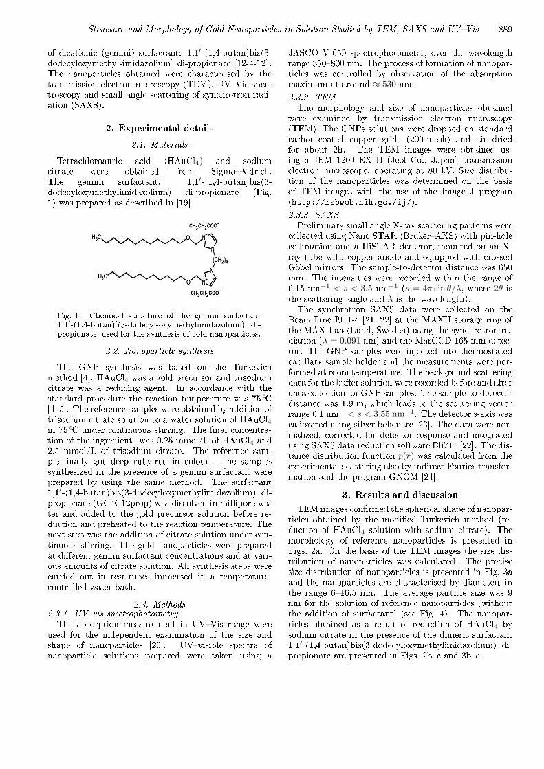
Tetrachloroauric acid (HAuCl4) and sodiumcitrate were obtained from Sigma�Aldrich.The gemini surfactant: 1,1′-(1,4-butan)bis(3-dodecyloxymethylimidazolium) di-propionate (Fig.1) was prepared as described in [19].
Fig. 1. Chemical structure of the gemini surfactant1,1′-(1,4-butan)′(3-dodecyl-oxymethylimidazolium) di-propionate, used for the synthesis of gold nanoparticles.
2.2. Nanoparticle synthesis
The GNP synthesis was based on the Turkevichmethod [4]. HAuCl4 was a gold precursor and trisodiumcitrate was a reducing agent. In accordance with thestandard procedure the reaction temperature was 75 ◦C[4, 5]. The reference samples were obtained by addition oftrisodium citrate solution to a water solution of HAuCl4in 75 ◦C under continuous stirring. The �nal concentra-tion of the ingredients was 0.25 mmol/L of HAuCl4 and2.5 mmol/L of trisodium citrate. The reference sam-ple �nally got deep ruby-red in colour. The samplessynthesized in the presence of a gemini surfactant wereprepared by using the same method. The surfactant1,1′-(1,4-butan)bis(3-dodecyloxymethylimidazolium) di-propionate (GC4C12prop) was dissolved in millipore wa-ter and added to the gold precursor solution before re-duction and preheated to the reaction temperature. Thenext step was the addition of citrate solution under con-tinuous stirring. The gold nanoparticles were preparedat di�erent gemini surfactant concentrations and at vari-ous amounts of citrate solution. All synthesis steps werecarried out in test-tubes immersed in a temperature-controlled water bath.
2.3. Methods2.3.1. UV�vis spectrophotometryThe absorption measurement in UV�Vis range were
used for the independent examination of the size andshape of nanoparticles [20]. UV�visible spectra ofnanoparticle solutions prepared were taken using a
JASCO V-650 spectrophotometer, over the wavelengthrange 350�800 nm. The process of formation of nanopar-ticles was controlled by observation of the absorptionmaximum at around ≈ 530 nm.
2.3.2. TEMThe morphology and size of nanoparticles obtained
were examined by transmission electron microscopy(TEM). The GNPs solutions were dropped on standardcarbon-coated copper grids (200-mesh) and air driedfor about 2h. The TEM images were obtained us-ing a JEM 1200 EX II (Jeol Co., Japan) transmissionelectron microscope, operating at 80 kV. Size distribu-tion of the nanoparticles was determined on the basisof TEM images with the use of the Image J program(http://rsbweb.nih.gov/ij/).
2.3.3. SAXSPreliminary small angle X-ray scattering patterns were
collected using Nano STAR (Bruker�AXS) with pin-holecollimation and a HiSTAR detector, mounted on an X-ray tube with copper anode and equipped with crossedGöbel mirrors. The sample-to-detector distance was 650mm. The intensities were recorded within the range of0.15 nm−1 < s < 3.5 nm−1 (s = 4π sin θ/λ, where 2θ isthe scattering angle and λ is the wavelength).The synchrotron SAXS data were collected on the
Beam Line I911-4 [21, 22] at the MAXII storage ring ofthe MAX-Lab (Lund, Sweden) using the synchrotron ra-diation (λ = 0.091 nm) and the MarCCD 165 mm detec-tor. The GNP samples were injected into thermostatedcapillary sample holder and the measurements were per-formed at room temperature. The background scatteringdata for the bu�er solution were recorded before and afterdata collection for GNP samples. The sample-to-detectordistance was 1.9 m, which leads to the scattering vectorrange 0.1 nm− < s < 3.55 nm−1. The detector s-axis wascalibrated using silver behenate [23]. The data were nor-malized, corrected for detector response and integratedusing SAXS data reduction software Bli711 [22]. The dis-tance distribution function p(r) was calculated from theexperimental scattering also by indirect Fourier transfor-mation and the program GNOM [24].
3. Results and discussion
TEM images con�rmed the spherical shape of nanopar-ticles obtained by the modi�ed Turkevich method (re-duction of HAuCl4 solution with sodium citrate). Themorphology of reference nanoparticles is presented inFigs. 2a. On the basis of the TEM images the size dis-tribution of nanoparticles was calculated. The precisesize distribution of nanoparticles is presented in Fig. 3aand the nanoparticles are characterised by diameters inthe range 6�16.5 nm. The average particle size was 9nm for the solution of reference nanoparticles (withoutthe addition of surfactant) (see Fig. 4). The nanopar-ticles obtained as a result of reduction of HAuCl4 bysodium citrate in the presence of the dimeric surfactant1,1′-(1,4-butan)bis(3-dodecyloxymethylimidazolium) di-propionate are presented in Figs. 2b�e and 3b�e.

890 M. Murawska, A. Skrzypczak, M. Kozak
Fig. 2. TEM images of gold nanoparticles for: reference sample (without gemini surfactant) a); and samples preparedwith: b) 0.0088% c) 0.0165% d) 0.035% and e) 0.07% gemini surfactant.
Fig. 3. Particle size distribution for: reference sample (without gemini surfactant) a); and samples prepared with: b)0.0088% c) 0.0165% d) 0.035% and e) 0.07% gemini surfactant.
Even a small addition of a gemini surfactant initiateschanges in the shape of nanoparticles and single tetra-hedral particles can be observed. In the samples withconcentration 0.0165% and 0.035% (w/w) of dicationicsurfactant several types of shapes are visible: spheri-cal, tetrahedral, double tetrahedral pyramid, pentagonalpyramid and truncated tetrahedron (see Fig. 2c�d). Thenanoparticles reached the maximum average size (28.5nm) for the surfactant concentration of 0.035% (Figs. 3dand 4). The greatest problem is quite high heterogeneityof shapes and sizes in samples obtained in the presence ofa gemini surfactant. The size range calculated for thesenanoparticles is from 5 nm to even 60 nm within one
sample. An important feature of metallic nanoparticlesis the enhancement of their optical properties under thein�uence of the light, known as the surface plasmon res-onance.
The surface plasmon resonance is observed at a speci�cfrequencies of incident light and depends on the particlesize, shape, dielectric constant of metal and the mediumaround nanoparticles [25]. The typical frequency of plas-mon resonance for spherical gold nanoparticles variesfrom 520 nm to 530 nm [25, 26]. The formation of AuNP was con�rmed by UV�VIS spectroscopy at a wave-length close to 530 nm. The reference solution of Au NP(without gemini surfactant) gave maximum absorbance

Structure and Morphology of Gold Nanoparticles in Solution Studied by TEM, SAXS and UV�Vis 891
Fig. 4. Average particle size versus gemini surfactantconcentration.
at 523 nm (Fig. 5). After the addition of a gemini surfac-tant the absorption peak is shifted towards longer wave-lengths (Fig. 5). The absorbance peak for NP synthesizedin the presence of 0.0088% gemini surfactant is observedat 540 nm. The broadening of the peak shows substan-tial changes in the nanoparticles morphology. For Aunanoparticles obtained with 0.035% of gemini surfactantthe largest broadening of the absorbance is visible. Thisindicates a high heterogeneity of size and shape withinthe sample.
Fig. 5. UV-Vis spectra of gold nanoparticles a) refer-ence sample (without gemini surfactant); and samplesprepared with: b) 0.0088% c) 0.0165% d) 0.035% ande) 0.07% gemini surfactant.
Figure 6 presents the SAXS scattering curve for thereference solution of gold nanoparticles obtained usingsynchrotron radiation on the beam line I9-11 of MAX-lab. The SAXS data are consistent with literature [5].On the basis of the experimental curve the pair distancedistribution p(r) function (Fig. 7) was calculated. Themaximum size of nanoparticles Dmax = 15 nm is con-sistent with the maximum size of nanoparticles obtainedfrom the size distribution estimated on the basis of TEMdata. The SAXS data for all samples of gold nanoparti-cles synthesized with and without the addition of a sur-factant are shown in Fig. 6.On the basis of �ts of SAXS data to the Guinier equa-
tion [26] the gyration radii characterizing systems stud-
Fig. 6. SAXS scattering curves for the reference sam-ple and gold nanoparticles prepared with the additionof surfactant.
Fig. 7. The particle distance distribution function p(r)determined from SAXS curve for the gold nanoparticleswithout the addition of gemini surfactant.
ied were determined (Fig. 8). For the reference sys-tem the radius of gyration RG was 8.7 nm. But fornanoparticles obtained in the presence of gemini surfac-tant radii of gyration, were higher reaching: 12.0 nm(0.0088% GC4C12prop) 13.1 nm (0.0165% GC4C12prop)17.1 nm (0.035% GC4C12prop) and 13.9 nm (0.07%GC4C12prop). The observed changes of gyration radii
Fig. 8. Fits of SAXS data to the Guinier equation [26].

892 M. Murawska, A. Skrzypczak, M. Kozak
were connected with increasing surfactant concentrationand are in good agreement with size distribution calcu-lated from TEM. The shapes of the SAXS curves ob-tained for samples prepared in the presence of geminisurfactant exhibit high polydispersity of nanoparticleswhich is consistent with TEM results. Observed highpolydispersity prevented the calculation of the p(r) func-tion for these samples. The morphology of nanoparticlesobtained in presence of GC4C12prop were also similarto morphology of nanoparticles obtained by Bakshi et al.[27] using gemini surfactant trimethylene-1,3-bis (dode-cyldimethylammonium bromide).
4. Conclusions
The phenomenon of plasmon resonance was observedfor the GNP samples obtained in the wavelength rangecorresponding to the presence of gold nanoparticles withsizes from 5�100 nm [20]. TEM images con�rmed thespherical morphology of the reference nanoparticles andshowed the presence of gold nanoparticles of di�erentshapes (e.g. tetrahedral and pentagonal) in solutions ob-tained in the presence of gemini surfactants. The shapeand size of nanoparticles were found dependent on theconcentration of the gemini surfactant. The average par-ticle sizes increases with surfactant concentration (up to0.035%) reaching 28.5 nm for the surfactant concentra-tion 0.035%. However, for gemini surfactant concentra-tions of 0.0165%, 0.035% and 0.07% a large dispersionof particle sizes and a heterogeneity of their shapes wasobserved. Also the SAXS studies con�rmed the sizesof nanoparticles obtained from analysis of TEM images.The future studies with other surfactants should improvehomogeneity of shape and size of nanoparticles within onesample.
Acknowledgments
The study reported was carried out with �nancial sup-port from the Ministry of Science and Higher Educa-tion (grant nr N N202 127237). The research (the datacollection at Beam Line I911) leading to these resultshas received funding from the European Community'sSeventh Framework Programme (FP7/2007�2013) undergrant agreement no. 226716.
References
[1] P. Yanez-Sedeno, J.M. Pingarron, Anal. Bioanal.Chem. 382, 884 (2005).
[2] S. Zeng, K.T. Yong, I. Roy, X.Q. Dinh, X. Yu,F. Luan, Plasmonics 6, 491 (2011).
[3] N. Zheng, J. Fan, G.D. Stucky, J. Am. Chem. Soc.128, 6550 (2006).
[4] J. Turkevich, P.C. Stevenson, J. Hillier, Discuss Fara-day Soc 11, 55 (1951).
[5] J. Polte, T.T. Ahner, F. Delissen, S. Sokolov, F. Em-merling, A.F. Thunemann, R. Kraehnert, J. Am.Chem. Soc. 132, 1296 (2010).
[6] T.C. Chiu, S.H. Chiou, M.M. Hsieh, Y.T. Chen, H.T.Chang, J. Nanosci. Nanotechnol. 5, 2128 (2005).
[7] J. Perez-Juste, I. Pastoriza-Santos, L.M. Liz-Marzan,P. Mulvaney, Coord. Chem. Rev. 249, 1870 (2005).
[8] M. Hu, J.Y. Chen, Z.Y. Li, L. Au, G.V. Hartland,X.D. Li, M. Marquez, Y.N. Xia, Chem. Soc. Rev. 35,1084 (2006).
[9] S.S. Shankar, A. Rai, B. Ankamwar, A. Singh, A. Ah-mad, M. Sastry, Nature Materials 3, 482 (2004).
[10] M.A. Faramarzi, H. Forootanfar, Coll. Surf. B-Biointerfaces 87, 23 (2011).
[11] T. Sakai, P. Alexandridis, Langmuir 20, 8426 (2004).[12] J. Wagner, T.R. Tshikhudo, J.M. Kohler, Chem. En-
gineering J. 135, 104 (2008).[13] S.D. Perrault, W.C.W. Chan, J. Am. Chem. Soc 131,
17042 (2009).[14] M. Burst, M. Walker, D. Bethel, D.J. Schi�rin, J.
Chem. Soc. Chem. Commun. 7, 801 (1994).[15] R. Zana, Adv. Coll. Interface Sci. 97, 205 (2002).[16] R. Zana, M. Benrraou, R. Rue�, Langmuir 7, 1072
(1991).[17] F.M. Menger, C.A. Littau, J. Am. Chem. Soc. 113,
1451 (1991).[18] T.S. Selvam, K.M. Chi, J. Nanopart. Res. 13, 1769
(2011).[19] Z. Pietralik, M. Taube, A. Skrzypczak, M. Kozak,
Acta Phys. Polon. A 117, 311 (2010).[20] D.E. Mustafa, T. Yang, Z. Xuan, S. Chen, H. Tu,
A. Zhang, Plasmonics 5, 221 (2010).[21] Y. Cerenius, K. Ståhl, L.A. Svensson, T. Ursby,
Å. Oskarsson, J. Albertsson, A. Liljas, J. Synch. Rad.7, 203 (2000).
[22] M. Knaapila, C. Svensson, J. Barauskas, M. Zackris-son, S.S. Nielsen, N. Nørgaard Toft, B. Vestergaard,L. Arleth, Y. Cerenius, J. Synch. Rad. 16, 498 (2009).
[23] T.C. Huang, H. Toraya, T.N. Blanton, Y. Wu, J.Appl. Cryst. 26, 180 (1993).
[24] A.V. Semenyuk, D.I. Svergun, J. Appl. Crystallogr.24, 537 (1991).
[25] S.R. Grobmyer, B.M. Moudgil, Cancer nanotechnol-ogy: methods and protocols, Humana Press, New York2010.
[26] A. Guinier, Ann. Phys. 12, 166 (1939).[27] M.S. Bakshi, P. Sharma, T.S. Banipal,Materials Lett.
61, 5004 (2007).