structure and dynamics of helix-0 of the n-bar domain in ... · und psychologie, universita¨t...
TRANSCRIPT

Structure and Dynamics of Helix-0 of the N-BAR Domain in LipidMicelles and Bilayers
Christian Low,* Ulrich Weininger,* Hwankyu Lee,y Kristian Schweimer,z Ines Neundorf,§
Annette G. Beck-Sickinger,§ Richard W. Pastor,z and Jochen Balbach*{
*Institut fur Physik, Biophysik, Martin-Luther-Universitat Halle-Wittenberg, D-06120 Halle (Saale), Germany; yLaboratory ofComputational Biology, National Heart, Lung, and Blood Institute, National Institutes of Health, Bethesda, MD 20892; zLehrstuhlBiopolymere, Universitat Bayreuth, 95447 Bayreuth, Germany; §Institut fur Biochemie, Fakultat fur Biowissenschaften, Pharmazieund Psychologie, Universitat Leipzig, D-04103 Leipzig, Germany; and {Mitteldeutsches Zentrum fur Struktur und Dynamik der Proteine(MZP), Martin-Luther-Universitat Halle-Wittenberg, Germany
ABSTRACT Bin/Amphiphysin/Rvs-homology (BAR) domains generate and sense membrane curvature by binding the nega-tively charged membrane to their positively charged concave surfaces. N-BAR domains contain an N-terminal extension (helix-0)predicted to form an amphipathic helix upon membrane binding. We determined the NMR structure and nano-to-picoseconddynamics of helix-0 of the human Bin1/Amphiphysin II BAR domain in sodium dodecyl sulfate and dodecylphosphocholinemicelles. Molecular dynamics simulations of this 34-amino acid peptide revealed electrostatic and hydrophobic interactions withthe detergent molecules that induce helical structure formation from residues 8–10 toward the C-terminus. The orientation in themicelleswas experimentally confirmed by backboneamide proton exchange. The simulation and the experiment indicated that theN-terminal region is disordered, and the peptide curves to adopted the micelle shape. Deletion of helix-0 reduced tubulation ofliposomes by the BAR domain, whereas the helix-0 peptide itself was fusogenic. These findings support models for membranecurving by BAR domains in which helix-0 increases the binding affinity to the membrane and enhances curvature generation.
INTRODUCTION
Proteins play fundamental roles inmodulating the structure of
lipid bilayers. Processes such as membrane fusion, budding,
or tubulation are associated with changes in membrane cur-
vature. The banana-shapedBARdomains have been identified
throughout eukarya as regulators of membrane remodeling
processes. They sense and curve membranes, and participate
in numerous cytoskeletal and nuclear processes, such as
clathrin–mediated endocytosis or organization of the T-tubule
network in the muscle (1–10). Point mutations in the BAR
domain of Bin1 found in patients with centronuclear myop-
athy cause a dysfunction of the latter process (11).
The crystal structures of the human and Drosophila am-
phiphysin BAR domain (12,13) reveal a crescent-shaped ho-
modimerwith apositively chargedconcave surface.Thisfinding
suggests that driving and/or sensing curvatures of membranes
by BAR domains occurs by binding of negatively charged
membranes to this positively charged surface (13). Some
BAR domains (denoted N-BAR) contain an N-terminal ex-
tension with an amphipathic character that is predicted to
undergo a random coil to helix transition by binding to the
membrane (13). This extension, termed helix-0, shows no
electron density in the crystal structure (14). In vitro, BAR
domains can induce curvature in liposomes, which results in
narrow tubes (tubulation) (15). Recent experimental and
theoretical studies (16) with N-BAR domains indicate that
helix-0 embeds in the lipid bilayer and strongly increases the
ability to tubulate liposomes. The insertion of amphipathic
helices into hydrophobic phases of the bilayer has been pro-
posed to be a general mechanism for curvature generation
during vesicle budding as shown in amphiphysin (13) and
other examples (17–19). Experimental evidence for structure
induction and insertion of the amphipathic helix has been de-
rived from circular dichroism (CD) and electron paramagnetic
resonance spectroscopy (EPR) (13,14,20,21). There are cur-
rently three candidate curvature-generating mechanisms: 1),
the local spontaneous curvature; 2), the bilayer coupling; and
3), the scaffolding (22). The scaffoldmechanism assumes that
the intrinsic curvature of the BAR domain forces the mem-
brane shape, as opposed to a deformation of the lipid bilayer
by a shallow (spontaneous curving) or deep (bilayer-cou-
pling) insertion of an amphipathic helix.
To obtain further insights into the predicted N-terminal
amphipathic helix of N-BAR domains, we studied helix-0 of
the human Bin1/Amphiphysin II BAR domain (N-BAR) in
detergent and lipid environments by high-resolution NMR
spectroscopy and molecular dynamics (MD) simulations.
Structure calculation, dynamicmeasurements, and a fast amide
proton exchange confirmed the earlier proposed amphipathic
character of the induced helix but also revealed a disordered
N-terminal part of the amphipathic helix that is highly flexible
doi: 10.1529/biophysj.108.134155
Submitted March 28, 2008, and accepted for publication July 3, 2008.
Address reprint requests to Jochen Balbach Institut fur Physik, Fachgruppe
Biophysik, Martin-Luther-Universitat, Halle-Wittenberg, D-06120 Halle
(Saale), Germany. Tel.: 49-345-55-28550; Fax: 49-345-55-27161; E-mail:
Abbreviations: BAR, bin/amphiphysin/rvs-homology; DPC, dodecylphos-
phocholine; EM, electron microscopy; 2D, two-dimensional; 3D, three-
dimensional; FRET, fluorescence resonance energy transfer; MEXICO,
measurements of exchange rates in isotopically labeled compounds; OG,
n-Octyl-b-D-glucopyranoside; SDS, sodium dodecyl sulfate.
Editor: Arthur G. Palmer 3rd.
� 2008 by the Biophysical Society
0006-3495/08/11/4315/09 $2.00
Biophysical Journal Volume 95 November 2008 4315–4323 4315
and exposed to the solvent. We also considered the balance of
electrostatic and hydrophobic interactions. Last, a tubulation
assay of liposomes analyzed by EM or FRET showed that the
isolated N-BAR peptide was fusogenic.
MATERIALS AND METHODS
Expression and purification of the N-BAR andBAR domain and the N-BAR peptide of humanBin1/Amphiphysin II
The plasmid of the BAR domain of human Bin1/Amphiphysin II was a kind
gift of E. D. Laue (Cambridge). The histidine-tagged recombinant protein
was expressed in E. coli BL21(DE3) and purified as described previously
(12). The N-BAR peptides were expressed as a SUMO fusion protein and
cleaved by the SUMO protease (23) or synthesized by solid-phase peptide
synthesis. Further details are provided in the SupplementaryMaterial, Data S1.
Liposomes and tubulation assay
Small unilamellar vesicles were prepared from total bovine brain lipids
(Folch fraction 1; Sigma B1502; Sigma-Aldrich, Munich, Germany) in 20
mMHepes, 150 mMNaCl, pH 7.4, by extrusion (pore size of 100 nm) using
a Liposofast extruder (Avestin, Ottawa, Ontario, Canada) as described pre-
viously (13). For tubulation assays, N-BAR domain and different constructs
(5 mM for N-BAR and BAR, 10 mM for N-BAR peptides) were mixed with
brain lipid liposomes (0.2 mg/ml) for 30 min at room temperature and then
processed for negative staining. For EM analysis, carbonized copper grids
(Plano,Wetzlar, Germany) were pretreated for 1 min with bacitracin (0.1 mg/
ml). After air-drying, protein lipid mixture diluted 5-fold with 20 mMHepes,
150 mM NaCl, pH 7.4, was applied for 3 min. Subsequently, grids were
again air-dried. Samples were negatively stained with 1% (w/v) uranyl-ac-
etate and visualized in an electron microscope (Zeiss EM 900; Carl Zeiss
GmbH, Jena, Germany) operating at 80 kV.
FRET assay of membrane fusion
Membrane fusion was measured by FRET using an FP-6500 spectrofluo-
rometer (Jasco, Groß-Umstadt, Germany). Two populations of liposomes
composed of bovine brain lipids, one unlabeled and one labeled with 2%
each of n-[7-nitro-2-1,3-benzoaxadiazole-4-yl]-egg-phosphatidylethanolamine
and n-[lissamine rhodamine B]-egg-phosphatidylethanolamine, were mixed
at a 9:1 unlabeled/labeled ratio and 0.25 mg/ml total lipid in 20 mM Hepes,
150 mMNaCl, pH 7.4, at 25�C in the presence of different concentrations of
N-BAR and various constructs (concentration range: 1–15 mM for N-BAR
and BAR, 5–50 mM for N-BAR peptide constructs). The excitation wave-
length was 450 nm, and the emission spectrum was recorded from 480 to
700 nm after several time points. To obtain a value for donor fluorescence,
1% Triton X-100 was added. Kinetics of membrane fusion was followed by
fluorescence increase of the donor fluorescence at 530 nm after excitation at
450 nm.
CD
Far-ultraviolet (UV) CD spectra of BAR domain and mutants were measured
in the presence and absence of Folch liposomes in 20 mM Hepes, 150 mM
NaCl, pH 7.4, at 15�C with a J815A spectropolarimeter (Jasco). 10 mM
protein was incubated with 0.2 mg/ml Folch liposomes and degassed for
5 min before measurement. The N-terminal peptide was measured in the
presence of different detergents (SDS, OG, DPC) and Folch liposomes. The
signals from pure liposomes or micelles were subtracted from the sample
spectra as blanks.
Sample preparation for NMR
N-BAR peptide was dissolved in 20 mM sodium phosphate, pH 7.4 (90%
H2O/10%2H2O) containing either d38-DPC or d25-SDS micelles and 0.03%
NaN3. The final 15N-labeled N-BAR peptide samples contained 1 mM
protein and 200 mM d38-DPC or 150 mM d25-SDS. A 1 mM 15N-labeled
N-BAR peptide sample without detergent was prepared as a reference.
NMR spectroscopy
NMR spectra were acquired with a Bruker Avance 800, Bruker Avance 700
equipped with a cryoprobe, and a Bruker Avance II 600 spectrometer (all
obtained from Bruker BioSpin, Rheinstetten, Germany) in 20 mM sodium
phosphate buffer, pH 7.4, containing 10% 2H2O at 25�C; the free N-BAR
peptide experiments, however, were carried out at 15�C and the extended
N-BAR peptide (1–44 residues) at pH 6.0 and at 15�C. The N-BAR peptide
in SDS and DPC micelles and the unbound form were assigned by 3D-15N
TOCSY-HSQC and 3D-15N-NOESY-HSQC (complete assignments were
deposited in the Biological Magnetic Resonance Bank database). For
structure calculation of SDS- and DPC-bound N-BAR peptide, an additional
2D NOESY spectrum was recorded and a 3D HNHA spectrum was recorded
for the SDS-bound form. For further investigations, a 15N heteronuclear
nuclear Overhauser enhancement (NOE) and a MEXICO proton-exchange
experiment (24) were performed. The ordinate in Fig. 5 corresponds to the
NMR crosspeak intensity at the respective exchange time divided by in-
tensity in a reference experiment. Spectra were processed with NMRpipe (25)
and analyzed with NMRView (26).
Structure calculation
Distance restraints were obtained from 3D 15N-NOESY-HSQC and 2D
NOESY and used as ambiguous constraints for structure calculation with
ARIA (27). Backbone dihedral restraints were calculated from chemical
shifts using TALOS (28). It should be noted that the random coil values of
TALOS are not optimized for the micellar environment. ARIA runs with and
without TALOS restraints, however, gave the same overall topology and
curvature, but root mean-square deviation values were reduced with these
restraints and therefore TALOS was included in the final run. Structure ge-
ometry was analyzed with PROCHECK (29). The preceding structure cal-
culations did not rely on any of the MD results.
Computational methods
In principle, the peptide micelle complex could be assembled from a solution
of dispersed lipids and an unfolded peptide. However, self-assembly of SDS
micelles from 70 to 100 monomers in ;10000 waters was incomplete in
40 ns simulations (data not shown). Consequently, initial conditions for the
simulations reported here were developed with fully formed and well-
equilibrated (40 ns) spherical micelles in ;5500 waters.
Experimental evidence indicates that the peptide is disordered in solution
and largely a-helical in the micelle. To explore potential force field (FF)
dependencies, 50 ns simulations of the peptide in water and in an a-helix
were carried out with CHARMM22/CMAP (30,31), GROMOS96 (53a6)
(32), and OPLS-AA (33). The a-helix remained stable over the entire tra-
jectory with CHARMM22/CMAP, rapidly converted to a stablep-helix with
GROMOS96, and became disordered within 13 ns with OPLA-AA. Based
on these results, the OPLS-AA FF was chosen for peptide, DPC, and SDS
(with one change noted below).
All simulations and analyses were performed using the GROMACS 3.3.1
simulation package (34,35). Coordinates of the SDS molecule were gener-
ated using the PRODRG2 server (36). The united-parameter set for lipids
was downloaded from http://moose.bio.ucalgary.ca, and charges of SDS
headgroup were set equal to those in the CHARMM FF (37) (these charges
4316 Low et al.
Biophysical Journal 95(9) 4315–4323
were not available in OPLS). All-atom OPLS parameters (33) were used for
the peptide. Five initial conditions were constructed (Table 1 and see Fig. 6).
SDS1, SDS2, and SDS3 contained 75 lipids (the experimentally established
aggregation number for SDS micelles) (38). SDS4 contained 40 lipids to
investigate effects of micelle size, andDPC contained 65 monomers of DPC.
Initial coordinates for the N-BAR peptide were those of the lowest energy
structure from the NMR ensemble, which is 76% a-helical. The peptides
were placed in different orientations and positions with respect to the micelle
as specified in Table 1. A hole for the inserting peptide was made by fol-
lowing the hole protocol (39). TheMSMS programwas used for scanning the
surface of peptides (40), and then a hole-making force was introduced to the
scanned surface of the peptide. The peptide was inserted into the resulting
hole, and then energy minimization was performed with position restraints
applied to the peptide. Approximately 16,000 TIP4P water molecules (41)
were placed around a mixture of the peptide and micelle to a thickness of
1 nm, forming a periodic box sized 83 83 8 nm3. Na1 ions were added to
neutralize charges from the SDS molecules. For DPC, the equilibrated 65-
surfactant DPC micelle was downloaded from http://moose.bio.ucalgary.ca,
and the same procedures were performed with 5 Cl� ions added to neutralize
the peptide. A cutoff of 1.1 nmwas set for the van derWaals interactions with
particle mesh Ewald summation used for electrostatic interactions (42). A
pressure of 1 bar and a temperature of 298 K were maintained in an NPT
ensemble by applying the Berendsen coupling method with the time con-
stants of 0.1 and 1.0 ps for temperature and pressure, respectively (43). After
energy minimization with 200 steps of steepest descent, unrestrained trajec-
torieswere carried out for 60 nswith a time step of 2 fs. Coordinates were saved
every picosecond, and averages were obtained from the last 20 ns. The sec-
ondary structure of the peptide was calculated using the DSSP program (44).
Protein structure accession number
The coordinates of the structure of the N-BAR peptide in DPC and SDS
micelles have been deposited in the Protein Data Bank under the accession
numbers 2RND and 2RMY.
RESULTS AND DISCUSSION
The predicted N-terminal amphipathic helix-0 of the human
Bin1/Amphiphysin II BAR domain comprises residues 1–33
(14,45). This part of the molecule is disordered in the absence
of lipids and thereby unresolved in the crystal structure (12).
The N-BAR peptide studied here (1MAEMGSKGV10TAG-
KIASNVQ20KKLTRAQEKV30LQKLY) contains an addi-
tional tyrosine residue at the C-terminus of helix-0 for
spectroscopic reasons. DPC and SDS micelles were chosen
because they have been successfully used for other peptide
and protein structure determinations by NMR spectroscopy
(46–49).
Structure induction upon membrane binding
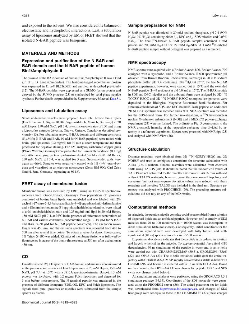
Far-UV CD spectra of the full-length N-BAR domain re-
vealed an increased helicity after adding brain lipid liposomes
(Fig. 1 a, solid lines). The helical content of a deletion mutant
lacking the first 31 residues (BAR) did not change in the pres-
ence of lipids. The isolated N-BAR peptide is unstructured in
aqueous solution (Fig. 1 b, solid black line). As in the full-
length protein, the ellipticity minima at 208 and 222 nm in-
dicate that the peptide takes on a helical structure when bound
to liposomes or micelles. The CD spectra of the N-BAR
peptide in brain lipid liposomes or DPC or SDS micelles are
virtually identical, indicating a similar secondary structure
under these conditions (Fig. 1 b). In 60% tetrafluoroethylene,
the helical content increased further, indicating that not all
residues of theN-BARpeptide are in a helical conformation in
the presence of detergents or lipids. A helical wheel projection
(see Fig. 3d) of the peptide highlights its amphipathic char-
acter (that is, hydrophobic and charged/polar residues are
located opposite to each other). Conspicuous is the high
number of lysine residues, implying that binding and structure
induction of theN-BAR peptide is driven by hydrophobic and
electrostatic interactions. This finding is further supported by
the observation that nonionic OG micelles as membrane mi-
metics do not lead to structuring of the peptide as observed by
the far-UV CD spectrum (Fig. 1 b, dashed light gray).
NMR structure and dynamics of N-BAR peptide
Binding of the N-BAR peptide to micelles results in a devi-
ation of the backbone and, more obviously, the side-chain
resonances in the 15N-HSQC spectrum (Fig. 2 b) from the
random coil chemical shifts dominating the spectrum in
TABLE 1 Composition and initial conditions for the N-BAR
peptide/micelle simulations
Name
Micelles
Initial position of the peptide
Type No. lipids Location
Orientation of
hydrophobic residues
SDS1 SDS 75 Water region Toward micelle
SDS2 SDS 75 Water region Away from micelle
SDS3 SDS 75 Inside micelle –
SDS4 SDS 40 Inside micelle –
DPC DPC 65 Inside micelle –
FIGURE 1 Far-UV CD spectra of the BAR domain and the N-terminal
N-BAR peptide in various solvent environments. (a) CD spectra of the
N-BAR (residues 1–241) (solid line) and the BAR (residues 32–241) domain
(dashed line) of human amphiphysin II in the presence and absence of brain
lipid liposomes (gray and black, respectively). Structure induction on
binding to liposomes is only seen for the N-BAR domain (solid gray
line), indicated by a significant signal decrease at 222 nm. (b) The N-BAR
peptide is unstructured in solution (solid black line). In the presence of
liposomes (dashed dark gray line), SDS (solid gray line) or DPC (blackdashed line) micelles, the peptide becomes structured. In the presence of OG
(dashed light gray line) micelles, however, no structure induction is
observed. A CD spectrum recorded in 60% trifluoroethanol (dotted line)
shows the greatest helical content.
Structure of Helix-0 of the N-BAR Domain 4317
Biophysical Journal 95(9) 4315–4323

aqueous solution (Fig. 2 a). This finding confirms the inter-
action of N-BAR with the micellar environment and the in-
duction of a defined secondary structure in SDS or DPC
observed by far-UV CD spectra. All backbone and side-chain
resonanceswere assigned asdescribed inMaterials andMethods.
More than 500 NOE distance constraints were derived from
two-dimensional NOESY and 15N-NOESY-HSQC spectra in
the presence of either SDS or DPC micelles. By using all
experimentally determined constraints (NOEs, dihedral an-
gles derived from J couplings, and chemical shifts), ensembles
of structures of the N-BAR peptide in SDS (Fig. 3 a) and DPC(Fig. 3 b) micelles were calculated; structural statistics are
provided in Table S2 in Data S1. Residues 8–34 in SDS and
10–34 in DPC micelles are well-ordered with a heavy atom
root mean-square deviation, 1.1 A. This a-helical content isconsistent with theCDdata (Fig. 1). Hence, the structured part
of the N-BAR peptide is an amphipathic helix with the neg-
atively charged side chains on the convex side and the hydro-
phobic side chains on the concave side (Fig. 3 c).Thedisorderingof theN-terminus results fromfast and large-
amplitude local dynamics confirmed by 15N-heteronuclear
NOE (hNOE) measurements (Fig. 4). hNOE values. 0.5 are
typical for structural elements in peptides and proteins that are
relatively rigid on a timescale of nanoseconds to picoseconds.
For the N-BAR peptide in SDS and DPC micelles, the hNOE
gradually decreases from T10 toward the N-terminus and is
even negative for the first residues. Therefore, the dynamic
data agree well with the loss of NOE constraints in the highly
flexible and disordered conformation at the N-terminus. In
comparison, all hNOE values of the N-BAR peptide in
aqueous solution (Fig. 4 c) are close to zero or negative. Takentogether with a lack of medium-range NOEs over the entire
sequence, a random coil conformation in the absence of de-
tergent and lipids can be concluded. The hNOEof an extended
FIGURE 2 2D 1H-15N HSQC spectra of the N-BAR peptide (a) in
aqueous solution and (b) bound to DPC micelles. The assigned crosspeaks
of the backbone amides are labeled using the one-letter amino acid code and
the sequence position. Boxes indicate resonance signals, which show
crosspeak intensities below the plotted contour level. The respective spec-
trum of N-BAR peptide in SDS micelles is shown in Fig. S11 in Data S1.
FIGURE 3 Structure ensembles of the N-BAR peptide backbone bound to
detergent micelles at 25�C: 10 lowest energy structures in (a) SDS micelles
and (b) DPCmicelles. (c) Electrostatic surface potential representation of the
N-BAR peptide in DPC micelles. Negative potentials are shown in red and
positive potentials in blue. (d) Helical wheel diagram for the N-BAR pep-
tide. The amino acid sequence is plotted clockwise. Hydrophobic residues
are shown in gray boxes, and positively and negatively charged residues are
shown in blue and red boxes, respectively.
4318 Low et al.
Biophysical Journal 95(9) 4315–4323

N-BAR peptide in DPC micelles with 44 amino acids drops
after K35 toward the C-terminus, indicating that the amphi-
pathic helix ends at position 35 and following residues form
the linker to helix-1 of the BAR domain.
Fast (millisecond) amide proton exchange
To determine which regions of the N-BAR peptide were
buried in the detergent micelle, fast (millisecond) amide
proton exchange was measured for each residue by NMR
(24); examples of exchange curves are depicted in Fig. 5. This
approach is straightforward compared to the use of spin labels
(50), because the NMR sample for structure determination
can be used without further modifications. N-terminal, polar,
and charged residues showed a pronounced signal change
during the experiment (Fig. 5, red), indicating fast-exchang-
ing amide protons because of an increased solvent accessi-
bility and a dynamic open and closing of the corresponding
hydrogen bonds on the millisecond-second timescale. Amide
protons of hydrophobic residues, however, did not exchange
at all (Fig. 5, blue), because they are buried in the micelle and
therefore shielded from the solvent. Furthermore, residues
1–11, the region with high, local fluctuations derived from
dynamic data (Fig. 4, b and c), showed low protection against
exchange of the amide protons.
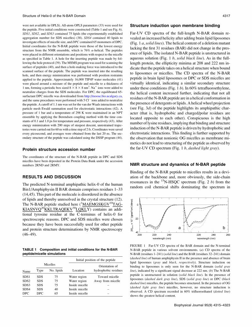
MD simulation of the N-BAR peptide
As an independent complement to the preceding experimental
results, MD simulations were carried out on the N-BAR
peptide in micelles with different surfactants, micelle sizes,
and initial configurations to investigate helical stability,
peptide orientation, and depth of insertion in the micelle en-
vironment. Fig. 6 shows snapshots at the beginning (left) andend (right) of the five 60 ns simulations of peptide/micelle
complex simulations (Table 1). In each case, the peptide mi-
grated to the surface of the micelle. The micelle remained
spherical, and the peptide curved. In pure water, the N-BAR
peptide partially unfolds in the first nanosecond and loses
almost all helicity by 13 ns (see Fig. S9 inData S1).Hence, the
micelle environment generally stabilizes the helix, leading to
a range ofa-helicities of 40 to 50% (SDS1: 48%; SDS3: 50%;
DPC: 40%) for residues 12–30 during the final simulated 20
ns. The helical instability of the N-terminal residues agrees
with the amide proton exchange and the NMR relaxation data
(Figs. 4 and 5). The SDS4 simulation (the smaller micelle)
yielded a slightly lower helical content (30%). It is possible
that the higher curvature imposed on the peptide for binding
led to this instability. As evident in Fig. 6, the N-BAR peptide
FIGURE 4 1H-15N heteronuclear NOEs of the N-BAR peptide in (a)SDS, (b) DPC, and (c) aqueous solution. (d) hNOE values of the extended
N-BAR peptide (1–44 residues).
FIGURE 5 NMR experiment to detect fast-exchanging amide protons
(MEXICO) of the N-BAR peptide bound to SDS and DPC micelles. Fast
amide proton exchange was followed on a residue by residue level. (a)
Exchange curves in SDS micelles are shown for T23 (solid red symbols),
S16 (open red symbols), L33 (solid blue symbols), and V29 (open bluesymbols). Fast-exchanging amides are colored in red. Amide protons, which
did not exchange within the timescale of the experiment (below dashed line;
see Fig. S12 in Data S1) are colored in blue. Exchange curves for residues in
gray could not been evaluated due to signal overlap or low signal intensity.
This color code was assigned to a ribbon representation of the lowest energy
NMR structure of the N-BAR peptide in (b) SDS and (c) DPC micelles.
Structure of Helix-0 of the N-BAR Domain 4319
Biophysical Journal 95(9) 4315–4323
bound to the micelle with little (6%) a-helicity in the SDS2
trajectory. This observation can be attributed to the initial
condition in which the cationic residues of the peptide were
oriented toward the micelle. When the peptide interacts with
the micelle, cationic and hydrophobic residues, respectively,
have favorable interactions with SDS headgroups and tails,
which may lead to a flip of the peptide and instability of the
helical structure. It is possible that the peptide in SDS2 would
refold to the helical form in much longer simulations, but this
observation is outside the scope of this study. Our results
indicate thatMDsimulations can represent the experimentally
measured stability of the peptide, although the final configu-
ration is partially determined by initial configuration and mi-
celle size. Further analyses arebasedonSDS1,SDS3, andDPC.
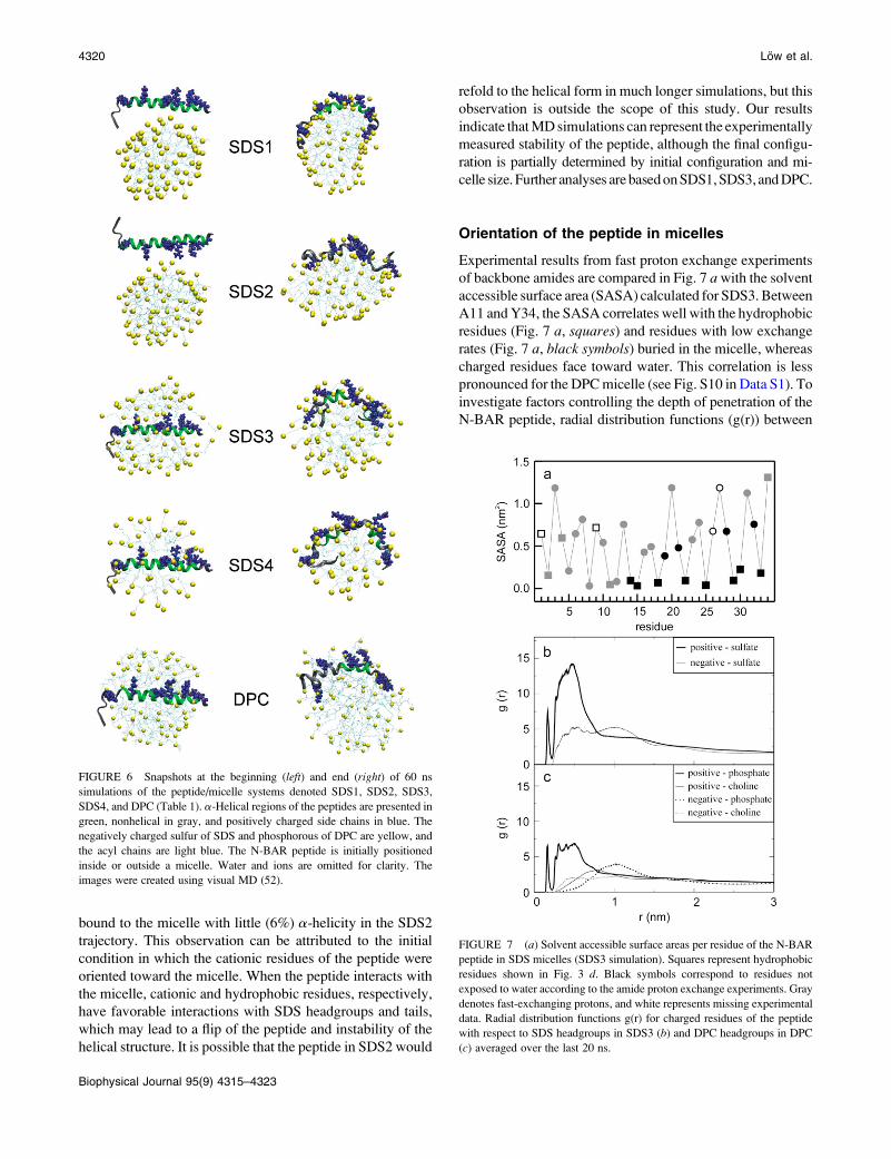
Orientation of the peptide in micelles
Experimental results from fast proton exchange experiments
of backbone amides are compared in Fig. 7 awith the solventaccessible surface area (SASA) calculated for SDS3. Between
A11 andY34, the SASA correlates well with the hydrophobic
residues (Fig. 7 a, squares) and residues with low exchange
rates (Fig. 7 a, black symbols) buried in the micelle, whereas
charged residues face toward water. This correlation is less
pronounced for the DPCmicelle (see Fig. S10 in Data S1). To
investigate factors controlling the depth of penetration of the
N-BAR peptide, radial distribution functions (g(r)) between
FIGURE 6 Snapshots at the beginning (left) and end (right) of 60 ns
simulations of the peptide/micelle systems denoted SDS1, SDS2, SDS3,
SDS4, and DPC (Table 1). a-Helical regions of the peptides are presented in
green, nonhelical in gray, and positively charged side chains in blue. The
negatively charged sulfur of SDS and phosphorous of DPC are yellow, and
the acyl chains are light blue. The N-BAR peptide is initially positioned
inside or outside a micelle. Water and ions are omitted for clarity. The
images were created using visual MD (52).
FIGURE 7 (a) Solvent accessible surface areas per residue of the N-BARpeptide in SDS micelles (SDS3 simulation). Squares represent hydrophobic
residues shown in Fig. 3 d. Black symbols correspond to residues not
exposed to water according to the amide proton exchange experiments. Gray
denotes fast-exchanging protons, and white represents missing experimental
data. Radial distribution functions g(r) for charged residues of the peptide
with respect to SDS headgroups in SDS3 (b) and DPC headgroups in DPC
(c) averaged over the last 20 ns.
4320 Low et al.
Biophysical Journal 95(9) 4315–4323
charged residues of the peptide and lipid headgroups were
calculated for SDS3 and DPC. The integral of g(r) over a
particular interval is proportional to the coordination number
in that interval. Fig. 7, b and c, shows that g(r) of the lipid
headgroups around cationic residues of the peptide is sub-
stantially higher than for anionic residues. Moreover, in the
DPC micelles, g(r) of the phosphate headgroups around cat-
ionic residues of the peptide have higher values compared to
the choline headgroups around both anionic and cationic
residues of the peptide. These results imply that cationic
residues of the peptide strongly interact with anionic lipid
headgroups and that the N-BAR peptide embeds deeper in the
DPC micelle compared to SDS.
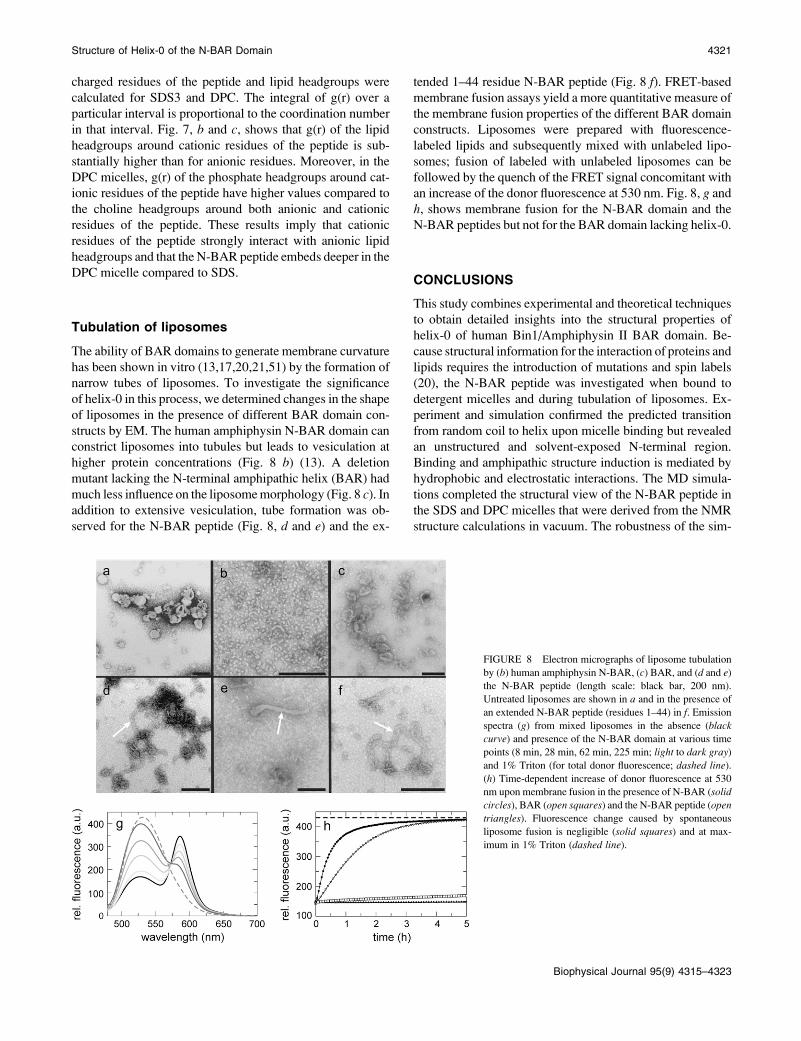
Tubulation of liposomes
The ability of BAR domains to generate membrane curvature
has been shown in vitro (13,17,20,21,51) by the formation of
narrow tubes of liposomes. To investigate the significance
of helix-0 in this process, we determined changes in the shape
of liposomes in the presence of different BAR domain con-
structs by EM. The human amphiphysin N-BAR domain can
constrict liposomes into tubules but leads to vesiculation at
higher protein concentrations (Fig. 8 b) (13). A deletion
mutant lacking the N-terminal amphipathic helix (BAR) had
much less influence on the liposomemorphology (Fig. 8 c). Inaddition to extensive vesiculation, tube formation was ob-
served for the N-BAR peptide (Fig. 8, d and e) and the ex-
tended 1–44 residue N-BAR peptide (Fig. 8 f). FRET-basedmembrane fusion assays yield a more quantitative measure of
the membrane fusion properties of the different BAR domain
constructs. Liposomes were prepared with fluorescence-
labeled lipids and subsequently mixed with unlabeled lipo-
somes; fusion of labeled with unlabeled liposomes can be
followed by the quench of the FRET signal concomitant with
an increase of the donor fluorescence at 530 nm. Fig. 8, g andh, shows membrane fusion for the N-BAR domain and the
N-BAR peptides but not for the BAR domain lacking helix-0.
CONCLUSIONS
This study combines experimental and theoretical techniques
to obtain detailed insights into the structural properties of
helix-0 of human Bin1/Amphiphysin II BAR domain. Be-
cause structural information for the interaction of proteins and
lipids requires the introduction of mutations and spin labels
(20), the N-BAR peptide was investigated when bound to
detergent micelles and during tubulation of liposomes. Ex-
periment and simulation confirmed the predicted transition
from random coil to helix upon micelle binding but revealed
an unstructured and solvent-exposed N-terminal region.
Binding and amphipathic structure induction is mediated by
hydrophobic and electrostatic interactions. The MD simula-
tions completed the structural view of the N-BAR peptide in
the SDS and DPC micelles that were derived from the NMR
structure calculations in vacuum. The robustness of the sim-
FIGURE 8 Electron micrographs of liposome tubulation
by (b) human amphiphysin N-BAR, (c) BAR, and (d and e)the N-BAR peptide (length scale: black bar, 200 nm).
Untreated liposomes are shown in a and in the presence of
an extended N-BAR peptide (residues 1–44) in f. Emission
spectra (g) from mixed liposomes in the absence (blackcurve) and presence of the N-BAR domain at various time
points (8 min, 28 min, 62 min, 225 min; light to dark gray)
and 1% Triton (for total donor fluorescence; dashed line).
(h) Time-dependent increase of donor fluorescence at 530
nm upon membrane fusion in the presence of N-BAR (solid
circles), BAR (open squares) and the N-BAR peptide (open
triangles). Fluorescence change caused by spontaneous
liposome fusion is negligible (solid squares) and at max-
imum in 1% Triton (dashed line).
Structure of Helix-0 of the N-BAR Domain 4321
Biophysical Journal 95(9) 4315–4323
ulations is reflected by comparable final conformations of the
N-BAR peptide starting from different conditions and by the
peptide curvature, which was maintained during the simula-
tion and adapts to the micelle. A slightly increased curvature
of the N-BAR peptide in DPC micelles compared to SDS
found in the calculated NMR structures (Fig. 3, a and b) is aresult of several side-chain-side-chain and backbone-side-
chain NOEs, which were unambiguously identified. This
curvature might result from the more deeply embedded pep-
tide caused by the polar interactions with the zwitterionic
DPCheadgroups, if we assume the same spherical size of both
micelle types.
Recent experiments based on fluorescence measurements
imply an antiparallel dimer formation of the helix-0 of the
BRAP/Bin2 BAR domain (breast cancer-associated protein)
when bound to liposomes (14). In this study, no long-range
NOEs between N- and C-terminal residues of the N-BAR
peptide were observed in the NOESY spectra, which rules out
dimer formation. Nevertheless, oligomerization of the helix-0
under different conditions cannot be excluded because the
structure was determined in detergent micelles with a high
detergent/peptide ratio.
Recently, a point mutation (K35N) in the helix-0 of the
human Bin1/Amphiphysin II N-BAR domain found in pa-
tients suffering from autosomal recessive centronuclear myo-
pathy reduced the number and length of tubules in ex vivo
membrane assays (11). In isolation at least, extended N-BAR
peptides comprising 1–44 residues with K35 and N35 could
not be distinguished from the 1–34 peptide in the biophysical
measurements (CD and NMR spectra, tubulation) presented
here. The tubulation experiments, however, did reveal the
importance of helix-0 for changingmembrane morphology of
liposomes by itself or when present in the BAR domain.
In summary, this work highlights the importance of the am-
phipathic helix-0 for increasing the affinity of the N-BAR do-
main to lipidbilayers. It also helps differentiate currentmodels of
curvature generation by the N-BAR domain. Although these
findings do not definitively rule out the spontaneous curvature
and bilayer-coupling mechanics, they favor the scaffold mech-
anism. Therefore, we expect little curvature generation preced-
ing the main interaction with the entire N-BAR domain.
SUPPLEMENTARY MATERIAL
To view all of the supplemental files associated with this
article, visit www.biophysj.org.
We thank Paul Rosch for NMR spectrometer time at 600, 700, and 800 MHz;
Gerd Hause and Rolf Sachs for electron microscopy; Andreas Kerth for help
with liposome preparation; and Alfred Blume for helpful discussions.
This research was supported in part by a grant from the Deutsche
Forschungsgemeinschaft (Ba 1821/3-1 and GRK 1026); the Excellence
Initiative of the State Sachsen-Anhalt; the Intramural Research Program of
the National Institutes of Health (NIH), National Heart, Lung and Blood
Institute; and the CIT Biowulf/LoBoS3 cluster at the NIH for use of the
high-performance computational capabilities.
REFERENCES
1. David, C., P. S. McPherson, O. Mundigl, and P. de Camilli. 1996. Arole of amphiphysin in synaptic vesicle endocytosis suggested by itsbinding to dynamin in nerve terminals. Proc. Natl. Acad. Sci. USA.93:331–335.
2. Shupliakov, O., P. Low, D. Grabs, H. Gad, H. Chen, C. David, K.Takei, P. De Camilli, and L. Brodin. 1997. Synaptic vesicle endocy-tosis impaired by disruption of dynamin-SH3 domain interactions.Science. 276:259–263.
3. Ren, G., P. Vajjhala, J. S. Lee, B. Winsor, and A. L. Munn. 2006. TheBAR domain proteins: molding membranes in fission, fusion, andphagy. Microbiol. Mol. Biol. Rev. 70:37–120.
4. Di Paolo, G., S. Sankaranarayanan, M. R. Wenk, L. Daniell, E.Perucco, B. J. Caldarone, R. Flavell, M. R. Picciotto, T. A. Ryan, O.Cremona, and P. De Camilli. 2002. Decreased synaptic vesiclerecycling efficiency and cognitive deficits in amphiphysin 1 knockoutmice. Neuron. 33:789–804.
5. Zhang, B., and A. C. Zelhof. 2002. Amphiphysins: raising the BAR forsynaptic vesicle recycling and membrane dynamics. Bin-Amphiphysin-Rvsp. Traffic. 3:452–460.
6. Lee, E., M. Marcucci, L. Daniell, M. Pypaert, O. A. Weisz, G. C.Ochoa, K. Farsad, M. R. Wenk, and P. De Camilli. 2002. Amphiphysin2 (Bin1) and T-tubule biogenesis in muscle. Science. 297:1193–1196.
7. McMahon, H. T., and J. L. Gallop. 2005. Membrane curvature andmechanisms of dynamic cell membrane remodelling. Nature. 438:590–596.
8. Dawson, J. C., J. A. Legg, and L. M. Machesky. 2006. Bar domainproteins: a role in tubulation, scission and actin assembly in clathrin-mediated endocytosis. Trends Cell Biol. 16:493–498.
9. Shimada, A., H. Niwa, K. Tsujita, S. Suetsugu, K. Nitta, K. Hanawa-Suetsugu, R. Akasaka, Y. Nishino, M. Toyama, L. Chen, Z. J. Liu,B. C. Wang, M. Yamamoto, T. Terada, A. Miyazawa, A. Tanaka, S.Sugano, M. Shirouzu, K. Nagayama, T. Takenawa, and S. Yokoyama.2007. Curved EFC/F-BAR-domain dimers are joined end to end into afilament for membrane invagination in endocytosis. Cell. 129:761–772.
10. Henne, W. M., H. M. Kent, M. G. Ford, B. G. Hegde, O. Daumke, P. J.Butler, R. Mittal, R. Langen, P. R. Evans, and H. T. McMahon. 2007.Structure and analysis of FCHo2 F-BAR domain: a dimerizing andmembrane recruitment module that effects membrane curvature. Struc-ture. 15:839–852.
11. Nicot, A. S., A. Toussaint, V. Tosch, C. Kretz, C. Wallgren-Pettersson,E. Iwarsson, H. Kingston, J. M. Garnier, V. Biancalana, A. Oldfors,J. L. Mandel, and J. Laporte. 2007. Mutations in amphiphysin 2 (BIN1)disrupt interaction with dynamin 2 and cause autosomal recessivecentronuclear myopathy. Nat. Genet. 39:1134–1139.
12. Casal, E., L. Federici, W. Zhang, J. Fernandez-Recio, E. M. Priego,R. N. Miguel, J. B. DuHadaway, G. C. Prendergast, B. F. Luisi, andE. D. Laue. 2006. The crystal structure of the BAR domain fromhuman Bin1/amphiphysin II and its implications for molecular recog-nition. Biochemistry. 45:12917–12928.
13. Peter, B. J., H. M. Kent, I. G. Mills, Y. Vallis, P. J. Butler, P. R. Evans,and H. T. McMahon. 2004. BAR domains as sensors of membranecurvature: the amphiphysin BAR structure. Science. 303:495–499.
14. Fernandes, F. M., L. M. Loura, F. J. Chichon, J. L. Carrascosa, A.Fedorov, and M. Prieto. 2008. Role of Helix-0 of the N-BAR domainin membrane curvature generation. Biophys. J. 94:3065–3073.
15. Takei, K., V. I. Slepnev, V. Haucke, and P. De Camilli. 1999. Func-tional partnership between amphiphysin and dynamin in clathrin-mediated endocytosis. Nat. Cell Biol. 1:33–39.
16. Blood, P. D., and G. A. Voth. 2006. Direct observation of Bin/amphiphysin/Rvs (BAR) domain-induced membrane curvature bymeans of molecular dynamics simulations. Proc. Natl. Acad. Sci.USA. 103:15068–15072.
17. Farsad, K., N. Ringstad, K. Takei, S. R. Floyd, K. Rose, and P. DeCamilli. 2001. Generation of high curvature membranes mediated bydirect endophilin bilayer interactions. J. Cell Biol. 155:193–200.
4322 Low et al.
Biophysical Journal 95(9) 4315–4323

18. Ford, M. G., I. G. Mills, B. J. Peter, Y. Vallis, G. J. Praefcke, P. R.
Evans, and H. T. McMahon. 2002. Curvature of clathrin-coated pits
driven by epsin. Nature. 419:361–366.
19. Lee, M. C., L. Orci, S. Hamamoto, E. Futai, M. Ravazzola, and R.
Schekman. 2005. Sar1p N-terminal helix initiates membrane curvature
and completes the fission of a COPII vesicle. Cell. 122:605–617.
20. Gallop, J. L., C. C. Jao, H. M. Kent, P. J. Butler, P. R. Evans, R.
Langen, and H. T. McMahon. 2006. Mechanism of endophilin N-BAR
domain-mediated membrane curvature. EMBO J. 25:2898–2910.
21. Masuda, M., S. Takeda, M. Sone, T. Ohki, H. Mori, Y. Kamioka, and N.
Mochizuki. 2006. Endophilin BAR domain drives membrane curvature by
twonewly identified structure-basedmechanisms.EMBOJ. 25:2889–2897.
22. Zimmerberg, J., and M. M. Kozlov. 2006. How proteins produce
cellular membrane curvature. Nat. Rev. Mol. Cell Biol. 7:9–19.
23. Bosse-Doenecke, E., U. Weininger, M. Gopalswamy, J. Balbach, S.
Moller Knudsen, and R. Rudolph. 2008. High yield production of
recombinant native and modified peptides exemplified by ligands for
G-protein coupled receptors. Protein Expr. Purif. 58:114–121.
24. Koide, S., W. Jahnke, and P. E. Wright. 1995. Measurement of intrinsic
exchange rates of amide protons in a 15N-labeled peptide. J. Biomol.NMR. 6:306–312.
25. Delaglio, F., S. Grzesiek, G. W. Vuister, G. Zhu, J. Pfeifer, and A. Bax.
1995. NMRPipe: a multidimensional spectral processing system based
on UNIX pipes. J. Biomol. NMR. 6:277–293.
26. Johnson, B. A. 2004. Using NMRView to visualize and analyze the
NMR spectra of macromolecules. Methods Mol. Biol. 278:313–352.
27. Linge, J. P., M. Habeck, W. Rieping, and M. Nilges. 2003. ARIA:
automated NOE assignment and NMR structure calculation. Bioinfor-matics. 19:315–316.
28. Cornilescu, G., F. Delaglio, and A. Bax. 1999. Protein backbone angle
restraints from searching a database for chemical shift and sequence
homology. J. Biomol. NMR. 13:289–302.
29. Laskowski, R. A., J. A. Rullmannn, M. W. MacArthur, R. Kaptein, and
J. M. Thornton. 1996. AQUA and PROCHECK-NMR: programs for
checking the quality of protein structures solved by NMR. J. Biomol.NMR. 8:477–486.
30. MacKerell, A. D., Jr., M. Feig, and C. L. Brooks 3rd. 2004. Extending
the treatment of backbone energetics in protein force fields: limitations
of gas-phase quantum mechanics in reproducing protein conforma-
tional distributions in molecular dynamics simulations. J. Comput.Chem. 25:1400–1415.
31. Brooks, B. R., R. E. Bruccoleri, B. D. Olafson, D. J. States, S.
Swaminathan, and M. J. Karplus. 1983. CHARMM: a program for mac-
romolecular energy, minimization, and dynamics calculations. J. Comp.Chem. 4:187–217.
32. Oostenbrink, C., A. Villa, A. E. Mark, and W. F. van Gunsteren. 2004.
A biomolecular force field based on the free enthalpy of hydration and
solvation: the GROMOS force-field parameter sets 53A5 and 53A6.
J. Comput. Chem. 25:1656–1676.
33. Jorgensen, W. L., and J. Tirado-Rives. 1988. The OPLS potential
functions for proteins. Energy minimization for crystals of cyclic
peptides and crambin. J. Am. Chem. Soc. 110:1657–1666.
34. Lindahl, E., B. Hess, and D. van der Spoel. 2001. GROMACS 3.0: a
package for molecular simulation and trajectory analysis. J. Mol. Mod.7:306–317.
35. van der Spoel, D., E. Lindahl, B. Hess, G. Groenhof, A. E. Mark, andH. J. Berendsen. 2005. GROMACS: fast, flexible, and free. J. Comput.Chem. 26:1701–1718.
36. Schuttelkopf, A. W., and D. M. van Aalten. 2004. PRODRG: a tool forhigh-throughput crystallography of protein-ligand complexes. ActaCrystallogr. D Biol. Crystallogr. 60:1355–1363.
37. MacKerell, A. D., Jr. 1995. Molecular dynamics simulation analysis of asodium dodecyl sulfate micelle in aqueous solution: decreased fluidity ofthe micelle hydrocarbon interior. J. Phys. Chem. 99:1846–1855.
38. Bales, B. L., and M. Almgren. 1995. Fluorescence quenching of pyreneby copper (II) in sodium dodecyl sulfate micelles. Effect of micelle size ascontrolled by surfactant concentration. J. Phys. Chem. 99:15153–15162.
39. Faraldo-Gomez, J. D., G. R. Smith, and M. S. Sansom. 2002. Settingup and optimization of membrane protein simulations. Eur. Biophys. J.31:217–227.
40. Sanner, M. F., A. J. Olson, and J. C. Spehner. 1996. Reduced surface: anefficient way to compute molecular surfaces. Biopolymers. 38:305–320.
41. Jorgensen, W. L., J. Chandrasekhar, J. D. Madura, R. W. Impey, andM. L. Klein. 1983. Comparison of simple potential functions forsimulating liquid water. J. Chem. Phys. 79:926–935.
42. Essmann, U., L. Perera, M. L. Berkowitz, T. Darden, H. Lee, and L. G.Pedersen. 1995. A smooth particle mesh Ewald method. J. Chem. Phys.103:8577–8593.
43. Berendsen, H. J. C., J. P. M. Postma, W. F. van Gunsteren, A. Dinola,and J. R. Haak. 1984. Molecular dynamics with coupling to an externalbath. J. Chem. Phys. 81:3684–3690.
44. Kabsch, W., and C. Sander. 1983. Dictionary of protein secondarystructure: pattern recognition of hydrogen-bonded and geometricalfeatures. Biopolymers. 22:2577–2637.
45. Gallop, J. L., and H. T. McMahon. 2005. BAR domains and membranecurvature: bringing your curves to the BAR. Biochem. Soc. Symp. 223–231.
46. Han, X., J. H. Bushweller, D. S. Cafiso, and L. K. Tamm. 2001.Membrane structure and fusion-triggering conformational change ofthe fusion domain from influenza hemagglutinin. Nat. Struct. Biol.8:715–720.
47. Liang, B., and L. K. Tamm. 2007. Structure of outer membrane proteinG by solution NMR spectroscopy. Proc. Natl. Acad. Sci. USA.104:16140–16145.
48. Kessler, H., D. F. Mierke, J. Saulitis, S. Seip, S. Steuernagel, T. Wein,and M. Will. 1992. The structure of Ro 09–0198 in different environ-ments. Biopolymers. 32:427–433.
49. Koppitz, M., B. Matha, and H. Kessler. 1999. Structure investigation ofamphiphilic cyclopeptides in isotropic and anisotropic environments-Amodel study simulating peptide-membrane interactions. J. Pept. Sci.5:507–518.
50. Neumoin, A., B. Arshava, J. Becker, O. Zerbe, and F. Naider.2007. NMR studies in dodecylphosphocholine of a fragmentcontaining the seventh transmembrane helix of a G-protein-cou-pled receptor from Saccharomyces cerevisiae. Biophys. J. 93:467–482.
51. Richnau, N., A. Fransson, K. Farsad, and P. Aspenstrom. 2004. RICH-1has a BIN/Amphiphysin/Rvsp domain responsible for binding to mem-brane lipids and tubulation of liposomes. Biochem. Biophys. Res.Commun. 320:1034–1042.
52. Humphrey, W., A. Dalke, and K. Schulten. 1996. VMD: visual mole-cular dynamics. J. Mol. Graph. 14:33–38.
Structure of Helix-0 of the N-BAR Domain 4323
Biophysical Journal 95(9) 4315–4323