structurally and mechanistically novel oligomers …
TRANSCRIPT
CHARACTERIZATION OF THE PLASMA AND BLOOD ANTICOAGULANT POTENTIAL OF
STRUCTURALLY AND MECHANISTICALLY NOVEL OLIGOMERS OF 4-HYDROXYCINNAMIC
ACIDS@
Brian L. Henry,†§ Jay N. Thakkar,†§ Erika J. Martin,‡ Donald F. Brophy¶
and
Umesh R. Desai†§
From the Departments of †Medicinal Chemistry and ¶Pharmacy, §Institute for Structural
Biology and Drug Discovery, and ‡Coagulation Special Studies Laboratory, Virginia
Commonwealth University, Richmond, Virginia
Running Title: Anticoagulant Activity of Sulfated DHPs
Address correspondence to: Umesh R. Desai, Virginia Commonwealth University, Institute for
Structural Biology and Drug Discovery, 800 East Leigh Street, Richmond, VA 23219 Ph. 804-
828-7328, Fax 804-827-3664, e-mail: [email protected]
@This work was supported by the National Heart, Lung and Blood Institute (RO1 HL069975 and
R41 HL081972) and the American Heart Association National Center (EIA 0640053N).
Word count for text: 3491
Word count for abstract: 154
2
Abstract
Recently, we designed sulfated dehydropolymers (DHPs) of 4-hydroxycinnamic acids
that displayed interesting anticoagulant properties (Henry et al. J. Biol. Chem. 2007; 282: 31891-
9). Structurally and mechanistically, sulfated DHPs are radically different from all the
anticoagulants studied to-date. To assess whether their unique mechanism and structure is worth
exploiting for further rational design of homogeneous DHP-based molecules, we investigated
their anticoagulant potential in human plasma and blood using a range of clotting assays.
Sulfated DHPs prolong plasma clotting times, prothrombin and activated partial thromboplastin
times, at concentrations comparable to the clinically used low molecular weight heparin,
enoxaparin. Fibrin formation studies in human plasma show that there is a structural dependence
of anticoagulant action. Human whole blood studies using thromboelastography and Hemostasis
Analysis System indicate that they are 17–140-fold less potent than enoxaparin. The results
demonstrate that sulfated DHPs possess good in vitro and ex vivo activity, which will likely be
improved through rational design.
Keywords
Anticoagulant agents, dehydropolymers, thrombin inhibition; clotting times;
thromboelastography; Hemostasis analysis system
3
Abbreviations
APTT, activated partial thromboplastin time
CEM, clot elastic modulus
DHP, dehydropolymer
DTI, direct thrombin inhibitor
FXa, factor Xa
HAS, hemostasis analysis system
LMWH, low molecular weight heparin
MA, maximum amplitude of a TEG signal
MR, average molecular weight
PCF, platelet contractile force
PEG, polyethyleneglycol
PT, prothrombin time
T50, time to reduce fibrin formation by 50%
TC, time to clot
TEG, thromboelastography
TGT, thrombin generation time
UFH, unfractionated heparin
4
Introduction
Thrombin and factor Xa (FXa), two key serine proteases of the coagulation cascade, have
been the prime target of rational drug design for the past decade (1). Both proteases can be
targeted through either the antithrombin -dependent (indirect) or -independent (direct) inhibition
pathways. The direct inhibitors include peptides or peptidomimetics, e.g., hirudin, argatroban,
and ximelagatran, while indirect inhibitors include heparin, low-molecular weight heparins and
fondaparinux. Heparins work through antithrombin, a plasma serine proteinase inhibitor (serpin)
and a major natural regulator of clotting. Full-length heparin, or unfractionated heparin (UFH)
(Fig. 1A), greatly enhances the rate of antithrombin inhibition of thrombin, FXa and factor IXa
under physiological conditions, which is the major mechanism involved in its anticoagulant
action (2). Yet, UFH suffers from several limitations including bleeding risk, variable patient
response, heparin-induced thrombocytopenia and the inability to inhibit clot-bound thrombin (3,
4). Low molecular weight heparins (LMWHs), derivatives of UFH with reduced polymeric
length, and fondaparinux, a specific sequence of five saccharide residues (Fig. 1A), have been
introduced in the past two decades as agents with higher specificity for FXa. Another heparin
pentasaccharide, idraparinux, is likely to be introduced shortly [5,6]. Yet, each newer agent
retains bleeding risk and is unable to inhibit clot-bound thrombin (7,8).
The problems of heparin-based therapy arguably arise from the structure of UFH. The
large number of sulfate groups introduces phenomenal anionic character in the polysaccharide.
The average molecular weight (MR) of UFH is ~15,000 implying the presence of ~65–85
negative charges on average on a single chain (9). In addition to this polyanionic character,
heparin biosynthesis results in millions of sequences that differ from each other in the placement
of sulfate groups, thereby generating considerable microheterogeneity and polydispersity. Both
5
these structural features introduce a large number of interactions with plasma proteins and cells
(10), which are potentially the cause of heparin’s adverse effects.
With respect to the other pathway of regulating thrombin and fXa, the direct inhibition
pathway, several molecules have been put forward such as argatroban, ximelagatran and
dabigatran for thrombin and, rivaroxaban, DX9065a and razaxaban for factor Xa [11-13]. Direct
thrombin inhibitors (DTIs) and factor Xa inhibitors form a major class of clotting regulators that
are considered to be superior to heparins primarily because of the expectation that they can
inhibit both circulating and clot-bound enzymes. Yet, challenges exist in the development of
these inhibitors including establishing enzyme-binding affinity that is not associated with
excessive bleeding and avoiding liver toxicity (14).
We reasoned that reducing UFH’s high negative charge density would reduce its adverse
effects. At the same time, enhancing its hydrophobic character would possibly induce greater
specificity of action. Thus, we designed sulfated dehydropolymers (DHPs) of 4-
hydroxycinammic acids as advanced mimics of UFH and LMWH (Fig. 1B). Sulfated DHPs are
typically prepared in high yields in two simple steps, an enzymatic coupling of 4-
hydroxycinnamic acid monomers followed by a chemical sulfation step (15). Initial studies
suggested that the designed sulfated DHPs can reduce clotting (15). Recently, we discovered that
although designed as mimics of heparin, sulfated DHPs do not prefer to utilize the indirect
pathway of thrombin and factor Xa inhibition. In fact, the dominant mechanism of inhibition is
direct allosteric inhibition of thrombin and factor Xa (16). Allosteric inactivation of the two key
proteinases arises from binding in anion-binding exosite II. This is the first observation of
thrombin inhibition arising from exclusive exosite II interaction. The dominant mechanism of
factor Xa inhibition remains unstudied, but is likely to involve its anion-binding exosite II.
6
In addition to this unique mechanistic feature, sulfated DHPs are also structurally distinct.
Except for the presence of sulfate groups, DHPs possess a scaffold unlike any other
anticoagulant investigated to-date. The unique mechanism of action and novel structure of DHPs
may lead to a homogeneous molecule that exhibits more specificity and reduced side-effects in
comparison to current agents. However, before a homogeneous molecule can be designed, it is
important to answer the question whether DHPs possess sufficient anticoagulant activity in
plasma and blood for further rational design. To answer this question, we studied the
anticoagulant potential of DHPs in several in vitro and ex vivo systems including activated partial
thromboplastin time (APTT), prothrombin time (PT), thromboelastography (TEG®) and
Hemostasis Analysis System (HAS™) and compared it to a clinically used anticoagulant,
enoxaparin. Our studies show that sulfated DHPs are fairly potent anticoagulants in human
plasma and blood. These results support the need to explore the DHP scaffold in the design of
novel anticoagulants.
Methods
Proteins, Chemicals and Coagulation Assay Conditions
Sulfated DHPs, CDSO3, FDSO3 and SDSO3 (Fig. 1B) were prepared in two steps from
4-hydroxycinnamic acid monomers, caffeic acid, ferulic acid and sinapic acid, as described
previously (15). Stock solutions of sulfated DHPs were prepared in deionized water and stored at
–80OC. Pooled normal human plasma for coagulation time assays was purchased from Valley
Biomedical (Winchester, VA). Activated partial thromboplastin time reagent containing ellagic
acid (APTT-LS), thromboplastin-D and 25 mM CaCl2 were obtained from Fisher Diagnostics
(Middletown, VA). Thromboelastograph® Coagulation Analyzer 5000 (TEG®), disposable cups
7
and pins, and 200 mM stock CaCl2 were obtained from Haemoscope Corporation (Niles, IL).
LMWH (MR 5,060) was purchased from Sigma (St. Louis, MO), while enoxaparin (MR 4,500)
was from Aventis Pharmaceuticals. All other chemicals were analytical reagent grade from either
Sigma Chemicals (St. Louis, MO) or Fisher (Pittsburgh, PA) and used as obtained.
` Inhibition of CaCl2-initiated Fibrin Formation in Plasma by Sulfated DHPs
A 650 µL aliquot of freshly thawed pooled human plasma was co-incubated with 10 µL
sulfated DHP (either 3 or 5 mg/ml in H2O) and 200 µL APTT-LS reagent at 37 OC for 5 minutes.
Following incubation, 850 µL of this sample was transferred to a PEG 20000-coated polystyrene
cuvette. Fibrin formation was initiated by rapidly adding 50 μl of 20 mM Tris-HCl buffer, pH
7.4, containing 100 mM NaCl, 25 mM CaCl2, and 0.1% PEG8000, and 200 µl of 25 mM CaCl2.
Following initiation, the transmittance at 600 nm was continuously monitored until a plateau was
reached corresponding to the formation of a solid fibrin polymer.
Inhibition of Thromboplastin-D-initiated Fibrin Formation in Plasma by Sulfated
DHPs
A 200 µL aliquot of freshly thawed citrated human plasma was co-incubated with 10 µL
sulfated DHP (or the reference molecule) and 390 µL of water at 37 OC for 5 minutes. Following
incubation, 600 µL of the sample was transferred to a PEG 20000-coated polystyrene cuvette.
Clotting was initiated by rapidly adding 400 µL of pre-warmed thromboplastin-D reagent and the
decrease in transmittance at 600 nm was monitored continuously until a plateau was reached.
The time to clot, or the lag time, was calculated as the time necessary for the transmittance to
decrease by 1% from the initial value. Likewise, the time necessary for a 50% decrease in
transmittance from the initial value was also obtained.

8
Prothrombin Time and Activated Partial Thromboplastin Time
Clotting time was determined in a standard 1-stage recalcification assay with a BBL
Fibrosystem fibrometer (Becton-Dickinson, Sparles, MD). For PT and APTT assays, the
reagents were pre-warmed to 37°C. For PT assays, 10 µL sulfated DHP (or the reference
molecule) was mixed with 90 μL of citrated human plasma, incubated for 30 s at 37 OC followed
by addition of 200 µL pre-warmed thromboplastin. For APTT assays, 10 µL sulfated DHP was
mixed with 90 µL citrated human plasma and 100 µL 0.2% ellagic acid. After incubation for 220
s, clotting was initiated by adding 100 µL of 25 mM CaCl2. Each experiment was performed at
least twice. The averaged data was fitted by a quadratic equation to calculate the concentration of
the anticoagulant necessary to double the clotting time (2×APTT or 2×PT).
Thromboelastograph (TEG®) Analysis of Clot Formation in the Presence of
Sulfated DHPs
The TEG® assays were performed essentially as reported earlier (17). Briefly, the assays
were initiated by transferring 20 μL of 200 mM CaCl2 into the HaemoscopeTM disposable cup,
oscillating through 40 45’ angle at 0.1 Hz, followed by the addition of a mixture of 340 μL of
sodium citrated whole blood containing 10 μL sulfated DHP or dH2O (control) at 37 OC. This
recalcification initiates clot formation in the TEG® coagulation analyzer, which operates until all
necessary data collection (R, K, α and MA) is completed in an automated manner.
Hemostasis Analysis System (HASTM) Analysis of Clot Formation in the Presence of
Sulfated DHPs
Analysis of platelet function and clot structure was performed using the HASTM
(Hemodyne, Inc., Richmond, VA). A mixture of 700 μl of citrated whole blood and 10 μl
sulfated DHP or ddH2O (control) was co-incubated at room temperature for 5 minutes and then
9
700 μl was placed in a disposable cup. To initiate clotting, 50 μl of 150 mM CaCl2 was added to
700 μl of the blood – DHP mixture to give a final CaCl2 concentration of 10 mM, while the cone
was simultaneously lowered into the recalcified blood sample. As the clotting proceeds, platelets
attach to both surfaces generating tension within the fibrin meshwork. This tension is measured
with a displacement transducer in terms of platelet contractile force (PCF). The onset of PCF is a
measure of thrombin generation time (TGT), while clot elastic modulus (CEM) is the ratio of the
applied force (stress) by the transducer to the measured displacement (strain). The HASTM
system operates in an automated manner until all data is collected.
Results and Discussion
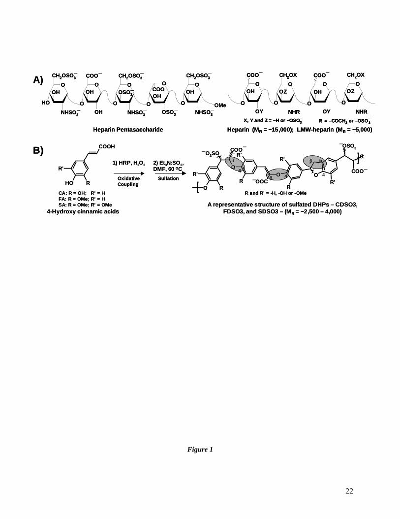
Structure of Sulfated Dehydropolymers (DHPs) of 4-Hydroxycinnamic Acids
Three synthetic sulfated DHPs – CDSO3, FDSO3 and SDSO3 (Fig. 1B) – were studied.
The molecules were prepared in two steps from caffeic acid, ferulic acid and sinapic acid, each
of which contains a common scaffold, the 4-hydroxycinnamic acid monomer (Fig. 1B) (15).
Briefly, the sulfated DHPs (MR ~2,500 – 4,000) are a mixture of oligomeric chains that contain 4
– 15 monomers suggesting that the molecules are comparable in size to enoxaparin (MR ~5,000)
(15). In addition, the DHPs contain several types of inter-monomeric linkages (Fig. 1B), thereby
generating polydispersity and heterogeneity, a property they share with LMWHs. Yet, sulfated
DHPs are significantly less sulfated than heparins. Whereas sulfated DHPs contain an average of
0.33 sulfate group per monomer, LMWHs possess an average of 1 – 1.3 sulfate groups for every
saccharide residue. More importantly, sulfated DHPs possess a large number of aromatic rings in
the backbone, while heparins have none. Thus, sulfated DHPs are significantly more
hydrophobic than the LMWHs.

10
Effect of Sulfated DHPs on Fibrin Formation in Normal Human Plasma
To determine whether our sulfated DHPs prolong fibrin formation in plasma, we utilized
in vitro transmittance assays. Addition of CaCl2 to normal pooled human plasma under APTT-
like conditions triggers ‘coagulation’ resulting in the synthesis of fibrin, which blocks the
passage of light through the sample. A characteristic decrease in transmittance at 600 nm as a
function of time is observed from which the time to clot (TC) and the time it takes to reduce the
transmittance, i.e., clotting, by 50% (T50) can be measured. The presence of all three sulfated
DHPs prolonged fibrin synthesis, as shown by the delayed decrease in transmittance at 600 nm
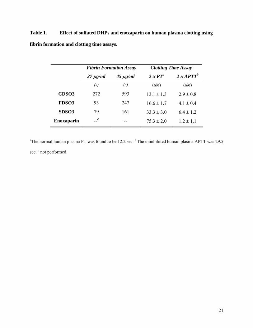
(Fig. 2A). For SDSO3, the T50 value changed from 79 s to 161 s as the concentration was
increased from 27 to 45 μg/ml (Fig. 2A, Table 1). Similarly, the T50 values were 272 and 593 s,
and 93 s and 247 s at 27 and 45 μg/ml for CDSO3 and FDSO3, respectively. These initial results
demonstrated that sulfated DHPs prolong fibrin formation in a dose-dependent manner. The
results also suggest that the anticoagulation potency varied with the structure of the sulfated DHP
(see Fig. 1).
To assess whether anticoagulant potency of sulfated DHPs is retained if coagulation is
initiated through the extrinsic pathway, thromboplastin-D was used as an initiator of clotting.
Once again, the presence of all three sulfated DHPs significantly slowed down the formation of
fibrin in normal plasma suggesting that three molecules inhibit clotting (not shown). Figure 2B
and 2C show the change in TC and T50 as a function of the concentration of each sulfated DHP
(and reference molecule, enoxaparin). Both TC and T50 increase as the concentration of sulfated
DHP increases. The increase is not linear and is accelerated at higher concentrations of the
anticoagulant. More interestingly, the three sulfated DHPs display an anticoagulation profile
similar to enoxaparin. While, CDSO3 is more potent than enoxaparin, FDSO3 and SDSO3 are
11
less potent. For example, the concentration of anticoagulant needed to double the time for 50%
fibrin formation was found to be ~3.5 μM for CDSO3 and 6, 11 and >25 μM for enoxaparin,
FDSO3 and SDSO3, respectively.
Effect of Sulfated DHPs on Clotting Times
PT and APTT are commonly used to assess the coagulation status of human plasma (18).
All three sulfated DHPs exhibited a significant concentration-dependent prolongation of PT and
APTT (not shown). A typical parameter for describing anticoagulant activity in these assays is
the concentration of the anticoagulant needed for doubling the normal plasma clotting time
(2×PT or 2×APTT). The 2×PT value for sulfated DHPs ranged from 13.1–33.3 μM, while that
for enoxaparin was 75.3 μM suggesting the new molecules are 2.3–5.7-fold more potent in the
PT assay (Table 1). The doubling of APTT required 2.9–6.4 μM concentration of the three
sulfated DHPs, while enoxaparin required 1.2 μM. This indicates that the sulfated DHPs are
~2.4–5.3-fold weaker anticoagulants in the APTT assay as compared to enoxaparin and the order
of activity is CDSO3 > FDSO3 > SDSO3 (Table 1).
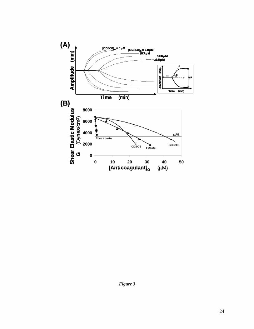
Thromboelastographic Measurement of Effect of Sulfated DHPs on Whole Blood
Clotting
Whole blood clotting is a dynamic process that involves many components including
cells, which may alter anticoagulant potency. To compare sulfated DHPs and enoxaparin in a
whole blood system, we employed thromboelastography (TEG®), a technique used in clinical
settings for following anticoagulation with LMWHs (19-21). TEG® measures various responses
of a formed clot to shearing force. In this technique, a pin is inserted into an oscillating cup
containing whole blood. As fibrin polymerizes, the pin starts to move with the oscillating cup
and the movement of the pin is recorded as amplitude, which in time reaches maximum

12
amplitude (MA) (Fig. 3A). The stronger the clot, the more the pin moves with the cup and the
higher the MA. Shear elastic modulus strength (G), a measure of clot stiffness, is calculated from
MA. Additionally reaction time R and angle α (Fig. 3A) are also obtained in a TEG® experiment.
R is the time required for the initial fibrin formation, while α is the acute angle in degrees
between an extension of the R tracing and the tangent of the maximum slope produced by the
TEG® tracing during clot stiffening. Angle α is a measure of the rate of formation of three-
dimensional fibrin network. Parameters that affect MA include fibrin concentration and
structure, concentration and functional state of platelets, deficiency of coagulation factors and
presence of clotting inhibitors (22).
All three sulfated DHPs affect R, α, MA and G parameters in a dose-dependent manner
(see Table S1 in Supplementary Material). Briefly, as the concentration of CDSO3 increases
from 0 to 24.3 μM, R increases from 7.0 to 21.5 min. This effect parallels the time to clot results
obtained in the plasma assay. Likewise, sulfated DHPs lower the value of angle α from 59° for
normal blood to 13.5–17° at the highest concentrations studied. This indicates that the kinetics of
fibrin polymerization and networking is significantly retarded by the presence of sulfated DHPs.
Enoxaparin exhibits similar characteristics, except that it is 23–51-fold more potent than sulfated
DHPs when comparisons are made at doubling the R value from its value in the absence of any
anticoagulants (not shown). Likewise, enoxaparin is 17–32-fold and 18–37-fold more potent
when comparisons are made for a 50% reduction in the angle α and shear elastic modulus G,
respectively (Fig. 3B).
13
Effect of Sulfated DHPs on Whole Blood Coagulation as Evaluated by Hemostasis
Analysis System
To further compare the whole blood anticoagulant potential of the sulfated DHPs with
enoxaparin, we performed an ex-vivo study using HAS™, which measures the forces generated
by platelets within a clot (23). In this technique, the clot is allowed to form between a
temperature-controlled lower surface (cup) and a parallel upper surface (cone). As the clot
grows, it attaches to both the surfaces pulling the fibrin strands inward. This pull is measured by
a displacement transducer, which produces an electrical signal on the cone proportional to the
amount of force generated by the platelets. HASTM also provides detailed information on clot
structure through the measurement of clot elastic modulus (CEM), which is the ratio of stress
induced by platelets to strain arising from the change in clot thickness (24). PCF is observed to
increase as soon as thrombin is formed suggesting that appearance of PCF can be used as
surrogate marker for TGT (thrombin generation time), the minimal time required for production
of thrombin following initiation of clotting (23).
In addition to its dependence on thrombin, PCF is sensitive to platelet number, platelet
metabolic status, presence of thrombin inhibitors and degree of GPIIb/IIIa exposure (20,25-27).
Likewise, CEM is a complex parameter that is sensitive to changes in clot structure, fibrinogen
concentration, the rate of thrombin generation and red blood cell flexibility, while TGT is
sensitive to clotting factor deficiencies, antithrombin concentration and presence of
anticoagulants. Low PCF and low CEM coupled with a prolonged TGT are associated with
increased bleeding risk, while elevated PCF and CEM paired with a decreased TGT are
associated with thrombotic disease states.

14
All three DHPs affect TGT, PCF and CEM parameters in a dose-dependent manner (see
Table S2 in Supplementary Material). For example, as the concentration of FDSO3 increases
from 0 to 23.8 μM, the TGT value increases from 235 seconds to 465 seconds (Fig. 4A). This
effect parallels the results obtained in the plasma thrombogenesis assay and TEG®. More
importantly, the presence of sulfated DHPs in blood decreases PCF from 7.6 Kdynes to 2.4–1.2
Kdynes at 14–37 μM (Fig. 4B), while enoxaparin induces a PCF of 0.9 Kdynes at 0.44 μM.
When comparisons are made for a 50% reduction in PCF, enoxaparin is 63–140-fold more
potent. Likewise, sulfated DHPs decrease CEM from 21.6 Kdynes/cm2 for normal blood to 4.5–
1.3 Kdynes/cm2 at the highest concentrations studied. Comparison of CEM values indicates that
enoxaparin is 43–90-fold more potent than sulfated DHPs (Fig. 4C). These results confirm that
sulfated DHPs behave in a manner similar to enoxaparin, except for the concentration at which
these are effective.
Conclusions and Significance
The major conclusion of this work is that sulfated DHPs display whole blood
anticoagulation properties similar to a clinically used anticoagulant, enoxaparin, although the
effective concentration range is different. In TEG® and HAS™ assays, sulfated DHPs are 17–51-
fold and 43–140-fold less active than enoxaparin, respectively. Considering that the structure and
mechanism of action of sulfated DHPs is radically different from all known anticoagulants
(15,16), this is a significant observation. It implies that a new class of more potent molecules,
most likely homogeneous and synthetically accessible, may be possible to design from the DHP
scaffold. We expect that this class of molecules will utilize allosteric modulation of thrombin and
factor Xa activity through binding in exosite II, the first molecules to display this unique
mechanism (16). In addition, our previous results using enzyme inhibition assays show that
15
unsulfated DHPs also possess significant anticoagulation potential (15). This suggests that it may
be possible to design un-sulfated, synthetic molecules with a unique mechanism of action. A
specific advantage expected of these un-sulfated homogeneous DHP-based structures is that
absence of sulfate group would make the molecules orally bioavailable.
The work presented here shows that in the PT assay, the three sulfated DHPs are effective
at concentrations in the range of enoxaparin, while in the APTT assay they are only 2–6-fold
weaker. Despite major mechanistic differences, both sulfated DHPs and enoxaparin prolong
APTT better than PT. Inhibition of fibrin formation in plasma shows that CDSO3 was
comparable to enoxaparin. Yet, sulfated DHPs are much weaker in whole blood than enoxaparin.
It is possible that the significant hydrophobic character of sulfated DHPs induces binding to cells
resulting in significant sequestering of active agent. It is likely that this non-specific binding will
be reduced with homogeneous, synthetic small molecules.
Overall, the results demonstrate that sulfated DHPs possess good plasma and whole
blood anticoagulation activity. This does not imply that our novel molecules will be clinically
effective. Toxicity studies will have to be performed to ascertain that these novel structures do
not induce abnormal effects. An important point to note in this regard is that in vivo enoxaparin
does not prolong PT and APPT at concentrations sufficient to anticoagulate suggesting that in
vitro or ex vivo potency does not translate directly into in vivo effectiveness. Yet, the results
described here suggest that the novel structure and mechanism of sulfated DHPs may lead to a
new class of potent anticoagulants.
16
References
1. Weitz JI, Hirsh J. New anticoagulant drugs. Chest 2001; 119: 95S-107S.
2. Olson ST, Swanson R, Raub-Segall E, et al. Accelerating ability of synthetic
oligosaccharides on antithrombin inhibition of proteinases of the clotting and fibrinolytic
systems. Comparison with heparin and low-molecular-weight heparin. Thromb. Haemost.
2004; 92: 929-939.
3. Menajovsky LB. Heparin-induced thrombocytopenia: clinical manifestations and
management strategies. Am. J. Med. 2005; 118 Suppl 8A: 21S-30S.
4. van Dongen CJ, van den Belt AG, Prins MH, et al. Fixed dose subcutaneous low molecular
weight heparins versus adjusted dose unfractionated heparin for venous thromboembolism.
Cochrane Database Syst Rev 2004; (4), CD001100.
5. Buller HR, Cohen AT, Davidson B, Decousus H, Gallus AS, Gent M, Pillion G, Piovella F,
Prins MH, Raskob GE. Idraparinux versus standard therapy for venous thromboembolic
disease. N. Engl. J. Med. 2007; 357: 1094-1104.
6. Buller HR, Cohen AT, Davidson B, Decousus H, Gallus AS, Gent M, Pillion G, Piovella F,
Prins MH, Raskob GE. Extended prophylaxis of venous thromboembolism with
idraparinux. N. Engl. J. Med. 2007; 357: 1105-1112.
7. Bauersachs RM. Fondaparinux: an update on new study results. Eur. J. Clin. Invest. 2005;
35 Suppl 1: 27-32.
8. Turpie AG. The safety of fondaparinux for the prevention and treatment of venous
thromboembolism. Expert Opin. Drug Saf. 2005; 4: 707-721.
9. Desai UR. New antithrombin-based anticoagulants. Med. Res. Rev. 2004; 24: 151-81.
17
10. Capila I, Linhardt RJ. Heparin-protein interactions. Angew. Chem. Int. Ed. 2002; 41: 391-
412.
11. Kikelj D. Peptidomimetic thrombin inhibitors. Pathophysiol. Haemost. Thromb. 2003; 33:
487-491.
12. Gerotziafas GT, Samama MM. Heterogeneity of synthetic factor Xa inhibitors. Curr.
Pharm. Des. 2005; 11: 3855-3876.
13. Perzborn E, Kubitza D, Misselwitz F. Rivaroxaban. A novel, oral, direct factor Xa inhibitor
in clinical development for the prevention and treatment of thromboembolic disorders.
Hamostaseologie 2007; 27: 282-289.
14. Nutescu EA, Shapiro NL, Chevalier A. New anticoagulant agents: direct thrombin
inhibitors. Clin. Geriatr. Med. 2006; 22: 33-56.
15. Monien BH, Henry BL, Raghuraman A, et al. Novel chemo-enzymatic oligomers of
cinnamic acids as direct and indirect inhibitors of coagulation proteinases. Bioorg. Med.
Chem. 2006; 14: 7988-7998.
16. Henry BL, Monien BH, Bock PE, et al. A novel allosteric pathway of thrombin inhibition.
Exosite II mediated potent inhibition of thrombin by chemo-enzymatic, sulfated
dehydropolymers of 4-hydroxycinnamic acids. J. Biol. Chem. 2007; 282: 31891-31899.
17. Prasa D, Svendsen L, Sturzebecher J. The ability of thrombin inhibitors to reduce the
thrombin activity generated in plasma on extrinsic and intrinsic activation. Thromb.
Haemost. 1997; 77: 498-503.
18. Bajaj SP, Joist JH. New insights into how blood clots: implications for the use of APTT
and PT as coagulation screening tests and in monitoring of anticoagulant therapy. Semin.
Thromb. Hemost. 1999; 25: 407-418.
18
19. Salooja N, Perry DJ. Thromboelastography. Blood Coagul. Fibrinol. 2001; 12: 327-337.
20. Carr ME Jr, Martin EJ, Kuhn JG, et al. Monitoring of hemostatic status in four patients
being treated with recombinant factor VIIa. Clin. Lab. 2004; 50: 529-538.
21. Klein SM, Slaughter TF, Vail PT, et al. Thromboelastography as a perioperative measure
of anticoagulation resulting from low molecular weight heparin: a comparison with anti-Xa
concentrations. Anesth. Analg. 2000; 91: 1091-1095.
22. Chandler WL. The thromboelastography and the thromboelastograph technique. Semin.
Thromb. Hemostasis 1995; 21 Suppl 4: 1-6.
23. Carr ME, Martin EJ, Kuhn JG, et al. Onset of force development as a marker of thrombin
generation in whole blood: the thrombin generation time (TGT). J. Thromb. Haemost.
2003; 1: 1977-1983.
24. Carr ME. Development of platelet contractile force as a research and clinical measure of
platelet function. Cell Biochem. Biophys. 2003; 38: 55-78.
25. Carr ME, Carr SL, Greilich PE. Heparin ablates force development during platelet
mediated clot retraction. Thromb. Haemost. 1996; 75: 674-678.
26. Carr ME, Carr SL, Tildon T, et al. Batroxobin-induced clots exhibit delayed and reduced
platelet contractile force in some patients with clotting factor deficiencies. J. Thromb.
Haemost. 2003; 1: 243-249.
27. Carr ME, Carr SL, Hantgan RR, et al. Glycoprotein IIb/IIIa blockade inhibits platelet-
mediated force development and reduces gel elastic modulus Thromb. Haemost. 1995; 73:
499-505.
19
Figure Legends
Figure 1. Structures of heparins (A) and sulfated DHPs (B). A) Fondaparinux is based
on heparin pentasaccharide, while heparin and LMWHs are polydisperse,
heterogeneous mixture of polysaccharide chains (MR ~15,000 and ~5,000 Da,
respectively) arising due to variations in X, Y, Z and R groups. B) Sulfated DHPs
possess a radically different structure from the heparins (and other anticoagulants)
and are synthesized in two steps from the corresponding 4-hydroxycinnamic acid
monomers, caffeic acid (CA), ferulic acid (FA) or sinapic acid (SA). The MR of
the sulfated DHPs is in the range of 2,500–4,000. Linkages, β-O-4 and β-5, are
commonly present in sulfated DHPs (shown as shaded ovals).
Figure 2 Inhibition of fibrin formation in pooled normal human plasma in the
presence of sulfated DHPs. Fibrin formation as a function of time was monitored
using the decrease in transmittance of light at 600 nm following initiation of
‘clotting’ under APTT conditions (A) and PT conditions (B). A) CDSO3 (bold
black lines), FDSO3 (bold grey lines) and SDSO3 (thin black lines).
Concentrations of sulfated DHPs used were either 27 μg/ml (denoted as ‘L’) or 45
μg/ml (‘H’). B) Time to clot (TC) and C) time needed for 50% formation of fibrin
(T50) values are plotted as a function of the concentration of sulfated DHP and
enoxaparin. Solid lines in both B) and C) are trendlines, not non-linear
regressions. Δ = CDSO3; ♦ = enoxaparin; ● = FDSO3; = SDSO3. See text for
details.
Figure 3 Comparison of the effect of sulfated DHPs and enoxaparin on clot formation
in whole blood using TEG®. Inset in (A) shows a typical thromboelastogram
20
expected of any anticoagulant. MA, R, α and G are parameters obtained from
TEG® analysis. See Methods for details. (B) shows the variation in G as a
function of concentration of the sulfated DHPs and enoxaparin. Solid lines are
trendlines (not regression fits) from which concentration of anticoagulant needed
to reduce shear elastic modulus G by 50% (shown as shaded line) of the starting
value was derived.
Figure 4 Comparison of the effect of sulfated DHPs and enoxaparin on platelet
function in whole blood using HAS™. A) shows selected HAS™ profiles
obtained with FDSO3, B) and C) show the variation in PCF and CEM,
respectively, as a function of concentration of the sulfated DHPs and enoxaparin.
Solid lines are trendlines from which the concentration of anticoagulant needed to
reduce PCF or CEM by 50% (shaded line) of the starting value was derived.
21
Table 1. Effect of sulfated DHPs and enoxaparin on human plasma clotting using
fibrin formation and clotting time assays.
Fibrin Formation Assay Clotting Time Assay
27 μg/ml 45 μg/ml 2 × PTa 2 × APTTb
(s) (s) (μM) (μM)
CDSO3 272 593 13.1 ± 1.3 2.9 ± 0.8
FDSO3 93 247 16.6 ± 1.7 4.1 ± 0.4
SDSO3 79 161 33.3 ± 3.0 6.4 ± 1.2
Enoxaparin --c -- 75.3 ± 2.0 1.2 ± 1.1
aThe normal human plasma PT was found to be 12.2 sec. b The uninhibited human plasma APTT was 29.5
sec. c not performed.
22
Figure 1
O
O
CH2OSO3
NHSO3
OH
OMe
O
OSO3
OHO
COOO
O
CH2OSO3
NHSO3
OSO3
O
OH
OH
O
COOO
OH
CH2OSO3
NHSO3
OH
_ _ _ _
_
_ _ _ _
_
Heparin Pentasaccharide
O
O
CH2OX
NHR
OZO
OY
OH
O
COOO
O
CH2OX
NHR
OZO
OY
OH
O
COO_ _
X, Y and Z = –H or –OSO3
_R = –COCH3 or –OSO3
_
Heparin (MR = ~15,000); LMW-heparin (MR = ~5,000)
A)
B)1) HRP, H2O2
OxidativeCoupling
CA: R = OH; R’ = HFA: R = OMe; R’ = HSA: R = OMe; R’ = OMe
OH R
R'
COOH_
O R
COO
O
R
R'O3SO
OOCO
R
R'
R'O
OSO3
COO
R'
A representative structure of sulfated DHPs – CDSO3, FDSO3, and SDSO3 – (MR = ~2,500 – 4,000)
__
_
R and R’ = -H, -OH or -OMe
2) Et3N:SO3, DMF, 60 OC
Sulfation
4-Hydroxy cinnamic acids
_β
β
β
44
5
4
O
O
CH2OSO3
NHSO3
OH
OMe
O
OSO3
OHO
COOO
O
CH2OSO3
NHSO3
OSO3
O
OH
OH
O
COOO
OH
CH2OSO3
NHSO3
OH
_ _ _ _
_
_ _ _ _
_
Heparin Pentasaccharide
O
O
CH2OSO3
NHSO3
OH
OMe
O
OSO3
OHO
COOO
O
CH2OSO3
NHSO3
OSO3
O
OH
OH
O
COOO
OH
CH2OSO3
NHSO3
OH
_ _ _ _
_
_ _ _ _
_
Heparin Pentasaccharide
O
O
CH2OX
NHR
OZO
OY
OH
O
COOO
O
CH2OX
NHR
OZO
OY
OH
O
COO_ _
X, Y and Z = –H or –OSO3
_R = –COCH3 or –OSO3
_
Heparin (MR = ~15,000); LMW-heparin (MR = ~5,000)
O
O
CH2OX
NHR
OZO
OY
OH
O
COOO
O
CH2OX
NHR
OZO
OY
OH
O
COO_ _
X, Y and Z = –H or –OSO3
_X, Y and Z = –H or –OSO3
_R = –COCH3 or –OSO3
_R = –COCH3 or –OSO3
_
Heparin (MR = ~15,000); LMW-heparin (MR = ~5,000)
A)
B)1) HRP, H2O2
OxidativeCoupling
CA: R = OH; R’ = HFA: R = OMe; R’ = HSA: R = OMe; R’ = OMe
OH R
R'
COOH_
O R
COO
O
R
R'O3SO
OOCO
R
R'
R'O
OSO3
COO
R'
A representative structure of sulfated DHPs – CDSO3, FDSO3, and SDSO3 – (MR = ~2,500 – 4,000)
__
_
R and R’ = -H, -OH or -OMe
2) Et3N:SO3, DMF, 60 OC
Sulfation
4-Hydroxy cinnamic acids
_β
β
β
44
5
4

23
Figure 2
0
50
100
150
200
250
0 5 10 15 20 250
50
100
150
200
250
0 5 10 15 20 25
0.4
0.5
0.6
0.7
0.8
0.9
1
0 200 400 600 800Time (s)
Tran
smitt
ance
(%
×10
-2) CDSO3
FDSO3SDSO3
L HLH
LH
L = 27 μg/mlH = 45 μg/ml
[Anticoagulant]O (μM)
TC
(s)
T 50
(s)
A)
B) C)CDSO3 CDSO3
SDSO3 SDSO3
FDSO3
FDSO3
EnoxaparinEnoxaparin
0
50
100
150
200
250
0 5 10 15 20 250
50
100
150
200
250
0 5 10 15 20 25
0.4
0.5
0.6
0.7
0.8
0.9
1
0 200 400 600 800Time (s)
Tran
smitt
ance
(%
×10
-2) CDSO3
FDSO3SDSO3
L HLH
LH
L = 27 μg/mlH = 45 μg/ml
0.4
0.5
0.6
0.7
0.8
0.9
1
0 200 400 600 800Time (s)
Tran
smitt
ance
(%
×10
-2) CDSO3
FDSO3SDSO3
L HLH
LH
L = 27 μg/mlH = 45 μg/ml
[Anticoagulant]O (μM)
TC
(s)
T 50
(s)
A)
B) C)CDSO3 CDSO3
SDSO3 SDSO3
FDSO3
FDSO3
EnoxaparinEnoxaparin
24
Figure 3
(A)
(B)
0
2000
4000
6000
8000
0 10 20 30 40 50[Anticoagulant]O (μM)
Shea
r Ela
stic
Mod
ulus
G
(
Dyn
es/c
m2 )
50%
CDSO3 FDSO3SDSO3
Enoxaparin
Time (min)
Am
plitu
de
(mm
)
R αMA
Time (min)
Am
plitu
de
(mm
)
R αMA
[CDSO3]O = 0 μM [CDSO3]O = 7.8 μM15.7 μM
19.6 μM23.5 μM
Time (min)
Am
plitu
de
(mm
) [CDSO3]O = 0 μM [CDSO3]O = 7.8 μM15.7 μM
19.6 μM23.5 μM
Time (min)
Am
plitu
de
(mm
)
(A)
(B)
0
2000
4000
6000
8000
0 10 20 30 40 50[Anticoagulant]O (μM)
Shea
r Ela
stic
Mod
ulus
G
(
Dyn
es/c
m2 )
50%
CDSO3 FDSO3SDSO3
Enoxaparin
0
2000
4000
6000
8000
0 10 20 30 40 50[Anticoagulant]O (μM)
Shea
r Ela
stic
Mod
ulus
G
(
Dyn
es/c
m2 )
50%
CDSO3 FDSO3SDSO3
Enoxaparin
Time (min)
Am
plitu
de
(mm
)
R αMA
Time (min)
Am
plitu
de
(mm
)
R αMA
[CDSO3]O = 0 μM [CDSO3]O = 7.8 μM15.7 μM
19.6 μM23.5 μM
Time (min)
Am
plitu
de
(mm
) [CDSO3]O = 0 μM [CDSO3]O = 7.8 μM15.7 μM
19.6 μM23.5 μM
Time (min)
Am
plitu
de
(mm
)
Time (min)
Am
plitu
de
(mm
)
R αMA
Time (min)
Am
plitu
de
(mm
)
R αMA
[CDSO3]O = 0 μM [CDSO3]O = 7.8 μM15.7 μM
19.6 μM23.5 μM
Time (min)
Am
plitu
de
(mm
) [CDSO3]O = 0 μM [CDSO3]O = 7.8 μM15.7 μM
19.6 μM23.5 μM
Time (min)
Am
plitu
de
(mm
)

25
Figure 4
PCF
(kdy
nes)
[Anticoagulant]O (μM)
0
2
4
6
8
1 3 5 7 9 11 13 15 17 19
0 μM FDSO3
9.5 μM
12.7 μM
15.9 μM
23.8 μM
Time (min)
A)
0 23.8
TGT
9.5 15.912.7 μM FDSO3
0
6
12
18
24
0 10 20 30 400
2
4
6
8
10
0 10 20 30 40
Enoxaparin
CDSO3 FDSO3
SDSO3
B)
EnoxaparinCDSO3 FDSO3
SDSO3
PCF
(kdy
nes)
CEM
(kdy
nes/
cm2 )
50%50%
C)
PCF
(kdy
nes)
[Anticoagulant]O (μM)
0
2
4
6
8
1 3 5 7 9 11 13 15 17 19
0 μM FDSO3
9.5 μM
12.7 μM
15.9 μM
23.8 μM
Time (min)
A)
0 23.8
TGT
9.5 15.912.7 μM FDSO3
0
2
4
6
8
1 3 5 7 9 11 13 15 17 19
0 μM FDSO3
9.5 μM
12.7 μM
15.9 μM
23.8 μM
Time (min)
A)
0 23.8
TGT
9.5 15.912.7 μM FDSO3
0
6
12
18
24
0 10 20 30 400
2
4
6
8
10
0 10 20 30 40
Enoxaparin
CDSO3 FDSO3
SDSO3
B)
EnoxaparinCDSO3 FDSO3
SDSO3
PCF
(kdy
nes)
CEM
(kdy
nes/
cm2 )
50%50%
C)
0
6
12
18
24
0 10 20 30 400
2
4
6
8
10
0 10 20 30 40
Enoxaparin
CDSO3 FDSO3
SDSO3
B)
EnoxaparinCDSO3 FDSO3
SDSO3
PCF
(kdy
nes)
CEM
(kdy
nes/
cm2 )
50%50%
C)