structural characterization and immunostimulatory activity
TRANSCRIPT

This is an Open Access document downloaded from ORCA, Cardiff University's institutional
repository: http://orca.cf.ac.uk/100798/
This is the author’s version of a work that was submitted to / accepted for publication.
Citation for final published version:
Wang, Junqiao, Nie, Shaoping, Cui, Steve W., Wang, Zhijun, Phillips, Aled O., Phillips, Glyn O.,
Li, Yajing and Xie, Mingyong 2017. Structural characterization and immunostimulatory activity of a
glucan from natural Cordyceps sinensis. Food Hydrocolloids 67 , pp. 139-147.
10.1016/j.foodhyd.2017.01.010 file
Publishers page: http://dx.doi.org/10.1016/j.foodhyd.2017.01.010
<http://dx.doi.org/10.1016/j.foodhyd.2017.01.010>
Please note:
Changes made as a result of publishing processes such as copy-editing, formatting and page
numbers may not be reflected in this version. For the definitive version of this publication, please
refer to the published source. You are advised to consult the publisher’s version if you wish to cite
this paper.
This version is being made available in accordance with publisher policies. See
http://orca.cf.ac.uk/policies.html for usage policies. Copyright and moral rights for publications
made available in ORCA are retained by the copyright holders.

1
Structural characterization and immunostimulatory activity of a glucan 1
from natural Cordyceps sinensis 2
3
Junqiao Wang a
, Shaoping Nie a*
, Steve W. Cui a,b
, Zhijun Wang a
, Aled O. Phillips c
, 4
Glyn O. Phillips d
, Y ajing Li e
, Mingyong X ie a*
5
a
State Key Laboratory of Food Science and Technology, Nanchang University, 235 Nanjing East 6
Road, Nanchang, J iangxi Province, 330047, China 7
b
Guelph Food Research Centre, Agriculture and Agri-Food Canada, 93 Stone Road West, Guelph, 8
Ontario, N1G 5C9, Canada 9
c
School of Medicine, University of Cardiff, Wales, UK 10
d
Phillips Hydrocolloids Research Centre, Glyndwr University, Wrexham, LL11 2AW Wales, UK 11
e
Qinghai Ta Er Sheng Gu Agricultural Science and Technology Co. Ltd., Xining 810003, China 12
13
14
15
16
* Corresponding authors: 17
[email protected] (Professor Shaoping Nie, PhD), Tel. & Fax: +86-791-88304452. 18
[email protected] (Professor Mingyong X ie, PhD), Tel. & Fax: +86-791-83969009. 19
20
21
22

2
Abstract 23
A water-soluble polysaccharide, named NCSP-50, was obtained from natural Cordyceps sinensis 24
by hot water extraction and ethanol fractionation precipitation. It was eluted as a single 25
symmetrical peak and had an average molecular weight of 9.76×10⁵
Da. The structure was 26
determined by monosaccharide composition, methylation analysis, 1D/2D NMR spectroscopy, and 27
enzymatic hydrolysis and characterization of the oligosaccharides by MALDI-TOF mass 28
spectrometry. The repeating unit of this polysaccharide was proposed as follows: 29
30
This glucan showed potent immunostimulatory activity on the basis of its significant abilities to 31
promote macrophage proliferation, enhance NO production, as well as and cytokines (IL-1β and 32
TNF-α ) secretion. 33
34
K eywords: natural Cordyceps sinensis; α -glucan; caterpillar fungus; immunostimulatory 35
36
37

3
1. Introduction 38
Cordyceps sinensis (Berk.) Sacc. is a parasitic fungus growing on the larva of the caterpillar, 39
which was also called “Dong-Chong-X ia-Cao” in Chinese. As a famous traditional Chinese 40
medicine, C. sinensis has a long history being used as food/medicine, especially in “lung 41
protectorate” and “kidney improvement”, as well as “Y in/Yang double invigorant” (Zhu, Halpern, 42
& Jones, 1998a, 1998b). In China, it was mainly distributed at Qinghai, Tibet, Sichuan, Yunnan 43
and Gansu plateau, at the elevation of 3500-5000 metres in the prairie soil. The growth of natural 44
C. sinensis needs a restricted habitat, so the yield is limited each year. But the production is 45
decreasing gradually during the recent years because of serious damage to ecological environment 46
and reckless harvesting. The demand of the market, on the contrary, experiences a constant 47
increase owing to a raising awareness of its multi-biological properties to the public. The 48
pharmacological effect of C. sinensis might be attributed to its chemical constituents and bioactive 49
ingredients, including polysaccharides, amino acids, minerals, nucleosides, cordycepic acid, 50
cordycepin, etc. (Wang, et al., 2015). Among them, polysaccharides, which account for 3-8% of 51
the total dry weight (Zhao, X ie, Wang, & L i, 2014), have been demonstrated to exhibit a wide 52
range of bioactivities, such as antioxidant (L i, L i, Dong, & Tsim, 2001), anti-tumor (Chen, Shiao, 53
Lee, & Wang, 1997), liver and kidney protection (L iu, Zuo, Tao, & L iu, 2013; Wang, et al., 2014; 54
Wang, et al., 2010), anti-fibrosis (Yao, et al., 2014) and immunomodulatory effect (Nie, Cui, X ie, 55
Phillips, & Phillips, 2013; Sheng, Chen, Li, & Zhang, 2011; Wu, et al., 2014). In our previous 56
study, a hydrophilic polysaccharide fraction (CBHP) mainly made up of glucose (95.15%) from 57
cultured C. sinensis was demonstrated to exhibit potent antifibrotic effect against renal fibrosis 58
(Nie et al., 2011; Zhang, L iu, Al-Assaf, Phillips, & Phillips, 2012). 59

4
Besides, in vivo and in vitro immuomodulating properties of polysaccharide from cultured C. 60
sinensis have been also well documented over the past decades. UM01 PS, a polysaccharide from 61
mycelia of C. sinensis fungus UM01, could significantly promote cell proliferation, phagocytic 62
ability, NO release, as well as multiply cytokines and chemokine production in macrophages 63
(Meng, et al., 2014). Cordysinocan, an exopolysaccharide from cultured Cordyceps UST 2000, 64
showed a stimulating effect on the human T-lymphocytes was demonstrated as well (Cheung, et 65
al., 2009). Zhang et al. reported that the polysaccharide could enhance the immunity of 60
Co 66
radiation-induced immunosuppression mice through reducing oxidative injury and modulating 67
cytokine production (Zhang, et al., 2011). It was evidenced that these polysaccharides with 68
effective immunodulating activity was mainly made up of galactose, glucose and mannose. 69
However, there are few reports demonstrating such effect of polysaccharides from natural 70
occurring C. sinensis at present. Additionally, significant differences in terms of chemical 71
composition and molecular weight of water-extracted polysaccharides between natural C. sinensis 72
and the cultured mycelium have been observed in our recent study (Wang et al., accepted). 73
Therefore, in this study, we aimed to characterize the detailed chemical structure of a glucan from 74
natural C. sinensis using methylation analysis, enzymatic hydrolysis, MALDI-TOF and 1D/2D 75
NMR spectroscopy, and further evaluate the immunostimulatory effect with regard to cell 76
proliferation assay, production of NO and cytokines in RAW 264.7 cells. This work will provide 77
useful information on the advanced structural characteristics of the polysaccharides from C. 78
sinensis, and will be helpful for further studying the structure and activity relationship. 79
2. Materials and methods 80
2.1 Materials 81
5
Natural C. sinensis was sampled from Qinghai province, China. T-series dextrans (T-10, T-40, 82
T-70, T-500 and T-2000) were purchased from Pharmacia Biotech (Uppsala, Sweden) and 83
monosaccharide standards (fucose, rhamnose, arabinose, galactose, glucose, mannose, xylose, 84
fructose, ribose, galacturonic acid and glucuronic acid), lipopolysaccharide (LPS) and super DHB 85
were from Sigma-Aldrich (St. Louis, MO, USA). Deuterium oxide (D2O) and sodium 86
borodeuteride (NaBD4, 98 atom% D) were from Acros Organics (New Jersey, USA). α -amylase 87
was purchased from Megazyme (Wicklow, Ireland) and HPLC grade methanol was from Merk 88
(Darmstadt, Germany). All other reagents were of analytical grade unless specified. 89
2.2 Isolation and purification 90
The natural C. sinensis was grounded and defatted with 80% ethanol overnight. Subsequently, the 91
dried ethanol-insoluble residues were extracted three times with distilled water (1:20, w/v) at 95˚C, 92
2 h each time. After centrifugation, all the supernatant was concentrated and precipitated with 93
ethanol until reaching a final concentration of 80%. The resulting precipitate was collected by 94
centrifugation and lyophilization, giving the crude polysaccharide. It was then removed protein by 95
Sevag reagent (chloroform/1-butanol, v/v = 4:1), resulting a white polysaccharide named as 96
NCSP. 97
NCSP was then purified by a stepwise fractionated precipitation with ethanol. Specifically, 98
anhydrous ethanol was added slowly to the polysaccharide solution (5 mg/mL) until the final 99
concentration of ethanol reached 30%. The solution was then kept stationary overnight, followed 100
by centrifugation at 4800 rpm for 20 min. The precipitate was collect and repeatedly washed with 101
anhydrous ethanol three times. The supernatant, on the other hand, was subjected to the next step 102
of precipitation with a higher ethanol concentration. In this way, the precipitated fractions were 103

6
obtained successively at final ethanol concentration of 30%, 50% and 70%, designated as 104
NCSP-30, NCSP-50 and NCSP-70, respectively. The final supernatant fraction, namely 105
NCSP-S70, was also collected. 106
2.3 Assay for structural analysis 107
2.3.1 Homogeneity and molecular weight determination 108
The homogeneity and molecular weight distribution of polysaccharide fractions were determined 109
by HPGPC on an Agilent 1260 LC instrument equipped with a refractive index detector (RID), a 110
variable wavelength detector (V WD), coupled with an UltrahydrogelTM
1000 column (7.8 mm × 111
300 mm, Waters, USA) and an UltrahydrogelTM
L inear column (7.8 mm × 300 mm, Waters, USA). 112
Polysaccharide solution was filtered through 0.45 μm filter prior to injection, with 0.1 M 113
NaCl/0.02% NaN3 aqueous solution as mobile phase at a flow rate of 0.6 mL/min. The molecular 114
weight of polysaccharides was estimated using a standard curve prepared by T-series dextrans. 115
According to the information obtained from HPGPC that would be discussed later in this study, 116
we selected NCSP-50 for the following analysis. 117
2.3.2 Monosaccharide composition analysis 118
The identification and quantification of monosaccharide composition of NCSP-50 was achieved 119
by high performance anion exchange chromatography coupled with pulsed amperometric 120
detection (HPAEC-PAD) (Dionex ICS-5000 System, Dionex Corporation, CA). NCSP-50 (5 mg) 121
were dissolved in 0.5 mL 12M H2SO4 at an ice bath for 30 min, and then diluted to 3 mL (2 M 122
H2SO4) to further hydrolysis 2 h at 100˚C. Separation was performed on a CarboPac PA20 column 123
(3 mm×150 mm, Dionex, CA) and a CarboPac PA20 Guard (3 mm × 30 mm, Dionex, CA) with a 124
gradient elution procedure at a flow rate of 0.5 mL/min at 30˚C. The eluents consisted of 250 mM 125

7
NaOH solution (A), distilled water (B) and 1M sodium acetate (C). Initially, 0.8% A was eluted 126
for 20 min, and then a gradient increase from 5% C to 20% C while maintaining 0.8% A. Finally, 127
80% A was eluted to regenerate the column for 20 min. Chromeleon software was used to process 128
the data. 129
2.3.3 Methylation analysis 130
Methylation analysis was carried out according to the method of Ciucanu and K erek (1984) with 131
slight modification. Briefly, dried NCSP-50 was completely dissolved in anhydrous DMSO and 132
then added dried NaOH powder to the solution with further stirring for 3 h. Iodomethane was 133
added to react with the solution in order to get the methlylated polysaccharide. A complete 134
methylation was confirmed by the disappearance of O-H absorption (3200-3700 cm-1
) in IR 135
spectrum. The methylated polysaccharide was hydrolyzed, reduced and acetylated to produce 136
partial methylated alditol acetates (PMAAs). Finally, the PMAAs were analyzed by GC-MS 137
(Agilent Technology 7890A/5975C, USA), equipped with a SP-2330 capillary column (30 m × 138
0.25 mm, 0.2 mm film thickness, Supelco, Bellefonte, Pa). The GC temperature program was 139
isothermal at 160˚C, followed by 2 ˚C/min gradient up to 210˚C and 5 ˚C /min up to 240 ˚C. The 140
individual peaks of the PMAAs were identified by their characteristic GC retention times 141
(Biermann & McGinnis, 1988) and fragmentation patterns, as well as by comparison with mass 142
spectrum patterns from literature (Sassaki, Gorin, Souza, Czelusniak, & Iacomini, 2005). 143
2.3.4 NMR spectroscopy 144
NCSP-50 (30 mg) was dissolved in D2O and then freeze dried. This procedure was repeated two 145
times to completely exchange H2O with D2O, and polysaccharide was finally dissolved in 1 mL 146
D2O at room temperature for 3h before NMR analysis. Both 1
H and 13
C spectrum were recorded 147
8
on a Bruker Avance 600 MHz NMR spectrometer (Brucker, Rheinstetten, Germany) at 294 K . 148
NCSP-50 was further subjected to 2D NMR spectroscopy, including homonuclear 1
H/1
H 149
correlation (COSY, TOCSY ), heteronuclear single-quantum coherence (HSQC) and heteronuclear 150
multiple-bond correlation (HMBC) experiments through the standard Bruker pulse sequence. 151
2.3.5 Enzymatic hydrolysis and matrix-assisted laser desorption/ionization time-of-flight 152
(MALDI-TOF) analysis 153
NCSP-50 (5mg) was dissolved in 5 mL distilled water and digested for 36 h at 37°C with 100 µ L 154
of α -Amylase (EC3.1.1.1 from Bacillus amyloliquefaciens). The enzymatic reaction was 155
terminated by heating the solution at 100°C for 15 min. This solution was injected into HPLC to 156
obtain the profile of molecular weight distribution after enzyme digestion. On the other hand, the 157
solution was precipitated with four volumes of anhydrous ethanol and then centrifuged. The 158
resulting precipitation was collected and lyophilized to harvest a mixture of oligosaccharide 159
named NCSP-50-E. NCSP-50-E was dissolved in water and further analyzed by MALDI-TOF. 160
For MALDI-TOF analysis, mass spectrum was recorded on an AB SCIEX TOF/TOFTM
5800 161
System (Framingham, MA 01701, USA) equipped with nitrogen laser operating at 337 nm. Super 162
DHB was used as the matrix at a concentration of 10 mg/mL dissolved in 0.1% Trifluoroacetic 163
acid (TFA) 50% methanol-water solution. NCSP-50-E (10µ L) was mixed with 10 µ L of the matrix 164
solution and a total of 1 µ L of this mixture was applied to a stainless steel plate and allowed to dry 165
under vacuum at room temperature. Spectra were acquired both in the linear and reflector mode. 166
2.4 Immunostimulatory activity in vitro 167
2.4.1 Cell culture 168
Murine macrophage cell line RAW 264.7 (Shanghai Institute of Cell Biology, Shanghai, China) 169

9
was cultured in RPMI 1640 medium containing 10% fetal bovine serum (FBS) and 100 U/mL 170
penicillin and 100 μg/mL streptomycin under a humidified incubator (37˚C, 5% CO2). 171
2.4.2 Macrophage proliferation assay 172
The effect of NCSP-50 on the viability of RAW 264.7 cells was determined by a WST-8 Cell 173
Counting K it-8 (Beyotime Biotechnology, J iangsu, China). The cells (100 μL) were seeded into a 174
96-well plate at a density of 1.0 × 10⁵
cells/mL and incubated for 4 h at 37˚C in a humidified 175
incubator with 5% CO2. Subsequently, 100 μL RPMI 1640 medium in the presence of 176
polysaccharide solutions was added to each well reaching a final concentration of 0, 25, 50, 100 177
and 200 μg/mL and incubated for 24 h. LPS (1 μg/mL) was used as the positive control, RPMI 178
1640 medium in the absence of polysaccharide and LPS was used as the normal control, and 179
RPMI 1640 medium without cells was used as blank. At the end of incubation, CCK -8 solution 180
(10 μL) was added to each well and the plate was further incubated for 2 h. Absorbance was 181
recorded at 450 nm on the microplate reader (Varioskan Flash, Thermo Fisher Scientific, USA). 182
2.4.3 Nitric oxide (NO) production 183
The RAW 264.7 cells were suspended in the RPMI 1640 medium and adjusted to a density of 5.0 184
× 105
cell/mL, followed by pipetting into 24-well plate in a volume of 1 mL. After pre-incubation 185
for 4 h, different concentrations of NCSP-50 or starch (0, 25, 50, 100 and 200 μg/mL), as well as 186
LPS (1 μg/mL) were treated for another 24 h. Afterwards, the conditioned medium was collected 187
and analyzed using a commercial-available NO assay kit (Beyotime Biotechnology, J iangsu, 188
China) according to the manufacturer’s protocol. 189
2.4.4 Cytokine secretion 190
For cytokine determination, RAW 264.7 cells (5.0 × 10⁵
cells/well) were cultured in the presence 191

10
of different concentrations of polysaccharides (0, 25, 50, 100 and 200 μg/mL) and LPS (1 μg/mL) 192
for 24 h, and the culture supernatant was collected to determine the concentrations of various 193
cytokines (IL -1β and TNF-α ) by ELISA kits (Boster Bio-engineering L imited Company, Wuhan, 194
China) according to the manufacturer’s instruction. 195
2.5 Statistical analysis 196
All data was expressed as the mean ± standard deviation (SD). Comparison of the data was 197
conducted using one-way analysis of variance (ANOVA) followed by the Student-Newman-K euls 198
test. A value of P<0.05 was considered to be statistically significant. A ll statistical analysis was 199
performed through statistical software (SPSS, Version 17.0). 200
3. R esults and discussion 201
3.1 Isolation, purification and composition of NCSP-50 202
A crude polysaccharide (NCSP) from natural C. sinensis was obtained by hot water extraction and 203
ethanol precipitation, followed by removing protein, with a yield of 2.60% (w/w). A fter stepwise 204
ethanol precipitation, the subsequent yields of NCSP-30, NCSP-50, NCSP-70 and NCSP-S70 205
were 11.82%, 45.39%, 13.69% and 17.72% (w/w), respectively. The molecular weight distribution 206
of these four fractions was showed in Fig. 1A. NCSP-50, the major fraction obtained from NCSP, 207
exhibited only one symmetrical peak in HPGPC (Fig. 1A), indicating that the polysaccharide was 208
homogeneous. The other three fractions, however, should be processed for further purification 209
before structural identification. Therefore, we targeted NCSP-50 for the following analysis in this 210
study. The molecular weight of NCSP-50 was estimated to be 9.76×10⁵
Da based on a calibration 211
curve prepared with standard dextrans. In addition, a small absorption at 280 nm was observed as 212
revealed by UV detector (Fig. 1B), with the retention time similar to that of the signal obtained for 213

11
NCSP-50 in RI detector, indicating that the small amount of protein may be conjugated with 214
NCSP-50. Monosaccharide composition analysis revealed that NCSP-50 consisted of only glucose 215
and no uronic acid was found. These results suggested that NCSP-50 was a highly purified, 216
water-soluble neutral glucan. However, in the previous reports, several studies had reported the 217
presence of glucose, galactose and mannose in the polysaccharides from C. sinensis. Miyazaki, 218
Oikawa, and Yamada (1977) revealed that the polysaccharide from ascocarps of C. sinensis was 219
composed of galactose and mannose with a molar ratio of 1:1. K iho, Tabata, Ukai, and Hara (1986) 220
also purified a galactomannan from a 5% sodium carbonate extract of C. sinensis with a molecular 221
weight of about 2.3 kDa and the molar ratio between mannose and galactose was 3:5. Wu et al. 222
(2014) pointed out that the polysaccharide of C. sinensis collected from Sichuan province was 223
mainly composed of mannose, galactose and glucose with a molar ratio of 4.4:3.8:1.0 and had a 224
molecular weight of 22.45 kDa determined by SEC-MALLS. But the molecular weight of the 225
PSCS fraction, a polysaccharide from C. sinensis produced in Qinghai province, was about 100 226
kDa (Chen, et al., 1997). Nie et al. found that the CBHP, fractionated from water soluble extracts 227
from cultured C. sinensis through DIAION HP-20 resin, was mainly composed of glucose 228
(95.19%), along with trace amount of mannose (0.91%) and galactose (0.61%) (Nie et al., 2011). 229
It seemed that differences in extraction processes and the origins might result in the discrepancies 230
of monosaccharide composition and molecular weight. 231
3.2 Methylation analysis 232
Based on the analysis of PMAAs, the linkage patterns of NCSP-50 were summarized in Table 1. 233
The result showed the presence of three major derivatives, 1,5-O-Ac2-2,3,4,6-Me4-glucitol, 234
1,4,5-O-Ac2-2,3,6-Me4-glucitol and 1,4,5,6-O-Ac2-2,3-Me2-glucitol, in a molar ratio of nearly 235

12
1:4:1, suggesting that NCSP-50 was an O-6-branched (1→ 4)-D-glucan. 236
3.3 1
H NMR, 13
C NMR and 2D NMR 237
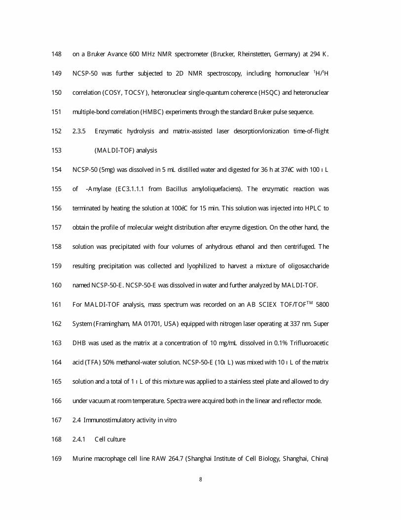
The 1
H NMR spectrum of the polysaccharide NCSP-50 exhibited three anomeric proton signals at 238
δ 5.27, δ 5.23 and δ 4.85 ppm, and labeled as A , B and C, respectively, according to their 239
decreasing chemical shifts (Fig. 2A). Based on 13
C NMR spectrum (Fig. 2B) and the cross peaks 240
in the HSQC spectrum (Fig. 2E), the anomeric carbon signal at 100.10 ppm (overlapped) was 241
correlated to both the anomeric proton signals at 5.27 and 5.23 ppm, and the anomeric carbon 242
signal at 99.01 ppm was correlated to the anomeric proton signal at 4.85 ppm. The chemical shifts 243
of anomeric proton and carbon signals indicated that all the three residues were presented in 244
α -configuration. All the 1
H and 13
C chemical shifts (Table 2) were completely assigned using 245
COSY, TOCSY, HSQC and HMBC experiments. 246
There was a high degree of signal overlapping between residue A and residue B in TOCSY 247
spectrum. This issue, however, was addressed by examining the well-resolved cross peaks in 248
COSY spectrum. The proton chemical shifts of residue A obtained were δ 5.27, 3.48, 3.82, 3.53 249
and 3.70 ppm for H-1, H-2, H-3, H-4 and H-5, respectively, from COSY spectrum (Fig. 2D and 250
Table 2). The chemical shifts of H-6/6’ (δ 3.65 and 3.73 ppm) and C-6 (δ 60.85 ppm), on the other 251
hand, were confirmed by HSQC spectrum (Fig. 2E). The corresponding chemical shifts of the 252
other carbon, also revealed by HSQC spectrum, were 100.09, 72.01, 73.51, 77.28 and 71.52 ppm 253
for C-1, C-2, C-3, C-4 and C-5, respectively (Fig. 2E and Table 2). Theses assignments were also 254
supported by previous reports (Niu, Yan, Lv, Yao, & Yu, 2013; Petersen, Motawie, Møller, 255
Hindsgaul, & Meier, 2015; Shan, et al., 2014). The downfield shift of C-4 (77.28 ppm) confirmed 256
that residue A was → 4)-α -D-Glcp-(1→ . 257

13
L ikewise, for residue B, the chemical shifts from H-1 to H-5 were assigned from COSY spectrum 258
(δ 5.23, 3.45, 3.56, 3.52 and 3.63 ppm) (Fig. 2D) and part of these was confirmed by TOCSY 259
spectrum (Fig. 2C and Table 2). Based on the proton chemical shifts, 13
C chemical shifts obtained 260
by HSQC spectrum were 100.09, 72.11, 73.22, 77.28 and 72.91 ppm, respectively (Fig. 2E). 261
According to the results from methylation analysis (Table 1), along with the literature data (Patra, 262
et al., 2013), the residue B was assigned to → 4,6)-α -D-Glcp-(1→ . 263
In the case of residue C, the chemical shifts of H-1, H-2, H-3, H-4 and H-5 was successfully 264
obtained from the COSY (Fig. 2D), which was 4.85, 3.43, 3.61, 3.29 and 3.59 ppm, respectively. 265
The specific allocation of H-6/6’ chemical shifts were supported by HSQC spectrum (Fig. 2E). 266
According to TOCSY spectrum (Fig. 2C), only cross peaks of H-1/H-2 and H-2/H-3 were 267
available due to the weak correlation between the adjacent protons. A ll the 13
C chemical shifts of 268
residue C were achieved from HSQC spectrum (Fig. 2E). Comparison of proton and carbon 269
chemical shifts with the literature values (Petersen, et al., 2015; C. Zhao, L i, Luo, & Wu, 2006) 270
allowed assigning residue C to α -D-Glcp-(1→ . 271
The HMBC experiment was carried out to enable us to identify glycosidic linkages between sugar 272
residues, as shown in Fig. 2F. Examining the cross peaks of both anomeric 1
H and 13
C of each 273
sugar residue could help to identify the sequence of residues in the polysaccharide. Cross peak 274
between H-1 (5.27 ppm) of residue A and C-4 (77.28 ppm) of residue A; H-4 (3.53 ppm) of 275
residue A and C-1 (100.09 ppm) of residue A; H-1 (5.23 ppm) of residue B and C-4 (77.28 ppm) 276
of residue A were observed, indicating that → 4)-α -D-Glcp-(1→ and → 4,6)-α -D-Glcp-(1→ were 277
linked to each other through 1,4-O-glycosidic bonds as the main chain of the polysaccharide. 278
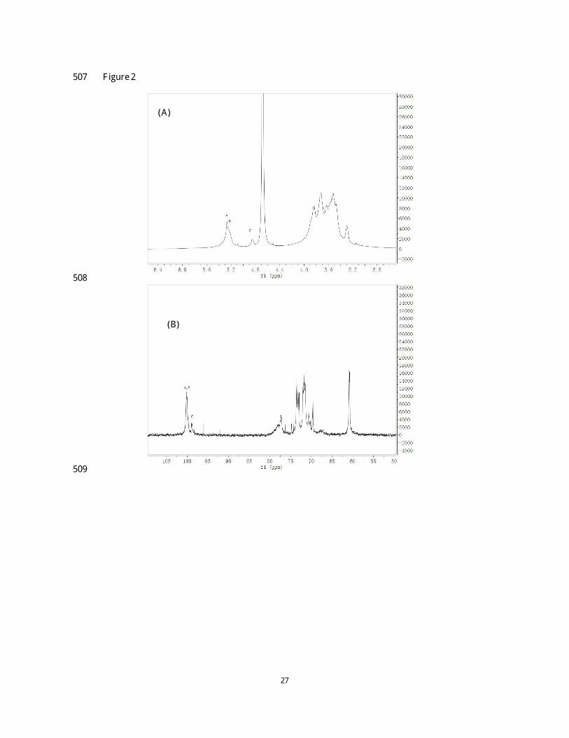
3.4 Enzymatic hydrolysis and MALDI-TOF analysis 279

14
In order to confirm the proposed chemical structure of NCSP-50, a specific enzymatic hydrolysis 280
procedure was performed. The enzymatic hydrolysate was investigated using HPLC so as to 281
monitor the changes of molecular weight distribution after treating with α -amylase. As is shown in 282
Fig. 3A, it was obvious to see that the molecular weight of NCSP-50 was significantly decreased, 283
suggesting that the polysaccharide was very sensitive to α -amylase. Then, we removed the 284
corresponding digests by precipitation with 80% ethanol followed by centrifugation to isolate the 285
polysaccharide, designated as NCSP-50-E. The MALDI-TOF profile of NCSP-50-E was shown in 286
Fig. 3B and 3C. The distance between the adjacent peaks was 162 mass units, corresponding to the 287
hexose residue in this polysaccharide. Pentose, such as arabinose, xylose, which has a 288
peak-to-peak mass difference of 132 Da, were not presented in this fraction, in agreement with the 289
aforementioned result. A maximum degree of polymerization of NCSP-50-E was 30 (m/z 4901), 290
indicating that NCSP-50 was successfully hydrolyzed by α -amylase. Therefore, the result proved 291
that (1→ 4)-linked α -D-Glcp existed in the backbone of NCSP-50. 292
The structure of NCSP-50 seems to be similar to that of pant reserve α -1,4-linked glucans. 293
However, the C-6 linked side chains in NCSP-50 were constituted by single α -glucose unit, on 294
every forth of the main chain. It seems that the structural feature of NCSP-50 was similar to that of 295
amylose which was also a kind of linear α -1,4-linked glucan. Moreover, it is acknowledged that 296
starch polysaccharides are hardly dissolved in cold water and have a high viscosity. NCSP-50, on 297
the contrary, was soluble in cold water displaying a milk white, transparent solution. The 298
discrepancies in physicochemical properties between NCSP-50 and amylose might be attributed to 299
their differences in structure characteristics. 300
In our previous study, the structure of CBHP has been characterized, which had a main chain of 301

15
(1→ 4)-linked α -D-Glcp together with small amount of (1→ 3)-linked α -D-Glcp and the branching 302
points were located at O-2 or O-6 with α -terminal-linked Glcp as side chain (Nie et al., 2011). 303
Obviously, CBHP had higher degree of branching compared to NCSP-50. Another difference 304
between them was the small amount of (1→ 3)-linked α -D-Glcp residues presented in the main 305
chain of CBHP. We speculated that the differences of the raw material and extraction procedures 306
might account for the varieties of the chemical structure between the two polysaccharides. 307
3.5 Immunostimulatory activities on macrophages 308
3.5.1 Effect of NCSP-50 on macrophage proliferation 309
Macrophages are presented in virtually all tissues and have long been considered as an important 310
component of host defense against microbial invaders and malignancies (Dunn, Barke, Ewald, & 311
Simmons, 1987). Additionally, macrophages can respond not only to endogenous stimuli 312
generated by injury or infection, but also to signals produced by antigen-specific immune cells 313
(Mosser & Edwards, 2008). Therefore, to characterize the immunostimulatory effect of NCSP-50 314
in an in vitro macrophage cell model, we firstly investigated the influence of cells proliferation in 315
the presence of polysaccharide with various concentrations (Fig. 4A). After 24 h incubation with 316
the polysaccharide solutions (25, 50, 100 and 200 μg/mL), the proliferation rate of RAW 264.7 317
cells was determined by the WST-8 assay. As shown in Fig. 4A, NCSP-50 exhibited a significant 318
stimulatory effect on RAW 264.7 cells proliferation. In the concentration of 50-200 μg/mL, the 319
proliferation rates of polysaccharide-treated groups were significantly higher than that of the 320
positive control group (p<0.01). 321
3.5.2 Effect of NCSP-50 on NO production in macrophages 322
NO is reported to be associated with macrophages activation in the host defense against tumor 323

16
cells and microorganisms (Schepetkin & Quinn, 2006). In order to investigate the effects of 324
NCSP-50 on macrophage response, the NO production of RAW 264.7 cells was determined by 325
Griess assay. As is shown in Fig. 4B, the NO concentration of the culture supernatant was 326
significantly increased in a dose-dependent manner by treatment with NCSP-50 (25-200 μg/mL, 327
P<0.01). The level of NO reached 22.32 μmol/L after treatment by 50 μg/mL of NCSP-50, similar 328
to that of the positive control (LPS, 1 μg/mL). In addition, in order to figure out the difference 329
between NCSP-50 and starch, the effect of starch on NO production was also evaluated (Fig. 4C). 330
After 24 h incubation in the presence of various concentrations of starch, it was obviously to see 331
that the production of NO was not significantly enhanced as compared to the control group (0 332
μg/mL). The results demonstrated that starch, although had a similar α -1,4-glucan backbone 333
structure, showed no effect on upregulating NO secretion in RAW 264.7 cells. 334
3.5.3 Effect of NCSP-50 on IL-1β and TNF-α secretion in macrophages 335
Cytokines are intercellular signaling proteins or peptides with relatively low molecular weight that 336
are released by the cells altering either their own function (autocrine) or those of adjacent cells 337
(paracrine) (Haddad, 2002). They are important mediators involved in modulating immune 338
response and inflammatory reactions, particularly during infection and trauma. In addition to 339
regulating cells of the innate and adaptive immune system, cytokines affect cell proliferation, 340
differentiation and functions (Hopkins, 2003). IL-1β and TNF-α are two typical pro-inflammatory 341
cytokines, which can be secreted by activated macrophages with immunomodulatory properties. It 342
is of significance that TNF-α could stimulate the production of genotoxic molecules, such as NO 343
and reactive oxygen species that could lead to DNA damage and mutations (Hussain, Hofseth, & 344
Harris, 2003). In the present study, the stimulatory effect of NCSP-50 on the production of IL -1β 345

17
and TNF-α by RAW 264.7 cells was determined by ELISA . As shown in Fig. 4D and 4E, 346
NCSP-50 could significantly promote RAW 264.7 cells to release IL -1β and TNF-α . With respect 347
to IL-1β, it is obviously that NCSP-50 could increase the IL-1β production in a dose-dependent 348
manner. Compared with the control group, the IL-1β concentration was significantly increased by 349
NCSP-50 treatment (25 μg/mL, p < 0.05; 50, 100 and 200 μg/mL, p < 0.01) and reached up to 350
51.47 pg/mL at a concentration of 200 μg/mL, slightly lower than that induced by LPS (54.95 351
pg/mL). On the other hand, with regard to TNF-α secretion, NCSP-50 also showed a notable 352
promotion effect, with the highest level of 74905.42 pg/mL at a concentration of 100 μg/mL. In 353
contrast, the influence of starch on TNF-α production was not significant at all concentrations as 354
evident in Fig. 4F. These results indicated that NCSP-50 could remarkably promote the secretion 355
of cytokines in RAW 264.7 cells, whereas starch had no effect. 356
Therefore, it was confirmed that the potent immunostimulatory activity of NCSP-50 should be 357
caused and influenced by its structure characteristics, different from that of the starch. The 358
discrepancy might be attributed to the degree of substitution on the main chain, the length of side 359
chains and the conformation etc., between NCSP-50 and starch. 360
4. Conclusion 361
In the present study, the structure properties of a water-soluble polysaccharide NCSP-50 from 362
natural C. sinensis were elucidated. HPGPC results showed that the molecular weight of NCSP-50 363
was 9.76×10⁵
Da. Using monosaccharide composition, methylation analysis, enzymatic hydrolysis, 364
MALDI-TOF analysis and NMR spectroscopy, the structure of NCSP-50 was deduced to be a 365
homogenous glucan, comprised a main chain of (1→ 4)-linked-α -D-Glcp with a single α -D-Glcp 366
branch substituted at C-6. Unlike starch, NCSP-50 was revealed to significantly stimulate the 367

18
proliferation of macrophages, promote nitric oxide production and enhance cytokine secretion. 368
Our results demonstrated that NCSP-50 had the potential to be an immunopotentiating agent, and 369
the in-deep research on the related mechanism, on the other hand, will be conducted in our future 370
work. 371
Acknowledgment 372
The financial support from the National Natural Science Foundation of China for Excellent Young 373
Scholars (31422042), the outstanding science and technology innovation team project in J iangxi 374
Province (20133BCB24001), the Project of Science and Technology of J iangxi Provincial 375
Education Department (K JLD13004) and Research Project of State K ey Laboratory of Food 376
Science and Technology (SK LF-ZZB-201508, SK LF-ZZA-201611) is gratefully acknowledged. 377
378
R eference 379
Biermann, C. J ., & McGinnis, G. D. (1988). Analysis of Carbohydrates by GLC and MS: CRC 380
Press. 381
Chen, Y. J ., Shiao, M. S., Lee, S. S., & Wang, S.-Y. (1997). Effect of Cordyceps sinensis on the 382
proliferation and differentiation of human leukemic U937 cells. Life Sciences, 60(25), 383
2349-2359. 384
Cheung, J . K ., L i, J ., Cheung, A . W., Zhu, Y., Zheng, K . Y., Bi, C. W., et al. (2009). Cordysinocan, 385
a polysaccharide isolated from cultured Cordyceps, activates immune responses in 386
cultured T-lymphocytes and macrophages: Signaling cascade and induction of cytokines. 387
J ournal of Ethnopharmacology, 124(1), 61-68. 388
Ciucanu, I., & K erek, F. (1984). A simple and rapid method for the permethylation of 389

19
carbohydrates. Carbohydrate Research, 131(2), 209-217. 390
Dunn, D. L., Barke, R. A., Ewald, D. C., & Simmons, R. L . (1987). Macrophages and 391
translymphatic absorption represent the first line of host defense of the peritoneal cavity. 392
Archives of Surgery, 122(1), 105-110. 393
Haddad, J . J . (2002). Cytokines and related receptor-mediated signaling pathways. Biochemical 394
and Biophysical Research Communications, 297(4), 700-713. 395
Hopkins, S. J . (2003). The pathophysiological role of cytokines. Legal Medicine, 5, S45-S57. 396
Hussain, S. P., Hofseth, L . J ., & Harris, C. C. (2003). Radical causes of cancer. Nature Reviews 397
Cancer, 3(4), 276-285. 398
K iho, T., Tabata, H., Ukai, S., & Hara, C. (1986). A minor, protein-containing galactomannan from 399
a sodium carbonate extract of Cordyceps sinensis. Carbohydrate Research, 156, 189-197. 400
L i, S. P., L i, P., Dong, T. T. X ., & Tsim, K . W. K . (2001). Anti-oxidation activity of different types 401
of natural Cordyceps sinensis and cultured Cordyceps mycelia. Phytomedicine, 8(3), 402
207-212. 403
L iu, Y., Zuo, J ., Tao, Y., & L iu, W. (2013). Protective effect of Cordyceps polysaccharide on 404
hydrogen peroxide-induced mitochondrial dysfunction in HL-7702 cells. Molecular 405
Medicine Reports, 7(3), 747-754. 406
Meng, L .-Z., Feng, K ., Wang, L .-Y., Cheong, K .-L ., Nie, H., Zhao, J ., et al. (2014). Activation of 407
mouse macrophages and dendritic cells induced by polysaccharides from a novel 408
Cordyceps sinensis fungus UM01. J ournal of Functional Foods, 9, 242-253. 409
Miyazaki, T., Oikawa, N., & Yamada, H. (1977). Studies on fungal polysaccharides. X X . 410
Galactomannan of Cordyceps sinensis. Chemical and Pharmaceutical Bulletin, 25(12), 411

20
3324-3328. 412
Mosser, D. M., & Edwards, J . P. (2008). Exploring the full spectrum of macrophage activation. 413
Nature Reviews Immunology, 8(12), 958-969. 414
Nie, S. P., Cui, S. W., Phillips, A . O., X ie, M.-Y., Phillips, G. O., A l-Assaf, S., et al. (2011). 415
Elucidation of the structure of a bioactive hydrophilic polysaccharide from Cordyceps 416
sinensis by methylation analysis and NMR spectroscopy. Carbohydrate Polymers, 84(3), 417
894-899. 418
Nie, S., Cui, S. W., X ie, M., Phillips, A . O., & Phillips, G. O. (2013). Bioactive polysaccharides 419
from Cordyceps sinensis: Isolation, structure features and bioactivities. Bioactive 420
Carbohydrates and Dietary F ibre, 1(1), 38-52. 421
Niu, Y., Yan, W., Lv, J ., Yao, W., & Yu, L . (2013). Characterization of a novel polysaccharide from 422
tetraploid Gynostemma pentaphyllum Makino. J ournal of Agricultural and Food 423
Chemistry, 61(20), 4882-4889. 424
Patra, S., Patra, P., Maity, K . K ., Mandal, S., Bhunia, S. K ., Dey, B., et al. (2013). A heteroglycan 425
from the mycelia of Pleurotus ostreatus: Structure determination and study of antioxidant 426
properties. Carbohydrate Research, 368, 16-21. 427
Petersen, B. O., Motawie, M. S., Møller, B. L ., Hindsgaul, O., & Meier, S. (2015). NMR 428
characterization of chemically synthesized branched α -dextrin model compounds. 429
Carbohydrate Research, 403, 149-156. 430
Sassaki, G. L ., Gorin, P. A., Souza, L . M., Czelusniak, P. A ., & Iacomini, M. (2005). Rapid 431
synthesis of partially O-methylated alditol acetate standards for GC–MS: Some relative 432
activities of hydroxyl groups of methyl glycopyranosides on Purdie methylation. 433

21
Carbohydrate Research, 340(4), 731-739. 434
Schepetkin, I. A ., & Quinn, M. T. (2006). Botanical polysaccharides: macrophage 435
immunomodulation and therapeutic potential. International Immunopharmacology, 6(3), 436
317-333. 437
Shan, J ., Sun, G., Ren, J ., Zhu, T., J ia, P., Qu, W., et al. (2014). An α -glucan isolated from root of 438
Isatis Indigotica, its structure and adjuvant activity. Glycoconjugate J ournal, 31(4), 439
317-326. 440
Sheng, L ., Chen, J ., L i, J ., & Zhang, W. (2011). An exopolysaccharide from cultivated Cordyceps 441
sinensis and its effects on cytokine expressions of immunocytes. Applied Biochemistry 442
and Biotechnology, 163(5), 669-678. 443
Wang, J ., K an, L ., Nie, S., Chen, H., Cui, S. W., Phillips, A . O., et al. (2015). A comparison of 444
chemical composition, bioactive components and antioxidant activity of natural and 445
cultured Cordyceps sinensis. LWT-Food Science and Technology, 63(1), 2-7. 446
Wang J ., Nie S., K an L ., Chen H., Cui S., Phillips A . O., et al. Comparison of structural features 447
and antioxidant activity of polysaccharides from natural and cultured Cordyceps sinensis. 448
Food Science and Biotechnology, Accepted. 449
Wang, Y., L iu, D., Zhao, H., J iang, H., Luo, C., Wang, M., et al. (2014). Cordyceps sinensis 450
polysaccharide CPS-2 protects human mesangial cells from PDGF-BB-induced 451
proliferation through the PDGF/ERK and TGF-β1/Smad pathways. Molecular and 452
Cellular Endocrinology, 382(2), 979-988. 453
Wang, Y., Y in, H., Lv, X ., Wang, Y., Gao, H., & Wang, M. (2010). Protection of chronic renal 454
failure by a polysaccharide from Cordyceps sinensis. F itoterapia, 81(5), 397-402. 455

22
Wu, D.T., Meng, L .Z., Wang, L .Y., Lv, G. P., Cheong, K . L., Hu, D. J ., et al. (2014). Chain 456
conformation and immunomodulatory activity of a hyperbranched polysaccharide from 457
Cordyceps sinensis. Carbohydrate Polymers, 110, 405-414. 458
Yao, X ., Meran, S., Fang, Y., Martin, J ., Midgley, A ., Pan, M.-M., et al. (2014). Cordyceps sinensis: 459
In vitro anti-fibrotic bioactivity of natural and cultured preparations. Food Hydrocolloids, 460
35, 444-452. 461
Zhang, J ., Yu, Y., Zhang, Z., Ding, Y., Dai, X ., & L i, Y. (2011). Effect of polysaccharide from 462
cultured Cordyceps sinensis on immune function and anti-oxidation activity of mice 463
exposed to 60
Co. International Immunopharmacology, 11(12), 2251-2257. 464
Zhang, X ., L iu B., Al-Assaf, S., Phillips, G. O., & Phillips, A. O. (2012). Cordyceps sinensis 465
decreases TGF-β1 dependent epithelial to mesenchymal transdifferentiation and 466
attenuates renal fibrosis. Food Hydrocolloids, 28(1), 200-212. 467
Zhao, C., L i, M., Luo, Y., & Wu, W. (2006). Isolation and structural characterization of an 468
immunostimulating polysaccharide from fuzi, Aconitum carmichaeli. Carbohydrate 469
Research, 341(4), 485-491. 470
Zhao, J ., X ie, J ., Wang, L . Y., & L i, S. P. (2014). Advanced development in chemical analysis of 471
Cordyceps. J ournal of Pharmaceutical and Biomedical Analysis, 87, 271-289. 472
Zhu, J .-S., Halpern, G. M., & Jones, K . (1998a). The scientific rediscovery of a precious ancient 473
Chinese herbal regimen: Cordyceps sinensis Part II. The J ournal of Alternative and 474
Complementary Medicine, 4(4), 429-457. 475
Zhu, J .-S., Halpern, G. M., & Jones, K . (1998b). The scientific rediscovery of an ancient Chinese 476
herbal medicine: Cordyceps sinensis Part I. The J ournal of Alternative and 477

23
Complementary Medicine, 4(3), 289-303. 478
479
480

24
TABL E S 481
Table 1 482
GC-MS of alditol acetate derivatives from the methylated products of NCSP-50 483
Methylated sugar RT(min) Deduced linkage Molar ratio a
1,5-O-Ac2-2,3,4,6-Me4-glucitol 15.333 D - Glcp-(1→ 15.99
1,4,5-O-Ac2-2,3,6-Me3-glucitol 23.856 → 4)- D -Glcp-(1→ 66.87
1,4,5,6-O-Ac2-2,3-Me2-glucitol 29.311 → 4,6)- D -Glcp-(1→ 17.14
a
Relative molar ratio, calculated from the ratio of peak areas. 484
485
486

25
Table 2 487
The 1
H NMR and 13
C NMR chemical shifts for NCSP-50 isolated from natural Cordyceps sinensis 488
in D2O at 295K 489
Chemical shifts (ppm)
Glycosidic linkage H1/C1 H2/C2 H3/C3 H4/C4 H5/C5 H6/C6
A → 4)-α -D-Glcp-(1→ 5.27 3.48 3.82 3.53 3.70 3.73a
3.65b
100.09 72.01 73.51 77.28 71.52 60.85
B → 4,6)-α -D-Glcp-(1→ 5.23 3.45 3.56 3.52 3.63 3.30 -
100.09 72.11 73.22 77.28 72.91 69.73
C α -D-Glcp-(1→ 4.85 3.43 3.61 3.29 3.59 3.72a
3.63b
99.01 72.16 73.28 69.7 73.07 60.74
a,b
interchangeable 490
491
492
493

26
F IGURE S 494
F igure 1 495
496
497
498
499
500
501
502
503
504
505
506
27
F igure 2 507
508
509
(A)
(B)

28
510
511
(C)
(D)

29
512
513
514
515
516
517
(E)
(F)
30
F igure 3 518
519
520
521
522
523
(A)
(B)
(C)

31
F igure 4 524
525
526
527
528
529

32
530
531
532
533
534