structural characteristics of an insect group i chitinase...
TRANSCRIPT
research papers
932 doi:10.1107/S1399004713033841 Acta Cryst. (2014). D70, 932–942
Acta Crystallographica Section D
BiologicalCrystallography
ISSN 1399-0047
Structural characteristics of an insect group Ichitinase, an enzyme indispensable to moulting
Lei Chen,a‡ Tian Liu,a,b‡ Yong
Zhou,c Qi Chen,a Xu Shend and
Qing Yanga*
aSchool of Life Science and Biotechnology,
Dalian University of Technology, 2 Linggong
Road, Dalian, Liaoning 116024, People’s
Republic of China, bState Key Laboratory for
Biocontrol, Sun Yat-Sen University, Higher
Education Mega Center, Guangzhou,
Guangdong 510006, People’s Republic of
China, cSchool of Software, Dalian University of
Technology, 321 Tuqiang Street, Dalian,
Liaoning 116620, People’s Republic of China,
and dState Key Laboratory of Drug Research,
Shanghai Institute of Materia Medica, Chinese
Academy of Sciences, 555 Zuchongzhi Road,
Shanghai 201203, People’s Republic of China
‡ These authors made equal contributions.
Correspondence e-mail: [email protected]
Insects possess a greater number of chitinases than any other
organisms. This work is the first report of unliganded and
oligosaccharide-complexed crystal structures of the insect
chitinase OfChtI from Ostrinia furnacalis, which is essential
to moulting. The obtained crystal structures were solved at
resolutions between 1.7 and 2.2 A. A structural comparison
with other chitinases revealed that OfChtI contains a long
substrate-binding cleft similar to the bacterial chitinase
SmChiB from Serratia marcescens. However, unlike the exo-
acting SmChiB, which has a blocked and tunnel-like cleft,
OfChtI possesses an open and groove-like cleft. The
complexed structure of the catalytic domain of OfChtI
(OfChtI-CAD) with (GlcNAc)2/3 indicates that the reducing
sugar at subsite �1 is in an energetically unfavoured ‘boat’
conformation, a state that possibly exists just before the
completion of catalysis. Because OfChtI is known to act from
nonreducing ends, (GlcNAc)3 would be a hydrolysis product
of (GlcNAc)6, suggesting that OfChtI possesses an endo
enzymatic activity. Furthermore, a hydrophobic plane
composed of four surface-exposed aromatic residues is
adjacent to the entrance to the substrate-binding cleft.
Mutations of these residues greatly impair the chitin-binding
activity, indicating that this hydrophobic plane endows
OfChtI-CAD with the ability to anchor chitin. This work
reveals the unique structural characteristics of an insect
chitinase.
Received 18 November 2013
Accepted 13 December 2013
PDB references:
OfChtI-CAD, 3w4r; complex
with (GlcNAc)2/3, 3wl1;
E148A mutant, complex with
(GlcNAc)2, 3wl0; E148Q
mutant, 3wkz
1. Introduction
The glycosyl hydrolase family 18 (GH18) chitinases (EC
3.2.1.14) are enzymes that hydrolyze chitin, a �-1,4-linked
N-acetylglucosamine (GlcNAc) linear polymer, into chito-
oligosaccharides. These enzymes are widely distributed in
many organisms, including bacteria, fungi, insects, plants and
mammals, and play roles in immunity and defence, digestion,
pathogenicity and arthropod moulting (Arakane & Muthu-
krishnan, 2010). Understanding the structure–function rela-
tionship of these enzymes is essential for disease control and
drug design.
The crystal structures of the catalytic domains (CADs) of
free or ligand-complexed chitinases have been determined,
including GH18 chitinases from bacteria (Perrakis et al., 1994;
van Aalten et al., 2001; Papanikolau et al., 2003; Songsiri-
ritthigul et al., 2008; Tsuji et al., 2010; Hsieh et al., 2010;
Pantoom et al., 2011; Payne et al., 2012; Busby et al., 2012;
Malecki et al., 2013), fungi (Hollis et al., 2000; Rao et al., 2005;

Hurtado-Guerrero & van Aalten, 2007; Schuttelkopf et al.,
2010; Rush et al., 2010; Yang et al., 2010), plants (Cavada et al.,
2006; Ohnuma, Numata, Osawa, Mizuhara, Lampela et al.,
2011; Ohnuma, Numata, Osawa, Mizuhara, Varum et al., 2011)
and mammals (Fusetti et al., 2002; Olland et al., 2009;
Sutherland et al., 2011), as well as GH19 chitinases from
bacteria (Hoell et al., 2006; Kezuka et al., 2006) and plants
(Song & Suh, 1996; Hahn et al., 2000; Ubhayasekera et al.,
2007, 2009; Huet et al., 2008; Ohnuma et al., 2012) and a GH23
chitinase from a bacterium (Arimori et al., 2013). The afore-
mentioned structural information has shown that all GH18
chitinases employ a substrate-assisted retaining mechanism
during catalysis, in which the C2-acetamido group of the
substrate acts as the catalytic nucleophile (Tews et al., 1997;
Brameld & Goddard, 1998a). However, the GH19 and GH23
family chitinases use an inverting mechanism (Brameld &
Goddard, 1998b; Arimori et al., 2013).
Unfortunately, there is no structural information available
for insect chitinases. Insects possess a greater number of
chitinases than any other organisms; in fact, an insect may
possess as many as eight groups of genes encoding GH18
chitinases, most of which are involved in chitinolytic processes
(Zhu, Arakane, Banerjee et al., 2008; Arakane & Muthu-
krishnan, 2010). These chitinases differ in their domain
compositions, enzymatic properties, expression patterns and
tissue localizations. Although the crystal structure of the
chitinase-like protein imaginal disc growth factor 2 (IDGF-2)
from the fruit fly Drosophila melanogaster (DmIDGF2) has
been determined (Varela et al., 2002), the reference value of
this protein is low because DmIDGF2 is not an active enzyme.
DmIDGF2 lacks the catalytic residues and has a blockage in
the substrate-binding cleft (Varela et al., 2002). For these
reasons, DmIDGF2 is referred to as a regulator rather than an
enzyme.
Among the eight groups of insect chitinases, group I chit-
inases have been enzymatically well characterized and have
been shown to function in a chitin-degradation process that
is closely associated with insect moulting (Zhu, Arakane,
Beeman et al., 2008). When the transcription level of the group
I chitinase TcCht5 from the red flour beetle Tribolium casta-
neum was downregulated during the larval, pharate pupal and
pupal stages, the insects failed to shed their old cuticle and
died during eclosion (Zhu, Arakane, Beeman et al., 2008). Our
previous work reported on the group I insect chitinase OfChtI
(previously called OfCht5) from Ostrinia furnacalis, a species
of moth that is the most destructive insect affecting corn
production. OfChtI is highly active in the hydrolysis of chito-
oligosaccharides, colloidal chitin and �-chitin, and exhibits
kinetic properties that differ from those of bacterial and plant
chitinases (Wu et al., 2013). In this study, the catalytic domain
of OfChtI (OfChtI-CAD) was crystallized both alone and
complexed with oligosaccharides, providing the first structural
insights into free or oligosaccharide-complexed insect chit-
inase. The structural investigations, together with site-directed
mutagenesis studies, reveal that the insect chitinase OfChtI
possesses several unique structural features that distinguish
this enzyme from other known chitinases.
2. Materials and methods
2.1. Gene cloning and site-directed mutagenesis
The gene fragment encoding OfChtI-CAD (residues 19–
407) was amplified from the full-length cDNA of OfChtI
(GenBank ID DQ294305) with the primers 50-TGAAGC-
TTACGTAGAATTCGCGGAGTCGGACAGCAGAGCG-30
(forward) and 50-CCGCCCTAGGGAATTCTTAATGAT-
GATGATGATGATGAGAACGCGGTGGTGGAACAG-30
(reverse). EcoRI restriction sites (bold) and a C-terminal
6�His affinity tag were introduced. The resulting PCR frag-
ment was ligated into the pPIC9 vector (Invitrogen, Carlsbad,
Califonia, USA) with an in-fusion HD cloning kit (Clontech,
Palo Alto, California, USA).
The OfChtI-CAD E148Q, E148A, F159A, F194A, W241A,
Y290A, F194A/W241A, F159A/Y290A and F159A/F194A/
W241A/Y290A mutants were produced using the Quik-
Change site-directed mutagenesis kit (Stratagene, La Jolla,
California, USA), according to the manufacturer’s instruc-
tions. The mutated genes were sequenced to confirm that the
desired mutations had been inserted.
2.2. Protein expression and purification
The plasmid containing the OfChtI-CAD gene was trans-
formed into Pichia pastoris GS115 cells (Invitrogen, Carlsbad,
California, USA) for the overexpression of recombinant
OfChtI-CAD. The cells were first grown in buffered glycerol
complex medium (BMGY; 1% yeast extract, 2% peptone, 1%
glycerol, 1.34% yeast nitrogen, 0.2% biotin, 100 mM potas-
sium phosphate pH 6.0) at 303 K to an optical density (OD) of
2.0 at 600 nm. After reaching this OD, the cells were collected
and resuspended in buffered methanol complex medium
(BMMY; 1% yeast extract, 2% peptone, 1% methanol, 1.34%
yeast nitrogen, 0.2% biotin, 100 mM potassium phosphate pH
6.0). Methanol [1%(v/v)] was added at 24 h intervals as an
inducer. After 72 h of fermentation, the culture supernatant
was harvested and subjected to ammonium sulfate precipita-
tion (75% saturation) at 277 K. The precipitate was dissolved
in buffer A (20 mM sodium phosphate, 0.5 M NaCl, 20 mM
imidazole pH 7.4) and the sample was then loaded onto a
HisTrap HP affinity column (5 ml; GE Healthcare, USA)
pre-equilibrated with buffer A. After washing the column, the
target protein was eluted with buffer B (20 mM sodium
phosphate, 0.5 M NaCl, 200 mM imidazole pH 7.4). The purity
of the eluted protein was analyzed by SDS–PAGE and found
to be >95%. The mutants were expressed and purified using
the same procedure as used for wild-type OfChtI-CAD.
2.3. Crystallization and data collection
Crystallization screening of recombinant OfChtI-CAD was
performed using the commercially available Index, Crystal
Screen and Crystal Screen 2 (Hampton Research, Riverside,
California, USA) screens as well as The JCSG Core Suites
I–IV (Qiagen, Valencia, California, USA). Pure OfChtI-CAD
was spin-concentrated to 10 mg ml�1 in 50 mM HEPES
pH 8.0, 100 mM NaCl. The hanging-drop vapour-diffusion
research papers
Acta Cryst. (2014). D70, 932–942 Chen et al. � Group I chitinase 933

crystallization experiments were set up at 277 K by mixing 1 ml
OfChtI-CAD and 1 ml reservoir solution. The protein crys-
tallized after five months from a crystallization cocktail
consisting of 100 mM HEPES pH 7.5, 25%(w/v) PEG 3350.
Crystals of the OfChtI-CAD–(GlcNAc)2/3 complex were
obtained by transferring native crystals to a stabilizing solu-
tion consisting of 5 mM (GlcNAc)6 (N0,N00,N000,N0000,N00000,
N000000-hexaacetylchitohexaose; Santa Cruz Biotechnology Inc.,
Dallas, Texas, USA), 100 mM HEPES pH 7.5, 25%(w/v) PEG
3350. The crystals were soaked for approximately 30 or 60 min
at room temperature. The E148A and E148Q mutants (at
10 mg ml�1) were co-crystallized with 5 mM (GlcNAc)6 in
100 mM HEPES pH 7.9, 23%(w/v) PEG 3350 and in 100 mM
HEPES pH 8.0, 21%(w/v) PEG 3350, respectively.
These crystals were soaked for several minutes in reservoir
solution containing 25%(v/v) glycerol as a cryoprotection
agent and were subsequently flash-cooled in liquid nitrogen.
Diffraction data were collected on BL-17U at the Shanghai
Synchrotron Radiation Facility in China and the diffraction
data were processed using the HKL-2000 package (Otwi-
nowski & Minor, 1997).
2.4. Structure determination and refinement
The structure of free OfChtI-CAD was solved by molecular
replacement with Phaser (McCoy et al., 2007) using the
structure of human acidic mammalian chitinase (PDB entry
3fxy; Olland et al., 2009) as a model. The subsequent structures
of the E148Q mutant and the complexes
with oligosaccharides were solved using
the coordinates of free OfChtI-CAD
as a model. The PHENIX suite of
programs (Adams et al., 2010) was used
for structure refinement. The molecular
models were manually built and
extended using Coot (Emsley et al.,
2010). The stereochemistry of the
models was checked by PROCHECK
(Laskowski et al., 1993). The coordi-
nates of OfChtI-CAD, OfChtI-CAD
with (GlcNAc)2/3, the E148A mutant
with (GlcNAc)2 and the E148Q mutant
were deposited in the Protein Data
Bank as entries 3w4r, 3wl1, 3wl0 and
3wkz, respectively. All structural
figures were generated using PyMOL
(DeLano, 2002). The data-collection
and structure-refinement statistics are
summarized in Table 1.
2.5. Isothermal titration calorimetry
Isothermal titration calorimetry
(ITC) experiments were performed at
303 K using a MicroCal iTC200 System
(MicroCal, Northampton, Massachu-
setts, USA). For the experiments with
(GlcNAc)3–6, 0.2 mM E148Q mutant in
50 mM HEPES buffer pH 8.0 was placed in a reaction cell with
a total volume of 202 ml, and 1–2 mM (GlcNAc)3–6 in the same
buffer as the E148Q mutant protein was placed in the ITC
syringe. Aliquots of 2–3 ml were injected into the reaction cell
at 120 s intervals with a stirring speed of 1000 rev min�1. The
titrations were typically completed after 13 or 20 injections.
The background was measured by injecting (GlcNAc)3–6 into
the buffer.
The ITC data were processed by the MicroCal Origin v.7.0
software that accompanied the ITC200 system. Prior to further
data analysis, all data were corrected for the heat of dilution
by subtracting the background. Using a nonlinear least-
squares algorithm, the data were analyzed using the single-site
binding model in the Origin software. Using this model, the
stoichiometry (n), binding association constant (Ka) and
enthalpy change (�H) of the reaction were obtained. For
(GlcNAc)3 binding, a two-site binding model was used. The
reaction free-energy change (�G), the dissociation constant
(Kd) and the entropy change (�S) could be calculated from
�H and Ka using �G = �RT lnKa = RT lnKd = �H � T�S,
where T is the absolute temperature in kelvin and R repre-
sents the gas constant (1.98 cal K�1 mol�1).
2.6. Enzymatic assays and chitin-binding assays
The enzymatic activities of wild-type OfChtI-CAD and
its mutants (F159A, F194A, W241A, Y290A, F194A/
W241A, F159A/Y290A and F159A/F194A/W241A/Y290A)
research papers
934 Chen et al. � Group I chitinase Acta Cryst. (2014). D70, 932–942
Table 1X-ray data-collection and structure-refinement statistics.
Values in parentheses are for the highest resolution shell.
OfChtI-CAD E148QE148A–(GlcNAc)2
OfChtI-CAD–(GlcNAc)2/3
PDB entry 3w4r 3wkz 3wlo 3wl1Space group P65 P65 P65 P65
Unit-cell parametersa = b (A) 93.8 94.2 94.1 93.8c (A) 122.0 122.2 122.3 122.2
Wavelength (A) 0.979228 0.978686 0.978686 0.978686Temperature (K) 100 100 100 100Resolution (A) 50.0–1.70
(1.73–1.70)50.0–2.00
(2.03–2.00)50.0–2.20
(2.23–2.20)50.0–1.77
(1.80–1.77)Unique reflections 66488 41398 30971 58863Observed reflections 1491430 702768 524755 952096Rmerge 0.094 (0.361) 0.102 (0.367) 0.090 (0.368) 0.058 (0.424)Average multiplicity 22.5 (22.5) 17.0 (17.0) 16.9 (17.0) 16.2 (15.0)hI/�(I)i 30.15 (12.80) 22.17 (10.77) 24.81 (12.32) 36.93 (8.55)Completeness (%) 99.78 (99.65) 99.95 (99.83) 99.89 (98.93) 99.66 (99.05)R/Rfree 0.1467/0.1594 0.1585/0.1856 0.1523/0.1777 0.1428/0.1686Protein atoms 3086 3081 3077 3081Water molecules 557 411 342 436Other atoms 28 28 57 100R.m.s. deviation from ideal
Bond lengths (A) 0.021 0.008 0.007 0.019Bond angles (�) 1.790 1.160 1.100 1.760
Wilson B factor (A2) 11.21 22.09 23.51 16.78Average B factor (A2) 15.70 21.00 22.20 22.00B factor, protein atoms (A2) 12.80 19.30 20.80 19.40B factor, water molecules (A2) 31.10 32.50 32.60 35.70Ramachandran plot (%)
Favoured 93.3 93.9 93.3 92.0Allowed 6.7 6.1 6.7 8.0Outliers 0.0 0.0 0.0 0.0

were determined using 4-nitrophenyl-�-chitobioside [pNP-
(GlcNAc)2] and crystalline �-chitin as substrates. For pNP-
(GlcNAc)2, the reaction mixture (60 ml) consisted of 20 nM
enzyme and 0.1 mM pNP-(GlcNAc)2 in 50 mM sodium phos-
phate buffer pH 6.5. After incubation at 303 K for 10 min,
60 ml of 0.5 M sodium carbonate was added to the sample to
stop the reaction. The amount of 4-nitrophenyl released was
determined by measuring the absorbance at 405 nm. For
crystalline �-chitin, the reaction mixture (100 ml) consisted of
40 nM enzyme and 1.0 mg ml�1 �-chitin in 50 mM sodium
phosphate buffer pH 6.5. The reaction mixtures were incu-
bated for 6 h at 303 K and then centrifuged at 13 000g for
5 min. The production of reducing sugars in the supernatant
was followed with potassium ferriferrocyanide, and the levels
of potassium ferriferrocyanide consumption, corresponding to
the amount of reducing-sugar production, were quantified by
measuring the absorbance at 420 nm (Imoto & Yagishita,
1971).
Binding assays were performed using crystalline �-chitin
as the substrate. The reaction mixtures (500 ml) consisted of
1.0 mg ml�1 �-chitin and 0.3 mg ml�1 purified wild-type
OfChtI-CAD or its mutants in 50 mM sodium phosphate
buffer pH 6.5 at 303 K with rotation at 250 rev min�1. At
different time points, the samples were centrifuged at 13 000g
for 5 min and the protein concentrations were determined
from Bradford assays calibrated against bovine serum albumin
(BSA).
3. Results
3.1. Crystal structure of OfChtI-CAD
OfChtI is a member of the insect group I chitinases, which
are composed of an N-terminal signal region (residues 1–18), a
CAD (residues 19–407), a PEST-like linker region (residues
408–491) and a C-terminal chitin-binding domain (residues
research papers
Acta Cryst. (2014). D70, 932–942 Chen et al. � Group I chitinase 935
Figure 1Overall structures of unliganded and oligosaccharide-complexed OfChtI-CADs. (a, b) Cartoon representation of OfChtI-CAD (a) and the E148Qmutant (b). The structure consists of two domains: a core domain with an (�/�)8 TIM-barrel fold (cyan, �-helices; blue, �-strands) and an insertiondomain (orange). The catalytic residues and the N-GlcNAc residues at the N-glycosylation sites are shown as sticks with green and yellow C atoms,respectively. (c, d) Surface representations of E148A complexed with (GlcNAc)2 (c) and OfChtI-CAD complexed with (GlcNAc)2/3 (d). The ligand isshown as a stick with yellow C atoms. The aromatic residues that stack with the sugar rings are shown in blue. The numbers indicate the subsite to whichthe sugar is bound. (e, f ) Stereoview of the substrate-binding cleft with details of the interactions between (GlcNAc)2 and E148A (e) and between(GlcNAc)2/3 and OfChtI-CAD ( f ). The ligand is represented as a stick with yellow C atoms and the 2Fo � Fc electron-density map around the ligand iscontoured at the 1.0� level. The catalytic residues and the amino acids that interact with the ligand are labelled and are shown as sticks with green andblue C atoms, respectively. The numbers indicate the subsite to which the sugar is bound. Hydrogen bonds are drawn as dashed lines.

492–553). Our experiments showed that full-length recombi-
nant OfChtI was not stable and underwent autocleavage when
incubated with the crystallization reagent, leaving a stable
43 kDa form with full catalytic activity towards the pNP-
(GlcNAc)2 substrate. Thus, the truncated form of OfChtI
(OfChtI-CAD; residues 19–407) was cloned, expressed and
purified for crystallization.
OfChtI-CAD crystals were obtained by vapour diffusion
and the structure was determined by X-ray diffraction. The
crystals belonged to space group P65 and the structure was
solved at a resolution of 1.7 A. The crystal contained one
molecule in the asymmetric unit. The final solved structure
contained residues Arg24–Arg406 with two N-GlcNAc resi-
dues at the N-glycosylation sites Asn87 and Asn305.
Like most reported chitinases, OfChtI-CAD folds into two
distinct domains: a core domain and an insertion domain
(CID). The core domain is composed of two separate regions
(residues 24–271 and 350–406), forming a classical (�/�)8-
barrel fold consisting of eight �-strands (�1–�8) at the centre
tethered to eight �-helices (�1–�8). The CID (residues 272–
349), which connects �7 and �7 of the core domain, is
composed of five antiparallel �-strands flanked by two short
�-helices, forming one of the walls of the active pocket
(Fig. 1a). The chitinase signature motif ‘DxDxE’ (Asp144–
Glu148) is located in the loop between strand �4 and helix
�4 (Supplementary Fig. S11). According to the catalytic
mechanism (van Aalten et al., 2001; Lu et al., 2002; Synstad
et al., 2004; Vaaje-Kolstad et al., 2004), the glutamate residue
Glu148 is the catalytic acid/base,
the aspartic acid residue Asp146
is critical for stabilizing the
enzyme–substrate intermediate,
and Asp144 appears to be crucial
for keeping Asp146 protonated
(Supplementary Fig. S2).
Based on sequence alignment
with other GH18 chitinases,
OfChtI-CAD shares 40%
sequence identity with HsCht
from Homo sapiens, 30% with
NtChiV from the plant Nicotiana
tabacum, 30% with the fungal
AfChiB1 from Aspergillus fumi-
gatus and 27% with the bacterial
SmChiA and SmChiB from
Serratia marcescens. The overall
structure of OfChtI-CAD is
similar to these chitinases, with an
r.m.s. deviation of 1.07 A (349 C�
atoms) with HsCht (PDB entry
1guv; Fusetti et al., 2002), 1.79 A
(317 C� atoms) with NtChiV
(PDB entry 3alf; Ohnuma et al.,
2011), 1.56 A (325 C� atoms) with
AfChiB1 (PDB entry 1w9p; Rao et al., 2005), 1.53 A (328 C�
atoms) with SmChiA (PDB entry 1ctn; Perrakis et al., 1994)
and 1.80 A (328 C� atoms) with SmChiB (PDB entry 1e15; van
Aalten et al., 2000) (Supplementary Table S1).
A long substrate-binding cleft was observed on the surface
of OfChtI-CAD and its volume was estimated to be 1628 A3,
with a surface area of 999 A2, using the CASTp software
(Dundas et al., 2006). The cleft is composed of a set of aligned
aromatic residues that includes Trp34, Tyr37, Phe61, Trp107,
Phe194, Trp223, Trp241, Tyr243 and Trp372 (Fig. 2).
Surprisingly, a unique flat plane characterized by four
aromatic residues, Phe159, Phe194, Trp241 and Tyr290, was
observed on the surface of OfChtI-CAD. These residues are
solvent-exposed and located separately on the chain, but they
come together to form a plane adjacent to the reducing end of
the long substrate-binding cleft (Fig. 2a; aromatic residues are
highlighted in cyan). The possible function of these residues is
discussed in x3.3.
3.2. Crystal structures of OfChtI-CAD and its mutants withchitooligosaccharides
To investigate the substrate-binding mode, three proteins,
OfChtI-CAD and its E148Q and E148A mutants (referred to
in the following as E148Q and E148A, respectively), were
crystallized by either co-crystallization or soaking with
substrates. E148Q and E148A are variants with the catalytic
residues mutated. Chitohexaose (GlcNAc)6, the longest chito-
oligosaccharide commercially available, was used as the
substrate. The subsite nomenclature adopts the �n and +n
nomenclature as utilized by Davies et al. (1997), where subsite
research papers
936 Chen et al. � Group I chitinase Acta Cryst. (2014). D70, 932–942
Figure 2The substrate-binding clefts of the OfChtI-CAD (a) and SmChiB (b) complexes. The carbohydrate-bindingmodule and the linker of SmChiB are not shown. The (GlcNAc)2/3 bound to OfChtI-CAD and the(GlcNAc)5 bound to SmChiB (PDB entry 1e6n; van Aalten et al., 2001) are shown as sticks with yellow Catoms. The numbers indicate the subsites to which the sugar is bound. The aromatic residues in thesubstrate-binding clefts of OfChtI-CAD and SmChiB are labelled and are shown as blue sticks. In OfChtI-CAD, the four residues forming the hydrophobic plane are shown in cyan. In SmChiB, loop 311–322forming the roof of the tunnel and residues 14–29 forming the blunted nonreducing end are labelled andshown in orange.
1 Supporting information has been deposited in the IUCr electronic archive(Reference: DW5087).
research papers
Acta Cryst. (2014). D70, 932–942 Chen et al. � Group I chitinase 937
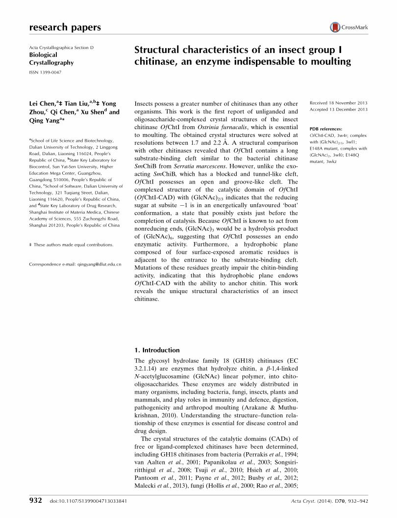
Figure 3The effect of mutations of the four solvent-exposed aromatic residues on the hydrolytic activity and binding affinity. (a) The four aromatic residues,highlighted in cyan on the surface of OfChtI-CAD, form a plane to anchor the plane of the crystalline chitin. A model of chitin is shown in orange. (b)The relative hydrolytic activities of the OfChtI-CAD mutants for pNP-(GlcNAc)2 and crystalline �-chitin compared with wild-type OfChtI-CAD. (c)The decrease in free protein concentration after binding of the wild-type and mutant OfChtI-CADs to crystalline �-chitin was determined at differenttime points over 18 h.
�n represents the nonreducing end and +n represents the
reducing end and cleavage occurs between the �1 and +1
subsites.
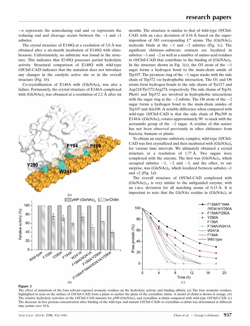
The crystal structure of E148Q at a resolution of 2.0 A was
obtained after a six-month incubation of E148Q with chito-
hexaose. Unfortunately, no substrate was found in the struc-
ture. This indicates that E148Q possesses partial hydrolytic
activity. Structural comparison of E148Q with wild-type
OfChtI-CAD indicates that the mutation does not introduce
any changes in the catalytic active site or in the overall
structure (Fig. 1b).
Co-crystallization of E148A with (GlcNAc)6 was also a
failure. Fortunately, the crystal structure of E148A complexed
with (GlcNAc)2 was obtained at a resolution of 2.2 A after six
months. The structure is similar to that of wild-type OfChtI-
CAD, with an r.m.s. deviation of 0.16 A based on the super-
imposition of 383 corresponding C� atoms. The (GlcNAc)2
molecule binds at the �1 and �2 subsites (Fig. 1c). The
significant chitinase–substrate contacts are localized in
subsites�1 and�2 as well as a number of amino-acid residues
in OfChtI-CAD that contribute to the binding of (GlcNAc)2.
In the structure shown in Fig. 1(e), the O3 atom of the �1
sugar forms a hydrogen bond to the main-chain amide of
Trp107. The pyranose ring of the �1 sugar stacks with the side
chain of Trp372 via hydrophobic interaction. The O1 and O6
atoms form hydrogen bonds to the side chains of Tyr217 and
Asp218/Tyr272/Arg274, respectively. The side chains of Trp34,
Phe61 and Trp372 are involved in hydrophobic interactions
with the sugar ring at the �2 subsite. The O6 atom of the �2
sugar forms a hydrogen bond to the main-chain amides of
Trp107 and Ala108. A notable difference when compared with
wild-type OfChtI-CAD is that the side chain of Phe309 in
E148A–(GlcNAc)2 rotates approximately 90� to stack with the
acetamido group of the �2 sugar. A residue of this nature
has not been observed previously in other chitinases from
bacteria, humans or plants.
To obtain an enzyme–substrate complex, wild-type OfChtI-
CAD was first crystallized and then incubated with (GlcNAc)6
for various time intervals. We ultimately obtained a crystal
structure at a resolution of 1.77 A. Two sugars were
complexed with the enzyme. The first was (GlcNAc)3, which
occupied subsites �1, �2 and �3, and the other, to our
surprise, was (GlcNAc)2, which localized between subsites +1
and +2 (Fig. 1d).
The overall structure of OfChtI-CAD complexed with
(GlcNAc)2/3 is very similar to the unliganded enzyme, with
an r.m.s. deviation for all matching atoms of 0.13 A. It is
important to note that the GlcNAc residue in (GlcNAc)3 at
the �1 subsite is in an unfavoured ‘boat’ 1,4B conformation
(Fig. 1f). Additionally, both the C2-acetamido group and O1
of this �1 sugar adopt different conformations from those in
E148A–(GlcNAc)2. The O1 of the �1 sugar, which is the
scissile glycosidic O atom, is within a distance of 2.5 A of the
catalytic O atom of the �-carboxyl side chain of the catalytic
proton donor Glu148. The C2-acetamido group of the �1
sugar is in a conformation that facilitates its O atom being
3.1 A away from the C1, and this O atom forms a hydrogen
bond to Tyr217. The C2-acetamido group of the �1 sugar is
also stabilized by the side chains of the catalytic residues
Glu148 and Asp146. The side chain of Asp146 rotates
approximately 91� and then faces Glu148 (Fig. 1f). This
conformational change breaks the interaction between
Asp146 and Asp144, but forms a hydrogen bond with the side
chain of Glu148 instead. The sugar residue at the �2 subsite
adopts the same conformation as observed in E148A–
(GlcNAc)2. The interactions in this subsite are similar in the
two complexed structures. The pyranose ring of the �3 sugar
interacts with the aromatic residue Trp34 via a stacking
interaction. For the interactions between (GlcNAc)2 and
subsites +1 and +2, the O3 and O4 atoms of the +1 sugar form
hydrogen bonds to residues Asp218 and Tyr149, respectively,
the pyranose ring of the +1 sugar stacks with Trp107, and the
pyranose ring of the +2 sugar stacks with Trp223 (Fig. 1f).
3.3. Roles of the residues Phe159, Phe194, Trp241 andTyr290
The chitin-binding electrophoresis experiments indicated
that OfChtI-CAD has an affinity for crystalline �-chitin
(Supplementary Fig. S3). The interaction between OfChtI-
research papers
938 Chen et al. � Group I chitinase Acta Cryst. (2014). D70, 932–942
Figure 4Thermograms and binding isotherms with theoretical fits for the binding of (GlcNAc)3–6 to the OfChtI-CAD E148Q mutant: (a) (GlcNAc)3, (b)(GlcNAc)4, (c) (GlcNAc)5, (d) (GlcNAc)6. (e) Free-energy changes for (GlcNAc)3–6 binding to OfChtI-CAD E148Q and (GlcNAc)4–6 binding toSmChiB E144Q (from Norberg et al., 2010) relative to the number of GlcNAc units in the saccharide chain. (GlcNAc)3 binds to OfChtI-CAD E148Q attwo sites. S1 is the strong binding site with a low Kd value and S2 is the weak binding site with a high Kd value.

CAD and �-chitin could not be destroyed by a high concen-
tration of salt and acetic acid, suggesting that the affinity was
strong (Supplementary Fig. S3). Based on the crystal struc-
tures, we suspect that a hydrophobic plane formed by the four
solvent-exposed residues Phe159, Phe194, Trp241 and Tyr290
might contribute to the binding of OfChtI-CAD to chitin
(Fig. 3a).
To confirm whether these surface aromatic residues parti-
cipate in chitin binding, four single-residue mutants (F159A,
F194A, W241A and Y290A), two double-residue mutants
(F194A/W241A, F159A/Y290A) and one four-residue mutant
(F159A/F194A/W241A/Y290A) were constructed. All of the
OfChtI-CAD variants were expressed and purified to homo-
geneity.
The enzymatic assays indicated that all of these mutants
exhibit the same hydrolytic activities towards pNP-(GlcNAc)2,
suggesting that the catalytic activity was not impaired when
a short oligosaccharide was used as the substrate (Fig. 3b).
However, if the long crystalline �-chitin was used as the
substrate, the mutations caused differing degrees of reduction
in the enzymatic activity (Fig. 3b). Phe194, which is the closest
residue to the substrate-binding cleft, is the most crucial for
chitin-hydrolysis activity. Mutation of any of the other three
residues affected the chitin-hydrolysis activity to the same
degree. Therefore, we deduce that Phe194 may be responsible
for binding a single chitin chain during hydrolysis, while the
other three residues are more likely to anchor the chitin plane.
The binding capacities of wild-type OfChtI-CAD and its
mutants were tested by incubating the proteins with �-chitin.
The amount of protein bound to �-chitin was analyzed by
determining the concentration of free protein in the super-
natant at different time points. As shown in Fig. 3(c), the
binding capacity decreased dramatically when the four resi-
dues were mutated. For the four-residue mutant, approxi-
mately 30% of the protein was retained in the supernatant
even after an 18 h incubation. However, for the wild type, no
free protein was left after a 6 h incubation. To determine the
contribution of each residue, the binding ability of the four
single-residue mutants was tested and concluded to be in the
following order: Y290A < F159A < W241A < F194A. The
mutation of Y290A and F159A caused a more drastic decrease
than the mutation of W241A and F194A. The single-residue
mutation of Y290A or F159A caused a more significant
decrease than the double-residue F194A/W241A mutation,
which indicated that residues Tyr290 and Phe159 are the major
contributors to the binding ability. Mutation of Phe194 had
very little effect on chitin binding, meaning that it was the
smallest contributor.
The above results prove that the four residues forming a
hydrophobic plane may not be involved in the catalytic
process but instead in binding the chitin substrate.
3.4. Free-energy changes during the binding of OfChtI tosubstrates
The substrate-binding energy changes for E148Q were
measured by ITC analysis using (GlcNAc)3–6 as ligands.
Because (GlcNAc)2 is the product of OfChtI, (GlcNAc)2 was
not included. The substrate binding was assessed in 50 mM
HEPES pH 8.0 at 303 K. The thermograms and titration
curves for (GlcNAc)3–6 are shown in Fig. 4. The derived
thermodynamic parameters of binding, Kd, �H, �S and �G,
for (GlcNAc)3–6 are summarized in Table 2.
As shown in Fig. 4(a), the titration curve for (GlcNAc)3
binding to E148Q fit well to a two-site binding model with a
stoichiometry of 1.84. (GlcNAc)3 bound to E148Q with an
approximate fourfold affinity difference (Kd1 = 76.9 mM and
Kd2 = 284.1 mM) in the two binding sites. For convenience, we
named the site with the low Kd value the ‘strong binding site’
and the site with the high Kd value the ‘weak binding site’.
Both types of binding were exothermic (�H < 0) and were
entropically driven with an enthalpic contribution (�H < 0,
|�T�S| > |�H|; Table 2).
For (GlcNAc)4–6, the titration curves fitted to a single-site
binding model (Figs. 4b, 4c and 4d). According to the Kd
values of 11.3, 2.0 and 0.48 mM for (GlcNAc)4, (GlcNAc)5 and
(GlcNAc)6, respectively, the highest Kd value indicates that
the binding of (GlcNAc)4 is the weakest, and an increase in
the polymerization degree of GlcNAc by one unit results in a
gain in affinity of approximately fivefold. The higher affinity
for (GlcNAc)6 compared with (GlcNAc)5 suggests that there
are at least six sugar-binding subsites in the substrate-binding
groove, which is in accordance with the crystal structural
data. The enthalpy values are in the range �1.45 to
�2.89 kcal mol�1, indicating that the binding of (GlcNAc)4–6
is exothermic. Based on the entropy values (�T�S) of �4.67,
�5.00 and �7.31 kcal mol�1 for (GlcNAc)4, (GlcNAc)5 and
(GlcNAc)6, respectively, together with the fact that |�T�S| >
|�H| (Table 2), we determined that the binding of all oligo-
saccharides examined is entropically driven. This evidence
suggests that substrate binding is accompanied by desolvation
and conformational changes of both the protein and its
ligands.
research papers
Acta Cryst. (2014). D70, 932–942 Chen et al. � Group I chitinase 939
Table 2Thermodynamic parameters of (GlcNAc)3–6 binding to the mutant E148Q derived from ITC.
n Kd (mM)�G(kcal mol�1)
�H(kcal mol�1)
�T�S(kcal mol�1)
(GlcNAc)3 1.84 � 0.12 Kd1, 76.9 � 18.9;Kd2, 284.1 � 97.4
�G1, �5.70;�G2, �4.91
�H1 �1.47 � 0.15;�H2, �1.21 � 0.34
�T�S1, �4.23;�T�S2, �3.70
(GlcNAc)4 0.93 � 0.01 11.3 � 1.5 �6.84 �2.17 � 0.05 �4.67(GlcNAc)5 0.92 � 0.02 2.0 � 0.2 �7.89 �2.89 � 0.23 �5.00(GlcNAc)6 0.93 � 0.02 0.48 � 0.09 �8.76 �1.45 � 0.02 �7.31
To analyze the binding-energy changes of each ligand, the
free energies of binding of (GlcNAc)3–6 were plotted against
the polymerization degree of GlcNAc (Fig. 4e). Notably, the
binding energy of (GlcNAc)3 in the strong binding site, as well
as the binding energies of (GlcNAc)4, (GlcNAc)5 and
(GlcNAc)6, correlated well with the function y = �1.02x �
2.69 (where y is the free-energy change and x is the number of
sugar residues, i.e. the polymerization degree). As shown
in Fig. 4(e), an increase in x, namely an increase in the
polymerization degree of GlcNAc [from (GlcNAc)3 to
(GlcNAc)6], results in an average free-energy gain of
approximately �1.0 kcal mol�1 per GlcNAc residue. Addi-
tionally, the binding of (GlcNAc)6 to OfChtI releases
1.0 kcal mol�1 more free energy than the binding of
(GlcNAc)5 to OfChtI, demonstrating that OfChtI possesses
at least six substrate-binding subsites, as deduced from the
crystal structures.
4. Discussion
Here, we report the crystal structures of unliganded and
oligosaccharide-complexed OfChtI-CADs, which represent
the first examples of insect GH18 chitinases.
4.1. Substrate binding sites
Based on the ITC analysis of OfChtI-CAD E148Q binding
(GlcNAc)4–6, an increase in the polymerization degree of
GlcNAc [from (GlcNAc)3 to (GlcNAc)6] results in an average
free-energy gain of approximately �1.0 kcal mol�1 per
GlcNAc residue. Similarly, according to Norberg et al. (2010),
the binding of chitooligosaccharides to bacterial SmChiB
E144Q showed that the free energies for binding (GlcNAc)4,
(GlcNAc)5 and (GlcNAc)6 were �7.4, �8.3 and
�9.2 kcal mol�1, respectively. The binding energies for
(GlcNAc)4, (GlcNAc)5 and (GlcNAc)6 correlated well with
the function y = �0.9x � 3.8, indicating that the free-energy
gain for each added GlcNAc residue was �0.9 kcal mol�1
(Fig. 4e). The above results demonstrate that the insect
OfChtI binds oligosaccharide substrates in similar manner as
bacterial chitinase and that both enzymes possess at least six
substrate-binding subsites.
4.2. OfChtI-CAD possesses endo-acting activity
Although most aromatic residues along the cleft are highly
conserved, a detailed structural comparison between OfChtI-
CAD and the well characterized SmChiB indicates that these
two enzymes possess different structural characteristics in the
substrate-binding cleft. SmChiB has a tunnel-like cleft with a
blunted nonreducing end. A unique loop (residues 311–322)
forms the roof of the tunnel (Fig. 2b), and the Asp316 residue
in the loop directly interacts with Trp97 on the other side of
the active pocket. The nonreducing end adjacent to subsite�3
is blocked by a short helix and a loop (residues 14–29).
Moreover, the side chain of Phe12 at subsite �3 is positioned
perpendicular to the sugar plane, making its interaction with
the �3 sugar via a stacking interaction impossible (Fig. 2b). In
contrast, OfChtI-CAD has a groove-like cleft with both the
reducing end and the nonreducing end open. No short helices
or loops are found at the ends of the cleft, and the cleft is
exposed to solvent by the lack of the roof that is present in
SmChiB (Fig. 2a). The Trp34 residue at the deduced subsite
�3 provides a strong stacking interaction with the pyranose
ring of the �3 sugar (Fig. 2a).
SmChiB is a well known exo-acting chitinase. The hypoth-
esis is that an exo-chitinase, such as SmChiB, may be char-
acterized by a tunnel-like and blunted substrate-binding cleft.
Therefore, it follows that an endo-chitinase may lack these
characteristics. For OfChtI-CAD, the cleft is groove-like with
both the nonreducing end and the reducing end open. The
crystal structure of wild-type OfChtI-CAD complexed with
(GlcNAc)2/3 indicates that (GlcNAc)3 binds at subsites�1,�2
and �3, and the reducing sugar at subsite �1 sits in an ener-
getically unfavoured ‘boat’ conformation. According to the
deduced mechanism (Supplementary Fig. S2), this ‘boat’
conformation of the �1 sugar is likely to be a state that occurs
just before the completion of catalysis, in which the product
has left and the catalysis-associated Asp146 residue is still
interacting with the �1 sugar. Because OfChtI shows activity
from the nonreducing ends of substrates (Wu et al., 2013), the
(GlcNAc)3 with a reducing sugar at subsite �1 suggests that
(GlcNAc)3 could be a hydrolysis product of (GlcNAc)6. Thus,
we provide direct evidence that a chitinase possessing a
groove-like and open-ended cleft, such as OfChtI-CAD, may
possess endo-chitinase activity.
Taken together, although the GH18 chitinases employ the
same catalytic mechanism and the substrate-binding cleft is
constituted by a number of conserved aromatic residues, slight
differences in the substrate-binding clefts could confer
different cleavage modes of chitinases during chitinolytic
processes.
4.3. Insect group I chitinase CAD possesses a surfacehydrophobic plane
Experimental results indicate that mutations of the four
planar residues (Phe159, Phe194, Trp241 and Tyr290) resulted
in a significant reduction in the hydrolysis of crystalline
�-chitin, but did not affect the hydrolysis of the short oligo-
saccharide substrate pNP-(GlcNAc)2. Moreover, the muta-
tions seriously affected the binding affinity of �-chitin.
Because the four residues are located far from the catalytic
centre, we deduce that mutations causing a reduction in
hydrolytic activity are related to the decrease in substrate-
binding ability, which may be associated with a substrate-
proximity effect. These results are the first to reveal the
importance of surface-exposed aromatic residues of the cata-
lytic domain during crystalline chitin hydrolysis.
According to a crystal structure-based alignment, the four
aromatic residues are highly conserved in the insect group I
chitinases from species including dipterans, lepidopterans,
coleopterans, hemipterans and hymenopterans, but are absent
in bacteria, fungi, plants and humans (Supplementary Fig. S1).
This indicates that the four residues forming the hydrophobic
research papers
940 Chen et al. � Group I chitinase Acta Cryst. (2014). D70, 932–942
plane to anchor the chitin are a conserved and unique char-
acteristic of the group I insect chitinases.
5. Conclusions
The first crystal structure of insect chitinase OfChtI-CAD has
now been described. The oligosaccharide-complexed crystal
structure with the �1 reducing sugar sitting in a ‘boat’
conformation may provide evidence that a chitinase with
a groove-like and open-ended cleft possesses endo-acting
activity. A unique conserved hydrophobic plane in this insect
chitinase increases the chitin-binding affinity. Because OfChtI
plays a vital role in insect moulting, this work provides a
possibility for the development of new technologies for pest
control and pest management.
This work was supported by the National Key Project
for Basic Research (2010CB126100), the National Natural
Science Foundation of China (31070715, 31101671 and
61202252), the National High Technology Research and
Development Program of China (2011AA10A204), the Open
Research Fund of the State Key Laboratory for Biocontrol
at Sun Yat-Sen University (SKLBC13KF01), the National
Key Technology R&D Program (2011BAE06B05) and the
Specialized Research Fund for the Doctoral Program of
Higher Education of China (20120041120052).
References
Aalten, D. M. F. van, Komander, D., Synstad, B., Gaseidnes, S., Peter,M. G. & Eijsink, V. G. H. (2001). Proc. Natl Acad. Sci. USA, 98,8979–8984.
Aalten, D. M. F. van, Synstad, B., Brurberg, M. B., Hough, E., Riise,B. W., Eijsink, V. G. H. & Wierenga, R. K. (2000). Proc. Natl Acad.Sci. USA, 97, 5842–5847.
Adams, P. D. et al. (2010). Acta Cryst. D66, 213–221.Arakane, Y. & Muthukrishnan, S. (2010). Cell. Mol. Life Sci. 67,
201–216.Arimori, T., Kawamoto, N., Shinya, S., Okazaki, N., Nakazawa, M.,
Miyatake, K., Fukamizo, T., Ueda, M. & Tamada, T. (2013). J. Biol.Chem. 288, 18696–18706.
Brameld, K. A. & Goddard, W. A. III (1998a). J. Am. Chem. Soc. 120,3571–3580.
Brameld, K. A. & Goddard, W. A. III (1998b). Proc. Natl Acad. Sci.USA, 95, 4276–4281.
Busby, J. N., Landsberg, M. J., Simpson, R. M., Jones, S. A.,Hankamer, B., Hurst, M. R. & Lott, J. S. (2012). J. Mol. Biol. 415,359–371.
Cavada, B. S. et al. (2006). FEBS J. 273, 3962–3974.Davies, G. J., Wilson, K. S. & Henrissat, B. (1997). Biochem. J. 321,
557–559.DeLano, W. L. (2002). PyMOL. http://www.pymol.org.Dundas, J., Ouyang, Z., Tseng, J., Binkowski, A., Turpaz, Y. & Liang,
J. (2006). Nucleic Acids Res. 34, W116–W118.Emsley, P., Lohkamp, B., Scott, W. G. & Cowtan, K. (2010). Acta
Cryst. D66, 486–501.Fusetti, F., von Moeller, H., Houston, D., Rozeboom, H. J., Dijkstra,
B. W., Boot, R. G., Aerts, J. M. & van Aalten, D. M. F. (2002). J.Biol. Chem. 277, 25537–25544.
Gouet, P., Robert, X. & Courcelle, E. (2003). Nucleic Acids Res. 31,3320–3323.
Hahn, M., Hennig, M., Schlesier, B. & Hohne, W. (2000). Acta Cryst.D56, 1096–1099.
Hoell, I. A., Dalhus, B., Heggset, E. B., Aspmo, S. I. & Eijsink,V. G. H. (2006). FEBS J. 273, 4889–4900.
Hollis, T., Monzingo, A. F., Bortone, K., Ernst, S., Cox, R. & Robertus,J. D. (2000). Protein Sci. 9, 544–551.
Hsieh, Y.-C., Wu, Y.-J., Chiang, T.-Y., Kuo, C.-Y., Shrestha, K. L.,Chao, C.-F., Huang, Y.-C., Chuankhayan, P., Wu, W.-G., Li, Y.-K. &Chen, C.-J. (2010). J. Biol. Chem. 285, 31603–31615.
Huet, J., Rucktooa, P., Clantin, B., Azarkan, M., Looze, Y., Villeret, V.& Wintjens, R. (2008). Biochemistry, 47, 8283–8291.
Hurtado-Guerrero, R. & van Aalten, D. M. F. (2007). Chem. Biol. 14,589–599.
Imoto, T. & Yagishita, K. (1971). Agric. Biol. Chem. 35, 1154–1156.Kezuka, Y., Ohishi, M., Itoh, Y., Watanabe, J., Mitsutomi, M.,
Watanabe, T. & Nonaka, T. (2006). J. Mol. Biol. 358, 472–484.Laskowski, R. A., MacArthur, M. W., Moss, D. S. & Thornton, J. M.
(1993). J. Appl. Cryst. 26, 283–291.Lu, Y., Zen, K. C., Muthukrishnan, S. & Kramer, K. J. (2002). Insect
Biochem. Mol. Biol. 32, 1369–1382.Malecki, P. H., Raczynska, J. E., Vorgias, C. E. & Rypniewski, W.
(2013). Acta Cryst. D69, 821–829.McCoy, A. J., Grosse-Kunstleve, R. W., Adams, P. D., Winn, M. D.,
Storoni, L. C. & Read, R. J. (2007). J. Appl. Cryst. 40, 658–674.Norberg, A. L., Karlsen, V., Hoell, I. A., Bakke, I., Eijsink, V. G. H. &
Sørlie, M. (2010). FEBS Lett. 584, 4581–4585.Ohnuma, T., Numata, T., Osawa, T., Inanaga, H., Okazaki, Y., Shinya,
S., Kondo, K., Fukuda, T. & Fukamizo, T. (2012). FEBS J. 279,3639–3651.
Ohnuma, T., Numata, T., Osawa, T., Mizuhara, M., Lampela, O.,Juffer, A. H., Skriver, K. & Fukamizo, T. (2011). Planta, 234,123–137.
Ohnuma, T., Numata, T., Osawa, T., Mizuhara, M., Varum, K. M. &Fukamizo, T. (2011). Plant Mol. Biol. 75, 291–304.
Olland, A. M., Strand, J., Presman, E., Czerwinski, R., Joseph-McCarthy, D., Krykbaev, R., Schlingmann, G., Chopra, R., Lin, L.,Fleming, M., Kriz, R., Stahl, M., Somers, W., Fitz, L. & Mosyak, L.(2009). Protein Sci. 18, 569–578.
Otwinowski, Z. & Minor, W. (1997). Methods Enzymol. 276, 307–326.Pantoom, S., Vetter, I. R., Prinz, H. & Suginta, W. (2011). J. Biol.
Chem. 286, 24312–24323.Papanikolau, Y., Tavlas, G., Vorgias, C. E. & Petratos, K. (2003). Acta
Cryst. D59, 400–403.Payne, C. M., Baban, J., Horn, S. J., Backe, P. H., Arvai, A. S., Dalhus,
B., Bjøras, M., Eijsink, V. G. H., Sørlie, M., Beckham, G. T. & Vaaje-Kolstad, G. (2012). J. Biol. Chem. 287, 36322–36330.
Pei, J., Kim, B.-H. & Grishin, N. V. (2008). Nucleic Acids Res. 36,2295–2300.
Perrakis, A., Tews, I., Dauter, Z., Oppenheim, A. B., Chet, I., Wilson,K. S. & Vorgias, C. E. (1994). Structure, 2, 1169–1180.
Rao, F. V., Houston, D. R., Boot, R. G., Aerts, J. M., Hodkinson, M.,Adams, D. J., Shiomi, K., Omura, S. & van Aalten, D. M. F. (2005).Chem. Biol. 12, 65–76.
Rush, C. L., Schuttelkopf, A. W., Hurtado-Guerrero, R., Blair, D. E.,Ibrahim, A. F., Desvergnes, S., Eggleston, I. M. & van Aalten,D. M. F. (2010). Chem. Biol. 17, 1275–1281.
Schuttelkopf, A. W., Gros, L., Blair, D. E., Frearson, J. A., van Aalten,D. M. F. & Gilbert, I. H. (2010). Bioorg. Med. Chem. 18, 8334–8340.
Song, H. K. & Suh, S. W. (1996). Acta Cryst. D52, 289–298.Songsiriritthigul, C., Pantoom, S., Aguda, A. H., Robinson, R. C. &
Suginta, W. (2008). J. Struct. Biol. 162, 491–499.Sutherland, T. E., Andersen, O. A., Betou, M., Eggleston, I. M.,
Maizels, R. M., van Aalten, D. & Allen, J. E. (2011). Chem. Biol. 18,569–579.
Synstad, B., Gaseidnes, S., Van Aalten, D. M. F., Vriend, G., Nielsen,J. E. & Eijsink, V. G. H. (2004). Eur. J. Biochem. 271, 253–262.
Tews, I., Terwisscha van Scheltinga, A. C., Perrakis, A., Wilson, K. S.& Dijkstra, B. W. (1997). J. Am. Chem. Soc. 119, 7954–7959.
Tsuji, H., Nishimura, S., Inui, T., Kado, Y., Ishikawa, K., Nakamura, T.& Uegaki, K. (2010). FEBS J. 277, 2683–2695.
research papers
Acta Cryst. (2014). D70, 932–942 Chen et al. � Group I chitinase 941

Ubhayasekera, W., Rawat, R., Ho, S. W. T., Wiweger, M., Von Arnold,S., Chye, M.-L. & Mowbray, S. L. (2009). Plant Mol. Biol. 71,277–289.
Ubhayasekera, W., Tang, C. M., Ho, S. W. T., Berglund, G., Bergfors,T., Chye, M.-L. & Mowbray, S. L. (2007). FEBS J. 274, 3695–3703.
Vaaje-Kolstad, G., Houston, D. R., Rao, F. V., Peter, M. G., Synstad,B., van Aalten, D. M. F. & Eijsink, V. G. H. (2004). Biochim.Biophys. Acta, 1696, 103–111.
Varela, P. F., Llera, A. S., Mariuzza, R. A. & Tormo, J. (2002). J. Biol.Chem. 277, 13229–13236.
Wu, Q., Liu, T. & Yang, Q. (2013). Insect Sci. 20, 147–157.Yang, J., Gan, Z., Lou, Z., Tao, N., Mi, Q., Liang, L., Sun, Y., Guo, Y.,
Huang, X., Zou, C., Rao, Z., Meng, Z. & Zhang, K.-Q. (2010).Microbiology, 156, 3566–3574.
Zhu, Q., Arakane, Y., Banerjee, D., Beeman, R. W., Kramer, K. J. &Muthukrishnan, S. (2008). Insect Biochem. Mol. Biol. 38, 452–466.
Zhu, Q., Arakane, Y., Beeman, R. W., Kramer, K. J. & Muthu-krishnan, S. (2008). Proc. Natl Acad. Sci. USA, 105, 6650–6655.
research papers
942 Chen et al. � Group I chitinase Acta Cryst. (2014). D70, 932–942