structural basis for the function of stringent starvation protein a as
TRANSCRIPT

Structural Basis for the Function of Stringent Starvation Protein Aas a Transcription Factor*
Received for publication, February 7, 2005Published, JBC Papers in Press, February 25, 2005, DOI 10.1074/jbc.M501444200
Anne-Marie Hansen‡§, Yijun Gu¶�, Mi Li**, Michelle Andrykovitch¶‡‡, David S. Waugh¶,Ding Jun Jin‡§§, and Xinhua Ji¶ ¶¶
From the ‡Transcription Control Section, Gene Regulation and Chromosome Biology Laboratory, NCI-Frederick, NationalInstitutes of Health, Frederick, Maryland 21702, the §Department of Biochemistry and Molecular Biology, University ofSouthern Denmark, Campusvej 55, DK-5230 Odense M, Denmark, the ¶Macromolecular Crystallography Laboratory,NCI-Frederick, National Institutes of Health, Frederick, Maryland 21702, and the **Basic Research Program, SAIC-Frederick, Frederick, Maryland 21702
Stringent starvation protein A (SspA) of Escherichiacoli is an RNA polymerase-associated transcriptional ac-tivator for the lytic development of phage P1 and isessential for stationary phase-induced acid tolerance ofE. coli. We report the crystal structure of Yersinia pestisSspA, which is 83% identical to E. coli SspA in aminoacid sequence and is functionally complementary insupporting the lytic growth of phage P1 and acid resist-ance of an E. coli sspA mutant. The structure revealsthat SspA assumes the characteristic fold of glutathioneS-transferase (GST). However, SspA lacks GST activityand does not bind glutathione. Three regions of SspA areflexible, the N and C termini and the �2-helix. The struc-ture also reveals a conserved surface-exposed pocketcomposed of residues from a loop between helices �3and �4. The functional roles of these structural featureswere investigated by assessing the ability of deletionand site-directed mutants to confer acid resistance ofE. coli and to activate transcription from a phage P1 latepromoter, thereby supporting the lytic growth of phageP1. The results indicate that the flexible regions are notcritical for SspA function, whereas the surface pocket isimportant for both transcriptional activation of thephage P1 late promoter and acid resistance of E. coli.The size, shape, and property of the pocket suggest thatit mediates protein-protein interactions. SspA orthologsfrom Y. pestis, Vibrio cholerae, and Pseudomonas aerugi-nosa are all functional in acid resistance of E. coli,whereas only Y. pestis SspA supports phage P1 growth.
Control of transcription is a key step in the regulation of geneexpression in all living cells. Escherichia coli RNA polymerase
(RNAP)1 (1, 2) is a multisubunit enzyme (3) responsible for thetranscription process, which consists of initiation, elongation,termination, and recycling steps (for reviews, see Refs. 4–6).One of the major control points is transcription initiation, inwhich transcription factors modulate the activity of RNAPeither positively or negatively at a particular promoter. TheE. coli stringent starvation protein A (SspA) is an RNAP-associated protein (7) required for the transcriptional activa-tion of phage P1 late genes (8), of which the expression isimportant for the lytic development of phage P1 (9). Althoughat present its role in transcription is unknown, SspA is knownto be implicated in survival during nutrient starvation andprolonged stationary phase (10). Recently, SspA was shown toplay an important role in the stationary phase-induced stressresponse including acid tolerance by down-regulating the levelof the global regulator H-NS (68), which negatively regulatesmultiple stress defense systems (11, 12). In addition, SspA ishighly conserved among Gram-negative bacteria. SspA or-thologs in Neisseria gonorrhoeae, Francisella novicida, Fran-cisella tularensis, and Vibrio cholerae were shown to affect theexpression of genes involved in pathogenesis (13–17).
To gain insight into the structural basis for the function ofSspA in transcriptional activation of the phage P1 late pro-moter and in stationary phase induced acid tolerance of E. coli,we determined the three-dimensional structure of SspA. Tothis end, we cloned, expressed, purified, and characterizedSspA from E. coli, Y. pestis, V. cholerae, and Pseudomonasaeruginosa (18). All four SspA proteins were subjected to crys-tallization trials. E. coli SspA crystallized but failed to diffractx-rays well enough for structure determination. P. aeruginosaSspA also did not form crystals that were suitable for datacollection, and V. cholerae SspA failed to crystallize under anyconditions tested. Only Y. pestis SspA crystallized readily, andthe crystals diffracted to 2.0 Å. Here, we report the crystalstructure of Y. pestis SspA, a homologous model of E. coli SspA,and the structural basis of E. coli SspA function in the devel-opment of phage P1 and in stationary phase-induced acid re-sistance of E. coli.
EXPERIMENTAL PROCEDURES
X-ray Diffraction Data Collection and Processing—Crystallizationand preliminary x-ray diffraction experiments have been reported else-
* This work was supported in part with Federal funds from the NCI,National Institutes of Health, under Contract No. NO1-CO-24000. Thecosts of publication of this article were defrayed in part by the paymentof page charges. This article must therefore be hereby marked “adver-tisement” in accordance with 18 U.S.C. Section 1734 solely to indicatethis fact.
The atomic coordinates and structure factors (code 1YY7) have beendeposited in the Protein Data Bank, Research Collaboratory for Struc-tural Bioinformatics, Rutgers University, New Brunswick, NJ(http://www.rcsb.org/).
� Current address: Shanghai Innovative Research Center, 439Chunxiao Road, Shanghai 201203, China.
‡‡ Current address: Agentase, LLC. 3636 Boulevard of the Allies,Pittsburgh, PA 15213.
§§ To whom correspondence may be addressed: NCI-Frederick, 1050Boyles St., Bldg. 469, Rm. 127, Frederick, MD 21702-1201. Tel.: 301-846-7684; Fax: 301-846-1456; E-mail: [email protected].
¶¶ To whom correspondence may be addressed: NCI-Frederick, 1050Boyles St., Bldg. 539, Rm. 124, Frederick, MD 21702-1201. Tel.: 301-846-5035; Fax: 301-846-6073; E-mail: [email protected].
1 The abbreviations used are: RNAP, RNA polymerase; PDB, ProteinData Bank; SspA, stringent starvation protein A; MAD, multiwave-length anomalous diffraction; MIR, multiple isomorphous replacement;GSH, glutathione; GST, glutathione S-transferase; GST A1-1, alpha-class GST subunit-type 1; CDNB, 1-chloro-2,4-dinitro-benzene; IPTG,isopropyl-thio-�-D-galactoside; GST S1-1, sigma-class GST subunit type1; GST B1-1, beta-class GST subunit type 1; r.m.s., root mean square.
THE JOURNAL OF BIOLOGICAL CHEMISTRY Vol. 280, No. 17, Issue of April 29, pp. 17380–17391, 2005Printed in U.S.A.
This paper is available on line at http://www.jbc.org17380

where (18). The SspA crystals belong to space group P6522 (unit celldimensions a � b � 100.4 Å, c � 181.9 Å). Native data, at the resolutionof 2.0 Å, were collected at the beamline X9B of the National Synchro-tron Light Source (Brookhaven National Laboratories) using an ADSCQuantum-4 CCD detector. The Pt-derivative crystal was obtained bysoaking native crystals in a solution that was similar to the motherliquor and contained 1.0 mM K2PtCl4. The 2.4-Å Pt-derivative data setwas collected with an in-house MAR-345 image plate system mountedon a Rigaku rotating anode operated at 50 kV and 100 mA. Multiwave-length anomalous diffraction (MAD) data collection for the Pt-deriva-tive crystal was attempted at the National Synchrotron Light Source.Unfortunately, the crystal diffracted to only 2.8 Å. All crystals wereflash frozen and maintained at �173 °C for experiments carried outboth in-house and at the synchrotron facilities. The crystallizationmother liquor plus 10% MPD was used as the cryoprotectant for datacollection. The raw data were processed with Denzo and Scalepack (19).Further data processing was performed with the CCP4 package (20).X-ray data statistics are summarized in Table I.
Crystal Structure Determination—Several phasing options were ex-plored, including MAD phasing at 2.8 Å, single isomorphous replace-ment with anomalous scattering at 2.4 Å, and multiple isomorphousreplacement (MIR) using both 2.4- and 2.8-Å Pt-derivative data sets.SOLVE (21) was used in all phasing trials; the MIR approach resultedin the best electron density map. Four common Pt sites were identifiedin both Pt derivatives. Additionally, two minor sites were located ineach derivative. The heavy atom sites were then refined with SHARP(22), and the phases were improved with DM (23) and Solomon (24).ARP/WARP (25) was used to refine and extend the phases to 2.0 Å andto build the initial model, which contained 389 out of 426 residues.Multiple rounds of refinement were performed using CNS (26) with5.0% of the data reserved for cross-validation. As the refinement pro-gressed, more residues were located and built into the model and watermolecules were identified as peaks in the Fo � Fc electron density mapequal to or higher than 3.0 �. The refinement was coupled with bulksolvent correction. Model building was carried out with O (27). The finalmodel contains 410 amino acids, 441 water molecules, and 2 citrateions. The average B factor for all atoms is 25.2 Å2. The quality of thestructure was assessed using PROCHECK (28). A total of 93.4% of themain chain torsion angles are in most favored regions and none indisallowed regions. The refinement statistics can be found in Table I.
Molecular Modeling—The crystal structure of Y. pestis SspA wasused for the construction of an initial model of E. coli SspA. Watermolecules in the Y. pestis structure were excluded. A monomer of E. coliSspA was built with the mutation facility embedded in O (27). Then adimer of E. coli SspA was generated by mimicking the dimeric Y. pestisprotein. The dimer was subjected to geometry optimization using theconjugate gradient method of Powell (29) embedded in CNS (30). Thegeometric parameters of Engh and Huber (31) were used as the basis ofthe force field.
Glutathione Binding Test—The SspA crystals were soaked in a so-lution that was similar to the crystallization mother liquor and con-tained 1–5 mM glutathione (GSH) or S-hexyl-GSH for 6–24 h beforethey were frozen for data collection at the National Synchrotron LightSource. Details of data acquisition were as described above. For refine-ment, the final structure of Y. pestis SspA was used as the starting
model. For each data set, one round of refinement using simulatedannealing (30) was followed by Difference Fourier synthesis using CNS(26). Visual inspection of the resulting electron density maps was per-formed with O (27). Neither GSH nor S-hexyl-GSH was identified in thestructures; therefore, no further refinement was pursued.
BLAST Searches and Multiple Amino Acid Sequence Alignment—BLAST searches were performed to identify orthologs of SspA ingenomic sequences available on GenBankTM sites of the National Cen-ter for Biotechnology Information (www.ncbi.nlm.nih.gov/) using thetBLASTn program with Blosum 62 matrix; Gap penalties were set at 11(Existence) and 1 (Extension). In addition, genome data on Vibriofischeri were obtained from the website www.integratedgenomics.com.The multiple sequence alignment was done using ClustalW (32) with aBlosum 30 matrix (gap opening penalty 10 and gap extension penalty0.05) and refined manually using the GeneDoc program (www.psc.edu/biomed/genedoc, Ref. 33).
Measurement of Glutathione S-Transferase Activity—The glutathi-one S-transferase (GST) activity of Y. pestis SspA, E. coli SspA, RpoS,and alpha-class GST subunit-type 1 (GST A1-1) (PanVera) was meas-ured as their ability to catalyze the conjugation of 1-chloro-2,4-dinitro-benzene (CDNB) to GSH using a GST Detection Module (AmershamBiosciences) according to the manufacturer’s instructions. Specific GSTactivity was calculated as �mol�1 min�1 mg protein�1.
Bacterial Strains, Plasmids, Media, and Techniques—Bacterialstrains used in this study were derivatives of E. coli K12 strainMG1655. Bacterial media, agar plates, and techniques were carried outas described (34). Standard DNA techniques, agar plates and liquidmedia were used as described (35). Restriction endonucleases (NewEngland Biolabs, Beverly, MA) and the Expand High Fidelity PCRSystem (Roche Applied Science) used to amplify DNA by PCR, wereused according to the manufacturer’s instructions.
Deletions and Site-directed Mutagenesis of E. coli sspA—Open read-ing frames encoding N- and C-terminal deletion mutants of SspA,SspA�1–9, and SspA�204–212, were cloned under the control of anarabinose inducible promoter in pBAD24 (36), resulting in plasmidspDJ602 and pDJ603, respectively.
pDJ602 (pBADsspA�1–9): A DNA fragment encoding a 27-bp 5�-enddeletion of sspA was cloned into the EcoRI/HindIII sites of pBAD24. TheDNA fragment was PCR-amplified from MG1655 chromosomal DNAusing the oligos sspA�1–9 (5�-CAGGACGAATTCACCATGGTAATGA-CGCTGTTTTCCGGTCCT-3�) and sspABADDS (5�-CAGGAC-AAGCTTAACTCCGGCCCAGACGC-3�).
pDJ603 (pBADsspA�204–212): A DNA fragment encoding a 27-bp3�-end deletion of sspA was cloned into the EcoRI/HindIII sites ofpBAD24. The DNA fragment was PCR-amplified from MG1655 chro-mosomal DNA using the oligos sspABADUS (5�-TAGCAGAATTCAC-CATGGCTGTCGCTGCCAACAAACGTTCG-3�) and sspA�204–212(5�-CAGGACAAGCTTAACGTTCTGCTTCAGTTAAAGAAGC-3�).
The following mutants were generated by site-directed mutagenesisof sspA encoded by pDJ600 (68): SspAR82A (pDJ604), SspAP84A(pDJ605), SspAH85A (pDJ606), SspAP86A (pDJ607), SspAY92A(pDJ609), SspAH85A/Y92A (pDJ610), and SspAP84A/H85A/P86A(pDJ611). Site-directed mutagenesis of sspA was carried out using theQuikChange II site-directed mutagenesis kit (Stratagene) as recom-mended by the manufacturer.
TABLE ISummary of X-ray diffraction data and structure refinement of Y. pestis SspA
Native Pt-1 Pt-2
Resolution range (Å) 30.0–2.0 30.0–2.8 30.0–2.4Space group P6522 P6522 P6522Cell dimension (Å) (a � b, c) 100.4 181.9 100.1 180.2 100.6 183.5Completeness (%), overall/last shella 99.5/97.4 96.6/92.6 97.6/95.1Redundancy 7.0 3.4 6.8I/�(I), overall/last shell 16.1/2.8 7.5/1.9 12.6/3.3Rscaling,
b overall/last shell 0.108/0.519 0.130/0.506 0.103/0.289Data used for refinement 36,005Data used for R-free calculations 1,785Final R-factorc 0.179Final R-free 0.215R.m.s. deviations from ideal geometry:
Bond distances (Å) 0.009Bond angles (°) 1.3
a 2.02–2.09, 2.90–2.80, 2.40–2.44 Å, for native, Pt-1, and Pt-2, respectively.b Rscaling� ��I � �I��/�I.c Crystallographic R-factor � �hkl ��Fo� � �Fc��/�hkl Fo.
Structure and Function of Transcription Factor SspA 17381

pQEsspAR82A (pDJ604): Mutagenesis of sspA resulting in the aminoacid substitution R82A was performed using oligos sspAR82AFW(5�-GAATATCTGGATGAGGCTTTCCCGCATCCGCCACTG-3�) andsspAR82ABW (5�-CAGTGGCGGATGCGGGAAAGCCTCATCCA-GATATTC-3�).
pQEsspAP84A (pDJ605): Mutagenesis of sspA resulting in the aminoacid substitution R82A was performed using oligos sspAP84AFW1 (5�-TATCTGGATGAGCGTTTCGCGCATCCGCCACTGATGCCTGTT-3�)and sspAP84ABW1 (5�-AACAGGCATCAGTGGCGGATGCGCGAAAC-GCTCATCCAGATA-3�).
pQEsspAH85A (pDJ606): Mutagenesis of sspA resulting in the aminoacid substitution H85A was performed using oligos sspAH85AFW (5�-CTGGATGAGCGTTTCCCGGCTCCGCCACTGATGCCT-3�) andsspAH85ABW (5�-AGGCATCAGTGGCGGAGCCGGGAAACGCTCA-TCCAG-3�).
pQEsspAP86A (pDJ607): Mutagenesis of sspA resulting in the aminoacid substitution P86A was performed using oligos sspAP86AFW (5�-GATGAGCGTTTCCCGCATGCGCCACTGATGCCTGTT-3�) andsspAP86ABW (5�-AACAGGCATCAGTGGCGCATGCGGGAAACG-CTCATC-3�).
pQEsspAY92A (pDJ609): Mutagenesis of sspA resulting in the aminoacid substitution Y92A was performed using oligos sspAY92AFW (5�-CTGATGCCTGTTGCCCCGGTAGCTCGCGGTGAA-3�) and sspAY-92ABW (5�-TTCACCGCGAGCTACCGGGGCAACAGGCATCAG-3�).
pQEsspAH85A/Y92A (pDJ610): Mutagenesis of sspA resulting insubstitutions of H85A and Y92 with alanine was performed on pDJ606using oligos sspAY92AFW and sspAY92ABW.
pQEsspAP84A/H85A/P86A (pDJ611): Mutagenesis of sspA resulting insubstitutions of P84A, H85A, and P86A was performed using oligossspAP84A/H85A/P86AFW (5�-GGAATAT CTGGATGAGCGTTTCGCG-GCTGCGCCACTGATGCCTGT-3�) and sspAP84A/H85A/P86ABW(5�-ACAGGCATCAGTGGCGCAGCCGCGAAACGCTCATCCAGAT-ATTCC-3�).
Mutant pQEsspABP84A/H85A/P86A (pDJ612) were generated bysite-directed mutagenesis of sspA encoded by pDJ706. Mutagenesis ofsspA resulting in substitution of P84A, H85A, and P86A was performedusing oligos sspAP84A/H85A/P86AFW and sspAP84A/H85A/P86ABW.
Cloning of Y. pestis sspA, V. cholerae sspA, and P. aeruginosa sspA—The constructs pBADsspAYp (pDJ613), pBADsspAVc (pDJ614), andpBADsspAPa (pDJ615) encode Y. pestis sspA, V. cholerae sspA, andP. aeruginosa sspA, respectively, under the control of an arabinoseinducible promoter in pBAD24.
pBADsspAYp (pDJ613): A DNA fragment encoding Y. pestis sspAwas PCR-amplified from pRILSspAYp (18) using the oligos YpVcssp-AUS1 (5�-CCATCGGAATTCACCATGGCTGTCGCTG CCAACAACGT-3�) and YpsspADS (5�-TTTA CTAAGCTTTTAGCTCCGAGTTTTCAGA-TG-3�) and cloned into the EcoRI/HindIII sites of pBAD24.
pBADsspAVc (pDJ614): A DNA fragment encoding V. cholerae sspAwas PCR amplified from pRILsspAVc (18) using the oligos YpVcssp-AUS1 (5�-TAGCAGGGATCCATGGCTGTCGCTGCCAACAAACGT-3�)and VcSspADS (5�-TTTACTAAGCTTTTAGCTGCGAGCCAG ACGCA-TCTC-3�) and cloned into the EcoRI/HindIII sites of pBAD24.
pBADsspAPa (pDJ615): A DNA fragment encoding P. aeruginosasspA was PCR-amplified from pRILsspAPa (18) using the oligos Passp-AUS1 (5�-CCATCGGAATTCACCATGGCTTCAATCAACAAGCTGAC-C-3�) and PasspADS (5�-TTTACTAAGCTTTTAGCTCCGAGTTTTCAG-ATG-3�) and cloned into the EcoRI/HindIII sites of pBAD24.
Acid Resistance Test—The acid resistance test was performed asdescribed previously (68). Overnight cultures (18 h) grown in LB at37 °C were diluted 1:1000 in warm LB, adjusted to pH 2.5 with HCl (6N), and incubated for 2 h at 37 °C. The number of colony forming unitsper milliliter was determined by plating serial dilutions of cells inphosphate-buffered saline (pH 7.4) on LB agar plates before and afterexposure to acid. The LB agar plates were incubated overnight at 37 °C.Acid survival was calculated as the percentage of cells remaining afteracid treatment compared with the initial number of cells. Each exper-iment was repeated independently at least three times.
The ability of E. coli wild-type and mutant SspA proteins, as well asthe Y. pestis, V. cholerae, and P. aeruginosa SspA proteins to comple-ment stationary phase acid intolerance of the sspA mutant strainDJ6000 (68) was determined by expressing sspA in trans from plasmids.The E. coli wild-type (pQEsspA (pDJ600) (68)) and the site-directedmutant sspA genes (pQEsspAR82A, pQEsspAH85A, pQEsspAP86A,pQEsspAY92A, and pQEsspAP84A/H85A/P86A) were expressed from atac promoter in the absence of the inducer isopropyl-thio-�-D-galacto-side (IPTG). The E. coli sspA deletion mutants (pBADsspA�1–9 and
pBADsspA�204–212), as well as the wild-type genes from E. coli(pBADsspABEc (pDJ608)), Y. pestis (pBADsspAYp), V. cholerae(pBADsspAVc), and P. aeruginosa (pBADsspAPa) were expressed in thepresence of 0.02% arabinose (8). The expression of plasmid-encodedsspA in the sspA mutant strain was confirmed by Western blot analysisusing a polyclonal antibody against E. coli SspA.
Complementation of Phage P1 Growth on the sspA Mutant Strain—The assay was performed in two ways. In the first approach the growthof phage P1 was followed by looking at plaque formation on a solid plate,whereas in the second approach lytic growth was observed as cell lysisof a culture. The ability of the E. coli SspA mutants and the wild-typeSspA from E. coli, Y. pestis, V. cholerae, and P. aeruginosa to comple-ment phage P1 growth of the sspA mutant DJ6000 (68) was determinedby expressing sspA in trans as described above in cells grown to themid-log phase LB medium containing 20 mM CaCl2, and when appro-priate, 100 �g/ml ampicillin. Strains were then infected with differentdilutions of P1-vir (laboratory collection) for 20 min at room tempera-ture. After 5 ml of 0.7% molten top agar containing 20 mM CaCl2 wereadded, the cells were poured onto LB agar plates. The plates wereincubated overnight at 37 °C, and the number of plaques and the plaquesize were determined. The complementation test was carried out in atleast three independent experiments for each strain.
The ability of the wild-type and mutant E. coli SspA to support lyticgrowth of the thermal-inducible prophage P1Cmclr.100 (37) in the sspAmutant DJ6000 (68) was determined by growing cells in LB medium at30 °C to an A600 value of 0.4 prior to induction of the prophage to lyticgrowth by a temperature shift to 42 °C. Lytic growth of phage P1 wasdetermined by optically measuring the degree of cell lysis at 600 nmevery 10 min after prophage induction. The wild-type and mutantE. coli SspA were expressed in trans in the absence of the inducer IPTG.
In Vitro Transcription—In vitro transcription of the P1 late promoterPs was performed as described (8). The DNA template pHAL66 (2 nM)(8) encoding Ps was incubated at room temperature for 10 min withE. coli RNAP, Lpa (800 nM), and SspA (200 nM E. coli SspA; 400 nM
Y. pestis, V. cholerae, and P. aeruginosa SspA in transcription buffer(300 mM potassium glutamate, 40 mM Tris-glutamate, 50 mM Mg-glutamate, 5 mM dithiothreitol, and 0.1 mg ml�1 bovine serum albu-min). The transcription reaction was initiated by the addition of NTPs(0.2 mM of ATP, GTP, and CTP; 0.02 mM for UTP including �0.25 �Ciof [�-32P]UTP with the specific activity 3000 Ci/mM). The reactions werestopped after 10 min by addition of 5 �l of stop solution (0.25 M EDTA,50% glycerol, 0.25% (w/v) bromphenol blue, and 0.25% (w/v) xylenecyanol). The transcription products were denatured at 90 °C for 2 minand resolved on an 8% denaturing acrylamide gel. Proteins used in thetranscription reactions were purified as previously described: RNAPholoenzyme (38), E. coli SspA (8), Y. pestis, V. cholerae, and P. aerugi-nosa SspA (18).
RESULTS
Crystal Structure of Y. pestis SspA—The SspA protein as-sumes the fold of the cytosolic GST (39) (Fig. 1 and Table II),which is in agreement with the observation that SspA displayssequence similarity with GST family members (40, 41). GST, aphase II detoxification enzyme, is functional as a dimer (42).The enzyme is present in all eukaryotes and several bacteria.
FIG. 1. Overall structure of Y. pestis SspA. The dimeric moleculeis illustrated as ribbon diagrams (helices as spirals, �-strands as ar-rows, and loops as pipes) with the two domains of each subunit coloredin cyan and orange, respectively. The figure was prepared with MOL-SCRIPT (65) and Raster3D (66).
Structure and Function of Transcription Factor SspA17382

To date, at least thirteen distinct classes of cytosolic GSTisozymes have been characterized, including alpha, beta, delta,kappa, lambda, mu, omega, phi, pi, sigma, theta, tau, and zeta,(43, 44). Most GST family members are catalytically activetoward CDNB. However, proteins that adopt a GST fold butlack this activity, such as hematopoietic prostaglandin D syn-thase (HPGDS) (45), the cephalopod major lens protein S-crystallin (46–49), and the globular region of the yeast prionUre2p (50–52), have also been identified. HPGDS and S-crys-tallin are similar to sigma-class GST subunit type 1 (GST S1-1)(PDB code 1GSQ, Ref. 53), whereas Ure2p and SspA (this work)are similar to beta-class GST subunit type 1 (GST B1-1) (PDBcode 1A0F, Ref. 54). Detailed secondary structural elementassignments for the above-mentioned GSTs and GST-like pro-teins can be found in Table II.
Two molecules of Y. pestis SspA, Mol A and Mol B, exist inthe asymmetric unit as a dimer (Fig. 1). The two subunits arerelated by a 2-fold axis. Each subunit is composed of twodomains, a small N-terminal 3-layer ��� sandwich and a largeC-terminal up-down helix bundle. The sandwich is centered bya 4-stranded �-sheet flanked by three helices in the order�1-�1-�2-�2/310-�3-�4-�3. Two �-helices, �1 and �3, are lo-cated on one side of the �-sheet, whereas on the other sideexists an �-helix (�2) in Mol A, but a 310 helix in Mol B (Fig. 1and Table II). It should be noted that three and nine residuesat the N termini of Mol A and Mol B, respectively, are absent inthe structure. The superposition of Mol A and Mol B indicatesthat the conformation of the N-terminal domain is significantlydifferent in the two molecules inasmuch as �2 in Mol A isreplaced by a 310 helix in Mol B (Fig. 2A). It appears that the �2helix exists also as either a �-strand or a 310 helix in differentGSTs and GST-like proteins (Fig. 2 and Table II). The C-terminal domain of Y. pestis SspA consists of five �-helices, �4through �8, and two 310 helices, located at the C-terminal endof �5 and �8, respectively (Fig. 1). All five �-helices are presentin cytosolic GSTs and GST-like proteins (Fig. 2 and Table II).
SspA Is Impaired in GST Activity and Binding of GSH—Todetermine whether SspA has GST activity, we measured theability of SspA to catalyze the addition of the tripeptide thiolGSH to electrophilic functional groups in endogenous and xe-nobiotic substrates exemplified by CDNB (42). A positive con-trol, GST A1-1, could catalyze the reaction as expected;
whereas neither Y. pestis nor E. coli SspA could do so, behavinglike E. coli RpoS, a negative control (Table III).
Structure-based sequence alignment indicated that the cat-alytic residue Cys10 of GST B1-1 is replaced with Phe21 andTyr21 in Y. pestis and E. coli SspA, respectively (Table IV),which might account for the lack of GST activity. Nevertheless,E. coli SspA with Tyr21 replaced by a cysteine residue,SspAY21C, is still catalytically inactive toward CDNB (TableIII), suggesting that the structure of SspA further differs fromthat of GST B1-1. Fig. 3 depicts the superposition of fourGSH-binding sites, including that of GST B1-1 in complex withGSH (PDB code 2PMT, Ref. 55), Ure2p in complex with GSH(PDB code 1K0D, Ref. 56), Y. pestis SspA (this work), and anE. coli SspA model (this work). It is known that Ure2p bindsGSH (56) because all protein-GSH interactions are conserved;however, Ure2p is inactive toward CDNB (50, 57, 58) because itlacks the catalytic Cys residue (Table IV and Fig. 3). In theputative GSH-binding site of SspA, two residues important forprotein-GSH interactions are missing at positions 21 and 58(Table IV). At position 21, residue Phe (Y. pestis SspA) or Tyr(E. coli SspA) can neither form a hydrogen bond with the -SHgroup of GSH, nor can they facilitate the GSH conjugationreaction with CDNB (Fig. 3). In addition, the bulky hydropho-bic side chain of Phe or Tyr may cause a steric hindrance for thebinding of GSH. At position 58, the side chains of residues Thr(Y. pestis SspA) and Ser (E. coli SspA), respectively, are shorterthan that of the corresponding residues Gln in GST B1-1 andArg in Ure2p (Table IV). Therefore, the residue at position 58 ofSspA not only eliminates a hydrogen bond between the proteinand GSH (Fig. 3), but also dramatically changes the shape ofthe GSH-binding site.
Taken together, we predicted that SspA does not bind GSH.Indeed, we attempted soaking either GSH or a GSH analog,S-hexyl-GSH, into the Y. pestis SspA crystal, but were not ableto identify any bound ligands. Furthermore, SspA did not bindto a GSH-Sepharose matrix (data not shown). Thus, SspAassumes the GST fold but lacks any GST function that requiresGSH as a cofactor.
Structural Indication of Functional Regions—The superpo-sition of Mol A and Mol B of Y. pestis SspA reveals three flexibleregions, the N and C termini and �2 (Fig. 2A). The conforma-tions of these three regions in SspA also differ significantly
TABLE IISecondary structural elements of SspA, GST B1-1, Ure2p, GST S1-1, and HPGDS
GSTa Mol Ab Mol B GST B1-1c Ure2pd GST S1-1e HPGDSf
310: 5–7 �: 100–107�1 11–15 11–15 2–5 114–118 3–7 4–9�1 20–32 20–32 12–20 123–134 15–22 16–24�2 36–40 37–40 26–31 139–143 29–32 29–34�2 48–53 310: 48–50 �: 36–37 310: 150–152 310: 154–157 310: 38–41 310: 42–44 310: 46–48 310: 39–42 310: 43–45�3 61–64 61–64 54–56 167–170 52–55 53–56�4 67–70 67–70 62–64 176–178 58–60 59–62�3 72–82 72–82 66–76 181–196 63–74 64–71
310: 77–80 310: 76–78�4 93–119 93–119 90–113 206–235 81–105 82–99�5 122–138 122–138 122–139 242–273 112–136 109–135
310: 142–145 �: 141–145 �: 279–284 310: 289–291�6 156–171 156–171 153–168 308–323 151–166 148–163
310: 327–330 310: 168–171�7 180–191 180–191 176–186 332–342 175–186 172–183�8 194–199 194–199 189–197 345–352 188–196 185–193
310: 202–205 310: 202–205a The secondary structural elements common for most cytosolic GSTs.b SspA.c PDB code 1A0F, Ref. 54.d PDB code 1G6W, Ref. 51.e PDB code 1GSQ, Ref. 53.f PDB code 1PD2, Ref. 45.
Structure and Function of Transcription Factor SspA 17383

from those in GST B1-1 (Fig. 2B) and Ure2p (Fig. 2C). In GSTs,the �2-�3 loop (Tables II and IV) is involved in GSH binding,and the C-terminal region is involved in the binding of xenobi-otic substrates (39, 59). In Ure2p, although the �2-�3 loop isalso involved in GSH binding as in GSTs (Tables II and IV), theN-terminal region modulates the capacity of Ure2p to assembleinto prion filaments (56), and the �2 and C-terminal regions actto inhibit the formation of prions (52). In addition, Ure2p has a32-residue insertion that forms the flexible cap (Fig. 2C), apreferred region for protein-protein interactions, which islikely to be involved in the assembly of Ure2p into amyloidfibrils (51). Therefore, to understand the relationship betweenstructure and function of SspA, the role of the three flexibleregions needs to be elucidated.
A multiple sequence alignment of 50 SspA orthologs revealeda high degree of sequence similarity, with 20 residues strictlyconserved (Fig. 4). Among the 20 residues, Pro60, Leu62, Tyr78,and Arg100 are of structural importance for the GST fold (41),whereas Pro84, His85, Pro86, and Leu88, which are located in theloop region between helices �3 and �4, are specific to SspA. The
�3-�4 loop region (Fig. 1) is comprised of surface-exposed res-idues (Fig. 5, panels A and C). The side chains of these residuesin the dimer form a surface pocket from which the rings of His85
and Tyr92 of each subunit protrude (Fig. 5, panels B and D).The dimensions of the surface pocket are �23 22 10 Å, andthe residues forming the pocket are mainly hydrophobic, sug-gesting that it may serve as a site for protein-protein interac-tions. In addition, both His85 and Tyr92 can act as hydrogenbond donors and thereby enhance the interactions betweenSspA and its partner protein.
Mutational Analysis of the Structural Basis for SspA Func-tion—Modeling of E. coli SspA on the basis of the Y. pestis SspAstructure resulted in a similar structure (The entire E. coliSspA model is not shown; The GSH-binding site of the E. coli SspAmodel is shown in Fig. 3). Since the function of Y. pestis SspA isunknown, we used E. coli SspA to study the structural basis forSspA function. The function of E. coli SspA was assayed for its twoknown functions: to confer acid resistance of E. coli; (68) and tosupport the lytic growth of phage P1 by activating transcription ofphage P1 late genes (8, 9).
To test the functional importance of the flexible N and Ctermini of SspA, we deleted nine amino acid residues from eachterminus, resulting in the deletion mutants SspA�1–9 andSspA�204–212, respectively. When supplied in trans frompBADsspA�1–9 and pBADsspA�204–212, the deletion mu-tants fully complemented acid resistance of the sspA E. colimutant strain upon exposure to low pH, with survival ratescomparable to wild-type SspA, and they supported phage P1growth (data not shown), indicating that the N and C terminiare not required for SspA function. We did not pursue struc-tural perturbation of the �2 region as it is implicated for GSH
FIG. 2. Stereoviews showing struc-tural comparisons. A, comparison be-tween Mol A (blue) and Mol B (cyan) ofY. pestis SspA. B, comparison betweenMol A of Y. pestis SspA (blue, this work)and one subunit of GST B1-1 in complexwith GSH (green, PDB code 2PMT, Ref.55). C, comparison between Mol A of Y.pestis SspA (blue, this work) and Ure2p(red, PDB code 1G6W, Ref. 51). The pro-tein is illustrated as ribbon diagrams(helices as spirals, �-strands as arrows,and loops as pipes). The figure was pre-pared with MOLSCRIPT (65).
TABLE IIIGST activity measurement of Y. pestis SspA, E. coli SspA,
RpoS, and GST A1-1
Protein GST activity S.D.
�mol�1 min�1 mg protein�1
GST A1-1 55 5RpoS 7 2Y. pestis SspA 8 2E. coli SspA 7 1E. coli SspAY21C 10 1
Structure and Function of Transcription Factor SspA17384

binding in GST and Ure2p and for prion activity in Ure2p.To assess the functional importance of the surface-exposed
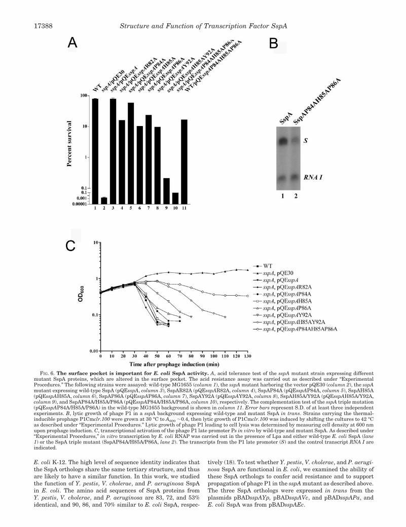
pocket, we made single alanine substitutions of residues Arg82,Pro84, His85, Pro86, and Tyr92, a double alanine substitutionH85A/Y92A, and a triple alanine substitution P84A/H85A/P86A. The ability of each of these alanine-substituted mutantsto complement acid-resistance of E. coli and support phage P1growth, when expressed in trans from plasmids in an sspAbackground, was determined (Fig. 6A). Whereas the wild-typestrain was acid-resistant, and the sspA mutant was acid-sensitive, the wild-type SspA protein provided in trans con-ferred acid tolerance of the sspA mutant as described (68) (Fig.6A, columns 1–3). Among the single alanine substitution SspAmutants, the mutant P84A behaved almost like wild-type SspA(Fig. 6A, compare columns 3 and 5), whereas the ability ofmutants R82A, H85A, P86A, and Y92A to support acid resist-ance was reduced 4–8-fold compared with the wild type (Fig.6A, compare columns 4, 6–8 with column 3). Interestingly,whereas elimination of the ring structure of either His85 orTyr92 decreased the complementation of acid resistance 8-fold,substitution of both His85 and Tyr92 with alanine (H85A/Y92A)reduced SspA activity about 40-fold (Fig. 6A, column 9), sug-gesting a cooperative action by residues His85 and Tyr92 in theSspA function. Furthermore, the triple substitution that re-placed SspA residues Pro84, His85, and Pro86 with alanine(P84A/H85A/P86A) almost abolished SspA complementation ofacid resistance (Fig. 6A, column 10), indicating that the overallstructure of the pocket, in addition to the ring systems of His85
and Tyr92, is important for SspA function.To determine whether sspA encoding the triple mutant
SspAP84A/H85A/P86A is dominant over wild-type sspA, wedetermined the acid tolerance of the wild-type sspA backgroundexpressing the triple mutant SspA in trans. Apparently thetriple mutant sspA gene is dominant over the wild-type alleleas wild-type cells became partially acid sensitive (survival rateof 17% compared with 80% for the wild type), when expressingSspA/P84A/H85A/P86A in trans (Fig. 6A, column 11). Thisresult is consistent with the notion that SspA functions as adimer of which the surface-exposed pocket is formed by resi-dues from both subunits (Fig. 5).
In addition, we examined the ability of these alanine-substi-tuted SspA proteins to support the lytic growth of phage P1 in
a sspA mutant strain (Table V). In this assay, the lytic devel-opment of phage P1 was examined as the ability of P1-vir toform plaques on a sspA background expressing mutant SspA. AP1-vir phage was used because it forms clear plaques on a lawnof wild-type cells. Overall, the phenotype of the SspA alaninemutants with respect to phage P1 growth was less severe thanthat of acid resistance. For instance, although the triple muta-tion P84A/H85A/P86A abolished the ability of the sspA mutantto confer acid resistance, it was still in part able to support P1growth. However, the plaques of phage P1 grown on the sspAmutant strain expressing SspA/P84A/H85A/P86A appearedturbid and were only about one-fourth of the wild type in size,indicating that the triple mutant supports the growth of phageP1 inefficiently. Other SspA mutants either affected P1 growthless significantly (P86A, Y92A, and H85A/Y92A) or behavedlike the wild type in phage P1 plaque formation (R82A, P84A,and H85A).
To further analyze the effects of these sspA mutations on thegrowth of phage P1, we performed a kinetic assay of phagegrowth (Fig. 6B), which is more sensitive compared with theassay described above. In this assay, the ability of the thermal-inducible P1 prophage P1Cmclr.100 (37) to undergo lyticgrowth in a sspA background expressing mutant SspA wasdetermined by optically measuring the degree of cell lysis (Fig.6B). Under the conditions used, phage P1 completed a life cyclein the wild-type strain 30–40 min after thermal induction ofthe prophage to lytic growth, leading to cell lysis as reported(60). Similarly, it took phage P1 30–40 min after prophageinduction to undergo lytic growth in the sspA mutant express-ing either wild-type SspA, SspAR82A, SspAP84A, SspAH85A,or SspAP86A in trans (Fig. 6B), whereas the sspA mutantharboring a vector did not lyse even during prolonged incuba-tion as previously reported (9). Though, the degree of cell lysisof the sspA background expressing the mutants P84A andP86A appeared less efficient compared with that of the wildtype. However, the SspA mutants Y92A, H85A/Y92A, andP84A/H85A/P86A were defective in supporting lytic growth ofphage P1 in the sspA mutant as cell lysis was delayed. Cell lysisof the sspA mutant expressing Y92A appeared 40–50 min afterprophage induction, whereas cell lysis was further delayed 10min in strains expressing SspAH85A/Y92A or SspAP84A/H85A/P86A. The growth assay allowed distinction between the
TABLE IVMajor GSH-interacting residues in GST B1-1, Ure2p, Y. pestis SspA, and E. coli SspA
GST B1-1 Ure2p Y. pestis SspA E. coli SspA
Catalytic residue Cys10 Asn124 Phe21 Tyr21
VDW and H-bonding Gln51 Arg164 Thr58 Ser58
H-bonding Val52 Val165 Val59 Val59
Cis-conformation Pro53 Pro166 Pro60 Pro60
Unusual �/� angels Glu65 Glu180 Glu71 Glu71
Amide H-bonding Gly66 Ser181 Ser72 Ser72
FIG. 3. GSH-binding site of GST and GST-like proteins. Stereoview showing the superposition of the GSH-binding site in GST B1-1 (green,PDB code 2PMT, Ref. 55), Ure2p (orange, PDB code 1K0D, Ref. 56), Y. pestis SspA (blue, this work), and E. coli SspA model (purple, this work).Amino acid residues and GSH are illustrated as ball-and-stick models in atomic color scheme (carbon in black, nitrogen in blue, oxygen in red, andsulfur in yellow). Dotted lines indicate functionally significant hydrogen bonds. This figure was prepared with MOLSCRIPT (65) and Raster3D (66).
Structure and Function of Transcription Factor SspA 17385

FIG. 4. Multiple sequence alignment of 50 SspA orthologs. Residues strictly conserved or chemically similar in at least 80% of the sequencesare shown on black and gray backgrounds, respectively. Secondary structure elements shown above the alignment are in accordance to thestructure of Y. pestis SspA (Table II). Species abbreviations: Acife, Acidithiobacillus ferrooxidans; Actac, Actinobacillus actinomycetemcomitans;
Structure and Function of Transcription Factor SspA17386

P1 growth phenotypes of the sspA mutant expressing the mu-tants H85A/Y92A and P84A/H85A/P86A. Cells expressing theH85A/Y92A mutant eventually lysed with a rate comparablewith that of the cells expressing Y92A. The most severe effecton phage growth was observed for the SspA triple mutantP84A/H85A/P86A, which only displayed a minor degree of lysiscompared with the strains encoding other SspA mutants.
To confirm that the defect of the SspA triple mutant insupporting the lytic growth of phage P1 is caused by its inabil-ity to activate transcription from the phage P1 late promoter,we performed in vitro transcription of the late promoter Psusing purified proteins (Fig. 6C). Indeed, whereas wild-typeSspA and the phage-encoded factor Lpa-activated transcriptionof the P1 late promoter as previously reported (8), SspA84A/
H85A/P86A showed a 2–3-fold decrease in transcriptional ac-tivity compared with wild-type SspA (Fig. 6C), indicating thatthe surface-exposed pocket is involved in transcription activa-tion by SspA. The residual transcription activity of the triplemutant SspA could account for the weak complementation ofphage P1 growth on a sspA mutant by SspAP84A/H85A/P86A(Table V). Taken together, residues of the surface pocket, es-pecially residues Pro84, His85, and Pro86, are important for theability of SspA to support the lytic growth of phage P1.
SspA orthologs from Y. pestis, V. cholerae, and P. aeruginosaare functional in E. coli—SspA is highly conserved amongGram-negative bacteria as shown in the multiple sequencealignment (Fig. 4). The 50 SspA orthologs included in thealignment display 40–100% sequence identity to SspA from
Actpl, Actinobacillus pleuropneumoniae; Azovi, Azotobacter vinelandii; Borbr, Bordetella bronchiseptica; Borpa, Bordetella parapertussis; Borpe,Bordetella pertussis; Burma, Burkholderia mallei; Chrvi, Chromobacterium violaceum; Colps, Colwellia psychrerythraea 34H; Coxbu, Coxiellaburnetii; Dicno, Dichelobacter nodosus; EcolK, E. coli K-12; EcolC, E. coli CFT073; EcolO, E. coli O157:H7; Erwca, Erwinia carotovora; Erwch,Erwinia chrysanthemi; Haedu, Haemophilus ducreyi; Haein, Haemophilus influenzae; Haeso, Haemophilus somnus; Klepn, Klebsiella pneumonia;Legpn, Legionella pneumophila; Metca, Methylococcus capsulatus; Micde, Microbulbifer degradans; Neigo, Neisseria gonorrhoeae; Neime, Neis-seria meningitides; Niteu, Nitrosomonas europaea; Pasmu, Pasteurella multocida; Phoas, Photorhabdus asymbiotica; Pholu, Photorhabdusluminescens; Prost, Providencia stuartii; Pseae, P. aeruginosa; Psefl, Pseudomonas fluorescens; Psepu, Pseudomonas putida; Psesy, Pseudomonassyringae; Ralso, Ralstonia solanacearum; Salen, Salmonella enterica; Salty, Salmonella typhimurium LT2; Serma, Serratia marcescens; Sheon,Shewanella oneidensis; Shidy, Shigella dysenteriae; Shifl, Shigella flexneri; Shiso, Shigella sonnei; Vibch, V. cholerae; Vibfi, Vibrio fischeri; Vibpa,Vibrio parahemeolyticus; Vibvu, Vibrio vulnificus; Xanax, Xanthomonas axonopodis; Xanca, Xanthomonas campestris; Xylfa, Xylella fastidiosa;and Yerpe, Y. pestis.
FIG. 5. Functionally important pocket on the surface of dimeric Y. pestis SspA. A and B, side views showing the edge of the pocket. Cand D, top views showing the bottom of the pocket. In panels A and C, the protein is illustrated as ribbon diagrams (helices as spirals, �-strandsas arrows, and loops as pipes) with the two subunits colored in cyan and orange, respectively. The side chains of pocket-defining residues are shownas ball-and-stick models in atomic color scheme (carbon in black, nitrogen in blue, and oxygen in red). In panels B and D, the pocket is visualizedwith space-filling models. The figure was prepared with MOLSCRIPT (65), Raster3D (66), and GRASP (67).
Structure and Function of Transcription Factor SspA 17387
E. coli K-12. The high level of sequence identity indicates thatthe SspA orthologs share the same tertiary structure, and thusare likely to have a similar function. In this work, we studiedthe function of Y. pestis, V. cholerae, and P. aeruginosa SspAin E. coli. The amino acid sequences of SspA proteins fromY. pestis, V. cholerae, and P. aeruginosa are 83, 72, and 53%identical, and 90, 86, and 70% similar to E. coli SspA, respec-
tively (18). To test whether Y. pestis, V. cholerae, and P. aerugi-nosa SspA are functional in E. coli, we examined the ability ofthese SspA orthologs to confer acid resistance and to supportpropagation of phage P1 in the sspA mutant as described above.The three SspA orthologs were expressed in trans from theplasmids pBADsspAYp, pBADsspAVc, and pBADsspAPa, andE. coli SspA was from pBADsspAEc.
FIG. 6. The surface pocket is important for E. coli SspA activity. A, acid tolerance test of the sspA mutant strain expressing differentmutant SspA proteins, which are altered in the surface pocket. The acid resistance assay was carried out as described under “ExperimentalProcedures.” The following strains were assayed: wild-type MG1655 (column 1), the sspA mutant harboring the vector pQE30 (column 2), the sspAmutant expressing wild-type SspA (pQEsspA, column 3), SspAR82A (pQEsspAR82A, column 4), SspAP84A (pQEsspAP84A, column 5), SspAH85A(pQEsspAH85A, column 6), SspAP86A (pQEsspAP86A, column 7), SspAY92A (pQEsspAY92A, column 8), SspAH85A/Y92A (pQEsspAH85A/Y92A,column 9), and SspAP84A/H85A/P86A (pQEsspAP84A/H85A/P86A, column 10), respectively. The complementation test of the sspA triple mutation(pQEsspAP84A/H85A/P86A) in the wild-type MG1655 background is shown in column 11. Error bars represent S.D. of at least three independentexperiments. B, lytic growth of phage P1 in a sspA background expressing wild-type and mutant SspA in trans. Strains carrying the thermal-inducible prophage P1Cmclr.100 were grown at 30 °C to A600 �0.4, then lytic growth of P1Cmclr.100 was induced by shifting the cultures to 42 °Cas described under “Experimental Procedures.” Lytic growth of phage P1 leading to cell lysis was determined by measuring cell density at 600 nmupon prophage induction. C, transcriptional activation of the phage P1 late promoter Ps in vitro by wild-type and mutant SspA. As described under“Experimental Procedures,” in vitro transcription by E. coli RNAP was carried out in the presence of Lpa and either wild-type E. coli SspA (lane1) or the SspA triple mutant (SspAP84A/H85A/P86A, lane 2). The transcripts from the P1 late promoter (S) and the control transcript RNA I areindicated.
Structure and Function of Transcription Factor SspA17388
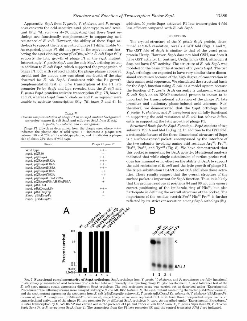
Apparently, SspA from Y. pestis, V. cholerae, and P. aerugi-nosa converts the acid-sensitive sspA phenotype to acid-resis-tant (Fig. 7A, columns 4–6), indicating that these SspA or-thologs are functionally complementary in supporting acidresistance of E. coli. However, the ability of these SspA or-thologs to support the lytic growth of phage P1 differ (Table V).As expected, phage P1 did not grow in the sspA mutant har-boring the sspA cloning vector pBAD24, while E. coli SspA fullysupports the lytic growth of phage P1 in the sspA mutant.Interestingly, Y. pestis SspA was the only SspA ortholog tested,in addition to E. coli SspA, which supported the propagation ofphage P1, but with reduced ability; the phage plaque appearedturbid, and the plaque size was about one-fourth of the sizeobserved for E. coli SspA. Consistent with the P1 growthcomplementation test, in vitro transcription of the P1 latepromoter Ps by SspA and Lpa revealed that the E. coli andY. pestis SspA proteins activate transcription (Fig. 7B, lanes 1and 2), whereas SspA from V. cholerae and P. aeruginosa wereunable to activate transcription (Fig. 7B, lanes 3 and 4). In
addition, Y. pestis SspA activated P1 late transcription 4-foldless efficient compared with E. coli SspA.
DISCUSSION
The crystal structure of the Y. pestis SspA protein, deter-mined at 2.0-Å resolution, reveals a GST fold (Figs. 1 and 2).The GST fold of SspA is similar to that of the yeast prionprotein Ure2p. However, SspA does not bind GSH, nor does ithave GST activity. In contrast, Ure2p binds GSH, although itdoes not have GST activity. The structure of E. coli SspA wasmodeled on the basis of the structure of Y. pestis SspA. The twoSspA orthologs are expected to have very similar three-dimen-sional structures because of the high degree of conservation intheir amino acid sequences. We elucidated the structural basisfor the SspA function using E. coli as a model system becausethe function of Y. pestis SspA currently is unknown, whereasE. coli SspA as an RNAP-associated protein is known to beimportant for transcriptional activation of the phage P1 latepromoter and stationary phase-induced acid tolerance. Fur-thermore, we demonstrated that the SspA orthologs fromY. pestis, V. cholerae, and P. aeruginosa are all fully functionalin supporting the acid resistance of E. coli but behave differ-ently in supporting the lytic growth of phage P1.
Structural Basis for the SspA Function—SspA consists of twosubunits Mol A and Mol B (Fig. 1). In addition to the GST fold,a noticeable feature of the three-dimensional structure of SspAis a surface-exposed pocket, encompassed by the interface ofthe two subunits involving amino acid residues Arg82, Pro84,His85, Pro86, and Tyr92 (Fig. 5). We have demonstrated thatthis pocket is important for SspA activity. Mutational analysisindicated that while single substitution of surface pocket resi-dues has minimal or no effect on the ability of SspA to supportthe acid resistance of E. coli and the lytic growth of phage P1,the triple substitution P84A/H85A/P86A abolishes these activ-ities. These results suggest that the overall structure of thesurface pocket is important for SspA function. Thus, it is likelythat the proline residues at positions 84 and 86 not only ensurecorrect positioning of the imidazole ring of His85, but alsoparticipate in defining the overall structure of the pocket. Theimportance of the residue stretch Pro84-His85-Pro86 is furtherreflected by its strict conservation among SspA orthologs (Fig.
FIG. 7. Functional complementarity of SspA orthologs. SspA orthologs from Y. pestis, V. cholerae, and P. aeruginosa are fully functionalin stationary phase-induced acid tolerance of E. coli but behave differently in supporting phage P1 lytic development. A, acid tolerance test of theE. coli sspA mutant strain expressing different SspA orthologs. The acid resistance assay was carried out as described under “ExperimentalProcedures.” The following strains were assayed: wild-type E. coli MG1655 (column 1), the sspA mutant containing the vector pBAD24 (column 2),and the sspA mutant expressing the sspA gene from E. coli (pBADsspAEc, column 3), Y. pestis (pBADsspAYp, column 4), V. cholerae (pBADsspAVc,column 5), and P. aeruginosa (pBADsspAPa, column 6), respectively. Error bars represent S.D. of at least three independent experiments. B,transcriptional activation of the phage P1 late promoter Ps by different SspA orthologs in vitro. As described under “Experimental Procedures,”in vitro transcription by E. coli RNAP was carried out in the presence of Lpa and either E. coli SspA (lane 1), Y. pestis SspA (lane 2), V. choleraeSspA (lane 3), or P. aeruginosa SspA (lane 4). The transcripts from the P1 late promoter (S) and the control transcript RNA I are indicated.
TABLE VGrowth complementation of phage P1 in an sspA mutant background
expressing mutant E. coli SspA and wild-type SspA from E. coli,Y. pestis, V. cholerae, and P. aeruginosa
Phage P1 growth as determined from the plaque size, where ���indicates the plaque size of wild type, �� indicates a plaque sizebetween 50 and 75% of the wild-type plaque, and � indicates a plaquesize of about 25% that of wild type.
Strain Phage P1 growtha
Wild type ���sspA, pQE30 �sspA, pQEsspA ���sspA, pQEsspAR82A ���sspA, pQEsspAP84A ���sspA, pQEsspAH85A ���sspA, pQEsspAP86A ��sspA, pQEsspAY92A ��sspA, pQEsspAH85AY92A �sspA, pQEsspAP84AH85AP86A �sspA, pBAD24 �sspA, pBADsspAEc ���sspA, pBADsspYp �SspA, pBADsspVc �SspA, pBADsspPa �
Structure and Function of Transcription Factor SspA 17389

4). Since the structure of the surface pocket suggests that it isa region for potential protein-protein interactions and that it isimportant for SspA function in both E. coli and phage P1 latetranscription, we speculate that the pocket serves as a bindingsite for RNAP. The effect of mutations in the surface pocket onthe ability of SspA to complement phage P1 growth appearedless severe compared with the ability to support acid resistance(Table V and Fig. 6). This could be caused by the possibilitythat the phage-encoded co-activator Lpa stabilizes the SspA-RNAP interaction. In addition, the three-dimensional structureof SspA reveals the flexible N and C termini (Fig. 2). Unlike theN- and C-terminal regions of the GST-like Ure2p protein,which are functionally important (51, 61), deletions of the Nand C termini of SspA do not affect SspA function.
Because the formation of the surface-exposed pocket, whichis important for SspA activity, requires both Mol A and Mol B,SspA must function as a dimer. In support of this notion, weshowed that the triple sspA mutation P84A/H85A/P86A exhib-its a dominant negative phenotype in a wild-type background(Fig. 6A, compare columns 1 and 11). Note that residues thatparticipate in the dimer formation of Ure2p are also importantfor transcription regulation (61), suggesting that Ure2p is alsofunctional as a dimer. However, Ure2p does not contain asurface-exposed pocket like that of SspA. Instead, a cleft be-tween the two subunits of Ure2p, which is next to the GSH-binding site, has been suggested to serve as the interaction sitefor proteins that bind to Ure2p (52). It remains to be elucidatedwhether the corresponding cleft in SspA is involved in the SspAfunction.
SspA Orthologs Are Functionally Complementary—SspA ishighly conserved among Gram-negative bacteria. We havedemonstrated that the SspA orthologs from Y. pestis, V. chol-erae, and P. aeruginosa are fully functional in supporting acidresistance of E. coli, indicating that these SspA orthologs sharea common function in the cell. However, only Y. pestis SspAcould partially support the growth of phage P1 in E. coli.Consistent with this, Y. pestis SspA shares the highest degreeof sequence conversation with E. coli SspA among the SspAorthologs tested. Thus, it is likely that phage P1 evolved to useSspA for its development in E. coli.
Therefore, SspA is most likely involved in acid tolerance ofY. pestis, V. cholerae, P. aeruginosa, and possibly other speciesencoding sspA as well. Acid tolerance is important for the ability ofenteric bacteria such as E. coli O157:H7 to survive the low pHenvironment they encounter upon passage through the gastroin-testinal tract during infection of the mammalian host (62–64).Since SspA is required for acid resistance (68), it most likely playsan important role in bacterial pathogenesis. The finding that thesurface pocket residues Pro84, His85, and Pro86, which are impor-tant for activity of E. coli SspA, are strictly conserved in 50 SspAorthologs (Fig. 4), suggests that the pocket is important for SspAfunction in general. Thus, this surface pocket provides a potentialtarget for putative inhibitors of SspA function.
Acknowledgment—We thank Zbigniew Dauter for assistance duringx-ray data acquisition.
REFERENCES
1. Zhang, G., Campbell, E. A., Minakhin, L., Richter, C., Severinov, K., andDarst, S. A. (1999) Cell 98, 811–824
2. Vassylyev, D. G., Sekine, S., Laptenko, O., Lee, J., Vassylyeva, M. N., Borukhov, S.,and Yokoyama, S. (2002) Nature 417, 712–719
3. Burgess, R. R., Erickson, B., Gentry, D. R., Gribskov, M., Hager, D., Lesley, S.,Strickland, M., and Thompson, N. (1987) in RNA Polymerase and theRegulation of Transcription (Reznikoff, W. S., ed), pp. 3–15, Elsevier Sci-ence Publishing, New York
4. Mooney, R. A., Artismovitch, I., and Landick, R. (1998) J. Bacteriol. 180,3265–3275
5. von Hippel, P. H. (1998) Science 281, 600–6056. Nudler, E., and Gottesman, M. E. (2002) Genes Cells 7, 755–7687. Ishihama, A., and Saitoh, T. (1979) J. Mol. Biol. 129, 517–530
8. Hansen, A. M., Lehnherr, H., Wang, X., Mobley, V., and Jin, D. J. (2003) Mol.Microbiol. 48, 1621–1631
9. Williams, M. D., Fuchs, J. A., and Flickinger, M. C. (1991) Gene (Amst.) 109,21–30
10. Williams, M. D., Ouyang, T. X., and Flickinger, M. C. (1994) Mol. Microbiol. 11,1029–1043
11. Atlung, T., and Ingmer, H. (1997) Mol. Microbiol. 24, 7–1712. Hommais, F., Krin, E., Laurent-Winter, C., Soutourina, O., Malpertuy, A., Le
Caer, J. P., Danchin, A., and Bertin, P. (2001) Mol. Microbiol. 40, 20–3613. De Reuse, H., and Taha, M. K. (1997) Res. Microbiol. 148, 289–30314. Baron, G. S., and Nano, F. E. (1998) Mol. Microbiol. 29, 247–25915. Merrell, D. S., Hava, D. L., and Camilli, A. (2002) Mol. Microbiol. 43,
1471–149116. Xu, Q., Dziejman, M., and Mekalanos, J. J. (2003) Proc. Natl. Acad. Sci.
U. S. A. 100, 1286–129117. Lauriano, C. M., Barker, J. R., Yoon, S. S., Nano, F. E., Arulanandam, B. P.,
Hassett, D. J., and Klose, K. E. (2004) Proc. Natl. Acad. Sci. U. S. A. 101,4246–4249
18. Andrykovitch, M., Routzahn, K. M., Li, M., Gu, Y., Waugh, D. S., and Ji, X.(2003) Acta Crystallogr. Sect. D 59, 881–886
19. Otwinowski, Z., and Minor, W. (1997) Methods Enzymol. 276, 307–32620. Collaborative Computational Project Number 4. (1994) Acta Crystallogr. Sect.
D 50, 760–76321. Terwilliger, T. C., and Berendzen, J. (1999) Acta Crystallogr. Sect. D 55,
849–86122. de La Fortelle, E., and Bricogne, G. (1997) Methods Enzymol. 276, 472–49423. Cowtan, K. (1994) Joint CCP4 and ESF-EACBM Newsletter on Protein Crys-
tallography 31, 34–3824. Abrahams, J. P., and Leslie, A. G. W. (1996) Acta Crystallogr. Sect. D 52,
30–4225. Lamzin, V. S., and Wilson, K. S. (1997) Methods Enzymol. 277, 269–30526. Brunger, A. T., Adams, P. D., Clore, G. M., DeLano, W. L., Gros, P., Grosse-
Kunstleve, R. W., Jiang, J. S., Kuszewski, J., Nilges, M., Pannu, N. S., Read,R. J., Rice, L. M., Simonson, T., and Warren, G. L. (1998) Acta Crystallogr.Sect. D 54, 905–921
27. Jones, T. A., and Kjeldgaard, M. (1997) Methods Enzymol. 277, 173–20828. Laskowski, R. A., MacArthur, M. W., Moss, D. S., and Thornton, J. M. (1993)
J. Appl. Crystallogr. 26, 283–29129. Powell, M. J. D. (1977) Math. Prog. 12, 241–25430. Brunger, A. T., and Rice, L. M. (1997) Methods Enzymol. 277, 243–26931. Engh, R. A., and Huber, R. (1991) Acta Crystallogr. Sect. A 47, 392–40032. Thompson, J. D., Higgins, D. G., and Gibson, T. J. (1994) Nucleic Acids Res. 22,
4673–468033. Nicholas, K. B., Nicholas, H. B., Jr., and Deerfield, D. W. (1997) in EMBNEWS
Vol. 4, p. 1434. Miller, J. H. (1972) Experiments in Molecular Genetics, Cold Spring Harbor
Laboratory, Cold Spring Harbor, NY35. Sambrook, J., Fritsch, E. F., and Maniatis, T. (1989) Molecular Cloning: A
Laboratory Manual, Cold Spring Harbor Laboratory Press, New York36. Guzman, L. M., Belin, D., Carson, M. J., and Beckwith, J. (1995) J. Bacteriol.
177, 4121–413037. Rosner, J. L. (1972) Virology 48, 679–68038. Sukhodolets, M. V., and Jin, D. J. (1998) J. Biol. Chem. 273, 7018–702339. Dirr, H., Reinemer, P., and Huber, R. (1994) Eur. J. Biochem. 220, 645–66140. Toung, Y. P., and Tu, C. P. (1992) Biochem. Biophys. Res. Commun. 182,
355–36041. Rife, C. L., Parsons, J. F., Xiao, G., Gilliland, G. L., and Armstrong, R. N.
(2003) Proteins: Struct. Funct. Genet. 53, 777–78242. Hayes, J. D., and Pulford, D. J. (1995) Crit. Rev. Biochem. Mol. Biol. 30,
445–60043. Sheehan, D., Meade, G., Foley, V. M., and Dowd, C. A. (2001) Biochem. J. 360,
1–1644. Dixon, D. P., Lapthorn, A., and Edwards, R. (2002) Genome Biol 3, Reviews,
3004.1–1045. Kanaoka, Y., Ago, H., Inagaki, E., Nanayama, T., Miyano, M., Kikuno, R.,
Fujii, Y., Eguchi, N., Toh, H., Urade, Y., and Hayaishi, O. (1997) Cell 90,1085–1095
46. Tomarev, S. I., and Zinovieva, R. D. (1988) Nature 336, 86–8847. Tomarev, S. I., Zinovieva, R. D., and Piatigorsky, J. (1991) J. Biol. Chem. 266,
24226–2423148. Tomarev, S. I., Chung, S., and Piatigorsky, J. (1995) J. Mol. Evol. 41,
1048–105649. Chuang, C. C., Wu, S. H., Chiou, S. H., and Chang, G. G. (1999) Biophys. J. 76,
679–69050. Coschigano, P. W., and Magasanik, B. (1991) Mol. Cell. Biol. 11, 822–83251. Bousset, L., Belrhali, H., Janin, J., Melki, R., and Morera, S. (2001) Structure
(Camb) 9, 39–4652. Umland, T. C., Taylor, K. L., Rhee, S., Wickner, R. B., and Davies, D. R. (2001)
Proc. Natl. Acad. Sci. U. S. A. 98, 1459–146453. Ji, X., von Rosenvinge, E. C., Johnson, W. W., Tomarev, S. I., Piatigorsky, J.,
Armstrong, R. N., and Gilliland, G. L. (1995) Biochemistry 34, 5317–532854. Nishida, M., Harada, S., Noguchi, S., Satow, Y., Inoue, H., and Takahashi, K.
(1998) J. Mol. Biol. 281, 135–14755. Rossjohn, J., Polekhina, G., Feil, S. C., Allocati, N., Masulli, M., De Illio, C.,
and Parker, M. W. (1998) Structure 6, 721–73456. Bousset, L., Belrhali, H., Melki, R., and Morera, S. (2001) Biochemistry 40,
13564–1357357. Perrett, S., Freeman, S. J., Butler, P. J., and Fersht, A. R. (1999) J. Mol. Biol.
290, 331–34558. Choi, J. H., Lou, W., and Vancura, A. (1998) J. Biol. Chem. 273, 29915–2992259. Armstrong, R. N. (1997) Chem. Res. Toxicol. 10, 2–1860. Yarmolinsky, M., and Sternberg, N. (1988) in The Bacteriophages (Calendar,
Structure and Function of Transcription Factor SspA17390

R., ed) pp. 291–438, Plenum Press, New York61. Kulkarni, A. A., Abul-Hamd, A. T., Rai, R., El Berry, H., and Cooper, T. G.
(2001) J. Biol. Chem. 276, 32136–3214462. Lin, J., Lee, I. S., Frey, J., Slonczewski, J. L., and Foster, J. W. (1995) J.
Bacteriol. 177, 4097–410463. Bearson, S., Bearson, B., and Foster, J. W. (1997) FEMS Microbiol. Lett. 147,
173–180
64. Merrell, D. S., and Camilli, A. (2002) Curr. Opin. Microbiol. 5, 51–5565. Kraulis, P. J. (1991) J. Appl. Crystallogr. 24, 946–95066. Merritt, E. A., and Bacon, D. J. (1997) Methods Enzymol. 277, 505–52467. Nicholls, A., Sharp, K. A., and Honig, B. (1991) Proteins: Struct. Funct. Genet.
11, 281–29668. Hansen, A., Qiu, Y., Yeh, N., Blattner, F. R., Durfee, T., and Jin, D. J. (2005)
Mol. Microbiol. 56, 713–734
Structure and Function of Transcription Factor SspA 17391