structural basis for specific recognition of substrates by …€¦ · · 2017-06-16structural...
TRANSCRIPT

ORIGINAL RESEARCH ARTICLEpublished: 05 September 2012doi: 10.3389/fmicb.2012.00312
Structural basis for specific recognition of substrates bysapovirus protease
MasaruYokoyama1‡,Tomoichiro Oka2,3‡, Hirotatsu Kojima4,Tetsuo Nagano4,Takayoshi Okabe4,Kazuhiko Katayama2,Takaji Wakita2,Tadahito Kanda1† and Hironori Sato1*1 Pathogen Genomics Center, National Institute of Infectious Diseases, Tokyo, Japan2 Department of Virology II, National Institute of Infectious Diseases, Tokyo, Japan3 Food Animal Health Research Program, Ohio Agricultural Research and Development Center, Department of Veterinary Preventive Medicine, The Ohio State
University, Wooster, OH, USA4 Open Innovation Center for Drug Discovery, The University of Tokyo, Japan
Edited by:Hiroyuki Toh, National Institute ofAdvanced Industrial Science andTechnology, Japan
Reviewed by:Hiroyuki Toh, National Institute ofAdvanced Industrial Science andTechnology, JapanMing Tan, Cincinnati Children’sHospital Medical Center, USA
*Correspondence:Hironori Sato, Pathogen GenomicsCenter, National Institute of InfectiousDiseases, 4-7-1 Gakuen,MusashiMurayama-shi, Tokyo208-0011, Japan.e-mail: [email protected]†Present address:Tadahito Kanda, The Center ofResearch Network for InfectiousDiseases, RIKEN, Tokyo, Japan.‡Masaru Yokoyama and TomoichiroOka have contributed equally to thiswork.
Sapovirus (SaV) protease catalyzes cleavage of the peptide bonds at six sites of a viralpolyprotein for the viral replication and maturation. However, the mechanisms by whichthe protease recognizes the distinct sequences of the six cleavage sites remain poorlyunderstood. Here we examined this issue by computational and experimental approaches.A structural modeling and docking study disclosed two small clefts on the SaV proteasecavity that allow the stable and functional binding of substrates to the catalytic cavityvia aromatic stacking and electrostatic interactions. An information entropy study and asite-directed mutagenesis study consistently suggested variability of the two clefts underfunctional constraints. Using this information, we identified three chemical compounds thathad structural and spatial features resembling those of the substrate amino acid residuesbound to the two clefts and that exhibited an inhibitory effect on SaV protease in vitro.Theseresults suggest that the two clefts provide structural base points to realize the functionalbinding of various substrates.
Keywords: sapovirus protease, substrate recognition, P1 and P4 amino acid residues, 3-D models, amino aciddiversity, mutagenesis, 3-D pharmacophore, inhibitor screening
INTRODUCTIONSapovirus (SaV) is a non-enveloped RNA virus that belongs tothe family Caliciviridae and causes gastroenteritis in humans andswine (Chiba et al., 1979, 2000; Guo et al., 1999; Hansman et al.,2007). The SaV genome is a single-stranded RNA that encodestwo or three open reading frames (ORFs; Liu et al., 1995; Noelet al., 1997; Numata et al., 1997; Guo et al., 1999; Robinson et al.,2002). The ORF1 encodes six non-structural proteins (NS1, NS2,NS3, NS4, NS5, and NS6-7) and a structural protein, the capsidprotein (VP1; Oka et al., 2006, 2009). The NS6-7 protein containsthe chymotrypsin-like protease domain (the 3C-like protease; Okaet al., 2005a,b, 2007; Robel et al., 2008) and the RNA-dependentRNA polymerase domain (the 3-D-like polymerase; Fullertonet al., 2007; Bull et al., 2011). The ORF1 precursor protein is post-translationally cleaved at six sites by the 3C-like protease (Okaet al., 2005b, 2006).
The SaV 3C-like protease domain comprises 146 amino acidresidues (Oka et al., 2007). This enzyme cleaves the peptide bondsof specific dipeptides, such as the glutamic acid/glycine (E/G), glu-tamine/glycine (Q/G), and glutamic acid/alanine (E/A; Oka et al.,2006). However, these dipeptide motifs exist in the non-cleaved
sites of the ORF1 polyprotein, indicating that additional aminoacid residues are required for the specific recognition of sub-strates (Oka et al., 2006). In this regard, calicivirus proteaseshave a large cavity that can accommodate substrate peptides withseveral amino acid in lengths (Nakamura et al., 2005; Zeitleret al., 2006; Oka et al., 2007). It is conceivable that these sub-strate amino acid residues around the cleavage sites, termed theP4, P3, P2, P1, P1′, P2′, P3′, and P4′ sites, are all involved tosome extent, either directly or indirectly, in the recognition andcleavage by protease. However, there must be a division of roles:previous studies on the calicivirus proteases consistently suggestmore extensive involvement of the substrate amino acid residuesupstream of the peptide bond of the cleavage sites in the cleav-age by proteases (Wirblich et al., 1995; Sosnovtsev et al., 1998;Hardy et al., 2002; Belliot et al., 2003; Scheffler et al., 2007; Robelet al., 2008). In the case of SaV, the substrate P1 and P4 aminoacid residues in particular are physicochemically more conservedamong different SaV strains (Oka et al., 2009) and more sensi-tive to the substitutions (Robel et al., 2008; Oka et al., 2009).Therefore, these amino acid residues may provide the specific con-tact sites with SaV protease. However, due to the lack of structural
www.frontiersin.org September 2012 | Volume 3 | Article 312 | 1

Yokoyama et al. Substrate recognition by SaV protease
information on SaV protease and its substrates, such interactionremains unclear.
Recent advances in the hardware and software for biomolecularsimulation and bioinformatics have rapidly improved the preci-sion and performance of these techniques. We have applied someof these techniques, in combination with experimental methods,to understand the structural and evolutionary basis of the viro-logical phenomena (Oka et al., 2007, 2009; Motomura et al., 2008,2010; Naganawa et al., 2008; Shirakawa et al., 2008;Yokoyama et al.,2010, 2012; Ode et al., 2011; Sakuragi et al., 2012). In this study, bycombining methods of homology modeling, the automated liganddocking, Shannon entropy analysis, site-directed mutagenesis, andin silico screening of SaV inhibitors, we studied the structural basisfor the substrate recognition by SaV protease.
MATERIALS AND METHODSSTRUCTURAL MODELING OF SaV PROTEASE DOCKED TO THESUBSTRATE OCTAPEPTIDESWe first constructed a ligand-free protease domain model of theSaV Mc10 strain (Oka et al., 2005b; GenBank accession number:AY237420) by the homology modeling technique (Sanchez et al.,2000; Baker and Sali, 2001) as described previously (Oka et al.,2007). The modeling was performed using tools available in theMolecular Operating Environment (MOE; Chemical ComputingGroup, Inc., Montreal, QC, Canada). As the modeling template,we used the high-resolution crystal structure of norovirus 3C-like protease at a resolution of 1.50 Å [Protein Data Bank (PDB)code: 2FYQ; Zeitler et al., 2006] because, like SaV, the norovirusbelongs to the family Caliciviridae, and thus the protease sequenceshows a higher identity to the SaV protease sequences (about 25%identity) than to the other available 3C-like protease sequencesof viruses. We applied the multiple sequence alignment approach(Baker and Sali, 2001) using the reported 3C-like proteases tominimize misalignments of the target and template sequences, asdescribed previously (Oka et al., 2007; Shirakawa et al., 2008). Thesequences used for the alignment included those of the rhinovirus3C-like protease (PDB code: 1CQQ; Matthews et al., 1999), thepoliovirus 3C-like protease (PDB code: 1L1N; Mosimann et al.,1997), and the hepatitis A virus 3C-like protease (PDB code: 1QA7;Bergmann et al., 1999). The alignment was done with the align-ment tool MOE-Align, and homology modeling was done with thetool MOE-Homology in MOE. We optimized the 3-D model ther-modynamically via energy minimization using the MOE and anAMBER99 force field (Ponder and Case, 2003). We further refinedthe physically unacceptable local structure of the models basedon a Ramachandran plot evaluation using MOE. The 3-D modelsof the six octapeptides corresponding to the six cleavage sites ofthe ORF1 precursor protein of the SaV Mc10 strain (NS1/NS2,NS2/NS3, NS3/NS4, NS4/NS5, NS5/NS6-7, and NS6-7/VP1) wereconstructed using the Molecular Builder module in MOE. Subse-quently, the thermodynamically and physically optimized proteasemodels were used to construct protease-substrate complex mod-els. Individual octapeptide models were docked with the optimizedSaV protease domain model described above, using the automatedligand docking program ASEDock2005 (Goto et al., 2008) oper-ated in MOE as described previously (Yokoyama et al., 2010).Default setting in ASEDock2005 was applied for the search of
the candidate docking structures, and the structures with the bestdocking score expressed by the arbitrary docking energy (U dock)in ASEDock2005 (Kataoka and Goto, 2008) were selected for theanalysis of the protease-substrate interaction sites.
ANALYSIS OF AMINO ACID DIVERSITY WITH INFORMATION ENTROPYThe amino acid diversity at individual sites of the SaV pro-tease domain was analyzed with Shannon entropy scores asdescribed previously (Sander and Schneider, 1991; Mirny andShakhnovich, 1999; Oka et al., 2009). The amino acid sequencesof the protease domain of various human SaV strains fromdifferent geographic regions in the world were obtained fromGenBank (the number of sequences is 19; accession numbers:X86560, AY694184, AY237422, AY237423, AY646853, AY646854,AJ249939, AY237420, AY237419, AY646855, AY603425, AJ786349,DQ058829, DQ125333, AY646856, DQ125334, DQ366344,DQ366345, DQ366346). The amino acid diversity within theSaV protease population was calculated using Shannon’s formula(Shannon, 1948):
H (i) = −∑
xi
p (xi) log2p (xi) (xi = G, A, I , V , . . . . . .) ,
where H(i), p(xi), and i indicate the amino acid entropy (H ) scoreof a given position, the probability of occurrence of a given aminoacid at the position, and the number of the position, respectively.An H score of zero indicates absolute conservation, whereas 4.4bits indicates complete randomness. The H scores were displayedon the 3-D structure of the SaV protease model constructed above.
We also calculated the Shannon entropy by considering thephysicochemical properties of amino acid residues, i.e., the chem-ical properties and size of side chains as described previously (Okaet al., 2009). For analysis of the diversity in the chemical properties,the amino acid residues were classified into seven groups: acidic(D,E), basic (R,K,H), neutral hydrophilic (S, T, N, Q), aliphatic(G, A, V, I, L, M), aromatic (F, Y, W), thio-containing (C), andimine (P). For analysis of the diversity in the size of side chains,the amino acid residues were classified into four groups: small (G,A, C, S), medium-small (T, V, N, D, I, L, P, M), medium-large (Q,E, R, K), and large (H, F, Y, W). The H scores were plotted on the3-D structure of the SaV protease model.
SITE-DIRECTED MUTAGENESIS OF THE SaV PROTEASE DOMAINThe detailed strategy of the mutagenesis for the SaV pro-tease domain has been described previously (Oka et al., 2005b,2006). Briefly, we used the full-length cDNA clone of thegenome of the SaV strain Mc10 (pUC19/SaV Mc10 full-length; GenBank accession number: AY237420) as a start-ing material for the mutagenesis. We constructed nine SaVMc10 full-length mutant cDNA clones. Site-directed mutagene-sis was performed using a GeneTailor Site-Directed Mutagene-sis System (Invitrogen). The oligonucleotides used for the site-directed mutagenesis were as follows (the codons correspond-ing to changed amino acid(s) are indicated in lowercase): forT1085A, 5′-GTGGTTGTCACAGTTgcaCACGTGGCCTCTGCG-3′; for Y1156A, 5′-ATCACGGTCCAGGGGgctCACCTGCGCATC
Frontiers in Microbiology | Virology September 2012 | Volume 3 | Article 312 | 2

Yokoyama et al. Substrate recognition by SaV protease
ATA-3′; for K1167A, 5′-ATGGATACCCAACAgcgCGTGGGGACTGTGGCAC-3′; for R1168A,5′-GATACCCAACAAAGgctGGGGACTGTGGCACAC-3′; for K1167E, 5′-ATGGATACCCAACAgagCGTGGGGACTGTGGCAC-3′; for R1168E, 5′-ATGGATACCCAACAAAGgagGGGGACTGTGGCACAC-3′; for K1167AR1168A, 5′-ATGGATACCCAACAgcggcgGGGGACTGTGGCAC-3′; and forK1167ER1168E, 5′-ATGGATACCCAACAgaggagGGGGACTGTGGCAC-3′. The T1085AY1156A mutant was generated with theabove Y1156A primer using methylated DNA of the T1085A asthe template. All the mutant clones constructed were subjected tothe sequencing of the entire genomic cDNA region to verify theabsence of unnecessary mutations leading to amino acid changes.
IN VITRO TRANSCRIPTION-TRANSLATION ASSAYIn vitro transcription-translation with a rabbit reticulocytesystem was performed using the TNT T7 Quick for PCRDNA kit (Promega, Madison, WI, USA) as described previ-ously (Oka et al., 2005b). Briefly, a template for the in vitrotranscription-translation, containing the entire ORF1, was pre-pared by PCR amplification using the full-length cDNA clone.The primers used for the amplification were as follows. The for-ward primer containing a T7 promoter sequence (underlined)and a translation initiation codon (bold) was 5′-GGATCCTAATACGACTCACTATAGGGAACAGCCACCATG gcttccaagccattctacccaatagag-3′; and the antisense primer containing a stop codon(bold) was 5′-T30TTA-ttctaagaacctaacggcccgg. The PCR product(3 µl) was mixed with 20 µl of TNT T7 PCR Quick Master Mix(Promega) and 2 µl of Redivue Pro-mix L- [35S] in vitro cell-labeling mix (GE Healthcare Biosciences, Piscataway, NJ, USA).The mixture was incubated at 30˚C for 3 or 16 h and subjected toSDS-polyacrylamide gel electrophoresis (SDS-PAGE). The trans-lation products separated by electrophoresis were blotted onto aPVDF membrane (Immobilon-P; Millipore, Bedford, MA, USA)using a semi-dry electroblotting apparatus (ATTO; Tokyo). Theradiolabeled proteins were detected by a BAS 2500 BioimageAnalyzer (Fuji Film, Tokyo).
IMMUNOPRECIPITATIONFor the detection of NS1 (p11) and NS5 (VPg), which wereundetectable with the above assay system, we performed immuno-precipitation before the SDS-PAGE as described previously (Okaet al., 2005b, 2006, 2009). Briefly, 10 µl of the in vitro transcription-translation reaction mixture was diluted with 80 µl of RIPA lysisbuffer containing 50 mM Tris, pH 7.4, 150 mM NaCl, 0.25%deoxycholic acid, 1% NP40, and 1 mM EDTA (Upstate, LakePlacid, NY, USA) and incubated with 5 µg of anti-A (anti-NS1)or ant-D (anti-NS5) antibodies raised against E. coli-expressedrecombinant proteins (aa 1–231 for NS1 and aa 941–1055 forNS5; Oka et al., 2005b). After incubation for 1 h on ice, 25 µlof a suspension of Protein A Magnetic Beads (New EnglandBiolabs) and 900 µl of RIPA buffer were added. The mixturewas gently rotated at 4˚C for 1 h and then washed three timeswith 1 ml of RIPA lysis buffer. The immunoprecipitated pro-teins were resuspended in 20 µl of SDS-PAGE sample bufferand heated at 95˚C for 5 min prior to analysis with 5 to 20%Tris-Gly polyacrylamide gel. The proteins were blotted onto anImmobilon-P polyvinylidene difluoride membrane (Millipore).
Immunoprecipitated radioactive proteins were detected with aBioimage Analyzer BAS 2500 (Fuji Film).
THE CHEMICAL COMPOUND LIBRARYChemical compounds (139,369 compounds, molecular weights42–2986) were obtained from the Open Innovation Center forDrug Discovery (The University of Tokyo, Tokyo, Japan). Thecompound library database of this center provides informationon the molecular formula, molecular weight, hydrogen-bonddonor-acceptor numbers, topological polar surface area (TPSA),and other physicochemical parameters of the compounds forpharmacophore-based in silico drug screening.
PHARMACOPHORE-BASED IN SILICO SCREENINGPharmacophore-based in silico screening was done using toolsavailable in the MOE. We created a pharmacophore query witha substrate feature using the Pharmacophore Query Editor toolin MOE. Pharmacophore-based in silico screening was done bythe Pharmacophore Search module in the MOE using the createdquery.
DRUG SUSCEPTIBILITY ASSAYThe susceptibility of SaV protease to the synthetic small chem-ical compound was determined by means of an in vitro transcleavage assay as follows. A radiolabeled full-length Mc10 ORF1polyprotein containing a defective protease (Promut; Oka et al.,2005b) or a non-radiolabeled partial Mc10 ORF1 polyprotein(NS6-7-VP1) containing a functional protease (Prowt; Oka et al.,2006) was separately expressed using the in vitro transcrip-tion/translation system (Oka et al., 2011). The PCR primer pairsused for the preparation of DNA template for the expressionof the NS6-7-VP1 were as follows. The forward primer was 5′-GGATCCTAATACGACTCACTATAGGGAACAGCCACCATGgctcccacaccaattgttac-3′, including the T7 promoter sequence (under-lined) and a start codon (bold); and the antisense primer was5′-T30TTA-ttctaagaacctaacggcccgg, including a stop codon (bold).Twenty microliter of the non-radiolabeled products containingProwt was mixed with 1 µl of 2 mM inhibitor candidate in DMSOand incubated for 10 min at room temperature. Then 10 µl of theradiolabeled full-length ORF1 polyprotein (Promut) was added tothe Prowt-inhibitor mixture and incubated at 30˚C for 20 h, andsubjected to the SDS-PAGE analysis as described above. To quan-titate the proteolytic activity of the SaV protease, we measured theintensity of the band corresponding to the NS4-NS5 intermediateprocessing product with Typhoon 7500 (GE Healthcare), due tothe lack of overlapping non-specific products of the in vitro trans-lation around the NS4-NS5. The chemical compound concentra-tions resulting in a 50% reduction of the NS4-NS5 intermediateprotein production of the drug-free control were determined onthe basis of the dose-response curve and defined as the IC50 valuesof the SaV proteolysis activity.
STRUCTURAL MODELING OF SaV PROTEASE DOCKED TO CHEMICALCOMPOUNDSStructural models of the chemical compounds were constructedusing the Molecular Builder tool in MOE. Individual compoundswere docked with the SaV protease domain model using the auto-mated ligand docking program ASEDock2005 (Goto et al., 2008)operated in MOE as described above.
www.frontiersin.org September 2012 | Volume 3 | Article 312 | 3

Yokoyama et al. Substrate recognition by SaV protease
RESULTSSTRUCTURAL MODELING OF SaV PROTEASES DOCKED TO THESUBSTRATE OCTAPEPTIDESTo obtain structural insights into the protease-substrate interac-tions at the atomic level, we constructed a 3-D model of the intactprotease domain of the SaV Mc10 strain, which were docked tooctapeptides corresponding to the six authentic cleavage sites (P4–P4′ sites) of the ORF1 polyprotein of the Mc10 strain (see Materialsand Methods for details; Figure 1). The amino acid sequences ofthe six octapeptides are very different from each other (Figure 1A).Despite the variation, the octapeptides bound to the protease withthe same orientation in the clefts of the protease (Figure 1B);the P1–P4 amino acid residues bound to the cleft between theN- and C-terminal domains, whereas the P1′–P4′ amino acidresidues bound to the cleft on the C-terminal domain. The dock-ing positions were functionally reasonable, because they allowedthe cleavage sites of the octapeptides to be placed near the aminoacids essential for the catalytic activity of the SaV protease, i.e.,H31, E52, C116, and H131 (Oka et al., 2005b). Other docking posi-tions caused docking results with very poor docking scores and didnot fulfill the functional requirement for the catalytic reaction.
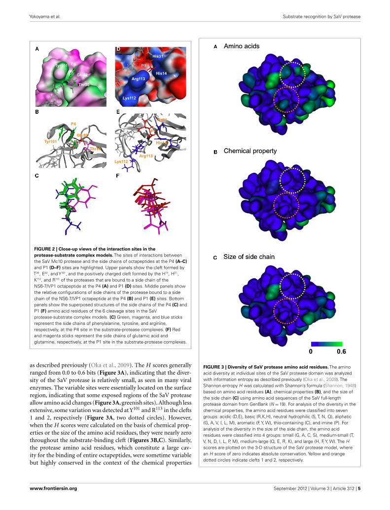
The protease-peptide complex models disclosed two interac-tion sites that were common to the six bound peptides. First, thesubstrate amino acid residues at the P4 position were exclusivelyplaced in a thin cleft, termed cleft 1, that was formed by threo-nine (T), glutamic acid (E), and tyrosine (Y) at positions 30, 52,and 101 of the protease domain (T30, E52, and Y101, respectively;Figures 2A–C). An aromatic ring of the phenylalanine (F) or Yat P4 of the octapeptides of the NS1/NS2, NS2/NS3, NS3/NS4,NS5/NS6-7, and NS6-7/VP1 cleavage sites (Figure 1A) was posi-tioned such that an aromatic stacking could be generated with theY101 in the protease cleft 1 (Figures 2A–C). The steric configura-tion of the aromatic rings of the P4 amino acid residues in thebound state was very similar except for the Y of the NS5/NS6-7cleavage site (Figure 2C). In the case of the NS4/NS5 peptide, theP4 amino acid is the arginine (R; Figure 1A) and was arrangednear the side chain of the E52 (Figure 2C).
Second, the substrate amino acid residues at the P1 site wereexclusively placed in a small positively charged cleft, termed cleft2, that was formed by the histidine (H), H, lysine (K), and R atpositions 14, 31, 112, and 113 of the protease domain (H14, H31,K112, and R113, respectively; Figures 2D–F). In four out of thesix cleavage sequences the P1 amino acid is negatively charged (E;Figure 1A) that could interact electrostatically with the side chainsof the positively charged cleft 2 of the protease (Figure 2D). In thecase of the NS2/NS3 and NS3/NS4, the P1 amino acid was glut-amine (Q; Figure 1A) which is hydrophilic and thus could causeelectrostatic interactions via a polarized charge. The steric configu-ration of the side chains of the P1 amino acid residues at the boundstate was very similar (Figure 2F). The simulated docking betweenthe protease and the substrate having alanine substitutions at P1and P4 positions resulted in a docking position similar to that forthe wild-type substrate, whereas the docking score was reduced toabout 1/2. Collectively, these results suggest that the interactions atthe P1 and P4 sites of the substrates play a key role in the substraterecognition, as suggested in the previous experiments (Robel et al.,2008; Oka et al., 2009).
FIGURE 1 | Structural models of SaV protease docked to the substrateoctapeptides. (A) Sequences of the six cleavage sites of the SaV ORF1polyprotein are shown with one-letter amino acid codes. Slashes representthe peptide bonds cleaved by the protease. (B) Structural models of theSaV protease-substrate complex. The 3-D structural model of the proteasedomain was constructed by homology modeling and thermodynamicallyand physically refined as described previously (Oka et al., 2007). The 3-Dstructural models of the octapeptides corresponding to the six authenticcleavage sites of the SaV Mc10 ORF1 were constructed by using theMolecular Builder tool in MOE. The optimized protease model was dockedto individual octapeptides using the automated ligand docking programASEDock2005 (Goto et al., 2008) operated in MOE as described previously(Yokoyama et al., 2010). Red and orange sticks indicate main and side chainsof the octapeptides, respectively.
AMINO ACID DIVERSITY OF HUMAN SaV PROTEASETo obtain evolutionary insights into the protease-substrate inter-actions, we analyzed the amino acid diversity of the proteasedomain among various human SaV strains in the public data-base. Full-length human SaV protease domain sequences werecollected from GenBank (N = 19) and used to calculate the Shan-non entropy scores, H (Shannon, 1948), in order to analyze thediversity of individual amino acid residues in the SaV population
Frontiers in Microbiology | Virology September 2012 | Volume 3 | Article 312 | 4
Yokoyama et al. Substrate recognition by SaV protease
FIGURE 2 | Close-up views of the interaction sites in theprotease-substrate complex models. The sites of interactions betweenthe SaV Mc10 protease and the side chains of octapeptides at the P4 (A–C)and P1 (D–F) sites are highlighted. Upper panels show the cleft formed byT30, E52, and Y101, and the positively charged cleft formed by the H14, H31,K112, and R113 of the proteases that are bound to a side chain of theNS6-7/VP1 octapeptide at the P4 (A) and P1 (D) sites. Middle panels showthe relative configurations of side chains of the protease bound to a sidechain of the NS6-7/VP1 octapeptide at the P4 (B) and P1 (E) sites. Bottompanels show the superposed structures of the side chains of the P4 (C) andP1 (F) amino acid residues of the 6 cleavage sites in the SaVprotease-substrate complex models. (C) Green, magenta, and blue sticksrepresent the side chains of phenylalanine, tyrosine, and arginine,respectively, at the P4 site in the substrate-protease complexes. (F) Redand magenta sticks represent the side chains of glutamic acid andglutamine, respectively, at the P1 site in the substrate-protease complexes.
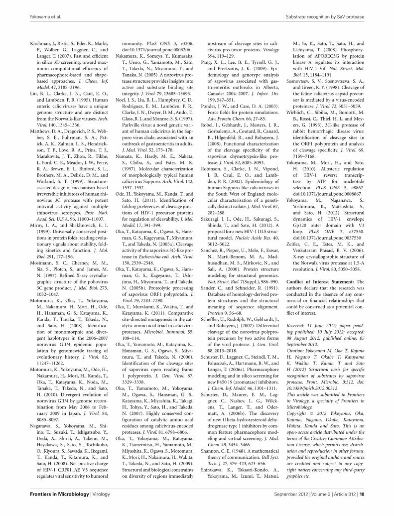
as described previously (Oka et al., 2009). The H scores generallyranged from 0.0 to 0.6 bits (Figure 3A), indicating that the diver-sity of the SaV protease is relatively small, as seen in many viralenzymes. The variable sites were essentially located on the surfaceregion, indicating that some exposed regions of the SaV proteaseallow amino acid changes (Figure 3A, greenish sites). Although lessextensive, some variation was detected at Y101 and R113 in the clefts1 and 2, respectively (Figure 3A, two dotted circles). However,when the H scores were calculated on the basis of chemical prop-erties or the size of the amino acid residues, they were nearly zerothroughout the substrate-binding cleft (Figures 3B,C). Similarly,the protease amino acid residues, which constitute a large cav-ity for the binding of entire octapeptides, were sometime variablebut highly conserved in the context of the chemical properties
FIGURE 3 | Diversity of SaV protease amino acid residues. The aminoacid diversity at individual sites of the SaV protease domain was analyzedwith information entropy as described previously (Oka et al., 2009). TheShannon entropy H was calculated with Shannon’s formula (Shannon, 1948)based on amino acid residues (A), chemical properties (B), and the size ofthe side chain (C) using amino acid sequences of the SaV full-lengthprotease domain from GenBank (N =19). For analysis of the diversity in thechemical properties, the amino acid residues were classified into sevengroups: acidic (D,E), basic (R,K,H), neutral hydrophilic (S, T, N, Q), aliphatic(G, A, V, I, L, M), aromatic (F, Y, W), thio-containing (C), and imine (P). Foranalysis of the diversity in the size of the side chain, the amino acidresidues were classified into 4 groups: small (G, A, C, S), medium-small (T,V, N, D, I, L, P, M), medium-large (Q, E, R, K), and large (H, F, Y, W). The Hscores are plotted on the 3-D structure of the SaV protease model, wherean H score of zero indicates absolute conservation. Yellow and orangedotted circles indicate clefts 1 and 2, respectively.
www.frontiersin.org September 2012 | Volume 3 | Article 312 | 5

Yokoyama et al. Substrate recognition by SaV protease
and sizes of the side chains (Figures 1B and 3). Thus theSaV protease appears to restrict extensive changes in the shapeand chemical properties of the substrate-binding surface for itssurvival.
SITE-DIRECTED MUTAGENESIS OF SaV PROTEASEConsistent with the above structural and diversity data, we pre-viously reported that the E52 in cleft 1, as well as H14 and H31
in cleft 2, are essential to maintain proper processing by SaVprotease (Oka et al., 2007). To obtain further insights into thebiological roles of clefts 1 and 2 in the proteolysis of the SaV pre-cursor polyprotein, we performed additional site-directed muta-genesis using a full-length clone of SaV Mc10 strain (Oka et al.,2005b). The Mc10 ORF1 encodes a polypeptide of 2278 amino
acid residues, where the six cleavage sites have been experimen-tally determined (Oka et al., 2006; Figure 4A). A total of ninemutants of the SaV protease domain were constructed using theMc10 ORF1. Full-length ORF1 precursor proteins having a sin-gle or double mutations in the protease domain were expressedusing the in vitro transcription-translation system, and the pro-cessing products were analyzed by gel electrophoresis as describedpreviously (Oka et al., 2005b, 2006, 2007, 2009). The Mc10 func-tional protease (Prowt) and a defective mutant completely lackingthe proteolysis activity (Promut; Oka et al., 2005b) were used aspositive and negative controls of the proteolysis, respectively.
When the ORF1 containing the Prowt was expressed, nine prod-ucts corresponded in size to the mature proteins NS1, NS2, NS3,NS4, NS5, and VP1, and relatively stable intermediate proteins,
FIGURE 4 | Site-directed mutagenesis of the substrate interaction sitesof SaV Mc10 protease. (A) Proteolytic cleavage map of the SaV Mc10 ORF1polyprotein and the processing intermediates (Oka et al., 2006). Black barsindicate the protein segments, A and D, used to raise polyclonal antibodies fordetection of the NS1 and NS5 proteins, respectively. (B) SDS-PAGE of35S-labeled in vitro translation products of SaV Mc10 ORF1 containing variousprotease mutants. NS1 and NS5 were detected by immunoprecipitation using
anti-A or anti-D polyclonal antibodies as described previously (Oka et al.,2005b, 2006, 2009). Mc10 ORF1 containing functional protease (Prowt) and adefective mutant lacking in the proteolysis activity (Promut) were included asdescribed previously (Oka et al., 2005b). Newly appearing products whencompared to Prowt are indicated by asterisks. Size markers are shown on theleft. Mc10 ORF1-specific proteins (Oka et al., 2005b, 2006) are shown on theright.
Frontiers in Microbiology | Virology September 2012 | Volume 3 | Article 312 | 6

Yokoyama et al. Substrate recognition by SaV protease
such as NS2-3, NS4-5, and NS4-5-6-7 were detected (Figure 4B,lane Prowt, black arrowheads; Oka et al., 2005b, 2006, 2009).These products were undetectable in the Promut ORF1 sample,and instead a product corresponding to the ORF1 polyproteinwas detected (Figure 4B, lane Promut, open triangle; Oka et al.,2005b, 2006, 2009). A single alanine substitution at T30 in the cleft1, K112 in the cleft 2, or R113 in the cleft 2 of viral protease resultedin a processing pattern similar to that of Prowt (Figure 4B, lanesT30A, K112A, and R113A). On the other hand, a single alaninesubstitution at Y101 in the cleft 1 (Y101A), a single acidic substitu-tion at K112 or R113 in the cleft 2 (K112E and R113E), and doublemutations in each cleft (T30AY101A and K112ER113E) resultedin abnormality of the precursor processing, i.e., an increase inaccumulation of the full-length ORF1 polyprotein and/or theNS4-5-6-7-VP1 intermediate protein (Figure 4B, asterisks). Inthe samples expressing ORF1 with T30A/Y101A or K112E/R113Edouble mutations, processing products corresponding to the NS5and NS4-5 disappeared almost completely (Figure 4B, lanes 5 and11, respectively).
PHARMACOPHORE-BASED IN SILICO SCREENING FOR THE LEADCOMPOUNDS OF SaV PROTEASE INHIBITORSTo further assess the role of the clefts 1 and 2 in the ligand bind-ing, we performed a pharmacophore-based in silico screeningof protease inhibitors. A total of 139,369 compounds (molecu-lar weights 42–2986) were screened for the lead molecules thatcontain an aromatic-ring-like portion resembling the P4 aminoacid, a negatively charged portion resembling the P1 amino acid,and a hydrophobic portion resembling the P1′ amino acid, beingarranged at similar 3-D positions with the authentic substrates(Figure 5). The hydrophobic portion resembling the P1′ aminoacid was included to better mimic the authentic substrate struc-tures. A total of 151 lead compounds matched to the categorywere then subjected to the in vitro trans cleavage assay of the SaVMc10 ORF1 polyprotein. With this screening, we could obtainthree compounds that inhibited processing of the SaV ORF1 atIC50 values of 18.4–26.5 µM (Figure 6).
We then analyzed how the lead compounds bound to the SaVMc10 protease by docking simulation (Figure 7). As expected,these compounds were predicted to bind to the protease at thesame interaction sites by which the authentic substrates boundto the protease. The aromatic-ring-like portion resembling the P4amino acid bound to the thin cleft formed by T30, E52, and Y101 forthe binding of the side chain of the P4 amino acid. The negativelycharged portion resembling the P1 amino acid bound to the smallpositively charged pocket formed by the H14, H31, K112, and R113
for the binding of the side chain of the P1 amino acid.
DISCUSSIONThe viral proteins that support viral replication and make up theviral particle are often translated as part of polyprotein precursors.Viral protease catalyzes cleavage of the precursor protein and thusplays an essential role in the viral life cycle. In this study, by com-bining computational and experimental approaches, we studiedthe structural basis for the substrate recognition by SaV protease.The results obtained in this study were consistent with each other
FIGURE 5 | Flow chart for screening chemical compounds against SaVprotease. Pharmacophore-based in silico screening (Schuster et al., 2006a,b;Kirchmair et al., 2007) was applied to extract the lead compounds havingstructural features that resembled those of the substrates of SaV protease.One hundred and fifty-one compounds exhibiting some similarities to theauthentic substrates were further assessed with respect to their inhibitoryactivity against SaV protease. With this strategy, we could obtain threecompounds that inhibited the processing of the SaV ORF1 polyprotein.
and disclosed novel structural base points of the protease for theattractive interactions with specific structures of ligands.
Using a homology modeling and a docking tool, we firstexamined the physical interactions of SaV protease and octapep-tides corresponding to the six authentic cleavage sites of the SaVORF1 polyprotein. Despite the marked sequence variation of theoctapeptides, they were bound to the protease in the same orien-tation in the structural models (Figure 1). The results suggestedthat there might be common interaction sites that served as ful-crums to direct the orientation of the octapeptides. Consistently,the models disclosed two interaction sites that were shared with thesix peptides and support the stable and functional binding of sub-strates to the catalytic cavity; the variable side chains at the P4 andP1 sites of the peptides were consistently bound to the two smallclefts, termed clefts 1 and 2, respectively (Figure 2). The former
www.frontiersin.org September 2012 | Volume 3 | Article 312 | 7

Yokoyama et al. Substrate recognition by SaV protease
FIGURE 6 | Dose-response curves of the inhibitors against SaVprotease. The inhibitory effects of the three chemical compounds that werescreened for their structural similarity to the authentic substrates of SaVprotease were determined with an in vitro trans cleavage assay. Aradiolabeled full-length Mc10 ORF1 polyprotein containing a defectiveprotease (Promut; Oka et al., 2005b) or a non-radiolabeled partial Mc10 ORF1polyprotein (NS6-7-VP1) containing a functional protease (Prowt; Oka et al.,2006) was separately expressed using the in vitro transcription/translationsystem. The translation products were mixed and incubated in the presenceof increasing concentrations of the indicated compounds at 30˚C for 20 h.The intensity of the radioactive band corresponding to the NS4-NS5 productwas measured with Typhoon 7500 and plotted in relation to the compoundconcentrations. (A) Compound No.50. (B) Compound No.100. (C)Compound No.116.
participated in aromatic stacking interactions, whereas the latterparticipated in electrostatic interactions. These results are consis-tent with the previous findings that the P4 and P1 amino acidresidues of the substrates play key roles in efficient proteolysis bySaV protease (Robel et al., 2008; Oka et al., 2009) and predicted thatthese two clefts could play a key role in substrate recognition viainteractions with the P4 and P1 amino acid residues of substrates.
This prediction was assessed by several analyses. If the cleftsplayed essential roles in recognition of substrates, spontaneousmutations that alter profoundly the physicochemical properties ofthe clefts should be suppressed for viral survival. Consistently ourShannon entropy study using protease sequences of various SaV
FIGURE 7 | Structural models of SaV protease docked to the inhibitors.Molecular formulas of the inhibitors (left) and structural models of theinhibitor-protease complexes (right) are shown. Molecular models of thethree chemical compounds having anti-SaV-protease activity wereconstructed using the Molecular Builder tool in MOE. Individual compoundswere docked to the SaV protease domain model using the automatedligand docking program ASEDock2005 (Goto et al., 2008). Light blue sticksin the protease indicate inhibitors. Greenish and bluish portions of theprotease indicate an aromatic and hydrophobic site and positively chargedsite, respectively. (A) Compound No.50. (B) Compound No.100. (C)Compound No.116.
strains from the world shows that the amino acid residues formingthe clefts 1 and 2 are variable but highly conserved in terms of thechemical properties or the sizes of side chains (Figure 3). Theresults indicate that these clefts tolerate mutations in nature butresist a range of mutations that markedly alter the chemical prop-erties or the shapes of the cleft surface. The findings are consistentwith the above structure-based prediction on the function of theclefts 1 and 2. These clefts are located on the surface of the largecavity of the protease. Therefore, the restrictions in the variationin two clefts are likely to be caused by functional constraints forthe essential interactions.
Moreover, we examined whether a range of mutations thatmarkedly alter the physicochemical properties of the clefts indeedcould result in aberrant processing of the SaV precursor polypro-tein. Our site-directed mutagenesis study showed that a singlemutation in cleft 1 (T30A) or in cleft 2 (K112A or R113A) causedlittle detectable damage in the processing of the viral precursor
Frontiers in Microbiology | Virology September 2012 | Volume 3 | Article 312 | 8

Yokoyama et al. Substrate recognition by SaV protease
polyprotein, showing a tolerance to mutations as indicated by ourinformation entropy study. Notably, however, (i) a single mutationthat causes a loss of aromatic stacking interaction (Y101A) in thecleft 1, (ii) a single mutation that causes a loss of the electrosta-tic interaction in the cleft 2 (K112E or R113E), and (iii) doublemutations within the clefts unexceptionally resulted in incompleteprocessing (Figure 4). The results indicate that the abnormal pro-cessing was caused only by single mutations that could extensivelyalter the chemical properties of the clefts. The data agree with theentropy data and again suggest the acceptability of variation in thetwo clefts under functional constraints.
Finally, we performed in silico screening of SaV proteaseinhibitors on the basis of the above structural and biological infor-mation. The screening of the 139,369 compounds in silico led tothe identification of the 151 compounds that resembled the struc-tural and spatial features of the P4 and P1 amino acid residuesof authentic substrates (Figure 5). From them, we could experi-mentally identify the three compounds that inhibited proteolysisof the SaV precursor polyprotein in vitro (Figure 6). As expected,these compounds were predicted to bind to the SaV protease atthe two clefts via similar attractive interactions with the authenticligands (Figure 7). These results provide additional evidence thattwo clefts on the SaV protease cavity play a key role in the ligandrecognition by providing the structural base points for the specificattractive interactions.
Notably, six cleavage sites of SaV precursor polyprotein alsodiffer with respect to their susceptibility to the SaV protease, withthe NS2/NS3, NS4/NS5, and NS5/NS6-7 sites being consistentlymore resistant to the cleavage than the NS1/NS2, NS3/NS4, andNS6-7/VP1 sites (Oka et al., 2005b, 2006, 2009). In this regard,it is of note that the P4 position of the NS4/NS5 site of humanSaV is exclusively arginine instead of an aromatic amino acid(Figure 1A) and that this arginine is conserved in all humanSaV strains reported thus far (Oka et al., 2005b, 2006, 2009). Thissubstitution at P4 position will abolish the aromatic stacking inter-action in the cleft 1 and thus will attenuate attractive interactions
between protease and the NS4/NS5 cleavage site. This possibilityis well consistent with the experimental findings; the cleavage ofthe NS4/NS5 site is less efficient than that of the other sites (Okaet al., 2005b, 2006, 2009) and is more sensitive to the cleft 1 muta-tions than the other cleavage sites are (Figure 4, lane 5, NS5).Moreover, the attenuation of cleavage of the NS4/NS5 site wasreversed simply by replacing the arginine with phenylalanine atthe P4 site (Oka et al., 2009). These findings strongly suggest thatthe well-preserved arginine at the P4 position of the SaV NS4/NS5cleavage site plays a key role in maintaining the distinct cleavabilityof precursor polyprotein by SaV protease.
In this study, we disclosed a novel 3-D pharmacophore con-taining two clefts on the cavity of the SaV protease, which can beused to identify the lead compounds of SaV protease inhibitors.SaV is one of the commonly detected pathogens in the acute gas-troenteritis of both children and adults (Johansson et al., 2005;Harada et al., 2009; Iturriza-Gomara et al., 2009; Pang et al., 2009).Diarrhea is one of the greatest causes of mortality in childrenunder age 5 in many countries (Boschi-Pinto et al., 2008), andthe outbreaks of the acute gastroenteritis often seriously affectsthe clinical, economic, and social activities. Therefore, anti-viralcompounds against SaV may be beneficial to some at-risk pop-ulations or communities. Thus far no anti-SaV inhibitors forthe clinical use have been developed. Our findings will provideimportant clues to the unique specificity of the SaV protease, theregulation of SaV maturation, and the rationale design of anti-SaVinhibitors.
ACKNOWLEDGMENTSWe thank Mami Yamamoto and Kana Miyashita for their techni-cal assistance with the mutagenesis. This work was supported bya grant from the Japan Health Science Foundation for Researchon Health Sciences Focusing on Drug Innovation, and grants forResearch on Emerging and Re-emerging Infectious Diseases andFood Safety from the Ministry of Health, Labour and Welfareof Japan.
REFERENCESBaker, D., and Sali, A. (2001). Protein
structure prediction and structuralgenomics. Science 294, 93–96.
Belliot, G., Sosnovtsev, S. V., Mitra,T., Hammer, C., Garfield, M., andGreen, K. Y. (2003). In vitro pro-teolytic processing of the MD145norovirus ORF1 nonstructuralpolyprotein yields stable precursorsand products similar to thosedetected in calicivirus-infected cells.J. Virol. 77, 10957–10974.
Bergmann, E. M., Cherney, M. M., Mck-endrick, J., Frormann, S., Luo, C.,Malcolm, B. A., Vederas, J. C., andJames, M. N. (1999). Crystal struc-ture of an inhibitor complex ofthe 3C proteinase from hepatitis Avirus (HAV) and implications forthe polyprotein processing in HAV.Virology 265, 153–163.
Boschi-Pinto, C., Velebit, L., andShibuya, K. (2008). Estimating childmortality due to diarrhoea in
developing countries. Bull. WorldHealth Organ. 86, 710–717.
Bull, R. A., Hyde, J., Mackenzie, J. M.,Hansman, G. S., Oka, T., Takeda, N.,and White, P. A. (2011). Compari-son of the replication properties ofmurine and human calicivirus RNA-dependent RNA polymerases. VirusGenes 42, 16–27.
Chiba, S., Nakata, S., Numata-Kinoshita, K., and Honma, S.(2000). Sapporo virus: historyand recent findings. J. Infect. Dis.181(Suppl. 2), S303–S308.
Chiba, S., Sakuma, Y., Kogasaka, R., Aki-hara, M., Horino, K., Nakao, T., andFukui, S. (1979). An outbreak ofgastroenteritis associated with cali-civirus in an infant home. J. Med.Virol. 4, 249–254.
Fullerton, S. W., Blaschke, M., Coutard,B., Gebhardt, J., Gorbalenya, A.,Canard, B., Tucker, P. A., andRohayem, J. (2007). Structuraland functional characterization of
sapovirus RNA-dependent RNApolymerase. J. Virol. 81, 1858–1871.
Goto, J., Kataoka, R., Muta, H., andHirayama, N. (2008). ASEDock-docking based on alpha spheresand excluded volumes. J. Chem. Inf.Model. 48, 583–590.
Guo, M., Chang, K. O., Hardy, M. E.,Zhang, Q., Parwani,A. V., and Saif, L.J. (1999). Molecular characterizationof a porcine enteric calicivirus genet-ically related to Sapporo-like humancaliciviruses. J. Virol. 73, 9625–9631.
Hansman, G. S., Oka, T., Katayama,K., and Takeda, N. (2007). Humansapoviruses: genetic diversity,recombination, and classification.Rev. Med. Virol. 17, 133–141.
Harada, S., Okada, M., Yahiro, S.,Nishimura, K., Matsuo, S., Miyasaka,J., Nakashima, R., Shimada, Y.,Ueno, T., Ikezawa, S., Shinozaki, K.,Katayama, K., Wakita, T., Takeda,N., and Oka, T. (2009). Surveillanceof pathogens in outpatients with
gastroenteritis and characterizationof sapovirus strains between 2002and 2007 in Kumamoto Prefecture,Japan. J. Med. Virol. 81, 1117–1127.
Hardy, M. E., Crone, T. J., Brower, J.E., and Ettayebi, K. (2002). Substratespecificity of the Norwalk virus 3C-like proteinase. Virus Res. 89, 29–39.
Iturriza-Gomara, M., Elliot, A. J., Dock-ery, C., Fleming, D. M., and Gray,J. J. (2009). Structured surveillanceof infectious intestinal disease inpre-school children in the commu-nity: “The Nappy Study.” Epidemiol.Infect. 137, 922–931.
Johansson, P. J., Bergentoft, K., Lars-son, P. A., Magnusson, G., Widell,A., Thorhagen, M., and Hedlund, K.O. (2005). A nosocomial sapovirus-associated outbreak of gastroenteri-tis in adults. Scand. J. Infect. Dis. 37,200–204.
Kataoka, R., and Goto, J. (2008). ASE-Dock – docking based on the shapeof binding site. Mol. Sci. 2, NP008.
www.frontiersin.org September 2012 | Volume 3 | Article 312 | 9
Yokoyama et al. Substrate recognition by SaV protease
Kirchmair, J., Ristic, S., Eder, K., Markt,P., Wolber, G., Laggner, C., andLanger, T. (2007). Fast and efficientin silico 3D screening: toward max-imum computational efficiency ofpharmacophore-based and shape-based approaches. J. Chem. Inf.Model. 47, 2182–2196.
Liu, B. L., Clarke, I. N., Caul, E. O.,and Lambden, P. R. (1995). Humanenteric caliciviruses have a uniquegenome structure and are distinctfrom the Norwalk-like viruses. Arch.Virol. 140, 1345–1356.
Matthews, D. A., Dragovich, P. S., Web-ber, S. E., Fuhrman, S. A., Pat-ick, A. K., Zalman, L. S., Hendrick-son, T. F., Love, R. A., Prins, T. J.,Marakovits, J. T., Zhou, R., Tikhe,J., Ford, C. E., Meador, J. W., Ferre,R. A., Brown, E. L., Binford, S. L.,Brothers, M. A., Delisle, D. M., andWorland, S. T. (1999). Structure-assisted design of mechanism-basedirreversible inhibitors of human rhi-novirus 3C protease with potentantiviral activity against multiplerhinovirus serotypes. Proc. Natl.Acad. Sci. U.S.A. 96, 11000–11007.
Mirny, L. A., and Shakhnovich, E. I.(1999). Universally conserved posi-tions in protein folds: reading evolu-tionary signals about stability, fold-ing kinetics and function. J. Mol.Biol. 291, 177–196.
Mosimann, S. C., Cherney, M. M.,Sia, S., Plotch, S., and James, M.N. (1997). Refined X-ray crystallo-graphic structure of the poliovirus3C gene product. J. Mol. Biol. 273,1032–1047.
Motomura, K., Oka, T., Yokoyama,M., Nakamura, H., Mori, H., Ode,H., Hansman, G. S., Katayama, K.,Kanda, T., Tanaka, T., Takeda, N.,and Sato, H. (2008). Identifica-tion of monomorphic and diver-gent haplotypes in the 2006–2007norovirus GII/4 epidemic popu-lation by genomewide tracing ofevolutionary history. J. Virol. 82,11247–11262.
Motomura, K., Yokoyama, M., Ode, H.,Nakamura, H., Mori, H., Kanda, T.,Oka, T., Katayama, K., Noda, M.,Tanaka, T., Takeda, N., and Sato,H. (2010). Divergent evolution ofnorovirus GII/4 by genome recom-bination from May 2006 to Feb-ruary 2009 in Japan. J. Virol. 84,8085–8097.
Naganawa, S., Yokoyama, M., Shi-ino, T., Suzuki, T., Ishigatsubo, Y.,Ueda, A., Shirai, A., Takeno, M.,Hayakawa, S., Sato, S., Tochikubo,O., Kiyoura, S., Sawada, K., Ikegami,T., Kanda, T., Kitamura, K., andSato, H. (2008). Net positive chargeof HIV-1 CRF01_AE V3 sequenceregulates viral sensitivity to humoral
immunity. PLoS ONE 3, e3206.doi:10.1371/journal.pone.0003206
Nakamura, K., Someya, Y., Kumasaka,T., Ueno, G., Yamamoto, M., Sato,T., Takeda, N., Miyamura, T., andTanaka, N. (2005). A norovirus pro-tease structure provides insights intoactive and substrate binding siteintegrity. J. Virol. 79, 13685–13693.
Noel, J. S., Liu, B. L., Humphrey, C. D.,Rodriguez, E. M., Lambden, P. R.,Clarke, I. N., Dwyer, D. M., Ando, T.,Glass, R. I., and Monroe, S. S. (1997).Parkville virus: a novel genetic vari-ant of human calicivirus in the Sap-poro virus clade, associated with anoutbreak of gastroenteritis in adults.J. Med. Virol. 52, 173–178.
Numata, K., Hardy, M. E., Nakata,S., Chiba, S., and Estes, M. K.(1997). Molecular characterizationof morphologically typical humancalicivirus Sapporo. Arch. Virol. 142,1537–1552.
Ode, H., Yokoyama, M., Kanda, T., andSato, H. (2011). Identification offolding preferences of cleavage junc-tions of HIV-1 precursor proteinsfor regulation of cleavability. J. Mol.Model. 17, 391–399.
Oka, T., Katayama, K., Ogawa, S., Hans-man, G. S., Kageyama, T., Miyamura,T., and Takeda, N. (2005a). Cleavageactivity of the sapovirus 3C-like pro-tease in Escherichia coli. Arch. Virol.150, 2539–2548.
Oka, T., Katayama, K., Ogawa, S., Hans-man, G. S., Kageyama, T., Ushi-jima, H., Miyamura, T., and Takeda,N. (2005b). Proteolytic processingof sapovirus ORF1 polyprotein. J.Virol. 79, 7283–7290.
Oka, T., Murakami, K., Wakita, T., andKatayama, K. (2011). Comparativesite-directed mutagenesis in the cat-alytic amino acid triad in calicivirusproteases. Microbiol. Immunol. 55,108–114.
Oka, T., Yamamoto, M., Katayama, K.,Hansman, G. S., Ogawa, S., Miya-mura, T., and Takeda, N. (2006).Identification of the cleavage sitesof sapovirus open reading frame1 polyprotein. J. Gen. Virol. 87,3329–3338.
Oka, T., Yamamoto, M., Yokoyama,M., Ogawa, S., Hansman, G. S.,Katayama, K., Miyashita, K., Takagi,H., Tohya, Y., Sato, H., and Takeda,N. (2007). Highly conserved con-figuration of catalytic amino acidresidues among calicivirus-encodedproteases. J. Virol. 81, 6798–6806.
Oka, T., Yokoyama, M., Katayama,K., Tsunemitsu, H., Yamamoto, M.,Miyashita, K., Ogawa, S., Motomura,K., Mori, H., Nakamura, H., Wakita,T., Takeda, N., and Sato, H. (2009).Structural and biological constraintson diversity of regions immediately
upstream of cleavage sites in cali-civirus precursor proteins. Virology394, 119–129.
Pang, X. L., Lee, B. E., Tyrrell, G. J.,and Preiksaitis, J. K. (2009). Epi-demiology and genotype analysisof sapovirus associated with gas-troenteritis outbreaks in Alberta,Canada: 2004–2007. J. Infect. Dis.199, 547–551.
Ponder, J. W., and Case, D. A. (2003).Force fields for protein simulations.Adv. Protein Chem. 66, 27–85.
Robel, I., Gebhardt, J., Mesters, J. R.,Gorbalenya, A., Coutard, B., Canard,B., Hilgenfeld, R., and Rohayem, J.(2008). Functional characterizationof the cleavage specificity of thesapovirus chymotrypsin-like pro-tease. J. Virol. 82, 8085–8093.
Robinson, S., Clarke, I. N., Vipond,I. B., Caul, E. O., and Lamb-den, P. R. (2002). Epidemiology ofhuman Sapporo-like caliciviruses inthe South West of England: mole-cular characterisation of a geneti-cally distinct isolate. J. Med. Virol. 67,282–288.
Sakuragi, J. I., Ode, H., Sakuragi, S.,Shioda, T., and Sato, H. (2012). Aproposal for a new HIV-1 DLS struc-tural model. Nucleic Acids Res. 40,5012–5022.
Sanchez, R., Pieper, U., Melo, F., Eswar,N., Marti-Renom, M. A., Mad-husudhan, M. S., Mirkovic, N., andSali, A. (2000). Protein structuremodeling for structural genomics.Nat. Struct. Biol. 7(Suppl.), 986–990.
Sander, C., and Schneider, R. (1991).Database of homology-derived pro-tein structures and the structuralmeaning of sequence alignment.Proteins 9, 56–68.
Scheffler, U., Rudolph, W., Gebhardt, J.,and Rohayem, J. (2007). Differentialcleavage of the norovirus polypro-tein precursor by two active formsof the viral protease. J. Gen. Virol.88, 2013–2018.
Schuster, D., Laggner, C., Steindl, T. M.,Palusczak, A., Hartmann, R. W., andLanger, T. (2006a). Pharmacophoremodeling and in silico screening fornew P450 19 (aromatase) inhibitors.J. Chem. Inf. Model. 46, 1301–1311.
Schuster, D., Maurer, E. M., Lag-gner, C., Nashev, L. G., Wilck-ens, T., Langer, T., and Oder-matt, A. (2006b). The discoveryof new 11beta-hydroxysteroid dehy-drogenase type 1 inhibitors by com-mon feature pharmacophore mod-eling and virtual screening. J. Med.Chem. 49, 3454–3466.
Shannon, C. E. (1948). A mathematicaltheory of communication. Bell Syst.Tech. J. 27, 379–423, 623–656.
Shirakawa, K., Takaori-Kondo, A.,Yokoyama, M., Izumi, T., Matsui,
M., Io, K., Sato, T., Sato, H., andUchiyama, T. (2008). Phosphory-lation of APOBEC3G by proteinkinase A regulates its interactionwith HIV-1 Vif. Nat. Struct. Mol.Biol. 15, 1184–1191.
Sosnovtsev, S. V., Sosnovtseva, S. A.,and Green, K. Y. (1998). Cleavage ofthe feline calicivirus capsid precur-sor is mediated by a virus-encodedproteinase. J. Virol. 72, 3051–3059.
Wirblich, C., Sibilia, M., Boniotti, M.B., Rossi, C., Thiel, H. J., and Mey-ers, G. (1995). 3C-like protease ofrabbit hemorrhagic disease virus:identification of cleavage sites inthe ORF1 polyprotein and analysisof cleavage specificity. J. Virol. 69,7159–7168.
Yokoyama, M., Mori, H., and Sato,H. (2010). Allosteric regulationof HIV-1 reverse transcrip-tase by ATP for nucleotideselection. PLoS ONE 5, e8867.doi:10.1371/journal.pone.0008867
Yokoyama, M., Naganawa, S.,Yoshimura, K., Matsushita, S.,and Sato, H. (2012). Structuraldynamics of HIV-1 envelopeGp120 outer domain with V3loop. PLoS ONE 7, e37530.doi:10.1371/journal.pone.0037530
Zeitler, C. E., Estes, M. K., andVenkataram Prasad, B. V. (2006).X-ray crystallographic structure ofthe Norwalk virus protease at 1.5-Aresolution. J. Virol. 80, 5050–5058.
Conflict of Interest Statement: Theauthors declare that the research wasconducted in the absence of any com-mercial or financial relationships thatcould be construed as a potential con-flict of interest.
Received: 11 June 2012; paper pend-ing published: 10 July 2012; accepted:08 August 2012; published online: 05September 2012.Citation: Yokoyama M, Oka T, KojimaH, Nagano T, Okabe T, KatayamaK, Wakita T, Kanda T and SatoH (2012) Structural basis for specificrecognition of substrates by sapovirusprotease. Front. Microbio. 3:312. doi:10.3389/fmicb.2012.00312This article was submitted to Frontiersin Virology, a specialty of Frontiers inMicrobiology.Copyright © 2012 Yokoyama, Oka,Kojima, Nagano, Okabe, Katayama,Wakita, Kanda and Sato. This is anopen-access article distributed under theterms of the Creative Commons Attribu-tion License, which permits use, distrib-ution and reproduction in other forums,provided the original authors and sourceare credited and subject to any copy-right notices concerning any third-partygraphics etc.
Frontiers in Microbiology | Virology September 2012 | Volume 3 | Article 312 | 10