structural and molecular basis of znrf3/rnf43
TRANSCRIPT

Structural and molecular basis of ZNRF3/RNF43 transmembrane ubiquitin ligase inhibition by the Wnt agonist R-spondin
CitationZebisch, Matthias, Yang Xu, Christos Krastev, Bryan T. MacDonald, Maorong Chen, Robert J. C. Gilbert, Xi He, and E. Yvonne Jones. 2013. “Structural and molecular basis of ZNRF3/RNF43 transmembrane ubiquitin ligase inhibition by the Wnt agonist R-spondin.” Nature Communications 4 (1): 2787. doi:10.1038/ncomms3787. http://dx.doi.org/10.1038/ncomms3787.
Published Versiondoi:10.1038/ncomms3787
Permanent linkhttp://nrs.harvard.edu/urn-3:HUL.InstRepos:11879669
Terms of UseThis article was downloaded from Harvard University’s DASH repository, and is made available under the terms and conditions applicable to Other Posted Material, as set forth at http://nrs.harvard.edu/urn-3:HUL.InstRepos:dash.current.terms-of-use#LAA
Share Your StoryThe Harvard community has made this article openly available.Please share how this access benefits you. Submit a story .
Accessibility
ARTICLE
Received 6 Aug 2013 | Accepted 17 Oct 2013 | Published 14 Nov 2013
Structural and molecular basis of ZNRF3/RNF43transmembrane ubiquitin ligase inhibitionby the Wnt agonist R-spondinMatthias Zebisch1, Yang Xu2,3, Christos Krastev1, Bryan T. MacDonald2, Maorong Chen2,
Robert J.C. Gilbert1, Xi He2 & E. Yvonne Jones1
The four R-spondin (Rspo) proteins are secreted agonists of Wnt signalling in vertebrates,
functioning in embryogenesis and adult stem cell biology. Through ubiquitination and
degradation of Wnt receptors, the transmembrane E3 ubiquitin ligase ZNRF3 and related
RNF43 antagonize Wnt signalling. Rspo ligands have been reported to inhibit the ligase
activity through direct interaction with ZNRF3 and RNF43. Here we report multiple crystal
structures of the ZNRF3 ectodomain (ZNRF3ecto), a signalling-competent Furin1–Furin2
(Fu1–Fu2) fragment of Rspo2 (Rspo2Fu1–Fu2), and Rspo2Fu1–Fu2 in complex with ZNRF3ecto, or
RNF43ecto. A prominent loop in Fu1 clamps into equivalent grooves in the ZNRF3ecto and
RNF43ecto surface. Rspo binding enhances dimerization of ZNRF3ecto but not of RNF43ecto.
Comparison of the four Rspo proteins, mutants and chimeras in biophysical and cellular
assays shows that their signalling potency depends on their ability to recruit ZNRF3 or RNF43
via Fu1 into a complex with LGR receptors, which interact with Rspo via Fu2.
DOI: 10.1038/ncomms3787 OPEN
1 Division of Structural Biology, Wellcome Trust Centre for Human Genetics, University of Oxford, Oxford OX3 7BN, UK. 2 F.M. Kirby Neurobiology Center,Department of Neurology, Boston Children’s Hospital, Harvard Medical School, Boston, Massachusetts 02115, USA. 3 Key Laboratory for MolecularEnzymology and Engineering of Ministry of Education, College of Life Science, Jilin University, Changchun 130012, China. Correspondence and requests formaterials should be addressed to E.Y.J. (email: [email protected]).
NATURE COMMUNICATIONS | 4:2787 | DOI: 10.1038/ncomms3787 | www.nature.com/naturecommunications 1
& 2013 Macmillan Publishers Limited. All rights reserved.
Rspo (R-spondin, also roof plate-specific spondin) proteinsare evolutionarily conserved from fish to humans and havewell-documented roles in a broad range of developmental
and physiological processes resulting from enhancement ofcanonical and non-canonical Wnt signalling1–3. Rspo1 isinvolved in mammalian sex determination4 and is a potentstimulator of epithelial repair in the gastrointestinal tract2,5.Rspo2 has recently been identified as a major determinant ofsusceptibility to infectious diarrhoea in mice, linking infectionand intestinal homoeostasis6. Gene fusions involving RSPO2 andRSPO3 have been found in 10% of primary colon cancers7,whereas mutations in RSPO4 underlie inherited anonychia, adisorder in nail development8–10. The leucine-rich repeatcontaining G protein-coupled receptors 4, 5 and 6 (LGR4/5/6)are conserved high-affinity cell surface receptors for Rspoproteins11–15; however, the molecular mechanisms by whichRspo proteins function have remained obscure.
Recently published work has indicated that Rspo proteins canexert their potentiating effects on Wnt signalling through directinteraction with the extracellular regions of ZNRF3 or RNF43,ultimately inducing formation of a complex comprising ZNRF3/RNF43, Rspo and LGR4/5/6 (ref. 16). Similar to the Rspo proteins,ZNRF3 and RNF43 are highly conserved in vertebrates. Loss-of-function mutations of RNF43 in pancreatic cancer have implicatedit as a tumour suppressor17. ZNRF3 and RNF43 comprise anamino-terminal extracellular region of uncharacterized topologyand moderate sequence conservation of 39% identity between thetwo proteins, a transmembrane region and a cytoplasmic regionthat bears the hallmark sequence of a really interesting new gene(RING)-type E3 ubiquitin ligase. Similar to LGR4/5/6 receptors,ZNRF3/RNF43 have been reported to associate in the membranewith the Wnt receptor Frizzled and LRP5/6 coreceptors13,16.ZNRF3/RNF43 specifically targets these Wnt receptors forubiquitination and turnover, hence reducing Wnt signallingresponses16,18. Direct extracellular interaction with Rspoproteins inhibits ZNRF3/RNF43 activity16. These observationshave led to the suggestion that Rspo acts to physically bridgebetween its two receptor types ZNRF3/RNF43 and LGR4/5/6 (ref.16). Current models suggest that membrane clearance of ZNRF3/RNF43 through this ternary complex relieves turnover of Wntreceptors and hence enhances Wnt responsiveness.
Here we report a molecular level analysis of the ZNRF3/RNF43ectodomain structure and its interactions with Rspo proteins. Ourstudy provides mechanistic insight into this key control point inthe Wnt signalling pathway.
ResultsStructure determination. Sequence analyses suggest a putativedomain structure for the Rspo proteins comprising two furin-likecysteine-rich regions (Fu domains) plus a thrombospondin type 1repeat domain3 (Fig. 1a). Our own and published data point to theinvolvement of the Fu domains in the potentiation of canonicalWnt signalling by Rspo proteins1,19–21 (Supplementary Fig. S1).We therefore engineered constructs to express the region spanningthe two Fu domains of Rspo2 proteins from several species. Wealso generated secreted forms of the corresponding ZNRF3 andRNF43 ectodomains. The Rspo2Fu1–Fu2 and respective ZNRF3ecto
or RNF43ecto molecules migrated together in gel filtrationchromatography indicating high-affinity binding (data notshown), substantiating their ligand–receptor relationship. Byusing a combination of heavy atom and molecular-replacement-based phasing strategies, we determined multiple crystal structuresfor Xenopus (x) Rspo2Fu1–Fu2 (highest resolution 2.2 Å),xZNRF3ecto, zebrafish (z) ZNRF3ecto and mouse (m) ZNRF3ecto,(highest resolutions 2.4, 1.6 and 2.0 Å, respectively), plus
complexes comprising xZNRF3ecto–xRspo2Fu1–Fu2, mZNRF3ecto–mRspo2Fu1–Fu2, mZNRF3ecto–xRspo2Fu1–Fu2 and xRNF4F3ecto–xRspo2Fu1–Fu2 (at 2.1, 2.8, 2.4 and 2.7 Å, respectively; seeMethods, Table 1 and Supplementary Table S1). In thefollowing sections and figures, the highest resolution structures(Table 1 and Fig. 1) for the apo ligand, apo receptor and ligand–receptor complex will be used unless otherwise stated.
Structure of the ZNRF3 ectodomain. The ZNRF3ecto crystalstructures revealed a distinctive variant of the protease-associateddomain topology22. Two b-sheets (comprising b2, b1, b7, b3and b4, b5, b6 strands, respectively; Fig. 1b) splay apart,accommodating an a-helix (aC) at the open edge; two additionala-helices (aA and aB) pack against the b4, b5 and b6 face of thisdistorted b-sandwich. A disulphide bridge, conserved acrossspecies, links two structurally elaborate loops, b3–b4 and b4–aA.The resultant single-domain structure is relatively compact. Thecrystal structures for apo xZNRF3ecto, zZNRF3ecto and mZNRF3ecto
showed no major differences in the main chain conformation(Supplementary Fig. S2). Comparisons of ZNRF3ecto structures forproteins crystallized in several different crystal lattices, or forcrystals containing multiple copies in the asymmetric unit,consistently highlighted an acidic region (N105-E114; residuenumbering is for mouse sequences unless otherwise stated) withinthe b3–b4 loop, the short aC–b7 loop and the extended b1–b2hairpin as flexible elements of the fold (Supplementary Fig. S2). Asearch of the Protein Data Bank for structures with a similartopology yielded the ectodomain of GRAIL (gene related to anergyin lymphocytes) as the closest match (deposited as an unpublishedcrystal structure by J.R. Walker and colleagues, StructuralGenomics Consortium; Protein Data Bank ID code 3ICU).GRAIL is a single-span transmembrane E3 ubiquitin ligase,which localizes to the endosomal compartment and promotesCD3 ubiquitinylation, acting as an essential regulator of T-celltolerance23,24. The sequence identity between ZNRF3 and GRAILectodomains is low (13.4% for 127 residues); however, structuralsuperposition revealed a shared three-dimensional fold consistentwith a common evolutionary origin (r.m.s.d. 2.5 Å for 131equivalent Ca pairs; Supplementary Fig. S3a).
Our crystallographic data provided independent structures formultiple copies of the ZNRF3 ectodomain in eight differentcrystal forms (Table 1 and Supplementary Table S1). All but twoof these crystal structures reveal an extensive interface (averageinterface area 992±109 Å2; Supplementary Table S2) formedbetween two ZNRF3ecto polypeptide chains. This dimer isconserved, and pairwise structural superpositions yieldedr.m.s.d. values of o1.3 Å (for 275 equivalent Ca pairs;Supplementary Tables S3 and S4). The interaction is twofoldsymmetric; strands b3 and b7 of the ‘subunits’ abut face-to-face atthe core of the dimer (Fig. 1c and 2a). The b1–b2 hairpin forms asecond interface by reaching out to embrace helix aA and the b3–b4 loop in the opposing subunit (Fig. 2b). Intriguingly, thesestructural features interact in a parallel (cis) fashion consistentwith ZNRF3 associating as a dimer on the cell surface.
Structure of a signalling-competent fragment of Rspo2. In thecrystal structure of the isolated RspoFu1–Fu2 protein (Table 1) thetwo Fu domains arrange sequentially to form a ladder-likestructure of b-hairpins (Fig. 1d). Each Fu domain comprises threeb-hairpins rigidified by four disulphide bridges (Fig. 1e), similarto the cysteine-rich regions found in members of the epidermalgrowth factor receptor family (Supplementary Fig. S3b).The connection between the Fu domains shows considerablerotational freedom, allowing a 50�–60� variation in the relativeinterdomain orientation (Supplementary Fig. S4). The N terminal
ARTICLE NATURE COMMUNICATIONS | DOI: 10.1038/ncomms3787
2 NATURE COMMUNICATIONS | 4:2787 | DOI: 10.1038/ncomms3787 | www.nature.com/naturecommunications
& 2013 Macmillan Publishers Limited. All rights reserved.

of the two domains, Fu1, is distinguished by the extension of thesecond b-hairpin (Fig. 1d,e). This prominent loop presents asolvent exposed methionine (M68) at its tip, which we term the‘Met-finger’.
Structure of liganded complexes of ZNRF3ecto and RNF43ecto.The crystal structures of the ZNRF3ecto–Rspo2Fu1–Fu2 andRNF43ecto–Rspo2Fu1–Fu2 complexes (Table 1 and SupplementaryTable S1) revealed a 1:1 interaction between Fu1 of the Rspo2Fu1–
TM
a
b d f
e
c
r.m.s.d. = 2.8 Å
Q195
I191
V192A198
I95
M68
90°
SP PAD RING
SP Fu1 Fu2 TSR BR
g h
90°
C104
7
CT
CT
NT
CT CT
NT NT
NT
Met-fingerM68
Figure 1 | Unliganded and complexed structures of ZNRF3 and Rspo proteins. (a) Schematic domain organization of Rspo (top) and ZNRF3/RNF43
proteins (bottom) roughly at scale. The domains included in the crystallization constructs are coloured in blue, red and orange. Disulphides are derived
from the crystal structure, except for those of the TSR domain of Rspo, which are based on a model48. (b) Cartoon representation of the fold of the
ZNRF3 ectodomain protomer. b-strands are numbered and a-helices are labelled in alphabetical order from the N to C terminus. (c) Structure of the
recurring ZNRF3ecto dimer with view parallel to the putative membrane layer and from top towards the membrane. An acidic region with sequence105NNNDEEDLYEY115 is highlighted in red in b and c. (d) The xRspo2Fu1–Fu2 structure. Both b-hairpins and disulphide bridges line up to form a ladder-like
structure. The second b-hairpin of Fu1 contains an exposed methionine side chain. (e) Fu1 and Fu2 share the same architecture, except that the second
b-hairpin of Fu1 is considerably longer. (f) The ZNRF3ecto–Rspo2Fu1–Fu2 complex as the same 2:2 symmetric complex in all seven crystallographic
observations. Shown are two views parallel to the putative membrane orientation. The RNF43ecto–Rspo2Fu1–Fu2 complex resembles one half of this complex
(Supplementary Fig. S5). (g) The ZNRF3ecto–Rspo2Fu1–Fu2 interface. xZNRF3ecto is shown in semi-transparent surface (orange) and ribbon, xRspo2Fu1–Fu2, is
depicted in blue. Residue side chains involved in the interface are shown as sticks and labelled (atom colouring: dark blue, nitrogen; red, oxygen; yellow,
sulphur). Dotted lines represent hydrogen bonds. A corresponding stereo figure with final electron density can be found in Supplementary Fig. S6.
(h) The Met-finger pocket. Structural features are represented as in g. BR, basic region; PAD, protease-associated domain; SP, signal peptide; TM,
transmembrane; TSR, thrombospondin-related domain.
NATURE COMMUNICATIONS | DOI: 10.1038/ncomms3787 ARTICLE
NATURE COMMUNICATIONS | 4:2787 | DOI: 10.1038/ncomms3787 | www.nature.com/naturecommunications 3
& 2013 Macmillan Publishers Limited. All rights reserved.

Fu2 and a single ZNRF3ecto or RNF43ecto chain (Fig. 1f). As will bediscussed below, all complex structures, except for the RNF43complex, reveal a conserved 2:2 stoichiometry (SupplementaryFig. S5a). The interaction interface between Rspo2Fu1–Fu2 and itstwo receptors RNF43 and ZNRF3 is essentially the same(Supplementary Fig. S5b). It involves an interface area of990±105 Å2 (Fig. 1g,h, Fig. 3, Supplementary Fig. S6 andSupplementary Table S2). Because of the availability of higherresolution data and multiplicity of data sets, we will first focus onthe Rspo2–ZNRF3 interaction. Neither the Rspo2Fu1–Fu2 nor theZNRF3ecto dimer show major conformational changes on com-plex formation (Supplementary Fig. S5). The first, extensive, areaof interaction involves hydrophobic interactions interspersed withhydrophilic (complementarily charged) patches contributed bythe first two b-hairpins of the Rspo Fu1 and the region imme-diately carboxy-terminal to the b3 strand of ZNRF3ecto (Fig. 1g).The Met-finger at the tip of the second b-hairpin of Fu1 nestlesinto a pocket formed between the b3 strand and the aC–b7 loopof the ZNRF3ecto, which is lined with hydrophobic residues (I95,I191, V192, A198; Fig. 1h). The aC–b7 loop is a flexible region inthe unliganded ZNRF3ecto crystal structures and moulds tointerface the RspoFu1–Fu2 M68 in the complex. Overall, theZNRF3ecto dimer structure appears less flexible in the complexstructures compared with the unliganded structures. The acidicregion of the b3–b4 loop (immediately adjacent to C104 of thedisulphide bridge) becomes more ordered in the ligand-boundZNRF3ecto structures (Supplementary Fig. S2), probably as aresult of electrostatic interactions with a positively charged patchon Rspo Fu1 (Fig. 3a).
Biophysical and cellular analyses support the structure data.Analytical ultracentrifugation results are consistent with
ZNRF3ecto dimer formation and analyses of several ZNRF3-Rspo2 and RNF43-Rspo2 interface mutants (Figs 1g,h and 3)using surface plasmon resonance (SPR)-binding assays confirmthe crystallographically determined complex structures(Fig. 4). The single-domain protein mRspo2Fu1 still boundmZNRF3ecto with high affinity, whereas no detectable bindingwas measured for mRspo2Fu2 (Fig. 4b). Consistent with the highlevel of surface residue conservation at the interface (Fig. 3a),the Fu1–Fu2 repeats for all four members of the Rspo familyshowed binding to ZNRF3ecto in SPR assays (Fig. 4b). However,the fine-grained differences in the interface-forming residues didimpact on the binding affinities; the stronger binding ofRspo2Fu1–Fu2 versus Rspo1Fu1–Fu2 and Rspo4Fu1–Fu2 appeared tobe conferred, in part, by the substitution of isoleucine formethionine at the tip of the second b-hairpin (Fig. 4c). Previouslyreported genetic and cancer-associated mutations further corro-borate the functional significance of the ZNRF3ecto–Rspo2Fu1–Fu2
interface as the generic interaction mode for Rspo1–4 andZNRF3/RNF43 (Fig. 4d–g). For example, in Rspo4, the equivalentof the R65W, Q70R and G72R mutations have been reported ininherited anonychia8–10. From an analysis of the interactioninterface, it is obvious that these mutations are not compatiblewith ZNRF3/RNF43 binding (Fig. 1g). Functional assays thatmeasure Rspo signalling activity in cells further support thesignificance of the Rspo–ZNRF3 interface (Fig. 5 andSupplementary Fig. S7). For example, a Met-finger mutation,M68E, which profoundly compromised Rspo–ZNRF3 interactionin SPR assays, exhibited much weaker signalling activity,whereas a conserved substitution, M68I, showed slightlyreduced binding to ZNRF3 and relatively normal (or slightlyreduced) signalling capacity (Fig. 5d). Other interface mutants inFu1, including the anonychia-associated mutations R65W, Q70Rand G72R, as well as N50R, each exhibited weakened signalling
Table 1 | Data collection and refinement statistics.
ZNRF3ecto/RNF43ecto zZNRF3 mZNRF3 xZNRF3 — — mZNRF3 mZNRF3 xZNRF3 xRNF43RspoFu1–Fu2 — — — xRSPO2 xRSPO2–Pt mRSPO2 xRSPO2 xRSPO2 xRSPO2
Data collectionSpace group P21 P21 P21 P41212 P41212 P212121 P1 P21 C2Cell dimensions
a, b, c (Å) 36.0, 53.3,72.5
47.3, 57.8,50.5
49.2, 58.7,52.7
97.1, 97.1,292.9
96.6, 96.6,290.1
59.8, 77.2,130.6
36.4, 71.0,72.0
56.0, 81.2,71.6
88.9, 35.8,87.9
a, b, g (�) 90, 102.1, 90 90, 97.6, 90 90, 93.6, 90 90, 90, 90 90, 90, 90 90, 90, 90 109.2, 101.7,101.3
90, 113.0, 90 90, 114.6, 90
Resolution (Å)* 42.59–1.60(1.63–1.60)
37.84–2.00(2.05–2.00)
37.66–2.40(2.49–2.40)
39.69–2.20(2.25–2.20)
39.46–3.20(3.46–3.20)
66.46–2.80(2.97–2.80)
38.89–2.40(2.49–2.40)
65.94–2.10(2.16–2.10)
32.76–2.70(2.83–2.70)
Rmerge 0.074 (0.359) 0.089 (0.353) 0.067 (0.478) 0.106 (0.852) 0.218 (1.443) 0.116 (1.345) 0.073 (0.605) 0.115 (0.788) 0.147 (0.550)I/sI 16.1 (2.3) 9.7 (1.9) 8.7 (1.8) 10.3 (2.0) 23.2 (2.5) 16.0 (2.3) 15.0 (2.6) 17.1 (2.6) 9.5 (2.2)
Completeness (%) 99.8 (99.0) 91.8 (78.6) 99.2 (99.3) 99.1 (99.9) 99.9 (99.9) 99.9 (100) 78.5 (29.2) 93.7 (56.5) 95.8 (97.8)Redundancy 11.0 (10.9) 3.6 (2.4) 3.0 (3.1) 6.8 (6.6) 50.6 (15.1) 14.4 (14.6) 6.6 (5.8) 17.0 (7.8) 3.8 (3.0)
RefinementResolution (Å)* 42.59–1.60
(1.63–1.60)37.84–2.00(2.05–2.00)
37.66–2.40(2.49–2.40)
39.69–2.20(2.25–2.20)
66.46–2.80(2.97–2.80)
38.89–2.40(2.49–2.40)
65.94–2.10(2.16–2.10)
32.76–2.70(2.83–2.70)
No. of reflections 34,163 15,439 12,423 67,674 14,706 18,529 31,015 6,517Rwork/Rfree 0.222/0.258 0.200/0.276 0.224/0.299 0.223/0.270 0.236/0.323 0.195/0.273 0.188/0.246 0.317/0.395No. of atoms
Protein 2,121 2,327 2,220 6,807 3,967 3,815 4,145 1,679Water 79 29 9 292 — — 220 —Ligands — — — — 1 — — —
B-factors (Å2)Protein 41.5 48.6 64.7 47.9 86.2 79.8 35.4 60.4Water 37.8 42.7 51.0 40.6 — — 36.8 —Ligands — — — — 79.1 — — —
r.m.s.d.Bond lengths (Å) 0.008 0.013 0.012 0.012 0.013 0.015 0.013 0.004Bond angles (�) 1.199 1.591 1.631 1.418 1.656 1.864 1.604 0.748Number of monomers or1:1 complexes
2 3 2 8 2 2 2 1
Dimeric architecture No Yes Yes — Yes Yes Yes NoProtein Data Bank code 4C84 4C86 4C8T 4C8V 4C99 4C9A 4C9R 4C9V
*Highest resolution shell is shown in parenthesis. Statistics of additional structures can be found in Supplementary Table S1.
ARTICLE NATURE COMMUNICATIONS | DOI: 10.1038/ncomms3787
4 NATURE COMMUNICATIONS | 4:2787 | DOI: 10.1038/ncomms3787 | www.nature.com/naturecommunications
& 2013 Macmillan Publishers Limited. All rights reserved.

a b
R199
D70
�1
Figure 2 | Dimerization interface of xZNRF3ecto. (a) View along the twofold axis away from the putative membrane. (b) xZNRF3ecto–xRspoFu1–Fu2 complex
with close-up view onto the b1–b2 hairpin arm (‘clamp’) embracing the respective other protomer. This interface is stabilized by binding of Rspo
to ZNRF3 and subsequent structuring of the acidic region of the b3–b4 loop drawn in red. Residue numbers refer to mouse proteins.
R65W
S53RG72R
N50R H86R(43) K108N(43)
RspoFu1–Fu2
RSPOFu1–Fu2180°
ZNRF3ecto
ZNRF3ecto
S85F(43),M101T(3)
E112K(3)
H86R(43)
C119R(43)
P118T(43)D132N(3)
A146G(43) K108N(43)D140H(43)
A169T(43)G166C(43)
N167I(43)
A78T(43)
I48T(43)
M173T(43)L82S(43)
H183R(43)
M83T(43) H86R(43)
P154S,L(43) R127P/Q(43)
P118T(43)
C119R(43)
W120L(3)
M83T(43)
�3–�4 acidic region
M68I/A/E
L63FE109K(3)
M98T(3)Q97E(3)
L82S(43)
D102N(4)
G131C(2)E127V(2), T121I(4)
P134Q(2)
P123L(4)
K113T(1)
G82E(1), G76A(4)
Y82H(2)
L63F(2)
R64Q(2)
D68N(1)
N67K(1)
R69C(2)
G72S(4), G72R(4*)
G52S(1), G51R/V(2)
180°
D50H(3)E44D(4)
S45P(2)
S53R(2)
Q57H(2)
R87M(4)
R86Q(2)
G84E(2)
F105L(2)
D102N(4)
G67W/E(2)
Q71P(1),F62S(2)
Q70H(2), Q65R(4*)
R65T(3), R60W(4*)
F105L(2)
G84E(2)
R86Q(2)
R87M(4)
Q57H(2)
S53R(2)
S45P(2)E44D(4)D50H(3)
a b
Figure 3 | Characteristics of the ZNRF3 dimer and Rspo–ZNRF3 complex interfaces. (a) An open book view of the ZNRF3–Rspo interface. The surface
contributing to the interface is coloured green on ZNRF3ecto and RspoFu1–Fu2; within this, surface mutants tested in this study are highlighted in red (top).
Rspo and ZNRF3ecto coloured by electrostatic surface potential from red (acidic) to blue (basic) (middle). Sequence conservation across species
coloured from white (not conserved) to black (conserved). (b) Disease-related mutations are plotted onto the molecular surface of Rspo (top) and ZNRF3/
RNF43 (bottom), and are concentrated at the Rspo–ZNRF3/RNF43 interaction interface. Tumour-associated missense mutations derived from the cosmic
database (http://cancer.sanger.ac.uk/cancergenome/projects/cosmic/) are shown in red and missense mutations causal for congenital anonychia on
RSPO4 are shown in orange. Sites in orange on ZNRF3 are mutations of RNF43 that map to the dimer interface of ZNRF3. Numbers 1–4 in parentheses
indicate mutations found in RSPO1 to RSPO4 (top). Number 3 and 43 in parentheses indicate mutations found in ZNRF3 and RNF43, respectively (bottom).
NATURE COMMUNICATIONS | DOI: 10.1038/ncomms3787 ARTICLE
NATURE COMMUNICATIONS | 4:2787 | DOI: 10.1038/ncomms3787 | www.nature.com/naturecommunications 5
& 2013 Macmillan Publishers Limited. All rights reserved.

ability that correlated with reduction in binding to ZNRF3(Fig. 5e).
Dimerization propensity of ZNRF3 versus monomeric RNF43.For all ZNRF3ecto–Rspo2Fu1–Fu2 complex structures we deter-mined (from five different combinations of species and crystal
forms), the dimer found in most of the unliganded ZNRF3ecto
crystal structures reoccurs (Fig. 1f, Supplementary Fig. S5 andSupplementary Table S2). The overall assembly thus comprises a2:2 complex of ZNRF3ecto–Rspo2Fu1–Fu2. The 2:2 complexresembles a crab with the ZNRF3ecto dimer forming the bodyfrom which the two Rspo2Fu1–Fu2 ligands diverge, without
80a
60
40
c (s
)
20
01.0 1.5
ZN
RF
3 ecto
Mon
omer
ZN
RF
3 ecto
-R
SP
O2 F
u1–F
u21:
1 co
mpl
ex
ZN
RF
3 ecto
-R
SP
O2 F
u1–F
u22:
2 co
mpl
ex
ZN
RF
3 ecto
dim
er
2.52.0 3.0
s (S)
3.5 4.0 4.5 5.0
120 h1 6.8 μMm2 25 nMh3 60 nMm4 300 μMm2Fu1 510 nM
m2Fu2 noresponse
100
80
60
Nor
mal
ized
res
pons
e
40
20
0
[mZNRF3ecto] (M)
1e–10 1e–9 1e–8 1e–7 1e–6 1e–5 1e–4 1e–3
mZNRF3ectomZNRF3ecto E92N, E94T
mZNRF3ecto S90CxRNF43ecto × xRSPO2Fu1–Fu2xZNRF3ecto × xRSPO2Fu1–Fu2
b
cwtM68I 85 nMM68E 23 μM
wtR65W 670 nMQ70R 22 μMG72R 27 μM
Nor
mal
ized
res
pons
e
120
100
80
60
40
20
0
[mZNRF3ecto] (M)
mZNRF3ecto hRNF43ecto mZNRF3ecto
[mZNRF3ecto] (M)
1e–10 1e–9 1e–8 1e–7 1e–6 1e–5 1e–4 1e–3
120
100
80
60
40
20
01e–10 1e–9 1e–8 1e–7 1e–6 1e–5 1e–4 1e–3
[mZNRF3ecto] (M)
120
100
80
60
40
20
0
1e–10 1e–9 1e–8 1e–7 1e–6 1e–5 1e–4 1e–3
wtL63F 109 nMS53R 182 nMN50R 5.6 μM
Nor
mal
ized
res
pons
e
120
100
80
60
40
20
0
[mRSPO2Fu1–Fu2] (M)
LGR5(chip: hLGR5ecto)
ZNRF3(chip: RSPO variant)
mZNRF3ecto
1e–10 1e–9 1e–8 1e–7 1e–6 1e–5 1e–4 1e–3
120
100
80
60
40
20
0
[mRSPO2Fu1–Fu2] (M)
1e–10 1e–9 1e–8 1e–7 1e–6 1e–5 1e–4 1e–3
120
100
80
60
40
20
0
[mRSPO2Fu1–Fu2] (M)
1e–10 1e–9 1e–8 1e–7 1e–6 1e–5 1e–4 1e–3
wt 290 nME109K 680 nMM98T 1.5 μMQ97E 5.6 μM
wt 25 nMK108N 62 nML82S 78 nMH86R no response
wt
E92N,E94T
S90C
650 nM
Nor
mal
ized
res
pons
e
120
100
80
60
40
20
0
[mRSPOFu1–Fu2 variant] (M)
1e–10 1e–9 1e–8 1e–7 1e–6 1e–5
120
100
80
60
40
20
0
[mZNRF3ecto] (M)
mR22 (wt) 14 nMmR44 (wt) 73 nM
mR24 25 nMmR42 240 nM
1e–10 1e–9 1e–8 1e–7 1e–6 1e–5 1e–4 1e–3 1e–10 1e–9 1e–8 1e–7 1e–6 1e–5 1e–4 1e–3
[hLGR5ecto, Ir × mRSPO2Fu1–Fu2] (M)
120
100
80
60
40
20
0
mR22 (wt) 25 nMmR21 71 nMmR24 162 nMmR42 3.6 μM
wtE92N,E94T
S90C
3.5 μM
d e
f g h
i j k
290 nM
35 nM
850 nM
206 nM
ARTICLE NATURE COMMUNICATIONS | DOI: 10.1038/ncomms3787
6 NATURE COMMUNICATIONS | 4:2787 | DOI: 10.1038/ncomms3787 | www.nature.com/naturecommunications
& 2013 Macmillan Publishers Limited. All rights reserved.

interacting with each other, as the pincers. In contrast, our singlestructure of RNF43 in complex with Rspo2Fu1–Fu2 displaysno dimeric architecture.
Dimerization of ZNRF3ecto is weak in solution and the proteindid not behave as a dimer in gel filtration. Still, a propensityof ZNRF3ecto to dimerize was evident from analytical ultra-centrifugation data. Broad peaks of ZNRF3ecto from ultravioletabsorbance data were highly indicative of a rapid equilibriumof self-association. In sedimentation velocity plots using the fasterinterference optics traces of a dimer could be detected (Fig. 4a).This rapid dimerization was not observed when a glycosylationsite E92N, E94T was engineered into the dimerization interfaceobserved in the crystal structures. Formation of the observedcrystallographic ZNRF3 dimer in solution is further supported bythe observation of almost quantitative spontaneous crosslinkingof the S90C variant of mZNRF3ecto that introduces a cysteineclose to the dimer symmetry axis (Supplementary Fig. S8).A crystal structure of this variant at 2.1 Å shows that thismutation and crosslinking is easily accommodated, requiringonly minor backbone distortions (Supplementary Table S1 andSupplementary Fig. S8d).
Formation of the ZNRF3ecto–Rspo2Fu1–Fu2 complex also leadsto increased dimerization in solution (Fig. 4a). An explanation forthis is found by careful analysis of the crystal structures. Asoutlined before, ligand binding leads to a structuring of the acidicregion of the b3–b4 loop (red in Figs 1b,c and 2b). This sameregion of the b3–b4 loop also interacts with the b1–b2 hairpin inthe opposing subunit of the ZNRF3ecto dimer (Fig. 2b). Notably,the b1–b2 hairpin shows less conformational variation in theliganded ZNRF3ecto dimer structures, always maintaining a tightembrace (Supplementary Figs S2b and S5a), consistent with theRspo2Fu1–Fu2 interactions contributing an indirect stabilizingeffect on the dimer via the b3–b4 loop. This stabilizing effectprovides an explanation for our results showing that Rspo2Fu1–Fu2
bound weaker to monomerized mZNRF3ecto E92N, E94T butstronger to predimerized mZNRF3ecto S90C than to the wtmZNRF3ecto (Fig. 4h).
No dimer is observed in solution for RNF43ecto, even afterbinding to mRspo2Fu1–Fu2 (Fig. 4a), and we also see only a minorpropensity for spontaneous cysteine crosslinking of the P77C(corresponding to S90C of mZNRF3ecto) variant of hRNF43ecto
(Supplementary Fig. S8b). We note that the residues involved inthe dimerization interface of ZNRF3, albeit conserved within theZNRF3 family, are not conserved between ZNRF3 and RNF43(Supplementary Fig. S5c). Furthermore, two glycosylation sitesexist in RNF43 and map to the acidic region of the b3–b4 loopand the b1–b2 clamp (Supplementary Figs S2 and S5). These sitesare not resolved in the RNF43 complex but might stericallyhamper dimerization.
Rspo interacts with LGRs via Fu2. Fu2, similar to Fu1, isessential for Rspo signalling function1 (Supplementary Fig. S1).
We therefore suspected that Fu2 might be involved in bindingto other components of the Rspo receptor complex, such asLGR4/5/6. Indeed, although Rspo1, Rspo1Fu1–Fu2 and Rspo1DFu2
were each co-immunoprecipitated with RNF43, confirming thatFu1 is critical for binding to ZNRF3/RNF43, Rspo1, Rspo1Fu1–Fu2
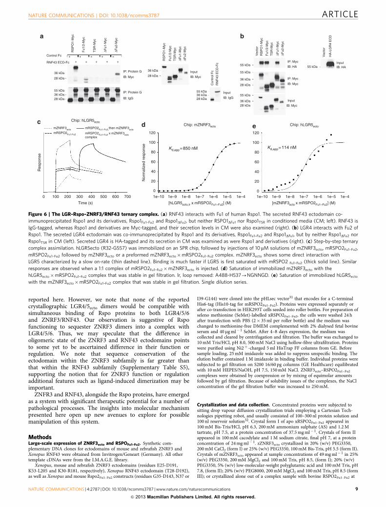
and Rspo1DFu1 each immunoprecipitated LGR4, indicating thatFu2 is the primary binding site for LGR receptors (Fig. 6a,b).Consistent with these co-immunoprecipitation data, Rspo2Fu1–Fu2
simultaneously bound to ZNRF3 and LGR5 in SPR bindingassays (Fig. 6c–e) with the monomerized ZNRF3ecto bindingweaker and the dimerized S90C variant binding stronger to apreformed LGR5ecto–RspoFu1–Fu2 complex (Fig. 4k). Our datatherefore support the model of a ZNRF3/RNF43–Rspo–LGR4/5/6complex assembled through ZNRF3/RNF43–RspoFu1 andRspoFu2–LGR4/5/6 interactions (Fig. 7).
Rspo–ZNRF3/RNF43 interaction determines signallingpotency. Although there is a clear requirement for both Furindomains in Rspo ternary complex formation and functionalactivation of the Wnt pathway, our results point to the ability ofRspoFu1 to recruit ZNRF3/RNF43 as the major determinant ofactivity (for example, Figs 4b and 5b). In biophysical assays, wild-type mRspo2 and -4 proteins, as well as their Fu1/Fu2 chimeras,bound with nanomolar affinity to hLGR5ecto (Fig. 4i), furthersuggesting that engagement of LGR is not the efficiency-deter-mining step. Cellular assays using Rspo2 Fu1/Fu2 chimerasshowed that a Fu1 repeat from a ‘strong’ Rspo (that is, Rspo2or -3) was sufficient to induce a higher Wnt response (Fig. 5c).On the other hand, in spite of an 80-fold (300 mM 43.6 mM)increase in binding efficiency to ZNRF3ecto, replacement of Fu2 ofRspo4 by that of a ‘strong’ Rspo was not able to enhance Wntsignalling when expressed at comparable levels (Fig. 5c). Hence, itcan be concluded that functional efficiency of the four Rspoligands is largely based on their ability to recruit ZNRF3 orRNF43 via Fu1 into a complex with LGRs.
DiscussionIn combination, the structural, biophysical and cell-based studieswe report here for the ZNRF3/RNF43–Rspo system reveal twomodes of interaction: receptor–ligand and receptor dimer. For theligand–receptor mode, our data define a generic architecture forthe interaction between the Rspo ligands, and the ZNRF3 andRNF43 transmembrane E3 ubiquitin ligases that is conservedacross evolution from fish to human. Indeed, the differences inbinding affinities, from highest affinity for Rspo2 to lowest forRspo4, appear to mirror the trend in biological activity of the fourRspo proteins (reviewed in ref. 3). Our results highlight the role ofthe Fu1 domain of the Rspo protein in ZNRF3/RNF43 binding.Both Fu domains together have been implicated in Rsposignalling. The primary role of Fu1 in ZNRF3/RNF43 bindingleaves a substantial surface available for the Rspo to mediate
Figure 4 | Biophysical characterization of the ZNRF3ecto dimer and interface mutants. (a) Sedimentation velocity experiments. A plot of c(s)
(in arbitrary units) against s (in svedbergs). Shown in each case are individual data points and the fit of an appropriate number of Gaussian distributions.
All samples were adjusted to a concentration of 350mM. Also shown arrowed are the expected sedimentation coefficients for the different complexes
observed in the crystal structures as predicted using HYDROPRO (see Methods). (b–h) SPR experiments using mZNRF3ecto (b–e) or mRspo2Fu1–Fu2 (f–h)
as analyte and interface mutants/variants as immobilized ligands. (b) mZNRF3ecto binds to mRspo2Fu1–Fu2 (I39-G144) and retains high affinity to Fu1
(I39-R95) but not to Fu2 (A94-G144). Fu1–Fu2 polypeptides of human or mouse homologues (hRspo1: I32-S143, hRspo3: R32-H147, mRspo4: T29-Q136)
bind with different affinity to mZNRF3ecto. (c) Mutations of the Met-finger impact affinity. (d) Anonychia mutations of RSPO4 introduced to mRspo2Fu1–Fu2
drastically impair binding. (e) Three additional interface mutants of which two (L63F and S53R) have been found in tumour tissues. (f) As the immobilized
ligand mZNRF3 binds with lower affinity to the mRspo2Fu1–Fu2 analyte. Of the three interface mutants, two (E109K and M98T) have been identified in
tumour tissues. (g) Three interface mutants of hRNF3ecto have been identified in tumours, one of which completely disrupts binding. (h) Binding of
mRspo2Fu1–Fu2 to ZNRF3ecto dimer interface mutants. (i) Binding of mRspo2Fu1–Fu2, mRspo4Fu1–Fu2 and chimeras to hLGR5ecto. Single dilution series.
(j) Binding of RspoFu1–Fu2 chimeras to ZNRF3ecto. (k) Binding of the preformed hLGR5ecto,lr–Rspo2Fu1–Fu2 complex to ZNRF3ecto dimer interface mutants.
NATURE COMMUNICATIONS | DOI: 10.1038/ncomms3787 ARTICLE
NATURE COMMUNICATIONS | 4:2787 | DOI: 10.1038/ncomms3787 | www.nature.com/naturecommunications 7
& 2013 Macmillan Publishers Limited. All rights reserved.

formation of a three component complex involving ZNRF3/RNF43, Rspo and LGR4/5/6 as postulated16. Indeed, our co-immunoprecipitation results focus LGR4/5/6-binding activityonto Fu2, consistent with Rspo proteins acting as complexassemblers (Fig. 7). Whilst we were preparing our paper forpublication, several crystal structures of Rspo1Fu1–Fu2 in complexwith LGR4/5ecto and a single Rspo1Fu1–Fu2–LGR5ecto–RNF43ecto
complex were reported25–28, which fully support this notion.Unexpectedly, our analyses reveal a dimerization mode for
ZNRF3ecto. The conservation of ZNRF3 ectodomain dimerizationacross evolution from fish to mammals suggests that thisinteraction has some role in the mechanism of action of ZNRF3.It is also noteworthy that three cancer-associated mutationsreported for RNF43 map to the corresponding dimer interfaceobserved in the ZNRF3ecto crystal structures (Fig. 3b), suggestingthe characteristics of this surface have functional relevance in
RNF43 as well. Many members of the E3 RING ubiquitin ligasesuperfamily have been reported to require dimerization forfunction (reviewed in ref. 29), a conclusion supported by recentinsights into the mechanism of action of the RING ligase RNF4(ref. 30). In ZNRF3, the ectodomain may, alongside cytoplasmicregions, contribute to functionally essential RING domaindimerization. However, neither our biophysical measurementsnor our structural data for an RNF43 ectodomain in complexwith Rspo2 provide any evidence of a similar dimerization modefor RNF43; a finding that argues against ectodomain dimerizationhaving a central role in ligase activity. The newly reportedstructure of the 1:1:1 complex of Rspo1–LGR5–RNF43 alsoreveals no RNF43 dimer25. Intriguingly at the level of a simplemodelling exercise, the ternary complex architecture appearscompatible with ZNRF3 dimerization (Fig. 7). All of the currentlyavailable structures are compatible with a dimeric ZNRF3 as
50
40
30
20
10
0
50
60a b
40
30
20
10
0
Sup
erT
opF
lash
(R
LU)
50d
40
30
20
10
0
50
40
30
20
10
0
50
40
30
20
10
0
Sup
erT
opF
lash
(R
LU)
Sup
erT
opF
lash
(R
LU)
50
60c
e
g h
f
40
30
20
10
0
Con
Con
19 kDa CMIB : His
15 kDa
15 kDaCM
IB : His
Lysate LysateIB : His IB : His
Con
Fu1
Fu2
hR1
hR3
mR
2
mR
4
2/1
2/3
2/4
4/2
Con
mR
2 w
t
N50
R
S53
R
L63F
M68
E
M68
I
R65
W
Q70
R
G72
R
mR
2 w
t
15 kDa
6 kDa
19 kDa
15 kDa
6 kDa
2/1 2/2(mR2 wt)
2/3 2/4 4/2
Sup
erT
opF
lash
(R
LU)
Sup
erT
opF
lash
(R
LU)
Sup
erT
opF
lash
(R
LU)
Con Con
Con mR2 wt
mR2 wt R65W Q70R G72R Con mR2 wt N50R S53R L63F
M68E M68I
hR1 hR3mR2 mR4mR2 Fu1 Fu2
Figure 5 | Activation of the Wnt pathway assayed by the SuperTopFlash reporter. (a–f) Co-transfected decreasing doses (25, 5 and 1 ng) of His-tagged
R-spondin constructs used for SPR experiments in Fig. 4. Error bars represent s.d. from three replicates. (g,h) Western blots showing expression
levels of the His-tagged R-spondin constructs from whole-cell lysate and conditioned media (CM). Expression for mRspo2 Fu1-His was poor and below the
level of detection; however, the individual Fu1 domain from RSPO1 was detected by western blotting and produced identical results (Supplementary
Fig. S1b,c). RLU, relative luciferase units.
ARTICLE NATURE COMMUNICATIONS | DOI: 10.1038/ncomms3787
8 NATURE COMMUNICATIONS | 4:2787 | DOI: 10.1038/ncomms3787 | www.nature.com/naturecommunications
& 2013 Macmillan Publishers Limited. All rights reserved.
reported here. However, we note that none of the reportedcrystallographic LGR4/5ecto dimers would be compatible withsimultaneous binding of Rspo proteins to both LGR4/5/6and ZNRF3/RNF43. Our observation is suggestive of Rspofunctioning to sequester ZNRF3 dimers into a complex withLGR4/5/6. Thus, we may speculate that the difference inoligomeric state of the ZNRF3 and RNF43 ectodomains pointsto some yet to be ascertained difference in their function orregulation. We note that sequence conservation of theectodomain within the ZNRF3 subfamily is far greater thanthat within the RNF43 subfamily (Supplementary Table S5),supporting the notion that for ZNRF3 function or regulationadditional features such as ligand-induced dimerization may beimportant.
ZNFR3 and RNF43, alongside the Rspo proteins, have emergedas a system with significant therapeutic potential for a number ofpathological processes. The insights into molecular mechanismpresented here open up new avenues to explore for possiblemanipulation of this system.
MethodsLarge-scale expression of ZNRF3ecto and RSPOFu1–Fu2. Synthetic com-plementary DNA clones for ectodomains of mouse and zebrafish ZNRF3 andXenopus RNF43 were obtained from Invitrogen/Geneart (Germany). All othertemplate cDNAs were from the I.M.A.G.E. library.
Xenopus, mouse and zebrafish ZNRF3 ectodomains (residues E25-D191,K53-L205 and K30-R181, respectively), Xenopus RNF43 ectodomain (T28-D192),as well as Xenopus and mouse Rspo2Fu1–Fu2 constructs (residues G35-D143, N37 or
I39-G144) were cloned into the pHLsec vector31 that encodes for a C-terminalHis6-tag (His10-tag for mRSPO2Fu1–Fu2). Proteins were expressed separately orafter co-transfection in HEK293T cells seeded into roller bottles. For preparation ofseleno methionine (SeMet)-labelled xRSPO2Fu1–Fu2, the cells were washed 24 hafter transfection with PBS (2� 35 ml per roller bottle) and the medium waschanged to methionine-free DMEM complemented with 2% dialysed fetal bovineserum and 40 mg ml� 1 SeMet. After 4–8 days expression, the medium wascollected and cleared by centrifugation and filtration. The buffer was exchanged to10 mM Tris/HCl, pH 8.0, 500 mM NaCl using hollow-fibre ultrafiltration. Proteinswere purified using Ni2þ -charged 5 ml HisTrap FF columns from GE. Beforesample loading, 25 mM imidazole was added to suppress unspecific binding. Theelution buffer contained 1 M imidazole in binding buffer. Individual proteins weresubjected to gel filtration on S200 16/60 pg columns (GE Healthcare) equilibratedwith 10 mM HEPES/NaOH, pH 7.5, 150 mM NaCl. ZNRF3ecto–RSPO2Fu1–Fu2
complexes were obtained by coexpression or by mixing of equimolar amountsfollowed by gel filtration. Because of solubility issues of the complexes, the NaClconcentration of the gel filtration buffer was increased to 250 mM.
Crystallization and data collection. Concentrated proteins were subjected tositting drop vapour diffusion crystallization trials employing a Cartesian Tech-nologies pipetting robot, and usually consisted of 100–300 nl protein solution and100 nl reservoir solution32. Crystal form I of apo xRSPO2Fu1–Fu2 appeared in100 mM Bis-Tris/HCl, pH 6.3, 200 mM ammonium sulphate (AS) and 1.2 Mtartrate, pH 7.5, at a protein concentration of 37.5 mg ml� 1. Crystals of form IIappeared in 100 mM cacodylate and 1 M sodium citrate, final pH 7, at a proteinconcentration of 24 mg ml� 1. zZNRF3ecto crystallized in 20% (w/v) PEG3350,200 mM CaCl2 (form I) or 25% (w/v) PEG3350, 100 mM Bis-Tris, pH 5.5 (form II).Crystals of mZNRF3ecto appeared at sample concentrations of 49 mg ml� 1 in 25%(w/v) PEG3350, 200 mM MgCl2 and 100 mM Tris, pH 8.5, (form I); 20% (w/v)PEG3350, 5% (w/v) low-molecular-weight polyglutamic acid and 100 mM Tris, pH7.8, (form II); 20% (w/v) PEG8000, 200 mM MgCl2 and 100 mM Tris, pH 8.5 (formIII); or crystallized alone out of a complex sample with bovine RSPO2Fu1–Fu2 at
55 kDa
28 kDa
IP: Protein G
IB: Myc
Input
IB: Myc
Con
trol
Fc
RN
F43
EC
D-F
c
+
RS
PO
1-M
yc
Fu1
/2-M
yc
TS
R-M
yc
ΔFu1
-Myc
ΔFu2
-Myc
RS
PO
1-M
yc
Fu1
/2-M
yc
TS
R-M
yc
ΔFu1
-Myc
ΔFu2
-Myc
RS
PO
1-M
ycV
ecto
r
Vec
tor
Fu1
/2-M
ycT
SR
-Myc
ΔFu1
-Myc
ΔFu2
-Myc
HA
-LG
R4
EC
D
+
+
+
+
+
+
++
+
RNF43 ECD-Fc
Control Fc
a b
c d e
Input
IB: IgGInput
IB: Myc
InputIB: HA
IP: Myc
IP: Myc
IB: HA
IB: Myc
IP: Protein G
IB: IgG
36 kDa28 kDa
36 kDa
36 kDa28 kDa
55 kDa36 kDa28 kDa
55 kDa
55 kDa
55 kDa55 kDa
36 kDa
36 kDa
28 kDa
28 kDa
0
Res
pons
e
100 200 300
Time (s)
500400 600 700
0
20
40
Nor
mal
ized
res
pons
e
60
100
120
Chip: mZNRF3ecto Chip: hLGR5ecto
Chip: hLGR5ecto
Kd,app=850 nM Kd,app=114 nM80
0
20
40
60
100
120
80
mZNRF3ecto
mRSPO2Fu1-Fu2
mRSPO2Fu1–Fu2,then mZNRF3ecto
mRSPO2Fu1–Fu2 x mZNRF3ectocomplex
1e–10 1e–9 1e–8
[hLGR5ecto,Ir x mRSPO2Fu1–Fu2] (M) [mZNRF3ecto x mRSPO2Fu1–Fu2] (M)
1e–7 1e–6 1e–5 1e–4 1e–10 1e–9 1e–8 1e–7 1e–6 1e–5 1e–4
Figure 6 | The LGR–Rspo–ZNRF3/RNF43 ternary complex. (a) RNF43 interacts with Fu1 of human Rspo1. The secreted RNF43 ectodomain co-
immunoprecipitated Rspo1 and its derivatives, Rspo1Fu1–Fu2 and Rspo1DFu2, but neither RSPO1DFu1 nor Rspo1TSR in conditioned media (CM; left). RNF43 is
IgG-tagged, whereas Rspo1 and derivatives are Myc-tagged, and their secretion levels in CM were also examined (right). (b) LGR4 interacts with Fu2 of
Rspo1. The secreted LGR4 ectodomain was co-immunoprecipitated by Rspo1 and its derivatives, Rspo1Fu1–Fu2 and Rspo1DFu1, but by neither Rspo1DFu2 nor
Rspo1TSR in CM (left). Secreted LGR4 is HA-tagged and its secretion in CM was examined as were Rspo1 and derivatives (right). (c) Step-by-step ternary
complex assimilation. hLGR5ecto (R32-G557) was immobilized on an SPR chip, followed by injections of 10mM solutions of mZNRF3ecto, mRSPO2Fu1–Fu2,
mRSPO2Fu1–Fu2 followed by mZNRF3ecto or a preformed mZNRF3ecto�mRSPO2Fu1–Fu2 complex. mZNRF3ecto shows some direct interaction with
LGR5 characterized by a slow on-rate (thin dashed line). Binding is much faster if LGR5 is first saturated with mRSPO2 Fu1–Fu2 (thick solid line). Similar
responses are observed when a 1:1 complex of mRSPO2Fu1–Fu2�mZNRF3ecto is injected. (d) Saturation of immobilized mZNRF3ecto with the
hLGR5ecto�mRSPO2Fu1–Fu2 complex that was stable in gel filtration. lr, loop removed: A488-H537-NGNNGD. (e) Saturation of immobilized hLGR5ecto
with the mZNRF3ecto�mRSPO2Fu1–Fu2 complex that was stable in gel filtration. Single dilution series.
NATURE COMMUNICATIONS | DOI: 10.1038/ncomms3787 ARTICLE
NATURE COMMUNICATIONS | 4:2787 | DOI: 10.1038/ncomms3787 | www.nature.com/naturecommunications 9
& 2013 Macmillan Publishers Limited. All rights reserved.

16.2 mg ml� 1 in 0.5 M Li2SO4 and 10% (w/v) PEG8000 (form IV). The S90Cvariant of mZNRF3ecto crystallized in 45% (v/v) 2-methyl-2,4-pentanediol, 200 mMammonium acetate and 100 mM Bis-Tris, pH 5.5, at a concentration of38.5 mg ml� 1. Apo xZNRF3ecto crystallized at 25 mg ml� 1 in 20% (w/v) PEG3350,100 mM Bis-Tris propane, pH 6.5, 200 mM NaBr (form I), and 20% (w/v)PEG3350 and 0.200 M NaCl (form II). Crystals composed of the complex ofmZNRF3ecto and mRSPO2Fu1–Fu2 were obtained at a concentration of 18 mg ml� 1
in 1.8 M AS, 100 mM Bis-Tris, pH 6.5, 2% (v/v) PEGMME550. Crystals of themixed species complexes of mZNRF3ecto and SeMet xRSPO2Fu1–Fu2 appeared in25% (w/v) PEG4000, 200 mM NaCl, 100 mM HEPES/NaOH, pH 7.5 (form I), and20% (w/v) PEG3350, 200 mM sodium citrate, 100 mM Bis-Tris propane, pH 6.5(form II). xZNRF3ecto–xRSPO2Fu1–Fu2 complexes crystallized in 20% (w/v)PEG3350, 200 mM (NH4)F (form I) and 20% (w/v) PEG6000, 100 mM MES, pH6.0 (form II). The complex of xRNF43ecto and xRSPO2Fu1–Fu2 was crystallized incondition A1 of the PACT premier screen from Molecular Dimensions. Forcryoprotection, crystals were transferred to mother liquor supplemented with 1.7 Msodium malonate, pH 7 (both apo xRSPO2Fu1–Fu2 crystals), with AS to 3 M (mouse/mouse complex), or with PEG200 to achieve total (PEG, polyethyleneglycol)430% (all other ZNRF3 apo and complex crystals) by incrementallyadjusting the concentration of the cryoprotectant. Crystal were then flash-cooled bydipping into liquid nitrogen. The xRNF43ecto–xRSPO2Fu1–Fu2 complex crystal wasfrozen directly and showed strong ice rings. Diffraction data were collected atDIAMOND synchrotron light source at the beamlines i02, i03, i04 and i24. Crystalforms II and III of apo mZNRF3ecto had been soaked with a platinum compound,but showed only low binding of heavy atoms.
Structure determination. The structure of xRspo2Fu1–Fu2 was solved using highlyredundant single-wavelength anomalous dispersion (SAD) data from a Pt(IV)-
soaked crystal that diffracted to 3.2 Å (Table 1). Ten strong anomalous sites couldbe identified by AUTOSHARP33. Refinement and subsequent density modificationwith SOLOMON lead to clearly interpretable electron density. A partial modelobtained from BUCCANEER34 was used to solve the high-resolution structure.The model was improved with iterative rounds of manual building in COOT35 andrefinement in REFMAC5 (ref. 36). The structure of mZNRF3ecto in complex withSeMet-labelled xRSPO2Fu1–Fu2 was solved from SAD data collected at the SeK-absorption edge. Albeit only one component of the complex was labelled and thecomplex being crystallized in the low-symmetry space group P1, the Se atomsubstructure (four sites) could be identified by PHENIX HYSS37 from averageredundancy data (Supplementary Table S1). An initial model generated byAUTOSOL was used to solve the high-resolution mZNRF3ecto structure(Supplementary Table S1). All other structures were solved by molecularreplacement with PHASER38 and completed by manual rebuilding in COOT andrefinement with REFMAC5. Models were validated with MOLPROBITY39.Superpositions were performed within CCP4 or COOT using the SSM algorithm.Electrostatics potentials were generated using APBS40, surface sequenceconservation was calculated using CONSURF41 and interface areas of proteinswere calculated using the PISA web server42. Figures were produced in PYMOLand assembled in PHOTOLINE32.
Analytical ultracentrifugation. xZNRF3ecto–xRSPO2Fu1–Fu2 and xRNF43ecto–xRSPO2Fu1–Fu2 complexes and apo mZNRF3ecto variants at 350 mM in 10 mMHEPES/NaOH, 250 mM NaCl were subjected to sedimentation velocity experi-ments at 20 �C using an Optima Xl-I analytical ultracentrifuge (Beckman) with3 mm or 12 mm double sector centerpieces in an An-60 Ti rotor (Beckman) at40,000 r.p.m. Sedimentation was monitored by ultraviolet absorption at 300 nmand by Rayleigh interference. Data were analysed using SEDFIT operating in c(s)and c(s,f/fo) modes (with a frictional coefficient range of 1–2 in the latter case and a
Limited Wnt responsedue to receptor turnover
UbRING
ZNRF3
Fu1 Fu2 Fu1 Fu2
Fz
WNT
L
R
P
5/6
LGR4/5/6
Fz
C C
N N
C
CN
N
C
WNT
L
R
P
5/6
Membraneclearance
Receptor stabilizationenhances Wntresponsiveness
RINGRING
C
N
a
b
N
Figure 7 | Modelling of a ternary 2:2:2 LGRecto–RspoFu1–Fu2–ZNRF3ecto complex and its implication for signalling. (a) The hLGR5ecto� hRSPO1Fu1–Fu2�mZNRF3ecto 2:2:2 complex was generated by superposing the ternary hLGR5ecto�RSPO1Fu1–Fu2� hRNF43ecto complex25 (Protein Data Bank ID code
4KNG) onto the mZNRF3ecto dimer from the mRSPO2Fu1–Fu2 complex. No clashes are observed. Glycosylation sites of LGR5 all point into the periphery
of the shown complex. (b) A model for regulation of Wnt signalling by RSPO and its receptors based on our results and those by Hao et al.16 The
schematic model takes into account the different binding sites of LGRs and ZNRF3/RNF43 on Rspos as determined by us and others25–28.
ARTICLE NATURE COMMUNICATIONS | DOI: 10.1038/ncomms3787
10 NATURE COMMUNICATIONS | 4:2787 | DOI: 10.1038/ncomms3787 | www.nature.com/naturecommunications
& 2013 Macmillan Publishers Limited. All rights reserved.

resolution in s of 100)43. The resulting sedimentation coefficient distributions wereplotted using ProFit (Uetikon am See, CH). The crystal structures were modelledhydrodynamically using the programme HYDROPRO44.
SPR equilibrium binding studies. Affinity between variants of mZNRF3ecto,human RNF43ecto and mRSPO2Fu1–Fu2 was measured at 25 �C in 10 mM HEPES/NaOH, pH 7.5, 150 mM NaCl, 0.005% Tween20 using a Biacore T200 machine (GEHealthcare). Synthetic DNA corresponding to mZNRF3(K53-L205), hRNF43(Q44-L188) and mRSPO2(I39-G144), as well as variants thereof, was obtained fromInvitrogen/Geneart (Germany) and cloned into a variant of the pHLsec vectorencoding a C-terminal recognition sequence for the Escherichia coli BirA enzyme.Biotinylation at this sequence tag was performed as described45. Experiments wereperformed as described before46, with the biotinylated variants immobilized to thechip surface precoupled with approximately 10,000 resonance units (RU) ofstreptavidin. Immobilized protein amounts varied between 350 and 1,000 RU(1 experiment with 1,650 RU). The amount of immobilized protein did not seem tostrongly influence the binding model. After each injection of analyte, the chipsurface was regenerated with 2 M MgCl2, 10 mM HEPES/NaOH, pH 7.5 (RSPOcoupled), 100 mM phosphate, pH 3.7, 2 M NaCl and 1% (v/v) Tween20 (RNF43 orZNRF3 coupled) or 25% ethylene glycol, 2 M NaCl, 100 mM HEPES/NaOH, pH7.5, 1% Tween20 (LGR coupled) to return to baseline levels. Data were fitted to aLangmuir adsorption model B¼BmaxC/(KdþC), where B is the amount of boundanalyte and C is the concentration of analyte in the sample. Data were thennormalized to a maximum analyte-binding value of 100. Unless stated otherwise,data points correspond to average from two independent dilution series.
Co-immunoprecipitation binding assays. Full-length human RSPO1-Myc(1–263), and domains of Fu1/Fu2 (1–147) and TSR (1–20, 144–263), wereoriginally reported in ref. 47. On the basis of the disulphide bond pattern resolvedin the crystal structure, new individual Fu domain deletions were generated:deltaFurin1 (del 39–94; Rspo1DFu1) and deltaFurin2 (del 97–142; Rspo1DFu2). Forimmunoprecipitation, conditioned medium from HEK293T cells transfected usingFugeneHD with RSPO1-Myc, Furin1/2-Myc, TSR-Myc, deltaFurin1-Myc or del-taFurin2-Myc was mixed with conditioned medium from cells transfected withIgG, Human RNF43 ECD-IgG (1–198) or mouse HA-Lgr4 ECD (from ref. 11), andincubated at 4 �C overnight. The mixture was then incubated with proteinG-agarose beads for 2 h at 4 �C and washed with buffer (150 mM NaCl, 1 mMEDTA, 2.5 mM EGTA, 10% glycerol, 0.1% Tween20 with protease inhibitors).Protein was eluted using 2� SDS sample buffer and separated by SDS–PAGE.Western blotting was performed by using horseradish peroxidase-conjugated anti-human IgG (Calbiochem), anti-HA or anti-c-Myc. For co-immunoprecipitationassays, anti-Myc (9E10, Santa Cruz) was used at a 1:100 concentration. For westernblots, primary antibodies were diluted 1:1000 and secondary antibodies werediluted 1:10000 from stocks.
Activity assays. To assess Rspo activation of the Wnt signalling pathway, atraditional dual-luciferase assay consisting of the Wnt-responsive SuperTopFlashreporter (normalized to a control promoter driving Renilla luciferase) was used aspreviously described46. Mammalian cell transfections were done in HEK293T(ATCC CRL-11268) cells and performed in triplicate for each sample condition.Cells were plated at 1� 105 per ml in 24-well plates and transfected the followingday with a total of 200 ng of DNA per well (50 ng SuperTopFlash, 10 ng TK-Renilla,experimental expression vectors and balanced with empty vector). Lysates werecollected 36 h post transfection and used with the Dual-luciferase reporter system(Promega). Firefly and Renilla luciferase activity was measured using the Wallac1420 multilabel counter in 96-well plates. Normalized data expressed in relativeluciferase units was averaged from triplicate assays and error bars reflect s.d.Representative results are shown from one of multiple independent experiments.
References1. Kazanskaya, O. et al. R-Spondin2 is a secreted activator of Wnt/beta-catenin
signaling and is required for Xenopus myogenesis. Dev. Cell 7, 525–534 (2004).2. Kim, K. A. et al. Mitogenic influence of human R-spondin1 on the intestinal
epithelium. Science 309, 1256–1259 (2005).3. de Lau, W. B., Snel, B. & Clevers, H. C. The R-spondin protein family. Genome
Biol. 13, 242 (2012).4. Parma, P. et al. R-spondin1 is essential in sex determination, skin
differentiation and malignancy. Nat. Genet. 38, 1304–1309 (2006).5. Zhao, J. et al. R-spondin1, a novel intestinotrophic mitogen, ameliorates
experimental colitis in mice. Gastroenterology 132, 1331–1343 (2007).6. Papapietro, O. et al. R-Spondin 2 signalling mediates susceptibility to fatal
infectious diarrhoea. Nat. Commun. 4, 1898 (2013).7. Seshagiri, S. et al. Recurrent R-spondin fusions in colon cancer. Nature 488,
660–664 (2012).8. Blaydon, D. C. et al. The gene encoding R-spondin 4 (RSPO4), a secreted
protein implicated in Wnt signaling, is mutated in inherited anonychia. Nat.Genet. 38, 1245–1247 (2006).
9. Chishti, M. S., Kausar, N., Rafiq, M. A., Amin, M. & Ahmad, W. A novelmissense mutation in RSPO4 gene underlies autosomal recessive congenitalanonychia in a consanguineous Pakistani family. Br. J. Dermatol. 158, 621–623(2008).
10. Khan, T. N. et al. Novel missense mutation in the RSPO4 gene in congenitalhyponychia and evidence for a polymorphic initiation codon (p.M1I). BMCMed. Genet. 13, 120 (2012).
11. Carmon, K. S., Gong, X., Lin, Q., Thomas, A. & Liu, Q. R-spondins function asligands of the orphan receptors LGR4 and LGR5 to regulate Wnt/beta-cateninsignaling. Proc. Natl Acad. Sci. USA 108, 11452–11457 (2011).
12. Carmon, K. S., Lin, Q., Gong, X., Thomas, A. & Liu, Q. LGR5 interacts andcointernalizes with Wnt receptors to modulate Wnt/beta-catenin signaling.Mol. Cell Biol. 32, 2054–2064 (2012).
13. de Lau, W. et al. Lgr5 homologues associate with Wnt receptors and mediateR-spondin signalling. Nature 476, 293–297 (2011).
14. Glinka, A. et al. LGR4 and LGR5 are R-spondin receptors mediating Wnt/beta-catenin and Wnt/PCP signalling. EMBO Rep. 12, 1055–1061 (2011).
15. Ruffner, H. et al. R-Spondin potentiates Wnt/beta-catenin signaling throughorphan receptors LGR4 and LGR5. PLoS One 7, e40976 (2012).
16. Hao, H. X. et al. ZNRF3 promotes Wnt receptor turnover in an R-spondin-sensitive manner. Nature 485, 195–200 (2012).
17. Wu, J. et al. Whole-exome sequencing of neoplastic cysts of the pancreasreveals recurrent mutations in components of ubiquitin-dependent pathways.Proc. Natl Acad. Sci. USA 108, 21188–21193 (2011).
18. Koo, B. K. et al. Tumour suppressor RNF43 is a stem-cell E3 ligase that inducesendocytosis of Wnt receptors. Nature 488, 665–669 (2012).
19. Kim, K. A. et al. R-Spondin family members regulate the Wnt pathway by acommon mechanism. Mol. Biol. Cell 19, 2588–2596 (2008).
20. Nam, J. S., Turcotte, T. J., Smith, P. F., Choi, S. & Yoon, J. K. Mouse cristin/R-spondin family proteins are novel ligands for the Frizzled 8 and LRP6 receptorsand activate beta-catenin-dependent gene expression. J. Biol. Chem. 281,13247–13257 (2006).
21. Li, S. J. et al. Loss-of-function point mutations and two-furin domainderivatives provide insights about R-spondin2 structure and function. CellSignal. 21, 916–925 (2009).
22. Mahon, P. & Bateman, A. The PA domain: a protease-associated domain.Protein Sci. 9, 1930–1934 (2000).
23. Anandasabapathy, N. et al. GRAIL: an E3 ubiquitin ligase that inhibits cytokinegene transcription is expressed in anergic CD4þ T cells. Immunity 18,535–547 (2003).
24. Nurieva, R. I. et al. The E3 ubiquitin ligase GRAIL regulates T cell tolerance andregulatory T cell function by mediating T cell receptor-CD3 degradation.Immunity 32, 670–680 (2010).
25. Chen, P. H., Chen, X., Lin, Z., Fang, D. & He, X. The structural basisof R-spondin recognition by LGR5 and RNF43. Genes Dev. 27, 1345–1350(2013).
26. Peng, W. C. et al. Structure of stem cell growth factor R-spondin 1 in complexwith the ectodomain of its receptor LGR5. Cell Rep. 3, 1885–1892 (2013).
27. Wang, D. et al. Structural basis for R-spondin recognition by LGR4/5/6receptors. Genes Dev. 27, 1339–1344 (2013).
28. Xu, K., Xu, Y., Rajashankar, K. R., Robev, D. & Nikolov, D. B. Crystal structuresof Lgr4 and its complex with R-spondin1. Structure 21, 1683–1689 (2013).
29. Deshaies, R. J. & Joazeiro, C. A. RING domain E3 ubiquitin ligases. Annu. Rev.Biochem. 78, 399–434 (2009).
30. Plechanovova, A. et al. Mechanism of ubiquitylation by dimeric RING ligaseRNF4. Nat. Struct. Mol. Biol. 18, 1052–1059 (2011).
31. Aricescu, A. R., Lu, W. & Jones, E. Y. A time- and cost-efficient system forhigh-level protein production in mammalian cells. Acta Crystallogr. D Biol.Crystallogr. 62, 1243–1250 (2006).
32. Walter, T. S. et al. A procedure for setting up high-throughput nanolitrecrystallization experiments. Crystallization workflow for initial screening,automated storage, imaging and optimization. Acta Crystallogr. D Biol.Crystallogr. 61, 651–657 (2005).
33. Vonrhein, C., Blanc, E., Roversi, P. & Bricogne, G. Automated structuresolution with autoSHARP. Methods Mol. Biol. 364, 215–230 (2007).
34. Cowtan, K. The Buccaneer software for automated model building. 1.Tracing protein chains. Acta Crystallogr. D Biol. Crystallogr. 62, 1002–1011(2006).
35. Emsley, P. & Cowtan, K. Coot: model-building tools for molecular graphics.Acta Crystallogr. D. Biol. Crystallogr. 60, 2126–2132 (2004).
36. Murshudov, G. N., Vagin, A. A. & Dodson, E. J. Refinement of macromolecularstructures by the maximum-likelihood method. Acta Crystallogr. D Biol.Crystallogr. 53, 240–255 (1997).
37. Adams, P. D. et al. PHENIX: a comprehensive Python-based system formacromolecular structure solution. Acta Crystallogr. D Biol. Crystallogr. 66,213–221 (2010).
38. McCoy, A. J. et al. Phaser crystallographic software. J. Appl. Crystallogr. 40,658–674 (2007).
NATURE COMMUNICATIONS | DOI: 10.1038/ncomms3787 ARTICLE
NATURE COMMUNICATIONS | 4:2787 | DOI: 10.1038/ncomms3787 | www.nature.com/naturecommunications 11
& 2013 Macmillan Publishers Limited. All rights reserved.

39. Davis, I. W. et al. MolProbity: all-atom contacts and structure validation forproteins and nucleic acids. Nucleic Acids Res. 35, W375–W383 (2007).
40. Baker, N. A., Sept, D., Joseph, S., Holst, M. J. & McCammon, J. A. Electrostaticsof nanosystems: application to microtubules and the ribosome. Proc. Natl Acad.Sci. USA 98, 10037–10041 (2001).
41. Ashkenazy, H., Erez, E., Martz, E., Pupko, T. & Ben-Tal, N. ConSurf 2010:calculating evolutionary conservation in sequence and structure of proteins andnucleic acids. Nucleic Acids Res. 38, W529–W533 (2010).
42. Krissinel, E. & Henrick, K. Inference of macromolecular assemblies fromcrystalline state. J. Mol. Biol. 372, 774–797 (2007).
43. Schuck, P. Size-distribution analysis of macromolecules by sedimentationvelocity ultracentrifugation and lamm equation modeling. Biophys. J 78,1606–1619 (2000).
44. Ortega, A., Amoros, D. & T. J., G. Prediction of hydrodynamic and othersolution properties of rigid proteins from atomic- and residue-level models.Biophys. J. 101, 892–898 (2011).
45. O’Callaghan, C. A. et al. BirA enzyme: production and application in the studyof membrane receptor-ligand interactions by site-specific biotinylation. Anal.Biochem. 266, 9–15 (1999).
46. Chen, S. et al. Structural and functional studies of LRP6 ectodomain reveal aplatform for Wnt signaling. Dev. Cell 21, 848–861 (2011).
47. Wei, Q. et al. R-spondin1 is a high affinity ligand for LRP6 and induces LRP6phosphorylation and beta-catenin signaling. J. Biol. Chem. 282, 15903–15911(2007).
48. Ayadi, L. Molecular modelling of the TSR domain of R-spondin 4.Bioinformation 3, 119–123 (2008).
AcknowledgementsWe thank Diamond Light Source for beamtime (proposal mx8423) and the staff ofbeamlines I02, I03, I04, I04-1 and I24 for assistance with crystal testing and data col-lection. We thank W. Lu and Y. Zhao for help with tissue culture, R. Chen for con-tributions to protein production, K. Harlos and T. Walter for assistance withcrystallization and T. Malinauskas for discussion. The analytical ultracentrifugationexperiments were performed in the Biophysical Facility at the Department of Bio-chemistry, University of Oxford. X.H. acknowledges F. Cong (Novartis) for humanZNRF3 and RNF43 constructs. This work was funded by Cancer Research UK, the UKMedical Research Council (to E.Y.J., A10976 and G9900061) and NIH (to X.H., RO1-
GM057603 and GM057603S1). M.Z. holds an IEF Marie Curie fellowship, C.K. is therecipient of a Wellcome Trust D. Phil. studentship and Y.X. is supported in part by agraduate studentship from the Chinese Scholarship Council. The Wellcome Trust Centrefor Human Genetics is supported by Wellcome Trust grant 090532/Z/09/Z. X.H. is anendowed research chair of Boston Children’s Hospital (BCH) and acknowledges supportby BCH Intellectual and Developmental Disabilities Research Center (P30 HD-18655).
Author contributionsAll authors contributed to the design of the project, data analysis and preparation of themanuscript. M.Z. cloned, purified and performed SPR and AUC experiments on Rspoproteins and ZNRF3, crystallized and solved the individual and complex structures. C.K.contributed to cloning, protein purification, SPR and diffraction data collection. R.J.C.G.contributed to AUC data collection and analysis. B.T.M., Y.X. and M.C. cloned andperformed functional assays and co-immunoprecipitation for Rspo proteins.
Additional informationAccession codes: Coordinates and structure factors for zZNRF3ecto, mZNRF3ecto,xZNRF3ecto, xRspo2Fu1–Fu2, mZNRF3ecto–mRspo2Fu1–Fu2, mZNRF3ecto–xRspo2Fu1-Fu,xZNRF3ecto–xRspo2v and xRNF43ecto–xRspo2Fu1–Fu2 crystal structures have beendeposited in the Protein Data Bank with the succession numbers 4C84, 4C85, 4C86,4C8A, 4C8C, 4C8F, 4C8P, 4C8T, 4C8U, 4C8V, 4C8W, 4C99, 4C9A, 4C9E, 4C9R, 4C9Uand 4C9V as also given in Table 1 and Supplementary Table S1.
Supplementary Information accompanies this paper at http://www.nature.com/naturecommunications
Competing financial interests: The authors declare no competing financial interests.
Reprints and permission information is available online at http://npg.nature.com/reprintsandpermissions/
How to cite this article: Zebisch, M. et al. Structural and molecular basis of ZNRF3/RNF43 transmembrane ubiquitin ligase inhibition by the Wnt agonist R-spondin.Nat. Commun. 4:2787 doi: 10.1038/ncomms3787 (2013).
This article is licensed under a Creative Commons Attribution 3.0Unported Licence. To view a copy of this licence visit http://
creativecommons.org/licenses/by/3.0/.
ARTICLE NATURE COMMUNICATIONS | DOI: 10.1038/ncomms3787
12 NATURE COMMUNICATIONS | 4:2787 | DOI: 10.1038/ncomms3787 | www.nature.com/naturecommunications
& 2013 Macmillan Publishers Limited. All rights reserved.