structural and functional linkages between subunit ... · structural and functional linkages...
TRANSCRIPT
doi:10.1006/jmbi.2001.4978 available online at http://www.idealibrary.com on J. Mol. Biol. (2001) 312, 525±540
Structural and Functional Linkages Between SubunitInterfaces in Mammalian Pyruvate Kinase
John O. Wooll, Robert H. E. Friesen, Mark A. WhiteStanley J. Watowich, Robert O. Fox, J. Ching Lee* andEdmund W. Czerwinski*
Department of HumanBiological Chemistry andGenetics and the Sealy Centerfor Structural Biology, TheUniversity of Texas MedicalBranch, Galveston, TX 77555-0647, USA
Present; address: R. H. E. FriesenTechnology Foundation, NijenborghGroningen, The Netherlands.
Abbreviations used: PK, pyruvateenolpyruvate; M1-PK, muscle PK iskidney PK isozyme; rRMPK, recomPK; FBP, fructose 1,6-bisphosphate;
E-mail addresses of the [email protected]; [email protected]
0022-2836/01/030525±16 $35.00/0
Mammalian pyruvate kinase (PK) is a four-domain enzyme that is activeas a homo-tetramer. Tissue-speci®c isozymes of PK exhibit distinct levelsof allosteric regulation. PK expressed in muscle tissue (M1-PK) showshyperbolic steady-state kinetics, whereas PK expressed in kidney tissue(M2-PK) displays sigmoidal kinetics. Rabbit M1 and M2-PK are isozymeswhose sequences differ in only 22 out of 530 residues per subunit, andthese changes are localized in an inter-subunit interface. Previous studieshave shown that a single amino acid mutation to M1-PK at either the Y(S402P) or Z (T340 M) subunit interface can confer a level of allostericregulation that is intermediate to M1-PK and M2-PK. In an effort to eluci-date the roles of the inter-subunit interaction in signal transmission andthe functional/structural connectivity between these interfaces, the S402Pmutant of M1-PK was crystallized and its structure resolved to 2.8 AÊ .Although the overall S402P M1-PK structure is nearly identical with thewild-type structure within experimental error, signi®cant differences inthe conformation of the backbone are found at the site of mutation alongthe Y interface. In addition, there is a signi®cant change along the Zinterface, namely, a loss of an inter-subunit salt-bridge between Asp177of domain B and Arg341 of domain A of the opposing subunit. Concur-rent with the loss of the salt-bridge is an increase in the degree ofrotational ¯exibility of domain B that constitutes the active site. Compari-son of previous PK structures shows a correlation between an increase inthis domain movement with the loss of the Asp177: Arg341 salt-bridge.These results identify the structural linkages between the Y and Z inter-faces in regulating the interconversion of conformational states of rabbitM1-PK.
# 2001 Academic Press
Keywords: pyruvate kinase; allosterism; subunit communication; structure;X-ray crystallography
*Corresponding authorsIntroduction
Maintenance of the ¯ux of energy and materialwithin a cell is essential for homeostasis and func-tion. Fundamental energy production in the cell is
, Biomade4, 9747 AG
kinase; PEP, P-ozyme; M2-PK,binant rabbit muscleYPK, yeast PK.ding authors:u
found within the glycolytic pathway, the regu-lation of which is primarily associated with twoallosteric enzymes. Phosphofructokinase coordi-nates the process down the pathway of glycolysis.Passage out of glycolysis is modulated by pyruvatekinase (PK; E.C. 2.7.1.40) that catalyzes the transferof the phosphate from P-enolpyruvate (PEP) toADP.1 ± 7
PEP�ADP()PKATP� Pyruvate
Mammalian PK is a homo-tetramer, and eachsubunit consists of four domains (Figure 1(a)). Thecatalytic activity of PK is proposed to be activatedby the ``clamping down'' or ``closing'' of the B
# 2001 Academic Press

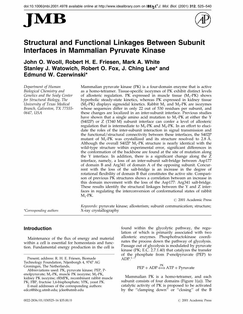
Figure 1. (a) View of the S402P monomer structureshowing overall view of the monomer with the domainscolored, A (yellow), B (blue), C (cyan) and N (green).The putative FBP binding region of the S402P structureis shown in red. (b) View of the S402P structure perpen-dicular to the Y interface separating monomers 1 and 3from monomers 2 and 4.5 The Z interface separatesmonomers 1 and 2 from 3 and 4. The Z interface is con-voluted as monomers 1 and 2 wrap around monomers 3and 4, respectively, like clasping hands. Large grayspheres represent the locations of the active site in eachmonomer. The smaller colored spheres represent thesites of mutation, which are �40 AÊ from their respectiveactive site. The putative FBP binding region of theS402P structure is shown in red.
526 Linkages in Mammalian Pyruvate Kinase
domain (residues 116-218) onto the A domain (resi-dues 43-115 and 219-387), thus dehydrating theinterceding cleft that makes up the active site.8,9
The role of the other two domains, C (residues
388-530) and N (residues 1-42) are less well charac-terized. But these domains are situated at sites ofinter-subunit contact, and therefore, may playessential roles in assembly and intermolecular com-munication (Figure 1(b)).
Central to this investigation are the differentiallyregulated PK isozymes muscle PK (M1-PK) andkidney PK (M2-PK) that are products of alternativesplicing of a single gene. In rabbit, M1 and M2-PKare isozymes with 530 amino acid residues in eachsubunit. They differ in 22 residues localized in theC domain along the Y inter-subunit interface.10 ± 12
M1-PK is the major isozyme of skeletal muscle. It isa stable homo-tetramer that shows hyperbolic stea-dy-state kinetics, and is relatively insensitive toeffectors, requiring millimolar concentrations to eli-cit an effect. M2-PK expression is more ubiquitous,and is the major isozyme in the kidney and leuko-cytes. It shows sigmodal steady-state kinetics. It isactivated by fructose 1,6-bisphosphate (FBP) and isinhibited by phenylalanine at micromolar concen-trations. Furthermore, M2-PK has been shown toundergo reversible dimer-tetramer association,probably allowing an added level of regulatorycontrol.12,13
Previous studies3,6 on these PK isozymes suggestthat the regulatory behavior can be described by atwo-state model.14 This model proposes an active(R) and an inactive (T) form of a macromoleculewith differential af®nities for ligands. The bindingof the inhibitor phenylalanine to the M1-PK hasbeen shown to produce a global structural change.7
The phenylalanine-bound enzyme exhibits areduced af®nity for the substrate, PEP. Theseobservations are consistent with an R! T tran-sition induced by phenylalanine binding.2,3,15 Inthe M2 system, transition between the R and Tform due to effector interaction is more pro-nounced. Not only is a similar behavior seen withthe inhibitor phenylalanine at lower concen-trations, it has been shown that PEP enhances sub-unit interactions while ADP has the oppositeeffect.12 It is evident that various metabolites bindto PK and elicit different signals transmittedthrough subunit interfaces. Understanding the dis-tribution of M1-PK and M2-PK between the activeand inactive state must ultimately link the differentmolecular behaviors of M1-PK and M2-PK to thedifferences in sequence encoded by these alterna-tively spliced exons.
A strategy was developed to elucidate the mol-ecular mechanism in conferring allostery in PKbased on the sequence differences and enzymeregulatory patterns of PK isozymes.10 In an effortto elucidate the role(s) of inter-subunit interactionsin signal transmission and the functional/structur-al connectivity between these interfaces, mutationsat these interfaces were generated in the context ofthe M1-PK sequence based on sequence differencesbetween the M1 and M2-PK isozymes. Thus, theinter-subunit interaction between the C domains ofadjacent subunits along the Y interface was modi-®ed by a S402P mutation.10 ± 12,16 Furthermore, the
Linkages in Mammalian Pyruvate Kinase 527
choice of a S402P mutation is based on the resultof a computational study which shows a signi®-cant structural perturbation at the point ofmutation. The perturbation leads to a formation ofan alternative inter-subunit salt-bridge involvingthe conserved Lys421.10 Converting Ser402 to Prochanges neither the secondary, nor the tetramericstructure, as measured by circular dichroism andsedimentation velocity, respectively. However, theS402P mutant exhibits steady-state kinetic behaviorthat indicates that the mutant is more responsiveto regulation by effectors. In another study toprobe the Z interface, the inter-subunit interactionbetween the A and B domain of adjacent subunitswas perturbed with a T340M mutation.11 Althoughthe mutations are at different interfaces, qualitat-ively the T340M and S402P M1-PK mutants elicitsimilar kinetic effects, namely, greater sensitivity toinhibition by phenylalanine and decreased respon-siveness to activation by FBP. More signi®cantly,the T340 M mutation enhances the af®nity of sub-unit-subunit interaction along the Y interface inM2-PK.12,13,16 These results indicate that the twodifferent subunit interfaces are functionally con-nected, and that the pathways of signal trans-mission traverse both interfaces. A detailedunderstanding of the molecular mechanism of sig-nal transmission and allostery in PK requires struc-tural information of PK mutants at these subunitinterfaces. Thus, the crystal structures of the recom-binant rabbit muscle M1-PK (rRMPK) wild-typeenzyme and the S402P M1-PK mutant were deter-mined. A comparison between these structures isreported.
Results
Quality of the structures
The P1 unit cell of the S402P M1-PK and rRMPKcrystals each contain two PK tetramers arrangedalong a pseudo 2-fold axis nearly co-incident withthe crystallographic a axis. Although the orien-tation of the tetramers is similar in the two pro-teins, lattice contacts differ in the two crystals.Most notably, there are inter-monomeric contactsin the rRMPK crystal, which may account for thereorientation of residues 514 to 518 observed in therRMPK crystal but not in the S402P M1-PK or the1PKN structure.17 Comparison of the rRMPK and1PKN structures, crystallized under similar con-ditions, con®rms the high degree of structural simi-larity even at the sites of the sequence
Table 1. Effect of non-crystallographic symmetry density ave
NCS symmetry (N-fold)
Octomer 1Tetramer 2Dimer 1,2 4Dimer 1,3 4Monomer 8
discrepancies (D233E, E234Q, and S400A).11 The 8-fold, non-crystallographic symmetry re®nement ofthe rRMPK structure, in which all monomers areidentical, resulted in R-factor and Rfree of 0.239 and0.247, respectively. Although the 1PKN structurewas solved to a slightly higher resolution than therRMPK structure (2.9 AÊ versus 3.0 AÊ ) the rmsd ofthe Ca positions in the a-helices between singlemonomers is less than 0.2 AÊ . As there is virtuallyno difference between the rRMPK and 1PKN struc-tures, the rRMPK structure was used to comparewith the S402P M1-PK structure. The ®rst 11 resi-dues are not visible in either the rRMPK or theS402P M1-PK electron-density maps. These sameresidues are also not visible in the 1PKNstructure.17
Analysis of the S402P M1-PK crystallographicdata shows that a speci®c single mutation in themonomer increases the ¯exibility and dynamics ofthe tetramer. Attempts to model the S402P M1-PKstructure with 8-fold symmetry failed, primarilybecause there is an increase in the conformationalvariability of the B domain and an increase in thedynamic behavior of the 399-407 loop regions.
To determine the optimal re®nement procedurefor the S402P PK data, various non-crystallo-graphic, symmetry-averaging models were testedat the same step. The results indicate that theS402P M1-PK is best modeled using 2-fold, non-crystallographic symmetry re®nement (identicaltetramers; Table 1). The R-factor and Rfree conver-gence of the re®nement at 2.8 AÊ are 0.239 and0.273, respectively, as shown in Table 2. The initialelectron density maps were readily interpretable inone region of the asymmetric unit, but unclear inthe 2-fold non-crystallographic symmetry-relatedregion. The density averaging technique iterativelyaverages the electron density across the non-crys-tallographic symmetry elements until the phaseangles converge. In this study the 2-fold densityaveraging improves the phases and yields electron-density maps that are clearer and interpretable.This conclusion is illustrated by the analysis of theregion de®ned by residues 126 to 132. In the 1PKNstructure, residues 126-130 could not be modeledbecause there was insuf®cient electron density inthat region of the map. The unaveraged electrondensity map of the S402P structure is largely dis-continuous and uninterpretable. Following 2-folddensity averaging, the electron density around resi-dues 126-130 in the S402P structure is nearly con-tinuous and interpretable. The ®nal R-factor
raging on re®nement of the S402P structure
Rfree R-factor
0.345 0.2080.313 0.2370.336 0.2850.314 0.2660.308 0.278
Table 2. Re®nement results
A. S402P structureRfree (# reflections) 0.273 (9149)R-factor (# reflections) 0.239 (83018)Number of non-hydrogen atoms/tetramer 15936Number of water molecules/tetramer 102rms (bond distances in AÊ ) 0.008rms (bond angles in deg.) 1.3Estimated coordinate error (Luzzati) 0.38 AÊ
Average B-factors/tetramer 38.3 AÊ 2
Domain A 31.7 AÊ 2
Domain B 56.5 AÊ 2
Domain C 36.9 AÊ 2
Domain N 32.1 AÊ 2
Waters 29.5 AÊ 2
Ramachandran plot (PROCHECK) 1 2 3 4Most favored 390 (85.2 %) 397 (86.7 %) 396 (86.5 %) 392 (85.6 %)Allowed 60 (13.1 %) 56 (12.2 %) 58 (12.7 %) 56 (12.2 %)Generously allowed 4 (0.9 %) 2 (0.4 %) 2 (0.4 %) 4 (0.9 %)Disallowed 4 (0.9 %) 3 (0.7 %) 2 (0.4 %) 6 (1.3 %)
B. rRMPK structureRfree (# reflections) 0.247 (7985)R-factor (# reflections) 0.239 (71333)Number of non-hydrogen atoms/
monomer 3984Number of water molecules 0rms (bond distances in AÊ ) 0.007rms (bond angles in deg.) 1.3Estimated coordinate error (Luzzati) 0.39 AÊ
Average B-factors/monomer 48.0 AÊ 2
Domain A 38.4 AÊ 2
Domain B 81.4 AÊ 2
Domain C 40.5 AÊ 2
Domain N 38.6 AÊ 2
Ramachandran plot (PROCHECK) 1Most favored 397 (86.5 %)Allowed 57 (12.4 %)Generously allowed 4 (0.9 %)Disallowed 1 (0.2 %)
528 Linkages in Mammalian Pyruvate Kinase
overall from map averaging was 0.113 with anoperator correlation coef®cient of 0.963. There are102 ordered water molecules included in the S402PM1-PK structure. The 126-132 loop is the mostdynamic region of the protein with residues 128and 129 sometimes in the generously allowedregion of the Ramachandran plot.
Monomer conformation
Comparison of domain-domain and monomer-monomer orientations in the S402P M1-PK struc-ture to all available rabbit muscle PK structures,rRMPK (this study), 1PKN,17 1AQF,18 1A5U19
demonstrates that, within a few percent, thesestructures adopt a similar tertiary and quaternaryalignment. The least squares alignment of the Ca
positions in the a-helices between the four mono-mers of the S402P M1-PK and the single rRMPKmonomer structure are ca 0.2 AÊ rmsd, indicatingan overall agreement in the peptide backbone pos-itions of the helices. Signi®cant differences betweenthe S402P M1-PK and rRMPK structures are seenin the loop containing the site of mutation, thedynamic loops of the B domain and the putativeFBP binding region20 near the C terminus(Figure 2). The rmsd values of the Ca positions are
greater than 1.5 AÊ in the 401-406 loop, greater than1.9 AÊ in the 120-154 loop in the B domain, andgreater than 0.9 AÊ in the putative FBP bindingregion. A comparison of all of the known PK struc-tures shows a similar arrangement of the a-helicesin the monomers. Least squares alignment of theCa positions in the a-helices of the monomers ofthe known PK structures against the monomer ofthe S402P M1-PK structure, results in rmsd valuesof <0.1 AÊ , well within experimental error. Thus,any structural differences between the PK struc-tures will be manifested in the non-a-helical por-tions.
Further observation, using the procedure out-lined by Gerstein,21 reveals that, in addition tosimilarities in the higher-order structure, the rela-tive orientation of the 17 helices (as de®ned in the1PKN structure) show high similarity within eachmonomer of all available rabbit muscle PK struc-tures, as well as the yeast,20 Leishmania mexicana22
and Escherichia coli23 enzymes.
B domain
Domain B is a very ¯exible region of the mono-mer of PK. The B-factors of the B domains of theS402P M1-PK and rRMPK structures are signi®-
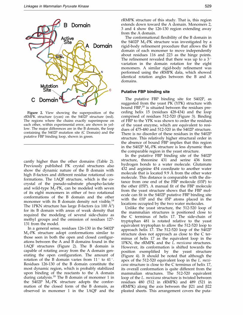
Figure 2. View showing the superposition of therRMPK structure (cyan) on the S402P structure (red).The regions where the chains exactly superimpose oneach other, within experimental error, are shown in yel-low. The major differences are in the B domain, the loopcontaining the S402P mutation site (C Domain) and theputative FBP binding loop, shown in green.
Linkages in Mammalian Pyruvate Kinase 529
cantly higher than the other domains (Table 2).Previously published PK crystal structures alsoshow the dynamic nature of the B domain withhigh B-factors and different residue rotational con-formations. The 1AQF structure, which is the co-crystal of the pseudo-substrate phospho-lactateand wild-type M1-PK, can be modeled with sevenof its eight monomers in either of two rotationalconformations of the B domain and the othermonomer with its B domain density not visible.18
The 1PKN structure has large B-factors (ca 100 AÊ 2)for its B domain with areas of weak density thatrequired the modeling of several side-chains asmethyl groups and the omission of residues 127-131 from the model.
In a general sense, residues 126-130 in the S402PM1-PK structure adopt conformations similar tothose seen in both the open and closed con®gur-ations between the A and B domains found in the1AQF structures (Figure 2). The B domain iscapable of rotating away from the A domain gen-erating the open con®guration. The amount ofrotation of the B domain varies from 11 � to 41 �.Residues 126-130 of the B domain constitute themost dynamic region, which is probably stabilizedupon binding of the reactants to the A domainduring catalysis.18 The B domain of monomer 1 inthe S402P M1-PK structure adopts the confor-mation of the closed form of the B domain, asobserved in monomer 1 in the 1AQF and the
rRMPK structure of this study. That is, this regionextends down toward the A domain. Monomers 2,3 and 4 show the 126-130 region extending awayfrom the A domain.
The conformational ¯exibility of the B domain inthe S402P M1-PK structure was investigated by arigid-body re®nement procedure that allows the Bdomain of each monomer to move independentlyabout residues 116 and 223 as the hinge points.The re®nement revealed that there was up to a 3 �variation in the domain rotation for the eightmonomers. A similar rigid-body re®nement wasperformed using the rRMPK data, which showedidentical rotation angles between the B and Adomains.
Putative FBP binding site
The putative FBP binding site for S402P, assuggested from the yeast PK (YPK) structure withbound FBP,20 is situated between the residues pre-ceding helix 15 (residues 428-434) and the loopcomprised of residues 512-520 (Figure 3). Bindingof FBP to the YPK was shown to order the residuesof the yeast enzyme, which are equivalent to resi-dues of 475-480 and 512-520 in the S402P structure.There is no disorder of these residues in the S402Pstructure. This relatively higher structural order inthe absence of bound FBP implies that this regionin the S402P M1-PK structure is less dynamic thanthe comparable region in the yeast structure.
In the putative FBP binding site of the S402Pstructure, threonine 431 and serine 436 formhydrogen bonds to a water molecule. Glutamate432 and arginine 454 coordinate to another watermolecule that is located 9.9 AÊ from the other watermolecule. This distance is comparable with the dis-tance from one end of the FBP molecule (03P) tothe other (05P). A manual ®t of the FBP moleculefrom the yeast structure shows that the FBP mol-ecule can ®t in the S402P putative FBP binding sitewith the 03P and the 05P atoms placed in thelocations occupied by the two water molecules.
Unlike the yeast structure, the 512-520 loop ofthe mammalian structures is positioned close tothe C terminus of helix 17. The side-chain oftryptophan 481 is rotated relative to the yeastequivalent tryptophan to allow the 512-520 loop toapproach helix 17. The 512-520 loop of the S402Pstructure does not approach as close to the C ter-minus of helix 17 as the equivalent loop in the1PKN, the rRMPK and the L. mexicana structures.However, its conformation is shifted towards theposition exempli®ed by the yeast structure(Figure 4). It should be noted that although theapex of the 512-520 equivalent loop in the L. mexi-cana structure is close to the C terminus of helix 17,its overall conformation is quite different from themammalian structures. The 512-520 equivalentloop of the L. mexicana structure is twisted betweenresidues 480 (512 in rRMPK) and 489 (521 inrRMPK) along the axis between the b21 and b22pleated sheets. This arrangement covers the puta-

Figure 3. Stereo view of the putative FBP binding region of the S402P structure. The inset shows the location of theFBP binding region (red) in the monomer. Shown are residues 430-436, 454, 480-489 and 510-520. Contours are at 1s. The two water molecules (blue spheres) closely coincide with the positions of the phosphate oxygen atoms of theFBP molecule as seen in the yeast enzyme. Arginine 454 interacts with the water molecule and glutamate 432, whichalso interacts with the water molecule.
530 Linkages in Mammalian Pyruvate Kinase
tive FBP binding site, and causes a shift of the area
where the FBP could bind.22 In addition, the N ter-
minus of helix 17 is shortened in the L. mexicana
structure, and the course of the polypeptide chain
between residues 443 (475 in rRMPK) and 453 (485
in rRMPK) is quite different from the S402P and
the other mammalian structures. Lys453 is postu-
lated to form the contact with the FBP. In the
S402P structure, residue 453 is a valine residue,
and is in the middle of helix 17 and cannot interact
with a FBP molecule. However, the Arg454 of the
S402P structure is positioned to possibly bind to
FBP.
Figure 4. Stereo view of the FBP binding region of the ye(cyan), S402P (green) and the L. mexicana (orange) structurescolored) of the four structures. The courses of the polypeptid(rRMPK numbering) are shown on the upper-right of theatoms), the sulfate from the L. mexicana structure (sulfur, orS402P structure are also shown. The remainder of the S402P
The active site
Comparing the active-site orientations in thefour monomers of the S402P M1-PK and the mono-mer of the M1-PK structure reveals the high degreeof similarity in each of the subunits. The Rama-chandran plot shows that the re®ned S402P M1-PKstructure possesses a reasonable geometry (Table 2)with more than 99 % of f-c angles in the allowedregions. In all monomers Thr327 is found near orin the unallowed region. This allows Thr327 tostrongly coordinate to the pyruvate in the activesite. The arrangement of the active-site atoms ofmonomer 1 differs from that of the other threemonomers of the tetramer. Monomer 1 does not
ast enzyme (yellow) with the RMPK (magenta), rRMPKsuperimposed by least-squares ®tting of helix 17 (multi-e chains of the structures between residues 475 and 485®gure. The FBP of the yeast structure (yellow carbonange) and the water molecules (oxygen, blue) from thestructure is shown in gray.
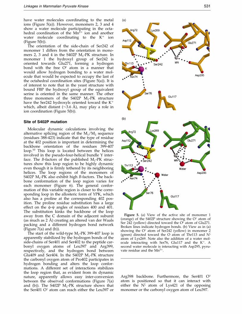
Figure 5. (a) View of the active site of monomer 1(orange) of the S402P structure showing the Og atom ofSer 242 (yellow) directed toward the Oe atom of Glu271.Broken lines indicate hydrogen bonds. (b) View as in (a)showing the Og atom of Ser242 (yellow) in monomer 2(green) directed toward the O atom of Thr113 and Nz
atom of Lys269. Note also the addition of a water mol-ecule interacting with Ser76, Glu117 and the K�. Asecond water molecule is interacting with Asp295, pyru-vate residue and the Mn2�.
Linkages in Mammalian Pyruvate Kinase 531
have water molecules coordinating to the metalions (Figure 5(a)). However, monomers 2, 3 and 4show a water molecule participating in the octa-hedral coordination of the Mn2� ion and anotherwater molecule coordinating to the K� ion(Figure 5(b)).
The orientation of the side-chain of Ser242 ofmonomer 1 differs from the orientation in mono-mers 2, 3 and 4 in the S402P M1-PK structure. Inmonomer 1 the hydroxyl group of Ser242 isoriented towards Glu271, forming a hydrogenbond with the free Oe atom in a manner thatwould allow hydrogen bonding to a water mol-ecule that would be expected to occupy the last ofthe octahedral coordination sites (Figure 5(a)). It isof interest to note that in the yeast structure withbound FBP the hydroxyl group of the equivalentserine is oriented in the same manner. The otherthree monomers of the S402P M1-PK structurehave the Ser242 hydroxyls oriented toward the K�
which, albeit distant (�3.6 AÊ ), may play a role inion coordination (Figure 5(b)).
Site of S402P mutation
Molecular dynamic calculations involving thealternative splicing region of the M1/M2 sequence(residues 388-423) indicate that the type of residueat the 402 position is important in determining thebackbone orientation of the residues 399-407loop.10 This loop is located between the helicesinvolved in the pseudo-four-helical bundle Y inter-face. The B-factors of the published M1-PK struc-tures show this loop region to be highly dynamiceven though it is ®rmly tethered by its neighboringhelices. The loop regions of the monomers ofS402P M1-PK also exhibit high B-factors. The back-bone conformation of the loop region varies foreach monomer (Figure 6). The general confor-mation of this variable region is closer to the corre-sponding loop in the allosteric form of YPK, whichalso has a proline at the corresponding 402 pos-ition. The proline residue substitution has a largeeffect on the f-c angles of residues 400 and 401.The substitution kinks the backbone of the loopaway from the C domain of the adjacent subunit(as much as 2 AÊ ) creating an altered van der Waalspacking and a different hydrogen bond network(Figure 7(a) and (b)).
The start of the wild-type M1-PK 399-407 loop isapparently stabilized by the hydrogen bonds of theside-chains of Ser401 and Ser402 to the peptide car-bonyl oxygen atoms of Leu397 and Arg399,respectively, and the hydrogen bond betweenGlu409 and Ser404. In the S402P M1-PK structurethe carbonyl oxygen atom of Pro402 participates inhydrogen bonding and alters the loop confor-mations. A different set of interactions stabilizesthe loop region that, as evident from its dynamicnature, apparently allows easy inter-conversionbetween the observed conformations (Figure 7(a)and (b)). The S402P M1-PK structure shows thatthe Ser401 Og atom can reach either the Leu397 or
Arg398 backbone. Furthermore, the Ser401 Og
atom is positioned so that it can interact witheither the Nz atom of Lys421 of the opposingmonomer or the carbonyl oxygen atom of Leu397.

Figure 6. View of the Ca positions of residues 391-406containing the S402P mutation site between helix 13(residues 390-400) and helix 14 (residues 407-421). Thestructures of monomer 1 (orange), monomer 3 (yellow),monomer 4 (magenta), and rRMPK (cyan) are shownsuperimposed on monomer 2 (green). The regions ofhelices 13 and 14 are included to illustrate the highdegree of conformational identity inherent in the struc-tures.
532 Linkages in Mammalian Pyruvate Kinase
In monomer 1, the hydrogen bond between theOg atom of Ser401 and the Nz atom of Lys421 ofthe opposing monomer 2 limits the proline inducedloop protrusion into the Y interface found in theother three monomers. The absence of detectablehydrogen bonds in the residues 400-405 of mono-mer 3 are consistent with this loop having the lar-gest conformational deviation, and gauged from itsB-factors, the lowest stability.
Alteration in the backbone conformations of theS402P loops occurs with different sets of hydrogenbonds to the negatively charged residues lining thecavity. The loop residues Asp406 and Glu409 enterinto a hydrogen bond network with residuesGln439, Arg442, Tyr443, and the water moleculefound near Glu409. The rRMPK structure showsthe side-chain of Asp406 directed into the solventand the side-chain of Glu409 oriented toward the399-407 loop, allowing the formation of hydrogenbonds with Ser404 and possibly Lys421. The S402PM1-PK structure has both residues 406 and 409oriented into the cavity and entering into hydrogenbonds. The extent of contact varies amongst thefour monomers. One carboxyl oxygen atom ofGlu409 in monomer 1 forms hydrogen bonds withthe hydroxyl group of Tyr443 and one hydrogenbond to Arg442 and a water molecule. Monomers2, 3, and 4 have one Glu409-Tyr443, one Glu409-Lys421, one Glu409-Arg442 and one Glu409-Waterhydrogen bond.
Dimer interface
There are two interfaces (Figure 1(b)) in the PKtetramer.5,17 The Y interface involves the C and Ndomains of monomers 1 and 2 and monomers 3and 4. The Z interface is approximately perpen-dicular to the Y interface, and involves all fourdomains between monomer pairs 1-3 and 2-4. Sur-face area calculations reveal that less surface is bur-ied between the 1 and 2 monomers, the Y interface(2813 AÊ 2), than between the 1 and 3 monomers, theZ interface (5945 AÊ 2). This supports the proposalthat dissociation of the tetramer into dimers wouldoccur at the Y interface.16 The tetramer possessesapproximate 222 symmetry with the largest devi-ation of 3 � at the Z interface.
The 1 and 2 monomers align the respective Cdomains in a ``tail-to-tail'' fashion (Figure 1(b)).Helices 13 and 14 form a helix-loop-helix region(residues 390 to 421) in the C domain, which inter-acts in a pseudo-four-helical bundle with itscounterpart in the opposing monomer (Figure 8).This helix-loop-helix region, which includes resi-due 402, is in the alternatively spliced region of theM1/M2 encoded RNA, and must therefore carryinformation for conferring allosteric regulation. Aparticularly prominent feature in this region isLys421, which protrudes across the Y interfaceinto, and through, the loop between helices 13 and14. The 399-407 loop creates a cavity in the proteinthat has a negative electrostatic-potential intowhich the Lys421 of the opposing monomer pro-trudes. The Lys421 interacts with several residuesin the opposing monomer, notably Glu409 andTyr443 (Figure 7(a) and (b)). Lys421 has beenimplicated as an essential residue in inter-subunitcommunication.10 Several altered side-chain confor-mations are also found within the N domain in thecavity formed at the junction of the four mono-mers. Residues 389, 391, 399 adopt conformationsthat are either oriented into the solvent (includingre®ned water molecules) or produce no detectablealterations or new interactions.
The Z interface encompasses extensive contactsbetween the adjacent A domains with minor con-tacts with the B and N domains (Figures 1(b) and8). This interface also contains residues that ¯ankthe opening to the active site, which have beenproposed to contribute to the stabilization of theactive form of the enzyme.23 The main interactionsare an inter-subunit crossing of helix 11 (residues341-353) and the bulging portion of helix 10 (resi-dues 302-319) into the ®rst 34 amino acid residuesof the N terminus and the top half of helix 12 (resi-dues 368-378) of the opposing monomer. Helix 12spans both interfaces and extends into the activesite. The relative orientation of the monomers, theinteracting helices, and the general positioning ofthe side-chains are very similar between the S402PM1-PK and rRMPK structures.
Another key feature of the Z interface is a salt-bridge near the opening of the active site betweenthe invariant residues Arg341 and Asp177 in the

Figure 7. (a) Stereo view of the helix 13-loop-helix 14 site of monomer 1 (orange) with Lys421 of monomer 2(green) protruding into the loop. Note the conformations of the side-chains of Ser401 and Glu409 (yellow) which areas found in the T state structures. (b) Stereo view of the helix 13-loop-helix 14 site of monomer 2 (green) with Lys421of monomer 1 (orange) protruding into the loop. The conformations of the side-chains of Ser401 and Glu409 (yellow)differ from monomer 1 and are as found in the R-state structures.
Linkages in Mammalian Pyruvate Kinase 533
opposing A and B domains, respectively (Figure 9).It is interesting to note that this interaction is dis-rupted in the S402P M1-PK structure (Figure 10).The side-chain of Asp177 is instead reorientedtoward the active site of the same subunit. The w1,and w2 angles of Asp177 of all of the S402P M1-PKmonomers are similar to those found in the E. colistructure, which is proposed to be in the T form.9
The position of the Arg341 side-chain remainsrelatively unchanged with either a slight move-ment back toward the backbone as in monomers 1and 2, (where it hydrogen bonds with the carbonyloxygen atom of Gly294 from the opposing mono-mer) or with a tilt, as in monomer 3, which bringsthe NZ atom closer to the carbonyl oxygen atom ofPro339. It also appears that there is a loss of thehydrogen bond between the Arg338 NZ atom andGly297. The side-chain of Arg338 swings �50 � intothe solvent and does not appear to make anyspeci®c interactions. Concurrent with the loss ofthe 177-341 salt-bridge and the 338-297 hydrogenbond in the S402P M1-PK structure is an apparent
increase in the rotation of the B domain about thehinge point as discussed in ``B Domain''.
Another interesting perturbation of the intersu-bunit interactions involves Phe25. The S402P struc-ture differs from the wild-type M1-PK (1PKN) withan additional inter-monomeric interaction invol-ving the aromatic rings of Phe25 (Figure 11). ThePhe25 residues in the S402P M1-PK have w1 and w2
dihedral angles of ÿ85 � and 74 �, respectively; asopposed to the 1PKN values of ÿ180 � and 36 �.The rings of the S402P Phe25 are oriented with theCg atom of one ring almost over the Cz atom of thering of the opposing monomer across the Z inter-face (Figure 1(b)). The inter-planar distance is�3.6 AÊ , which is slightly greater than the minimaldistance for ring-ring interactions.24 ± 26 Changingthe orientation of the Phe25 rings to the edge toface interactions observed in the 1PKN structure,followed by energy re®nement, resulted in therings resuming the face-to-face orientation with thering plane normals making an angle of �8 �. ThePhe25 interactions of the S402P M1-PK structure
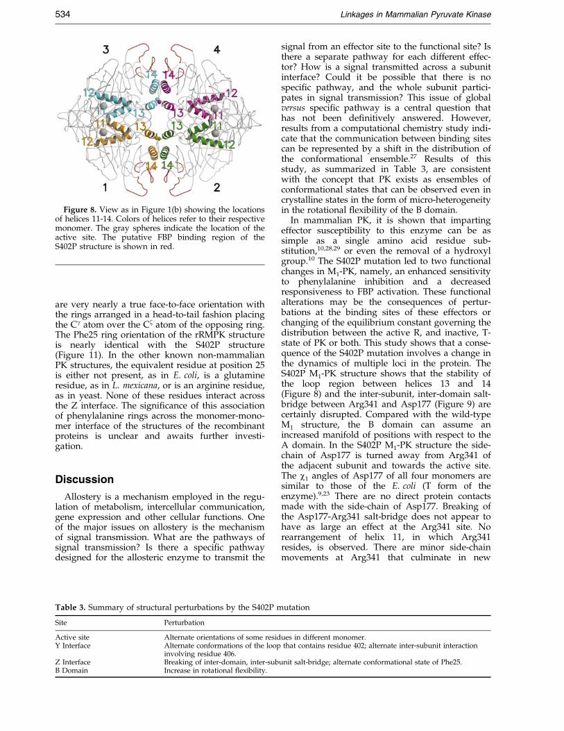
Figure 8. View as in Figure 1(b) showing the locationsof helices 11-14. Colors of helices refer to their respectivemonomer. The gray spheres indicate the location of theactive site. The putative FBP binding region of theS402P structure is shown in red.
534 Linkages in Mammalian Pyruvate Kinase
are very nearly a true face-to-face orientation withthe rings arranged in a head-to-tail fashion placingthe Cg atom over the Cz atom of the opposing ring.The Phe25 ring orientation of the rRMPK structureis nearly identical with the S402P structure(Figure 11). In the other known non-mammalianPK structures, the equivalent residue at position 25is either not present, as in E. coli, is a glutamineresidue, as in L. mexicana, or is an arginine residue,as in yeast. None of these residues interact acrossthe Z interface. The signi®cance of this associationof phenylalanine rings across the monomer-mono-mer interface of the structures of the recombinantproteins is unclear and awaits further investi-gation.
Discussion
Allostery is a mechanism employed in the regu-lation of metabolism, intercellular communication,gene expression and other cellular functions. Oneof the major issues on allostery is the mechanismof signal transmission. What are the pathways ofsignal transmission? Is there a speci®c pathwaydesigned for the allosteric enzyme to transmit the
Table 3. Summary of structural perturbations by the S402P m
Site Perturbation
Active site Alternate orientations of some residY Interface Alternate conformations of the loop
involving residue 406.Z Interface Breaking of inter-domain, inter-subB Domain Increase in rotational flexibility.
signal from an effector site to the functional site? Isthere a separate pathway for each different effec-tor? How is a signal transmitted across a subunitinterface? Could it be possible that there is nospeci®c pathway, and the whole subunit partici-pates in signal transmission? This issue of globalversus speci®c pathway is a central question thathas not been de®nitively answered. However,results from a computational chemistry study indi-cate that the communication between binding sitescan be represented by a shift in the distribution ofthe conformational ensemble.27 Results of thisstudy, as summarized in Table 3, are consistentwith the concept that PK exists as ensembles ofconformational states that can be observed even incrystalline states in the form of micro-heterogeneityin the rotational ¯exibility of the B domain.
In mammalian PK, it is shown that impartingeffector susceptibility to this enzyme can be assimple as a single amino acid residue sub-stitution,10,28,29 or even the removal of a hydroxylgroup.10 The S402P mutation led to two functionalchanges in M1-PK, namely, an enhanced sensitivityto phenylalanine inhibition and a decreasedresponsiveness to FBP activation. These functionalalterations may be the consequences of pertur-bations at the binding sites of these effectors orchanging of the equilibrium constant governing thedistribution between the active R, and inactive, T-state of PK or both. This study shows that a conse-quence of the S402P mutation involves a change inthe dynamics of multiple loci in the protein. TheS402P M1-PK structure shows that the stability ofthe loop region between helices 13 and 14(Figure 8) and the inter-subunit, inter-domain salt-bridge between Arg341 and Asp177 (Figure 9) arecertainly disrupted. Compared with the wild-typeM1 structure, the B domain can assume anincreased manifold of positions with respect to theA domain. In the S402P M1-PK structure the side-chain of Asp177 is turned away from Arg341 ofthe adjacent subunit and towards the active site.The w1 angles of Asp177 of all four monomers aresimilar to those of the E. coli (T form of theenzyme).9,23 There are no direct protein contactsmade with the side-chain of Asp177. Breaking ofthe Asp177-Arg341 salt-bridge does not appear tohave as large an effect at the Arg341 site. Norearrangement of helix 11, in which Arg341resides, is observed. There are minor side-chainmovements at Arg341 that culminate in new
utation
ues in different monomer.that contains residue 402; alternate inter-subunit interaction
unit salt-bridge; alternate conformational state of Phe25.

Figure 9. Stereo view as in Figure 1(b) rotated 90 � around the horizontal axis showing the locations of the salt-bridges across the Z interface between Asp 177 (small colored spheres) of one monomer and Arg 341 (larger spheres)of the other monomer. Colors of spheres refer to their respective monomer. Large gray spheres indicate the locationof the active site. The putative FBP binding region of the S402P structure is shown in red. Monomers 1 and 2 arelightly colored to distinguish them from monomers 3 and 4.
Linkages in Mammalian Pyruvate Kinase 535
hydrogen bonds with the carbonyl oxygen atom ofGly294 (from the opposing monomer) or carbonyloxygen atom of Pro399. The side-chain of Arg338swings (�50 �) into the solvent, thereby losing itshydrogen bond to Gly297. Concurrent with theloss of the Asp177-Arg341 salt-bridge in the S402PM1-PK structure is an apparent increase in the free-dom of rotation of the B domain about the hingepoint. Rigid-body re®nement of the B domain pro-duced a variation of up to 3 � in rotation for allmonomers. Similar re®nement of the rRMPK struc-ture, which retains the Asp177-Arg341 salt-bridge,shows essentially no difference in the rotation inseven of the eight monomers. This apparent corre-lation between salt-bridge formation and freedomof rotation of the B domain is a logical consequenceof the fact that the bridge is formed between the Band A domains of adjacent subunits. Formation ofthe inter-domain salt-bridge is expected to anchorthe movements of the B domain.
Previously published rabbit muscle structures,containing different ligands, also demonstrates therelation between the presence of the Asp177-Arg341 salt-bridge and the rotational ¯exibility oftheir B domain. The ®rst rabbit muscle PK struc-ture solved by Larsen et al.17 (1PKN) was crystal-lized under conditions similar to those employedfor S402P M1-PK and rRMPK. These conditionsinclude the presence of pyruvate, K� and Mn2�.The 1PKN structure is found to maintain the salt-bridge. The 1PKN structure was re®ned using amonomer with 8-fold, non-crystallographic sym-metry (two tetramers in the asymmetric unit). TheB-factors for the B domain are high but the struc-ture was re®ned to an R-factor of 22 %, indicatingsuf®cient uniformity of monomer conformations tomodel the eight B domains as one conformation.Thus, in the 1PKN and rRMPK structures the pre-
sence of the salt-bridge is correlated with uniform-ity of the B domain rotational conformation.
In another study of M1-PK that was crystallizedin the presence of the pseudo-substrate L-phospho-lactate and the counterions Mn2� and K�, thestructure was re®ned with three different orien-tations of the B domain within the two tetramersin the asymmetric unit.18 Six of the monomersshow the B domain in the open conformation andone in the closed conformation. One B domain wasnot resolved due to extensive disorder. In each ofthe seven resolved B domains the Asp177 isoriented away from the Arg341, and does not formthe salt-bridge. Only in the closed B domain is theside-chain of Asp177 oriented towards the activesite. It does not, however, enter into the network ofthe substrates or protein. Hence, in this structurethe absence of the salt-bridge is related to anincrease in the number of conformations assumedby the B domain, and all of them assume an openconformation, the conformation proposed by Cons-ler et al.8 and Mattevi et al.9 as the inactive T-form.Thus, a breakage of the salt-bridge apparentlyshifts the state equilibrium constant in favor of theinactive T-state.
The deepest penetration of the side-chain ofAsp177 into the active site is found with M1-PKcomplexed with Mg2�-ATP-Na�-oxalate (1A5U).19
Of the eight subunits found in the unit cell (twotetramers) six were fully liganded and two weremissing the ATP. Large conformational changeswere observed in the orientation of the B domain,and no subunit maintained the Asp177-Arg341interaction. The B domains of those monomerswith the ATP bound moved closer to the Adomain and the Asp177 penetrated to within �4 AÊ
of the Mg2�-ATP. Thus, once again a correlationcan be established in this structure between the

Figure 10. View of the Asp177-Arg341 inter-monomersalt-bridge, with Gln328. Residues 177, 328 and 341 ofthe S402P structure are shown (magenta and green)with the corresponding residues of the superimposedrRMPK structure (cyan). The ®t is quite good, except forthe side-chains of the Asp177 residues. The carboxylgroup of Asp177 of the S402P structure (magenta) pointstoward the active site. Whereas, the Asp177 of thesuperimposed rRMPK structure (cyan) points towardthe Gln328 of the same monomer and toward theArg341 of the opposite monomer. In the S402P struc-ture, a water molecule (black sphere) occupies the spacewhere the carboxyl moiety of Asp177 is situated in therRMPK structure.
Figure 11. View of the Phe25-Phe25 interaction in theS402P structure (orange and cyan). The residues fromthe rRMPK structure (yellow) and the 1PKN structure(blue) are also shown. See the text for details.
536 Linkages in Mammalian Pyruvate Kinase
disruption of the salt-bridge and a change in theorientation of the B domain.
In addition to mammalian PK, a similar patternof structural linkage between the formation of thesalt-bridge and dynamics in domain rotation isreported in YPK.20 Structure of YPK in the pre-sence and absence of the effector FBP (1A3W and1A3X, respectively) were determined. In both tetra-meric structures the Asp177-Arg341 salt-bridge(Asp147-Arg314 in the yeast sequence) is intact.Re®nement of the structures was successful byemploying non-crystallographic symmetry, i.e. tet-ramer re®nement using just one monomer. Thequality of the re®ned structures implies that allfour subunits had the same B domain orientation.Inspection of YPK without FBP shows that eventhough the B domain of the two monomers in theunit cell (monoclinic C2) differ in rotation by about4 �, they maintain the salt-bridge. In summary, allthe available structural information on PK indi-cates a linkage between the dynamics in the
rotational state of the B domain with the presenceof the Asp177-Arg341 salt-bridge. An increase indomain rotational freedom is apparently associatedwith the absence of the salt-bridge.
The relationship for B-domain stability and theAsp177-Arg341 salt-bridge as seen in the musclePK crystal structures also can be supported fromgenetic observations. Using information derivedfrom genetic evidence based on studies of erythro-cyte PK in hereditary non-spherocytic hemolyticpatients, mammalian PK mutants at residues 340were generated. Studies revealed that the mutationat the 340 residue, threonine to methionine residue,is detrimental to the function of PK.11 ± 13 The struc-tural effects (i.e. impediment of the Asp177-Arg341salt-bridge) of the T340M PK mutants have notbeen determined. Kinetic analysis of T340M M1
and M2-PK mutants revealed alterations in steady-state behavior similar to those found in the S402PM1-PK mutant. The mutation produces signi®-cantly reduced enzyme activity, indicated by adecrease in the Kcat and an increase in Km,app forPEP in the presence of 2 mM ADP. This result indi-cates a communication between residue 402 andthe active site, which resides about 40 AÊ apart. Nochange in the Km,app for ADP is observed. Inaddition, the T340M mutation effects a signi®cantenhancement in the association behavior at the Yinterface, site of the residue 402. In other words, amutation at one interface affects the associationpattern at another interface 40 AÊ away. Theseresults establish the functional linkage betweenthese two regions, the loop between helices 13 and14 and the turn at the beginning of helix 11 whichcontains residue 340. This structural study ident-i®es the nature of changes in those two regions.Thus, both structural and functional studies lead toa consistent conclusion in revealing the linkage ofthese regions in the allosteric regulation of PK.
Having established the correlation between theformation of the Asp177-Arg341 salt-bridge androtational motion of the B domain, it is importantto de®ne the functional relevance of these structur-

Linkages in Mammalian Pyruvate Kinase 537
al changes. Based on small angle neutron scatteringdata, Consler et al.8 proposed that the inactive,T-state can be represented by a rotation of theB-domain in opening of the cleft between the Band A domains. A closing of the cleft can representthe active, R-state. The increased dynamic of the Bdomain in the S402P M1-PK is consistent with achange in the equilibrium constant governing thedistribution of R and T-states most likely towardsthe T-state. It is, therefore, not surprising that theS402P mutation leads to an increase in sensitivityto phenylalanine, the binding of which shifts theequilibrium towards the T-state. The decreasedresponsiveness to FBP and lower apparent af®nityfor PEP are also consistent with the interpretationthat the S402P mutation favors the distributiontowards the T-state. By comparing the structures ofthe rabbit muscle (1PKN) and E. coli PK, Matteviet al.9 noticed the presence and absence of theAsp177-Arg341 salt-bridge, respectively. As thestructures of the mammalian and E. coli PK areinterpreted as representative of R and T-state,respectively, it was proposed that Asp177 plays animportant role in the T . R transition.
In a recent study of the structure of PK fromL. mexicana, it was shown that the region contain-ing residue 341 is in a more dynamic state whenthe enzyme assumes the inactive T-state.22 It wasfurther reported that there is signi®cant loopdynamics in the effector binding site. In conjunc-tion with this study, it seems that an increase indynamics in speci®c loci in the protein structure iscorrelated with the conferring of allostery in PK.Hence, a consistent structure-function correlation isemerging. It is most encouraging that the strategyof combining human genetic information with sol-ution biophysical and structural studies is yieldingconsistent information to elucidate the molecularmechanism of allosteric regulation in PK.
Concluding Remarks
Results from the literature and this study indi-cate the importance of the formation of the inter-subunit and inter-domain salt-bridge betweenAsp177 and Arg341 in de®ning the distribution ofthe PK conformation between the R and T-states.These conformations are intimately linked to themovement of the B domain towards or away fromthe A domain. It is not surprising to observe anincrease in the freedom of the B domain forrotation with a breakage of the salt-bridge, becausethis interaction is inter-domain in nature. Further-more, nature's selection of this particular salt-bridge is logical. Formation of this inter-domainsalt-bridge anchors the B domain to the A domainvia Arg341, which is highly connected to theactive-site residues.30 As this salt-bridge also con-stitutes an inter-subunit interaction between twodomains of adjacent subunits, it is not surprisingthat the consequences of transmission of infor-mation through this particular structural element
are global in nature. It seems that allosteric pro-teins might be engineered to have clusters of struc-tural elements that are highly connected in amanner analogous to the pattern of inter-molecularinteractions observed in cells.31
There is ample evidence in this study to indicatethat PK can exist in ensembles of microstates,namely, the various numbers of microstatesobserved in the active sites, rotational ¯exibility ofthe B domain and orientation of the Phe25 ring.Mutations or ligand bindings can perturb the dis-tribution of these microstates.
Materials and Methods
Materials
Succinic acid, pyruvic acid, and Hepes were pur-chased from Sigma Biochemical and used withoutfurther puri®cation. Polyethylene glycol 8000 wasobtained from Fisher Scienti®c.
Overexpression and purification of rRMPK
Construction and expression of the S402P M1-PKmutant and the wild-type rabbit muscle PK were asdescribed.10,11 The proteins were puri®ed using the pro-cedure described by Cheng et al.11 FBP (0.4 mM) anddithiothreitol (0.2 mM) were added to all buffer solutionsto enhance protein stability and reduce oxidative effects.Protein purity was estimated to be at least 95 % (w/v)from Coomassie blue staining of SDS-PAGE gels.
Crystallization conditions
The rRMPK and S402P M1-PK enzymes were crystal-lized and processed under identical conditions. Prior tothe crystallization process, approximately 2-3 mg of pre-cipitated protein (2 mg/ml) was removed from the 70 %ammonium sulfate storage buffer and re-suspended in200 ml of dialysis buffer (100 mM KCl, 10 mM Hepes(pH 6.0)). The solution was passed through a Superdex200 HR 10/30 FPLC (Pharmacia) column at a ¯ow rateof 1 ml/min for desalting purposes and as an additionalpuri®cation step. Peak fractions were collected and dia-lyzed for four hours against one liter of dialysis buffer at4 �C.
Crystallization of the proteins was achieved using thehanging drop method at ca 18 �C. The crystallization sol-ution consisted of 2.9 mM sodium pyruvate, 1.2 mMMnCl2, 0.45 M KCl, 30 mM succinate adjusted to pH 6.0,9 to 11 % (w/v) polyethylene glycol 8000, and 1.5 mg/ml of protein. A stream of N2 gas at 15 psi was bubbledinto the wells before sealing to slow oxidation effectsfrom the air. Crystals appeared in two to three days, andwere fully formed in four to ®ve days.
Data collection
X-ray data were collected using a MacScience DIP2030H area detector mounted on a MacScience M06HFrotating anode X-ray generator running at 50 kV and 90mA equipped with MAXOS optics. Crystals were soakedin solutions containing increasing glycerol concentrationsranging from 0.5 % to 20 % (w/v) in six steps. The crys-tals were ¯ash-cooled using nitrogen boil-off from aCryo Industries of America Cryo Cooler. Data sets con-

538 Linkages in Mammalian Pyruvate Kinase
sisting of 400 one-half degree frames were collected fromone crystal of each protein. The S402P M1-PK PK andrRMPK crystals nominally diffracted to 2.33 and 3.0 AÊ
resolution, respectively. Analysis of the S402P M1-PKdata collection statistics indicated that the resolutionlimit of useable data did not exceed 2.8 AÊ . No other crys-tal of S402P M1-PK diffracted as well.
Data analysis and structure refinement
The diffraction images were integrated using the pro-grams DENZO and SCALEPACK.32 Data were later cor-rected as described by White et al.33 Data collectionstatistics for the S402P M1-PK and rRMPK crystals aresummarized in Table 4.
Initial phases were obtained using the molecularreplacement procedure of XPLOR34. The muscle PKstructure,17 PDB entry 1PKN, was used as the searchmodel. Subunit and secondary structure numbering fol-low those described for 1PKN. Molecular replacementrevealed the location of the two tetramers in the asym-metric unit. Subsequent re®nement consisted of cycles ofrigid-body positioning, positional minimization, B-factoroptimization, density averaging and model building.Initial energy re®nements were carried out using XPLORwith CNS used in the ®nal steps.34,35 Rigid-body re®ne-ments were performed using shells of increasing resol-ution (12-8! 10-6! 8-4! 6-2.5 AÊ ) with increasingnumbers of rigid bodies in the order of tetramers, mono-mers, domains and then full positional re®nement, withnon-crystallographic symmetry applied between thedomains. Non-crystallographic symmetry matricesobtained from the ®nal rigid-body re®nement wereapplied in all individual atomic re®nements. Initially,strict 8-fold symmetry of one monomer was employedfor both structures. The rRMPK structure was fullyre®ned with strict 8-fold, non-crystallographic symmetry.However, it was determined during re®nement that theS402P M1-PK monomers did not adhere to strict 8-foldaveraging. At the beginning of the re®nement process,
Table 4. Data collection statistics
S402P structureSpace group P1
Unit cell
a � 82.1 AÊ ;b � 108.7 AÊ ;c � 144.4 AÊ
a � 95.9 �; b � 92.5 �;g � 111.7 �
Unique reflections 92,167Resolution 90-2.8Rmerge (last shell) 0.132 (.256)Redundancy (last shell) 1.6 (1.7)Completeness (last shell) 81.8 % (79.2 %)I/s(I) (last shell) 11.0 (4.2)
rRMPK structureSpace group P1
Unit cell
a � 79.0 AÊ ;b � 105.4 AÊ ;c � 143.5 AÊ
a � 95.9 �; b � 92.8 �;g � 111.9 �
Unique reflections 79,318Resolution 90-3.0Rmerge (last shell) 0.082 (.258)Redundancy (last shell) 2.4 (2.2)Completeness (last shell) 93.2 % (82.9 %)I/s(I) (last shell) 9.1 (3.1)
monomer 1 had been arbitrarily chosen for 8-fold, non-crystallographic symmetry averaging. Inspection of thesubsequent 8-fold non-crystallographic symmetry aver-aged electron density maps indicated that several regionsof the maps corresponding to monomers 2, 3 and 4 dif-fered from monomer 1. Calculations using 1, 2, 4 and 8-fold, non-crystallographic symmetry averaging showedthat 2-fold, non-crystallographic symmetry averaginggave better results (Table 1). Steadily improving re®ne-ment cycles using 2-fold, non-crystallographic symmetryaveraging veri®ed this conclusion (Figures 5 to 7). Afterthe ®rst round of modeling an anisotropic B-factor cor-rection was made to the observed scattering factors. Onecycle of simulated annealing was then carried out usingXPLOR. Solvent ¯attening, individual atomic B-factorsand positional re®nement in CNS were used in ®nalrounds of re®nement. An additional round of simulatedannealing was performed on the S402P M1-PK structurewhen the non-crystallographic symmetry was reducedfrom 8 to 2-fold. Ordered water molecules were addedto the S402P M1-PK structure using the water-pick anddelete function of CNS. Water molecules were kept ifthey were within hydrogen-bonding distance as deter-mined by the contact protocol of employing distance,geometry and neighbor constraints. After each round ofenergy re®nement Fo ÿ Fc and 2Fo ÿ Fc electron-densitymaps were produced using a locally modi®ed version ofthe density-averaging program RAVE.36,37 The electron-density map chosen for model building was determinedby calculating the rmsd of the phase angle from onecycle of RAVE to the next. When the phase angle con-verged, the electron-density map from that particularaveraging cycle was chosen.
Manual intervention
The energy-re®ned coordinates were manually ®ttedinto averaged electron density maps using the programO.38 Because only 2-fold averaging was possible with theS402P M1-PK structure, the region around the site ofmutation and regions of the B-domain which were notobserved in the 1PKN structure, were in areas of weakelectron density. These areas of the model were rebuiltusing omit maps. Final re®nement statistics are summar-ized in Table 2.
Protein Data Bank accession numbers
Coordinates of the rRMPK and the S402P structureshave been deposited in the Protein Data Bank with entrynumbers 1F3W and 1F3X, respectively.
Acknowledgements
Figure 3 was drawn with XTALVIEW.39 All other®gures were drawn with MOLSCRIPT.40 All ®gureswere rendered with RASTER3D.41 A critical review ofthe manuscript by Drs Xiaodong Cheng and WernerBraun is appreciated. The research was supported byNIH grant GM-45579 and Robert A. Welch Foundationgrants H-0013 and H-1238 to J.C.L., and H-1345 toR.O.F.

Linkages in Mammalian Pyruvate Kinase 539
References
1. Ainsworth, S. & MacFarlane, N. (1973). A kineticstudy of rabbit muscle pyruvate kinase. Biochem. J.131, 223-236.
2. Consler, T. G., Woodward, S. H. & Lee, J. C. (1989).Effects of primary sequence differences on the globalstructure and function of an enzyme: a study of pyr-uvate kinase isozymes. Biochemistry, 28, 8756-8764.
3. Consler, T. G., Jennewein, M. J., Cai, G. Z. & Lee,J. C. (1992). Energetics of allosteric regulation inmuscle pyruvate kinase. Biochemistry, 31, 7870-7878.
4. Hall, E. R. & Cottam, G. L. (1978). Isozymes of pyr-uvate kinase in vertebrates: their physical, chemical,kinetic and immunological properties. Int. J. Biochem.9, 785-793.
5. Muirhead, H., Clayden, D. A., Barford, D., Lorimer,C. G., Fothergill-Gilmore, L. A., Schiltz, E. &Schmitt, W. (1986). The structure of cat muscle pyru-vate kinase. EMBO J. 5, 475-481.
6. Oberfelder, R. W., Barisas, B. G. & Lee, J. C. (1984).Thermodynamic linkages in rabbit muscle pyruvatekinase: analysis of experimental data by a two-statemodel. Biochemistry, 23, 3822-3826.
7. Oberfelder, R. W., Lee, L.-Y. & Lee, J. C. (1984).Thermodynamic linkages in rabbit muscle pyruvatekinase: kinetic, equilibrium, and structural studies.Biochemistry, 23, 3813-3821.
8. Consler, T. G. & Lee, J. C. (1988). Domain inter-action in rabbit muscle pyruvate kinase. II. Smallangle neutron scattering and computer simulation.J. Biol. Chem. 263, 2787-2793.
9. Mattevi, A., Bolognesi, M. & Valentini, G. (1996).The allosteric regulation of pyruvate kinase. FEBSLetters, 389, 15-19.
10. Friesen, R. H. E., Castellani, R. J., Lee, J. C. & Braun,W. (1998a). Allostery in rabbit pyruvate kinase:development of a strategy to elucidate the mechan-ism. Biochemistry, 37, 15266-15276.
11. Cheng, X., Friesen, R. H. E. & Lee, J. C. (1996).Effects of conserved residues on the regulation ofrabbit muscle pyruvate kinase. J. Biol. Chem. 271,6313-6321.
12. Friesen, R. H. E., Chin, A. J., Ledman, D. W. & Lee,J. C. (1998b). Interfacial communications in recombi-nant rabbit kidney pyruvate kinase. Biochemistry, 37,2949-2960.
13. Friesen, R. H. E. (1998). Effects of conserved residueson the allosteric regulation of pyruvate kinase: astructure-function study of rabbit muscle andkidney-pyruvate kinase. PhD thesis, Department ofHuman Biological Chemistry and Genetics, Univer-sity of Texas Medical Branch at Galveston, TX.
14. Monod, J., Wyman, J. & Changeux, J.-P. (1965). Onthe nature of allosteric transitions: a plausiblemodel. J. Mol. Biol. 12, 88-118.
15. Heyduk, E., Heyduk, T. & Lee, J. C. (1992). Globalconformational changes in allosteric proteins. Astudy of Escherichia coli cAMP receptor protein andmuscle pyruvate kinase. J. Biol. Chem. 267, 3200-3204.
16. Friesen, R. H. E. & Lee, J. C. (1998). The negativedominant effects of T340M mutation on mammalianpyruvate kinase. J. Biol. Chem. 273, 14772-14779.
17. Larsen, T. M., Laughlin, L. T., Holden, H. M.,Rayment, I. & Reed, G. H. (1994). Structure of rabbitmuscle pyruvate kinase complexed with Mn2�, K�,and pyruvate. Biochemistry, 33, 6301-6309.
18. Larsen, T. M., Benning, M. M., Wesenberg, G. E.,Rayment, I. & Reed, G. H. (1997). Ligand-induceddomain movement in pyruvate kinase: structure ofthe enzyme from rabbit muscle with Mg2�, K�, andL-phospholactate at 2.7 AÊ resolution. Arch. Biochem.Biophys. 345, 199-206.
19. Larsen, T. M., Benning, M. M., Rayment, I., . &Reed, G. H. (1998). Structure of the bis(Mg2�)-ATP-oxalate complex of the rabbit muscle pyruvatekinase at 2.1 AÊ resolution: ATP binding over a bar-rel. Biochemistry, 37, 6247-6255.
20. Jurica, M. S., Mesecar, A., Heath, P. J., Shi, W.,Nowak, T. & Stoddard, B. L. (1998). The allostericregulation of pyruvate kinase by fructose-1,6-bispho-sphate. Structure, 6, 195-210.
21. Gerstein, M. (1992). A resolution-sensitive procedurefor comparing protein surfaces and its application tothe comparison of antigen-combining sites. ActaCrystallog. sect. A, 48, 271-276.
22. Rigden, D. J., Phillips, S. E. V., Michels, P. A. M. &Fothergill-Gilmore, L. A. (1999). The structure ofpyruvate kinase from Leishmania mexicana revealsdetails of the allosteric transition and unusual effec-tor speci®city. J. Mol. Bol. 291, 615-635.
23. Mattevi, A., Valentini, G., Rizzi, M., Speranza, M. L.,Bolognesi, M. & Coda, A. (1995). Crystal structureof Escherichia coli pyruvate kinase type I: molecularbasis of the allosteric transition. Structure, 3, 729-741.
24. Burley, S. K. & Petsko, G. A. (1988). Weakly polarinteractions in proteins. Advan. Protein Chem. 39,125-189.
25. Hunter, C. A. & Sanders, J. K. M. (1990). The natureof p-p interactions. J. Am. Chem. Soc. 112, 5525-5534.
26. Hunter, C. A., Singh, J. & Thornton, J. M. (1991). p-pInteractions: the geometry and energetics of phenyl-alanine-phenylalanine interactions in proteins. J. Mol.Biol. 218, 837-846.
27. Pan, H., Lee, J. C. & Hilser, V. J. (2000). Bindingsites in Escherichia coli dihydrofolate reductase com-municate by modulating the conformational ensem-ble. Proc. Natl Acad. Sci. USA, 97, 12020-12025.
28. Ikeda, Y., Tanaka, T. & Noguchi, T. (1997). Conver-sion of non-allosteric pyruvate kinase isozyme intoan allosteric enzyme by a single amino acid substi-tution. J. Biol. Chem. 272, 20495-20501.
29. Ikeda, Y. & Noguchi, T. (1998). Allosteric regulationof pyruvate kinase M2 isozyme involves a cysteineresidue in the intersubunit contact. J. Biol. Chem. 273,12227-12233.
30. Allen, S. C. & Muirhead, H. (1996). Re®ned three-dimensional structure of cat-muscle (M1) pyruvatekinase at a resolution of 2.6 AÊ . Acta Crystallog. sect.D, 52, 499-504.
31. Jeong, H., Mason, S. P., Barabasi, A.-L. & Oltvai,Z. N. (2001). Lethality and centrality in protein net-works. Nature, 411, 41-42.
32. Otwinowski, Z. (1993). Denzo. In Data Collection andProcessing (Sawyer, L., Isaacs, N. & Bailey, S., eds),pp. 56-62, SERC Daresbury Laboratory, Warrington,UK.
33. White, M. A., Watowich, S. J. & Fox, R. O. (1999).Calibration of spiral readout image plate detectors.J. Appl. Crystallog. 32, 65-70.
34. BruÈ nger, A. T. (1992). X-PLOR Version 3.1. A Systemfor X-ray Crystallography and NMR, Yale UniversityPress, New Haven, CT.
35. BruÈ nger, A. T., Adams, P. D., Clore, G. M., Delano,W. L., Gros, P. & Grosse-Kunstleve, R. W. et al.(1998). Crystallography and NMR system: a new

540 Linkages in Mammalian Pyruvate Kinase
software suite for macromolecular structure determi-nation. Acta Crystallog. sect. D, 54, 905-921.
36. Kleywegt, G. J. & Jones, T. A. (1994). Halloween . . .masks and bones. In From First Map to Final Model(Bailey, S., Hubbard, R. & Waller, D., eds), pp. 59-66, SERC Daresbury Laboratory, Warrington, UK.
37. Collaborative Computational Project Number 4(1994). The CCP4 suite: programs for protein crystal-lography. Acta Crystallog. sect. D, 50, 760-763.
38. Jones, T. A., Zou, J. Y., Cowan, S. W. & Kjelgaard,M. (1991). Improved methods for building proteinmodels in electron density maps and the location of
errors in theses models. Acta Crystallog. sect. A, 47,110-119.
39. McRee, D. E. (1999). XtalView/X®t - a versatile pro-gram for manipulating atomic coordinates and elec-tron density. J. Struct. Biol. 125, 156-165.
40. Kraulis, P. J. (1991). MOLSCRIPT: a program to pro-duce both detailed and schematic plots of proteinstructures. J. Appl. Crystallog. 24, 946-950.
41. Merritt, E. A. & Bacon, D. J. (1997). Raster3D: photo-realistic molecular graphics. Methods Enzymol. 277,505-524.
Edited by R. Huber
(Received 2 February 2001; received in revised form 23 July 2001; accepted 30 July 2001)