stroke, critical care, & infections - university … critical care, & infections an...
TRANSCRIPT

Stroke, Critical Care, & Infections
An Introduction
The LSU-Shreveport Department of NeurosurgeryCreated 12-20-2003
Revisions: None to Date
Presenting Authors: Neurosurgery Residents & Faculty
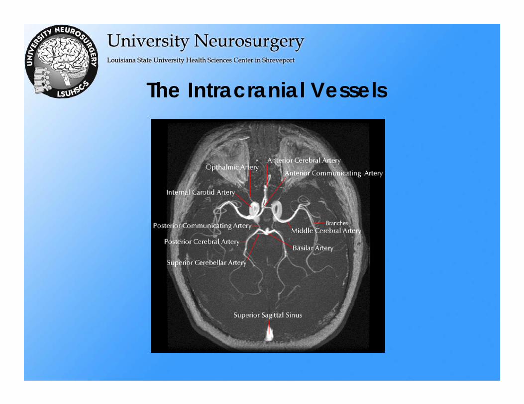
The Intracranial Vessels

Stroke
• Ischemic– Thromboembolic sources:
• Heart• Carotid artery
– Lacunar– For academic purposes, distinguish between TIA vs. RIND vs.
Stroke
• Hemorrhagic– Hypertensive– Aneurysm– Arteriovenous Malformation

Stroke• Ischemic Stroke
– Thromboembolic sources:• Heart (70% of cases)• Carotid artery (30% of cases)
CT Head showing stroke as a hypodensity

Stroke
• Treatment Options for ischemic stroke– TPA (if w/i 3 hrs) – injected intra-arterially, done with angiogram– Heparin short-term, then long-term anticoagulation (e.g. ASA,
ticlopidine (Ticlid), clopidogrel (Plavix), and/or warfarin(Coumadin))
– If carotid disease:• Internal carotid artery with >=70% stenosis in symptomatic patients
patient would benefit from carotid endarterectomy• Internal carotid artery with >=60% stenosis in ASYMPTOMATIC patients
patient would benefit from carotid endarterectomy• Thus, although the study has not been done, >=60% for any patient may
be inferred to benefit from CAROTID ENDARTERECTOMY.
• Prognosis is generally better for patients who are surgically treated with >=60% stenosis, according to NASCET & ACAS trials, in comparison to medical anticoagulation therapy

Diagnostic Studies
• Diagnostic studies– CT Head – r/o bleed
• Usually “normal” for 12-24 hrs following ischemic stroke– Angiogram – find occlusion, +/- TPA if within 3 hours– ECHOcardiogram – To find thrombus– MRI/MRA to better define stroke early, with major vessel occlusion– Blood panel for more common disorders, e.g.
cholesterol/LDL/HDL/Triglycerides– Blood panel – for rare disorders
• Antiphospholipid antibodies (APL), Anticardiolipin antibodies (ACL), Lupus anticoagulant (LA); Blood culture; Cardiac enzymes: Troponin, Creatinekinase (CPK, CK), LDH isoenzymes; Coagulation factors: Antithrombin III, Protein C, Protein S; Factor VIII; activated Protein C resistance (Factor V Leiden); Erythrocyte sedimentation rate (ESR); Hemoglobin electrophoresis; Homocysteine; Syphilis serology (VDRL, FTA, others); Toxicology screen (serum or urine )
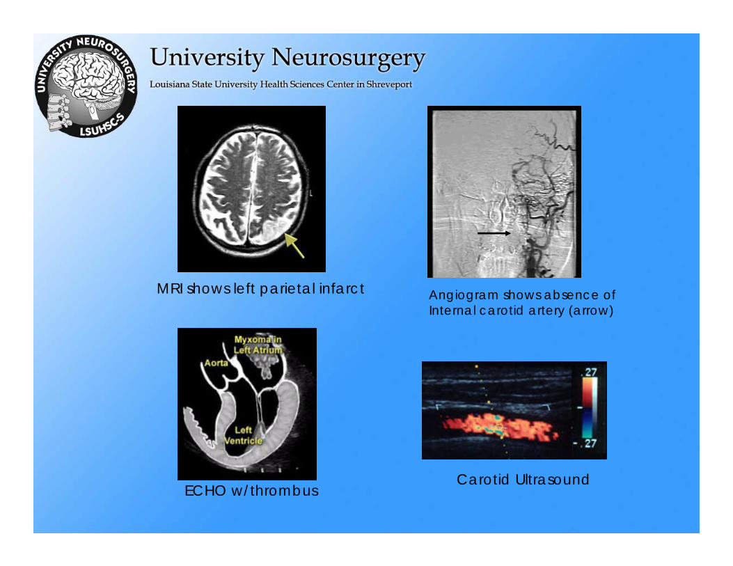
MRI shows left parietal infarct Angiogram shows absence ofInternal carotid artery (arrow)
Carotid UltrasoundECHO w/thrombus

Carotid Endarterectomy
• A candidate is a patient with the following findings
– Case study 1: this patient had findings of a right middle cerebral artery stroke, with carotid artery stenosis.
http://www3.us.elsevierhealth.com/Mosby/Stark-Bradley/C88_055c.html

Carotid Endarterectomy
– Case study 2: Intraoperative plaque and the left carotid bifurcation anatomy are pictured.
Plaque
Anatomy
http://www.healthatoz.com/healthatoz/Atoz/ency/endarterectomy.htmlhttp://gensurg.co.uk/cea%20-%20operation.htm

Stroke• Lacunar Stroke
– A stroke resulting from small vessel occlusion (“lake”) –shown as a small hypodensity on CT scan (arrows)
http://www.texmed.org/cme/spt/hours1and2/stroke20.asp

Stroke• Hemorrhagic Stroke
– Hypertensive hemorrhage:• Typically results in bleed into:
– Basal ganglia– Thalamus– Pons– Cerebellum
• Treatment includes blood pressure control (but not too low), and if superficial can remove blood clot
• Surgical procedures performed in only 10% of cases.
http://www.emedicine.com/radio/topic99.htm

Stroke - Hemorrhagic– Aneurysmal Subarachnoid Hemorrhage
• May cause symptoms by mass effect compression, e.g. III n. palsy in posterior communicating aneurysm, or by bleed (typically subarachnoidhemorrhage)
• 8% of us have an aneurysm; thankfully, most never rupture
• After an aneurysm ruptures, 1/3 people Die, 1/3 are significantly disabled, and 1/3 recover reasonably
• Treatment includes coiling or clipping• A delayed risk (occurring from 3-21 days, but most
commonly 7-10 days) after bleed is Vasospasm –due to unknown substances from blood, a vessel spasms and can cause serious stroke-like symptoms
• Vasospasm is treated by hypervolemia, hypertension, and hemodilution (Triple-H), +/-angioplasty

Stroke – Hemorrhagic• Arteriovenous Malformation
– Abnormal tangle of blood vessels which shunts blood from arteries to veins without capillaries
– 4%/year risk of bleed, usually in lobar locations
– Treatment is surgical excision or Gamma Knife
http://www.vh.org/adult/provider/radiology/RCW/1024962/T2MRI.html
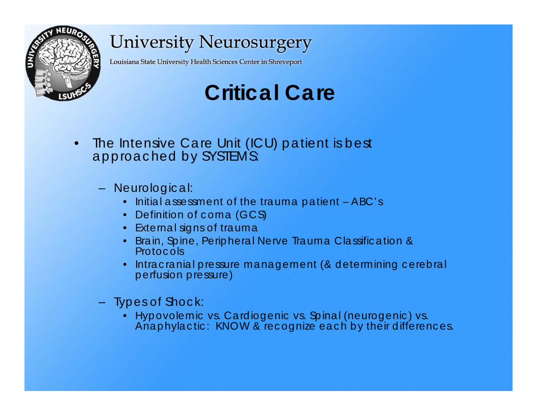
Critical Care
• The Intensive Care Unit (ICU) patient is best approached by SYSTEMS:
– Neurological:• Initial assessment of the trauma patient – ABC’s• Definition of coma (GCS)• External signs of trauma• Brain, Spine, Peripheral Nerve Trauma Classification &
Protocols• Intracranial pressure management (& determining cerebral
perfusion pressure)
– Types of Shock:• Hypovolemic vs. Cardiogenic vs. Spinal (neurogenic) vs.
Anaphylactic: KNOW & recognize each by their differences.
Critical Care
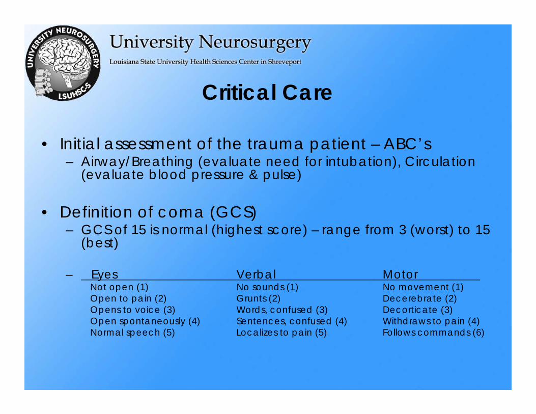
• Initial assessment of the trauma patient – ABC’s– Airway/Breathing (evaluate need for intubation), Circulation
(evaluate blood pressure & pulse)
• Definition of coma (GCS)– GCS of 15 is normal (highest score) – range from 3 (worst) to 15
(best)
– Eyes Verbal MotorNot open (1) No sounds (1) No movement (1)Open to pain (2) Grunts (2) Decerebrate (2)Opens to voice (3) Words, confused (3) Decorticate (3)Open spontaneously (4) Sentences, confused (4) Withdraws to pain (4)Normal speech (5) Localizes to pain (5) Follows commands (6)
Critical Care
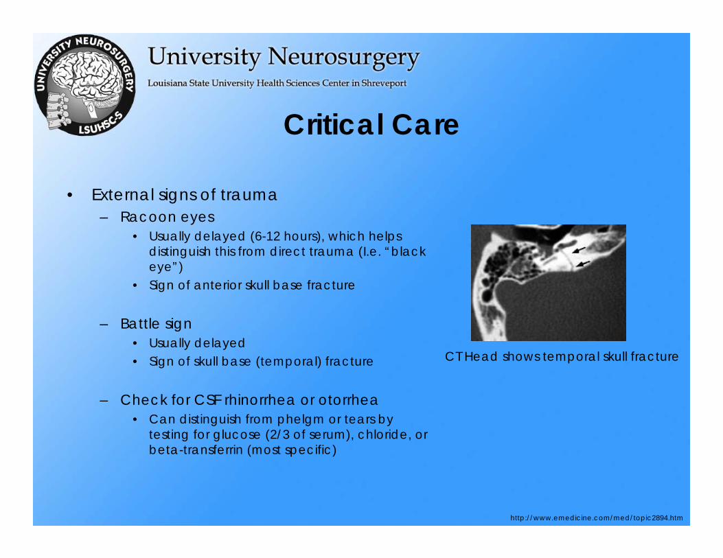
• External signs of trauma– Racoon eyes
• Usually delayed (6-12 hours), which helps distinguish this from direct trauma (I.e. “black eye”)
• Sign of anterior skull base fracture
– Battle sign• Usually delayed• Sign of skull base (temporal) fracture
– Check for CSF rhinorrhea or otorrhea• Can distinguish from phelgm or tears by
testing for glucose (2/3 of serum), chloride, or beta-transferrin (most specific)
CT Head shows temporal skull fracture
http://www.emedicine.com/med/topic2894.htm
Critical Care
Traumatic Brain Injury
– ClassificationSevere – GCS 3-8 Moderate – GCS 9-11
Mild – GCS 12-15
– Protocols for treatmentEstablish ABC’sNeed for CT scan if h/o loss of consciousness, external signs of trauma, or
neurological deficitGCS 8 or below (severe brain injury) may need intracranial pressure (ICP)
monitorDO NOT use D5-containing fluids – Glucose is TOXIC to brainEnsure pCO2 is NOT HIGH, as this can increase ICPIf CT scan shows bleed or comatose patient not improved, repeat CT
Head to f/u
Critical Care
• Spine Trauma– Classification
• Determine if a fracture is “stable or unstable”– Any subluxation is considered unstable, and immobilizatoin
is key– The spine can be developed into 3 columns – if the injury
affects 2 of 3 columns the fracture may be unstable and require surgical fixation
– Flexion-Extension films, in a mild fracture, when okayed by Neurosurgery team, can help detect instabiltiy
– A fracture of the lateral mass of C2-C6 should promote worry about vertebral artery injury, and an MRA should be done at minimum
– Protocols for Treatment
Spine Trauma• Protocols for Treatment
– Clearing a Cervical Collar after trauma– The C-collar can be removed WITHOUT FILMS, INDEPENDENT OF
MECHANISM, if the patient is» Awake, alert, and has had no medications to influence sensorium» Has no distracting injury (e.g. femur fracture)» Is completely non-tender and has full range of motion of the
cervical spine– If all of these are true but the patient is tender, s/he should undergo
AP/Lateral/Odontoid films, plus a CT scan with reconstruction to r/o fracture if no fracture, then flexion/extension C-spine films if all films normal, can remove C-collar
– Any patient with spinal cord injury within ** 8 hours ** must receive a steroid protocol of methylprednisolone for 24 hours

Peripheral Nerve Trauma
• Peripheral Nerve Trauma
– Classification• Neuropraxia: crush injury of nerve, usually recovers
without intervention • Axontemesis: crush injury of axon, with Wallerian
degeneration – axon must grow back @ 1mm/day• Neurontmesis: Nerve functionally severed – if no return
of function in 6 months, may need surgical apposition
– EMG/NCV studies indicate nerve continuity/function – can only get these after 2 weeks post-injury (due to artifact)
Peripheral Nerve Trauma
– Protocols for treatment• If any function present, do not intervene surgically• If no function present, do not intervene surgically, but wait for 6
months get EMG/NCV studies then, and if STILL no function (conduction) then can intervene to surgically splice nerve
• Exception to this is stab wound (as discussed)
– Case Study: Stab vs. Gunshot wound resulting in peripheral nerve injury
• Following a stab wound, nerve re-approximation can be IMMEDIATELY performed
• Following a gunshot wound, however, nerve reapproximationwill not help as a greater portion of the nerve than immediatelyknown may die; also, sometimes the injury can be due to concussive effect of the bullet, so waiting is best
Critical Care
• What is the normal intracranial pressure (ICP)?– Normal ICP is 5-20 cm H20, or 5-15 mm Hg
• Intracranial pressure management (& determining cerebral perfusion pressure)– Definition of CPP
• CPP = MAP minus ICP– How do you figure out MAP?
• MAP = 1/3 (systolic BP) plus 2/3(diastolic BP) for a “normal blood pressure” of 120/80, the MAP is 93
• So, for a normal person, normal CPP is 93 minus 10, or 83
Critical Care
• What causes ICP to increase?• Anxiety/Stress, elevated pCO2, brain edema, bleed/other mass lesions
• What interventions can be used to decrease ICP for each of these reasons?– Mannitol, an osmotic diuretic, can help acutely– The Monro-Kelly Doctrine states that there are 3 things in the
head – BRAIN, CEREBROSPINAL FLUID, & BLOOD– Of these, we can reduce the size of one or more
components to decrease intracranial pressure• First choice – cerebrospinal fluid via ventriculstomy• Second choice – reduce the amount of blood slightly (but not enough
to cause a stroke) – since pCO2 controls cerebral blood flow, a pCO2 of 30-35 is maintained (normal is 40; less than 30 can cause stroke)
• Final choice – which is controversial, but performed in the posterior fossaoccasionally, is removal of damaged brain to reduce ICP (last resort)
Critical Care
• Shock– Definition: Critical lack of perfusion to tissues– Types of Shock:
• Hypovolemic vs. Cardiogenic vs. Spinal (neurogenic) vs. Anaphylactic: Recognize features and treatment for each.
– HYPOVOLEMIC: HR high, BP low, CVP low, Hemoglobin low– CARDIOGENIC: HR high, BP low, CVP high– SPINAL (NEUROGENIC): HR low, BP low– ANAPHYLACTIC: BP low, HR high, Hemoglobin not low (usually + h/o
exposure)
– Treatment for hypovolemic shock and spinal shock is volume first HEART RATE indicates which type is occuring, and if heart rate still low, needs PRESSORS to correct vascular tone

Infections
• Outline
– Meningitis– Brain abscess– Subdural empyema– Epidural abscess– Osteomyelitis– Soft tissue infection overlying central nervous system
structure– Special consideration: The immunocompromised
patient (e.g. HIV+ or s/p transplant)
CNS Infections
• Present in a variety of ways• May result in death or severe morbidity if not treated
promptly• Usually occur from hematogenous spread• Can also occur from direct extension from adjacent bone,
soft tissue or sinsuses• Pathogens include bacteria, viruses, and fungi• Most common: Acute bacterial meningitis and cerebral
abscess

Infections
• Meningitis– Serious life threatening infection of meninges– Viral meningitis is more common, but is usually self-limiting– Common infective organisms are related to the patient’s age
and any underlying disease– A few organism account for most of the cases but a wide
variety may be responsible.• Neonate (0-4 weeks) - Group B streptococcus, E. Coli• 4-12 weeks - Group B streptococcus, Streptococcus pneumonia,
Salmonella, H. Influenza, Listeria Monocytogenes• 3 months-5 years - H. Influenza, Streptococcus pneumonia,
Neisseria meningitidis• Over 5 years and adults - Streptoccous pneumonia, Neisseria
Meningitidis
Meningitis
• Major presenting symptoms include:– High fever– Meningismus including headache, neck stiffness, photophobia and
vomiting– As illness progresses, mental status changes can ensue– Results from septic effect on brain, septic thrombosis and possible
infarction
• Infants and neonates: Listlessness and irritability; fever and neck stiffness may be absent; Meningococcal infections frequently have a coexisting petechial rash.
• Complications of bacterial meningitis: Cerebral edema, Seizures, Hydrocephalus, Subdural empyema, Brain abscess
• Original source of infection may be evident, e.g. sinusitis, bacterial endocarditis, otitis media, mastoiditis
Meningitis
• Diagnosis is made by examination of the CSF by lumbar puncture
• If there are signs of increased intracranial pressure or focal neurological signs, a preceding CT scan of the head is needed to exclude a mass lesion
• CSF features of meningitis:– Raised cell count (5 - 10,000)– Elevated protein ( >45 mg/dl)– Decreased glucose (<30 mg/dl)– Positive gram stain
• Treatment:– High dose antibiotics should be commenced immediately and then
tailored based on culture results– Duration of antibiotics 5-14 days, or longer for some cases

Brain Abscess• Brain abscess
– Can occur at any age, single or multiple, usually supratentorial– Can result from hematogenous or direct spread from paranasal
sinuses – MAY OCCUR in ABSENCE of MENINGITIS– May present with fever, malaise, seizures, neurological deficit– Appear as ring-enhancing regions on MRI, usually at grey-white
junction – starts as cerebritis then becomes encapsulated– RUPTURE INTO VENTRICLE results in >90% MORTALITY
Brain Abscess• Diagnosis
– Blood cultures usually negative, LP usually negative and risk, so BIOPSY & CX
• Offending Organism:– Preantibiotic era: Staph. Aureus and Streptococci– Today, Streptococci, specifically Strep. milleri, are isolated from 80%
of brain abscesses. Major habitat of Strep. milleri is the alimentary tract, including the mouth and dental plaques
– Otogenic abscesses usually yield a mixed flora, including bacteroides, various streptococci and enterobacter species
– Staph aureus is often the patbogen in abscesses resulting from trauma
• Treatment:– If > 2 mm., may need to evacuate/aspirate – otherwise continue Abx
(Vanco + chloramphenicol or cefotaxime and metronidazole); repeat imaging to eval. resolution
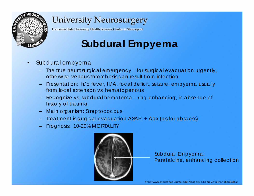
Subdural Empyema• Subdural empyema
– The true neurosurgical emergency – for surgical evacuation urgently, otherwise venous thrombosis can result from infection
– Presentation: h/o fever, H/A, focal deficit, seizure; empyema usually from local extension vs. hematogenous
– Recognize vs. subdural hematoma – ring-enhancing, in absence of history of trauma
– Main organism: Streptococcus– Treatment is surgical evacuation ASAP, + Abx (as for abscess)– Prognosis: 10-20% MORTALITY
Subdural Empyema:Parafalcine, enhancing collection
http://www.medschool.lsumc.edu/Nsurgery/subempy.html#anchor958872

Epidural Abscess
• Epidural abscess– Sx: Back pain, fever, spine tenderness– Risk Factors: Diabetes, IVDA– Presentation: dependent upon region
may result in neurological decline– Treatment: MUST take for surgical
evacuation if patient has worsening neurological function; RARELY can observe small epidural abscess, if no neurological decline. Need Abx.
– If associated with osteomyelitis may need surgical stabilization procedure also
– Usually due to hematogenous spread (S. aureus) can biopsy (CT-guided) for ID
http://www.aafp.org/afp/20020401/1341.html
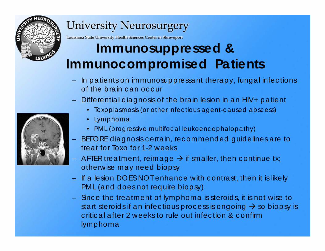
Immunosuppressed & Immunocompromised Patients
– In patients on immunosuppressant therapy, fungal infections of the brain can occur
– Differential diagnosis of the brain lesion in an HIV+ patient• Toxoplasmosis (or other infectious agent-caused abscess)• Lymphoma• PML (progressive multifocal leukoencephalopathy)
– BEFORE diagnosis certain, recommended guidelines are to treat for Toxo for 1-2 weeks
– AFTER treatment, reimage if smaller, then continue tx; otherwise may need biopsy
– If a lesion DOES NOT enhance with contrast, then it is likely PML (and does not require biopsy)
– Since the treatment of lymphoma is steroids, it is not wise to start steroids if an infectious process is ongoing so biopsy is critical after 2 weeks to rule out infection & confirm lymphoma
References
• http://www.strokecenter.org/pat/diagnosis/• Greenberg MS: Handbook of Neurosurgery, 4th ed., 1997.

The End