stress redistribution in individual ultrathin strained ... · pdf filestress redistribution in...
TRANSCRIPT

Stress redistribution in individual ultrathin strained silicon nanowires: a high-resolution
polarized Raman study
This article has been downloaded from IOPscience. Please scroll down to see the full text article.
2013 New J. Phys. 15 053042
(http://iopscience.iop.org/1367-2630/15/5/053042)
Download details:
IP Address: 132.207.45.217
The article was downloaded on 29/05/2013 at 20:37
Please note that terms and conditions apply.
View the table of contents for this issue, or go to the journal homepage for more
Home Search Collections Journals About Contact us My IOPscience

Stress redistribution in individual ultrathin strainedsilicon nanowires: a high-resolution polarizedRaman study
Alvarado Tarun1, Norihiko Hayazawa1,4,5,Maria Vanessa Balois1,4, Satoshi Kawata1, Manfred Reiche2
and Oussama Moutanabbir1,2,3,5
1 Near-field Nanophotonics Research Team, RIKEN, The Institute of Physicaland Chemical Research, 2-1 Hirosawa, Wako, Saitama 351-0198, Japan2 Max Planck Institute of Microstructure Physics, Weinberg 2, Halle (Saale)D-06120, Germany3 Departement de Genie Physique, Ecole Polytechnique de Montreal, Montreal,CP 6079, Succursale Centre-Ville, Montreal, Quebec H3C 3A7, Canada4 Department of Electronic Chemistry, Graduate School of Science andEngineering, Tokyo Institute of Technology, 4259 Nagatsuta, Midori-ku,Yokohama, Kanagawa 2268502, JapanE-mail: [email protected] and [email protected]
New Journal of Physics 15 (2013) 053042 (15pp)Received 7 January 2013Published 28 May 2013Online at http://www.njp.org/doi:10.1088/1367-2630/15/5/053042
Abstract. Strain nano-engineering provides valuable opportunities to createhigh-performance nanodevices by a precise tailoring of semiconductor bandstructure. Achieving these enhanced capabilities has sparked a surge of interest incontrolling strain on the nanoscale. In this work, the stress behavior in ultrathinstrained silicon nanowires directly on oxide is elucidated using background-free, high-resolution polarized Raman spectroscopy. We established a theoreticalframework to quantify the stress from Raman shifts taking into account theanisotropy associated with the nanowire quasi-one-dimensional morphology.The investigated nanowires have lateral dimensions of 30, 50 and 80 nm anda length of 1 µm top-down fabricated by patterning and etching 15 nm thickbiaxially tensile strained silicon nanomembranes generated using heteroepitaxy
5 Authors to whom any correspondence should be addressed.
Content from this work may be used under the terms of the Creative Commons Attribution 3.0 licence.Any further distribution of this work must maintain attribution to the author(s) and the title of the work, journal
citation and DOI.
New Journal of Physics 15 (2013) 0530421367-2630/13/053042+15$33.00 © IOP Publishing Ltd and Deutsche Physikalische Gesellschaft

2
and ultrathin layer transfer. The concern over the contribution of Ramanscattering at the nanowire 〈110〉 oriented sidewalls is circumvented by preciselyselecting the incident polarization relative to the sidewalls of the nanowire,thus enabling an accurate and rigorous analysis of stress profiles in individualnanowires. Unlike suspended nanowires, which become uniaxially strained as aresult of free surface-induced relaxation, we demonstrated that stress profiles insingle nanowires are rather complex and non-uniform along different directionsdue to the oxide–nanowire interface. As a general trend, higher stresses areobserved at the center of the nanowire and found to decrease linearly as afunction of the nanowire width. Using multi-wavelength high-resolution Ramanspectroscopy, we also extracted the stress profiles at different depths in thenanowire. The residual stress in the top ∼10 nm of the nanowire was foundto be nearly uniaxial and increase from the edge toward the center, whichremains highly strained. In contrast, the average stress profiles measured overthe whole nanowire thickness exhibit different behavior characterized by aplateau in the region ∼200 nm away from the edges. Our observations indicatethat the lattice near the newly formed free surface moves inwards and dragsthe underlying substrate leading to a complex redistribution of stress. Thisnanoscale patterning-induced relaxation has direct implications for electricaland mechanical properties of strained silicon nanowires and provides myriadopportunities to create entirely new strained-engineered nanoscale devices.
Contents
1. Introduction 22. Experimental details 3
2.1. Sample preparation . . . . . . . . . . . . . . . . . . . . . . . . . . . . . . . . 32.2. Experimental configuration . . . . . . . . . . . . . . . . . . . . . . . . . . . . 4
3. Analytical methods for stress calculation in nanowires 64. Results and discussion 85. Conclusion 14Acknowledgments 14References 14
1. Introduction
Silicon (Si) nanowires are quasi-one-dimensional nanostructures providing a wealth of oppor-tunities to implement a variety of nanoscale technologies [1–5]. Particularly, nanowire-basedarchitectures have been introduced to extend the lifetime of Si traditional electronics byachieving lower power consumption, higher performance and scalable devices to respond to therelentless course toward the miniaturization imposed by Moore’s law [6]. In this perspective,to overcome the limitations faced in the two-dimensional planar devices, transistors withnanowire-like channels are currently used in the fabrication of high-performance microproces-sors and memory devices for wireless systems such as mobile phones and radars [6]. Unlikefreestanding nanowire-based devices [7], these new technologies employ nanowires havingthree free facets while the fourth one forms an interface with the underlying substrate [5, 6].
New Journal of Physics 15 (2013) 053042 (http://www.njp.org/)

3
Both of these categories of nanowires suffer, however, from degradation in the carrier velocityas a result of quantum confinement and scattering at the sidewalls, which hinders the deviceperformance [8, 9]. Stress nano-engineering has emerged as a powerful stratagem to alleviatethese limitations and extend the capabilities of nanowire-based electronics [7].
Strain engineering has been used in the microelectronics industry since the 90 nmtechnology node. The main approach currently employed consists of depositing stressor layerson top of transistors. However, as the dimensions of nanowire-based transistors are becomingsmaller than the required thickness of such an overlayer, it is difficult to implement this approachinto smaller nodes or other nanowire-based architectures. Recently, it was demonstrated thatultrathin, globally strained silicon layers (nanomembranes) are the material of choice to generatestrained Si nanowires using top-down nanofabrication processes [7, 10, 11]. Interestingly, uponnanoscale patterning of biaxial strained nanomembranes—a crucial step in the fabrication ofstrained nanowires—the formation of free surfaces induces a local relaxation of strain due torearrangement of lattice atoms near the newly formed edges [10, 12–16]. In general, the extent ofthis phenomenon depends on dimension and geometry [12–16]. However, it is widely admittedthat due to nanowire geometry (i.e. the high aspect ratio) the post-patterning stress becomesuniaxial, which means that the stress is fully relaxed along the width of the nanowire [7, 11].However, as demonstrated in this work, this is not the case for nanowires fabricated directlyon oxide. More precisely, the nanowire–substrate interface makes the strain redistributionrather complex as one of the four facets is stabilized by the underlying oxide layer. Exploringand understanding this subtle but important phenomenon is crucial for accurate strain nano-engineering and a precise prediction of the performance of strained Si nanowire-based devices.
In order to accurately probe the evolution of strain in nanoscale, we have developeda method of high-resolution polarized Raman spectroscopy that allows the analysis of theindividual contributions of longitudinal-optical (LO) and transverse-optical (TO) phonons andtheir profiles in a single nanowire [10]. However, the use of a single excitation wavelength(532 nm) in that work can only permit the measurement of phonon profiles averaged over thewhole thickness of a nanowire. Moreover, while simultaneous detection of LO and TO phononmodes can directly characterize anisotropic stress relaxations at each position, TO phonondetection is still challenging due to its strong sensitivity to the laser polarization relative tosample geometry and thus the allowed LO phonon is always mixed. Herein, to circumvent theselimitations, we employ two excitation wavelengths with linear polarizations corresponding todifferent penetration depths in silicon, thereby achieving LO phonon detection as a local stressprobe at different depths within a single nanowire. We also discuss and take into considerationthe effects of the scattering at the nanowire sidewalls on the obtained Raman data and presenta method for an accurate determination of stress in nanoscale structures. The effect of thenanowire lateral dimension on the stress relaxation is also addressed.
2. Experimental details
2.1. Sample preparation
The Si nanowires were generated by patterning and etching a 15 nm thick biaxially tensile-strained Si nanomembrane by combining electron beam lithography and dry reactive ionetching. The strained nanomembrane was synthesized by growing epitaxially an ultrathinlayer of Si on a ∼500 nm thick Si0.84Ge0.16 relaxed buffer layer grown on Si (001) substrateusing reduced pressure chemical vapor deposition. The obtained strained-Si/Si0.84Ge0.16/Si
New Journal of Physics 15 (2013) 053042 (http://www.njp.org/)

4
heterostructure was subsequently capped by an SiO2 layer deposited by plasma-enhancedchemical vapor deposition. A second substrate was prepared by deposition of a ∼120 nm thickGe layer on Si (001) using solid-source molecular beam epitaxy. This wafer was also covered bya ∼200 nm thick SiO2 layer. The ultrathin strained Si layer was transferred from the first waferonto the second one using direct wafer bonding followed by ion-induced slicing and selectivechemical etching of SiGe leading to strained Si/SiO2/Ge/Si heterostructure. The intermediateGe layer was introduced to prevent the excitation laser from reaching the Si handle substrate inthe case of excitation by the laser in the visible range or when the region between nanowiresis exposed to the laser. Thus, the background from the underlying Si substrate is eliminatedand only the Raman signal from the strained nanowire is detected, thereby achieving a highersensitivity to local phonons, which is critical for a precise analysis of stress. Ordered arrays ofnanowires having a fixed length of 1000 nm and widths of 30, 50 or 80 nm were fabricated.The nanowire arrays were patterned on a negative resist using electron-beam lithography.Reactive ion etching was applied to transfer the pattern to the strained layer. The nanowiresare aligned along the 〈110〉 direction and separated by 500 nm from each other to allow theexposure of a single nanowire to the laser beam during Raman analysis. The 〈110〉 nanowireswere investigated because of their practical importance as high-performance channels for fieldeffect transistors. The etching was performed at −60◦C using a mixture of SF6 (100 sccm)and O2 (5 sccm) with a relatively low power of 40 W. These conditions were optimized toeliminate possible damage during the etching process. Here the chemical reactivity is dominant.High-resolution transmission electron microscopy investigations (not shown here) confirmedthe absence of any damage near the newly formed edges.
2.2. Experimental configuration
Figure 1 displays typical atomic force microscopy (top) and scanning electron microscopy(bottom) images of arrays of nanowires having a width of 80 and 30 nm, respectively. Thefigure also depicts the sidewalls orientation of the nanowires relative to the polarization ofthe excitation laser. The long axis of the nanowire is aligned along the 〈110〉 direction. Thecrystal and the sample coordinate are symbolized as x,y,z and x ′, y′, z′, respectively. Portonotation, z(x ′x ′)z or z(y′y′)z, is used to represent the polarization conditions. The laser beam(λ = 442 or 355 nm) passes through a linear polarizer and a half-wave plate. The linearpolarizer sets the incoming polarization from the laser. The half-wave plate is used to alignthe incident polarized beam parallel (x ′) or perpendicular (y′) to the nanowire long axis. Thebeam is then expanded and focused onto the nanowires using an oil-immersion objective lens(numerical aperture = 1.4, ×125). The surface of the strained Si is directly immersed in oil. Thisconfiguration improves the laser focusing and minimizes index mismatch-induced sphericalaberrations. According to the Rayleigh criterion, the diameter of the laser spot is λ/2. Thebackscattered Raman signal is collected by the same objective lens and passes through a pinhole(φ = 50 µm), which leads to an edge filter to block the strong Rayleigh signal in order for thebackscattered Raman signal to be detected. The backscattered light goes through an analyzerbefore reaching a spectrometer (grating = 1800 g mm−1, slit width = 50 µm) equipped with athermo-electronically cooled charge coupled device (CCD) camera. A single Lorentz functionwas fitted to the Si Raman peak for all spectra to accurately determine its frequency, widthand intensity. The system spectral resolution is 0.02 nm and the accuracy of our analysis afterLorentzian fitting is ∼0.02 cm−1. The samples with nanowire arrays were mounted on an x ′–y′
New Journal of Physics 15 (2013) 053042 (http://www.njp.org/)

5
Figure 1. Polarized Raman setup in backscattering configuration showingthe AFM image of an array of nanowires with d = 80 nm (top). Top viewschematic of the nanowire with crystal (x,y,z) and sample (x ′, y′, z′) coordinatesdepicting that the x ′- and y′-axes are parallel to the long axis of the nanowire(inset). Shown is the SEM image of an array of nanowires with d = 30 nm(bottom).
translation stage and were scanned with a 25 nm step while exposed to the focused laser beam.At each step, a Raman spectrum was recorded. To prevent laser heating, Raman spectra wereacquired at 1.0 mW power with a 5 s exposure time using the 442 nm lasers. An exposure of5 min and a power of 5 µW were used for the 355 nm laser. Note that due to the dependence ofthe Si absorption coefficient on photon frequency [17], the laser penetration depth (= λ/4πk)is calculated to be ∼10 nm at 355 nm and ∼168 nm at 442 nm where λ and k are the wavelengthand the imaginary part of the refractive index, respectively. The latter permits probing the stressprofiles for the total thickness of the nanowire, whereas the shallower penetration allows theanalysis of the top 10 nm of the nanowire.
New Journal of Physics 15 (2013) 053042 (http://www.njp.org/)

6
3. Analytical methods for stress calculation in nanowires
The local stress in the Si lattice is obtained from Raman shifts by solving the well-known secularequation. In the case of the unpatterned nanomembrane, the equi-biaxial (σxx = σyy = σ0) modelof stress-induced Raman shift for doublet (1ωxy: transverse optical TO phonons) and singlet(1ωz: longitudinal optical LO phonons) is [18]
1ωxy = [p(S11 + S12) + q(S11 + 3S12)] σ0/2ω0 = −3.42 × σ0, (1)
1ωz = [pS12 + q(S11 + S12)] (σxx + σyy)/2ω0 = −4.60 × σ0, (2)
where 1ωxy = ωε-Si − ωSi and 1ωz = ωε-Si − ωSi are expressed in the Raman frequency in eachmode in the presence (ωε-Si) and absence (ωSi) of stress. The S11 = 7.68 and S12 = −2.14(in GPa) are the elastic compliance tensor elements, while p = −1.85 and q = −2.31 are thephonon deformation potentials for bulk Si at room temperature [19].
The stress relaxation ratio upon patterning at any position in the nanowire is calculatedusing R(x, y) = 1 − σnanowire(x, y)/σ0, where σ0 and σnanowire correspond to the initial stressbefore nanopatterning and the measured stress in nanowires, respectively [22]. The initialstress, σ0, in the nanomembrane is calculated from the measured Raman shift using equation(2). However, it is important to note that equation (2) cannot be used for the extraction ofstress in nanowires, σnanowire, mainly because the assumption that shear stress is zero used toobtain the stress is no longer valid as the stress is no longer bi-isotropic in the nanowire dueto geometrical constraints and non-uniform rearrangements of atoms near the newly formedfree surfaces. In fact, in a nanowire the shearing strain terms at the crystal coordinate are nolonger zero due to anisotropic strain relaxation along the nanowire axes. In order to obtain thenanowire stress, we first calculate the strain and stress components in the sample coordinateswhere shearing terms are all zero due to the symmetry of the structure at the sample coordinatesystem and then transform them into the crystal coordinate [20]. At the center of the nanowire,the strain component along the nanowire length (ε′
xx(x ′, d)) has almost the value of the originalmembrane, ε0, but the stress along the width (σ ′
yy(x ′, d)) will be dramatically relaxed due tothe reduced dimension in this direction. d represents the width of nanowires. The shear straincomponent becomes zero in the middle of the nanowire, and the stress component along z′ isequal to zero due to free surface boundary conditions; the strain and stress tensors are thenexpressed as [20]
{ε′(x ′, d)}=ε′
xx(x ′, d)
1 0 0
0sα(x ′, d)C11+C2
12−C11(C12−H/2)
C11(C11+H/2)−C212
0
0 0−sα(x ′, d)C12−2C12C44
C11(C11+H/2)−C212
, (3)
{σ ′(x ′, d)} =
C ′
11ε′
xx(x ′, d) + C ′
12ε′
yy(x ′, d) + C ′
13ε′
zz(x ′, d) 0 00 α(x ′, d)σ0 00 0 0
, (4)
where α(x ′, d) = σ ′
yy(x ′, d)/σ0, s = C11 + C12 −(2C2
12/C11
)and H = 2C44 + C12 − C11. The
elastic constants C11 = C22 = 166 GPa, C12 = C21 = 64 GPa, C44 = 79.6 GPa, C ′
12 = C12
− 0.5H = 35.4 GPa, C ′
11 = C11 + 0.5H = 194.6 GPa and C ′
13 = C12 = 64 GPa are obtainedfrom [20, 21]. The constant α(x ′, d) varies between 0 and 1 and depends on the diameter of
New Journal of Physics 15 (2013) 053042 (http://www.njp.org/)

7
the nanowire. At the limits, α(x ′, d) equals 0 for a uniaxial stress and 1 for a bi-isotropic stress.After substitution, the strain tensor in the sample coordinate reduces to
{ε′(x ′, d)} = ε′
xx(x ′, d)
1 0 00 1.06α(x ′, d) − 0.06 00 0 −0.41α(x ′, d) − 0.36
(5)
and after transformation to the crystal coordinate system:
{ε(x ′, d)}=ε′
xx(x ′, d)
0.53α(x ′, d)+0.47 −0.53α(x ′, d)+0.53 0−0.53α(x ′, d)+0.53 0.53α(x ′, d)+0.47 0
0 0 −0.41α(x ′, d)−0.36
. (6)
Using the strain components above, the solution to the well-known secular equation for the LOphonon mode is expressed as
1ωz =ωε-Si − ωSi =ωSi
2[pεzz(x ′, d) + 2qεyy(x ′, d)], (7)
1ωz = ωε-Si − ωSi =−ωSiε
′
xx(x ′, d)
2(1.70α(x ′, d) + 1.50). (8)
In equation (6), there are two unknown parameters, strain ε′
xx(x ′, d) along the nanowirelong x ′-axis and α(x ′, d), the stress relaxation factor along the nanowire width which wehave to extract from the Raman measurement. However, assuming we detect only the LOphonon mode by conventional backscattering geometry, it is impossible to determine the twounknown parameters from the single phonon mode. To circumvent this limitation and obtainthe stress in nanowires, it is widely accepted that the stress along the width of the nanowirecan be assumed to be fully relaxed, that is α(x ′, d) = 0 (see e.g. [13, 20, 21] and referencestherein). Herein, we demonstrate that this assumption is too simplistic leading to an inaccurateanalysis of stress. Using equation (5) and at α(x ′, d) = 0, the stress along the x ′-axis is givenby σ ′
xx(x ′, d) = 169.25 × ε′
xx(x ′, d). By substituting the strain components in equation (6),εzz = −0.36 × ε′
xx(x ′, d) and εxx = εyy = 0.47 × ε′
xx(x ′, d), into equation (7), the stress (in GPa)in the long axis can be expressed in terms of the measured Raman shift as
σ ′
xx(x ′, d) = −225.67 ×ωε-Si − ωSi
ωSi. (9)
In an alternative approach, we propose the introduction of an edge structure (edge parallel tothe y′-axis) consisting of a patterned nanomembrane with a practically semi-infinite dimensionalong y′ (>1 cm), which can be assumed as a hypothetical nanowire with an infinite width(d = ∞). Equation (8) indicates that there are two unknown parameters, ε′
xx(x ′, d) and α(x ′, d),while the experiment only provides the values of 1ωz (figure 2(b)). For α(x ′, d) 6= 0, ε′
xx(x ′, d)
can be determined using the reference structure (d = ∞). Here, the nanowire strain profile alongthe x ′-axis, ε′
xx(x ′, d), is comparable with the profile along the same direction in the referencestructure, ε′
xx(x ′, d = ∞) = ε′
xx(x ′, d), because at an identical length (1 µm) the effect of thenewly formed free surfaces should be similar in both systems. In the y′-axis, the referencesample is infinite and thus the initial strain is preserved (i.e. no relaxation does take place alongthis axis), suggesting that ε′
yy(x ′, d = ∞) is equivalent to the initial strain in the unpatternednanomembrane, ε0.
New Journal of Physics 15 (2013) 053042 (http://www.njp.org/)
8
Analogously to equation (8), we introduce an additional anisotropic stress relaxation factor,β(x ′, d = ∞) = σ ′
xx(x ′, d = ∞)/σ0, which represents the relative stress relaxation along the x ′-axis in the reference structure. Thus, the equation of Raman shift as a function of β(x ′, d = ∞)
can be written as
1ωz = ωε-Si − ωSi =−ωSiε
′
yy(x ′, d = ∞)
2(1.70β(x ′, d = ∞) + 1.50), (10)
where ε′
yy(x ′, d = ∞) is obtained from the Raman signal of the unpatterned nanomembrane,whereas 1ωz is obtained from Raman scans along the x ′-axis from the edge. By using theobtained values of ε′
yy(x ′, d = ∞) and 1ωz, we can determine β. Thus, the strain along thex ′-axis of the edge sample can be extracted similarly to equation (5) using β:
ε′
xx(x ′, d = ∞) = ε′
yy(x ′, d = ∞)[1.06β(x ′, d = ∞) − 0.06]. (11)
This strain is only a function of x ′ and varies from x ′= 0 (edge) to an x ′
= 0.5 µm aroundthe nanowire center. The strain component, ε′
yy(x ′, d = ∞) (parallel to the edge), remainsunchanged. This approach is analogous to the semi-infinitely wide line structures used in thetheoretical treatment developed earlier by Jain et al [20]. The measured Raman shifts from theedge structures were attributed mostly to the strain along x ′, ε′
xx(x ′, d = ∞), and the constantstrain component in the y′-direction, ε0, which is the same as the strain obtained from thenanomembrane. In the following, we test the validity of these two approaches.
4. Results and discussion
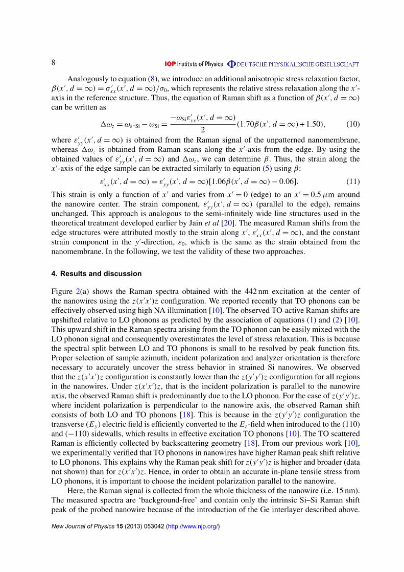
Figure 2(a) shows the Raman spectra obtained with the 442 nm excitation at the center ofthe nanowires using the z(x ′x ′)z configuration. We reported recently that TO phonons can beeffectively observed using high NA illumination [10]. The observed TO-active Raman shifts areupshifted relative to LO phonons as predicted by the association of equations (1) and (2) [10].This upward shift in the Raman spectra arising from the TO phonon can be easily mixed with theLO phonon signal and consequently overestimates the level of stress relaxation. This is becausethe spectral split between LO and TO phonons is small to be resolved by peak function fits.Proper selection of sample azimuth, incident polarization and analyzer orientation is thereforenecessary to accurately uncover the stress behavior in strained Si nanowires. We observedthat the z(x ′x ′)z configuration is constantly lower than the z(y′y′)z configuration for all regionsin the nanowires. Under z(x ′x ′)z, that is the incident polarization is parallel to the nanowireaxis, the observed Raman shift is predominantly due to the LO phonon. For the case of z(y′y′)z,where incident polarization is perpendicular to the nanowire axis, the observed Raman shiftconsists of both LO and TO phonons [18]. This is because in the z(y′y′)z configuration thetransverse (Ey) electric field is efficiently converted to the Ez-field when introduced to the (110)and (−110) sidewalls, which results in effective excitation TO phonons [10]. The TO scatteredRaman is efficiently collected by backscattering geometry [18]. From our previous work [10],we experimentally verified that TO phonons in nanowires have higher Raman peak shift relativeto LO phonons. This explains why the Raman peak shift for z(y′y′)z is higher and broader (datanot shown) than for z(x ′x ′)z. Hence, in order to obtain an accurate in-plane tensile stress fromLO phonons, it is important to choose the incident polarization parallel to the nanowire.
Here, the Raman signal is collected from the whole thickness of the nanowire (i.e. 15 nm).The measured spectra are ‘background-free’ and contain only the intrinsic Si–Si Raman shiftpeak of the probed nanowire because of the introduction of the Ge interlayer described above.
New Journal of Physics 15 (2013) 053042 (http://www.njp.org/)

9
Figure 2. (a) Raman spectra recorded at the center of nanowires with lateraldimensions of 30 nm (triangles), 50 nm (squares) and 80 nm (circles). TheRaman spectrum of the unpatterned strained Si is shown in solid black lines. Thevertical dashed line denotes the Si–phonon peak position in bulk (unstrained)Si. (b) Profile of the Raman peak for nanowires obtained under the z(x ′x ′)zconfiguration. The solid and dashed lines represent the Raman peak positionof the initial strained Si nanomembrane using the 442 nm (515.42 cm−1) and355 nm (515.75 cm−1) excitations, respectively. The error bars are smaller thanthe size of data points.
New Journal of Physics 15 (2013) 053042 (http://www.njp.org/)

10
Table 1. Summary of the Raman peak and spectral width at the center of singlenanowires.
Initial strainedlayer 15 nm thick (1xz = 515.42 cm−1 1t = 4.86 cm−1)
Nanowire width, d (nm) 80 50 30Peak, 1ωz (cm−1) 516.43 516.99 517.42Width, 1t(cm−1) 5.09 5.20 5.16
It should be noted that the dominant contribution to the measured Raman shift originated fromthe LO phonon mode from both the top (001) surface and the long axis sidewalls, {110}, basedon the Raman tensor calculation under x ′-polarized light. For the sake of comparison, the Ramanspectrum of the initial strained nanomembrane (solid black line) as well as the Si-phonon peakposition in bulk Si (broken vertical lines) is shown. Table 1 summarizes the Raman peak andspectral width for all the observed structures. As the width decreases, the Raman peaks shiftupward with respect to the position of the Si-phonon peak of the initial strained Si layer.
No significant broadening in the nanowire Si–Si Raman mode is, however, observed. It isnoticeable that the Si–Si peak position of the investigated nanowires is upshifted with respect toits position in the initial (1ωz = 515.42 cm−1) strained nanomembrane. This shift is indicativeof partial relaxation of stress during the process of nanowire patterning using reactive ionetching.
To obtain more insights into the complex redistribution of stress as nanowire dimensionshrinks, the profiles of the Raman shift for nanowires of varying width are plotted in figure 2(b).The nanowires were scanned along the x ′-axis with a step size of 25 nm using the z(x ′x ′)zconfiguration. The Raman shift for d = 30 nm using 355 nm (UV) excitation is also displayedin figure 2(b). At this excitation, only the top 10 nm of the nanowire thickness is probed.For comparison, the Raman shifts measured from the original membrane are indicated withsolid (442 nm) and dashed (355 nm) lines.
Figure 3 shows the stress profiles using (a) firstly, α(x ′, d) = 0 and (b) secondly,ε′
xx(x ′, d) = ε′
xx(x ′, d = ∞) approaches. Regardless of the approaches used, the profilesmeasured for all the nanowires display qualitatively similar behavior characterized by a morepronounced relaxation of stress near the nanowire edge. Moreover, as a general trend, theresidual stress profile shows plateau-like profiles in the region around the center. The breadthof this region is sensitive to the nanowire width and varies from ∼400 nm at d = 50 or 80 nmto ∼700 nm at d = 30 nm. The enhanced relaxation near the edges results from the additionalfree surface as compared to the rest of the nanowires (four versus three facets). As describedbelow, this constant stress value away from the edges can be attributed to the nearly constantand highly retained stress at the buried strained-Si/SiO2 interface.
The obtained initial stress from the nanomembrane using equation (2) was plotted (solidblack) to evaluate the validity of the first approach based on the assumption of a full relaxationalong the nanowire width (α(x ′, d) = 0). Strikingly, the stress profile along the long axisobtained using this approach is higher than the original stress in the nanomembrane. Thisdiscrepancy indicates that the assumption of a full relaxation along the width is inaccurate andthat stress is not uniaxial. In the following, we focus our analysis on the second approach.
New Journal of Physics 15 (2013) 053042 (http://www.njp.org/)

11
Figure 3. Stress profiles in single nanowires obtained under different excitationwavelengths using the first (a) and second (b) approaches described in the text.(c) Stress relaxation dependence estimated at the center of the nanowire as afunction of the nanowire diameter.
We first calculated the strain from equations (10) and (11) using the measured Raman shiftscanned from x ′
= 0 (edge) to x ′= 0.5 µm (towards the center) with semi-infinite width. The
strain values are then used in equations (8) and (9) to calculate the stress at each position.Figure 3(b) shows the obtained stress profiles σ ′
xx(x ′, d) and σ ′
yy(x ′, d). The calculated stressvalues along the x ′-direction are below the original stress of the membrane. It is also interestingto note that the profile of the stress along the x ′-direction of the nanowires is similar andwith stress values very close to stress of the edge sample, suggesting that stress is preservedat the center. Moreover, the stress along the x ′-direction at the edges (x ′
= 0 and 1.0 µm)is the same (σ ′
xx(x ′, d) ≈ 0.85 GPa) for all nanowires regardless of excitation wavelengths.This is not the case when a full relaxation is assumed along the width, which indicates thatthis assumption is fraught with large uncertainties. On the other hand, we can clearly seefrom the profile along the width, σ ′
yy(x ′, d), that the nanowire is still under a biaxial stress.
New Journal of Physics 15 (2013) 053042 (http://www.njp.org/)

12
The stress profile along the width is relatively constant through the whole length of the nanowireunlike along the long axis where pronounced relaxation is observed at the edges. Furthermore,a full relaxation of stress is only observed at the edge of the nanowire with d = 30 nm. Forthis set of nanowires, the stress at the center reaches a value of ∼200 MPa. This behavior isobserved for both excitation wavelengths. At d = 80 and 50 nm, the stress is well above thefully relaxed region (gray dashed zero line). This result indicates that the overall average stressin the nanowire is biaxial but not bi-isotropic. Note that at an excitation wavelength of 442 nmthe buried strained-Si/SiO2 interface is also probed. Here, the stress between 2006 x ′ 6 800 nmis comparable with the initial stress in the nanomembranes, whereas at 355 nm the initial stressis only preserved in the region 4506 x ′ 6 550 nm. Figure 3(c) exhibits the relaxation ratio atthe center of the nanowire as a function of nanowire diameter. It is noticeable that the extent ofthis relaxation along the width exhibits linear behavior as a function of the nanowire width:1σ ′
yy/σ0 = 1.01–5.77 × 10⁻³× d. The relaxation along the long axis is small and remains
unchanged with the nanowire width.Interestingly, the combination of two excitation wavelengths provides new insights into
the complex behavior of stress in nanowires. Figure 4 shows the depth dependence of stressprofiles along the two in-plane axes for a nanowire with a width of d = 30 nm under thez(x ′x ′)z condition using different excitation wavelengths (penetration depths), namely 442 nm(∼168 nm) and 355 nm (∼10 nm). It is noteworthy that Raman shifts measured using the 355 nmlaser are centered around higher wavenumbers as compared with those obtained using the442 nm excitation as shown in figure 2(b). Figure 4(a) also displays the original stress (lines)in the unpatterned nanomembrane obtained using the two-excitation wavelengths as well. Thesmall difference in the measured stress using the two excitations suggests that the region nearthe surface of the nanomembranes is slightly relaxed.
After patterning of the nanowires, the stresses are observed to be more relaxed in both the442 and 355 nm excitations. However, the interesting observation is that the stress relaxationbehavior as obtained by the 355 nm excitation decreases monotonously toward the center ofthe nanowire, which remains highly strained. At the 442 nm excitation, the stress relaxationplateaus in the region 2006 x 6 850 nm. This dissimilarity in stress behavior between the twoexcitation wavelengths is indicative of the non-uniform distribution of the in-plane stress alongthe nanowire thickness. More precisely, the fact that the stress measured using a laser with theshallower penetration depth is systematically smaller than the value averaged over the wholethickness provides direct evidence that the region near the nanowire–oxide interface maintainsa high level of stress. By combining the stress profiles measured at the 442 nm(σ ′15 nm
xx ) and355 nm σ ′10 nm
xx excitations, we extract the stress profile in the bottom 5 nm of the nanowire alongthe x ′-direction σ ′5nm
xx (σ ′5 nmxx = 3 × σ ′15 nm
xx − 2 × (σ ′10 nmxx )) (figure 4(a)). Similarly, the stress
profile along the y′-direction is depicted in figure 4(b). For the two in-plane axes, the obtainedstress profiles are qualitatively identical but remarkably different from the profiles measured forthe top 10 nm or over the whole nanowire thickness. Particularly, the part of the nanowire nearthe interface is under a higher stress especially in the region about 100–200 nm away from theedge, in qualitative agreement with finite element simulations (not shown). In this region, closestress values are recorded for the two in-plane directions, indicating that the interface with theoxide preserves almost the initial isotropy (i.e. a biaxial strain). Interestingly, the stress in thex ′-direction reaches values that are higher than the initial stress, indicating that the contractionof the lattice near the newly formed free surfaces is accompanied by a strong distortion closeto the interface with the oxide. This means that when the lattice near the edges moves inwards,
New Journal of Physics 15 (2013) 053042 (http://www.njp.org/)

13
(a)
Stre
ss,σ'
xx [
GPa
]
x’-Position [μm]
442nm
355nm
15nmTop 10nm
Bottom 5nmd = 30nm1.4
1.2
1.0
0.81.00.80.60.40.20.0
Stre
ss,σ'
yy [
GPa
]
x’-Position [μm]
15nmTop 10nm
Bottom 5nmd = 30nm1.0
0.8
0.6
0.4
0.2
0.0
-0.21.00.80.60.40.20.0
(b)
(c)
1.4
1.2
1.0
0.81.00.80.60.40.20.0
Stre
ss,σ'
edge
[G
Pa]
x’-Position [μm]
Edge
15nmTop 10nm
Bottom 5nm
Ref bottom 5nm
Figure 4. Stress profiles along the (a) x ′- and (b) y′-axis for nanowires witha width of d = 30 nm at different depths obtained using the two excitationwavelengths (penetration depths): 442 nm (∼168 nm) and 355 nm (∼10 nm). Thehorizontal solid (442 nm) and dashed (355 nm) lines marked the initial strainin the Si nanomembrane. (c) Stress profiles at different depths in the referencesample with a semi-infinite edge.
it drags the underlying substrate leading to a complex redistribution of stress. Above ∼200 nmaway from the edge, the stress decreases slightly along the nanowire length to stabilize around1.2 GPa. The decrease is more significant along the width. For this direction, the in-plane stressremains the same independently of the depth in the nanowire. Similar behavior is also observed
New Journal of Physics 15 (2013) 053042 (http://www.njp.org/)

14
for the edge sample as shown in figure 4(c). The only noticeable difference in the bottom 5 nmstress values along the long axis between the d = 30 nm nanowire and the reference sample islocated at the edges. The bottom 5 nm stress at the edge is ∼1.06 GPa, which is just above thestress measured with 355 nm excitation whereas for the d = 30 nm nanowire, the bottom 5 nmstress is 0.83 GPa. Toward the center, the bottom 5 nm stress values remain unchanged.
5. Conclusion
In summary, we presented a precise method to map the stress in individual strained Si nanowiresdirectly on oxide by using background-free, high-resolution polarized Raman spectroscopy withtwo excitation wavelengths. We also derived a theoretical framework to extract the stress fromRaman shifts, taking into account the stress anisotropy imposed by the nanowire quasi-one-dimensional morphology. We found that adjusting the incident polarization parallel to the longaxis of the nanowire is necessary for an accurate Raman probe. Our analysis demonstrates thatthe assumption of a full relaxation along the shortest dimension is not a valid approximationeven for a dimension as small as 30 nm. The obtained stress profiles unraveled the complexityof free surface-induced stress redistribution in patterned nanowires. Besides the anticipatedincrease in the importance of relaxation for narrower nanowires, the use of two excitationwavelengths provided unprecedented details on the behavior of stress. The obtained stressprofiles at different depths clearly demonstrate that the final stress distribution is rather complex,varying from nearly bi-isotropic to uniaxial within the same nanowire. This transition from aninitially uniform, isotropic stress to a heterogeneous distribution is the result of a simultaneouscontraction of the lattice near the newly formed free surfaces and a tension near the interfacewith the oxide. The fabrication and design of strained Si nanowire-based devices should takeinto account this non-uniform distribution of stress. Moreover, these observations provide thebasis for more accurate calculations and modeling of stress behavior in nanowires and theassociated changes in the physical properties.
Acknowledgments
AT acknowledges the financial support provided by the foreign postdoctoral researcher’s (FPR)program in RIKEN. NH gratefully acknowledges financial support from a Grant-in-Aid forYoung Scientist (A) No. 21686007 from The Ministry of Education, Culture, Sports, Scienceand Technology. OM acknowledges funding from NSERC, Canada Research Chair, and EcolePolytechnique de Montreal (PIED).
References
[1] Yan H, Choe H S, Nam S W, Hu Y, Das S, Klemic J F, Ellenbogen J C and Lieber C M 2011 Nature 470 240[2] Palacios T 2012 Nature 481 152[3] Kelzenberg M D, Boettcher S W, Petykiewicz J A, Turner-Evans D B, Putnam M C, Warren E L,
Spurgeon J M, Briggs R M, Lewis N S and Atwater H A 2010 Nature Mater. 9 239[4] Hochbaum A and Yang P 2010 Chem. Rev. 110 527[5] Colinge J-P et al 2010 Nature Nanotechnol. 9 225[6] Kuhn J K 2011 Proc. Int. Symp. on VLSI Technology, Systems and Applications (25–27 April 2011) pp 1–2[7] Hashemi P, Gomez L and Hoyt J L 2009 IEEE Electron Device Lett. 30 401
New Journal of Physics 15 (2013) 053042 (http://www.njp.org/)

15
[8] Uchida K, Koga J, Ohba R, Numata T and Takagi S-I 2001 IEDM Technical Digest 633[9] Gunawan O, Sekaric L, Majumdar A, Rooks M, Appenzeller J, Sleight J W, Guha S and Haensch W 2008
Nano Lett. 8 1566[10] Tarun A, Hayazawa N, Ishitobi H, Kawata S, Reiche M and Moutanabbir O 2011 Nano Lett. 11 4780[11] Minamisawa R A, Suess M J, Spolenak R, Faist J, David C, Gobrecht J, Bourdelle K K and Sigg H 2012
Nature Commun. 3 1096[12] Moutanabbir O, Reiche M, Hahnel A, Erfurth W, Gosele U, Motohashi M, Tarun A, Hayazawa N and
Kawata S 2010 Nanotechnology 21 134013[13] Ma F, Zhang T-W, Xu K-W and Chu P K 2011 Appl. Phys. Lett. 98 191907[14] Xiong G, Moutanabbir O, Huang X, Paknejad S A, Shi X, Harder R, Reiche M and Robinson I K 2011 Appl.
Phys. Lett. 99 114103[15] Poborchii V, Tada T, Usuda K and Kanayama T 2011 Appl. Phys. Lett. 99 191911[16] Moutanabbir O, Reiche M, Hahnel A, Oehme M and Kasper E 2010 Appl. Phys. Lett. 97 053105[17] Palik E D 1985 Handbook of Optical Constants of Solids (San Diego, CA: Academic)[18] De Wolf I 1996 Semicond. Sci. Technol. 11 139[19] Miyatake T and Pezzotti G 2011 J. Appl. Phys. 110 093511[20] Jain S C, Dietrich B, Richter H, Atkinson A and Harker A H 1995 Phys. Rev. B 52 6247[21] Chandrasekhar M, Renucci J B and Cardona M 1978 Phys. Rev. B 17 1623[22] Moutanabbir O, Reiche M, Hahnel A, Erfurth W, Gosele U, Motohashi M, Tarun A, Hayazawa N and
Kawata S 2010 Appl. Phys. Lett. 96 233105
New Journal of Physics 15 (2013) 053042 (http://www.njp.org/)