stress echo 2020: the international stress echo study in … · research open access stress echo...
TRANSCRIPT

RESEARCH Open Access
Stress echo 2020: the international stressecho study in ischemic and non-ischemicheart diseaseEugenio Picano1*, Quirino Ciampi2, Rodolfo Citro3, Antonello D’Andrea4, Maria Chiara Scali5, Lauro Cortigiani6,Iacopo Olivotto7, Fabio Mori7, Maurizio Galderisi8, Marco Fabio Costantino9, Lorenza Pratali1, Giovanni Di Salvo10,Eduardo Bossone3, Francesco Ferrara3, Luna Gargani1, Fausto Rigo11, Nicola Gaibazzi12, Giuseppe Limongelli13,Giuseppe Pacileo4, Maria Grazia Andreassi1, Bruno Pinamonti14, Laura Massa14, Marco A. R. Torres15,Marcelo H. Miglioranza16, Clarissa Borguezan Daros17, José Luis de Castro e Silva Pretto18, Branko Beleslin19,Ana Djordjevic-Dikic19, Albert Varga20, Attila Palinkas21, Gergely Agoston20, Dario Gregori22, Paolo Trambaiolo23,Sergio Severino24, Ayana Arystan25, Marco Paterni1, Clara Carpeggiani1 and Paolo Colonna26
Abstract
Background: Stress echocardiography (SE) has an established role in evidence-based guidelines, but recently itsbreadth and variety of applications have extended well beyond coronary artery disease (CAD). We lack aprospective research study of SE applications, in and beyond CAD, also considering a variety of signs in addition toregional wall motion abnormalities.
Methods: In a prospective, multicenter, international, observational study design, > 100 certified high-volume SElabs (initially from Italy, Brazil, Hungary, and Serbia) will be networked with an organized system of clinical,laboratory and imaging data collection at the time of physical or pharmacological SE, with structured follow-upinformation. The study is endorsed by the Italian Society of Cardiovascular Echography and organized in 10subprojects focusing on: contractile reserve for prediction of cardiac resynchronization or medical therapy response;stress B-lines in heart failure; hypertrophic cardiomyopathy; heart failure with preserved ejection fraction; mitralregurgitation after either transcatheter or surgical aortic valve replacement; outdoor SE in extreme physiology; rightventricular contractile reserve in repaired Tetralogy of Fallot; suspected or initial pulmonary arterial hypertension;coronary flow velocity, left ventricular elastance reserve and B-lines in known or suspected CAD; identification ofsubclinical familial disease in genotype-positive, phenotype- negative healthy relatives of inherited disease (such ashypertrophic cardiomyopathy).
Results: We expect to recruit about 10,000 patients over a 5-year period (2016-2020), with sample sizes rangingfrom 5,000 for coronary flow velocity/ left ventricular elastance/ B-lines in CAD to around 250 for hypertrophiccardiomyopathy or repaired Tetralogy of Fallot. This data-base will allow to investigate technical questions such asfeasibility and reproducibility of various SE parameters and to assess their prognostic value in different clinicalscenarios.(Continued on next page)
* Correspondence: [email protected] of Clinical Physiology, National Research Council, Pisa, ItalyFull list of author information is available at the end of the article
© The Author(s). 2017 Open Access This article is distributed under the terms of the Creative Commons Attribution 4.0International License (http://creativecommons.org/licenses/by/4.0/), which permits unrestricted use, distribution, andreproduction in any medium, provided you give appropriate credit to the original author(s) and the source, provide a link tothe Creative Commons license, and indicate if changes were made. The Creative Commons Public Domain Dedication waiver(http://creativecommons.org/publicdomain/zero/1.0/) applies to the data made available in this article, unless otherwise stated.
Picano et al. Cardiovascular Ultrasound (2017) 15:3 DOI 10.1186/s12947-016-0092-1
(Continued from previous page)
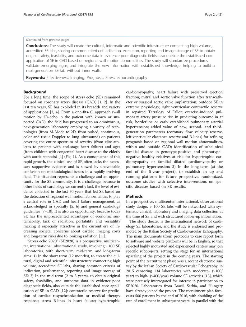
Conclusions: The study will create the cultural, informatic and scientific infrastructure connecting high-volume,accredited SE labs, sharing common criteria of indication, execution, reporting and image storage of SE to obtainoriginal safety, feasibility, and outcome data in evidence-poor diagnostic fields, also outside the established coreapplication of SE in CAD based on regional wall motion abnormalities. The study will standardize procedures,validate emerging signs, and integrate the new information with established knowledge, helping to build anext-generation SE lab without inner walls.
Keywords: Effectiveness, Imaging, Prognosis, Stress echocardiography
BackgroundFor a long time, the scope of stress echo (SE) remainedfocused on coronary artery disease (CAD) [1, 2]. In thelast ten years, SE has exploded in its breadth and varietyof applications [3, 4]. From a one-fits-all approach (wallmotion by 2D-echo in the patient with known or sus-pected CAD), the field has progressed to an omnivorous,next-generation laboratory employing a variety of tech-nologies (from M-Mode to 2D, from pulsed, continuous,color and tissue Doppler to lung ultrasound) on patientscovering the entire spectrum of severity (from elite ath-letes to patients with end-stage heart failure) and ages(from children with congenital heart disease to the elderlywith aortic stenosis) [4] (Fig. 1). As a consequence of thisrapid growth, the clinical use of SE often lacks the neces-sary supportive evidence and is slowed by unavoidableconfusion on methodological issues in a rapidly evolvingfield. This situation represents a challenge and an oppor-tunity for the SE community. It is a challenge, because inother fields of cardiology we currently lack the level of evi-dence collected in the last 30 years that led SE based onthe detection of regional wall motion abnormalities to playa central role in CAD and heart failure management, asacknowledged in specialty [5, 6] and general cardiologyguidelines [7–10]. It is also an opportunity, because todaySE has the unprecedented advantages of economic sus-tainability, lack of radiation, portability and versatilitymaking it especially attractive in the current era of in-creasing societal concerns about cardiac imaging costsand long-term risks due to ionizing radiation [11].“Stress echo 2020” (SE2020) is a prospective, multicen-
ter, international, observational study, involving > 100 SElaboratories, with short-term, mid-term, and long-termaims: 1) In the short term (12 months), to create the cul-tural, digital and scientific infrastructure connecting highvolume, accredited SE labs, sharing common criteria ofindication, performance, reporting and image storage ofSE; 2) In the mid-term (2 to 3 years), to obtain originalsafety, feasibility, and outcome data in evidence-poordiagnostic fields, also outside the established core appli-cation of SE in CAD [12]: contractile reserve for predic-tion of cardiac resynchronization or medical therapyresponse; stress B-lines in heart failure; hypertrophic
cardiomyopathy; heart failure with preserved ejectionfraction; mitral and aortic valve function after transcath-eter or surgical aortic valve implantation; outdoor SE inextreme physiology; right ventricular contractile reservein repaired Tetralogy of Fallot; exercise-induced pul-monary artery pressure rise in predicting outcome in atrisk, borderline or early established pulmonary arterialhypertension; added value of new, second- and third-generation parameters (coronary flow velocity reserve,left ventricular elastance reserve and B-lines) for refiningprognosis based on regional wall motion abnormalities,within and outside CAD; identification of subclinicalfamilial disease in genotype-positive and phenotype–negative healthy relatives at risk for hypertrophic car-diomyopathy or familial dilated cardiomyopathy orpulmonary hypertension; 3) In the long-term (at theend of the 5-year project), to establish an up andrunning platform for future prospective, randomized,outcome studies with selective interventions on spe-cific diseases based on SE results.
MethodsIn a prospective, multicenter, international, observationalstudy design, > 100 SE labs will be networked with sys-tematic clinical, laboratory and imaging data collection atthe time of SE and with structured follow-up information.The study theater is the international network of cardi-
ology SE laboratories, and the study is endorsed and pro-moted by the Italian Society of Cardiovascular Echography.The main documents (from protocols to case report formto software and website platform) will be in English, so thatselected highly motivated and experienced centers may joinspecific subprojects, setting the stage for an internationalupscaling of the project in the coming years. The startingpoint of the recruitment phase was a recent electronic sur-vey by the Italian Society of Cardiovascular Echography, in2015 censoring 134 laboratories with moderate- (>100/year) to high- (>400/year) volume SE activities [13], whichwere precisely interrogated for interest in participation toSE2020. Laboratories from Brazil, Serbia, and Hungaryhave already joined the project. The recruitment plan fore-casts 500 patients by the end of 2016, with doubling of therate of enrollment in subsequent years, in parallel with the
Picano et al. Cardiovascular Ultrasound (2017) 15:3 Page 2 of 21

increasing number of recruiting labs fulfilling quality con-trol criteria, reaching the target number of 100 at the endof the 5-year schedule.
Data collectionAs recommended by guidelines, we will adopt a 17-segment model of the left ventricle, with 1-to-4 segmentalscoring system [5, 6]. Stress protocols are harmonized ac-cording to recent European and North-American scien-tific societies' guidelines, with semi-supine exerciserecommended and pharmacological stress dosages up to40 mcg/kg/min for dobutamine, up to 0.84 mg/kg in6 min for dipyridamole, and up to a 4-min step of 200microg/kg/min for adenosine [5, 6]. With dobutamine, at-ropine (up to 1 mg) can be administered in patients withsuspected CAD (protocol 9), and it is associated with ahigher rate of complications in those with a history ofneuropsychiatric symptoms, reduced left ventricular func-tion, or small body habitus. The maximal allowed dobuta-mine dose is 20 mcg/kg/min in patients with aorticstenosis, in whom higher doses are less safe and prob-ably unnecessary [13]. All laboratories will share astandardized case report form coded in a databaseformat to facilitate retrieval and communication. For
applications outside CAD and for CAD testing withvasodilator stress, no atropine is given on top ofpharmacological stress.Although data collection with a dedicated project-
specific case report form is allowed, we encourage imple-menting a dedicated, free ad-hoc system for data storageand reporting developed at the National Research Council,Institute of Clinical Physiology. The software provides asuitable informatics infrastructure for the SE 2020 Italianmulticenter study, with an intuitive graphic interface, eye-catching graphic format and convenient reporting option.It could represent the trade-off between the comprehen-sive information required by scientific standards and thesmooth workflow priority of busy, high-volume, clinically-driven activities [14]. As an illustrative example, the reportpage for regional wall motion abnormalities and WallMotion Score Index is shown in Fig. 2. The softwarewas developed and tested in Italian and the transla-tion of the last release in other languages (English,Portuguese and Serbian) is currently in progress.
Data analysisData will be expressed as mean ± standard deviation(normally distributed data, such as wall motion score
Stress echo 2020: versatility
Ischemia
CFVR
Mitral Insufficiency
LV elastance
Extravascular lung water
Viability
200
10
8
.54
.2310
8
.54
.23
CAD HFrEFHFpEF
Valvular HD Congenital HD Extreme physiology Pulmonary arterialhypertension
HCM
Fig. 1 In the box, the contemporary spectrum of patients for whom SE can offer potentially unique diagnostic information: coronary arterydisease; heart failure (with either reduced or preserved left ventricular function); hypertrophic cardiomyopathy; valvular heart disease; extremephysiology; adult repaired congenital heart disease; early, at risk, or borderline pulmonary arterial hypertension. For each clinical condition, adifferent key SE parameter can be used, evaluated at rest (left column) and during stress (right column), maximizing the versatility of thetechnique. From top to bottom rows, regional wall motion (for ischemia and viability), coronary flow velocity reserve (CFVR), mitral insufficiency,end-systolic volume of the left ventricle (necessary to assess left ventricular elastance), and B-lines (a marker of extravascular lung water). Modifiedand adapted from ref 4 (Picano and Pellikka [4])
Picano et al. Cardiovascular Ultrasound (2017) 15:3 Page 3 of 21

index), median and inter-quartile (25th, 75th) range(non-normally distributed data, such as B-lines) or percent frequency (categorical data, such as presence orabsence of severe mitral regurgitation), with absolutenumbers. In patients with coronary angiographic informa-tion, diagnostic sensitivity, specificity, positive and nega-tive predictive value will be assessed for any combinationof the wall motion score index, coronary flow velocityreserve, left ventricular contractile reserve and B-lines.One-sample comparisons will be performed using
Wilcoxon test, and the chi-squared test without Fish-er's correction for categorical data. Event rates will beestimated with Kaplan–Meier curves and comparedby the log-rank test. Univariable analyses by Cox pro-portional hazards models will be performed to assessthe association between each candidate variable andoutcome. All variables with P <0.20 by univariableanalysis will be considered as candidate variables forthe multivariable analyses. Goodness of fit of themodels will be based on C-statistics and its variants,adjusting for optimism using bootstrap replications(at least 1000). A receiver operating characteristicanalysis will be used to obtain the best prognostic
predictor for the individual SE variables. We will alsoanalyze the data according to a clinically guided step-wise procedure, where the variables will be includedin the model in the same order in which they are ac-tually considered by the cardiologist. Statistical signifi-cance will be set at p < 0.05.
Quality controlIt is well-known that the diagnostic performance of SE isclosely related to the level of expertise of thecardiologist-echocardiographer performing the test,since the evaluation of regional wall motion is subjectiveand qualitative, with considerable variability even amongexperienced centers of undisputed reputation [15]. Thereproducibility and accuracy of wall motion reading canbe substantially increased with limited training [16] andthrough development of conservative, pre-specifiedreading criteria [17]. Therefore, quality control of thediagnostic performance in the various laboratories is amust in order to enter meaningful information in thedata bank. The burden of quality control is on the hubcenter of the principal investigator of each subproject,where various spoke centers may converge. For the
Fig. 2 The computerized case report form for the regional wall motion abnormalities of the SE 2020 study. The grading of the response isreported in tabular (right side, lower panel) and graphic (right side, upper panel) format, with normal values of Wall Motion Score Index in green,mild impairment in yellow, moderate in orange, and severe impairment in red
Picano et al. Cardiovascular Ultrasound (2017) 15:3 Page 4 of 21

general project, the hub center for regional wall motionanalysis is Pisa-CNR, in coordination with the principalinvestigator. There are five different levels of qualitycontrol, with increasing levels of complexity:
1. Level 1, pre-requisite: a volume activity of the lab of atleast 100 SE tests per year, which is the requirement forcredentialing of SE activity by scientific societies [18].
2. Level 2, spoke centers read hub SE images,consisting in 20 selected studies for regional wallmotion analysis. The concordance requiresidentification of test negativity/positivity and, inpositive tests, the correct localization of the ischemiczone. For each test, a multiple choice 6-answer test isgiven. The criterion of ≥ 90% concordance (at least 18out of 20 studies) is required, as previously describedfor first-generation SE multicenter studies [19, 20].
3. Level 3, hub centers read spoke centers studies,consisting in 20 any-quality consecutive studiesrecorded by the spoke center. The criterion of ≥ 80%concordance (at least 16 out of 20 studies) isrequired, as previously described for first-generationSE multicenter studies [19].
4. Level 4, core lab reading. All centers should grantfull access to images of SE studies entered in thedata bank for audit or reading by core lab laboratory,which is the standard for specific subprojects suchas number 10 for genetic SE, when every effortneeds to be made to minimize variability and asingle reader will analyze all studies acquired bydifferent centers, as required by recommendationsfor small-to-medium sample studies, when resourcesallow [20].
5. Level 5, specific protocols quality control. Althoughthe SE quality control has proved to work well forregional wall motion analysis, novel SE applicationsinvolve different parameters, methodology ofacquisition and reading criteria. Therefore, for eachsubproject, a web-based training session and qualitycontrol is organized by the specific hub center andprincipal investigator to assure consistency of data[21]. The principal investigator of each subprojectwill prepare a set of 20 studies with rest-stressimages. For each test, a multiple choice 6-answerstest is given (only 1 correct). The criterion of ≥90%concordance (at least 18 out of 20 studies) isrequired. The specific signs tested for certificationare: end-diastolic and end-systolic volume changes(protocol 1); B-lines (protocol 2, 4, 6 and 9); leftventricular outflow tract gradient (protocol 3 and 10);E/e' (protocol 4); mitral regurgitation quantitativeassessment (protocol 5); aortic stenosis quantitativeassessment (protocol 5); right ventricular function(protocol 7); pulmonary arterial systolic pressure
measurements during stress (protocol 8); coronaryflow velocity reserve (protocol 9); left ventricular elas-tance (protocol 9); global longitudinal strain (protocol4 and 10).
This study is also intended as a special level of voluntaryaccreditation and expertise in the specific field of interest,well above the volume activity criteria requested by guide-lines. The accreditation process is run and certified by theItalian scientific society of echocardiography strictly follow-ing criteria and procedures of the European association ofcardiovascular imaging to ensure standardization and inde-pendence of the process. When not otherwise specified,resting and SE measurements are performed according tothe latest joint recommendations of European and North-American societies [22]. A simplified view of each lab's roadto SE2020 is shown in Fig. 3: the essential pre-requisite isthe high-volume activity of the lab, with readers' certifica-tion of competence from national or international societiesand written declaration of interest in SE2020. After adop-tion of dedicated SE computerized software by the lab(allowing direct entry of the information in a format com-patible with the data bank) and voluntary certification forproject-specific SE reading, the center can start recruiting.
Overall study designWe will collect the experience of Italian, Brazilian,Hungarian and Serbian SE labs over the 5-year periodfrom 2016 to 2020. In this broader framework, 10sub-projects will address specific patients’ subsets.The target population ranges from 250-patient sam-ples for protocols focused on specific diseases (suchas protocol 7 in repaired tetralogy of Fallot) to 2,500for protocols on heart failure (number 2) to 5,000 toall-comers with known or suspected CAD tested withnovel indices (number 9) (Table 1).Different study projects will cover the entire spectrum
of disease, age and clinical status of current patients.The recruited participants are “the wellest of the well”(super-fit athletes entering project 6), the “worried well”(young first-degree relatives of patients with hyper-trophic cardiomyopathy or familiar forms of dilated car-diomyopathy or pulmonary arterial hypertension, inproject 10), the “suspected sick” (for instance patientswith suspected diastolic heart failure or CAD as in pro-jects 4 and 9), up to the "sickest of the sick" (for in-stance, patients with advanced heart failure or valvularheart disease entering projects 1, 2 and 5). Some degreeof overlap is unavoidably present for some projects, forinstance with subjects eligible for project 2 who are alsorecruitable for project 1 (if they undergo cardiac resyn-chronization therapy) or for project 5 (if they have heartfailure with preserved ejection fraction). Over time,patients may move from one project to another: for
Picano et al. Cardiovascular Ultrasound (2017) 15:3 Page 5 of 21
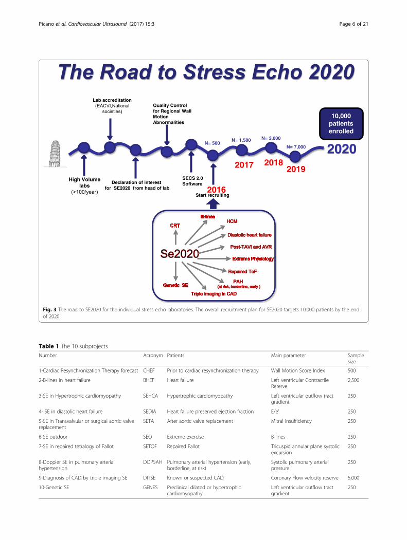
Fig. 3 The road to SE2020 for the individual stress echo laboratories. The overall recruitment plan for SE2020 targets 10,000 patients by the endof 2020
Table 1 The 10 subprojects
Number Acronym Patients Main parameter Samplesize
1-Cardiac Resynchronization Therapy forecast CHEF Prior to cardiac resynchronization therapy Wall Motion Score Index 500
2-B-lines in heart failure BHEF Heart failure Left ventricular ContractileRererve
2,500
3-SE in Hypertrophic cardiomyopathy SEHCA Hypertrophic cardiomyopathy Left ventricular outflow tractgradient
250
4- SE in diastolic heart failure SEDIA Heart failure preserved ejection fraction E/e’ 250
5-SE in Transvalvular or surgical aortic valvereplacement
SETA After aortic valve replacement Mitral insufficiency 250
6-SE outdoor SEO Extreme exercise B-lines 250
7-SE in repaired tetralogy of Fallot SETOF Repaired Fallot Tricuspid annular plane systolicexcursion
250
8-Doppler SE in pulmonary arterialhypertension
DOPSAH Pulmonary arterial hypertension (early,borderline, at risk)
Systolic pulmonary arterialpressure
250
9-Diagnosis of CAD by triple imaging SE DITSE Known or suspected CAD Coronary Flow velocity reserve 5,000
10-Genetic SE GENES Preclinical dilated or hypertrophiccardiomyopathy
Left ventricular outflow tractgradient
250
Picano et al. Cardiovascular Ultrasound (2017) 15:3 Page 6 of 21

instance, first-degree relatives of hypertrophic cardiomy-opathy patients with negative phenotype enrolled in pro-ject 10 may subsequently develop overt forms of diseaseand be enrolled in project 3. All these potential gray-zone situations will be readily identified in individual SEreports. The investigator is allowed to enter the patientin only one subproject at a given time.Although the setting will be mainly the Italian cardio-
logical community, all essential documents will be writtenin English and we plan to extend the project to other com-munities with long-standing history of cooperation andexperience in multicenter trials. Brazilian, Hungarian andSerbian centers are already recruiting and additional labora-tories from other countries are now entering the process ofaccreditation. The project is curiosity-driven, independentfrom sponsors, and clinically oriented. However, after theplanning and start-up phase, support from public or privatefunding agencies or industries is possible – provided that itis unrestricted and does not interfere in any way with datacollection and analysis.There is no bonus payment for subject recruitment
and subject referral. Enrolled patients are referred to theSE lab for clinically-driven indications. Each patientsigns an informed consent form allowing scientificutilization of data, respectful of privacy rights, at thetime of testing. The study project was submitted by thecoordinating center of the principal investigator on
January 31, 2016 and approved in its revised form by theRome-1 ethical committee on July 20, 2016 (protocolnumber 1487/Lazio1). Ethics committee approval will besought by each participating center, as needed.Inclusion criteria shared by all projects are: 1- age <
85 years and > 18 years (except for project 7 regardingrepaired Tetralogy of Fallot and project 10 regarding healthyrelatives of patients with familial disease, in which chil-dren > 10 years can enter the study after parental consent);2- technically acceptable acoustic window at rest (with atleast 14 segments well visualized in at least one projection).Exclusion criteria shared by all projects are: 1- pres-
ence of prognosis-limiting comorbidities, such as ad-vanced cancer, reducing life expectancy to < 1 year; 2-pregnancy/lactation; 3- unwillingness to give informedconsent and to enter a regular follow-up program.SE data will be available to the referring physician.A brief synopsis of each project is presented for each
sub-study, which places emphasis on different parame-ters tailored on the specific diagnostic question (Fig. 4).
CHEFCardiac resynchronization tHErapy Forecast.
BackgroundCardiac resynchronization therapy is increasingly usedin patients with heart failure, but the identification of
Fig. 4 The key echocardiographic parameter for each project, around the logo of SE2020. Clockwise: regional wall motion (project 1, butimportant in all other projects, mainly project 9); end-systolic volume as part of left ventricular elastance assessment (project 2); left ventricularoutflow tract gradient (project 3, but also important in project 10); E/e' ratio (project 4); mitral insufficiency (project 5, but also important in project2); B-lines (essential in project 6, but also important in project 2, 4 and 9); right ventricular function (project 7); pulmonary hemodynamics (project8 and 10); regional coronary flow velocity reserve (useful in project 9, but also important in projects 2, 3 and 10)
Picano et al. Cardiovascular Ultrasound (2017) 15:3 Page 7 of 21

“responders” remains challenging, since up to 1 in 3 pa-tients do not show symptomatic improvement with thiscostly and demanding electrical therapy [10]. QRS widthremains the single established criterion to assess intra-ventricular dyssynchrony according to guidelines; how-ever, there is no accepted consensus on which imagingparameter is best to predict cardiac resynchronizationtherapy response. Inconsistent results have been ob-tained with several echocardiographic indices of left ven-tricular dyssynchrony [23]. The presence of myocardialcontractile reserve assessed during SE predicted the re-sponse to cardiac resynchronization therapy: the use ofSE had obvious potential for a better selection of cardiacresynchronization therapy candidates, in whom a pre-served contractility is related to a higher percentage ofclinical and echocardiographic responders to cardiacresynchronization therapy [24–27].
AimsThe primary aim is to evaluate the feasibility of severalindices of SE (including the well- established, such aswall motion score index, and more innovative ones suchas left ventricular elastance) in the evaluation of patientcandidates for cardiac resynchronization therapy. Thesecondary aim is to assess the value of each of these pa-rameters (alone or in combination) in predicting thesymptomatic and functional improvement in the short-term (1 week to 1 month) after electrical or medicaltherapy. The tertiary aim is to assess the prognosticvalue of SE indices for prognostic stratification in themedium-long term.
MethodsPatients evaluated prior to cardiac resynchronizationtherapy, with class I, IIa or IIb for cardiac resynchroniza-tion therapy according to ESC 2016 guidelines [10], andtherefore with ejection fraction ≤ 35% (by 2-D Simpsonmethod, or real time 3D echocardiography) and QRSduration ≥ 130 ms. Contractile reserve will be assessedduring stress (exercise or dobutamine or vasodilators)through variations in Wall Motion Score Index and withmore advanced parameters such as left ventricularelastance reserve, as the peak stress/baseline ratio ofend-systolic pressure/ end-systolic volume [28]. Allother echocardiographic parameters of interest (leftventricular ejection fraction, E/e’, mitral regurgitation,tricuspid annular plane systolic excursion, septal flash,apical rocking, left anterior descending artery coronaryflow velocity whenever possible) will also be assessed atbaseline and peak stress. All patients will be followed-upwithin 6-months to 1 year also with resting echocardio-graphic examination to assess left ventricular remodellingand recovery of function. Any patient excluded from car-diac resynchronization therapy and kept on optimal
medical therapy will be included in the follow-up, sincethe presence of contractile reserve may predict the func-tional improvement in these patients as well [13].
Sample size calculationIf we assume a 60% response rate to cardiac resynchro-nization therapy, with doubling of likelihood of improve-ment in presence of a pre-test contractile reserve by SE,with a power of 80% and an alpha error of 5%, a samplesize of 277 patients is required. About the same numberis required to predict the response to medical therapy inpatients eventually not undergoing cardiac resynchroni-zation therapy.
Study hypothesisThe presence of contractile reserve is associated with abetter prognosis and greater chance of functional recov-ery with cardiac resynchronization therapy.
BHEFEvaluation of B-lines in HEart Failure patients withdepressed ejection fraction.
BackgroundB- lines are a semiquantitative sign of extravascular lungwater present in 1 out of 3 heart failure patients at restand in 1 out of 2 during stress, and potentially useful forrefining prognostic stratification and titrating diuretictherapy in these patients. Their prognostic power ishigher during stress than at rest, and integrates thepowerful stratification provided by dynamic assessmentof other established predictors such as mitral regurgita-tion, E/e’ as a surrogate of left ventricular filling pressure,ejection fraction and tricuspid annular plane systolic ex-cursion [29, 30].
AimsThe primary aim is to assess the feasibility of several in-dices of SE (including the well- established such as leftventricular ejection fraction and more innovative onessuch as B-lines or left anterior descending coronary flowreserve) in evaluating patients with known or suspectedheart failure with reduced ejection fraction. The secondaryaim is to assess the value of each of these parameters inpredicting the functional impairment, indicated by NewYork Heart Association class, cardiac natriuretic peptidesconcentration, peak VO2 and other indices. The tertiaryaim is to assess the prognostic value of SE indices forprognostic stratification in the medium-long -term.
MethodsWe will enrol patients referred to SE (with exercise, do-butamine or dipyridamole) with known or suspectedheart failure, with reduced (<40%) ejection fraction. B-
Picano et al. Cardiovascular Ultrasound (2017) 15:3 Page 8 of 21

lines will be scored with the 28-regions antero-lateralchest assessment as previously described at baseline andimmediately after stopping exercise [31]. A simplified 8-region or 4-region scan is also allowed in order to savetime without loss of critical information. All other echo-cardiographic parameters of interest (left ventricularejection fraction, E/e’, mitral regurgitation, tricuspid an-nular plane systolic excursion, left anterior descendingartery coronary flow velocity whenever possible) will alsobe assessed at baseline and peak stress [32]. All patientswill be followed-up for at least 1 year.
Sample size calculationIf we conservatively assume a 30% yearly incidence ofcomposite end-points (death, myocardial infarction, newhospital readmission, heart transplant, ventricular assistdevice implantation, aborted sudden death) and a 5% in-cidence of death, with doubling of likelihood of events inpresence of a positive SE (for B-lines increase duringstress or lack of contractile reserve or severe mitral in-sufficiency), with a power of 95% and an alpha error of5%, with an attrition rate of 15%, a sample size of about2500 patients is required if the effect on mortality isevaluated.
Study hypothesisThe integrated assessment of 5 major echocardiographicvariables during stress adds power to the prognosticstratification operated by isolated echocardiographic pre-dictors in heart failure patients. The prognostically mean-ingful signs explore 5 key links in the patho-physiologicchain behind cardiovascular response in heart failure: leftventricular systolic function – left ventricular diastolicfunction - mitral valve regurgitation - lung water accumu-lation - right ventricular function.
SEHCAStress Echo in Hypertrophic Cardiomyopathy.
BackgroundThe impact of SE in hypertrophic cardiomyopathy islimited by lack of standardization and outcome data.Current guidelines recommend SE solely for evaluationof left ventricular outflow tract obstruction [33]. How-ever, large-scale registry data show that SE positivity forischemic criteria (such as new wall motion abnormalitiesand coronary flow velocity reserve) rather than induciblegradients predict adverse outcome in hypertrophic car-diomyopathy [34]. Thus, important prognostic as well asfunctional information may be derived from SE, al-though their clinical impact remains limited due lack ofstandardized data collection and uniform protocols.Currently there are virtually no two centers performingSE in the same way in hypertrophic cardiomyopathy
patients, and the test remains grossly underutilized dueto (unjustified) concerns with safety. Prospective, stan-dardized, multicenter data collection is necessary toachieve comprehensive evaluation of SE potential in thisdisease. Of note, exercise limitation and breathlessnessmay be due to a number of different causes, includingleft ventricular outflow tract obstruction (which mayincrease, remain stable or decrease during exercise), func-tional mitral regurgitation, restrictive physiology, ampli-fied mechanical dyssynchrony, coronary microvasculardisease or – less frequently – ischemia associated withprognostically unfavorable stress-induced wall motion ab-normalities. Despite similar clinical manifestations, man-agement may differ substantially based on themechanisms [35]. SE is the only test with the potential todiscriminate the various components, allowing a targetedtreatment driven by pathophysiology, but prospective dataare required to substantiate this hypothesis. Of note,general consensus exists regarding the advisability ofpreferring physiological provocation with exercise overpharmacological stressors (particularly dobutamine due tosubstantial prevalence of false positives with regard to in-ducible obstruction).
AimsThe primary aim is to evaluate the feasibility of severalindices of SE (from the well- established such as left ven-tricular outflow tract gradient to more innovative featuressuch as B-lines or left anterior descending coronary flowreserve) in the evaluation of hypertrophic cardiomyopathypatients. The secondary aim is to assess the value of eachof these parameters in predicting functional impairment,indicated by New York Heart Association class, EuropeanSociety of Cardiology sudden cardiac death risk score andother indices. The tertiary aim is to assess the prognosticvalue of SE indices for prognostic stratification in themedium-long-term.
MethodsDiagnosis of hypertrophic cardiomyopathy will be based onexisting guidelines [33]. Phenocopies such as infiltrative/storage disease (eg, Fabry) will be excluded. Low-to-intermediate risk symptomatic or asymptomatic hyper-trophic cardiomyopathy patients will undergo exercise SEwith assessment at each stage and during recovery of wallmotion, mitral insufficiency, left ventricular outflow tractgradient (in orthostatic position) and E/e’. If feasible, coron-ary flow velocity reserve on left anterior descending andlung ultrasound B-lines will also be assessed. Only whenpatients are unable to exercise, or for the specific aim ofassessing coronary flow velocity reserve, will a vasodilatorstress (high dose dipyridamole or adenosine withoutatropine) be performed. All patients will be followed-up and the prognostic value of different rest and SE
Picano et al. Cardiovascular Ultrasound (2017) 15:3 Page 9 of 21

parameters (also compared to standard prognostic in-dices) will be assessed.
Sample size calculationThe composite end-point of the study comprises death(cardiovascular and all-cause), new hospital admissionfor acute heart failure, newly onset atrial fibrillation,aborted sudden death, heart transplant or ventricular as-sist device implantation. A pilot study showed an inci-dence of events around 8% per year [34]. If we assume apositivity rate to SE (by composite criteria) of 40%, withdoubling of likelihood of events in presence of SE posi-tivity (by any criteria), with a power of 80%, an alphaerror of 5%, and an attrition rate of 10%, a sample sizeof about 250 patients is required.
Study hypothesisHypertrophic cardiomyopathy patients with induciblewall motion abnormalities and reduced coronary flowvelocity reserve (or lung ultrasound B-lines) are at sub-stantially higher risk for subsequent unfavorable eventsthan patients with normal wall motion and preservedcoronary flow velocity reserve. Exercise capacity predictsoutcome in hypertrophic cardiomyopathy patients inde-pendent of the presence and magnitude of resting orprovocable obstruction.
SEDIAStress Echo in Diastolic Heart failure.
BackgroundDiastolic SE should be considered in the breathless pa-tient with normal left ventricular ejection fraction, highcardiac natriuretic peptides and low exercise tolerance,especially in presence of cardiovascular risk factors(advanced age, arterial systemic hypertension, diabetesmellitus, obesity and sedentary lifestyle) after rulingout other common cardiac (heart valve disease, coronaryartery disease) and non-cardiac causes of dyspnea (mainlyanemia and chronic obstructive pulmonary disease). « Redflag » indicators of possible diastolic dysfunction includeleft ventricular hypertrophy, enlarged left atrium and pul-monary hypertension. During SE, findings which mayaccount for unexplained dyspnea beyond diastolic dys-function are left ventricular outflow tract obstruction, dy-namic mitral regurgitation, and new onset regional wallmotion abnormalities [36]. The diagnostic SE criteria ofheart failure with preserved ejection fraction are reducedstroke volume and cardiac output reserve, high left ven-tricular filling pressures (average E/e’ ratio > 14) and pul-monary hypertension, which also identify patients withmore severe prognosis [37, 38]. In patients with heart fail-ure and either preserved (>50%) or mid-range (40-49%)ejection fraction, SE may allow the identification of
diastolic dysfunction in patients with parameters incon-clusive at rest [10]. According to the latest 2016 recom-mendations on the evaluation of left ventricular diastolicfunction by echocardiography [36], diastolic stress testingwould not be indicated in patients with either completelynormal diastolic function at rest (in whom a severe dia-stolic dysfunction is unlikely to develop during stress) andin patients with ≥ grade 2 diastolic dysfunction at rest(with elevated filling pressures at rest which will likely in-crease further during exercise). However, testing of thesepatients can be valuable to assess the incremental value, ifany, of newer candidate indices such as left ventricularend-diastolic volume reserve and B-lines. In addition, alsopatients with normal left ventricular diastolic function atrest can indeed develop increase in left ventricular fillingpressures with dyspnea when exercising.
AimsThe primary aim is to evaluate the feasibility of severalindices of SE (including more established such as E/e' ra-tio and more innovative ones such as B-lines or left ven-tricular diastolic volume reserve) in the evaluation ofpatients with known or suspected heart failure with pre-served or mid-range ejection fraction. The secondaryaim is to assess the value of each of these parameters inpredicting the functional impairment, indicated by NewYork Heart Association class, cardiac natriuretic pep-tides concentration, peak VO2 and other indices. Thetertiary aim is to assess the prognostic value of SE indi-ces for prognostic stratification of heart failure with pre-served ejection fraction in the medium-long-term.
MethodsPatients with known or suspected heart failure with pre-served ejection fraction by 2016 ESC criteria will be en-rolled and studied with cycle-ergometer in semi-supineSE (or treadmill). The test is especially indicated in pa-tients with dyspnea and grade 1 diastolic dysfunction atrest, identified as mitral E/A ratio ≤ 0.8, average E/e' ra-tio < 10, peak tricuspid regurgitant jet velocity <2.8 m/sand left atrium volume index normal (<34 mL/m2) or in-creased [36]. In patients unable to exercise, pharmaco-logical test of choice (dobutamine or vasodilator) isallowed, but not recommended. The diastolic assessmentshould be included into all exercise SE tests by measur-ing standard Doppler-derived mitral inflow velocity,pulsed Tissue Doppler of mitral annulus, and retrogradetricuspid gradient of tricuspid regurgitation. These mea-surements can be performed at intermediate load of ex-ercise and/or 1- 2 min after the end of the exercise, afterobtaining wall motion acquisitions, when the heart ratedecreases and mitral inflow E and A velocities appear tobe well separated. As a part of the “diastolic package”,we will also assess, at baseline, intermediate load (50
Picano et al. Cardiovascular Ultrasound (2017) 15:3 Page 10 of 21

watts) and peak-post stress [36]: diastolic left ventricularvolume index (to evaluate left ventricular diastolicvolume reserve, impaired in stiff hearts, which are lessdilated for any given filling pressure); systolic left ven-tricular volume index (for assessment of left ventricularelastance, which may unmask occult systolic dysfunctionwith normal ejection fraction increase); ejection fractionand both stroke volume and cardiac output (to assessconventional contractile reserve); mitral regurgitationand left ventricular outflow tract obstruction; pulmonaryartery systolic pressure (from velocity of tricuspid regur-gitation); B-lines during stress (to provide a direct im-aging of extra-vascular lung water accumulation as adirect cause of dyspnea), since they can be present atrest in diastolic heart failure [32]. Global longitudinalstrain could be optionally determined as the average ofthe regional longitudinal strain measured in 17-segments model from the apical long-axis, 4-chamberand 2-chamber view. Despite not being independent onpreload and afterload, global longitudinal strain could beconsidered as an appropriate, alternative parameter ofleft ventricular contractile reserve [22, 39]. On the basisof currently accepted criteria, the test is considered posi-tive for diastolic dysfunction when all of the followingthree conditions are met during exercise: average E/e' >14 or septal E/e' ratio > 15, peak tricuspid regurgitant jetvelocity >2.8 m/s and septal e' velocity < 7 cm/s at base-line [36].
Sample size calculationThe composite end-point of the study comprises death(cardiovascular and all-cause), new hospital admissionfor acute heart failure, aborted sudden death, hearttransplant or ventricular assist device implantation. Theexpected incidence of events is around 20% per year[10]. We assume a positivity rate to SE (by compositecriteria: increase in E/e' and/or increase in pulmonaryartery systolic pressure and/ or decrease in septal e' vel-ocity and/or increase in B-lines) of 30%, with doublingof likelihood of events in presence of SE positivity (byany criteria). With a power of 80%, an alpha error of 5%,and an attrition rate of 10% (for exams non-feasible and/or for exclusion criteria during the screening phase dueto previously unrecognized dynamic severe mitral regur-gitation or left ventricular outflow obstruction), a samplesize of about 250 patients is required.
Study hypothesisIn patients with known or suspected heart failure withreduced ejection fraction, higher left ventricular fillingpressures, more B- lines and lower increases in end-diastolic volumes during stress are associated with worseprognosis.
SETAStress Echo in Transcatheter or surgical Aortic ValveImplantation.
BackgroundTranscatheter Aortic Valve Implantation is an extraor-dinarily effective but still a relatively novel technology,and short and long term morbidity and mortality afterTranscatheter Aortic Valve Implantation remains signifi-cant. There is substantial interest in the identificationand modification of factors influencing prognosis beforeand after the procedure. The severity of concomitant mi-tral regurgitation improves after the procedure in 2 outof 3 patients, but baseline moderate-severe mitral regur-gitation and significant residual mitral regurgitation areassociated with an increase in mortality after Transcath-eter Aortic Valve Implantation and represent an import-ant group to target with medical or transcathetertherapies in the future [40]. Stress echo plays a pivotalrole in valvular heart disease [41], but its role after eitherTranscatheter Aortic Valve Implantation or surgical re-placement is still unsettled, although it has potential toidentify the presence and severity of mitral regurgitationand aortic stenosis after intervention, so as to refine theprognostic strategy and better tailor treatment.
AimsThe primary aim is to evaluate the feasibility of SEfocused on mitral and aortic reserve after aortic valve re-placement (with Transcatheter or surgical aortic valveimplantation). The secondary aim is to assess the pres-ence and entity of changes in valvular and ventricularfunction and their correlation with indices of functionalseverity (New York Heart Association Class, cardiacnatriuretic peptides, peak VO2, etc.).. The tertiary aim isto assess the prognostic value of SE indices for prognos-tic stratification in the medium-long-term.
MethodsPatients with previous (from 6 months to 10 years) sur-gical or Transcatheter Aortic Valve Implantation capableof exercising and with absent-to-moderate mitral insuffi-ciency will be enrolled and studied with semisupine SE.The full quantitative evaluation of mitral regurgitationand aortic stenosis will be performed according to rec-ommendations of the European Society of Cardiology. Inaddition, assessment of B- lines (as in protocol 2), rightventricular function (as in protocol 7), left ventricularelastance (as in protocol 1) will be performed at baselineand peak stress. Exercise will be started at 15 watts, with5-min steps and 15-watt increments per step [13]. Inpatients unable to exercise, a pharmacological test ofchoice (dobutamine or vasodilator) is allowed, but notrecommended.
Picano et al. Cardiovascular Ultrasound (2017) 15:3 Page 11 of 21

Sample size calculationThe expected incidence of SE positivity (by compositecriteria: increase in mean transaortic gradient > 20 mmHgand/or increase in mitral insufficiency > 1 grade and/orincrease in B-lines and/or increase in systolic pulmonaryartery pressure) is around 30% post- Transcatheter AorticValve Implantation or aortic valve surgical replacement[10]. With a power of 80%, an alpha error of 5%, and anattrition rate of 10%, a sample size of about 100 patients isrequired to detect a significant stress-induced increase inmitral regurgitation severity. For the prognostic analysis(tertiary end-point). If we conservatively assume a 20%yearly incidence of composite end-points (death, myo-cardial infarction, new hospital readmission, hearttransplant, ventricular assist device implantation,aborted sudden death), with doubling of likelihood ofevents in presence of a positive SE (for severe mitral in-sufficiency and/or abnormally increased transaortic gra-dient and/ or B-lines and/ or abnormal pulmonaryartery systolic pressure), with a power of 80% and analpha error of 5%, a sample size of about 250 patients isrequired with a 3 -year follow-up.
Study hypothesisIn patients with absent-to moderate mitral regurgitationin resting transthoracic echocardiography after eithersurgical or Transcatheter Aortic Valve Implantation,those with lower transaortic gradient and less mitral re-gurgitation during stress will have a more favourableoutcome than patients with higher gradients and moresevere residual mitral regurgitation during stress.
SEOStress Echo Outdoor in Extreme conditions.
BackgroundSE with B-lines can also be performed outdoors, withpocket size or portable instruments, in a setting of eco-logical stress entirely different from standard indoortesting [4]. The diagnostic target is the diagnosis, orearly subclinical identification, of life-threatening diseaseat high altitudes or any kind of environmental pulmon-ary edema. In this challenging but fascinating context,lung ultrasound detects B-lines in 20 to 40% of normaland/or super-fit subjects in extreme physiology settings,such as high altitude [42, 43], deep underwater apneadiving [44] or endurance exercise [45].
AimsThe primary aim is to evaluate the feasibility of outdoorSE focused on B-lines in the logistic setting of extremephysiology. The secondary aim is to assess the presenceand amount of B-lines increase at peak stress vs baselineconditions (pre-exercise; pre-apnea; pre-ascent) and to
correlate lung ultrasound findings with symptoms (suchas dyspnea, cough, fatigue). The tertiary aim is to assessthe prognostic value of SE indices for predicting spon-taneously occurring pulmonary edema.
MethodsSubjects involved in extreme sporting events (competi-tive triathlon, marathon, apnea diving etc.) or ordinaryexercise in extreme environments (trekking at high alti-tude) will undergo lung ultrasound scan for B-lines be-fore, soon after (within 10 min) and (when positive soonafter) later after (6 to 24 h) the acute extreme exercise.Additional clinical information will be collected inaddition to standard case report form (including AcuteMountain Sickness scores in subjects evaluated at highaltitude), and will include details on type and duration ofexercise (apnea diving vs ascent trekking vs strenuousexercise at sea level etc.) [43]; environmental conditions(temperature, humidity, setting, wind etc.); location andtiming of scanning, with possibility of limited scanning(8 regions instead of the standard 28) in more hostileenvironments.
Sample size calculationThe expected incidence of SE positivity (increase in B-lines >5 compared to rest) is around 30% [43]. With apower of 80%, an alpha error of 5%, and an attrition rateof 10%, a sample size of about 80 patients is required todetect a significant stress-induced increase in B-lines ineach of the three major study subgroups: high altitudetrekkers (n = 100); marathon and ultra marathon runners(n = 80) and apnea divers (n = 70).
Study hypothesisAsymptomatic subjects with evidence of B- lines aremore likely to develop clinically overt forms of environ-mental pulmonary edema with persistence of exposureor in the future when re-challenged under similarconditions.
SETOFStress Echo in operated Tetralogy of Fallot.
BackgroundTetralogy of Fallot is the most common cyanotic con-genital heart lesion, and since treatments became avail-able over 70 years ago, there are now a large number ofpatients with repaired Tetralogy of Fallot [46]. AfterTetralogy of Fallot repair, children often have residual le-sions (the most common being pulmonary regurgitation)which can be treated with surgical or catheter-based pul-monary valve replacement decreasing right ventricularsize but not yet correlated with improved outcome [46].Pulmonary regurgitation can cause progressive right
Picano et al. Cardiovascular Ultrasound (2017) 15:3 Page 12 of 21

ventricular dilatation and dysfunction. In Tetralogy ofFallot patients morbidity and mortality are strongly re-lated to right ventricular dysfunction. For this reason,the early detection of right ventricular dysfunction be-fore it reaches an irreversible stage remains crucial [47].Unfortunately, resting parameters have shown a limitedability to detect early impairment of right ventricularfunction. Recently, a few studies have suggested thatphysical or pharmacological stress may unmask abnor-malities of right ventricular function in patients withrepaired Tetralogy of Fallot, with normal right ventricu-lar function under resting conditions [48–50]. Physicalexercise SE allows the simultaneous assessment of rightand left ventricular global and regional function andDoppler parameters [51].
AimsThe primary aim is to evaluate the feasibility of rightventricular SE in patients with repaired tetralogy ofFallot. The secondary aim is to assess the presence andamount of right ventricular contractile reserve and itscorrelation with indices of functional severity (NYHAclass, cardiac natriuretic peptides, peak VO2, 6-minwalking test, etc.). The tertiary aim is to assess the prog-nostic value of SE indices for prognostic stratification inthe medium and long-term.
MethodsPatients with repaired Tetralogy of Fallot or Fallot-likepathology (double-outlet right ventricle Fallot type, tet-ralogy of Fallot with pulmonary atresia), evaluated atleast 1 year after the last surgical or percutaneous pro-cedure, will be recruited by regional reference centersfor congenital heart disease. Additional inclusion criteriaare age > 10 years, height > 140 cm, New York HeartAssociation class I or II. Right ventricular function willbe assessed at baseline and peak stress with variations(rest and peak stress) of tricuspid annular plane systolicexcursion, an index of right ventricular longitudinalfunction, and right ventricular fractional area change (aload-dependent index of right ventricular inlet function).Due to the influence of load on these measures, theytend to reflect right ventricular arterial coupling ratherthan measures of right ventricular contractility per se.To distinguish between genuine right ventricular dysfunc-tion and/or pathological increases in pulmonary vascularload, whenever possible we will combine systolic pulmon-ary artery pressure and right ventricular end-systolic areausing echocardiography to calculate right ventricularend-systolic pressure-area relation as a surrogate ofright ventricular contractility [52].Peak systolic tricuspid annulus velocity and conven-
tional indices of left ventricular systolic and diastolicfunction will also be measured at baseline and peak
stress. Left ventricular function will also be assessedthrough measurement of ejection fraction, wall motionscore index and E/e' at baseline and peak stress.
Sample size calculationThe expected incidence of SE positivity (by increase intricuspid annular plane systolic excursion < 5 mm) isaround 30% as shown by previous pilot studies [10].With a power of 80%, an alpha error of 5%, and an attri-tion rate of 10%, a sample size of about 250 patients isrequired to detect a significant stress-induced increasein tricuspid annular plane systolic excursion. For the ex-ploratory prognostic analysis (tertiary end-point), weconservatively assume a 20% yearly incidence of the pre-determined end-point (death, myocardial infarction, newhospital readmission, heart transplant, ventricular assistdevice implantation, aborted sudden death), with doub-ling of likelihood of events in presence of a positive SE(reduced right ventricular contractile reserve). With apower of 80% and an alpha error of 5%, a sample size ofabout 250 patients is required with a 3 -year follow-up.
Study hypothesisRepaired tetralogy of Fallot patients with better right(and possibly left) ventricular reserve will have lesschance of developing adverse events in their naturalhistory.
DOSPAHDoppler Stress echo in Pulmonary Arterial Hypertension.
BackgroundPatients at risk of pulmonary arterial hypertension at restmay show abnormal flow-adjusted increase in pulmonarypressures during exercise and are more likely to developsubsequent resting pulmonary hypertension [53, 54]. In pa-tients with established or borderline pulmonary arterialhypertension capable of exercising, the level of exercise-induced increase in systolic pulmonary artery pressure anda reduced right ventricular contractile reserve are associ-ated with a poorer prognosis [55]. The potential value ofexercise-stress echo can be diagnostic in patients at risk ofpulmonary arterial hypertension, and prognostic in patientswith early established or borderline pulmonary arterialhypertension (Group 1 of European Society of Cardiologyguidelines 2015) [56].
AimsThe primary aim is to evaluate the feasibility of SEfocused on pulmonary hemodynamics and right ven-tricular function in patients at risk, borderline or earlyestablished pulmonary arterial hypertension. The sec-ondary aim is to assess the presence and amount of rightventricular contractile reserve and its correlation with
Picano et al. Cardiovascular Ultrasound (2017) 15:3 Page 13 of 21

indices of functional severity (New York Heart Associ-ation Class, cardiac natriuretic peptides, peak VO2, etc.).The tertiary aim is to assess the prognostic value of SE forpredicting increase of resting systolic pulmonary arterypressure in the medium-term (2-years follow-up).
MethodsPatients at risk, borderline, or early established pulmon-ary hypertension capable of exercising will be recruitedby regional reference centers for pulmonary hyperten-sion. A physical stress with exercise will be performed. Athorough non-invasive hemodynamic assessment will beperformed, including evaluation of rest and peak stressof: 1) Systolic pulmonary artery pressure from maximalvelocity of tricuspid Doppler regurgitant jet adding thevalue of the right atrial pressure estimated on the basisof diameter and inspiratory collapse index of the inferiorvena cava [57]; 2) mean pulmonary artery pressure as0.6 x systolic pulmonary artery pressure +2 [58]; 3) car-diac output from the time-velocity integral of the leftventricular outflow tract [59]. SE positivity criteria willbe the absolute increase in systolic pulmonary arterypressure >50 mmHg or the delta mean pulmonary arter-ial pressure/cardiac output > 3 mmHg/L/min. When agood-quality tricuspid jet signal cannot be sampled, themean pulmonary artery pressure will be estimated basedon pulsed-Doppler measurement of the accelerationtime of pulmonary flow, sampled at the right ventricularoutflow tract as mean pulmonary arterial pressure =79 -(0.6 x Acceleration time) [60]. Right ventricular contractilereserve will be measured as described above in project 7.
Sample size calculationThe expected incidence of SE positivity is around 30% asshown by previous pilot studies [10]. With a power of80%, an alpha error of 5%, and an attrition rate of 10%, asample size of about 250 patients is required to detect asignificant stress-induced hemodynamic changes. Forthe prognostic analysis (tertiary end-point), if we conser-vatively assume a 15% yearly incidence of the pre-determined end-point (increase in resting systolicpulmonary artery pressure > 35 mmHg), with doubling oflikelihood of events in presence of a positive SE, with apower of 80% and an alpha error of 5%, a sample size ofabout 250 patients is required with a 3 -year follow-up.
Study hypothesisIn subjects at risk of pulmonary arterial hypertension,those with higher pulmonary artery systolic pressure andlower decrease in pulmonary vascular resistance (asdelta mean pulmonary arterial pressure/cardiac output>3 mmHg/L/min) during exercise will have more chanceof developing resting pulmonary hypertension in theirnatural history [59, 60]. In patients with established early
or borderline pulmonary arterial hypertension, thosewith lower decrease in pulmonary vascular resistancesand worse right ventricular contractile reserve duringexercise will have more chances of developing adverseevents in their natural history.
DITSEDiagnosis of CAD by Triple imaging Stress Echo (wallmotion, coronary flow reserve and left ventricular elas-tance) plus B-lines.
BackgroundThe cornerstone of diagnosis with SE is the finding ofreversible regional wall motion abnormalities. However,the potentially valuable, diagnostic and prognostic, infor-mation provided by SE extends well beyond regionalwall motion. In vasodilator and also during exercisestress, a clear step-up in diagnostic sensitivity (with amodest loss in specificity) and risk stratification capabil-ity is obtained with assessment of coronary flow velocityreserve in the left anterior descending coronary artery[61–63]. In exercise and dobutamine, and also withvasodilator stress, critical gains in sensitivity can beachieved by non invasive assessment of left ventricularcontractile reserve through changes in left ventricularelastance, a load-independent index of left ventricularcontractility more diagnostically and prognostically valuablethan changes in ejection fraction [64, 65]. Normal values ofcoronary flow velocity reserve are >2.0 for all stresses, whilethe contractile reserve normal values are >2.0 for exerciseand dobutamine but >1.0 for vasodilator stress. Further-more, evaluation of B-lines can introduce a variable of add-itional prognostic value indicating acute accumulation ofextra-vascular lung water [30, 31].
AimsThe primary aim is to evaluate the feasibility (with dif-ferent stresses and protocol, with or without contrast) ofrest and stress-induced integrated approach during SEwith the “quadruple imaging” approach: regional wallmotion abnormalities (standard, single imaging ap-proach), coronary flow velocity reserve on left anteriordescending (advanced, dual imaging approach); left ven-tricular elastance (derived from simple raw measures ofcuff sphygmomanometer systolic arterial pressures/end-systolic volume, triple imaging approach); extra-vascularlung water (derived from B-lines from lung sonography,quadruple imaging approach). The secondary aim is toassess the diagnostic value of each of these parameters(alone and in combination) in predicting underlying cor-onary anatomy independently assessed by cardiac com-puted tomography and/or invasive coronary angiography(required when cardiac computed tomography is posi-tive). The tertiary aim is to assess the prognostic value
Picano et al. Cardiovascular Ultrasound (2017) 15:3 Page 14 of 21

of the integrated SE in predicting events in the medium-term follow-up (up to 2 years)
MethodsAll patients (“allcomers”) referred to the SE lab with sus-pected CAD (history of chest pain or asymptomatic withprevious positivity of any stress test, different from SE)will be evaluated with standard regional wall motionanalysis and also – whenever feasible - with left ven-tricular coronary flow reserve (at least on the left anter-ior descending coronary artery) and left ventricularelastance reserve (whatever the stress: exercise, vasodila-tor or dobutamine). B-lines can be assessed at baselineand soon after stress. For each stress, all the four indices(regional wall motion, coronary flow velocity reserve, leftventricular elastance reserve and B-lines) can be obtained,if possible. For coronary flow velocity reserve assessmenton left anterior descending, both the maximum diastolicflow velocity and, when possible, the whole envelope to ex-trapolate the new parameter labelled coronary functionalreserve will be measured. Also patients with known CADreferred to SE for prognostic stratification will be includedand evaluated for prognostic outcome, including (whenfeasible) resting transthoracic echocardiography evaluationof regional and global left ventricular function to assessprogression to left ventricular dilation and dysfunction(ejection fraction decrease of > 15% and below 35%).Centers can recruit with any combination of dual im-
aging (regional wall motion plus at least one of the threemore innovative parameters: coronary flow velocity re-serve, left ventricular contractile reserve and B-lines) asdictated by the locally available technology and expertise.According to preliminary experience, the feasibility rateis expected to be highest for left ventricular contractilereserve and B-lines, and lower for more demanding cor-onary flow velocity reserve.
Sample size calculationThe expected incidence of SE positivity (by compositecriteria: regional wall motion abnormalities in 10%; reduc-tion in coronary flow reserve velocity in 30%, blunted leftventricular contractile reserve in 20%; B-line increase >5)is around 35% [10]. A subset of 500 patients with coronaryangiography verification (by invasive angiography or cor-onary CT) is required for reliable estimates of feasibility,imaging time and analysis time of each parameter. Forprognostic tertiary end-point, if we conservatively assumea 5% yearly incidence of composite end-points (death,myocardial infarction, new hospital readmission, hearttransplant, ventricular assist device implantation, abortedsudden death), with doubling of likelihood of events inpresence of a positive SE (for composite criteria), with apower of 90% and an alpha error of 5%, a sample size ofabout 2500 patients is required with a 3-year follow-up. If
only mortality is considered, a sample size of about 5,000patients will be required.
Study hypothesisQuadruple imaging combining coronary flow velocity re-serve on left anterior descending artery, left ventricularcontractility reserve evaluation with elastance assess-ment and B-lines in addition to conventional regionalwall motion analysis is feasible with reasonable successrate, and diagnostically useful in all patients with allstresses. Even in the absence of inducible regional wallmotion abnormalities, a lower coronary flow velocity re-serve on left anterior descending coronary artery and/ora reduced left ventricular contractile reserve and/or anincrease in B-lines will identify patients with worse out-come. When feasible, the combination of the quadrupleimaging (regional wall motion, coronary flow reserve,left ventricular elastance reserve and B-lines) yields moreprognostic information than any of the four parametersconsidered alone.
GENESGenetic Stress echocardiography.
BackgroundThe identification of phenotype-negative and genotypepositive carriers of pathologic mutations is an important,yet still elusive, target for clinical cardiologists, althoughencouraging preliminary results have been reported withSE allowing identification of dynamic gradients in hyper-trophic cardiomyopathy mutation carriers prior to devel-opment of hypertrophy [66], increased pulmonaryresistance in mutation carriers of familial pulmonaryhypertension with normal pulmonary pressure at rest[67], and higher resting end-diastolic volumes and pos-sibly reduced contractile reserve during stress in familialdilated cardiomyopathy mutation carriers with normalleft ventricular function at rest [68].
AimsThe primary aim is to evaluate the feasibility of SE ingenetically characterized (on the basis of existing recom-mendations) first-degree relatives of patients with genetic-ally transmitted cardiac diseases (such as hypertrophiccardiomyopathy, familial pulmonary arterial hypertension,and familial dilated cardiomyopathy). The secondaryaim is to assess the value of disease-specific sentinel-parameters during SE in predicting the carrier andnon-carrier status of healthy asymptomatic relatives,with the carrier status predicted by increased left ventricu-lar outflow tract gradient in hypertrophic cardiomyopathy,exaggerated rise in systolic pulmonary arterial pressure infamilial pulmonary arterial hypertension, and by bluntedleft ventricular contractile reserve in familial dilated
Picano et al. Cardiovascular Ultrasound (2017) 15:3 Page 15 of 21

cardiomyopathy. The tertiary, exploratory aim is to assessthe value of SE indices for predicting the phenotypic ex-pression of the disease in the medium-long term.
MethodsWe will initially select 75 patients (25 for each disease)with documented disease and mutant gene identified bynext-generation sequencing platform using targeted dis-ease gene panels in tertiary care centers of genetic cardi-ology (in Florence-Careggi, Naples-Monaldi, Pisa-CNRor Cattinara-Trieste); they will undergo standardized SEtesting. Furthermore, we will enroll around 250 first-degree relatives of the initially considered probands, withsimilar number for each disease, all with good-qualityechocardiographic imaging, normal findings at rest andage range preferentially between 10 and 21 years (since apathological phenotype is more likely to develop in thefollowing 5 years when a diseased genotype is present).The relatives will undergo both genetic testing for thegene identified in the proband and SE testing with cen-tralized core lab reading by observers blinded to genetictesting results. Each SE testing will be tailored on thespecific question: hypertrophic cardiomyopathy as inprotocol 3 (primary endpoint: orthostatic exercise in-duced change in left ventricular outflow tract gradient);pulmonary hypertension as in protocol 8 (primary end-point: rest-exercise, flow-adjusted, variation in pulmon-ary vascular resistances); dilated cardiomyopathy as inprotocol 1 (primary endpoint: left ventricular elastancechange following exercise). All enrolled subjects (bothgenotype-positive and genotype-negative) will undergoyearly clinical and resting transthoracic echocardiog-raphy follow-up. The criteria for disease detection inthese individuals include minimal phenotypes with lowcut-off values (a wall thickness > 13 mm in the anteriorseptum and/or posterior left ventricular wall for hyper-trophic cardiomyopathy; resting pulmonary artery systolicpressure > 40 mmHg verified by cath lab for primary pul-monary hypertension; a dilated left ventricular end-diastolicvolume index > 74 mL/m2 for men or 61 mL/m2 forwomen and/or ejection fraction < 45% for dilated cardiomy-opathy) [22]. In all subjects, coronary flow reserve duringexercise will be evaluated, since a coronary microvascularabnormality has been reported as a very early finding inboth dilated and hypertrophic cardiomyopathy, and thistechnique, albeit demanding, is feasible with last generationtechnology also during exercise [63].
Sample size calculationWe conservatively assume a significant increase in leftventricular outflow tract gradient > 50 mmHg during ex-ercise in 50% of mutation carriers (as shown in a pilotstudy, 66) versus 5% of non-carriers. A significant differ-ence in prevalence of left ventricular outflow tract
gradient will be observed, with a power of 80% and analpha error of 5% with an attrition rate of 10%, with asample size of about 80 patients for the hypertrophiccardiomyopathy study. A similar sample size estimationis applied to the other two subprojects of familial dilatedcardiomyopathy (primary SE endpoint: increase in leftventricular contractile reserve <2.0) and familial pul-monary hypertension (primary SE endpoint: increase insystolic pulmonary artery pressure > 50 mmHg), with atotal sample size of about 250 subjects for the wholesubproject.
Study hypothesisSE abnormalities predict carrier status in families withgenetic cardiomyopathies. In genetic carriers, SE abnor-malities may predict development of overt disease atmid-term follow-up.
DiscussionThe overarching aim of SE2020 is to provide data directlyrelevant to patient care by filling the existing evidence gapin several areas of SE. Due to health care rationing andshortage of resources for independent, patient-oriented re-search, the SE community should face the challenge tooptimize the use of infrastructural resources to providethe evidence base necessary for tailoring the right SE tothe right patient with the right technology, used by theright (properly trained and certified) cardiologist.There are several possible kinds of added values in this
project.
ClinicalOnly multicenter trials can provide the necessary infor-mation for validation of any diagnostic procedure in areasonable amount of time; otherwise, tests that are haz-ardous or unfeasible, or both, may become accepted be-fore inadequacies are recognized. SE is widely validatedand has stood the test of time, but this is true mainly (ifnot only) for CAD and heart failure applications basedon regional wall motion abnormalities. New applicationsare conceptually innovative, based on various parame-ters, applied to different patients, and we cannot skipthe chain of validation required to transform a promis-ing innovation into an established procedure [3, 4].
ScientificEnrollment of patients in 10 different studies of specialinterest will provide a considerable amount of unique in-formation in a relatively short time in several areas ofcritical scientific interest. In particular, in all of thesefields available prognostic data are absent or, when exist-ent, suffer from relatively small sample size and includesoft and heterogeneous end-points to document prog-nostic power. The study will also generate an intellectual
Picano et al. Cardiovascular Ultrasound (2017) 15:3 Page 16 of 21

and professional network which is the ideal platform forfuture randomized, outcome intervention trials to assessthe value of SE-based interventions, which is the highestlevel of evidence required by guidelines to changecurrent clinical practice and yet lacking to date for sev-eral new applications of cardiac imaging [3, 4].
EducationalWith a coordinated effort endorsed by the Italian Societyof Cardiovascular Echography, the stress echo communitywill develop, test in the field and disseminate a structuredsoftware for collecting medical history, demographic infor-mation, clinical, SE, other imaging techniques, and out-come data for patients entering the standardized Italianand international “SE lab without walls”. The participantsto this SE lab will voluntarily agree to share a commonlanguage of indications, execution, reporting, image stor-age and archiving SE information performed in accreditedcenters, which previously underwent quality control ofreading for specific SE skills. This will help to advance thefield of SE, minimizing the greatest weakness of the tech-nique, i.e., inter-laboratory variability in reading criteria,reporting heterogeneity and lack of permanent qualitycontrol standards [16, 17].
EconomicToday SE enjoys the advantages of economic sustainabil-ity and lack of radiation, making it potentially dominantin the current era of health care rationing [69]. However,to be cost-effective the indication must be appropriate,the exam must be performed by trained personnel, andwith robust evidence supporting its specific clinical use.Otherwise, SE becomes just another tree in the cardiac im-aging forest of inappropriate and redundant testing [70].
Comparison with previous studiesThe same conceptual and operative template of SE2020was put in place almost 30 years ago, at the beginning ofthe SE era, when the Italian first-generation multicenterstudies provided unique evidences for the use of pharma-cological testing with dipyridamole (EPIC; Echo-PersantineInternational Cooperative) study and dobutamine (EDIC,Echo-Dobutamine International Cooperative) study for thediagnosis of coronary artery disease. They addressed key as-pects of stress testing such as safety and prognostic value inspecific patient subsets [71–74]. From 1992 to 2012, over30 articles from this multicenter trial network were pub-lished in top peer-reviewed journals and more importantly,these results rapidly shaped clinical practice and scientificguidelines [5, 6], since practice is easier to change whenbased on evidence that someone contributed to building inhis/her own laboratory. In the footsteps of this template,the same paradigm will be followed today with SE2020(Table 2). At that time, the main focus was on CAD; today,
on conditions beyond CAD. Yesterday, the key sign waswall motion with 2-D, today an array of disease-specificdiagnostic markers (from B-lines with lung ultrasound tocoronary flow reserve with pulsed Doppler). At that time,early adopters were interested mainly for scientific reasons;today, virtually every lab can play a role, and is motivatedby clinical interest to share experiences and standardizelanguages in a widely deregulated field. Yesterday, the driv-ing force and core team of investigators came from a re-search institute (Italian National Research Council) with atop-down approach, from a research vision; today, from ascientific society (Italian Society of Echocardiography) net-working all interested clinical cardiologists with an inclusiveapproach generating a bottom-up strategy, where all differ-ent expertise is added in a common intellectual architec-ture. The rationale of first- and second-generation studiesis the same. As scientists and as clinicians, we need to acton the basis of effectiveness, real-world data populated byreal patients, real doctors and real problems rather than onpublished efficacy data collected in ideal conditions but notalways representative of true life. The seed of efficacyshould not be mistaken for the fruit of effectiveness, whichis the value of the technique when deployed in the field.We need the fruit of effectiveness, not the seed of efficacy,to feed our patients. We also learned from the decades ofexperience with first-generation multicenter trials that sim-ple protocols without economic induction can changeguidelines, and cardiologists interested in SE are generouswith their time and willing to do things that help them towork better and offer a contribution to clinically meaning-ful, patient-oriented research [19].One generation later, in a totally different economic
and scientific healthcare scenario, once again Italian andinternational cardiology creates a network to build upthe missing evidence. At least in principle, SE2020 fullyshares the four cornerstones of the landmark mega-trialGISSI, Gruppo Italiano Studio Streptochinasi nell’ Infarto,which can be summarized as follows [75]: 1) sponsorshipby a respected, independent, not-for profit national society:it was the national Association of Hospital Cardiologists for
Table 2 Stress echo multicenter trials
Years 1990-2010 2016-2030
Study acronym EPIC and EDIC SE 2020
Main focus CAD CAD and beyond
Enrolling centers criteria Selective Inclusive
Stress Dip and Dob Exercise, dip (ado) and dob
Key parameters RWMA CFVR, B-lines, E/e’, etc.
Participating centers 10+ 100+
Scientific societies role Absent Proactive
CAD coronary artery disease, CFVR coronary flow velocity reserve, EDIC echo-dobutamine international cooperative study, EPIC, echo-persantine internationalcooperative study, RWMA regional wall motion abnormalities
Picano et al. Cardiovascular Ultrasound (2017) 15:3 Page 17 of 21

GISSI, and now it is the Italian Society of Echocardiographyfor SE2020; 2) scientific coordination by a professional,public research institute: it was the Mario Negri of Milanfor GISSI, it is now the National Research Council of Pisafor SE2020; 3) inclusivity, with the involvement of nearly allthe interested professionals in the nation: it was over 300coronary care units in GISSI, it is now > 100 echocardi-ography labs in SE2020; and 4) emphasis on clinically rele-vant topic and outcomes that directly impact every daypatient care: it was thrombolytic therapy for GISSI, it isnow be cardiac imaging in and outside CAD for SE2020.Hopefully, the results will help improve the practice stan-dards for the Italian cardiology and echocardiographiccommunity across the nation and the available practice-changing scientific evidence worldwide. In a rapidly evolv-ing SE field, SE 2020 addresses the need for standardizationclearly identified by the American Society of Echocardiog-raphy roadmap to 2020 in its research and technology rec-ommendations [76]. The development of research registriesis identified as a potentially important tool for assessingand improving the quality of care and a valuable platformfor clinical research. Such registry data would be accessibleto the research community, facilitating a broad range ofclinical research on the effectiveness of echocardiography(in this case, of SE) for the improvement of patient manage-ment and outcome [76].
ConclusionThe study officially starts in the very same days whenAmerican poet and singer Bob Dylan is awarded the No-bel prize for literature in December 2016 – and perhapsalso for SE "the times they're in a-changin' ". SE2020 willcoordinate and channel the efforts of the echocardio-graphic community to keep SE where it stands now – atcenter stage in guidelines of CAD and heart failure –and to move on beyond CAD and above regional wallmotion abnormalities, filling in the gaps in a currentlyevidence-poor field: "The line it is drawn, the curse it is cast.The slow one now will later be fast, as the present now willlater be past, the order is rapidly fadin'. And the first onenow will later be last, for the times they are a changin'".
AbbreviationsCAD: Coronary artery disease; EDIC: Echo-dobutamine internationalcooperative study; EPIC: Echo-persantine international cooperative study;GISSI: Gruppo italiano studio streptochinasi nell’ infarto; SE: Stress echo;SIEC: Società italiana di ecocardiografia
Authors’ contributionsQC is the principal investigator responsible for coordinating all organizational,administrative and scientific aspects with the assistance of RC (deputy chief ofscientific activities of the Italian Society of Echocardiography). The Italian Societyof Echocardiography is a professional and scientific society recruiting membersamong hospital, extra-hospital and academic cardiologists, and PC isresponsible for recruiting centers, governing the accreditation processand disseminating activities of SE 2020 within the society. LC is responsible forcase report form harmonization of main core project and each of the 10 sub-projects; CC for data archiving and data quality control (with a technician, PL)
and data analysis (with a statistician, DG from Padua); PT for websiteconstruction and web-based learning (with web administrator computerscientist MDN); SS and AA for permanent quality control and accreditationprocess (with computer scientist MP); EP and QC for software development fordata archiving and reporting with a computer scientist (MP). The study steeringcommittee includes the liaison with scientific societies, such as EuropeanAssociation of Cardiovascular Imaging (MG), Brazilian Society of Echocardiography(MAT), Serbian Society of Cardiology (BB), Hungarian Society of Cardiology (AV).Each subproject has specific responsible investigators (appointed for a renewable1-year term): AD'A for project 1; MCS and MM for project 2; IO (with FM as accre-dited reader) for project 3; MG for project 4; MFC for project 5; LP for project 6;GdiS for project 7; EB and LG (with FF as accredited reader) for project 8; FR andNG for project 9; GL, MGA (geneticist) and BP (with LM as accredited reader) forproject 10. All the subproject leaders are also part of the steering committee. EP isthe project chairman, responsible for overall study design, scientific coordinationand final approval of all scientific, editorial and dissemination activities. All authorsread and approve the final manuscript.
Competing interestsThe authors declare that they have no competing interest.
Ethics approval and consent to participateThis study was approved by Ethical Committee of Lazio-1 on July 20h, 2016,with the reference number 1487/CE Lazio-1.
Author details1Institute of Clinical Physiology, National Research Council, Pisa, Italy.2Cardiology Division, Fatebenefratelli Hospital, Benevento, Italy. 3HeartDepartment, University Hospital “San Giovanni di Dio e Ruggi d’Aragona”,Salerno, Italy. 4Division of Cardiology, Monaldi Hospital, Second University ofNaples, Naples, Italy. 5Cardiology Department, Pisa University and Nottola(Siena) Hospital, Pisa, Italy. 6Cardiology Department, San Luca Hospital, Lucca,Italy. 7Cardiology Department, Careggi Hospital, Florence, Italy. 8Departmentof Advanced Biomedical Sciences, Federico II University Hospital, Naples,Italy. 9Cardiology Department, San Carlo Hospital, Potenza, Italy. 10PediatricCardiology Department, Brompton Hospital, London, UK. 11Division ofCardiology, Ospedale dell’Angelo Mestre-Venice, Mestre, Italy. 12CardiologyDepartment, Parma University Hospital, Parma, Italy. 13Pediatric CardiologyDepartment, Monaldi Hospital Clinics, Naples, Italy. 14Cardiology Department,University Hospital “Ospedale Riuniti”, Trieste, Italy. 15Hospital de Clinicas dePorto Alegre, Universidade Federal do Rio Grande do Sul, Porto Alegre, Brazil.16Cardiology Institute of Rio Grande do Sul, Porto Alegre, Brazil. 17CardiologyDivision, Hospital San José, Criciuma, Brazil. 18Hospital Sao Vicente de Paulo,Hospital de Cidade, Passo Fundo, Brazil. 19Cardiology Clinic, Clinical Center ofSerbia, Medical School, University of Belgrade, Belgrade, Serbia. 20Institute ofFamily Medicine, University of Szeged, Szeged, Hungary. 21Department ofInternal Medicine, Elisabeth Hospital, Hodmezovasarhely, Hungary.22Department of Biostatistics, University of Padua, Padua, Italy. 23Departmentof Cardiology, Sandro Pertini Hospital, Rome, Italy. 24Cardiology Department,Monaldi Hospital, Naples, Italy. 25RSE, Medical Centre Hospital of thePresident’s Affairs Administration of the Republic of Kazakhstan, Astana,Kazakhstan. 26Cardiology Hospital, Policlinico of Bari, Bari, Italy.
Received: 7 November 2016 Accepted: 12 December 2016
References1. Picano E. Stress echocardiography. From pathophysiological toy to
diagnostic tool. Point of view. Circulation. 1992;85:1604–12.2. Pellikka PA. Stress echocardiography in the evaluation of chest pain and
accuracy in the diagnosis of coronary artery disease. Prog Cardiovasc Dis.1997;39:523–32.
3. Picano E, Pibarot P, Lancellotti P, Monin JL, Bonow RO. The emerging roleof exercise testing and stress echocardiography in valvular heart disease.J Am Coll Cardiol. 2009;54:2251–60.
4. Picano E, Pellikka PA. Stress echo applications beyond coronary arterydisease. Eur Heart J. 2014;35:1033–40.
5. Pellikka PA, Nagueh SF, Elhendy AA, Kuehl CA, Sawada SG. AmericanSociety of Echocardiography recommendations for performance,interpretation, and application of stress echocardiography. J Am SocEchocardiogr. 2007;20:1021–4.
Picano et al. Cardiovascular Ultrasound (2017) 15:3 Page 18 of 21

6. Sicari R, Nihoyannopoulos P, Evangelista A, Kasprzak J, Lancellotti P,Poldermans D, Voigt JU, Zamorano JL, on behalf of the EuropeanAssociation of Echocardiography. Stress echocardiography expert consensusstatement. European Association of Echocardiography (EAE) (a registeredbranch of the ESC). Eur J Echocardiogr. 2008;9:415–37.
7. Montalescot G, Sechtem U, Achenbach S, Andreotti F, Arden C, Budaj A,Bugiardini R, Crea F, Cuisset T, Di Mario C, Ferreira JR, Gersh BJ, Gitt AK,Hulot JS, Marx N, Opie LH, Pfisterer M, Prescott E, Ruschitzka F, Sabaté M,Senior R, Taggart DP, van der Wall EE, Vrints CJ, ESC Committee for PracticeGuidelines, Hoes AW, Kirchhof P, Knuuti J, Kolh P, Lancellotti P, Linhart A,Nihoyannopoulos P, Piepoli MF, Ponikowski P, Sirnes PA, Tamargo JL,Tendera M, Torbicki A, Wijns W, Windecker S, Knuuti J, Valgimigli M, BuenoH, Claeys MJ, Donner-Banzhoff N, Erol C, Frank H, Funck-Brentano C,Gaemperli O, Gonzalez-Juanatey JR, Hamilos M, Hasdai D, Husted S, JamesSK, Kervinen K, Kolh P, Kristensen SD, Lancellotti P, Maggioni AP, Piepoli MF,Pries AR, Romeo F, Rydén L, Simoons ML, Sirnes PA, Steg PG, Timmis A,Wijns W, Windecker S, Yildirir A, Zamorano JL. 2013 ESC guidelines on themanagement of stable coronary artery disease: the Task Force on themanagement of stable coronary artery disease of the European Society ofCardiology. Eur Heart J. 2013;34:2949–3003.
8. Wolk MJ, Bailey SR, Doherty JU, Douglas PS, Hendel RC, Kramer CM, Min JK,Patel MR, Rosenbaum L, Shaw LJ, Stainback RF, Allen JM, Technical Panel,Brindis RG, Kramer CM, Shaw LJ, Cerqueira MD, Chen J, Dean LS, Fazel R,Hundley WG, Itchhaporia D, Kligfield P, Lockwood R, Marine JE, McCully RB,Messer JV, O'Gara PT, Shemin RJ, Wann LS, Wong JB, Appropriate UseCriteria Task Force, Patel MR, Kramer CM, Bailey SR, Brown AS, Doherty JU,Douglas PS, Hendel RC, Lindsay BD, Min JK, Shaw LJ, Stainback RF, Wann LS,Wolk MJ, Allen JM. American College of Cardiology Foundation AppropriateUse Criteria Task Force ACCF/AHA/ASE/ASNC/HFSA/HRS/SCAI/SCCT/SCMR/STS 2013 multimodality appropriate use criteria for the detection and riskassessment of stable ischemic heart disease: a report of the AmericanCollege of Cardiology Foundation Appropriate Use Criteria Task Force,American Heart Association, American Society of Echocardiography,American Society of Nuclear Cardiology, Heart Failure Society of America,Heart Rhythm Society, Society for Cardiovascular Angiography andInterventions, Society of Cardiovascular Computed Tomography, Society forCardiovascular Magnetic Resonance, and Society of Thoracic Surgeons. JAm Coll Cardiol. 2014;63:380–406.
9. Yancy CW, Jessup M, Bozkurt B, Butler J, Casey Jr DE, Colvin MM, DraznerMH, Filippatos G, Fonarow GC, Givertz MM, Hollenberg SM, Lindenfeld J,Masoudi FA, McBride PE, Peterson PN, Stevenson LW, American WC. Collegeof Cardiology Foundation; American Heart Association Task Force onPractice Guidelines 2013 ACCF/AHA guideline for the management of heartfailure: a report of the American College of Cardiology Foundation/American Heart Association Task Force on Practice Guidelines. J Am CollCardiol. 2013;62:e147–239.
10. Ponikowski P, Voors AA, Anker SD, Bueno H, Cleland JG, Coats AJ, Falk V,González-Juanatey JR, Harjola VP, Jankowska EA, Jessup M, Linde C,Nihoyannopoulos P, Parissis JT, Pieske B, Riley JP, Rosano GM, Ruilope LM,Ruschitzka F, Rutten FH, van der Meer P, et al. ESC guidelines for thediagnosis and treatment of acute and chronic heart failure: the Task Forcefor the diagnosis and treatment of acute and chronic heart failure of theEuropean Society of Cardiology. Eur J Heart Fail. 2016;18:891–975.
11. Picano E, Vañó E, Rehani MM, Cuocolo A, Mont L, Bodi V, Bar O, Maccia C,Pierard L, Sicari R, Plein S, Mahrholdt H, Lancellotti P, Knuuti J, Heidbuchel H,Di Mario C, Badano LP. The appropriate and justified use of medicalradiation in cardiovascular imaging: a position document of the ESCAssociations of Cardiovascular Imaging, Percutaneous CardiovascularInterventions and Electrophysiology. Eur Heart J. 2014;35:665–72.
12. Lancellotti P, Pellikka PA, Budts W, Chaudry F, Donal E, Dulgheru R, EdvarsenT, Garbi M, Ha JW, Kane G, Kreeger J, Mertens L, Pibarot P, Picano E, Ryan T,Tsutsui J, Varga A. Recommendations for the clinical use of stressechocardiography in non-ischemic heart disease: joint document of theEuropean Association of Cardiovascular imaging and the American Societyof Echocardiography. Eur Heart J Cardiov Imaging. 2016;190:1191–229.
13. Ciampi Q, Citro R, Severino S, Labanti G, Cortigiani L, Sicari R, Gaibazzi N,Galderisi M, Bossone E, Colonna P, Picano E. Stress echo in Italy: state of theart 2015. Eur Heart J Cardiovasc Imaging. 2016;17:23 (abstract).
14. Ciampi Q, Paterni M, Cortigiani L, Citro R, Sicari R, Galderisi M, Bossone E,Colonna P, Picano E. Next generation stress echo computerized software.Eur Heart J Cardiovasc Imaging. 2016;17:24 (abstract).
15. Hoffmann R, Lethen H, Marwick T, Arnese M, Fioretti P, Pingitore A, PicanoE, Buck T, Erbel R, Flachskampf FA, Hanrath P. Analysis of interinstitutionalobserver agreement in interpretation of dobutamine stressechocardiograms. J Am Coll Cardiol. 1996;27:330–6.
16. Picano E, Lattanzi F, Orlandini A, Marini C, L'Abbate A. Stressechocardiography and the human factor: the importance of being expert.J Am Coll Cardiol. 1991;17:666–9.
17. Varga A, Picano E, Dodi C, Barbieri A, Pratali L, Gaddi O, et al. Madness andmethod in stress echo reading. Eur Heart J. 1999;20:1271–5.
18. Popescu BA, Stefanidis A, Nihoyannopoulos P, Fox KF, Ray S, Cardim N, RigoF, Badano LP, Fraser AG, Pinto F, Zamorano JL, Habib G, Maurer G,Lancellotti P, Andrade MJ, Donal E, Edvardsen T, Varga A. Updatedstandards and processes for accreditation of echocardiographic laboratoriesfrom The European Association of Cardiovascular Imaging. Eur Heart JCardiovasc Imaging. 2014;15:717–27.
19. Picano E, Pingitore A, Sicari R, Minardi G, Gandolfo N, Seveso G, Chiarella F,Bolognese L, Chiarandà G, Sclavo MG, Previtali M, Margaria F, Magaia O,Bianchi F, Pirelli S, Severi S, Raciti M, Landi P, Vassalle C, Sousa BMJ, Felipe L,Duarte M, on behalf of the Echo Persantine International (EPIC) study group.Stress echocardiographic results predict risk of reinfarction early afteruncomplicated acute myocardial infarction: Large-scale multicenter study.J Am Coll Cardiol. 1995;26:908–13.
20. Gottdiener JS, Bednarz J, Devereux R, Gardin J, Klein A, Manning WJ.American Society of Echocardiography. American Society ofEchocardiography recommendations for use of echocardiography in clinicaltrials. J Am Soc Echocardiogr. 2004;17:1086–119.
21. Gargani L, Sicari R, Raciti M, Serasini L, Passera M, Torino C, Letachowicz K,Ekart R, Fliser D, Covic A, Balafa O, Stavroulopoulos A, Massy ZA, FiaccadoriE, Caiazza A, Bachelet T, Slotki I, Shavit L, Martinez-Castelao A, Coudert-KrierMJ, Rossignol P, Kraemer TD, Hannedouche T, Panichi V, Wiecek A,Pontoriero G, Sarafidis P, Klinger M, Hojs R, Seiler-Mußler S, Lizzi F,Onofriescu M, Zarzoulas F, Tripepi R, Mallamaci F, Tripepi G, Picano E,London GM, Zoccali C. Efficacy of a remote web-based lung ultrasoundtraining for nephrologists and cardiologists: a LUST trial sub-project.Nephrol Dial Transplant. 2016;31:1982-1988.
22. Lang RM, Badano LP, Mor-Avi V, Afilalo J, Armstrong A, Ernande L,Flachskampf FA, Foster E, Goldstein SA, Kuznetsova T, Lancellotti P, MuraruD. Recommendations for Cardiac Chamber Quantification byEchocardiography in Adults: An Update from the American Society ofEchocardiography and the European Association of Cardiovascular Imaging.Eur Heart J Cardiovasc Imaging. 2015;16:233–70.
23. Chung ES, Leon AR, Tavazzi L, Sun JP, Nihoyannopoulos P, Merlino J,Abraham WT, Ghio S, Leclercq C, Bax JJ, Yu CM, John Gorcsan IIIJ, St JohnSutton M, De Sutter J, Murillo J. Results of the Predictors of Response toCRT (PROSPECT) Trial. Circulation. 2008;117:2608–16.
24. Ciampi Q, Carpeggiani C, Michelassi C, Picano E. Left ventricular contractilereserve by stress echocardiography as a predictor of response to cardiacresynchronization therapy. A meta-analysis. Eur Heart J- Cardiovasc Imaging.2016;17:52 (abstract)
25. Sénéchal M, Lancellotti P, Magne J, Garceau P, Champagne J, Blier L, MolinF, Philippon F, Marie M, O'Hara G, Dubois M. Contractile reserve assessedusing dobutamine echocardiography predicts left ventricular reverseremodeling after cardiac resynchronization therapy: prospective validationin patients with left ventricular dyssynchrony. Echocardiography. 2010;27:668–76.
26. Chaudhry FA, Shah A, Bangalore S, DeRose J, Steinberg JS. Inotropiccontractile reserve and response to cardiac resynchronization therapy inpatients with markedly remodeled left ventricle. J Am Soc Echocardiogr.2011;24:91–7.
27. Gasparini M, Muto C, Iacopino S, Zanon F, Dicandia C, Distefano G, Favale S,Peraldo Neja C, Bragato R, Davinelli M, Mangoni L, Denaro A. Low-dosedobutamine test associated with interventricular dyssynchrony: a useful toolto identifycardiac resynchronization therapy responders: data from the LOwdose DObutamine stress-echo test in Cardiac Resynchronization Therapy(LODO-CRT) phase 2 study. Am Heart J. 2012;163:422–9.
28. Ciampi Q, Pratali L, Citro R, Villari B, Picano E, Sicari R. Clinical and PrognosticRole of Pressure-Volume Relationship in the Identification of Responders toCardiac Resynchronization Therapy. Am Heart J. 2010;160:906–14.
29. Picano E, Frassi F, Agricola E, Gligorova S, Gargani L, Mottola G. Ultrasoundlung comets: a clinically useful sign of extravascular lung water. J Am SocEchocardiogr. 2006;19:356–63.
Picano et al. Cardiovascular Ultrasound (2017) 15:3 Page 19 of 21

30. Agricola E, Picano E, Oppizzi M, Pisani M, Meris A, Fragasso G, Margonato A.Assessment of stress-induced pulmonary interstitial edema by chestultrasound during exercise echocardiography and its correlation with leftventricular function. J Am Soc Echocardiogr. 2006;19:457–63.
31. Scali MC, Cortigiani L, Simionuc A, Gregori D, Marzilli M, Picano E. Exercise-induced B-lines identify worse functional and prognostic stage in heartfailure patients with depressed left ventricular function. Eur J Heart Fail.2017;19. doi:10.1002/ejhf.776
32. Picano E, Pellikka PA. Ultrasound of extravascular lung water: a newstandard for pulmonary congestion. Eur Heart J. 2016;14:2091–104.
33. Elliott PM, Anastasakis A, Borger MA, Borggrefe M, Cecchi F, Charron P,Hagege AA, Lafont A, Limongelli G, Mahrholdt H, McKenna WJ, Mogensen J,Nihoyannopoulos P, Nistri S, Pieper PG, Pieske B, Rapezzi C, Rutten FH,Tillmanns C, Watkins H. 2014 ESC Guidelines on diagnosis and managementof hypertrophic cardiomyopathy: the Task Force for the Diagnosis andManagement of Hypertrophic Cardiomyopathy of the European Society ofCardiology (ESC). Eur Heart J. 2014;35:2733–79.
34. Ciampi Q, Olivotto I, Gardini C, Mori F, Peteiro J, Monserrat L, Fernandez X,Cortigiani L, Rigo F, Lopes LR, Cruz I, Cotrim C, Losi M, Betocchi S, Beleslin B,Tesic M, Dikic AD, Lazzeroni E, Lazzeroni D, Sicari R, Picano E. Prognosticrole of physical and pharmacological stress echocardiography inhypertrophic cardiomyopathy. The International Stress echo registry. Int JCardiol. 2016;219:331–8.
35. Cardim N, Galderisi M, Edvardsen T, Plein S, Popescu BA, D'Andrea A, BruderO, Cosyns B, Davin L, Donal E, Freitas A, Habib G, Kitsiou A, Petersen SE,Schroeder S, Lancellotti P, Camici P, Dulgheru R, Hagendorff A, Lombardi M,Muraru D, Sicari R. Role of multimodality cardiac imaging in themanagement of patients with hypertrophic cardiomyopathy: an expertconsensus of the European Association of cardiovascular imaging endorsedby the Saudi Heart Association. Eur Heart J Cardiovasc Imaging. 2015;16:280.
36. Nagueh SF, Smiseth OA, Appleton CP, Byrd BF 3rd, Dokainish H, EdvardsenT, Flachskampf FA, Gillebert TC, Klein AL, Lancellotti P, Marino P, Oh JK,Alexandru Popescu B, Waggoner AD. Recommendations for the evaluationof left ventricular diastolic function by echocardiography:an update fromthe American Society of Echocardiography and the European Association ofCardiovascular Imaging. Eur Heart J Cardiovasc Imaging 2016; doi:10.1093/ehjci/jew082
37. Donal E, Lund LH, Oger E, Reynaud A, Schnell F, Persson H, Drouet E, LindeC, Daubert C, KaRen investigators. Value of exercise echocardiography inheart failure with preserved ejection fraction : a substudy from the Karenstudy. Eur Heart J Cardiovasc Imaging. 2016;17:106–13.
38. Shimiaie J, Sherez J, Aviram G, Megidish R, Viskin S, Halkin A, Ingbir M,Nesher N, Biner S, Keren G, Topilsky Y. Determinants of effort intolerance inpatients with heart failure. JACC Heart Fail. 2015;3:803–14.
39. Mor-Avi V, Lang RM, Badano LP, Belohlavek M, Cardim NM, Derumeaux G,Galderisi M, Marwick T, Nagueh SF, Sengupta PP, Sicari R, Smiseth OA,Smulevitz B, Takeuchi M, Thomas JD, Vannan M, Voigt JU, Zamorano JL.Current and evolving echocardiographic techniques for the quantitativeevaluation of cardiac mechanics. Eur J Echocardiogr. 2011;12:325–37.
40. Chakravarty T, Van Belle E, Jilaihawi H, Noheria A, Testa L, Bedogni F, Rück A,Barbanti M, Toggweiler S, Thomas M, Khawaja MZ, Huttern A, AbramowitzY, Siegel RJ, Cheng W, Webb J, Leon MB, Makkar RR. Meta-analysis of theimpact of mitral regurgitation on outcomes after transcatheter aortic valveimplantation. Am J Cardiol. 2015;115:942–9.
41. Garbi M, Chambers J, Vannan M, Lancellotti P. Valve stressechocardiography: a practical guide for referral, procedure, reporting, andclinical implementation of results from the HAVEC group. JACC CardiovascImaging. 2015;8:724–36.
42. Fagenholz PJ, Gutman JA, Murray AF, Noble VE, Thomas SH, Harris NS. Chestultrasonography for the diagnosis and monitoring of high-altitudepulmonary edema. Chest. 2007;131:1013–8.
43. Pratali L, Cavana M, Sicari R, Picano E. Frequent subclinical high- altitudepulmonary edema detected by chest sonography as ultrasound lungcomets in recreational climbers. Crit Care Med. 2010;38:1818–23.
44. Frassi F, Pingitore A, Cialoni D, Picano E. Chest sonography detects lungwater accumulation in healthy elite apnea divers. J Am Soc Echocardiogr.2008;21:1150–5.
45. Pingitore A, Garbella E, Piaggi P, Menicucci D, Frassi F, Lionetti V, Piarulli A,Catapano G, Lubrano V, Passera M, Di Bella G, Castagnini C, Pellegrini S,Metelli MR, Bedini R, Gemignani A, L'Abbate A. Early subclinical increase inpulmonary water content in athletes performing sustained heavy exercise
at sea level: ultrasound lung comet tail evidence. Am J Physiol Heart CircPhysiol. 2011;301:H2161–7.
46. Gurvitz M, Burns KM, Brindis R, Broberg CS, Daniels CJ, Fuller SM, HoneinMA, Khairy P, Kuehl KS, Landzberg MJ, Mahle WT, Mann DL, Marelli A,Newburger JW, Pearson GD, Starling RC, Tringali GR, Valente AM, Wu JC,Califf RM. Emerging research directions in adult congenital heart disease. Areport from the NHLBI/ACHA working group. J Am Coll Cardiol. 2016;673:1956–64.
47. Sarubbi B, Pacileo G, Pisacane C, Ducceschi V, Iacono C, Russo MG, IaconoA, Calabrò R. Exercise capacity in young patients after total repair oftetralogy of Fallot. Pediatr Cardiol. 2000;21:211–5.
48. Apostolopoulou SC, Laskari CV, Tsoutsinos A, Rammos S. Doppler tissueimaging evaluation of right ventricular function at rest and duringdobutamine infusion in patients after repair of tetralogy of Fallot. Int JCardiovasc Imaging. 2007;23:25–31.
49. Brili S, Stamatopoulos I, Barbetseas J, Chrysohoou C, Alexopoulos N,Misailidou M, Bratsas A, Stefanadis C. Usefulness of dobutamine stressechocardiography with tissue Doppler imaging for the evaluation andfollow-up of patients with repaired tetralogy of Fallot. J Am SocEchocardiogr. 2008;21:1093–8.
50. Ait-Ali L, Siciliano V, Passino C, Molinaro S, Pasanisi E, Sicari R, Pingitore A,Festa P. Role of stress echocardiography in operated Fallot: feasibility anddetection of right ventricular response. J Am Soc Echocardiogr. 2014;142:1158–65.
51. Cifra B, Dragulescu A, Border WL, Mertens L. Stress echocardiography inpaediatric cardiology. Eur Heart J Cardiovasc Imaging. 2014;16:1051–9.
52. Claessen G, La Gerche A, Voigt JU, Dymarkowski S, Schnell F, Petit T, WillemsR, Claus P, Delcroix M, Heidbuchel H. Accuracy of echocardiography toevaluate pulmonary vascular and right ventricular function during exercise.JACC Cardiovasc Imaging. 2016;9:532–43.
53. Codullo V, Caporali R, Cuomo G, Ghio S, D'Alto M, Fusetti C, Borgogno E,Montecucco C, Valentini G. Stress Doppler echocardiography in systemicsclerosis:evidence for a role in the prediction of pulmonary hypertension.Arthritis Rheum. 2013;65:2403–11.
54. Kusunose K, Yamada H, Hotchi J, Bando M, Nishio S, Hirata Y, Ise T,Yamaguchi K, Yagi S, Soeki T, Wakatsuki T, Kishi J, Sata M. Prediction offuture overt pulmonary hypertension by 6-min walk stressechocardiography in patients with connective tissue disease. J Am CollCardiol. 2015;66:376–84.
55. Grünig E, Tiede H, Enyimayew EO, Ehlken N, Seyfarth HJ, Bossone E,D'Andrea A, Naeije R, Olschewski H, Ulrich S, Nagel C, Halank M, Fischer C.Assessment and prognostic relevance of right ventricular contractile reservein patients with severe pulmonary hypertension. Circulation. 2013;29(128):2005–15.
56. Galiè N, Humbert M, Vachiery J-L, Gibbs S, Lang I, Torbicki A, Simonneau G,Peacock A, Vonk Noordegraaf A, Beghetti M, Ghofrani A, Gomez SanchezMA, Hansmann G, Klepetko W, Lancellotti P, Matucci M, McDonagh T,Pierard LA, Trindade PT, Zompatori M, Hoeper M. 2015 ESC/ERS Guidelinesfor the diagnosis and treatment of pulmonary hypertension. Eur Heart J.2016;37:67–119.
57. Rudski LG, Lai WW, Afilalo J, Hua L, Handschumacher MD, Chandrasekaran K,Solomon SD, Louie EK, Schiller NB, et al. Guidelines for theechocardiographic assessment of the right heart in adults: a report from theAmerican Society of Echocardiography endorsed by the EuropeanAssociation of Echocardiography, a registered branch of the EuropeanSociety of Cardiology, and the Canadian Society of Echocardiography. J AmSoc Echocardiogr. 2010;23:685–713.
58. Chemla D, Castelain V, Humbert M, Hébert JL, Simonneau G, Lecarpentier Y,Hervé P. New formula for predicting mean pulmonary artery pressure usingsystolic pulmonary artery pressure. Chest. 2004;126:1313–7.
59. Lewis GD, Bossone E, Naeije R, Grünig E, Saggar R, Lancellotti P, Ghio S,Varga J, Rajagopalan S, Oudiz R, Rubenfire M. Pulmonary vascularhemodynamic response to exercise in cardiopulmonary diseases.Circulation. 2013;128:1470–9.
60. Bossone E, Ferrrara F, Grunig E. Echocardiography in pulmonaryhypertension. Curr Opin Cardiol. 2015;30:574–86.
61. Rigo F, Cortigiani L, Pasanisi E, Richieri M, Cutaia V, Celestre M, Raviele A,Picano E. The additional prognostic value of coronary flow reserve on leftanterior descending artery in patients with negative stress echo by wallmotion criteria. A Transthoracic Vasodilator Stress Echocardiography Study.Am Heart J. 2006;151:124–30.
Picano et al. Cardiovascular Ultrasound (2017) 15:3 Page 20 of 21

62. Cortigiani L, Rigo F, Gherardi S, Bovenzi F, Molinaro S, Picano E, Sicari R.Coronary flow reserve during dipyridamole stress echocardiography predictsmortality. JACC Cardiov Imaging. 2012;5:1079–85.
63. Zagatina A, Zhuravskaya A. The additive prognostic value of coronary flowvelocity reserve during exercise echocardiography. Eur Heart J CardiovascImaging. 2016;18 (in press).
64. Bombardini T, Gherardi S, Marraccini P, Schlueter MC, Sicari R, Picano E. Theincremental diagnostic value of coronary flow reserve and left ventricularelastance during high-dose dipyridamole stress echocardiography inpatients with normal wall motion at rest. Int J Cardiol. 2013;168:1683–4.
65. Grosu A, Bombardini T, Senni M, Duino V, Gori M, Picano E. End-systolicpressure/volume relationship during dobutamine stress echo: aprognostically useful non-invasive index of left ventricular contractility. EurHeart J. 2005;26:2404–12.
66. Lopes LR, Cotrim C, Cruz I, Picano E, Pinto F, Pereira H. Left ventricularoutflow tract obstruction as a primary phenotypic expression ofhypertrophic cardiomyopathy in mutation carriers without hypertrophy. IntJ Cardiol. 2014;176:1264–7.
67. Grünig E, Weissmann S, Ehlken N, Fijalkowska A, Fischer C, Fourme T, GaliéN, Ghofrani A, Harrison RE, Huez S, Humbert M, Janssen B, Kober J, KoehlerR, Machado RD, Mereles D, Naeije R, Olschewski H, Provencher S,Reichenberger F, Retailleau K, Rocchi G, Simonneau G, Torbicki A, TrembathR, Seeger W. Stress Doppler echocardiography in relatives of patients withidiopathic and familial pulmonary arterial hypertension. Results of amulticenter European analysis of pulmonary artery pressure response toexercise and hypoxia. Circulation. 2009;119:1747–57.
68. Fatkin D, Yeoh T, Hayward CS, Benson V, Sheu A, Richmond Z, Feneley MP,Keogh AM, Mc Donald PS. Evaluation of left ventricular enlargement as amarker of early disease in familial dilated cardiomyopathy. Circ CardiovGenet. 2011;4:341–8.
69. Picano E. Stress echocardiography: a historical perspective, Special article.Am J Med. 2003;114:126–30.
70. Picano E. Sustainability of medical imaging. Education and debate. BMJ.2004;328:578–80.
71. Picano E, Marini C, Pirelli S, Maffei S, Bolognese L, Chiriatti GP, Chiarella F,Orlandini A, Seveso G, Quarta Colosso M, Sclavo MG, Magaia O, Agati L,Previtali M, Lowenstein J, Torre F, Rosselli P, Ciuti M, Ostojic M, Gandolfo N,Margaria F, Giannuzzi P, Di Bello V, Lombardi M, Gigli G, Ferrara N, SantoroF, Lusa AM, Chiarandà G, Papagna D, Coletta C, Boccardi L, De Cristofaro M,Papi L, Landi P, on behalf of the EPIC study group. Safety of intravenoushigh-dose dipyridamole echocardiography. Am J Cardiol. 1992;70:252–6.
72. Picano E, Mathias Jr W, Pingitore A, Bigi R, Previtali M, on behalf of the EDICstudy group. Safety and tolerability of dobutamine-atropine stressechocardiography: a prospective, large scale, multicenter trial. Lancet. 1994;344:1190–2.
73. Picano E, Landi P, Bolognese L, Chiarandà G, Chiarella F, Seveso G, SclavoMG, Gandolfo N, Previtali M, Orlandini A, Margaria F, Pirelli S, Magaja O,Minardi G, Bianchi F, Marini C, Raciti M, Michelassi C, Severi S, Distante A, onbehalf of the EPIC study group. Prognostic value of dipyridamole-echocardiography early after uncomplicated myocardial infarction: a largescale multicenter trial. Am J Med. 1993;11:608–18.
74. Sicari R, Cortigiani L, Bigi R, Landi P, Raciti M, Picano E, on behalf of theEcho-Persantine International Cooperative (EPIC) and Echo-DobutamineInternational Cooperative (EDIC) Study Groups. The prognostic value ofpharmacological stress echo is affected by concomitant anti-ischemictherapy at the time of testing. Circulation. 2004;109:2428–31.
75. Braunwald E. Happy Birthday, GISSI! Am Heart J. 2004;148:187.76. Pellikka PA, Douglas PS, Miller JG, Abraham TP, Baumann R, Buxton DB, Byrd
BF 3rd, Chen P, Cook NL, Gardin JM, Hansen G, Houle HC, Husson S, Kaul S,Klein AL, Lang RM, Leong-Poi H, Lopez H, Mahmoud TM, Maslak S,McCulloch ML, Metz S, Nagueh SF, Pearlman AS, Pibarot P, Picard MH,Porter TR, Prater D, Rodriguez R, Sarano ME, Scherrer-Crosbie M, Shirali GS,Sinusas A, Slosky JJ, Sugeng L, Tatpati A, Villanueva FS, von Ramm OT,Weissman NJ, Zamani S. American society of echocardiographycardiovascular technology and research summit: a roadmap for 2020. J AmSoc Echocardiogr. 2013;26:325–37.
• We accept pre-submission inquiries
• Our selector tool helps you to find the most relevant journal
• We provide round the clock customer support
• Convenient online submission
• Thorough peer review
• Inclusion in PubMed and all major indexing services
• Maximum visibility for your research
Submit your manuscript atwww.biomedcentral.com/submit
Submit your next manuscript to BioMed Central and we will help you at every step:
Picano et al. Cardiovascular Ultrasound (2017) 15:3 Page 21 of 21