streptococcus laboratory general methods - cdc.gov
TRANSCRIPT
1
The reference used for compiling the methods in Section I is: Murray, P.R., Baron, E. J., Jorgensen, J.J., Pfaller, M.A., and Yolken, R.H. Manual of Clinical Microbiology, 8th ed. ASM Press: Washington, DC, 2003.
The Streptococcus species identification methods in Section II were compiled by Dr. Lynn Shewmaker. Also thanks to input from several individuals, including Richard Facklam and Lucia Teixeira.
Section I. 1. Accuprobe-Enterococcus Test………….………..42. Accuprobe-Pneumococcus Test …………..….….43. Acid formation in carbohydrate broth..................54. Arginine Hydrolysis……………………….……….65. Bacitracin Test……………………………………..76. Bile-esculin Test…………………………………...87. Bile solubility Test …………………………………98. CAMP Test…......................................................109. Catalase Test......................................................1110. Clindamycin test………………………………….1211. Esculin hydrolysis……………………………….. 1312. Gas from MRS broth……………………………...1413. Gram Stain………………………………………...1514. Growth at 10 & 45C……………………………. 1715. Hemolysis………………………………………….1816. Hippurate hydrolysis…………………………… 1917. Lancefield Group Antigen………………………..2018. Leucine amino peptidase (LAP)…………………2119. Litmus Milk Test…………………………………..2220. Motility………………………………………………2321. 6.5% NaCl Tolerace Test...................................2422. Optochin…………………………………………….2523. Pigmentation....................................................... 2624. Pyridoxal Requirement Test (Vitamin B6)……….2725. Pyrrolidonlarylamindase (PYR)............................2826. Pyruvate Utilization Test.......................................2927. Satellite Test…………..........................................3028. Starch hydrolysis..................................................3129. Sucrose agar (extracellular polysaccharides)….. 3230. Sucrose broth (5%).............................................. 3331. Tellurite tolerance.................................................3432. Urea hydrolysis.....................................................3533. Vancomycin test................................................... 3634. Voges-Proskauer.................................................. 37
Centers for Disease Control and Prevention Updated March, 2014
2
Section II. Identification Procedures for Catalase-Negative Gram-positive Cocci, page 54. A. General Discription of the genera identified in this lab, page 54.
Table 1. Identification of gram-positive genera...............................43 Beta-hemolytic streptococci............................................................48 Table 2. Identification and reporting of β-hemolytic streptococci....50 Table 3. Identification of atypical group A strep..............................51 Table 4. Identification of the S. anginous group..............................52 Non-beta-hemolytic streptococci.....................................................53 Streptococcus pneumoniae.............................................................53 Table 5. Identification non-β-hemolytic strep...................................54 Table 6. Characteristics of viridans streptococci.............................55 Table 7. Identification of nutritionally variant strep...........................56
B. Identification of Enterococcus and Vagococcus……………………………57 Table 8. Identification of Enterococcus and Vagococcus species…58 C. Identification of Lactococcus species..................................................61
Table 9. Identification of Lactococcus species.................................61 D. Identification of Gemella species..........................................................62
Table 10. Identification of Gemella species.....................................62 E. Identification of Pediococcus and Tetragenococcus species............62
Table 11. Identification of Pediococcus and Tetragenococcus.........63 F. Identification of Leuconostoc species..................................................64
Table 12. Identification of Leuconostoc species……………………..64 G. Identification of Aerococcus and Helococcus.....................................65
Table 13. Identification of Aerococcus and Helococcus…………….65 H. Identification of Globicatella..................................................................66
Table 14. Identification of Globicatella..............................................67 I. Identification of Dolosigranulum, Ignavigranum,Facklamia, and Alloicococcus
Table 15. Identification of Dolosigranulum, Ignavigranum, Facklamia, and Alloicococcus..........................................................68 Table 16. Rapid ID-32 for unusual gram positive bacteria................69
3
OUTLINE OF THE WORK FLOW FOR THE STREPTOCOCCUS LABORATORY
1. If the culture is an unidentified gram-positive coccus, an Enterococcus, viridans Streptococcus, or of unknown identity (basically includes all cultures other than pneumococci, ß-hemolytic streptococci, and nutritionally variant streptococci), inoculate the following media. Inoculate a trypticase soy 5% sheep blood agar plate by streaking a heavy inoculum onto one-fourth of the plate and streak the remaining portion for isolated colonies. Place a vancomycin disk on the heaviest part of the inoculum, and put the plate into a candle extinction jar or a CO2 incubator for 18 to 24 h at 35C. 2. If the culture is identified as a beta-hemolytic streptococcus, or group A, B, C, F, or G streptococci, inoculate a trypticase soy 5% sheep blood agar plate and place a bacitracin disk on the heavy part of the growth. All plates should be incubated in a candle extinction jar or CO2 incubator for 18 h at 35ΕC. For most cultures submitted as group A streptococci, those that appear pure on the submitted culture, a 30 ml and a 5 ml Todd-Hewitt broth (THB) should be inoculated. Place the 5 ml THB in a 30Ε C incubator or leave at room temperature for 1-3 days. The 5 ml THB is used for T-agglutination typing of group A streptococci. Place the 30 ml broth at 35ΕC for 16 to 18 h. Do not incubate longer than 18 h. For beta-hemolytic streptococci other than group A, a 30 ml THB can also be inoculated and placed at 35ΕC for 18 to 24 h. Some strains may take more than one day of incubation, no harm is done by incubating these broths longer than 24 to 72 h. The 30 ml THB is used for serogrouping and serotyping in some cases. 4. If the culture is submitted as a nutritionally deficient Streptococcus (NVS), inoculate an entire trypticase soy blood agar plate with the culture. Perpendicular to this streak, carefully make a single streak with a Staphylococcus aureus culture. This test will determine if the unknown culture forms satellite colonies adjacent to the staphylococci, a characteristic of all NVS. Incubate the plate in CO2 or a candle extinction jar at 35Ε C for 24 to 48 h. Examples of unusual or unexpected results: The blood agar plates used in the manner described above are for checking for purity of cultures. If any of the results are unusual or not expected or the culture is contaminated the test must be repeated.
Vancomycin resistant streptococci or other unknowns other than leuconostocs and pediococci, which are intrinsically vancomycin resistant.
4
AccuProbe-Enterococcus Test I. Principle The AccuProbe-Enterococcus test is used to aid in the identification of atypical enterococci and to help differentiate between Enterococcus and Lactococcus strains. II. Inoculum An overnight culture grown on blood agar incubated 35˚C in CO2. III. Reagents and Materials Genprobe Accuprobe Enterococcus Culture Identification Test, GEN-PROBE Inc. San Diego, CA IV. Procedure The test is performed according to the package insert instructions. V. Reading and Interpretaion Automated VI. Limitations Use care with the amount of colonies used. Too many colonies will result in a false positive test. VII. Quality Control Quality controls, positive and negative reactions are determined each day the test is determined. E. faecalis SS1273 and S. sanguinis SS910 are used as the positive and negative controls respecitively.
AccuProbe-Pneumococcus Test I. Principle The AccuProbe-Pneumococcus test is used to aid in the identification of atypical pneumococci and to help differentiate between viridans Streptococcus strains. II. Inoculum An overnight culture grown on blood agar incubated 35˚C in CO2. III. Reagents and Materials Genprobe Accuprobe Pneumococcus Culture Identification Test, GEN-PROBE Inc. San Diego, CA IV. Procedure The test is performed according to the package insert instructions. V. Reading and Interpretaion Automated VI. Limitations Use care with the amount of colonies used. Too many colonies will result in a false positive test. VII. Quality Control Quality controls, positive and negative reactions are determined each day the test is determined. S. pneumonia and S. sanguinis SS910 are used as the positive and negative controls respecitively.
5
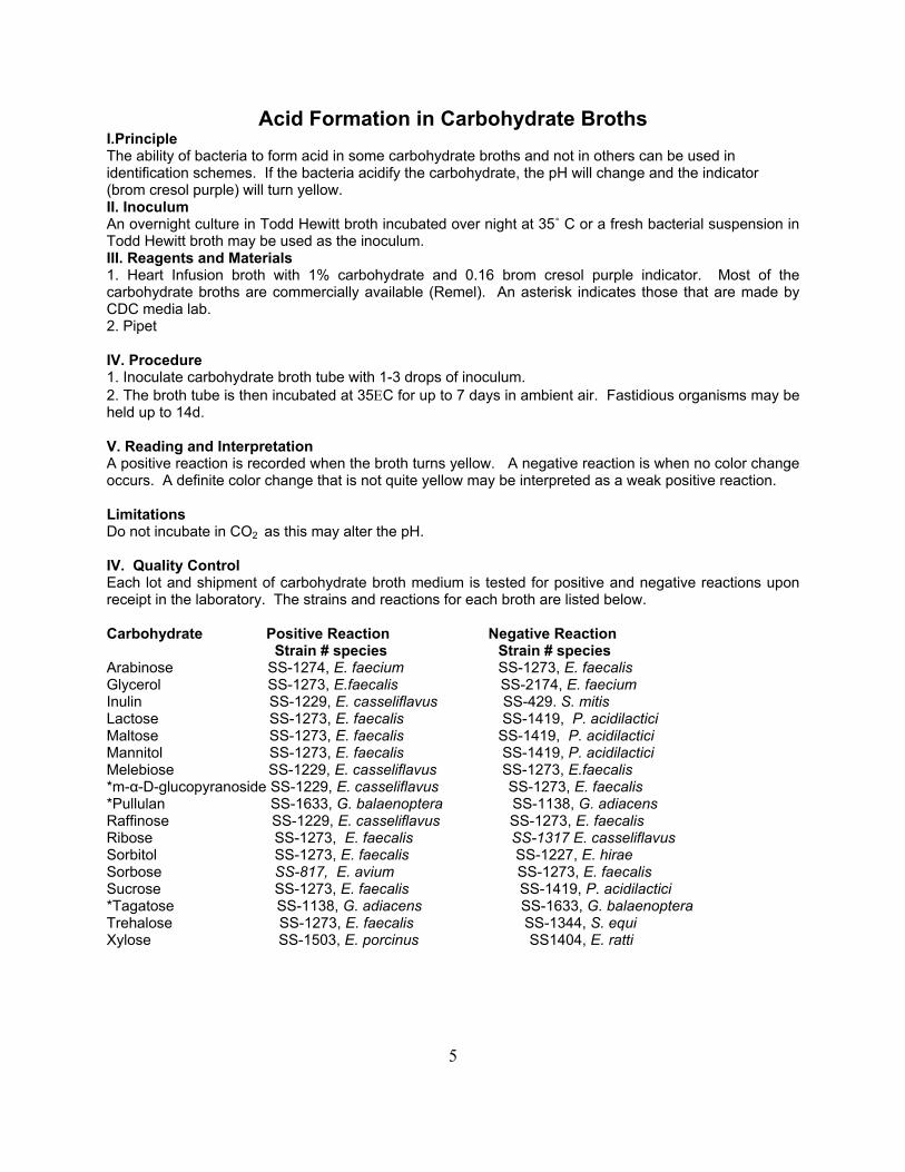
Acid Formation in Carbohydrate Broths I.Principle The ability of bacteria to form acid in some carbohydrate broths and not in others can be used in identification schemes. If the bacteria acidify the carbohydrate, the pH will change and the indicator (brom cresol purple) will turn yellow. II. Inoculum An overnight culture in Todd Hewitt broth incubated over night at 35˚ C or a fresh bacterial suspension in Todd Hewitt broth may be used as the inoculum. III. Reagents and Materials 1. Heart Infusion broth with 1% carbohydrate and 0.16 brom cresol purple indicator. Most of the carbohydrate broths are commercially available (Remel). An asterisk indicates those that are made by CDC media lab. 2. Pipet IV. Procedure 1. Inoculate carbohydrate broth tube with 1-3 drops of inoculum. 2. The broth tube is then incubated at 35ΕC for up to 7 days in ambient air. Fastidious organisms may be held up to 14d. V. Reading and Interpretation A positive reaction is recorded when the broth turns yellow. A negative reaction is when no color change occurs. A definite color change that is not quite yellow may be interpreted as a weak positive reaction. Limitations Do not incubate in CO2 as this may alter the pH. IV. Quality Control Each lot and shipment of carbohydrate broth medium is tested for positive and negative reactions upon receipt in the laboratory. The strains and reactions for each broth are listed below. Carbohydrate Positive Reaction Negative Reaction
Strain # species Strain # species Arabinose SS-1274, E. faecium SS-1273, E. faecalis Glycerol SS-1273, E.faecalis SS-2174, E. faecium Inulin SS-1229, E. casseliflavus SS-429. S. mitis Lactose SS-1273, E. faecalis SS-1419, P. acidilactici Maltose SS-1273, E. faecalis SS-1419, P. acidilactici Mannitol SS-1273, E. faecalis SS-1419, P. acidilactici Melebiose SS-1229, E. casseliflavus SS-1273, E.faecalis *m-α-D-glucopyranoside SS-1229, E. casseliflavus SS-1273, E. faecalis *Pullulan SS-1633, G. balaenoptera SS-1138, G. adiacens Raffinose SS-1229, E. casseliflavus SS-1273, E. faecalis Ribose SS-1273, E. faecalis SS-1317 E. casseliflavus Sorbitol SS-1273, E. faecalis SS-1227, E. hirae Sorbose SS-817, E. avium SS-1273, E. faecalis Sucrose SS-1273, E. faecalis SS-1419, P. acidilactici *Tagatose SS-1138, G. adiacens SS-1633, G. balaenoptera Trehalose SS-1273, E. faecalis SS-1344, S. equi Xylose SS-1503, E. porcinus SS1404, E. ratti
6
Arginine Hydrolysis I.Principle Certain bacteria contain the enzymes to hydrolyze arginine. This hydrolysis results in an alkaline change in the media results in a color change in the media. This test can be used for differentiated different bacteria. II. Specimen A drop of Todd Hewitt broth culture grown overnight is the preferred inoculum. Alternatively a suspension in Todd Hewitt broth from growth on a plate or a tiny amount of growth from a plate may be used as the inoculum. III. Reagents and Materials 1. Moeller’s decarboxylase broth containing arginine. The medium is commercially available. 2. Pipet or loop IV. Procedure 1. Add 1-3 drops of culture suspension to the tube of Moeller's decarboxylase medium containing arginine 2. Immediately overlay with sterile mineral oil (about 1 to 2 ml). 3. The medium is incubated at 35Ε C for up to 7 days in ambient air. (Some fastidious organism may be held up to 14d.) V. Reading and Interpretation. A positive reaction is recorded with the broth turns a deep purple color indicating an alkaline reaction, NH3 is released. The development of a yellow color or no change in color of the broth indicates a negative reaction. VI. Quality Control Each new lot and shipment of medium is tested for positive and negative reactions. E. faecalis strain SS1273 is used for determining positive reactions and S. avium strain SS817 is used for determining negative reactions.
7
Bacitracin Test
I. Principle The bacitracin disk is sensitivity test used to differentiate the beta- hemolytic Streptococcus. II. Inoculum An overnight culture grown on 5% sheep blood agar incubated 35˚C in CO2. III. Reagents and Materials 1. bacitracin “A” disk (BBL) IV. Procedure 1. Select a beta-hemolytic colony and heavily inoculate a quadrant of a 5% sheep blood agar plate. 2. Drop an “A” disk in the heaviest zone of inoculation. 3. Tap disk lightly to ensure that it adheres to the agar. 3. Incubate plate overnight in CO2 at 35˚C. V. Reading and Interpretation Any zone of inhibition is considered a positive test or sensitive test. Growth to the edge of the disk is interpreted as a negative test or resistant test. VI. Limitations VII.Quality Control Quality Control is performed on each shipment and lot of bacitracin disk. Streptococcus pyogenes is the positive (sensitive control) and Enterococcus faecalis SS1273 is the resistant or negative control. Results are recorded in the QC log book.
8
Bile Esculin Test I.Principle A selective and differential medium used in the identification of catalase-negative bacteria. The selective agent bile, inhibits most gram positive bacteria. The enterococci and Streptococcus bovis will grow. Esculin in the medium is hydrolyzed to esculetin and dextrose. The esculetin reacts with ferric chloride in the media to form a black-brown color. II. Inoculum An overnight culture in Todd Hewitt broth incubated over night at 35˚ C or a fresh bacterial suspension in Todd Hewitt broth may be used as the inoculum. An inoculating loopful of culture may also be used. III. Reagents and Materials 1. Bile esculin slant (Remel) IV. Procedure 1. Inoculate tube with 1 drop of inoculum allowing drop to run down slant. Alternatively, the slant may be inoculated with a loopful of growth from a blood agar plate. 2. The slant is then incubated at 35ΕC for 2 days in ambient air. Fastidious organisms may be held up to 14d. V. Reading and Interpretation The bile esculin test is positive when a black color forms over one-half or more of the slant. If no blackening occurs the test is negative. VI. Limitations Do not incubate medium in a carbon dioxide atmosphere. The increase in C02 will cause the viridans streptococci to grow better and increase the likelihood of a positive BE reaction. Streptococcus bovis and enterococci do not require C02 for good growth. VII. Quality Control Positive and negative reactions are determined on each new lot and shipment of media. Enterococcus faecalis strain SS-1273 is used for positive control reactions and Streptococcus sanguinis strain SS-910 is used for negative control reactions. Results are recorded in the QC log book.
9
Bile Solubility Test
I.Principle The purpose of the bile solubility test is to aid in the differentiation of S. pneumoniae from all other alpha-hemolytic streptococci. Sodium deoxycholate (2%) acts on the cell wall of pneumococci resulting in lysis. II. Inoculum An overnight culture grown on blood agar incubated 35˚C in CO2. III. Reagents and Materials 1. 2% deoxycholate (CDC Central Services Laboratory, formula #5333) 2. physiologic saline pH 7.0 3.13 X 100mm glass tube IV. Procedure 1. Make a 1.0 ml saline suspension of cells from growth on an agar plate. A turbidity equal to that of 1.0 to 2.0 McFarland density standard should be used. 2. After a satisfactory density is achieved, divide the suspension into 2 tubes with approximately 0.5 ml in each. 3. Add 0.5 ml of 2% sodium deoxycholate (bile salts) to one tube and 0.5 ml saline to the other tube. Mix by vigorous shaking. 4. Incubate the tubes at 35-37Ε C for up to 2 h.
V. Reading and Interpretation Examine for clearing of the turbidity periodically. A clearing of the turbidity in the bile tube but not in the saline control tube indicates a positive test, i.e., the pneumococcal cells have lysed ("solubilized"). If the tube containing the cells and bile have not cleared the test is negative. On occasion some strains of pneumococci are only partially soluble in the bile salts, that is, a partial clearing occurs. These strains must have the proper zone of inhibition around the optochin test to be called pneumococci. Partially soluble strains with zones of inhibition of less than 14 mm are not considered pneumococci. VI. Limitations The turbidity must be sufficient to detect a difference in the saline control tube. VII. Quality Control Each new lot of deoxycholate is tested for positive and negative reactions with S. pneumoniae strain ATCC-49619 (positive) and S. mitis strain SS-429 (negative). Results are recorded in QC log book.
10
Camp Test
I. Principle Some bacteria produce CAMP factor (a diffusible extracelluar protein) that synergistically acts with the beta-lysin of Staphylococcus aureus and enhances the lysis of red blood cells. The purpose of the CAMP test is to aid in the identification of nonhemolytic group B streptococci and other ß-hemolytic streptococci. II. Specimen Growth from a blood agar plate or any solid media. III. Reagents and Materials TSA-sheep blood agar IV. Procedure 1. The CAMP test is performed on TSA-sheep blood agar. A single streak of ß-lysin producing S. aureus made across the center of the plate. Strain SS-695 (Strep. Lab number is a ß-lysin producing strain of S. aureus. 2. A single colony of the unknown strain (beta hemolytic streptococci) is picked up with an inoculating loop and used to make a single streak perpendicular but not touching the S. aureus streak. A 2-3 mm space should remain between the streaks. 3. Incubate the inoculated plate normal atmosphere overnight at 35ΕC. Group B streptococci and a few other beta-streptococci produce an enhancement of the ß-lysin activity of the S. aureus strain. V. Reading and Interpretation This enhanced activity is in the shape of an arrowhead at the juncture of the two streaks, with the widest portion of the arrowhead on the group B side. VI. Limitations Do not incubate in an anaerobic environment or under CO2. Some S. pyogenes strains will give a positive reaction when incubated in CO2. VII. Quality Control Commercially available TSA-sheep blood agar does not always demonstrate the correct CAMP reaction. Therefore it is necessary to test a known group B streptococcus for CAMP reaction as a positive control on each test plate. Group B streptococcus strain SS-617 should be used as a positive control on each test plate.

11
Catalase Test
I. Principal Hydrogen peroxide is used (H2O2) to determine if bacteria produce the enzyme catalase.
II. Specimen Cultures that are grown on a blood free media or a colony grown on a blood agar plate that is carefully transferred to a slide without carry-over of any of the erythrocytes. Cultures are typically grown overnight at 35˚C in CO2. III. Reagents and Materials 1. Three percent hydrogen peroxide is obtained from a commercial drug store. 2. Pipet 3. Slides
IV. Procedure 1. The catalase test is best performed by flooding the growth of the bacteria (usually on an agar slant but blood free agar plates can be used) in question with 1.0 ml of 3% hydrogen peroxide and observing for effervescence (bubbling) which indicates a positive test. The bacteria must be grown on blood free medium. 2. Modifications of the catalase test may be performed by very carefully removing a colony of growth from a blood agar plate with a plastic needle or wooden applicator stick and transferring the colony to a glass slide. A drop of 3% hydrogen peroxide is added to the colony on the slide and observed for effervescence. V. Reading and Interpretation Any sign of bubbling is interpreted as a positive test. The absence of bubbling is interpreted as negative. VI. Limitations False positive results will result if any red bloods cell are transferred. Weak positive results should be repeated on a blood free medium. The catalase test gives the majority of differentiations very efficiently, however, there will be occasions when the catalase test and colony morphology will be misleading.
VII. Quality control The catalase quality control is performed once per lot and shipment. For positive reaction use a blood-free culture of Staphylococcus aureus: i. e., Cowen strain I but other confirmed Staphylococcal cultures can be used. For negative reaction use Streptococcus sanguinis strain SS-910 (ATCC-10556). Record in QC manual.

12
Clindamycin Test I. Principle Resistance of bacteria to clindamycin is determined by using a clindamycin disk at a concentration of 2µg/ml. This resistance is useful in differentiating the Lactococcus species. II. Inoculum Growth from a blood agar plate or any solid media. III. Reagents and Materials 1. clindamycin disk 2µg/ml 2. blood agar plate IV. Procedure Bacteria are spread onto a 5% sheep blood agar plate, the disk placed on the plate and the plate incubated at 35ΕC for 18-24 hr in CO2. V. Reading and Interpretation Any zone of inhibition around the disk is considered sensitive. Zones are usually ∃20mm. VI. Limitations VII. Quality Control The quality control is tested with each lot and shipment. Lactococcus lactis is used for the negative control (Sensitive) and L. garviae is used for the positive control (Resistant).
13
Esculin Hydrolysis
I.Principle A differential medium used in the identification of catalase-negative bacteria. Esculin in the medium is hydrolyzed to esculetin and dextrose. The esculetin reacts with ferric chloride in the media to form a black-brown color. II. Specimen An overnight culture in Todd Hewitt broth incubated over night at 35˚ C or a fresh bacterial suspension in Todd Hewitt broth may be used as the inoculum. An inoculating loopful of culture from a blood agar plate may also be used. III. Reagents and Materials 1. Esculin slant (Remel) IV. Procedure 1. Inoculate slant tube with 1-3 drops of inoculum allowing drop to run down slant. Alternatively, the slant may be inoculated with a loopful of growth from a blood agar plate. 2. The slant is then incubated at 35ΕC for 7 days in ambient air. Fastidious organisms may be held up to 14d. V. Reading and Interpretation The esculin test is positive when a black color forms over one-half or more of the slant. If no blackening occurs the test is negative. VI. Limitations Do not incubate medium in a carbon dioxide atmosphere. The increase in C02 will cause the viridans streptococci to grow better and increase the likelihood of a positive reaction. Streptococcus bovis and enterococci do not require C02 for good growth. VII. Quality Control Positive and negative reactions are determined on each new lot and shipment of media. Enterococcus faecalis strain SS-1273 is used for positive control reactions and Streptococcus mitis strain SS-429 is used for negative control reactions. Results are recorded in the QC log book.
14
GAS FROM GLUCOSE BROTH (MRS BROTH) I.Principle The production of gas from glucose is tested in Lactobacillus MRS broth. II. Specimen An overnight culture in Todd Hewitt broth incubated overnight at 35˚ C or a fresh bacterial suspension in Todd Hewitt broth may be used as the inoculum. An inoculating loopful of culture from a blood agar plate may also be used. III. Reagents and Materials The MRS broth is prepared in the CDC Central Services Laboratory, formula No. 9208. The petroleum jelly is also prepared in the CDC Central Services Laboratory, formula No. 9356. IV. Procedure The broth is inoculated with 2 or more colonies from a plate or with 1 to 2 drops of broth culture. The broth is then sealed with melted petroleum jelly and, the tube is incubated at ambient air 37˚ C up to 7 days. V. Results Gas production is indicated by the gas formation between the broth and the petroleum jelly plug which pushes the wax plug toward the top of the tube. Small bubbles that may accumulate over the incubation period are not read as positive, only when the wax plug is separated from the broth is the test read positive. Most leuconostoc strains are positive at 24 h but some strains may take longer. VI. Limitations VII. Quality Control Each new lot of MRS broth prepared by the CDC Central Services Laboratory is tested for positive (gas production) and negative (no gas production) reactions. Leuconostoc mesenteroides strain SS-1238 (ATCC-8293) is used for positive and Streptococcus sanguinis strain SS-910 (ATCC-10556) is used for negative gas production. Results are recorded in the QC log.
15
Gram Stain I. Principle The gram stain is used to differentiate between gram-positive and gram-negative bacteria. Cellular morphology can also be determined. Gram-positive and gram-negative bacteria are both stained by crystal violet. The addition of iodine forms a complex within the cell wall. Addition of a decolorizer removes the stain from gram-negative organisms due to their increased lipid content. These cells are stained pink with the counter stain safranin. II. Specimen The gram stain can be performed on the growth of any strain grown on any type of media. However, for this group of bacteria (gram-positive cocci), it is best performed on the growth of bacteria in thioglycolate broth at 24h incubation. The staining procedure is modified when preparing the smear from thioglycolate broth. The smear can not be fixed to the slide with hear but must be fixed with methanol. III. Reagents and Material (Store at room temperature) 1. Crystal Violet Stain 2. Gram Iodine (Combine Gram Iodine Concentrate to Gram Iodine Diluent) 3. Decolorizer Solution 4. Methanol 5. Slides 6. Inoculating loop 7. Microscope with Immersion Objective IV. Procedure 1. Spread single loop of culture from the thioglycolate broth to a microscope slide. Spread the culture over 1/3 to ½ to the total area of the slide. 2. Allow the smear to air dry. This may take up to 1 hour depending on the temperature and humidity of the room. 3. Cover the entire bacterial smear with 3 or 4 drops of methanol to fix the smear and allow to air dry. Again this may take up to an hour. 4. Cover the bacterial smear with crystal violet stain and allow to stand 1 minute. Gently was the stain off with cool tap water and drain water from slide. 5. Cover the smear with grams iodine and allow to stand 1 minute. Gently wash the iodine off with water and drain the water from the slide. 6. Rinse the bacterial smear with decolorizer solution for 10 seconds; decolorization is complete when the solution runs clear from the slide. Gently rinse with water and drain the slide. 7. Cover the bacterial smear with safranin stain, and allow to stand for 1 minute, then gently wash the stain from the slide. 8. Blot the slide dry with absorbent paper and examine the slide under oil immersion lens. V. Reading and Interpretation The gram stain is used to aid in the differentiation of the gram positive cocci. The arrangement of the cells is what helps to differentiate the genera. Bacteria that divide on random planes form grape-like clusters of cells. This is the type of arrangement commonly observed with staphylococci. Bacteria that divide on one plane form pairs and eventually form chains if the cells remain attached to each other. This type of cellular morphology is observed with streptococci. Bacteria that divide on two planes at right angles form packets of fours or tetrads. This type of arrangement is observed with the aerococci. One of the most difficult tasks that microbiologist have is determining whether or not the cellular morphology of the cells are actually cocci or short rods. Since many of the lactobacilli are gram positive short rods in chains, they are sometimes confused with the streptococci. The clinical sources and
16
colonial morphology on blood agar plates of the lactobacilli are also similar to the streptococci, especially members of the viridans streptococci. When reading the gram stain, remember that the cellular arrangement is never 100% chains, pairs, tetrads, or clusters. The microbiologist must determine the most common cellular arrangement. For example, for the Gemella species, one might observe some pairs and short chains as well as tetrads. If tetrads are observed in most fields under observation, then the strain is dividing on two planes and this should be recorded. VI. Limitations Younger cultures give more characteristic observations that older ones. Older cultures may stain gram negative. Stains exposed to antimicrobial reagents may have atypical morphology and are more susceptible to decolorization. Gram positive organisms that are over-decolorized will appear gram-negative. VII. Quality Control The gram stain quality control is performed once per week.Inoculate Streptococcus sanguinis strain SS910 and Escherichia coli 25922 into thioglycolate broth medium and incubate overnight at 35˚ C ambient air. Prepare the slide using 1 loopful of each culture on the same slide. Slides may be fixed in advance and stored. The completed procedure should show gram-positive cocci in chains and gram-negative rods. Record results in QC maual. VIII. References Murray, P.R., Baron, E. J., Jorgensen, J.J., Pfaller, M.A., and Yolken, R.H. Manual of Clinical Microbiology, 8th ed. ASM Press: Washington, DC, 2003.
17
Growth at 10C and 45C I.Principle Growth at 10C and 45C is determined in heart infusion broth base medium and can be used as differential test for catalase-negative gram positive cocci. II.Inoculum An overnight culture in Todd Hewitt broth incubated overnight at 35˚ C or a fresh bacterial suspension in Todd Hewitt broth may be used as the inoculum. An inoculating loopful of culture may also be used. III. Reagents and Materials 1. Heart infusion broth base medium Remel number 061030. IV. Procedure 1. Two of the broth tubes are inoculated with one or two colonies or one to two drops of an overnight Todd Hewitt broth culture. 2. Incubate each tube at the respective temperatures, 10C and 45C. For the 10C incubator, a refrigerator that can be adjusted to hold a temperature of 10C is satisfactory. The refrigerator can be used for storage of media and other materials. For the 45C incubator, a hot water bath that is adjusted to hold a temperature at 45C is best. 3. The tests are held a minimum of 7 days and up to 14 days in the case of slow growing strains. V. Reading and Interpretation An increase in turbidity indicates growth and a positive test. Color changes are not required for a positive test. VI. Limitations Be sure that the water level in the 45C water bath is above the level of media in the tubes. In addition, the caps of the medium tubes should be carefully sealed with parafilm or other sealer to prevent moisture and contamination from penetrating the medium tube.
VII. Quality control Each new lot of broth is tested for positive and negative reactions. Enterococcus faecalis strain SS-1273 is used for positive (growth) reactions at both 10C and 45C. Streptococcus sanguinis strain SS-910 is used for negative reactions (no growth) at both 10C and 45C.
18
Hemolysis I. Principle The hemolytic reaction is particularly useful in the differentiation of the Streptococci. The hemolytic reaction is determined on agar media containing 5% animal blood. The most commonly used base medium is trypticase soy agar and the most commonly used blood is sheep blood. The reason for the use of trypticase soy base is that it supports the growth of all the bacteria listed in Table 2. Other base media may be substituted if control strains of all genera are tested for growth. Sheep blood is used because of the convenience in testing throat swabs for ß-hemolytic streptococci. Sheep blood does not support the growth of Haemophilus haemolyticus which appears similar to streptococci on agar containing rabbit, horse, or human blood. II. Inoculum Pure culture on solid media. III. Reagents 1. Trypticase soy agar plates containing 5% sheep blood are obtained from Becton-Dickinson Microbiology Systems, Cockysville Md., product No. 21261. IV. Procedure 1. Streak culture for isolation on TSA plate with 5% sheep blood. 2. Incubate plate at 35˚C in CO2 for 24 h. V. Reading and Interpretation A beta-hemolytic reaction is interpreted as complete clearing around the colony. An alpha-hemolyic reaction is interpreted as greening around the colony and gamma hemolysis is interpreted as no change in the media surrounding the colony. The hemolytic reaction on blood agar is complex and subject to many variables. For the complete explanation and interpretation of the reactions the reader is referred to the 1977 publication, CDC Laboratory Update, Isolation and Identification of Streptococci. Part I. Collection, Transport, and Determination Hemolysis, Annex 1. Application of the interpretations of the hemolytic reactions for streptococci described in the Update to the other genera should not be a problem. VII. Limitatitions VIII. Quality control Under normal operating conditions, the blood agar plates are not quality controlled in our laboratory but are quality controlled by Becton-Dickinson Microbiology Systems. The hemolytic reaction of most bacteria submitted to this laboratory is known and identified with the culture upon submission. If a problem should be identified then quality control strains are used for testing the proper hemolytic reactions. Streptococcus pyogenes strain SS-103 is used for determining beta-hemolysis, Streptococcus sanguinis strain SS-910 is used for determining alpha-hemolysis, and Streptococcus bovis strain SS-752 is used for determining no reaction (neither beta or alpha hemolysis) on trypticase soy 5% sheep blood agar plates. All reactions are determined according to the 1977 CDC Laboratory Update, Isolation and Identification of Streptococci. Part I. Collection, Transport and Determination of Hemolysis (See annex 1).
19
Hipppurate Hydrolysis Test I. Principle Some bacteria produce the enzyme hippurate hydrolase which hydrolyzes sodium hippurate to form benzoic acid and glycine. The addition of ferric chloride to benzoic acid forms an insoluble brown ferric benzoate precipitate. II. Inoculum An overnight culture in Todd Hewitt broth incubated at 35˚ C or a fresh bacterial suspension in Todd Hewitt broth may be used as the inoculum. An inoculating loopful of culture from a blood agar plate may also be used. III. Reagents and Materials 1. Hippurate broth commercial suppliers. 2. Ferric choride (FeCl3) commercial supplies. Labeled as TDA if purchased for bioMereiux
IV. Procedure 1. The hippurate broth is inoculated with one drop of a fresh (16-20 h) Todd-Hewitt broth culture. 2. The broth is incubated for up to 7 days or until turbid growth is seen at 35ΕC. 3. The tube of broth is then centrifuged to sediment the bacteria. 4. Pipette 0.8 ml the clear supernatant to a small clear tube (13 x 100). 5. Add 0.2 ml of ferric chloride reagent to the supernatant. Mix well. V. Results and Interpretation A heavy precipitate that does not clear within 10 minutes indicates a positive test. A clear golden-brown liquid indicates a negative test.
VI. Limitations Growth should be turbid before testing. Some fastidious organisms may show poor growth. VII. Quality Control Group B Streptococcus strain SS-620 is used as a positive control and E. avium strain SS-817 is used as a negative control. QC is performed with each new lot number and shipment of hippurate broth and with each new lot and shipment of ferric chloride reagent. The quality control test results are recorded in the QC log.

20
Lancefield Group Antigen
I. Principle The purpose of determining the group antigen of ß-hemolytic streptococci is identify the species or species/group of streptococci as originally described by Rebecca Lancefield. Acid extraction is used to remove the serogroup from the cell. II. Inoculum An overnight 50 ml culture in Todd Hewitt broth incubated at 35˚ C. III. Reagents and Materials 1. 50 ml culture 2. 50 µl capillary tube 3. m-cresol purple 4. 13 X 100 mm test tube 5. boiling water bath 6. centrifuge 7. 1 cc vial 8. Serogrouping Reagents VI. Procedure 1. Centrifuge cells in 20 minutes at 2000 rpm to achieve a cell pellet. 2. Aspirate off the supernatant. 3. Add 2-3 drops m-cresol purple. Add .1 N HCl dropwise until a color change to pink. Vortex. 4. Place tube in a boiling water bath for 10 minutes. 5. After allowing to cool, transfer contents to a 13 X100 test tube. 6. Centrifuge at 2800 rpm for 10 minutes to remove any precipitate. 7. Transfer to a clean 13 X 100 mm test tube. 8. Add .5 N NaOH dropwise until a change to purple. 9. Centrifuge at 2800 rpm for 10 minutes to remove any precipitate. 10. Transfer to a 1ml screwcap storage vial. 11. Set of capillary tubes by adding approximately 1 cm of serogrouping reagent and 1cm of extract. Careful not to get any space or bubbles between the two! 12. Cap the end with clay and set in clay rack. 13. Observe for up to 30 minutes. V. Reading and Interpretation A definite line or zone of precipitation that forms between the two is considered positive. IV. Limitations Group D sometimes give weak reactions. VII. Quality Control Quality control is performed with the preparations of each batch of serogrouping reagent.
21
Leucine amino peptidase (LAP)
I.Principle Some bacteria produce leucine aminopeptidase which hydrolyzes the substrate leucine-β-naphthylamide to form β-naphthylamine. A pink to red color forms when p-dimethylaminocinnamaldehyde (PYR reagent) is added to β-naphthylamine. II. Inoculum Strains are grown on blood agar plates overnight at 35˚C in CO2 for most gram positive bacteria. More than 1 day of incubation may be necessary for more fastidious genera such as the gemellae, alloiococci, and helcococci. The strains to be tested are grown on a blood agar plate until sufficient growth is seen to heavily inoculate the disks. III. Reagents and Materials LAP disk (Remel) PYR reagent Loops Deionized Sterile water IV. Procedure The procedure that is used in the Streptococcus laboratory is modified from the package insert. The PYR test is usually done simultaneously. 1. Place the disks on blood agar plate in an area of little or no growth or on a slide. The moisture from the plate is usually sufficient to rehydrate the disk. If the disk is placed on a slide, then a tiny drop of sterile deionized water is added. (DO NOT OVERSATURATE THE DISK). It is convenient to place the LAP disk on the left (L=LAP). 2. Using a loop or wooden stick, inoculate the disks heavily. Using two or more loop-fulls of culture is necessary for satisfactory results. 3. Leave the plates with the disks on the bench at room temperature for 10 minutes. 4. Add the detection reagent and read after 3 minutes.
V. Reading and Interpretation The development of a red color within 3 minutes is positive. No change in color or a yellow color is negative. The color develops immediately. Discard the test after 10 minutes. VI. Limitations False negative reactions may result if too little inoculum is used. VII. Quality Control Each lot and shipment of LAP disks are tested for positive and negative reactions. Enterococcus faecalis strain SS-498 is used for positive reaction and Aerococcus viridans strain SS-1251 (ATCC-11563) is used for a negative reaction.
22
Litmus Milk Test I. Principle The purpose of the litmus milk test is to determine the acidification and clot of the milk in this test. This tests aids in the differentiation of the Leuconostoc species. II. Inoculum An overnight culture in Todd Hewitt broth incubated at 35˚ C or a fresh bacterial suspension in Todd Hewitt broth may be used as the inoculum. An inoculating loopful of culture may also be used. III. Reagents and Materials Litmus milk is obtained from commercial suppliers using their quality control. IV. Procedure 1. Tubes containing litmus milk are inoculated with one drop of an overnight Todd-Hewitt broth culture. 2. Incubate at 35ΕC for up to 7 days in ambient air. Some fastidious strains may be held for 14 d. V. Reading and Interpretation The tubes are inspected for color change. The tubes begin a light blue color. Acid formation is indicated by first a pink color and then changes to white on continued incubation. A negative reaction is indicated by no color change. The tubes are also examined for clot, or solidification of the tube contents. Partial or complete solidification of the tube contents indicates a positive reaction. A negative reaction is indicated by no change in the consistency of the tube contents. VI. Limitations VII. Quality Control Enterococcus faecalis SS-1273 is used as a positive control and E. hirae strain SS-1227 is used as a negative control. QC is performed with each new lot number and shipment. The quality control test results are recorded in the QC manual.
23
Motility Test I.Principle The ability of bacteria to move through a semisolid media is useful in differentiating bacteria. This test is particularly useful in differentiating the enterococci. II. Inoculum Strains are grown on blood agar plates overnight at 35˚C in CO2 for most gram positive bacteria. More than 1 day of incubation may be necessary for more fastidious genera such as the gemellae, alloiococci, and helcococci. III. Reagents and Materials 1. motility test medium (Remel, number 061408) 2. inoculating needle IV. Procedure 1. The medium is inoculated with an inoculating needle, not a loop. Apply a colony to the end of the needle from the agar plate. 2. The needle is inserted into the center of the medium in the tube for about one inch. 3. The inoculated tube is incubated at 30C in ambient air and incubated until good growth is observed, in most cases 24 to 48 h is sufficient. V. Reading and Interpretation Strains that are motile will show growth outward to the edge of the tube and downward toward the bottom of the tube. Negative strains will only show growth in the line of the stab. VI. Limitations Do not place the motility test at 37C. Some strains become nonmotile at 37C but are motile at temperatures 25C to 30C. VII.Quality Control Enterococcus gallinarium SS-1228 is used as a positive control and E. faecalis strain SS-1273 is used as a negative control. QC is performed with each new lot number and shipment.
24
6.5% NaCl Tolerance Test I. Principle Tolerance tests can be used in the differentiation of microorganisms. Some bacteria can grow in 6.5% NaCl and others are inhibited by these concentrations. Growth in broth containing 6.5% NaCl is determined in heart infusion broth base with the addition of 6% more NaCl. Heart infusion base contains 0.5% NaCl. To make the test easier to read we add 0.5% dextrose and brom cresol purple indicator. II. Inoculum A fresh inoculum grow in Todd Hewitt broth is preferred. Alternatively, a small loopful of growth from a blood agar plate may be used. III. Reagents and Materials 6.5% NaCl broth 5 ml Todd Hewitt broth The 6.5% NaCl broth is prepared by the CDC Central Services Laboratory, formula No. 1707. This formulation is identical to the modified 6.5% NaCl broth described in: Facklam, R. 1973. Comparison of several laboratory media for presumptive identification of enterococci and group D streptococci. Appl. Microbiol. 26:138-145. IV. Procedure 1. One or two colonies or one or two drops of an overnight broth culture is inoculated into the broth containing 6.5% NaCl. 2. The inoculated broth is incubated at 37ΕC in ambient air for up to a week or more depending upon the growth characteristics of the strain being tested. If 2 or 3 days were required for sufficient inoculum then the NaCl tolerance test should be incubated 10 to 14 days. In some cases (most enterococci) the test is positive after overnight incubation Reading and Interpretation When most strains grow the dextrose is fermented and the broth changes from purple to yellow color. However, the color change is not required for a positive test. If there is an obvious increase in turbidity, which indicates growth, without a change in color this is also read as a positive test. Limitations Other base media (brain heart infusion and trypticase soy) have been used for determining NaCl tolerance of the enterococci and viridans streptococci but these bases have not been tested with the other genera. Quality control Each new lot of media prepared by the CDC Central Services Laboratory is tested for positive (growth) and negative (no growth) reactions. Enterococcus faecalis strain SS-1273 and Streptococcus sanguinis strain SS-910 are used for positive and negative reactions respectively.
25
Optochin Test
I.Principle The purpose of the optochin test is to confirm the identification S. pneumoniae before serotyping and to aid in the differentiation of S. pneumoniae from viridans streptococci during surveillance studies. II. Inoculum Isolated alpha-hemolytic colonies suspected of being pneumococci III. Reagents and Materials 1. optochin AP@ disks purchased from Becton Dickinson Microbiology Systems, Cockeysville, Md. 2. blood agar plates IV. Procedure 1. Transferred an isolated colony and streak to a quarter of a blood agar plate. 2. Place the optochin “P” disk in the upper third of the inoculum. Tap the disk to insure that it stays on the media after the plate is inverted. 3. The plate is incubated overnight at 35-37ΕC in a candle extinction jar or carbon dioxide incubator. V. Reading and Interpretation If a 6 mm disk is used, a zone of inhibition of at least 14 mm in diameter is considered positive for identification of pneumococci. A zone of inhibition between 6 and 14 mm in diameter is considered questionable for identification of pneumococci and a bile solubility test should be performed. Bile soluble strains with optochin zones of inhibition between 6 and 14 mm are considered pneumococci, those strains that are not bile soluble with the same zone sizes are not considered pneumococci. VI. Limitations Cultures do not grow as well in normal atmospheres and larger zones of inhibition can cause misidentification. VII Quality Control Each new lot and shipment of optochin AP@ disks is tested for positive and negative reactions. S. pneumoniae strain ATCC-49619 is inhibited by optochin (positive reaction) and S. mitis strain SS-429 is not inhibited by optochin (negative reaction). Results are recorded in the QC log book.
26
Pigmentation Test
I. Principle Some bacteria produce pigment. The purpose of the pigmentation test is to aid in the identification of E. casseliflavus, E. mundtii, E. pallens, E. gilvus and E. sulfureus. These enterococci produce a yellow pigment that can be detected on several different media. II. Inoculum The unknown Enterococcus strain is grown on trypticase-soy 5% sheep blood agar plate for 24 h in a normal atmosphere at 35ΕC. III. Reagents and Materials 1. cotton swab IV. Procedure 1. Use a cotton swab to pick up a 50 mm smear of bacteria. 2. Examine the swab and smear for a bright yellow color. V. Reading and Interpretation A pale to bright yellow color is interpreted as positive. A cream, white, or grey color is negative. VI. Limitations The test should be repeated on the culture again after 48 h of incubation if there is any question about the results at 24 h. VII. Quality control E. casseliflavus strain SS-1229 is used for a positive control and E. faecalis strain SS-1273 is used for a negative control for determining pigmentation.
27
Pyridoxal Requirement Test (Vitamin B6) I. Principle Nutritionally variant streptococci (Abiotrophia and Granulicatella) are usually very fastidious and will grow only on supplemented media or enriched chocolate agar. These strains require pyridoxal for growth while non-NVS do not. A final concentration in broth of 0.001% of pyridoxal will support the growth of NVS. An alternative to performing the pyridoxal requirement test is the satellite test. II.Inoculum The cultures are typically received on chocolate agar slants. III. Reagents and Materials 1. 0.01% pyridoxal* 2. 2-TSA-sheep blood agar plate * It is convenient to keep a 0.01% solution of pyridoxal in the laboratory. This solution is prepared in purified water and filter sterilized. A 10 ml aliquot is dispensed into sterile tubes. This 0.01% solution should be kept frozen at -20Ε C. The aliquot in use may be stored at 4˚ C. IV. Procedure 1. Add a drop of the 0.01% solution onto the surface of one of the TSA-sheep blood agar plates. 2. Inoculate both plates with the suspected NVS strain. 3. Incubate at 35˚C in CO2 for 24 to 72 hours. V. Reading and Interpretation If growth is observed on the plate containing pyridoxal and not on the plate without pyridoxal the strain is a NVS. VI. Limitations VII. Quality Control There are no guidelines for quality control of the reagents or the media. Each lot of pyridoxal can be tested for supporting the growth of a known NVS strain.
28
Pyrrolidonylarylamidase Test (PYR)
I.Principle Some bacteria produce pyrrolidonyl arylamidase which hydrolyzes the substrate L- pyrrolidonyl -β-naphthylamide to form β-naphthylamine. A pink to red color forms when p-dimethylaminocinnam-aldehyde (PYR reagent) is added to β-naphthylamine. II. Inoculum Strains are grown on blood agar plates overnight at 35˚C in CO2 . More than 1 day of incubation may be necessary for more fastidious genera such as the gemellae, alloiococci, and helcococci. The strains to be tested are grown on a blood agar plate until sufficient growth is seen to heavily inoculate the disks. III. Reagents and Materials PYR disk (Remel) PYR reagent Loops Deionized Sterile water IV. Procedure The procedure that is used in the Streptococcus laboratory is modified from the package insert. The LAP test is usually done simultaneously. 1. Place the disks on blood agar plate in an area of little or no growth or on a slide. The moisture from the plate is usually sufficient to rehydrate the disk. If the disk is placed on a slide, then a tiny drop of sterile deionized water is added. (DO NOT OVERSATURATE THE DISK). 2. Using a loop or wooden stick, inoculate the disks heavily. Using two or more loop-fulls of culture is necessary for satisfactory results. 3. Leave the plates with the disks on the bench at room temperature for 10 minutes. 4. Add the detection reagent and read after 3 minutes.
V. Reading and Interpretation The development of a red color within 3 minutes is positive. No change in color or a yellow color is negative. The color develops immediately. Discard the test after 10 minutes. VI. Limitations False negative reactions may result if too little inoculum is used. VII. Quality Control Each lot and shipment of PYR disks are tested for positive and negative reactions. Enterococcus faecalis strain SS-498 is used for positive reaction and Streptococcus sanguinis strain SS-910 is used for a negative reaction.

29
Pyruvate Utilization Test
I. Principle Some bacteria possess the ability to utilize pyruvate which results in a change in pH. Bromthymol blue is added to the media as an indicator which results in a color change from blue-green to yellow. II. Inoculum A fresh inoculum grown in Todd Hewitt broth is preferred. Alternatively, a small loopful of growth from a blood agar plate may be used. III. Reagents and Materials Pyruvate broth (CDC Central Services Laboratory, formula # 1722) IV. Procedure 1. Incoculate the pyruvate broth with one drop of an overnight Todd-Hewitt broth culture of the unknown strain. 2. Incubate the tube at 35ΕC for 7 days in ambient air. Fastidious strains may be incubated for 14 days. V. Reading and Interpretation A positive reaction is indicated by the development of a yellow color. If the broth remains green or greenish yellow, the test result is negative. A yellow color with only a hint of green is usually a positive reaction. VI. Limitation VII. Quality Control Quality control tests are determined for each new batch of prepared medium. E. faecalis strain SS-1273 and S.sanguinis strain SS-910 are used as positive and negative controls respectively. Results are recorded in the QC log book.
30
Satellite Test (SAT) I. Principle Nutritionally variant streptococci (Abiotrophia and Granulicatella) are usually very fastidious and will grow only on supplemented media or enriched chocolate agar. The purpose of the SAT test is to determine if the unknown streptococcal strain is a nutritionally variant streptococci (NVS). These strains require pyridoxal for growth. Growth of Staphylococcus aureus on a TSA sheep blood agar plate releases these factors allowing NVS to grow. An alternative to performing the SAT test is to test for the requirement of vitamin B6 (pyridoxal). II.Inoculum The cultures are typically received on chocolate agar slants. III. Reagents and Materials 1. Staphylococcus aureus ATCC-25923 2. TSA-sheep blood agar plate IV. Procedure 1. Streak the submitted culture over the entire surface of a TSA-sheep blood agar plate with an inoculating loop. 2. A single streak of Staphylococcus aureus ATCC-25923 is then made across the middle of the agar plate. 3. Incubate the plate in a candle extinction jar or C02 incubator for 24 h or more. V. Reading and Interpretation If the strain is a NVS growth will appear only adjacent to the staphylococcus streak, about 2 to 5 mm wide. Streptococci that are not NVS will grow over the entire surface of the agar plate. VII. Quality Contol There are no quality control guidelines for testing the medium or the Staphylococcus strain for the satillitism test.
31
Starch Hydrolysis Test
I. Principle Some bacteria are able to hydrolyze starch on starch supplemented agar. When iodine is added to starch, it turns dark bluish-black. If starch has been hydrolyzed, then it is not available to react with the iodine and the area around the bacterial growth is clear. This test can be used to differentiate some bacteria. II. Inoculum Strains are grown on blood agar plates overnight at 35˚C in CO2 . More than 1 day of incubation may be necessary for more fastidious genera such as the gemellae, alloiococci, and helcococci. A drop of Todd Hewitt broth bacterial suspension may also be used to streak the plate. III. Reagents and Materials 1. Two percent starch agar (CDC Central Services Laboratory, formula #1710.) 2. Gram’s iodine IV. Procedure 1. Inoculate a starch agar plate with a heavy single streak of a fresh culture or run a drop of broth culture across the plate. 2. Incubate the plate in CO2 at 35ΕC for 48 h. Some strains may require longer incubation for sufficient growth. 3. After incubation, flood the plate with Gram’s iodine. V. Reading and Intepretation A clear zone surrounding the growth is positive test that the strain hydrolyzed starch. A deep purple to black or bluish color of the agar indicates that starch has not been hydrolyzed and thus a negative test. For negative tests the deep color develops in the agar right up to the growth. VI. Limitations Some fastidious strains show poor growth. VII. Quality Control Each new batch of starch agar is quality control tested. S. bovis strain SS-1224 is used for a positive control and E. avium strain SS-817 is used for a negative control.
32
5% Sucrose Agar Slime Formation I. Principle The purpose of growing the unknown strains on agar containing 5% sucrose is to determine the capacity of the strains to form extracellular polysaccharide (levans or dextrans) on the agar. II. Inoculum Strains are grown on blood agar plates overnight at 35˚C in CO2 . More than 1 day of incubation may be necessary for more fastidious genera such as the gemellae, alloiococci, and helcococci. A drop of Todd Hewitt broth bacterial suspension may also be used to streak the plate. III. Reagents and Materials 5% sucrose agar plates (CDC Central Services Laboratory, formula # 1714) IV. Procedure 1. Inoculate the 5% sucrose agar with a fresh inoculum and streak for isolated colonies. 2. Incubate the agar plate in CO2 at 35ΕC for 48 h. Some fastidious strains may require longer incubation for growth. 3. Examine the plate for levans and dextrans. A loop is used to scrape the colonies for viscosity or adherence. V. Reading and Interpretation Some bacteria produce a levan as the extracellular polysaccharide. The colonies appear very slimy, mucoidal and runny or as large gum drops on the agar. Some bacteria may produce dextrans in which the colonies are dry and adherent to the plate. A negative reaction is the failure to see extracellular material on the 5% sucrose agar by visual inspection or adherence with a loop. VI. Limitations VII. Quality Contol Quality control tests are performed on each batch of 5% sucrose agar. The following strains are use for QC: Leuconostoc mesenteroides strain SS-1238 (positive for slime production-levans), Streptococcus sanguinis SS910 (positive for dextrans-adherent) and Enterococcus faecalis strain SS-1273 (negative for levans and dextrans). Results are recorded in the QC log book.
33
5% Sucrose Broth I. Principle The purpose of growing the unknown strains on agar containing 5% sucrose is to determine the capacity of the strains to form extracellular polysaccharide (levans or dextrans) in the broth. II. Inoculum Strains are grown on blood agar plates overnight at 35˚C in CO2 . More than 1 day of incubation may be necessary for more fastidious genera such as the gemellae, alloiococci, and helcococci. A drop of Todd Hewitt broth bacterial suspension may also be used to streak the plate. III. Reagents and Materials 5% sucrose broth tubes (CDC Central Services Laboratory) IV. Procedure 1. Inoculate the 5% sucrose broth with a fresh inoculum. 2. Incubate broth at 35ΕC for 48 h or longer. Some fastidious strains may require longer incubation for growth. 3. Examine the broth for viscosity or a gel button. V. Reading and Interpretation Some bacteria produce a levan as the extracellular polysaccharide. The broth appears very thick and slimey. Some bacteria may produce dextrans in which a gel button adhers to the bottom or sides of the tube. A negative reaction is the failure to see viscosity or adherence in the 5% sucrose broth by visual inspection. There is only an increase in turbidity. VI. Limitations VII. Quality Contol Quality control tests are performed on each batch of 5% sucrose both. The following strains are use for QC: Leuconostoc mesenteroides strain SS-1238 (positive for slime production-levans), Streptococcus sanguinis SS910 (positive for dextrans-adherent gel button) and Enterococcus faecalis strain SS-1273 (negative for levans and dextrans). Results are recorded in the QC log book.
34
Tellurite Tolerance Test
I. Principle Tolerance to tellurite is determined on agar medium containing 0.04% potassium tellurite. Very few catalase-negative gram-positive cocci will grow on this medium. The primary purpose of the tellurite tolerance test is aid in the differentiation of E. faecalis and E. faecium and the other enterococci. II. Inoculum Strains are grown on blood agar plates overnight at 35˚C in CO2 . More than 1 day of incubation may be necessary for more fastidious genera such as the gemellae, alloiococci, and helcococci. A drop of Todd Hewitt broth bacterial suspension may also be used to streak the slant. III. Reagents and Materials 1.Tellurite agar slants (0.04% potassium tellurite, CDC Central Services Laboratory, formula # 9358) IV. Procedure 1. Inoculate the slant with one drop of a fresh (18-24 h) Todd-Hewitt broth culture or loopful of bacteria from a fresh plate. 2. Incubate at 35ΕC for 7 days in ambient air. V. Reading and Interpretation Tolerance (a positive result) is indicated whenever black colonies form on the surface. Typical and variant strains of E. faecalis usually form black colonies (positive tolerance) after 48 h of incubation. Some strains of E. faecium may form grey colonies (a negative reaction) but most stains fail to grow on tellurite medium. A slight blackening at the bottom of the slant is a negative result. VI. Limitations VII. Quality Control Quality control tests are performed on each new batch of medium supplied. E. faecalis strain SS-1273 and S. sanguinis strain SS-910 are used as positive and negative controls respectively. Results are recorded in the QC log book.

35
Urea Hydrolysis
I. Principle Urea provides a source of nitrogen for bacteria producing urease. The resulting change in pH causes the indicator, phenol red, to change from yellow to a red to pink-red color. The urease test is to particularly useful to aid in the identification of Streptococcus salivarius. II. Inoculum Strains are grown on blood agar plates overnight at 35˚C in CO2 . More than 1 day of incubation may be necessary for more fastidious genera such as the gemellae, alloiococci, and helcococci. A drop of Todd Hewitt broth bacterial suspension may also be used to streak the slant. III. Reagents and Materials 1. Christensen's urea agar (Remel) IV. Procedure 1. Inoculate slant with a loopful or drop of a fresh culture. 2. Incubated at 35Ε C for up to 7 days in ambient air. Fastidious strains are incubated 14 day. V. Reading and Interpretation A positive reaction is recorded when a light or dark pink color develops in the agar slant. A yellow color or no change in the straw colored slant indicates a negative test. VI. Limitations False positives may result due to protein hydrolysis but this has not been observed with the Streptococcus. VII. Quality Control Quality control is performed with each lot and shipment.S. salivarius, SS-908 and E. avium, SS-817 are used for positive and negative controls respectively.
36
Vancomycin Test I.Principle The vancomycin test is performed in a fashion similar to the bacitracin sensitivity test. Resistance to vancomycin can be used to differentiate a few of the catalase-negative gram-positve genera. II. Inoculum Strains are grown on blood agar plates overnight at 35˚C in CO2 . More than 1 day of incubation may be necessary for more fastidious genera such as the gemellae, alloiococci, and helcococci. III. Reagents and Materials 1. 30µg vancomycin disk (Becton Dickinson Microbiology Systems, Cockysville Md. Product No. 31353) The disks are stored according to the manufacturer's instructions. 2. trypticase soy sheep blood agar plates IV. Procedure 1. Transfer several colonies of the strain in question to one-half of a blood agar plate and steak heavily. 2. Place the vancomycin susceptibility testing disk (30 µg) in the heavy part of the streak. 3. Incubated the plate in a CO2 enhanced atmosphere at 37ΕC overnight. Some strains (alloiococci, gemellae, helcococci) may require 48 h or more to show sufficient growth to interpret the test. V. Reading and Interpretation Any zone of growth inhibition is considered positive (sensitive). The test is interpreted as resistant (negative) only if there is growth right up to the edge of the disk. This is not a susceptibility test, it is a sensitivity test for identification. VI. Limitations Sufficient inoculum is required for an accurate test. VII. Quality Control Each new lot and shipment of vancomycin disks is tested for positive (sensitive) and negative (resistant) reactions. Streptococcus bovis SS1224 is used as a positive (sensitive) reaction and Leuconostoc mesenteroides strain SS-1238 (ATCC-8293) is used for the negative (resistant) reaction. Results are recorded in the QC log book.
37
Voges-Proskauer Test (VP) I. Principle The Colbentz modification of the Voges-Proskauer (VP) test can be used to determine the production of acetylmethyl carbinol. Test reactions are used for differentiation of bacteria. The purpose of the VP test is to aid in the identification of Streptococcus anginosus strains of ß-hemolytic streptococci (Table 2). The VP test can also be used to aid in the differentiation of the viridans streptococci into Species/groups (Table 6).
II. Inoculum A fresh inoculum grown in Todd Hewitt broth is preferred. Alternatively, a small loopful of growth from a blood agar plate may be used. III. Reagents and Materials 1. Voges-Proskauer (VP) test broth (Remel) 2. 40% KOH (bioMerieux) 3. alpha-naphthol (bioMerieux) IV. Procedure 1. Inoculate VP broth tube with one drop or loopful of fresh inoculum. 2. Incubate 24-48 hrs at 35˚C in ambient air or until turbid growth is observed. 3. Transfer 0.5 ml to a 13X100 glass test tube. 4. Add twelve drops alpha naphthol and 4 drops 40% KOH. 5. Carefully vortex or shake tubes and observed for 30 minutes. The tube must be vigorously shaken several times during the 30 minute period. V. Reading and Interpretaion A positive reaction, a red color, is usually seen within 30 minutes. For streptococcal identification weak reactions (pink or rust colors) are interpreted as positive. VI. Limitations VII. Quality Control Each lot and shipment of media and reagents are tests. Enterococcus faecalis SS1273 is used as the positive control and Streptococcus sanguinis SS910 is used as the negative control. Results are recorded in the QC log book.
38
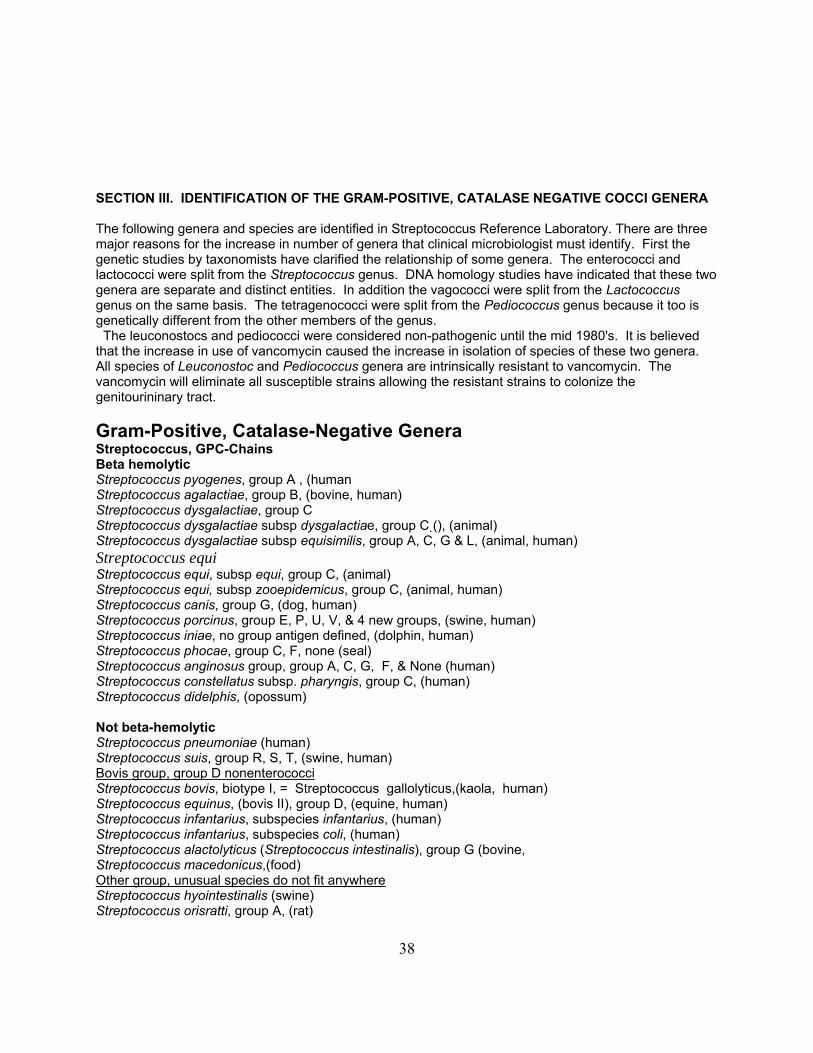
SECTION III. IDENTIFICATION OF THE GRAM-POSITIVE, CATALASE NEGATIVE COCCI GENERA The following genera and species are identified in Streptococcus Reference Laboratory. There are three major reasons for the increase in number of genera that clinical microbiologist must identify. First the genetic studies by taxonomists have clarified the relationship of some genera. The enterococci and lactococci were split from the Streptococcus genus. DNA homology studies have indicated that these two genera are separate and distinct entities. In addition the vagococci were split from the Lactococcus genus on the same basis. The tetragenococci were split from the Pediococcus genus because it too is genetically different from the other members of the genus. The leuconostocs and pediococci were considered non-pathogenic until the mid 1980's. It is believed that the increase in use of vancomycin caused the increase in isolation of species of these two genera. All species of Leuconostoc and Pediococcus genera are intrinsically resistant to vancomycin. The vancomycin will eliminate all susceptible strains allowing the resistant strains to colonize the genitourininary tract. Gram-Positive, Catalase-Negative Genera Streptococcus, GPC-Chains Beta hemolytic Streptococcus pyogenes, group A , (human Streptococcus agalactiae, group B, (bovine, human) Streptococcus dysgalactiae, group C Streptococcus dysgalactiae subsp dysgalactiae, group C (), (animal) Streptococcus dysgalactiae subsp equisimilis, group A, C, G & L, (animal, human) Streptococcus equi Streptococcus equi, subsp equi, group C, (animal) Streptococcus equi, subsp zooepidemicus, group C, (animal, human) Streptococcus canis, group G, (dog, human) Streptococcus porcinus, group E, P, U, V, & 4 new groups, (swine, human) Streptococcus iniae, no group antigen defined, (dolphin, human) Streptococcus phocae, group C, F, none (seal) Streptococcus anginosus group, group A, C, G, F, & None (human) Streptococcus constellatus subsp. pharyngis, group C, (human) Streptococcus didelphis, (opossum) Not beta-hemolytic Streptococcus pneumoniae (human) Streptococcus suis, group R, S, T, (swine, human) Bovis group, group D nonenterococciStreptococcus bovis, biotype I, = Streptococcus gallolyticus,(kaola, human) Streptococcus equinus, (bovis II), group D, (equine, human) Streptococcus infantarius, subspecies infantarius, (human) Streptococcus infantarius, subspecies coli, (human) Streptococcus alactolyticus (Streptococcus intestinalis), group G (bovine, Streptococcus macedonicus,(food) Other group, unusual species do not fit anywhereStreptococcus hyointestinalis (swine) Streptococcus orisratti, group A, (rat)

39
Streptococcus parauberis (bovine) Streptococcus thoraltensis (swine) Streptococcus uberis (bovine) Streptococcus urinalis, (human) Streptococcus waius (environment) Others (viridans-like) Streptococcus acidominimus, (food) Streptococcus hyovaginalis (swine) Streptococcus pluranimalium (bovine) Streptococcus thermophilus (food) Viridans streptococci, mutans group Streptococcus mutans (human plaque) Streptococcus cricetus (rodent plaque, human) Streptococcus downei (monkey plaque) Streptococcus ferus (rodent plaque) Streptococcus macaccae (monkey plaque) Streptococcus ratti (rodent, human plaque) Streptococcus sobrinus (human plaque) Viridans streptococci, oral groupStreptococcus salivarius (human) Streptococcus vestibularius (human) Streptococcus sanguinis (human) Streptococcus parasanguinis (human) Streptococcus gordonii (human) Streptococcus anginosus (human) Streptococcus constellatus (human) Streptococcus intermedius (human) Streptococcus mitis (human) Streptococcus oralis (human) Streptococcus crista (human) Streptococcus infantis (human) Streptococcus perois (human) Abiotrophia (Nutritionally Variant Streptococci), GPC-Chains Abiotrophia defectiva (human) Granulicatella (Abiotrophia) adiacens (human) Granulicatella (Abiotrophia) para-adiacens Granulicatella (Abiotrophia) elegans (human) Granulicatella (Abiotrophia) balaenopteriae (whales) Eremococcus coleocola (equine) Globicatella sanguinis, GPC-Chains, (human) Dolosicoccus paucivorans, GPC-Chains, (human) Facklamia species, GPC-Chains Facklamia hominus (human) CCUG 36813 1463 Facklamia ignava (human) CCUG 37419 1486 Facklamia sourekii (human) CCUG 31976 1533 Facklamia languida (human) CCUG 37420 1532 Facklamia tabacinasalis (tobacco) CCUG 30090 1566 Ignavigranum ruoffiae, GPC-Chains, (human) CCUG 37658 1483
40
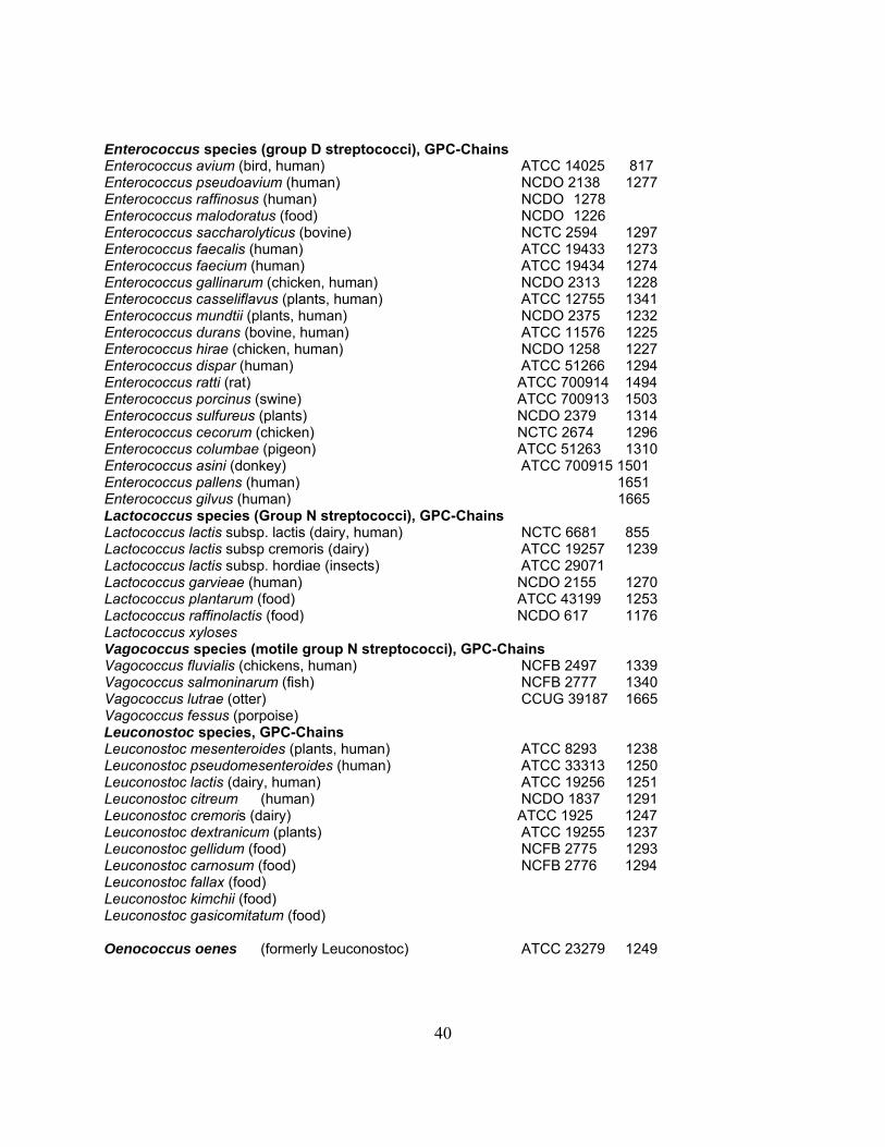
Enterococcus species (group D streptococci), GPC-Chains Enterococcus avium (bird, human) ATCC 14025 817 Enterococcus pseudoavium (human) NCDO 2138 1277 Enterococcus raffinosus (human) NCDO 1278 Enterococcus malodoratus (food) NCDO 1226 Enterococcus saccharolyticus (bovine) NCTC 2594 1297 Enterococcus faecalis (human) ATCC 19433 1273 Enterococcus faecium (human) ATCC 19434 1274 Enterococcus gallinarum (chicken, human) NCDO 2313 1228 Enterococcus casseliflavus (plants, human) ATCC 12755 1341 Enterococcus mundtii (plants, human) NCDO 2375 1232 Enterococcus durans (bovine, human) ATCC 11576 1225 Enterococcus hirae (chicken, human) NCDO 1258 1227 Enterococcus dispar (human) ATCC 51266 1294 Enterococcus ratti (rat) ATCC 700914 1494 Enterococcus porcinus (swine) ATCC 700913 1503 Enterococcus sulfureus (plants) NCDO 2379 1314 Enterococcus cecorum (chicken) NCTC 2674 1296 Enterococcus columbae (pigeon) ATCC 51263 1310 Enterococcus asini (donkey) ATCC 700915 1501 Enterococcus pallens (human) 1651 Enterococcus gilvus (human) 1665 Lactococcus species (Group N streptococci), GPC-Chains Lactococcus lactis subsp. lactis (dairy, human) NCTC 6681 855 Lactococcus lactis subsp cremoris (dairy) ATCC 19257 1239 Lactococcus lactis subsp. hordiae (insects) ATCC 29071 Lactococcus garvieae (human) NCDO 2155 1270 Lactococcus plantarum (food) ATCC 43199 1253 Lactococcus raffinolactis (food) NCDO 617 1176 Lactococcus xyloses Vagococcus species (motile group N streptococci), GPC-Chains Vagococcus fluvialis (chickens, human) NCFB 2497 1339 Vagococcus salmoninarum (fish) NCFB 2777 1340 Vagococcus lutrae (otter) CCUG 39187 1665 Vagococcus fessus (porpoise) Leuconostoc species, GPC-Chains Leuconostoc mesenteroides (plants, human) ATCC 8293 1238 Leuconostoc pseudomesenteroides (human) ATCC 33313 1250 Leuconostoc lactis (dairy, human) ATCC 19256 1251 Leuconostoc citreum (human) NCDO 1837 1291 Leuconostoc cremoris (dairy) ATCC 1925 1247 Leuconostoc dextranicum (plants) ATCC 19255 1237 Leuconostoc gellidum (food) NCFB 2775 1293 Leuconostoc carnosum (food) NCFB 2776 1294 Leuconostoc fallax (food) Leuconostoc kimchii (food) Leuconostoc gasicomitatum (food) Oenococcus oenes (formerly Leuconostoc) ATCC 23279 1249
41
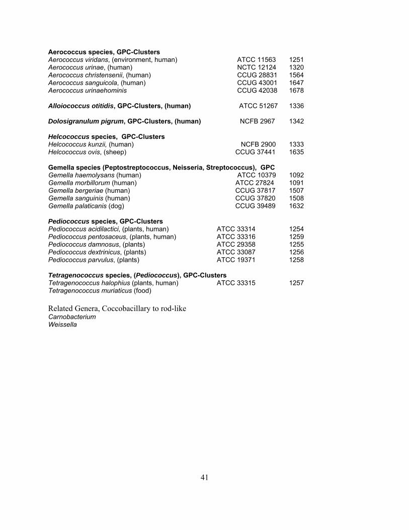
Aerococcus species, GPC-Clusters Aerococcus viridans, (environment, human) ATCC 11563 1251 Aerococcus urinae, (human) NCTC 12124 1320 Aerococcus christensenii, (human) CCUG 28831 1564 Aerococcus sanguicola, (human) CCUG 43001 1647 Aerococcus urinaehominis CCUG 42038 1678 Alloiococcus otitidis, GPC-Clusters, (human) ATCC 51267 1336 Dolosigranulum pigrum, GPC-Clusters, (human) NCFB 2967 1342 Helcococcus species, GPC-Clusters Helcococcus kunzii, (human) NCFB 2900 1333 Helcococcus ovis, (sheep) CCUG 37441 1635 Gemella species (Peptostreptococcus, Neisseria, Streptococcus), GPC Gemella haemolysans (human) ATCC 10379 1092 Gemella morbillorum (human) ATCC 27824 1091 Gemella bergeriae (human) CCUG 37817 1507 Gemella sanguinis (human) CCUG 37820 1508 Gemella palaticanis (dog) CCUG 39489 1632 Pediococcus species, GPC-Clusters Pediococcus acidilactici, (plants, human) ATCC 33314 1254 Pediococcus pentosaceus, (plants, human) ATCC 33316 1259 Pediococcus damnosus, (plants) ATCC 29358 1255 Pediococcus dextrinicus, (plants) ATCC 33087 1256 Pediococcus parvulus, (plants) ATCC 19371 1258 Tetragenococcus species, (Pediococcus), GPC-Clusters Tetragenococcus halophius (plants, human) ATCC 33315 1257 Tetragenococcus muriaticus (food) Related Genera, Coccobacillary to rod-like Carnobacterium Weissella

42
Table 1 lists the currently recognized genera of facultatively anaerobic gram positive cocci. Once it has been determined that the bacteria in question is a gram-positive, catalase-negative coccus, the next step is to determine to what genera the strain belongs. The genera that this laboratory identifies includes: Enterococcus, Leuconostoc/Weissella, Streptococcus, Pediococcus, Tetragenococcus, Aerococcus, Helcococcus, Vagococcus, Lactococcus, Abiotrophia, Granulicatella, Globicatella sanguinis, Dolosicoccus paucivorans. Alloiococcus, Dolosigranulum, Facklamia, and Ignavigranum species.
Initial separation into the appropriate genera is accomplished by determining the physiologic characteristics listed in Table 1. The extent to which each bacterial strain is tested is dependent upon the source of the strain. If the strain is from a normally sterile-site then all the tests listed below should be applied. There are some instances where all the tests do not need to be applied; i.e., the beta-hemolytic streptococci and the pneumococci. There are specific tests that clinical microbiologist perform when these pathogens are suspected. These situations and tests will be discussed in the section on streptococci.

43
Table 1. Phenotypic characteristics of facultatively anaerobic, catalase-negative, gram-positive cocci Phenotypic characteristicb
Genus
Gram
a
stain VAN GAS BE PYR LAP NaC
l 10oC 45oC HEM
Enterococcus groupc
ch S/R - + + + + + +d α/γ
Leuconostoc/Weissellae ch
R
+ V+
-
-
V+
V- V-
α/γ
Streptococcus ch S - -f -g + V- - V- α/β/γ
Nutritional var. Streph ch S - - + + - - V- α/γ
Unusual Strep/Genera i ch S - V+ V+ V+ V+ V- V- α/γ
Pediococcus cl/t R - + - + V- - V+ α
Tetragenococcus cl/t S - + - + + - + α
Aerococcus speciesj cl/t S - V+ V+ V- + - V- α
Helcococcus cl/t S - + + - + - - γ
Gemella cl/t/ch S - - + + - - - γ
Salt tolerant Gemella-likek cl/t/ch S - - + + + - - γ
aCell arrangement in gram strain: ch, chains; cl, clusters; t, tetrads. bVAN, vancomycin susceptibility screening test; GAS, gas production in MRS broth; BE, hydrolysis of esculin in the presence of bile; PYR, production of pyrrolidonylarylamidase; LAP, production of leucine aminopeptidase; NaCl, growth in broth containing 6.5% NaCl; 10oC and 45oC, growth at 10oC and 45oC; HEM, hemolytic activity on Trypticase soy 5% sheep blood agar. +, 85% or more of the strains are positive: -, 15% or less of the strains are positive; V+, variable positive (50 to 84% of the strains are positive); V-, variable negative (16 to 49% of strains are positive). cEnterococcus group includes all Enterococcus species, Vagococcus species and some Lactococcus species.
dSome strains of lactococci and vagococci grow very poorly at 45oC. eLeuconostoc and Weisella are often coccobacillary, sometimes appearing rod like in chains. fAll strains of S. bovis and approximately 10% of viridans streptococci are bile-esculin positive. gAll strains of S. pyogenes and most strains of S. porcinus and S. iniae are PYR positive. Other streptococci are all negative h Nutritional variant streptococci are now identified in two different genera Abiotrophia and Granulicatella
iUnusual strep/genera includes species of streptococci usually found in animals and Globicatella sanguinis and Dolosicoccus paucivorans. JAerococcus species includes A. viridans, A. urinae, A. sanguicola, and A. urinehominis. k Salt tolerant Gemella-like bacteria include Alloiococcus, Dolosigranulum, Facklamia, and Ignavigranum species.
44
STREPTOCOCCUS
In spite of the splitting of this genus into the streptococci, enterococci and lactococci it still remains a very diverse group of bacteria. The different species are found in every sort of human infection as well as being a commensal organism in the oral cavity and genitourinary tract. As stated above there are specific situations when all the tests listed in Table 2 do not have to be determined. The first situation is when ß-hemolysis is observed. This is most notable with throat swabs. ß-hemolysis is such a good indicator among the catalase negative gram positive cocci that after determining that the unknown isolate is gram positive, catalase negative, and ß-hemolytic you can apply the tests listed in the Table 2 for streptococcal species identification to determine what species of Streptococcus is present. The remaining tests in Table 2 do not have to be determined. The second situation occurs when the clinical microbiologist is determining whether or not S. pneumoniae is present in various body fluids. If the unknown strain is catalase negative, gram positive, and α-hemolytic then the tests used to identify pneumococci can be applied (Table 5). In all other situations all the tests listed in Table 2 must be performed to determine if the unknown strain is a Streptococcus. All streptococci are; vancomycin sensitive, do not form gas from MRS broth, produce LAP, do not grow at 10ΕC, and are non motile. Among the streptococci, group A streptococci, S. porcinus, S. iniae, and nutritionally variant streptococci are PYR positive. In the case of group A streptococci the PYR test is an excellent test for the presumptive identification of group A streptococci. The non-beta hemolytic streptococci (viridans, and non-enterococcal group D) do not grow in 6.5% NaCl broth; but some of the beta-hemolytic strains may grow in the broth. The bovis strains of streptococci usually grow at 45ΕC while the viridans streptococcal strains usually do not grow. The ß-hemolytic strains exhibit variable growth patterns at 45ΕC. The streptococci can be α, ß, or non hemolytic. The hemolysis reaction in blood agar plates is used to help differentiate the streptococcal species.
45
ENTEROCOCCUS Genetic evidence that the S. faecalis and S. faecium was sufficiently different from the other members of the Streptococcus genus was provided by Schleifer and Kilpper-Balz in 1984. It has been several years since this proposal and it is generally accepted that the genus Enterococcus is valid and used in the published literature. Since 1984, 25 other species have been proposed to be included in the Enterococcus genus. Genetic evidence for these proposals has been provided by DNA-DNA and DNA-rRNA hybridizations as well as reverse transcriptase sequencing of 16S ribosomal RNA. The enterococci are among the most ubiquitous bacteria known. They are found in the environment, human and other animal genitourinary tracts, and in most foods. These bacteria are not very invasive but are significant opportunists. The enterococci are usually sensitive to vancomycin but recently many strains have acquired resistance and in some cases the majority of strains identified may be vancomycin resistant because of nosocomial transmission of the strains. The resistance to vancomycin does not interfere with the identification of the strains because only the leuconostocs and pediococci are intrinsically resistant to vancomycin but all species of these two genera are PYR negative. Thus the only strains that are vancomycin resistant and PYR positive are the enterococci. Only rarely do enterococcal strains (less than 0.5%) produce gas from MRS broth. Nearly all strains produce LAP, grow in 6.5% NaCl, and grow at 10ΕC and 45ΕC. Some strains of E. avium or E. raffinosus do not grow at 10ΕC or produce LAP but this is not consistent or predictable. Some species are motile. Most strains are α or non-hemolytic; occasionally ß-hemolytic strains of E. faecalis or E. durans are identified.
LACTOCOCCUS
The transfer of the species of lactic streptococci formally known as group N streptococci to the genus Lactococcus was made in l985. The species included in this genus are thought to be nonpathogenic for man. In fact L. lactis (S. lactis) and L. cremoris (S. cremoris) are used in the dairy industries and are included in foodstuffs. We have identified several strains of lactococci from human sources but the clinical significance of these strains has not been evaluated.
The lactococci, formally called the lactic group of streptococci or the group N streptococci, were not thought to be pathogenic for man. Many of the lactococcal strains are used in the manufacturer of foods, such as cheese. However, several confirmed cases of infections including endocarditis have been attributed to lactococcal strains. The natural habitat of lactococci is thought to be the environment. The physiologic characteristics of the lactococci is very similar to the enterococci and most of the lactococcal strains associated with human infections in our culture collection now identified as lactococci were initially identified as enterococci. One of the criteria previously used to differentiate between the enterococci and lactococci was the inability of lactococci to grow at 45ΕC. However, with the inclusion of new species into the lactococcus genera, i. e., L. garvieae, some strains of lactococci grow at 45ΕC. All the lactococci are: vancomycin sensitive, do not produce gas in MRS broth, grow at 10ΕC, and are nonmotile. All strains give positive LAP reactions and most strains give positive PYR reactions, however, some L. lactis strains are negative. Variable reactions are observed in the 6.5% NaCl tolerance test. None of the strains are ß-hemolytic.
VAGOCOCCUS Vagococcal strains have rarely been isolated from human infections. The vagococcal strains were split from the Lactococcus genus. These organisms were previously known as motile lactococci. In fact, only the motility characteristic of the vagococci differentiates the vagococci from the lactococci. These bacteria are included in the identification of the enterococcus because of phenotypic similarities.
46
LEUCONOSTOC Like the lactococci the leuconostocs were once not thought to cause human infections. However, there are many reports of human infections caused by different Leuconostoc species. Species of the leuconostoc genus are the only catalase negative gram positive cocci that produce gas from MRS broth. As stated earlier all strains are intrinsically resistant to vancomycin. All strains of leuconostocs are negative in PYR and LAP tests. The combination of reactions, vancomycin resistance, and negative PYR and LAP is an excellent indicator of Leuconostoc identification. No other catalase negative, gram positive cocci has these characteristics. Like the lactococci, leuconostocs grow at 10ΕC but very poorly if at all at 45ΕC. Some strains grow in 6.5% NaCl broth while others do not. None of the strains are motile and none are ß-hemolytic.
PEDIOCOCCUS
Like the leuconostocs the pediococci are intrinsically resistant to vancomycin. They too were thought to be nonpathogenic for humans but there are several reports indicating that this is changing. The pediococcus strains appear very similar to the viridans streptococci on blood agar media and can be easily misidentified as viridans streptococci. All strains of pediococci tested have been resistant to vancomycin and all strains of viridans streptococci have been sensitive to vancomycin. Pediococci do not form gas in MRS broth, they are PYR negative but LAP positive, some strains grow in 6.5% NaCl broth while others do not. Most strains grow at 45ΕC but not at 10ΕC. All strains are nonmotile and appear α-hemolytic on blood agar plates.
GLOBICATELLA This new genus of gram positive cocci has just recently been described. The origin of this genus is from a collection of viridans-like streptococci that most closely resembled Streptococcus uberis. What makes Globicatella distinct from the viridans streptococci is that all the Globicatella strains were PYR positive, LAP negative and grow in broth containing 6.5% NaCl while all viridans species are PYR negative, LAP positive and fail to grow in 6.5% NaCl. All strains identified to date have been sensitive to vancomycin, PYR positive, LAP negative, grow in 6.5% NaCl broth, do not grow at 10ΕC or 45ΕC, are nonmotile, and are α-hemolytic.
TETRAGENOCOCCUS This genus contains only one species. T. halophilus was previously identified as Pediococus halophilus. The tetragenococci differ from the pediococci by vancomycin resistance. The pediococci are vancomycin resistant and the tetragenococci are vancomycin sensitive. Other characteristics are similar.
GEMELLA and GEMELLA-LIKE These bacteria grow very poorly on blood agar plates and will often take 48 h to grow. These bacteria may also resemble the viridans streptococci; occasionally the tetrads are not formed and only pairs and short chains are observed in the gram stain. One species was previously identified as a streptococci, G. morbillorum. On blood agar plates some of the strains give an wide-zone alpha hemolytic reaction after extended incubation. Identifying these bacteria is difficult and an extended set of physiologic characteristics may have to be determined before final identification is possible. These bacteria are characteristically negative in most tests listed in Table 1. Some strains are positive in both the PYR and LAP tests but other strains may fail to give a positive reaction in either. Salt tolerant Gemella-like genera include the Alloiococcus, Dolosigranulum, Facklamia, and Ignavigranum.
AEROCOCCUS
The Aerococcus sp. are sensitive to vancomycin, do not form gas from glucose, and are generally PYR positive but are LAP negative. All strains grow in broth containing 6.5% NaCl but do not grow at 10ΕC or 45ΕC. Strains are non motile and are strongly α-hemolytic. The strains grow well on blood agar media and form colonies somewhat smaller than the enterococci but larger than the viridans streptococci after overnight incubation.
47
ALLOIOCOCCUS Only one species of this genus is presently known, A. otitidis. These bacteria have been isolated from the ear fluids of children with otitis. Like the gemellae this bacterium is difficult to grow. Often 2 to 3 days are necessary for growth to develop on rabbit blood agar plates. Alloiococci are susceptible to vancomycin, do not form gas in MRS broth and are PYR and LAP positive. They are differentiated from the gemellae by the 6.5% NaCl test. Alloiococci grow in 6.5% broth but gemellae do not. These bacteria do not grow at 10ΕC or 45ΕC or thioglycolate broth, are not motile and are non-hemolytic on blood agar plates.
HELCOCOCCUS Two species of this genus are presently known. H. kurzii is the only one isolated from humans. These bacteria have been isolated from wound infections. Like the Alloiococci and gemellae these bacteria grow very slowly on blood agar media. The physiologic characteristics of helocococci are similar to the aerococci in that they are PYR positive, LAP negative, and grow in broth containing 6.5% NaCl. These bacteria grow more slowly and are not α-hemolytic on blood agar while the aerococci grow readily and are α-hemolytic on blood agar. Like the Aerococci these strains are vancomycin sensitive, do not form gas in MRS broth and fail to grow at 10ΕC and 45ΕC. All isolates have been non-motile.
NUTRITIONALLY VARIANT STREPTOCOCOCCUS Nutritionally variant streptococci (NVS) can be identified by demonstrating that the strain requires pyridoxal or grows on an agar plate only when a bacteria that satellites is present. In addition to this requirement, NVS also give positive PYR tests, which helps to differentiate these strains from viridans streptococci.

48
A IDENTIFICATION OF STREPTOCOCCAL SPECIES THE ß-HEMOLYTIC STREPTOCOCCI The most convenient way to begin to identify the streptococci is to determine the hemolysis of the bacteria on blood agar plates. As mentioned earlier the techniques for determining hemolysis is described in detail in; Isolation and Identification of streptococci, Part 1. Collection, transport, and determination of hemolysis, Annex 1. Once the streptococci are divided into ß-hemolytic and non-ß-hemolytic categories differentiation into species, groups, and categories can be made. The identification of most ß-hemolytic strains is performed by determining the antigenic characteristics of the culture; but, identification of the non-ß-hemolytic strains is performed by determining antigenic and physiologic characteristics of the culture.
Group A streptococci: Lancefield's group A Streptococcus is also known as Streptococcus pyogenes. Identification is confirmed by demonstrating the presence of the group A antigen on the streptococcal cells. All S. pyogenes have group A antigen; but, not all streptococci with group A antigen are S. pyogenes. Some strains of S. anginosus and S. dysgalactiae subsp. equisimilis may also have group A antigen. Non-pyogenes strains grow more slowly and form smaller colonies than do S. pyogenes strains. If the ß-hemolytic colonies appear small and growth is delayed or carbon dioxide is required for growth or the group A strain is PYRase negative the microbiologist should suspect that the strain may be S. anginous or S. dysgalactiae subsp. equisimilis, regardless of group reaction. When this occurs the strain should be tested for voges-proskauer (VP) reaction. See Table 2, page 67 for correct identification. Group B streptococci: Lancefield's Group B streptococci is also known as S. agalactiae. Like group A streptococci, identification is confirmed by demonstrating that the streptococcal cells contain group B antigen. Since the group B antigen is not identified with any other streptococcal strain the terms Lancefield group B and S. agalactiae are synonymous. Group C streptococci: The ß-hemolytic streptococci that contain group C antigen are shown in table on page 30. The group C antigen is found with several different species and the S. anginous group of bacteria, Table 2, page 67. Group G streptococci: The ß-hemolytic streptococci with group G antigen have not had an official taxonomic name. Some have suggested that these strains be called S. canis but this has not gained approval officially or in practical use. ß-hemolytic streptococci with group G antigen should be reported simply as Lancefield's group G streptococci. Group F streptococci: S. anginosus is used here to report streptococcal strains that may or may not have group antigen A, C, F, or G antigens. Most of these strains have group F antigen, the next most frequently identified strain will have no group antigen, and only rarely will an anginosus strain have group A, C, or G antigen. All these strains are VP positive and they are the only ß-hemolytic strains to have this characteristic. Technically some of these strains may be S. intermedius or S. constellatus. The DNA-DNA homology studies have shown that ß-hemolysis did not help to differentiate these strains, this characteristic was found among all 3 species. The majority of these strains will require additional carbon dioxide in the atmosphere for growth on blood agar plates. Often growth is not apparent until 48 h of incubation. Again for convenience the ß-hemolytic-VP positive strains should be reported as ß-hemolytic, S. anginosus, group ? (insert group reaction or none).
49
OTHER ß-HEMOLYTIC STREPTOCOCCI There are other ß-hemolytic streptococci but they are rarely found in human infections. Some of the strains are associated with infections of swine and they have specific group antigens. Using DNA-DNA homology the taxonomist have suggested that those ß-hemolytic strains with group E, P, U, V, and other experimental group antigens be called S. porcinus. ß-hemolytic strains with group L antigen have been associated with avian sources (chicken), it is suggested that these strains be called S. dysgalactiae. ß-hemolytic strains with group M antigen are not well studied and there is no suggested taxonomic name for these strains. A ß-hemolytic strain with a yet to be named group antigen (suggested X) isolated from fresh water dolphins, and occasionally in human infections, is called S. iniae. This bacterium may be submitted as a group A streptococci because it may react with the group A antibody in the latex slide agglutination assay. It is also PYRase positive but is not sensitive to bacitracin. Whenever a latex agglutination group A positive bacterium is submitted that is not sensitive to bacitracin the group reaction should be confirmed using the Lancefield extraction procedure. Some investigators have reported that strains with group R antigen can be ß-hemolytic. Using 5% sheep blood agar plates we have been unable to demonstrate that these strains are ß-hemolytic. These strains are α-hemolytic on blood agar plates containing 5% sheep blood. Strains with group R, S, and T antigens are very similar to each other physiologically and the taxonomist have suggested that these strains should be called S. suis. The strains are found commonly in swine and may be transmitted to man. There are several reports of farmers and abattoir workers being infected with group R (S. suis type II) streptococci. Identification of these bacteria is difficult without having knowledge that the infecting strain may be related to nonhuman sources. Demonstration of the group R, S, and T antigens with specific antisera is also difficult. It is suggested that if group R streptococci is the suspected agent in an infection the cells may have to be extracted with the formamide extraction technique in order to extract the group antigen. The formamide extraction technique is described in Annex 2 (Part II of Isolation and identification of streptococci). Note: S. phocae is maltose positive (data not shown) The serological and physiological tests listed in Table 2 can be used to identify nearly all of the ß-hemolytic streptococci isolated from human infections.

50
Table 2. Identification of the β-hemolytic streptococci. Species Lancefield
Group Bac NaCl PYR Camp VP Hip Arg Esc Str Sbl Tre Rib α-gal β-gal β-gur
Origin
S. pyogenes A + + + - - - + v - - na - Human
S. agalactiae B - + - + - + + - - - na na Human, bovine
S. dysgalactiae subsp. dysgalactiae subsp. equisimilis
C A,C,G,L
- v
+ +
- -
- -
- -
- -
+ +
v v
- v
v -
+ +
+ + - - +
animas human, animals
S. equi subsp. equi subsp. zoepidemicus subsp. ruminatorum
C C
- -
+ +
- -
- -
- -
- -
+ +
v v
+ +
- +
- v
na na
animals animals, human
S. canis G - + - v - - + + - - v na + + - dog, human
S. anginosus (group) A,C,G,F, None
- - - - + - + + - - + na Human
S. constellatus subsp. pharyngis subsp. constellatus
C C
- - - - + - + + - - + na Human
S. porcinus E,P,U,V, None, New
- v + + + v + + - + + na swine, human
S. iniae None - + + + - - V + + - na na dolphin, fish, human
S. phocae C,F, G, None + + - - - - - - - - - + Seals
S. didelphis None - + - - - - + - - - + na Opossum Abbreviations: Group, group carbohydrate antigen; Bac, S, susceptibile, R, resistant to bacitracin; NaCl, growth in 6.5% NaCl, Pyr, pyrrolidonylarylamidase; Camp, CAMP reaction; VP, voges-proskauer reaction; Hip, hydrolysis of hippurate; Arg, hydrolysis of arginine; Esc, hydrolysis of esculin; Str, hydrolysis of starch; Sbl, Tre, and Rib production of acid in sorbitol, trehalose, and ribose broth respectively. +, positive reaction >95%; -, negative reaction >95%; v, variable reaction 6-94% positive; na, not applicable; β-gal, β-galactosidase reaction; β-gur, β-glucoronidase reaction; α-gal, α-galcactosidase reaction.
- S. dysgalactiae subsp. dysgalactiae are not beta-hemolytic but are included in this table for taxomonic reasons. - The S. anginosus group includes beta-hemolytic strains of S. anginosus, S. constellatus, and S. intermedius. There is insufficient data to know the percent of each of these beta-hemolytic species that contain carbohydrate antigens.
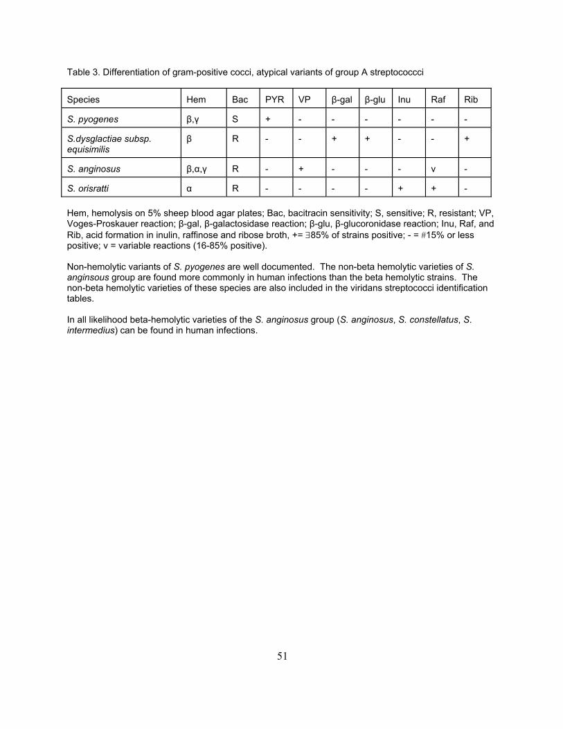
51
Table 3. Differentiation of gram-positive cocci, atypical variants of group A streptococcci Species Hem
Bac PYR VP β-gal β-glu Inu Raf Rib
S. pyogenes
β,γ S + - - - - - -
S.dysglactiae subsp. equisimilis
β R - - + + - - +
S. anginosus β,α,γ R - + - - - v - S. orisratti α R - - - - + + - Hem, hemolysis on 5% sheep blood agar plates; Bac, bacitracin sensitivity; S, sensitive; R, resistant; VP, Voges-Proskauer reaction; β-gal, β-galactosidase reaction; β-glu, β-glucoronidase reaction; Inu, Raf, and Rib, acid formation in inulin, raffinose and ribose broth, += ∃85% of strains positive; - = #15% or less positive; v = variable reactions (16-85% positive). Non-hemolytic variants of S. pyogenes are well documented. The non-beta hemolytic varieties of S. anginsous group are found more commonly in human infections than the beta hemolytic strains. The non-beta hemolytic varieties of these species are also included in the viridans streptococci identification tables. In all likelihood beta-hemolytic varieties of the S. anginosus group (S. anginosus, S. constellatus, S. intermedius) can be found in human infections.
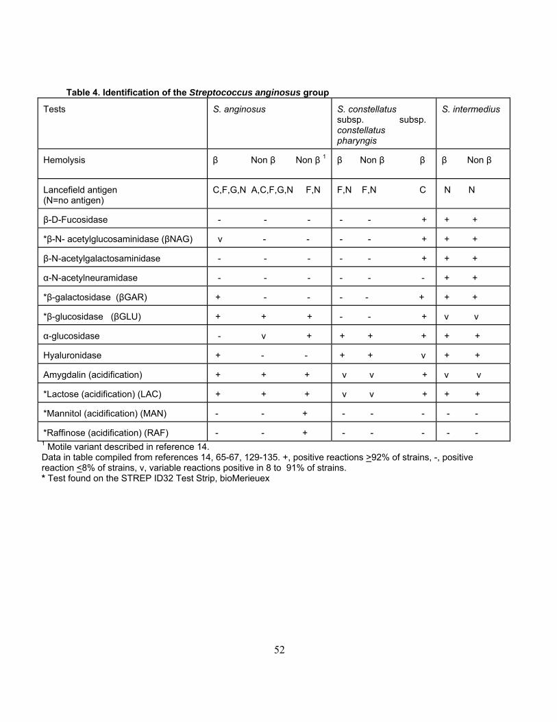
52
Table 4. Identification of the Streptococcus anginosus group Tests
S. anginosus
S. constellatus subsp. subsp. constellatus pharyngis
S. intermedius
Hemolysis
β Non β Non β 1
β Non β β
β Non β
Lancefield antigen (N=no antigen)
C,F,G,N A,C,F,G,N F,N
F,N F,N C
N N
β-D-Fucosidase
- - -
- - +
+ +
*β-N- acetylglucosaminidase (βNAG)
v - -
- - +
+ +
β-N-acetylgalactosaminidase
- - -
- - +
+ +
α-N-acetylneuramidase
- - -
- - -
+ +
*β-galactosidase (βGAR)
+ - -
- - +
+ +
*β-glucosidase (βGLU)
+ + +
- - +
v v
α-glucosidase
- v +
+ + +
+ +
Hyaluronidase
+ - -
+ + v
+ +
Amygdalin (acidification)
+ + +
v v +
v v
*Lactose (acidification) (LAC)
+ + +
v v +
+ +
*Mannitol (acidification) (MAN)
- - +
- - -
- -
*Raffinose (acidification) (RAF)
- - +
- - -
- -
1 Motile variant described in reference 14. Data in table compiled from references 14, 65-67, 129-135. +, positive reactions >92% of strains, -, positive reaction <8% of strains, v, variable reactions positive in 8 to 91% of strains. * Test found on the STREP ID32 Test Strip, bioMerieuex
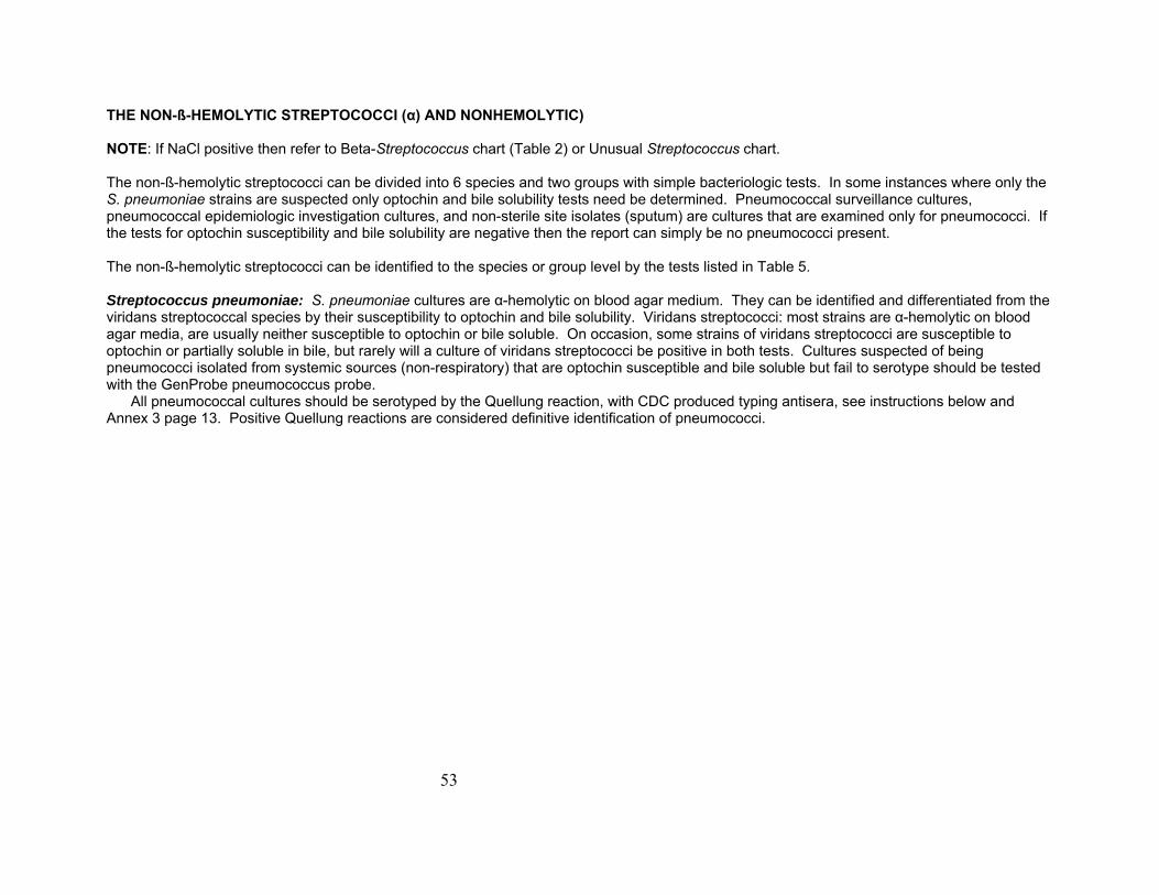
53
THE NON-ß-HEMOLYTIC STREPTOCOCCI (α) AND NONHEMOLYTIC) NOTE: If NaCl positive then refer to Beta-Streptococcus chart (Table 2) or Unusual Streptococcus chart. The non-ß-hemolytic streptococci can be divided into 6 species and two groups with simple bacteriologic tests. In some instances where only the S. pneumoniae strains are suspected only optochin and bile solubility tests need be determined. Pneumococcal surveillance cultures, pneumococcal epidemiologic investigation cultures, and non-sterile site isolates (sputum) are cultures that are examined only for pneumococci. If the tests for optochin susceptibility and bile solubility are negative then the report can simply be no pneumococci present. The non-ß-hemolytic streptococci can be identified to the species or group level by the tests listed in Table 5. Streptococcus pneumoniae: S. pneumoniae cultures are α-hemolytic on blood agar medium. They can be identified and differentiated from the viridans streptococcal species by their susceptibility to optochin and bile solubility. Viridans streptococci: most strains are α-hemolytic on blood agar media, are usually neither susceptible to optochin or bile soluble. On occasion, some strains of viridans streptococci are susceptible to optochin or partially soluble in bile, but rarely will a culture of viridans streptococci be positive in both tests. Cultures suspected of being pneumococci isolated from systemic sources (non-respiratory) that are optochin susceptible and bile soluble but fail to serotype should be tested with the GenProbe pneumococcus probe. All pneumococcal cultures should be serotyped by the Quellung reaction, with CDC produced typing antisera, see instructions below and Annex 3 page 13. Positive Quellung reactions are considered definitive identification of pneumococci.
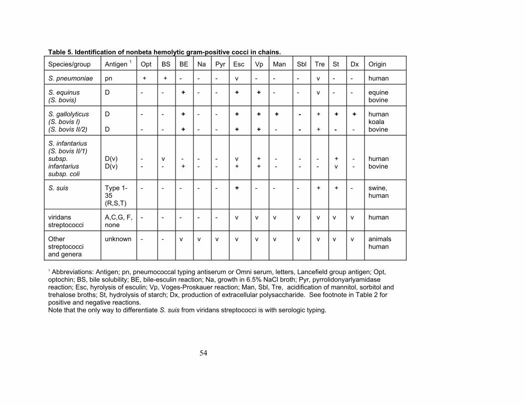
54
Table 5. Identification of nonbeta hemolytic gram-positive cocci in chains. Species/group
Antigen 1
Opt
BS
BE
Na
Pyr
Esc
Vp
Man
Sbl
Tre
St
Dx
Origin
S. pneumoniae
pn
+
+
-
-
-
v
-
-
-
v
-
-
human
S. equinus (S. bovis)
D
-
-
+
-
-
+
+
-
-
v
-
-
equine bovine
S. gallolyticus (S. bovis I) (S. bovis II/2)
D D
- -
- -
+ +
- -
- -
+ +
+ +
+ -
- -
+ +
+ -
+ -
human koala bovine
S. infantarius (S. bovis II/1) subsp. infantarius subsp. coli
D(v) D(v)
- -
v -
- +
- -
- -
v +
+ +
- -
- -
- -
+ v
- -
human bovine
S. suis
Type 1-35 (R,S,T)
-
-
-
-
-
+
-
-
-
+
+
-
swine, human
viridans streptococci
A,C,G, F, none
-
-
-
-
-
v
v
v
v
v
v
v
human
Other streptococci and genera
unknown
-
-
v
v
v
v
v
v
v
v
v
v
animals human
1 Abbreviations: Antigen; pn, pneumococcal typing antiserum or Omni serum, letters, Lancefield group antigen; Opt, optochin; BS, bile solubility; BE, bile-esculin reaction; Na, growth in 6.5% NaCl broth; Pyr, pyrrolidonyarlyamidase reaction; Esc, hyrolysis of esculin; Vp, Voges-Proskauer reaction; Man, Sbl, Tre, acidification of mannitol, sorbitol and trehalose broths; St, hydrolysis of starch; Dx, production of extracellular polysaccharide. See footnote in Table 2 for positive and negative reactions. Note that the only way to differentiate S. suis from viridans streptococci is with serologic typing.
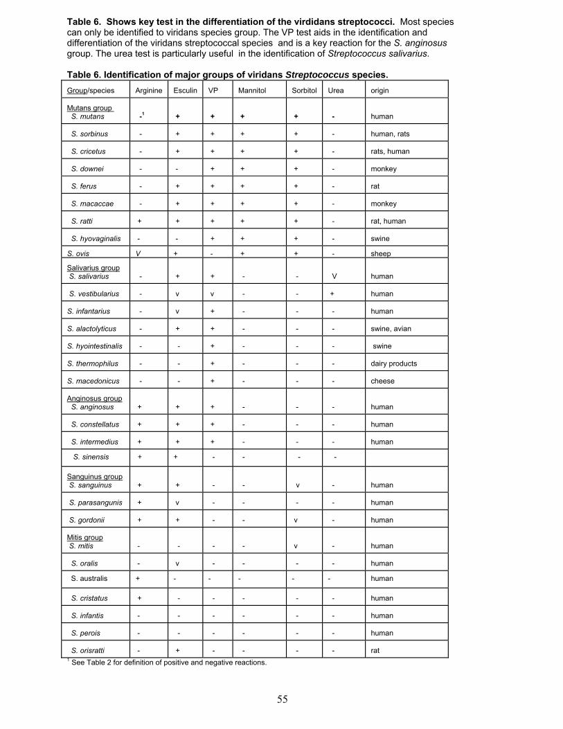
55
Table 6. Shows key test in the differentiation of the virdidans streptococci. Most species can only be identified to viridans species group. The VP test aids in the identification and differentiation of the viridans streptococcal species and is a key reaction for the S. anginosus group. The urea test is particularly useful in the identification of Streptococcus salivarius. Table 6. Identification of major groups of viridans Streptococcus species. Group/species
Arginine
Esculin
VP
Mannitol
Sorbitol
Urea
origin
Mutans group S. mutans
-1
+
+
+
+
-
human
S. sorbinus
-
+
+
+
+
-
human, rats
S. cricetus
-
+
+
+
+
-
rats, human
S. downei
-
-
+
+
+
-
monkey
S. ferus
-
+
+
+
+
-
rat
S. macaccae
-
+
+
+
+
-
monkey
S. ratti
+
+
+
+
+
-
rat, human
S. hyovaginalis
-
-
+
+
+
-
swine
S. ovis
V
+
-
+
+
-
sheep
Salivarius group S. salivarius
-
+
+
-
-
V
human
S. vestibularius
-
v
v
-
-
+
human
S. infantarius
-
v
+
-
-
-
human
S. alactolyticus
-
+
+
-
-
-
swine, avian
S. hyointestinalis
-
-
+
-
-
-
swine
S. thermophilus
-
-
+
-
-
-
dairy products
S. macedonicus
-
-
+
-
-
-
cheese
Anginosus group S. anginosus
+
+
+
-
-
-
human
S. constellatus
+
+
+
-
-
-
human
S. intermedius
+
+
+
-
-
-
human
S. sinensis
+
+
-
-
-
-
Sanguinus group S. sanguinus
+
+
-
-
v
-
human
S. parasangunis
+
v
-
-
-
-
human
S. gordonii
+
+
-
-
v
-
human
Mitis group S. mitis
-
-
-
-
v
-
human
S. oralis
-
v
-
-
-
-
human
S. australis
+
-
-
-
-
-
human
S. cristatus
+
-
-
-
-
-
human
S. infantis
-
-
-
-
-
-
human
S. perois
-
-
-
-
-
-
human
S. orisratti
-
+
-
-
-
-
rat
1 See Table 2 for definition of positive and negative reactions.
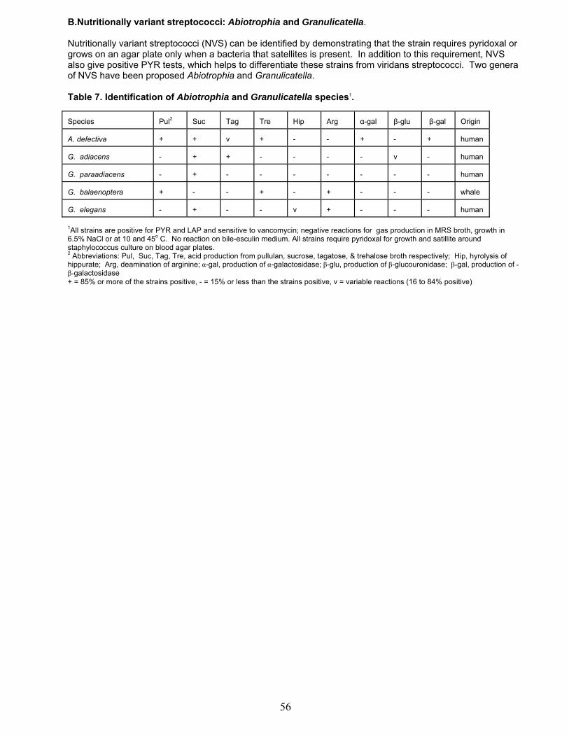
56
B.Nutritionally variant streptococci: Abiotrophia and Granulicatella. Nutritionally variant streptococci (NVS) can be identified by demonstrating that the strain requires pyridoxal or grows on an agar plate only when a bacteria that satellites is present. In addition to this requirement, NVS also give positive PYR tests, which helps to differentiate these strains from viridans streptococci. Two genera of NVS have been proposed Abiotrophia and Granulicatella. Table 7. Identification of Abiotrophia and Granulicatella species1. Species
Pul2
Suc
Tag
Tre
Hip
Arg
α-gal
β-glu
β-gal
Origin
A. defectiva
+
+
v
+
-
-
+
-
+
human
G. adiacens
-
+
+
-
-
-
-
v
-
human
G. paraadiacens
-
+
-
-
-
-
-
-
-
human
G. balaenoptera
+
-
-
+
-
+
-
-
-
whale
G. elegans
-
+
-
-
v
+
-
-
-
human
1All strains are positive for PYR and LAP and sensitive to vancomycin; negative reactions for gas production in MRS broth, growth in 6.5% NaCl or at 10 and 45o C. No reaction on bile-esculin medium. All strains require pyridoxal for growth and satillite around staphylococcus culture on blood agar plates. 2 Abbreviations: Pul, Suc, Tag, Tre, acid production from pullulan, sucrose, tagatose, & trehalose broth respectively; Hip, hyrolysis of hippurate; Arg, deamination of arginine; α-gal, production of α-galactosidase; β-glu, production of β-glucouronidase; β-gal, production of -β-galactosidase + = 85% or more of the strains positive, - = 15% or less than the strains positive, v = variable reactions (16 to 84% positive)

C. Identification of Enterococci and Vagococci The enterococci are gram positive cocci, occur in singles, pairs, and short chains. Cells are sometimes coccobacillary when gram stains are prepared from agar plate growth. Cells are more oval and in chains when gram stains are prepared from thioglycolate broth. The enterococci are facultatively anaerobic and optimum growth occurs at 35C. Most strains grow at 10C and 45C. All strains grow in broth containing 6.5% NaCl and hydrolyze esculin in the presence of 40% bile salts (bile-esculin medium). Motility is observed with some species. Enterococci hydrolyze pyrrolidonyl-ß-naphthylamide (PYR), the exceptions to this are E. cecorum, E. columbae, and E. saccharolyticus. Most strains produce leucine aminopeptidase (LAP). Some strains belonging to Group I enterococci give negative LAP tests. Enterococci do not contain cytochrome enzymes but on occasion the catalase test is positive. A pseudo catalase is sometimes produced and a weak effervescence is observed in the catalase test. Nearly all strains are homofermentive, gas is not produced and lactic acid is the end product of glucose fermentation. Most strains produce a cell-wall associated glycerol teichoic acid antigen that is identified as the streptococcal group D antigen. Detection of the group D antigen is sometimes difficult and depends upon the extraction procedure and the quality of the antiserum used.
Presumptive identification of a catalase negative gram positive cocci as an Enterococcus or Vagococcus can be accomplished by demonstrating that the unknown strain is vancomycin sensitive, PYR and LAP positive, and grows in 6.5% NaCl and at 45C. Species Identification Once established that the unknown catalase-negative gram-positive coccus is an Enterococcus the tests listed in Table 8 can be used to identify the species. The species into 5 Groups based on the reactions in acid formation in mannitol, sorbitol, and sorbose broths and hydrolysis of arginine. Group I consists of E. avium, E. malodoratus, E. raffinosus, E. pseudoavium, E. saccharolyticus, E. gilvus, and E. pallens PNS-E3 and Vagococcus lutrae. These species form acid in all three of the aforementioned Carbohydrate broths but do not hydrolyze arginine.
Group II consists of E. faecalis, E. faecium, E. casseliflavus, E. mundtii, and E. gallinarum E. haemoperoxidus, Lactococcus sp., PNS-E2. These species form acid in mannitol broth, hydrolyze arginine, but fail to form acid in sorbose broth and give variable reactions in sorbitol broth. Group III consists of E. durans, E. hirae, E. dispar, E. porcinus, E. ratti, E. faecium, and E. faecalis variant and E. villorum. These species hydrolyze arginine but do not form acid in mannitol, sorbitol or sorbose broths.
Group IV contains E. sulfureus, E. asini, and E. cecorum E. phoeniculicola, V. fessus, V. carniphilus, V. salmonarium, and PNS E1. These species are sorbose negative and not hydrolyze arginine. Group V consists of E. casseliflavus, E. gallinarum, E. faecalis, and E. columbae, E. hermanniensis, E. moraviensis, E. canis, V. fluvialis, and V. lutrae. The pigmentation test aids in the identification of E. casseliflavus, E. mundtii, E. pullins, E. gilvus and E. sulfureus. These enterococci produce a yellow pigment that can be detected on several different media. The pyruvate utilization test aids in the differentiation of E. faecalis and E. faecium. This test is also used to help differentiate between E. faecalis variant strains and E. hirae. The tellurite tolerance test aids in the differentiation of E. faecalis and E. faecium. E. haemoperoxidus is variable in the mannitol reaction and may be in group II or III.
57
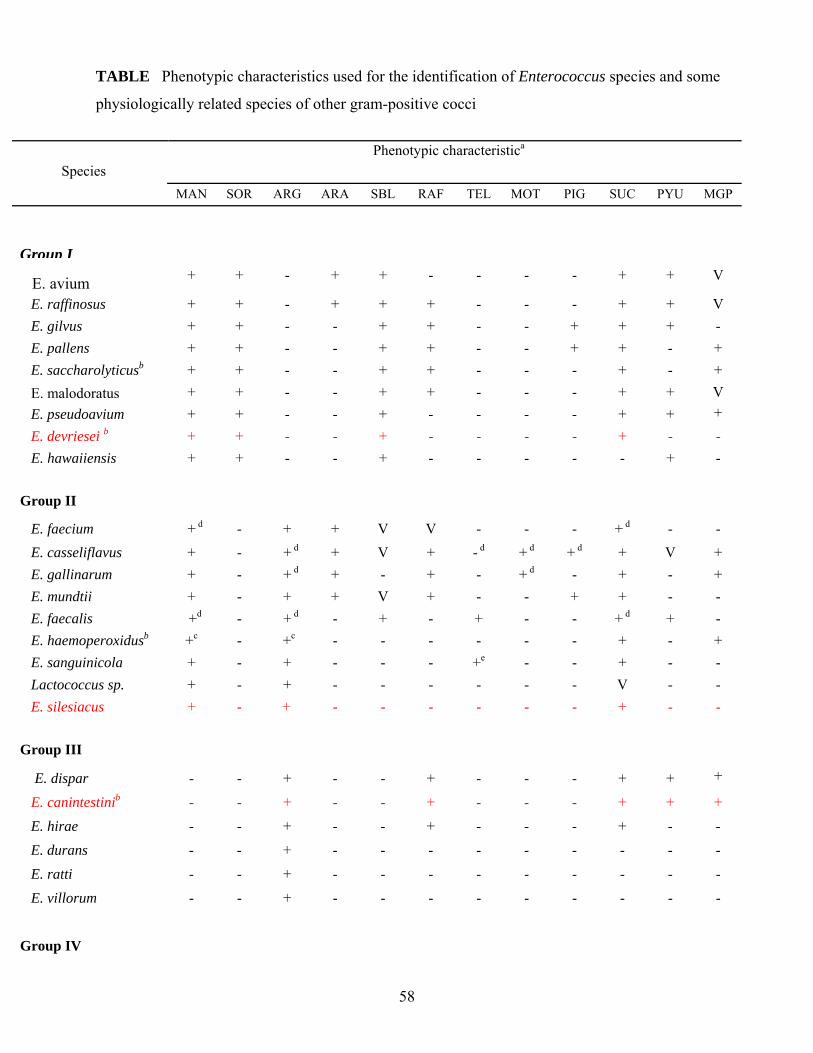
58
TABLE Phenotypic characteristics used for the identification of Enterococcus species and some
physiologically related species of other gram-positive cocci
Species Phenotypic characteristica
MAN SOR ARG ARA SBL RAF TEL MOT PIG SUC PYU MGP
Group I
E. avium + + - + + - - - - + + V
E. raffinosus + + - + + + - - - + + V E. gilvus + + - - + + - - + + + - E. pallens + + - - + + - - + + - + E. saccharolyticusb + + - - + + - - - + - + E. malodoratus + + - - + + - - - + + V E. pseudoavium + + - - + - - - - + + + E. devriesei b + + - - + - - - - + - - E. hawaiiensis + + - - + - - - - - + - Group II
E. faecium + d - + + V V - - - + d - - E. casseliflavus + - + d + V + - d + d + d + V + E. gallinarum + - + d + - + - + d - + - + E. mundtii + - + + V + - - + + - - E. faecalis +d - + d - + - + - - + d + - E. haemoperoxidusb +c - +c - - - - - - + - + E. sanguinicola + - + - - - +e - - + - - Lactococcus sp. + - + - - - - - - V - - E. silesiacus + - + - - - - - - + - - Group III
E. dispar - - + - - + - - - + + +
E. canintestinib - - + - - + - - - + + +
E. hirae - - + - - + - - - + - -
E. durans - - + - - - - - - - - -
E. ratti - - + - - - - - - - - -
E. villorum - - + - - - - - - - - -
Group IV
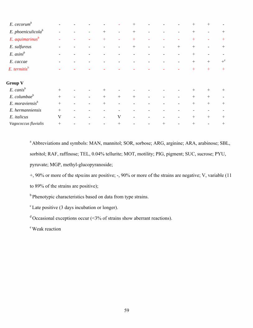
59
E. cecorumb - - - - - + - - - + + -
E. phoeniculicolab - - - + - + - - - + - +
E. aquimarinusb - - - + - + - - - + - +
E. sulfureus - - - - - + - - + + - +
E. asinib - - - - - - - - - + - -
E. caccae - - - - - - - - - + + +c
E. termitisb - - - - - - - - - + + +
Group V E. canisb + - - + - - - - - + + + E. columbaeb + - - + + + - - - + + - E. moraviensisb + - - + - - - - - + + + E. hermanniensis + - - - - - - - - - - - E. italicus V - - - V - - - - + + + Vagococcus fluvialis + - - - + - - + - + - +
a Abbreviations and symbols: MAN, mannitol; SOR, sorbose; ARG, arginine; ARA, arabinose; SBL,
sorbitol; RAF, raffinose; TEL, 0.04% tellurite; MOT, motility; PIG, pigment; SUC, sucrose; PYU,
pyruvate; MGP, methyl-glucopyranoside;
+, 90% or more of the stραins are positive; -, 90% or more of the strains are negative; V, variable (11
to 89% of the strains are positive);
b Phenotypic characteristics based on data from type strains.
c Late positive (3 days incubation or longer).
d Occasional exceptions occur (<3% of strains show aberrant reactions).
e Weak reaction
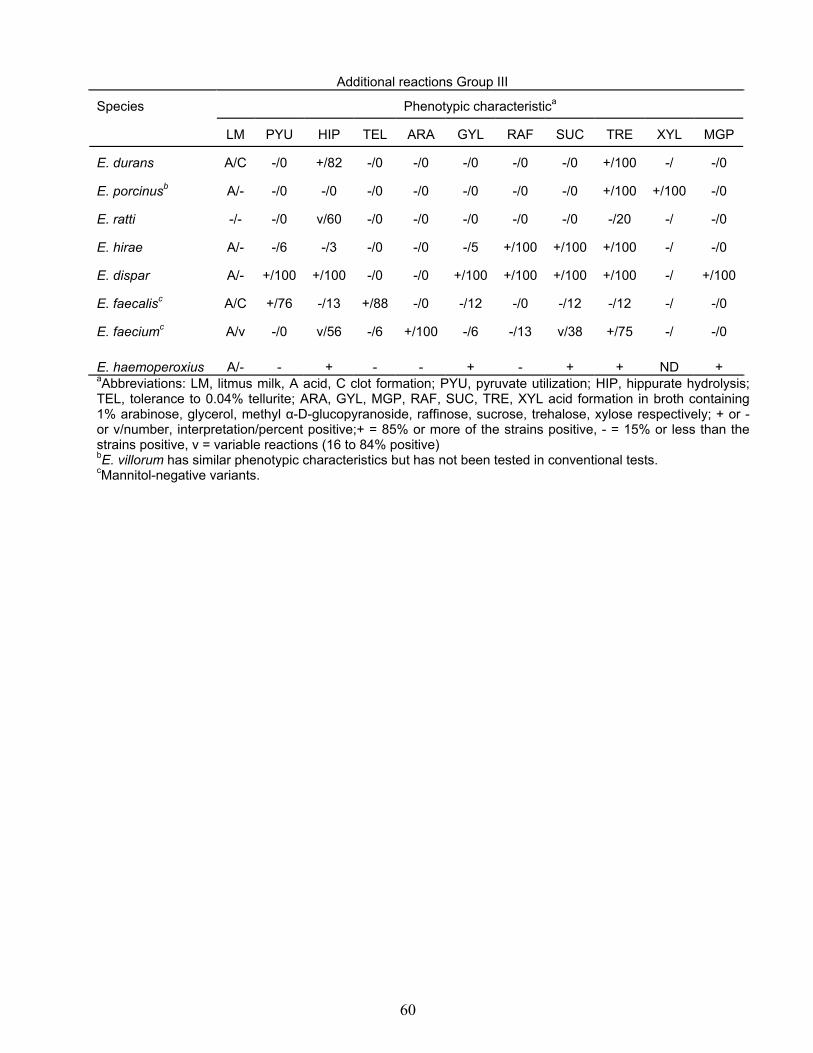
Additional reactions Group III
Phenotypic characteristica Species
LM
PYU
HIP
TEL
ARA
GYL
RAF
SUC
TRE
XYL
MGP
E. durans
A/C
-/0
+/82
-/0
-/0
-/0
-/0
-/0
+/100
-/
-/0
E. porcinusb
A/-
-/0
-/0
-/0
-/0
-/0
-/0
-/0
+/100
+/100
-/0
E. ratti
-/-
-/0
v/60
-/0
-/0
-/0
-/0
-/0
-/20
-/
-/0
E. hirae
A/-
-/6
-/3
-/0
-/0
-/5
+/100
+/100
+/100
-/
-/0
E. dispar
A/-
+/100
+/100
-/0
-/0
+/100
+/100
+/100
+/100
-/
+/100
E. faecalisc
A/C
+/76
-/13
+/88
-/0
-/12
-/0
-/12
-/12
-/
-/0
E. faeciumc
A/v
-/0
v/56
-/6
+/100
-/6
-/13
v/38
+/75
-/
-/0
E. haemoperoxius
A/-
-
+
-
-
+
-
+
+
ND
+
aAbbreviations: LM, litmus milk, A acid, C clot formation; PYU, pyruvate utilization; HIP, hippurate hydrolysis; TEL, tolerance to 0.04% tellurite; ARA, GYL, MGP, RAF, SUC, TRE, XYL acid formation in broth containing 1% arabinose, glycerol, methyl α-D-glucopyranoside, raffinose, sucrose, trehalose, xylose respectively; + or - or v/number, interpretation/percent positive;+ = 85% or more of the strains positive, - = 15% or less than the strains positive, v = variable reactions (16 to 84% positive) bE. villorum has similar phenotypic characteristics but has not been tested in conventional tests. cMannitol-negative variants.
60
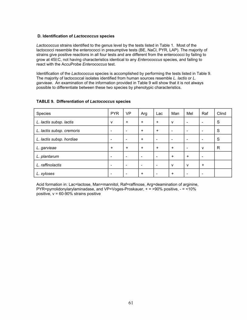
D. Identification of Lactococcus species Lactococcus strains identified to the genus level by the tests listed in Table 1. Most of the lactococci resemble the enterococci in presumptive tests (BE, NaCl, PYR, LAP). The majority of strains give positive reactions in all four tests and are different from the enterococci by failing to grow at 45ΕC, not having characteristics identical to any Enterococcus species, and failing to react with the AccuProbe Enterococcus test. Identification of the Lactococcus species is accomplished by performing the tests listed in Table 9. The majority of lactococcal isolates identified from human sources resemble L. lactis or L. garvieae. An examination of the information provided in Table 9 will show that it is not always possible to differentiate between these two species by phenotypic characteristics. TABLE 9. Differentiation of Lactococcus species Species
PYR
VP
Arg
Lac
Man
Mel
Raf
Clind
L. lactis subsp. lactis
v
+
+
+
v
-
-
S
L. lactis subsp. cremoris
-
-
+
+
-
-
-
S
L. lactis subsp. hordiae
-
-
+
-
-
-
-
S
L. garvieae
+
+
+
+
+
-
v
R
L. plantarum
-
-
-
-
+
+
-
L. raffinolactis
-
-
-
-
v
v
+
L. xyloses
-
-
+
-
+
-
-
Acid formation in: Lac=lactose, Man=mannitol, Raf=raffinose, Arg=deamination of arginine, PYR=pyrrolidonylarylaminadase, and VP=Voges-Proskauer, + = >90% positive, - = <10% positive, v = 60-90% strains positive
61
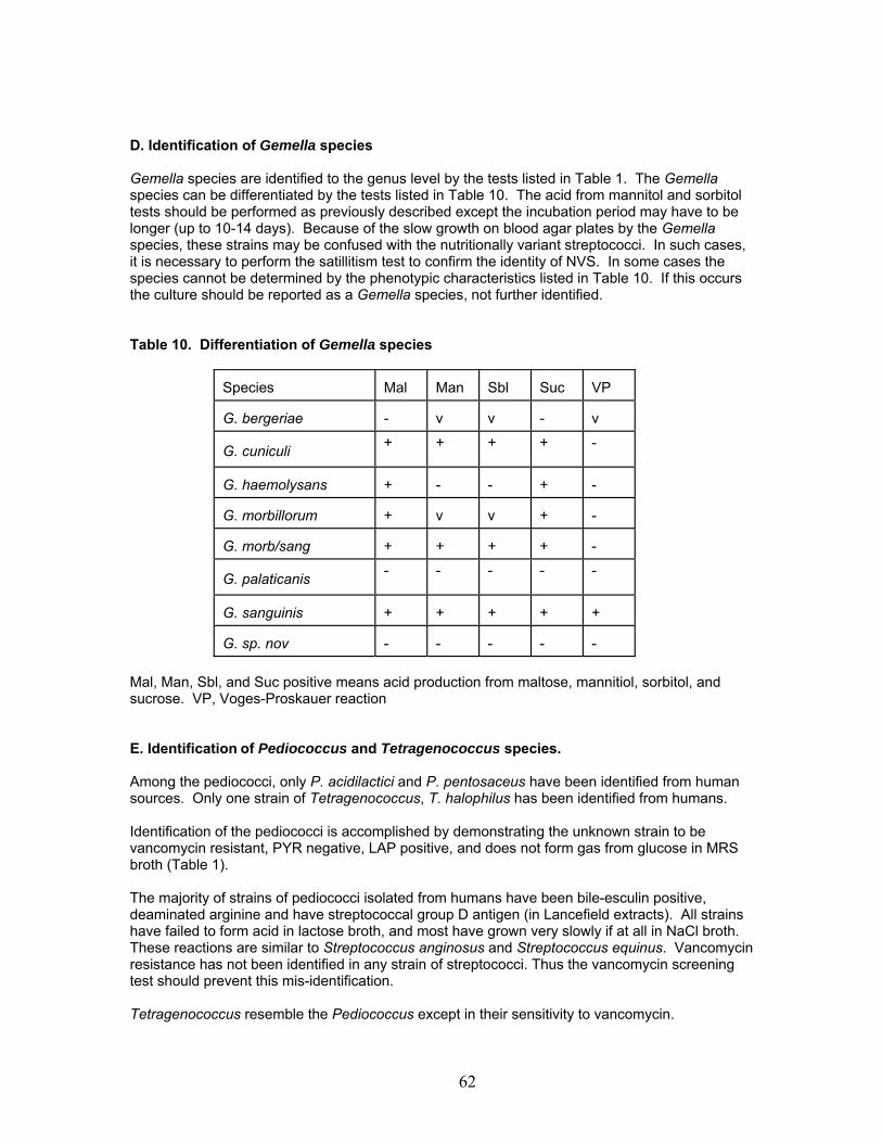
D. Identification of Gemella species
Gemella species are identified to the genus level by the tests listed in Table 1. The Gemella species can be differentiated by the tests listed in Table 10. The acid from mannitol and sorbitol tests should be performed as previously described except the incubation period may have to be longer (up to 10-14 days). Because of the slow growth on blood agar plates by the Gemella species, these strains may be confused with the nutritionally variant streptococci. In such cases, it is necessary to perform the satillitism test to confirm the identity of NVS. In some cases the species cannot be determined by the phenotypic characteristics listed in Table 10. If this occurs the culture should be reported as a Gemella species, not further identified.
Table 10. Differentiation of Gemella species
Species
Mal
Man
Sbl
Suc
VP
G. bergeriae
-
v
v
-
v
. cuniculi G
+ + + + -
G. haemolysans
+
-
-
+
-
G. morbillorum
+
v
v
+
-
G. morb/sang
+
+
+
+
-
. palaticanis G
- - - - -
G. sanguinis
+
+
+
+
+
G. sp. nov
-
-
-
-
-
Mal, Man, Sbl, and Suc positive means acid production from maltose, mannitiol, sorbitol, and sucrose. VP, Voges-Proskauer reaction E. Identification of Pediococcus and Tetragenococcus species. Among the pediococci, only P. acidilactici and P. pentosaceus have been identified from human sources. Only one strain of Tetragenococcus, T. halophilus has been identified from humans. Identification of the pediococci is accomplished by demonstrating the unknown strain to be vancomycin resistant, PYR negative, LAP positive, and does not form gas from glucose in MRS broth (Table 1). The majority of strains of pediococci isolated from humans have been bile-esculin positive, deaminated arginine and have streptococcal group D antigen (in Lancefield extracts). All strains have failed to form acid in lactose broth, and most have grown very slowly if at all in NaCl broth. These reactions are similar to Streptococcus anginosus and Streptococcus equinus. Vancomycin resistance has not been identified in any strain of streptococci. Thus the vancomycin screening test should prevent this mis-identification. Tetragenococcus resemble the Pediococcus except in their sensitivity to vancomycin.
62

TABLE 11. Identification of Pediococcus and Tetragenococcus species. Pediococcus sp. are vancomycin resistant and Tetragenococcus is vancomycin sensitive. Species
Van
Mal
Ara
Str
Suc
NaCl
Tre
Arg
CRS
P. acidilactici
R
-
+
-
-
V
V
+
(V)
P. pentosaceus
R
+
V
-
-
+
+
V
(+)
P. damnosus
R
+
-
-
+
-
+
-
P. dextrinicus
R
+
-
+
+
-
-
-
P. urinequi
R
+
-
-
+
+
P. parvulus
R
-
-
-
-
+
-
-
T. halophilus
S
+
-
-
+
-
Van= Vancomycin, S=sensitive, R= resistant, Mal = maltose, Ara= arabinose, Str = starch, Suc = sucrose, NaCl = 6.5% NaCl broth, Tre= trehalose, Arg= arginine, CRS = Chromogenic substrates either glucopyronidase or glucosaminidase + = >90% positive, - = <10% positive, v = 60-90% strains positive (-) most strains negative (+) most strains positive P. damnosus, P. dextrinicus, P. urinequi, and P. paruulus are not found in human infections, so far.
63
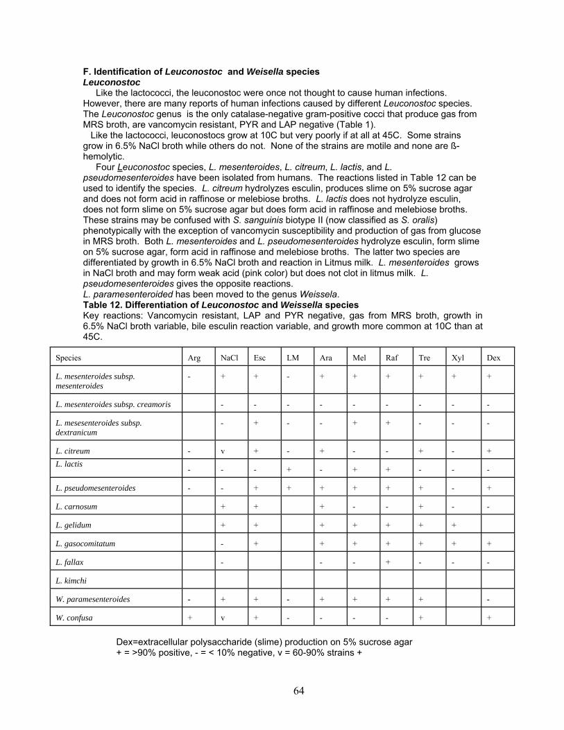
F. Identification of Leuconostoc and Weisella species Leuconostoc Like the lactococci, the leuconostoc were once not thought to cause human infections. However, there are many reports of human infections caused by different Leuconostoc species. The Leuconostoc genus is the only catalase-negative gram-positive cocci that produce gas from MRS broth, are vancomycin resistant, PYR and LAP negative (Table 1). Like the lactococci, leuconostocs grow at 10C but very poorly if at all at 45C. Some strains grow in 6.5% NaCl broth while others do not. None of the strains are motile and none are ß-hemolytic. Four Leuconostoc species, L. mesenteroides, L. citreum, L. lactis, and L. pseudomesenteroides have been isolated from humans. The reactions listed in Table 12 can be used to identify the species. L. citreum hydrolyzes esculin, produces slime on 5% sucrose agar and does not form acid in raffinose or melebiose broths. L. lactis does not hydrolyze esculin, does not form slime on 5% sucrose agar but does form acid in raffinose and melebiose broths. These strains may be confused with S. sanguinis biotype II (now classified as S. oralis) phenotypically with the exception of vancomycin susceptibility and production of gas from glucose in MRS broth. Both L. mesenteroides and L. pseudomesenteroides hydrolyze esculin, form slime on 5% sucrose agar, form acid in raffinose and melebiose broths. The latter two species are differentiated by growth in 6.5% NaCl broth and reaction in Litmus milk. L. mesenteroides grows in NaCl broth and may form weak acid (pink color) but does not clot in litmus milk. L. pseudomesenteroides gives the opposite reactions. L. paramesenteroided has been moved to the genus Weissela. Table 12. Differentiation of Leuconostoc and Weissella species Key reactions: Vancomycin resistant, LAP and PYR negative, gas from MRS broth, growth in 6.5% NaCl broth variable, bile esculin reaction variable, and growth more common at 10C than at 45C.
Species
Arg
NaCl Esc LM Ara Mel Raf
Tre
Xyl Dex
L. mesenteroides subsp. mesenteroides
-
+
+
-
+
+
+
+
+
+
L. mesenteroides subsp. creamoris
-
-
-
-
-
-
-
-
-
L. mesesenteroides subsp. dextranicum
-
+
-
-
+
+
-
-
-
L. citreum
-
v
+
-
+
-
-
+
-
+
L. lactis -
-
-
+
-
+
+
-
-
-
L. pseudomesenteroides
-
-
+
+
+
+
+
+
-
+
L. carnosum
+
+
+
-
-
+
-
-
L. gelidum
+
+
+
+
+
+
+
L. gasocomitatum
-
+
+
+
+
+
+
+
L. fallax
-
-
-
+
-
-
-
L. kimchi
W. paramesenteroides
-
+
+
-
+
+
+
+
-
W. confusa
+
v
+
-
-
-
-
+
+
Dex=extracellular polysaccharide (slime) production on 5% sucrose agar + = >90% positive, - = < 10% negative, v = 60-90% strains +
64

G. Identification of Aerococcus species, Helcococcus kunzi, Dolosigranulum pigrum, Tetragenococcus solitarius
The Aerococcus, Helcoccus, Dolosigranulum and Tetragenococcus all have the cellular arrangement of clusters and tetrads. All will grow in 6.5% NaCl with the exception of A. sanguicola and A. christensenii.
Table 13. Identification of Aerococcus species, Helcococcus kunzi, Dolosigranulum
pigrum, Tetragenococcus solitarius
Species
Lac
Mal
Man
Rib
Suc
Tre
Arg
β-
Esc
PYR
LAP
BE
NaCl
Hip
VP
A. viridans
v
v
v
v
+
+
-
+
+
+
-
-
+
v
-
A. urinae
-
v
+
v
+
-
-
v
v
-
+
-
+
+
-
A. sanguicola
v
+
-
+
+
+
-
+
+
+
+
+
+
+
-
A. christensenii
-
-
-
-
-
-
-
-
-
-
+
-
-
+
+
A. urinehominis
-
+
-
v
+
-
-
+
+
-
-
-
-
-
-
Dolosigranulum
pigrum
-
-
-
-
-
-
-
+
+
+
-
+
-
-
Helcoccus kunzi
-
-
-
-
-
-
-
+
+
+
-
-
+
-
-
Tetragenococcus
solitarius
-
+
+
+
+
+
+
+
+
+
+
+
-
+
Refer to previous tables for explanation of tests and results.
65
H. Identificaton of Globicatella species There is only one species in the genus Globicatella. Globicatella sanguinis closely resembles the
aerococci, streptococci, and enterococci phenotypically. The major differentiating characteristic between Globicatella and the aerococci is the cellular arrangement of the cells in the Gram stain. Globicatella forms chains while the aerococci form tetrads and clusters. The colonial morphology of Globicatella strains most closely resembles the viridans streptococci. However, these strains are readily distinguished with a negative leucine aminopeptidase reaction (LAP) and growth in the presence of 6.5% NaCl. The viridans streptococci are pyrridonyl arylamidase (PYR) negative, LAP positive and do not grow in the presence of 6.5% NaCl. The enterococci are PYR and LAP positive and grow at 10ΕC. None of the Globicatella isolates grew at 10ΕC or gave positive LAP reactions.
The biochemical characteristics of the most recently identified strains of A. viridans (28 strains) E. avium (28 strains) and S. uberis (9 strains, all nonhuman) were compared to the 28 strains of G. sanguinus. A. viridans is biochemically very similar to G. sanguinis (Table ). The colony morphologies are very similar on TSA with 5% sheep blood agar and both are LAP negative and grow in 6.5% NaCl. The cellular arrangement from growth in broth can be used to differentiate the two as A. viridans is arranged in clusters and tetrads, and G. sanguinis is arranged in short chains. Fermentation of inulin can be useful as the majority of G. sanguinis strains are positive and A. viridans strains are negative. The other key reactions for bile esculin, esculin and hippurate are of limited value in separating these two species since the PYR is a variable reaction for G. sanguinis and the bile esculin, esculin, and hippurate tests are variable reactions for Aerococcus viridans.
The LAP test is also useful in distinguishing enterococci from Globicatella strains. E. avium is further identified by positive pyruvate and sorbose tests and negative inulin and raffinose tests. G. sanguinis has the reverse reactions. The two species are phenotypically similar in the PYR, bile esculin, growth in 6.5% NaCl and at 45ΕC, hydrolysis of esculin and hippurate reactions, and acid production from lactose, maltose, mannitol, sorbitol, sucrose, and trehelose.
S. uberis has been included in the identification scheme with viridans streptococci for a number of years. The incorporation of the PYR and LAP tests in the identification scheme, as well as molecular studies have confirmed that many of the strains that are now Globicatella were previously reported as Streptococcus uberis-like. The key test for differentiation of the streptococci from Globicatella is the positive LAP reaction. S. uberis is also negative for esculin hydrolysis in the presence of bile, and positive for growth at 10ΕC. The majority of strains for both species are PYR positive, grow in 6.5% NaCl, hydrolyze esculin and hippurate, and produce acid from inulin, lactose, maltose, mannitol, raffinose, ribose, sorbitol, sucrose, and trehelose. Acid is not produced from glycerol and sorbose for both species.
66

Table 14. Phenotypic differences between G. sanguinis, A. viridans, S. uberis, and E.
avium.
Test
G. sanguinis (28 strains)
A. viridans (28 strains)
E. avium (28 strains)
S. uberis (9 strains)
Gram Stain
Pairs, short chains
clusters, tetrads
short chains
short chains
PYR
V (75)
+ (100)
+ (95)
+ (100)
LAP
- (0)
- (0)
+ (89)
+ (100)
Bile Esculin
+ (100)
V (79)
+ (100)
- (0)
6.5% NaCl
+ (100)
+ (96)
+ (100)
+ (90)
10ΕC
- (0)
- (0)
V (68)
+ (90)
45ΕC
+ (96)
V (25)
+ (100)
+ (90)
Esculin
+ (100)
V (82)
+ (100)
+ (100)
Hippurate
+ (100)
V (70)
V (30)
+(100)
Pyruvate
- (0)
- (0)
+ (86)
- (0)
Tellurite
V (71)
V (20)
- (4)
- (0)
Acid production from: Arabinose
V (42)
- (14)
+ (100)
- (0)
Inulin
+ (93, 11% w+)
- (7)
- (7)
+ (100)
Lactose
+ (93, 11% w+)
V (54)
+ (100)
+ (100)
Maltose
+ (100, 19% w+)
V (50)
+ (95)
+ (100)
Mannitol
+ (96, 11% w+)
V (25)
+ (100)
+ (100)
Melibiose
+ (100, 19% w+)
V (22)
V (50) V (30)
V (80) Raffinose
+ (100, 15% w+)
V (18)
- (0)
Ribose
V (75, 19% w+)
V (13)
+ (94)
+ (90)
Sorbitol
V (82, 11% w+) + (100)
- (14)
+ (100)
Sorbose
- (0)
- (7)
+ (100)
- (0)
Sucrose
+ (100, 15% w+)
V (68)
+ (100, 4% w+)
+ (100)
Trehelose
+ (100, 15% w+) V (54)
+ (100)
+ (100)
+, 85% or more of the strains positive; -, 15% or less of the strains positive; V, variable reaction 16 to 84% of the strains positive; ( ), first number in the parenthesis indicates the total number of strains giving strong or weakly positive reactions. Biochemicals were incubated at 37ΕC for 14 days.
67
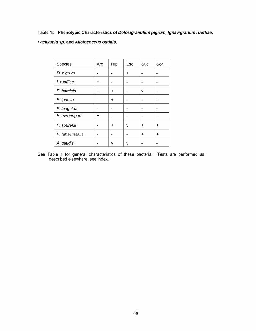
Table 15. Phenotypic Characteristics of Dolosigranulum pigrum, Ignavigranum ruoffiae,
Facklamia sp. and Alloiococcus otitidis.
Esc
Species
Arg
Hip
Suc Sor
D. pigrum
- - + - -
- - - I. ruoffiae +
-
F. hominis
+
+ -
v
-
F. ignava
-
- -
+
-
F. languida
-
-
-
- -
F. miroungae + - - - -
F. sourekii
-
+ v +
+
F. tabacinsalis - - - + + A. otitidis
-
v
v
-
-
See Table 1 for general characteristics of these bacteria. Tests are performed as
described elsewhere, see index.
68
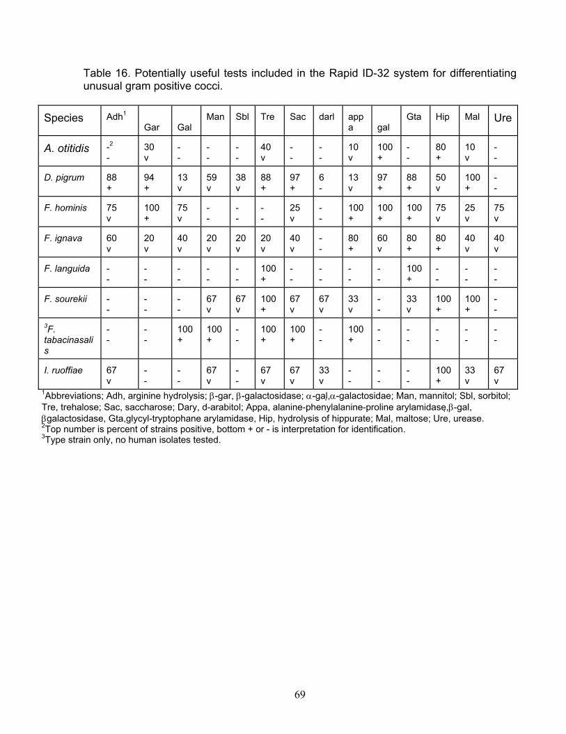
Table 16. Potentially useful tests included in the Rapid ID-32 system for differentiating unusual gram positive cocci.
Species Adh1 Gar Gal
Man Sbl Tre Sac darl appa gal
Gta Hip Mal Ure
A. otitidis - -2 -
30 v -
- -
- -
40 v
- -
- -
10 v
100 +
- -
80 +
10 v
- -
D. pigrum 88 +
94 +
13 v
59 v
38 v
88 +
97 +
6 -
13 v
97 +
88 +
50 v
100 +
- -
F. hominis 75 v
100 +
75 v
- -
- -
- -
100 +
25 v
75 v
25 v
- -
100 +
100 +
75 v
F. ignava v
60 60 v
20 v
40 v
20 v
20 20 v
40 v
- -
80 + v
80 +
80 +
40 v
40 v
F. languida - -
- -
- -
- -
- -
100 +
- -
- -
- -
- -
100 +
- -
- -
- -
F. sourekii - -
- -
- -
67 v
67 v
100 +
67 v
67 100 - v
33 v
- -
33 v
100 + +
-
- -
+ 100
- + 100 3F.
tabacinasalis
- -
100 +
- 100 +
- -
100 +
- -
- -
- -
- -
- -
I. ruoffiae 67 v
- -
- -
67 v
- -
67 v
67 v
33 v
- -
- -
- -
100 +
33 v
67 v
1Abbreviations; Adh, arginine hydrolysis; β-gar, β-galactosidase; α-gal,α-galactosidae; Man, mannitol; Sbl, sorbitol; Tre, trehalose; Sac, saccharose; Dary, d-arabitol; Appa, alanine-phenylalanine-proline arylamidase, β-gal, βgalactosidase, Gta,glycyl-tryptophane arylamidase, Hip, hydrolysis of hippurate; Mal, maltose; Ure, urease. 2Top number is percent of strains positive, bottom + or - is interpretation for identification. 3Type strain only, no human isolates tested.
69