strategies to prevent dopamine oxidation and related
TRANSCRIPT
ORIGINAL ARTICLE
Strategies to prevent dopamine oxidation and related cytotoxicityusing various antioxidants and nitrogenation
Devyesh Rana1 & Thibault Colombani1 & Halimatu S. Mohammed1& Loek J. Eggermont1 & Samantha Johnson2
&
Nasim Annabi1,3,4,5 & Sidi A. Bencherif1,2,6,7
Received: 30 May 2019 /Accepted: 28 June 2019 /Published online: 7 August 2019# Qatar University and Springer Nature Switzerland AG 2019
AbstractDopamine (DA) plays several important roles in the brain and body and has recently been used as a bioadhesive precursor formedical applications. However, DA oxidizes immediately when exposed to oxygen and rapidly polymerizes into polydopamine(PDA), leading to oxidative stress, cytotoxicity, and loss of DA functionalities. As a result, preventing rapid oxidation of DA is ofparamount importance but still remains a major challenge. Here, we report several strategies to impede DA oxidation in relevantaqueous solutions (i.e., water, PBS, and cell culture media). One strategy is based on using reducing agents or antioxidants suchas glutathione in its reduced state (GSH) and sodium tetraborate (commonly known as borax). Another strategy is based onnitrogenation, a method used to preserve DA in its reduced form by creating an oxygen-free environment. Our data suggest thatthe antioxidant properties of GSH and borax substantially decreased DA oxidation for up to 2 months. Nitrogenation or oxygenremoval further prevented DA oxidation, enhancing its shelf life for longer periods of time. When tested with mammalian cells,preventing DA oxidation with GSH dramatically improved viability of 3T3 fibroblasts and Tcells. These results demonstrate thatthe use of antioxidants, alone or in combination with nitrogenation, can help prevent DA oxidation and improve its stability forcell-based studies or for the design and development of biomaterials.
Keywords Dopamine . Oxidation . Reducing agents . Nitrogenation . Cytocompatibility
1 Introduction
Dopamine (DA), an innate catecholaminergic neurotransmit-ter, and its oxidized derivatives, including polydopamine(PDA), are of interest for their role in several biological pro-cesses and diseases [1–5]. For instance, DA oxidation, low
DA concentrations, and high PDA concentrations are indica-tors of neurological conditions, including Parkinson’s andAlzheimer’s diseases [6]. Biological processes regulate DAoxidation and its conversion into PDA to combat the onsetof these diseases [7]. In addition to these functionalities, evi-dence from clinical studies in humans suggests that a subunit
Electronic supplementary material The online version of this article(https://doi.org/10.1007/s42247-019-00037-5) contains supplementarymaterial, which is available to authorized users.
* Nasim [email protected]
* Sidi A. [email protected]
1 Department of Chemical Engineering, Northeastern University,Boston, MA, USA
2 Department of Bioengineering, Northeastern University,Boston, MA, USA
3 Harvard-MIT Division of Health Sciences and Technology,Massachusetts Institute of Technology, Cambridge, MA, USA
4 Department of Chemical and Biomolecular Engineering, Universityof California, Los Angeles, Los Angeles, CA 90095, USA
5 Center for Minimally Invasive Therapeutics (C-MIT), CaliforniaNanoSystems Institute (CNSI), University of California, LosAngeles, Los Angeles, CA 90095, USA
6 Harvard John A. Paulson School of Engineering and AppliedSciences, Harvard University, Cambridge, USA
7 Sorbonne University, UTC CNRS UMR 7338, Biomechanics andBioengineering (BMBI), University of Technology of Compiègne,Compiègne, France
Emergent Materials (2019) 2:209–217https://doi.org/10.1007/s42247-019-00037-5

of melanin polymerizes into PDA, which causes dark pigmen-tation on the skin, hair, and the substantia nigra of the brain[8]. However, in other organisms such as mussels, PDA playsa pivotal role in their adhesion in wet environments.
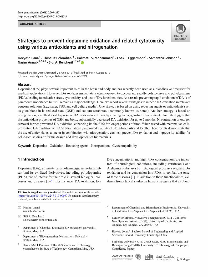
There are numerous reports on the rapid oxidation of DAunder physiological pH [5, 7, 9] and its effect on several pro-cesses, including adhesiveness of biomaterials [5, 7, 10–12],therapeutics [1–3], and conductive and adhesivematerial coat-ings [4, 9, 13–15]. In the body, self-polymerization of DA intoPDA occurs in the presence of oxygen or monoamine oxidase[10]. Furthermore, conversion of DA into PDA can be cata-lyzed by an oxidizing agent such as sodium periodate. Thischaracteristic oxidation is qualitatively observed by the black-ening of DA due to PDA formation, which has been associat-ed with mammalian cell death [6, 8, 16–20]. There is a largebody of evidence suggesting that PDA formation leads tooxidative stress–induced cytotoxicity due to the formation offree radicals [21]. Specifically, it was found that DA oxidationtriggers apoptosis, while the use of antioxidants could preventDA-related cytotoxicity [22]. Furthermore, PDA formationinhibits binding of DA to thiols, amines, and hydroxyls [10,23]. Thus, there is an urgent need to develop or reinforcestrategies to control the rapid oxidation of DA.
To address this oxidative challenge, Han et al. recentlyproposed a two-step polymerization technique to limit oxida-tion of dopamine by encapsulating DA in clay interlayersfollowed by controlled oxidation to form PDA [12].However, steric hindrance and physical restrictions limitedthe ability of DA to directly interact with tissues in this strat-egy [12]. Here, we have investigated three facile routes toprevent DA oxidation in relevant aqueous solutions (water,phosphate buffered saline (PBS), and cell culture media).Specifically, we hypothesize that both glutathione (GSH), anantioxidant present in most mammalian tissues, and sodiumtetraborate (borax), a chemically inert antioxidant, can be usedindependently and in combination with nitrogenation to pre-serve DA in its reduced state for further chemical modifica-tions or to limit oxidative stress–induced cell toxicity.
2 Results and discussion
2.1 Rate of DA oxidation and PDA formation
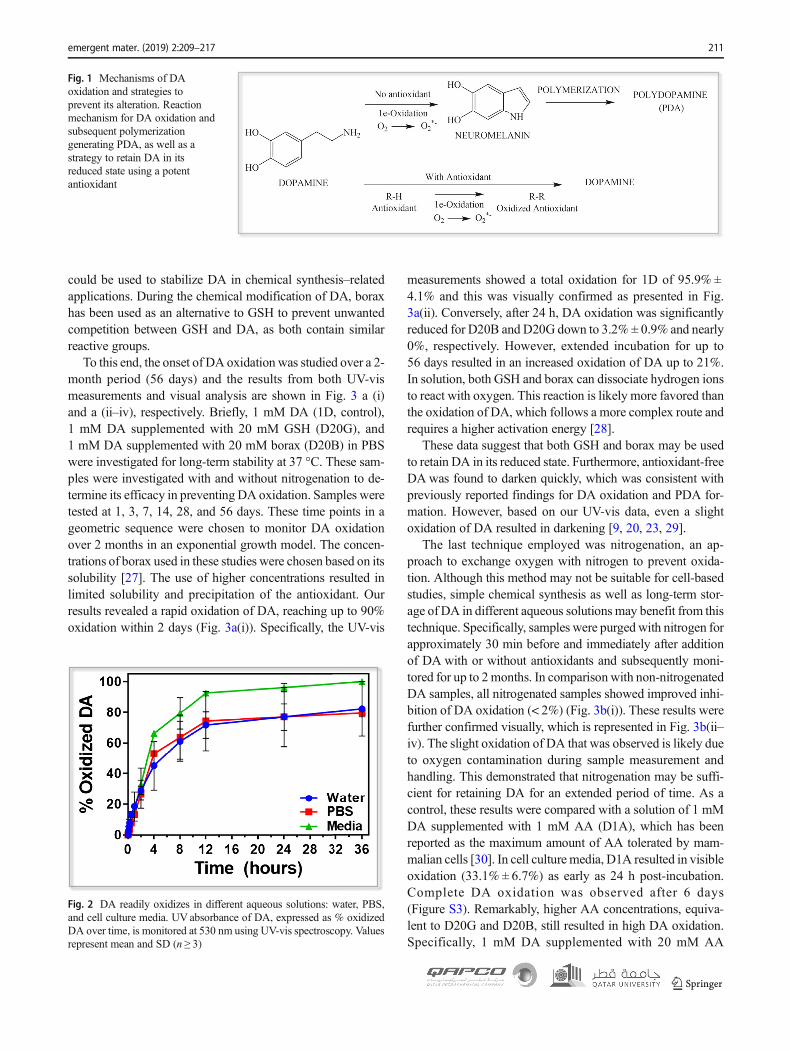
To determine the rate of DA oxidation and PDA formation(Fig. 1), absorbance of unmodified DA in water, PBS (pH7.4), and complete media were measured at various timepoints. UV-vis data was utilized and the rate of DA oxidation(rDA) was derived from a plot showing the percentage of ox-idized DA as a function of time. Within 1 h, low levels of DAoxidation were observed. The percentage of oxidation in wa-ter, PBS, and cell culture media was quantified to be 18.6% ±9.6%, 13.3% ± 1.7%, and 16.6% ± 1.9%, respectively (Fig. 2).
Prolonged incubation of DA-containing samples in their re-spective solvents intensified oxidation. Specifically, the per-centage of oxidized DA significantly increased to 71.6% ±8.6%, 74.3% ± 19.3%, and 92.7% ± 1.0% in water, PBS, andmedia, respectively, after 12 h.
Subsequently, the average reaction rate for DA oxidation at37 °C was determined as shown in Eq. 1:
DA½ � ¼ DA½ �0 � ekDAt; kDA ¼ 3:28� 10−3min−1 ð1Þ
The oxidation of unmodified DAwas visibly confirmed inall tested solutions (Fig. 2). DA in water and PBS resulted in76.9% ± 19.3% and 76.9% ± 8.7% oxidation within 24 h.Interestingly, the highest oxidation rate was observed in cellculture media, in which DA was almost completely oxidizedwithin the same time frame. These data can be fitted (correla-tion factor > 0.98) to Eq. 1. Furthermore, it was observed thatDA consumption follows first-order kinetics as shown in Eq. 2:
A½ � ¼ A½ �0 exp −katð Þ ð2Þ
In this equation, [A] is the final concentration of DA, [A]0 isthe starting concentration of DA, t is time, and ka is the rateconstant. The calculated kDA = 3.28 × 10⁻³ ± 0.001.2 ×10−3 min−1, where the rate constant is the average of the threesamples used and the error is the maximum deviation calcu-lated with n = 3. The rate of oxidation is in line with the ap-proximate simulated oxidation rate (rDA = 4 × 10⁻³[DA]1/2)[24]. To confirm this set of data, additional characterizationsby 1H nuclear magnetic resonance (NMR) spectroscopyRaman spectroscopy Fourier-transform infrared (FTIR),Spectroscopy and high-performance liquid chromatography(HPLC) are presented in the Supplementary Information(Figure S1 and Figure S2) [25]. In summary, these resultsconfirmed the instability and rapid oxidation of unmodifiedDA and its conversion into PDA in various oxygen-containingand antioxidant-free aqueous solutions.
2.2 Effect of antioxidants on DA oxidation
Three different strategies were proposed to prevent and limitthe rapid oxidation of DA. Specifically, nitrogenation wasemployed, as well as reduction reactions using two commonlyused antioxidants, borax and GSH. These results were com-pared with ascorbic acid (AA), a gold standard antioxidantused here as a control. AA, also known as vitamin C, wasused as it is and the most widely used antioxidant in cell-based in vitro studies. GSH was chosen for its highcytocompatibility and strong ability to reduce oxidative stress[26]. Although GSH has been used in a number of bioassays,there is limited data on its application for the stabilization ofDA. Finally, borax is known to be toxic to cells, which limitsits application for in vitro and in vivo studies. However, it
210 emergent mater. (2019) 2:209–217
could be used to stabilize DA in chemical synthesis–relatedapplications. During the chemical modification of DA, boraxhas been used as an alternative to GSH to prevent unwantedcompetition between GSH and DA, as both contain similarreactive groups.
To this end, the onset of DA oxidation was studied over a 2-month period (56 days) and the results from both UV-vismeasurements and visual analysis are shown in Fig. 3 a (i)and a (ii–iv), respectively. Briefly, 1 mM DA (1D, control),1 mM DA supplemented with 20 mM GSH (D20G), and1 mM DA supplemented with 20 mM borax (D20B) in PBSwere investigated for long-term stability at 37 °C. These sam-ples were investigated with and without nitrogenation to de-termine its efficacy in preventing DA oxidation. Samples weretested at 1, 3, 7, 14, 28, and 56 days. These time points in ageometric sequence were chosen to monitor DA oxidationover 2 months in an exponential growth model. The concen-trations of borax used in these studies were chosen based on itssolubility [27]. The use of higher concentrations resulted inlimited solubility and precipitation of the antioxidant. Ourresults revealed a rapid oxidation of DA, reaching up to 90%oxidation within 2 days (Fig. 3a(i)). Specifically, the UV-vis
measurements showed a total oxidation for 1D of 95.9% ±4.1% and this was visually confirmed as presented in Fig.3a(ii). Conversely, after 24 h, DA oxidation was significantlyreduced for D20B and D20G down to 3.2% ± 0.9% and nearly0%, respectively. However, extended incubation for up to56 days resulted in an increased oxidation of DA up to 21%.In solution, both GSH and borax can dissociate hydrogen ionsto react with oxygen. This reaction is likely more favored thanthe oxidation of DA, which follows a more complex route andrequires a higher activation energy [28].
These data suggest that both GSH and borax may be usedto retain DA in its reduced state. Furthermore, antioxidant-freeDA was found to darken quickly, which was consistent withpreviously reported findings for DA oxidation and PDA for-mation. However, based on our UV-vis data, even a slightoxidation of DA resulted in darkening [9, 20, 23, 29].
The last technique employed was nitrogenation, an ap-proach to exchange oxygen with nitrogen to prevent oxida-tion. Although this method may not be suitable for cell-basedstudies, simple chemical synthesis as well as long-term stor-age of DA in different aqueous solutions may benefit from thistechnique. Specifically, samples were purged with nitrogen forapproximately 30 min before and immediately after additionof DA with or without antioxidants and subsequently moni-tored for up to 2months. In comparison with non-nitrogenatedDA samples, all nitrogenated samples showed improved inhi-bition of DA oxidation (< 2%) (Fig. 3b(i)). These results werefurther confirmed visually, which is represented in Fig. 3b(ii–iv). The slight oxidation of DA that was observed is likely dueto oxygen contamination during sample measurement andhandling. This demonstrated that nitrogenation may be suffi-cient for retaining DA for an extended period of time. As acontrol, these results were compared with a solution of 1 mMDA supplemented with 1 mM AA (D1A), which has beenreported as the maximum amount of AA tolerated by mam-malian cells [30]. In cell culture media, D1A resulted in visibleoxidation (33.1% ± 6.7%) as early as 24 h post-incubation.Complete DA oxidation was observed after 6 days(Figure S3). Remarkably, higher AA concentrations, equiva-lent to D20G and D20B, still resulted in high DA oxidation.Specifically, 1 mM DA supplemented with 20 mM AA
Fig. 2 DA readily oxidizes in different aqueous solutions: water, PBS,and cell culture media. UV absorbance of DA, expressed as % oxidizedDA over time, is monitored at 530 nm using UV-vis spectroscopy. Valuesrepresent mean and SD (n ≥ 3)
Fig. 1 Mechanisms of DAoxidation and strategies toprevent its alteration. Reactionmechanism for DA oxidation andsubsequent polymerizationgenerating PDA, as well as astrategy to retain DA in itsreduced state using a potentantioxidant
emergent mater. (2019) 2:209–217 211

(D20A) resulted in 86.7% ± 6.1% DA oxidation after 6 days,while only 5% of DA was oxidized for 20GSH (Figure S3).Furthermore, 10mMGSH (10GSH) was also able to limit DAoxidation to levels below 10%.
This was confirmed by an observed slower reaction rateand rate constant (Eq. 3) of the DA oxidation reaction.Overall, the DA oxidation rate used is as follows:
DA½ � ¼ −1:51� 10−5 DA½ �0; kDA;Overall of 1:51� 10−5min−1
ð3Þ
Data was fit to a linear regression and by averaging theslope from multiple experiments (n = 3) to ascertainkDA,Overall. This result was also confirmed by enzyme-linkedimmunosorbent assay (ELISA) (Figure S4). GSH concentra-tions above 20 mM resulted in its precipitation and aggrega-tion and were not further considered for this reason. It shouldbe noted that, for typical cell culture–related experiments, me-dia is replaced within 5–7 days. This suggests that GSH maybe a suitable antioxidant for cell-based in vitro studies due toits longer stability.
A schematic of the experimental setup to prevent DA oxi-dation with GSH and nitrogenation can be found in Figs. 4 andFigure S5, respectively. In particular, DA-containing samplesthat were treated using nitrogenation could be stored for over2 months. For short-term storage or the chemical modification
of DA, borax was a viable option for samples that are sensitiveto functionalities of GSH (e.g., thiol, amine, or carboxylic acidgroups). Specifically, for applications in Ni/Co supercapacitorsor Fe-based anodes in which DA is used as a carbon source,controlling DA oxidation could have beneficial implications byimproving electron transfer within the carbon structures [31,32]. In these cases, borax can act as an inexpensive antioxidantfor controlling DA functionalization or controlled polymeriza-tion. Addition of GSH is a viable option for both long-term andshort-term sample preparation and storage for both benchtopand in vitro experiments. For biological applications,nitrogenation (or oxygen removal) is untenable, as it is condu-cive to cell hypoxia and atrophy. Similarly, the toxicity of boraxlimits its use for cell-based studies. Therefore, the use of GSHas antioxidant should be considered for biological assays.
2.3 In vitro cytocompatibility of DA
Preventing DA oxidation is critical for a number of biomedi-cal applications due to potential oxidative stress–induced cy-totoxicity [33]. Thus, we investigated the impact of DAwhenprotected with an antioxidant (GSH) on the viability and met-abolic activity of mammalian cells. To that end, 3T3 fibro-blasts were cultured in complete cell culture media alone(0D) or in combination with either 1 mM DA (1D), 1 mMDA + 20 mMGSH (D20G), or 1 mMDA + 1 mMAA (D1A)for 24 h at 37 °C. Prior to cell culture, these solutions were
Fig. 3 Prevention of DAoxidation with antioxidants andnitrogenation. a Plot of DAoxidation in % with respect totime. (i) Quantitative dataobtained from UVabsorbance at530 nm to record DA oxidationover time in normoxia.Representative images of (ii) 1D:1 mM DA in PBS, (iii) D20B:1 mM DA in 20 mM borax, and(iv) D20G: 1 mM DA in 20 mMGSH on days 0 and 28. bQuantitative data obtained fromUV-vis absorbance at 530 nm torecord DA oxidation over time inan oxygen-free environment(nitrogen purging).Representative images of (ii) 1D:1 mM DA in nitrogen-purgedPBS, (iii) D20B: 1 mM DA innitrogen-purged 20 mM borax,and (iv) D20G: 1 mM DA innitrogen-purged 20 mM GSH ondays 0 and 56. Values representmean and SD (n ≥ 3)
212 emergent mater. (2019) 2:209–217

incubated for 1, 3, 5, 7, and 14 days at 37 °C. As expected,antioxidant-free DA showed a high cytotoxicity with cell vi-abilities of only 46.7% ± 2.11%, 15.8% ± 3.22%, and 11.1%± 2.04% after 1, 3, and 5 days, respectively. Furthermore, all
cells died after 7 days (Fig. 5a, b), as compared with thecontrol (fibroblasts cultured in a DA-free media). PreventingDA oxidation using 20 mM GSH significantly increased cellviability to 87.3% ± 3.98%, 83.6% ± 6.97%, 78.3% ± 7.63%,
Fig. 5 GSH-protected DA retains high cell viability and metabolicactivity. a Confocal images of 3T3 fibroblasts showing fractions of live(green) and dead (red) cells when cultured for 24 h in a DA-free cellculture media (0D) or in cell culture media supplemented with 1 mMDA (1D), 1 mM DA + 20 mM GSH (D20G), or 1 mM DA + 1 mMAA (D1A). Pretreated DA solutions were allowed to sit for 1, 3, 5, 7, and
14 days prior to be mixed with the cell culture media and be exposed tocells. b Quantification of 3T3 cell viability and (c) metabolic activityfollowing 24 h incubation with the following cell culture media: 0D,1D, D20G, and D1A. The number sign means no cells. Valuesrepresent mean and SD (n = 3). p < 0.05
Fig. 4 Glutathione (GSH)prevents DA oxidation. a DAoxidation in antioxidant-freeaqueous solutions leads toneuromelanin pigment formationand darkening. b GSH preventsDA oxidation resulting incolorless solutions, confirmingthe stability of DA
emergent mater. (2019) 2:209–217 213

75.3% ± 8.85%, and 72.9% ± 3.32% after 1, 3, 5, 7, and14 days of culture, respectively. Nevertheless, low levels ofcytotoxicity were observed. This was attributed to antioxidantexhaustion over time leading to partial DA oxidation. Thiswas further evidenced by the negligible cytotoxicity observedwhen 3T3 cells were cultured in DA-free media supplementedwith 20 mM GSH (20G) (Figure S6). Surprisingly, using1 mM of AA (gold standard antioxidant) to prevent DA oxi-dation did not improve cell viability but rather increased celldeath. Fibroblast viability was 4.03% ± 3.7%, 5.89% ±2.31%, and 3.76% ± 2.64% after 1, 3, and 5 days, respectively.Furthermore, all cells died after 7 days. This is likely due toAA cytotoxicity, which is in accordance with the observedpoor 3T3 cell survival when cultured in DA-free media sup-plemented with 1 mMAA (1A) (Figure S6). This observationis also in agreement with previously published reports show-ing the high cell toxicity and necrosis-mediated cell deathinduced by AA concentrations between 1 and 4 mM [34].
To confirm these results, the metabolic activity of 3T3 fi-broblasts was assessed over time as shown in Fig. 5c. Cellswere cultured in cell culturemedia supplemented withDA andvarious antioxidants (0D, 1D, D20G, or D1A). Fibroblaststreated with 1D dramatically reduced their metabolic activityby 52.7%, 82.7%, 94.1%, and 100% after 1, 3, 5, and 7 and14 days, respectively. However, cells treated with D20G sig-nificantly improved cell metabolism, with up to 70% of met-abolic activity preserved even after 14 days. In agreement withthe cell viability data, 1 mM AA even further reduced cellmetabolic activity when compared with 1D, with an 18-foldand a 22-fold decrease after 1 day and 3 days, respectively.The cytotoxicity of AA limited its use in biological systems atconcentrations that are effective to prevent DA oxidation.Alternatively, GSH appeared to be a powerful and cell-friendly antioxidant. This is probably due to the suppressionor reduction of DA-mediated oxidative stress by scavengingfree radicals such as reactive oxygen species (ROS). OxidizedDA is known to trigger cytotoxicity and necrosis via deposi-tion of DA quinone and other catechol derivatives on proteins,altering their function [33].
Several studies have shown that DA is also a regulator of Tcell physiology and, in turn, immune responses [33, 35].However, oxidation of DA resulted in the generation ofROS, which are believed to alter T cell function and prolifer-ation. For this reason, low concentrations of ROS are a pre-requisite for maintaining T cell survival [35]. To demonstratethe potential of GSH to prevent dopaminergic-triggered apo-ptosis, murine spleen–derived primary T cells were cultured(200,000 cells/mL, 1 mL per well of a 24-well plate) in com-plete cell culture media alone (0D) or in combination witheither 1 mM DA (1D), 1 mM DA with 20 mM GSH(D20G), or 1 mM DA supplemented with 1 mM AA (D1A).Samples including 1D, D20G, and D1Awere incubated for 1,3, and 5 days at 37 °C prior to being exposed to T cells. After
24 h of incubation, T cell viability was determined by flowcytometry using a far-red fixable dead cell staining. While Tcells cultured in 0D maintained a high viability for 24 h, Tcells treated with 1D exhibited decreased cell viability, show-ing nearly complete cell death after 5 days (Fig. 6a, b). Asexpected, T cells cultured in the presence of D20G displayedincreased cell viability (> 80%), up to 821-fold when com-pared with DA after 5 days of incubation. Conversely, T cellstreated with D1A showed a dramatic decrease in cell viabilitywith nearly complete cell death after 5 d of incubation. Theseresults are in agreement with the data on cytotoxicity andmetabolic activity of 3T3 cells, and further confirmed the ca-pacity of GSH to prevent DA oxidation and ultimately oxida-tive stress–induced cell death.
Fig. 6 GSH-protected DA preserves high T cell viability. a First, DA-containing solutions were subjected to oxidation for 1, 3, and 5 days.Next, T cells were incubated for 24 h with the pretreated DA-containingconditions: DA-free RPMI media (0D, control) and in RPMI mediasupplemented with 1 mM DA (1D), 1 mM DA + 20 mM GSH (D20G),and 1 mM DA + 1 mMAA (D1A). b Representative histogram of CD3+
Tcell viability after 24 h of incubation in RPMI media that was pretreatedfor 24 h with 1D, D20G, andD1A using far-red fixable dead staining. Thehistograms show one representative experiment of triplicate treatment.The number sign # means no cells. Values represent mean and SD (n =3). p < 0.05
214 emergent mater. (2019) 2:209–217

3 Conclusion
Overall, we demonstrate three simple approaches to stabilizeDA and prevent or reduce its rapid oxidation. These strategiesare useful to extend DA lifetime and prevent oxidative stresscaused by its degradation. Our set of data demonstrates thatGSH, especially at high concentrations (up to 20 mM), sub-stantially prevents oxidation of DA (up to 99%) without anysignificant cytotoxicity. Unlike GSH, borax can provide a po-tential substitute when an antioxidant is utilized in amine-sensitive environments. Specifically, borax exhibits antioxi-dant properties in radical-rich environments due to its affinityfor hydroxyl groups and its ability to form diester bridgesbetween cis-hydroxyl-containing molecules [36].Comparatively, GSH involves the sulfhydryl group of the cys-teine interacting with radicals [37]. Finally, nitrogenation pro-vides a supplementary preventive option, reinforcing DA pro-tection against oxidation for up to 2 months. The strategies toprevent DA oxidation presented here can provide opportunitiesto further understand the activity of DA in its intact reducedstate. The ability to prevent oxidation for extended periods oftime (> 2 weeks) while retaining cytocompatibility has the po-tential to improve a number of biological studies, includinginteractions of DA in the nervous system [35]. Finally,preventing overoxidation of DA or its derivative, PDA, allowsthese molecules to retain sufficient functional groups and ulti-mately be used in tissue adhesive coatings and biomaterials
4 Materials and methods
4.1 Materials
Dopamine HCl, polydopamine, L-glutathione (98% purity;C10H17N3O6S), L-ascorbic acid (C6H8O6), acetonitrile,tetraflouroacetic acid (TFA), and trypsin-edta and DMSO-D6
were obtained fromMillipore-Sigma (Burlington, MA, USA).Sodium tetraborate decahydrate (borax 99.5% purity,Na2(B4O5(OH)4)·8H2O) was obtained from Thermo FisherScientific (Waltham, MA, USA). All chemicals were usedwithout further purification. Dulbecco’s Modification ofEagle’s Medium (DMEM) and Roswell Park MemorialInstitute (RPMI) medium were obtained from Corning LifeSciences (Corning, NY, USA) and supplemented with 10%v/v fetal bovine serum (Fisher Scientific), 100 μg/mL penicil-lin (Fisher Scientific), and 100 μg/mL streptomycin (FisherScientific) to form complete DMEM and RPMI cell culturemedia (50 μM β-mercaptoethanol (Fisher Scientific)).Fluorescent dyes, calcein AM and propidium iodide, wereboth purchased from GeneCopoeia (Rockville, MD, USA).Dopamine ELISA kit (KA1887) was obtained from NovusBiologicals (Littleton, CO, USA). Tcell viability was analyzedusing flow cytometry (BD FACSCalibur DXPUpgraded, Cytek
Biosciences) using ViaQuant™ fixable red dead cell staining(GeneCopoeia; Rockville, MD, USA) and anti-mouse CD3 an-tibody (17A2, FITC). Fibroblast 3T3 cells were labeled using aViaQuant™ Viability/Cytotoxicity Kit for Animal Cells(GeneCopoeia) using themanufacturer’s protocol by fluorescentmicroscopy (Zeiss Axio Observer Z1 inverted microscope).Finally, 3T3 cell metabolic activity was observed and were im-aged using after 1-day treatment (total post-seeding time of2 days) using Cell-QuantTM AlamarBlue Cell ViabilityReagent following manufacturer’s protocol.
4.2 1H NMR characterization of DA
Solutions of DA, PDA, and GSH in 1 mL DMSO-D6 wereeach prepared in a Bruker Biospin disposable NMR tube fol-lowing protocol developed elsewhere [10]. The respective 1HNMR spectra were collected on a 400-MHz Varian NMRspectrometer and evaluated by integrating peaks betweenδ5.6 and 5.8 ppm, corresponding to CH (Figure S1a) of thebenzene ring, and peaks at δ1.8 and δ2.0 ppm, correspondingto CH2 (Figure S1b, c) in DA. Similarly, shifts between δ6.5and 7.5 ppm were assigned to the H surrounding both thebenzene and pyrrole functional groups [38]. Finally, a shiftat δ1.6 ppm corresponded to the hydrogen-amine bonds inthe alkene chain in DA and the hydrogen-nitrogen bonds inthe pyrrole group.
4.3 High-performance liquid chromatography of DA
To make a DA stock solution, 20 mg of DAwas dissolved in1 mL of PBS and the pH was adjusted to 8.30 as previouslyreported [25]. This stock solution was then diluted to make a1 mM DA in PBS solution. In addition, both GSH (20 mM)and control PDA (1 mM) were each dissolved in 1 mL of PBS.The HPLC analyses were performed on an Agilent 1100 SeriesHPLC with quaternary pump, auto sampler, and UV detectorequipped with a Discovery BIOWide Pore C18 HPLC column(3-μm particle size; L, 5 cm; ID, 0.5 mm; Supelco Analytical aMillipore-Sigma Division, Burlington, MA, USA). The mobilephase was made up of MilliQ water with 0.1% TFA and ace-tonitrile in 0.1% TFA in an optimized ratio of 30:70, respec-tively. The various analytes were detected using UV-vis (280nm) at a flow rate of 0.3 mL/min. Elution times for DA andPDAwere detected at 5 min and 14.4 min, respectively, follow-ing UV absorbance [25]. Amount of DA by percentage wasobtained by relating the relative intensities of each peak to aknown concentration (i.e., 1 mM DA).
4.4 Raman and Fourier-transform infraredspectroscopy of DA oxidation
Raman spectroscopy was performed using a ThermoScientific DXR2xi Raman imaging microscope with an
emergent mater. (2019) 2:209–217 215
excitation wavelength of 785 nm, a resolution of 5 cm−1, and abeam diameter of 10 mm. Spectra were evaluated at 1485 cm−1 and 1525 cm−1 for the presence or disappearance of qui-none and pyrrole groups, respectively. Fourier-transform in-frared (FTIR) spectroscopy was obtained using a BrukerVertex 70 spectrometer. Spectra were evaluated at2150 cm−1 and 3150–3600 cm−1 for the presence or disap-pearance of the C–N–C and pyrrole groups, respectively.
4.5 DA oxidation study
Oxidation studies were conducted in 20-mL scintillation vialsequipped with rubber precision seal septum. Briefly, for thecontrols, 1 mMDA± antioxidant was dissolved either in 5 mLof water or PBS; after which, the vials were properly sealed.Samples were then monitored over 56 days and samples werecollected for UV-vis analysis. Absorbance measurements ofsamples were recorded at (day 0) and after 1, 3, 7, 14, 28, and56 days and the percent of DA oxidation was calculated fromUVabsorbance at 530 nm. For nitrogenated samples, 5 mL ofwater or PBS in scintillation vials was sealed with rubber sealsand subsequently purged with nitrogen for 30 min. A total of1 mM DAwith or without antioxidants were then added intothe vials using a syringe followed by 30 min of purging withnitrogen to ensure an oxygen-free sample prior to studies.Samples were then monitored over 56 days and measured asdescribed above. A control sample containing 1 mM PDA inPBS was taken as 100% oxidized. All absorbance values werethen scaled to control 1 mM PDA absorbance to represent100% DA oxidation and the set of data was used to determinecorresponding oxidation kinetics.
4.6 DA enzyme-linked immunosorbent assay
After 1, 3, and 5 days of DA oxidation at 37 °C and humid-ified at 5% CO2, 1 μL of a solution of 1 mL PBS with 1 mMDA, 1mMDA supplemented with 20mMGSH, or 1 mMDAsupplemented with 1 mM AA was collected, diluted 1000×,and then the concentration of DA was analyzed using a DAELISA kit, according to the manufacture’s guidelines.
4.7 DA stability in cell culture media
An assessment of DA stability was performed to evaluate thepotential oxidation of DA in media prior to any cytotoxicitystudies. Briefly, 1 mM DA solution was added to DMEMcontaining different concentrations of GSH (1, 5, 10, and20 mM) and AA (1 and 20 mM). Next, DA was monitoredboth visually and by UV-vis spectroscopy at 530 nm over a 7-day period. Plain media was used as control. Samples wereincubated at 37 °C with 5% CO2.
4.8 In vitro cytotoxicity studies
Mouse fibroblasts (NIH/3T3, CRL-1658, ATCC, Rockville,MD, USA) were cultured in complete DMEM at 37 °C in ahumidified 5% CO2/95% air containing atmosphere.Splenocytes were isolated from C57BL/6 mice and culturedin complete RPMI. For viability studies, cells were seeded in a24-well plate (200,000 cells/mL of complete media) for 24 hprior to incubation with the different conditions. All DA-containing conditions were subjected to oxidation for 1, 3,and 5 days before utilization. Conditions were as follows: noDA control (0D), 1 mM DA (1D), 1 mM DA with 10 mMGSH (D10G), 1 mM DA with 20 mM GSH (D20G), 1 mMDAwith 1 mM AA (D1A), and 1 mM DAwith 20 mM AA(D20A). The conditions were prepared in appropriate media(DMEM for fibroblasts, RPMI for T cells). Live/dead imagingand viability was determined using a ViaQuant Viability/Cytotoxicity Kit for Animal Cells (GeneCopoeia no. A017)according to the manufacturer’s protocol. Briefly, post-incu-bation for 24 h cells were washed with PBS and stained withpropidium iodide and calcein AM followed by more PBSwashing. The stained cells were immediately imaged on aZeiss Axio Fluorescent Microscope. Images were then proc-essed via ImageJ image processing software to filter and countstained cells. The percentage of viable cells was determined asthe ratio of viable cells to total number of cells. T cell viabilitywas analyzed using flow cytometry (BD FACS Calibur DXPUpgraded, Cytek Biosciences) using ViaQuant™ fixable reddead cell staining (GeneCopoeia; Rockville, MD, USA;640 nm) and anti-mouse CD3 antibody (17A2, FITC;488 nm). Cell metabolic activity (MA) was conducted usinga standard AlamarBlue assay (Thermo Fisher Scientific).Briefly, 100 μL of media was removed from a 24-well platecontaining cells with media and replaced with 100 uL ofAlamarBlue. MA was measured via fluorescence at 530–560 nm using an HTS-XT microplate reader.
4.9 Statistical analysis
Data analysis was carried out using a 1-way ANOVA test withGraphPad Prism 6.0 software. Error bars represent mean ±standard deviation (SD) of measurements (*p < 0.05,**p < 0.01).
Funding information S.A.B. received support from the NortheasternUniversity Seed Grant/Proof of Concept Tier 1 Research Grant,Burroughs Wellcome Fund (BWF) award, Thomas Jefferson/Face foun-dations award, DFCI/NU Joint ProgramGrant, and NSFCAREER award(1847843). N.A. acknowledges the support from the American HeartAssociation (AHA, 16SDG31280010) and the National Institutes ofHealth (NIH) (R01EB023052; R01HL140618), NortheasternUniversity, and the startup funds provided by the Department ofChemical Engineering, College of Engineering at NortheasternUniversity.
216 emergent mater. (2019) 2:209–217

References
1. D.J. F, K.M. Gray, Pharmacology and therapeutic use of low-dosedopamine. Pharmacotherapy: The Journal of Human Pharmacologyand Drug Therapy 6, 304–310 (1986)
2. B.M. Varsha, N.M. C, Dopamine and dobutamine in pediatric ther-apy. Pharmacotherapy 9, 303–314 (1989)
3. L.Y.W. Francis, Clinical pharmacology of dopamine agonists.Pharmacotherapy 20, 17S–25S (2000)
4. R.Myung-Hyun, L.Y.Min, P. Jung-Ki, C.J.Wook,Mussel-inspiredpolydopamine-treated polyethylene separators for high-power Li-ion batteries. Adv. Mater. 23, 3066–3070 (2011)
5. Y. Liu, K. Ai, L. Lu, Polydopamine and its derivative materials:synthesis and promising applications in energy, environmental, andbiomedical fields. Chem. Rev. 114, 5057–5115 (2014)
6. D.G. Graham, S.M. Tiffany, F.S. Vogel, The toxicity of melaninprecursors. J. Investig. Dermatol. 70, 113–116 (1978)
7. B. Uttara, A.V. Singh, P. Zamboni, R.T. Mahajan, Oxidative stressand neurodegenerative diseases: a review of upstream and down-stream antioxidant therapeutic options. Curr. Neuropharmacol. 7,65–74 (2009)
8. M. Lalkovičová, V. Danielisová, Neuroprotection and antioxidants.Neural Regen. Res. 11, 865–874 (2016)
9. Y. Cong, T. Xia, M. Zou, Z. Li, B. Peng, D. Guo, Z. Deng, Mussel-inspired polydopamine coating as a versatile platform for synthe-sizing polystyrene/Ag nanocomposite particles with enhanced anti-bacterial activities. J. Mater. Chem. B 2, 3450–3461 (2014)
10. J. Liebscher, R.Mrówczyński, H.A. Scheidt, C. Filip, N.D. Hădade,R. Turcu, A. Bende, S. Beck, Structure of polydopamine: a never-ending story? Langmuir 29, 10539–10548 (2013)
11. Z. Gao, L. Duan, Y. Yang, W. Hu, G. Gao, Mussel-inspired toughhydrogels with self-repairing and tissue adhesion. Appl. Surf. Sci.427, 74–82 (2018)
12. L. Han, X. Lu, K. Liu, K.Wang, L. Fang, L.-T. Weng, H. Zhang, Y.Tang, F. Ren, C. Zhao, G. Sun, R. Liang, Z. Li, Mussel-inspiredadhesive and tough hydrogel based on nanoclay confined dopaminepolymerization. ACS Nano 11, 2561–2574 (2017)
13. Y. Yang, P. Qi, Y. Ding, M.F. Maitz, Z. Yang, Q. Tu, K. Xiong, Y.Leng, N. Huang, A biocompatible and functional adhesive amine-rich coating based on dopamine polymerization. J. Mater. Chem. B3, 72–81 (2015)
14. R.H. Siddique, Y.J. Donie, G. Gomard, S. Yalamanchili, T.Merdzhanova, U. Lemmer, H. Hölscher, Bioinspired phase-separated disordered nanostructures for thin photovoltaic ab-sorbers. Sci. Adv. 3, e1700232 (2017)
15. E. Mazario, J. Sanchez-Marcos, N. Menendez, P. Herrasti, M.Garcia-Hernandez, A. Munoz-Bonilla, One-pot electrochemicalsynthesis of polydopamine coated magnetite nanoparticles. RSCAdv. 4, 48353–48361 (2014)
16. M.-V. Clement, L.H. Long, J. Ramalingam, B. Halliwell, The cy-totoxicity of dopamine may be an artefact of cell culture. J.Neurochem. 81, 414–421 (2002)
17. D. Offen, I. Ziv, S. Gorodin, A. Barzilai, Z. Malik, E. Melamed,Dopamine-induced programmed cell death in mouse thymocytes.Biochim. Biophys. Acta (BBA) Mol. Cell Res. 1268, 171–177(1995)
18. J. Segura-Aguilar, I. Paris, in Handbook of Neurotoxicity, ed. by R.M. Kostrzewa. (Springer New York, New York, NY, 2014), pp.865–883
19. B. Halliwell, Oxidative stress in cell culture: an under-appreciatedproblem? FEBS Lett. 540, 3–6 (2003)
20. J.K. Andersen, Oxidative stress in neurodegeneration: cause or con-sequence? Nat. Rev. Neurosci. 10, S18 (2004)
21. L. Shi, S. Santhanakrishnan, Y.S. Cheah, M. Li, C.L.L. Chai, K.G.Neoh, One-pot UV-triggered o-nitrobenzyl dopamine polymeriza-tion and coating for surface antibacterial application. ACS Appl.Mater. Interfaces 8, 33131–33138 (2016)
22. D. Offen, I. Ziv, H. Sternin, E. Melamed, A. Hochman, Preventionof dopamine-induced cell death by thiol antioxidants: possible im-plications for treatment of Parkinson’s disease. Exp. Neurol. 141,32–39 (1996)
23. Y.H. Ding, M. Floren, W. Tan, Mussel-inspired polydopamine forbio-surface functionalization. Biosurface Biotribol. 2, 121–136(2016)
24. M. Bisaglia, S.Mammi, L. Bubacco, Kinetic and structural analysisof the early oxidation products of dopamine: analysis of the inter-actions with α-synuclein. J. Biol. Chem. 282, 15597–15605 (2007)
25. S. Hong, Y.S. Na, S. Choi, I.T. Song, W.Y. Kim, H. Lee, Non-covalent self-assembly and covalent polymerization co-contributeto polydopamine formation. Adv. Funct. Mater. 22, 4711–4717(2012)
26. H. Sies, Oxidative stress: a concept in redox biology and medicine.Redox Biol. 4, 180–183 (2015)
27. H. Liu, T.A. Bruton, F.M. Doyle, D.L. Sedlak, In situ chemicaloxidation of contaminated groundwater by persulfate: decomposi-tion by Fe(III)- and Mn(IV) containing oxides and aquifer mate-rials. Environ. Sci. Technol. 48, 10330–10336 (2014)
28. P. Munoz, S. Huenchuguala, I. Paris, J. Segura-Aguilar, Dopamineoxidation and autophagy. Park. Dis. 2012, 13 (2012)
29. W.D. Bush, J. Garguilo, F.A. Zucca, A. Albertini, L. Zecca, G.S.Edwards, R.J. Nemanich, J.D. Simon, The surface oxidation poten-tial of human neuromelanin reveals a spherical architecture with apheomelanin core and a eumelanin surface. Proc. Natl. Acad. Sci.103, 14785–14789 (2006)
30. M.E. Rice, Ascorbate regulation and its neuroprotective role in thebrain. Trends Neurosci. 23, 209–216 (2000)
31. V. Veeramani, R. Madhu, S.-M. Chen, M. Sivakumar, Flower-likenickel-cobalt oxide decorated dopamine-derived carbon nanocom-posite for high performance supercapacitor applications. ACSSustain. Chem. Eng. 4, 5013–5020 (2016)
32. C. Lei, F. Han, D. Li, W.-C. Li, Q. Sun, X.-Q. Zhang, A.-H. Lu,Dopamine as the coating agent and carbon precursor for the fabri-cation of N-doped carbon coated Fe3O4 composites as superiorlithium ion anodes. Nanoscale 5, 1168–1175 (2013)
33. I. Miyazaki, M. Asanuma, Dopaminergic neuron-specific oxidativestress caused by dopamine Itself, vol 62 (2008), pp. 141–150
34. S.J. Padayatty, A. Katz, Y. Wang, P. Eck, O. Kwon, J.-H. Lee, S.Chen, C. Corpe, A. Dutta, S.K. Dutta, M. Levine, Vitamin C as anantioxidant: evaluation of its role in disease prevention. J. Am. Coll.Nutr. 22, 18–35 (2003)
35. P. Kesarwani, A.K. Murali, A.A. Al-Khami, S. Mehrotra, Redoxregulation of T-cell function: from molecular mechanisms to signif-icance in human health and disease. Antioxid. Redox Signal. 18,1497–1534 (2013)
36. I. Yerbolat Maratovich, K. Nurgul Narimanovna, I. IrinaVladimirovna, I. Marat Kapenovich, Protective action of sodiumtetraborate on chrom-induced hepato- and genotoxicity in rats.Biomed. Pharmacol. J. 10, 1239–1247 (2017)
37. H.J. Forman, H. Zhang, A. Rinna, Glutathione: overview of itsprotective roles, measurement, and biosynthesis. Mol. Asp. Med.30, 1–12 (2009)
38. D.R. Dreyer, D.J. Miller, B.D. Freeman, D.R. Paul, C.W.Bielawski, Elucidating the structure of poly(dopamine). Langmuir28, 6428–6435 (2012)
emergent mater. (2019) 2:209–217 217