stimulated brotic process in human tenon’s crucial for the
TRANSCRIPT

Page 1/14
Secreted protein, acidic and rich in cysteine iscrucial for the vascular endothelial growth factor-stimulated �brotic process in human Tenon’s�broblastsXiaoQiong Xiang
Shanghai Jiaotong University the First People's HospitalLiYing Luo
Shanghai Jiaotong University First People's HospitalYuHong Chen
Shanghai Jiaotong University the First People's HospitalFuXiang Ye
Shanghai Jiaotong University the First People's HospitalYang Fu
Shanghai Jiaotong University First People's HospitalMin Tang ( [email protected] )
Shanghai Jiaotong University the First People's Hospital
Research article
Keywords: Human Tenon’s �broblast (HTF), secreted protein, acidic and rich in cysteine (SPARC), vascularendothelial growth factor (VEGF), �brosis
Posted Date: September 17th, 2020
DOI: https://doi.org/10.21203/rs.2.14174/v2
License: This work is licensed under a Creative Commons Attribution 4.0 International License. Read Full License

Page 2/14
AbstractBackground Aberrant post-surgical scarring is responsible for failure of glaucoma �ltration surgery (GFS)and is attributed to strong �brotic process of human Tenon’s �broblasts (HTFs). Vascular endothelialgrowth factor (VEGF) and secreted protein, acidic and rich in cysteine (SPARC) contribute to angiogenesisand �brosis. However, whether SPARC can regulate the VEGF-mediated �brotic process in HTFs has notbeen clari�ed. This study aimed to examine how SPARC and VEGF crosstalk to regulate the expression ofCollagen-I and matrix metalloproteinase 9 (MMP9) as well as the ERK signalling in HTFs. MethodsHuman Tenon's capsule tissues were cultured for preparation of HTFs, which were characterized byimmuno�uorescence. The effects of VEGF treatment on SPARC, Collagen-I and MMP9 expression andERK phosphorylation were determined by Western blot, quantitative RT-PCR and immuno�uorescence.The proliferation and wound healing induced by VEGF were examined in HTFs and SPARC-silenced HTFs.Results Following successful passages, immuno�uorescent assays indicated that HTFs at passages 3-9displayed unique characters of �broblasts with Vimentin, but not keratin, expression. Treatment withVEGF signi�cantly up-regulated SPARC, Collagen-I and MMP9 expression and ERK phosphorylation, andpromoted the proliferation and wound healing of HTFs. The stimulatory effects of VEGF weresigni�cantly mitigated by SPARC silencing in HTFs. Conclusions Our data provided novel evidence thatSPARC was crucial for the VEGF-stimulated �brotic process in HTFs and may be a novel target for anti-�brotic therapies post GFS.
BackgroundGlaucoma can cause irreversible blindness and it may affect several millions of people in the near future1. Filtration surgery (FS) for glaucoma is usually effective. However, surgical failure occurs whenexcessive wound healing happens below the conjunctiva because human Tenon’s �broblasts (HTFs) canactivate and proliferate, contributing to �brotic process. Actually, many cytokines and biochemical factorscan activate HTFs and promote their �brotic process, leading to an aberrant scarring response 2-4. Thus,understanding the pathogenic process in HTFs will be of signi�cance in uncovering new therapeutictarget for prevention and intervention of post-FS scarring.
Vascular endothelial growth factor (VEGF) can regulate angiogenesis and �brosis, and has beenconsidered to be important for post-FS scar formation. Previous studies have shown that VEGF canpromote wound healing of HTFs and scar formation in glaucoma patients 5-6. High concentrations ofVEGF were detected in the aqueous humor of glaucoma patients and rabbits following FS 7. VEGF can beexpressed by �broblasts and stimulate the proliferation of Tenon’s �broblasts, contributing to �broticprocess 8. In addition, previous in vitro studies9 demonstrated that anti-VEGF antibody bevacizumab(BVC) has an antiproliferative effect on HTFs. However, the molecular mechanisms by which VEGFregulates the �brotic process in HTFs have not been clari�ed.
Secreted protein, acidic and rich in cysteine (SPARC) is one of the matricellular proteins that are crucialfor scar formation and tissue �brosis 9-10. SPARC can regulate the expression of Collagens and matrix

Page 3/14
metalloproteinases (MMPs), as well as the extracellular matrix (ECM) remodeling. SPARC is expressed inscarred of the Tenon’s capsule and may play a role in wound healing process after GFS. However, little isknown about whether SPARC regulates the VEGF-stimulated �brotic process in HTFs.
In this study, we prepared and characterized HTFs and tested how VEGF stimulated SPARC, collagen-Iand MMP9 expression and ERK activation as well as the wound healing and proliferation of HTFs.Furthermore, we examined the impact of SPARC silencing on the VEGF-stimulated �brotic process inHTFs.
MethodsPrimary cell culture and quality control
This study was approved by the Ethics Committee of Shanghai General Hospital (Registration number:2016KY034). All patients signed a written informed consent. Small biopsied Tenon’s capsule specimenswere isolated from �ve patients when they underwent a strabismus surgery. All patients were aged 10-15,three of them were girls, two were boys. After being washed, the tissues were cultured for preparation ofHTFs, as described previously11. The HTFs were cultivated in Dulbecco’s Modi�ed Eagle’s medium(DMEM) containing 10% of fetal bovine serum (FBS) at 37 C in a humidi��ed atmosphere of 5% CO2.Thecells were passaged every 3-5 days. Subsequently, their morphology and purity were examined byimmuno�uorescence using rabbit-anti-vimentin (1:400, ab92547, abcam), mouse-anti-keratin (1:50,ab185627, abcam), Alexa Fluor 488-conjugated goat-anti-rabbit or Alexa Fluor 647-conjugated goat-anti-mouse IgG and DAPI staining. HTFs between passage 3 and passage 9 were used for experiments afterstarving for 24 h.
Transduction
HTFs were cultured overnight and transduced with control lentivirus or lentivirus for expression of SPARC-speci�c shRNA at a multiplicity of infection (MOI) of 10 in the presence of puromycin (4 µg/ml) for fourdays to generate HTF-NC and HTF-SPARC-shRNA cells, respectively. The second generation of controllentivirus or lentivirus for SPARC-speci�c shRNA were generated by transfecting 293T cells withpL_shRNA_mKate2-SPARC-543 and Packaging Mix (cat. no. K4975-00, Invitrogen) using lipofectamine2000 (cat. no. 11668019, Invitrogen) 12. The e�cacy of SPARC silencing was determined by Western blot.
Cell viability assay
Cell viability was measured by MTS assay using a kit, according to the manufacturer’s instruction(Promega G3580). Brie�y, HTFs (1×10³ cells/mL) were cultured in 96-well plates and when reaching 80%con�uency, the cells were treated in sextuplicate with vehicle PBS or different concertration of VEGF (25-100 ng/mL, 96-100-20A-2, PeproTech) for 24 h. Individual wells were added with 20 μL of MTS andcultured at 37℃ for 2 h. The absorbance at 490 nm was measured using a microplate reader (Epoch™

Page 4/14
Bio-Tek Instruments, Winooski, VT, USA). Additional MTS assays were performed in HTF, HTF-NC andHTF-SPARC-shRNA cells using 50 ng/ml of VEGF for stimulation in the same way.
Western blot
The impact of VEGF on SPARC and other �brosis-related protein expression was determined by Westernblot. Brie�y, HTFs were stimulated with, or without, 25-100 ng/ml of VEGF for 24 h and their cell lysateswere prepared in RIPA buffer,and the samples were loaded onto SDS-PAGE gels and then the proteinswere transferred onto PVDF membranes. After determination of protein concentrations, cell lysates (50µg/lane) were analysed by western blot using primary antibodies against β-actin (1:1000, 60008-1-Ig,Proteintech), SPARC (1:1000, ab207743), Collagen-I (1:1000, ab138942), MMP9 (1:1000, ab76003),ERK1/2 (1:1000; ab184699, abcam) and p-REK1/2 (1:1000, #9101, Cell Signaling Technology) and HRP-goat anti-rabbit lgG (1:5000, #A27024) or HRP-goat anti-mouse lgG (1:5000, #A11357, Thermo�sher) aswell as an ECL assay kit. The data were analysed by densitometry using ImageJ software. AdditionalWestern blot assays about the proteins mentioned above were performed in HTF, HTF-NC and HTF-SPARC-shRNA cells using 50 ng/ml of VEGF for stimulation.
Immuno�uorescence
HTFs (1×10³ cells/mL) were grown on glass coverslips in 24-well plates and when reaching 80%con�uency, the cells were treated with VEGF(25-100 ng/mL) for 24 h. After being washed by phosphatebuffer saline,and �xed with 100% methanol at 4°C for 2 minutes.The cells were stained with primaryantibodies against SPARC, Collagen-I and MMP-9 for 24 h. followed by the cells were incubated withAlexa Fluor-488-conjugated secondary antibodies (1:1000, Invitrogen, Carlsbad, CA, USA) at roomtemperature for 1h,Then nuclearly stained with DAPI. The �uorescent signals were visualized using aconfocal microscope (Carl Zeiss) and analysed by Image J. Additional immuno�uorescent assays wereperformed in HTF, HTF-NC and HTF-SPARC-shRNA cells using 50 ng/ml of VEGF for stimulation.
Quantitative RT-PCR
The effect of VEGF on SPARC and other �brosis-related gene mRNA transcripts were determined byquantitative RT-PCR using special reagents including TRIzol for total RNA was extracted, the PrimeScript1st Strand cDNA Synthesis kit (RR036A, TaKaRa Bio, Dalian, China), the SYBR Premix Ex Taq (RR420,TaKaRaBio) in Viia7 Real-Time PCR System (Applied Biosystems, Life Techonogies, Carlsbad, CA, USA).The primers were list in Table 1.. The data were analyzed by 2-ΔΔCt. Expression was normalized to that ofthe housekeeping gene b-actin.Additional quantitative RT-PCR assays were performed in HTF, HTF-NCand HTF-SPARC-shRNA cells using 50 ng/ml of VEGF for stimulation.
Wound healing assay
Cell migration was assessed by wound healing assay. HTFs were cultured in twelve-well plates. When thecells reached full con�uency, they were scratched using a pipette tip. The cells were treated with, or

Page 5/14
without, 25-100 ng/ml of VEGF and the dynamic process of cell proliferation and migration wasmonitored longitudinally by photoimaging. The data were expressed as the percentage of wound healing.Additional wound healing assays were performed in HTF, HTF-NC and HTF-SPARC-shRNA cells using 50ng/ml of VEGF for stimulation.
Statistical analysis
Data are present as the means ± standard deviation (SD). The statistical comparisons were performed byStudent's t-test or ANOVA using SPSS version 17.0 for Windows (SPSS, USA). A P-value of <0.05 wasconsidered statistically signi�cant.
ResultsPreparation and characterization of HTFs.
Following cultured the tissue explants for 10 to 14 days, cells with the similar shape to HTFs migrated outfrom the tissue explants.. Immuno�uorescence revealed that all cells were positive for anti-vimentin, butnegative for anti-keratin staining, the hallmarks of �broblast-like cells (Fig. 1). These proved that the cellscultured from the tenon’s capsule were HTFs. We observed that cellular morphology and growthcharacteristics were similar between passage 3 and passage 9.
VEGF up-regulates the expression of �brotic proteins and promotes the proliferation and migration ofHTFs
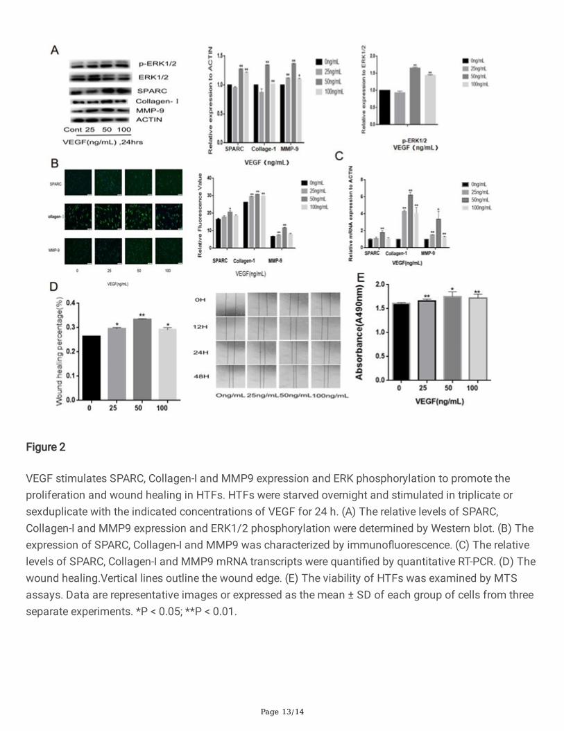
To understand the molecular mechanisms underlying the action of VEGF, we examined whether VEGFcould modulate the SPARC, Collagen-I, MMP9 expression and ERK1/2 activation in HTFs. Western blotindicated that treatment with 50 ng/ml of VEGF signi�cantly increased the relative levels of SPARC,Collagen-I, MMP9 expression and ERK phosphorylation in HTFs ( all the P<0.01, Fig. 2A ). However,treatment with VEGF at a lower or higher dose either did not signi�cantly up-regulate or only moderatelyincreased their expression and phosphorylation in HTFs. Similar patterns of VEGF-enhanced SPARC,Collagen-I and MMP9 expression were observed in HTFs by immuno�uorescence ( P<0.05, P<0.01 andP<0.01, Fig. 2B ). Similarly, treatment with VEGF increased the relative levels of SPARC, Collagen-I andMMP9 mRNA transcripts in HTFs ( P<0.01, P<0.01 and P<0.05, Fig. 2C ). Functionally, VEGF treatmentsigni�cantly promoted the proliferation and wound healing of HTFs ( P<0.05, P<0.01 and P<0.05, Fig. 2D ;P<0.01, P<0.05 and P<0.01, Fig.2E ). Hence, VEGF promoted the proliferation and wound healing of HTFsby enhancing �brotic protein expression and ERK1/2 activation in vitro.
SPARC silencing mitigates the VEGF-induced �brotic process in HTFs
SPARC can regulate ECM remodelling by modulating collagen expression and MMP activity 9,10. Tounderstand the regulatory role of SPARC in VEGF-induced �brotic process in HTFs, we generated stableSPARC-silenced HTFs by lentivirus-based shRNA technology. Following stimulation with VEGF (50

Page 6/14
ng/ml), we found that SPARC silencing not only decreased the VEGF-stimulated SPARC expression, butalso mitigated the VEGF-enhanced Collagen-1 and MMP expression and ERK phosphorylation in HTFs(Fig. 3A-C). Functionally, measurements of wound healing and cell proliferation revealed that followingVEGF stimulation, the percentages of wound healing and viability in the SPARC-silenced HTFs weresigni�cantly lower than that in the controls (Fig. 3D and E). Such data indicated that SPARC silencingmitigated the VEGF-stimulated wound healing and proliferation in HTFs in vitro. Collectively, these�ndings suggest that VEGF-stimulated �brotic process in HTFs may partially depend on inducing SPARCexpression in our experimental conditions. Meanwhile, our experiement showed that all the aspectsmentioned above of NC HTFs were sightly down regulated compared with normal HTFs. Probably, theprocess of infecting cells with virus would also affect the function of cells to some extent.
DiscussionFiltration surgery is one of the most effective treatments for glaucoma 14. Unfortunately, a failure after FSoccasionally occurs due to excessive post-surgical wound healing process and scar formation. Previousstudies have shown that VEGF121 and VEGF189 can induce the proliferation and migration of HTFs by
activating the ERK signaling 15, 5 and higher levels of VEGF expression in Tenon’s tissues are associatedwith post-FS �brosis 8. In this study, we prepared HTFs from human Tenon’s capsule tissues andcharacterized them. We found that almost all cells displayed typical morphology of �broblasts withVimentin, but not keratin, expression. Furthermore, we found that VEGF treatment stimulated theproliferation and would healing of HTFs, accompanied by up-regulating SPARC, Collagen-I and MMP9expression and ERK1/2 phosphorylation. Such data clearly demonstrated that VEGF promoted the �broticprocess of HTFs and suggested that SPARC might participate the process of VEGF-mediated �brosis.
SPARC is an ECM protein and its up-regulated expression is associated with �brosis. Actually, SPARC-/-
mice exhibit de�cient scarring and SPARC de�ciency increases successful rates of FS in a mouse modelof Glaucoma 16. In this study, we found that treatment with VEGF stimulated SPARC expression in HTFs,which extended previous observations in HMEC-1 cells and HUVECs17. Furthermore, VEGF also up-regulated Collagen-I and MMP9 expression in HTFs and promoted the proliferation and wound healing ofHTFs. More importantly, SPARC silencing signi�cantly mitigated the VEGF-stimulated Collagen-I andMMP9 expression in HTFs and attenuated the VEGF-stimulated proliferation and wound healing of HTFs.Previous studies have shown that SPARC can modulate MMP activity in U87MG glioma cells18 andaltered MMP activity can affect the ECM remodeling and �broblast proliferation and migration 19-20.
Moreover, SPARC can promote cell adhesion 21 and SPARC de�ciency inhibits cell migration in varioustypes of cells 22-23. It is possible that SPARC modulates MMP activity and wound healing, contributing tothe progression of �brosis 23. Our data further indicated that SPARC participated in the �brotic process inHTFs.
It is notable that SPARC can bind to collagen and acts as a chaperone to promote its post-translationalprocess 25, 26. Furthermore, SPARC can activate both platelet-derived growth factor (PDGF) and �broblast

Page 7/14
growth factor-2 (FGF2), which are crucial for Collagen-I expression 10,27. VEGF can activate the p38 andERK signaling in vascular endothelial cells 15,28. We found that VEGF treatment enhanced the ERK1/2phosphorylation in HTFs, consistent with a previous report 15. Interestingly, SPARC silencing signi�cantlymitigated the VEGF-enhanced ERK1/2 activation in HTFs. It is possible that SPARC silencing mayminimize PDGF and FGF2 activation, which cross-talks with the VEGF-related ERK1/2 signaling to reduceCollagen-I expression and �brotic process in HTFs. We are interested in further investigating the precisemolecular mechanisms underlying the action of SPARC in regulating Collagen-I and MMP9 expressionfollowing VEGF stimulation in HTFs.
There is still much research should be done in the future. Firstly, the characteristic of HTFs cultured fromstrabismus patients may be different from glaucoma patients in some extent. Secondly, we need moreevidences to prove that SPARC also plays a role in VEGF-stimulated �brotic process in HTFs in vivo.Besides, in our experiement, we reduced the expressiong of SPARC by transducing with lentivirus. Thoughthe effect of such SPARC-silencing is relatively lasting, it takes more time and needs to be more carefulwhen dealing with lentivirus. The usage of SPARC inhibitors, such as antibodies, seems to be anotherapproach, which is more safe and easy to control.
ConclusionsOur data indicated that VEGF treatment stimulated the wound healing and proliferation of HTFs by up-regulated SPARC, Collagen-I and MMP9 expression and ERK phosphorylation, which were signi�cantlymitigated by SPARC silencing. Such data suggest that SPARC may be crucial for the VEGF-stimulated�brotic process in HTFs. Conceivably, SPARC may be a promising therapeutic target for prevention andinvention of FS-related scarring.
List Of AbbreviationsGFS:Glaucoma Filtration Surgery
HTF:Human Tenon’s �broblast
VEGF:Vascular endothelial growth factor
SPARC:Secreted protein, acidic and rich in cysteine
MMP:Matrix metalloproteinase
FS: Filtration surgery
ECM:Extracellular Matrix
Declarations
Page 8/14
Ethics approval and consent to participate
This study was approved by the Ethics Committee of Shanghai General Hospital (Registration number:2016KY034). All patients signed a written informed consent.
Consent for publication
Not applicable
Availability of data and materials
The datasets used and/or analysed during the current study are available from the corresponding authoron reasonable request.
Competing interests
The authors declare that they have no competing interests.
Funding
This study was supported by a grant from the National Nature Science Foundation of China (GrantNo.81700845).
Authors' contributions
X X, F Y, and T M were primarily responsible for experimental concept and design. X X ,L L and Chen Yperformed the experiements and data acquisition.X X , Y F and F Y performed the data analysis as well asdrafting of the manuscript. All authors reviewed and approved the �nal manuscript.
Acknowledgement
We are grateful to the Shanghai Key Laboratory of Ocular Fundus Diseases of Shanghai General Hospitalfor providing the experimental equipments and device.
Authors' information
A�liations
Department of Ophthalmology, Shanghai General Hospital, Shanghai Jiao Tong University School ofMedicine, Shanghai, China
Corresponding author
Correspondence to Min Tang
CO-Correspondence to Yang Fu
Page 9/14
Reference1. Quigley HA, Broman AT. The number of people with glaucoma worldwide in 2010 and 2020. Br J
Ophthalmol 2006;90(3):262-7.
2. Masoumpour MB, Nowroozzadeh MH, Razeghinejad MR. Current and Future Techniques in WoundHealing Modulation after Glaucoma Filtering Surgeries. Open Ophthalmol J 2016;10:68-85.
3. Meyer-Ter-Vehn T, Klink T, Grehn F, Schlunck G. [Beyond TGF-beta: wound healing modulation in�ltering glaucoma surgery]. Klin Monbl Augenheilkd 2009;226(1):22-6.
4. Zada M, Pattamatta U, White A. Modulation of Fibroblasts in Conjunctival Wound Healing.Ophthalmology 2018;125(2):179-92.
5. Bao P, Kodra A, Tomic-Canic M, Golinko MS, Ehrlich HP, Brem H.. The role of vascular endothelialgrowth factor in wound healing. J Surg Res 2009;153(2):347-58.
�. Wilgus TA, Ferreira AM, Oberyszyn TM, Bergdall VK, Dipietro LA.. Regulation of scar formation byvascular endothelial growth factor. Lab Invest 2008;88(6):579-90.
7. Li Z1, Van Bergen T, Van de Veire S, Van de Vel I, Moreau H, Dewerchin M, Maudgal PC, Zeyen T,Spileers W, Moons L , et al. Inhibition of vascular endothelial growth factor reduces scar formationafter glaucoma �ltration surgery. Invest Ophthalmol Vis Sci 2009;50(11):5217-25.
8.Li Z, Hua W, Li X, Wang W. Suppression of Human Tenon Fibroblast Cell Proliferation by Lentivirus-Mediated VEGF Small Hairpin RNA. J Ophthalmol 2017;2017:7982051.
9. O'Neill, E. C. , Queena, Q. , Van, B. N. J. , Connell, P. P. , Sushil, V. , & Coote, M. A. , et al. (2010).Anti�brotic activity of bevacizumab on human tenon's �broblasts in vitro. Invest Ophthalmol Vis Sci.2010;51:6524.
10. Brekken RA, Sage EH. SPARC, a matricellular protein: at the crossroads of cell-matrix communication.Matrix Biol 2001;19(8):816-27.
11 Ding W, Pu W, Jiang S, Ma Y, Liu Q, Wu W, Chu H, Zou H, Jin L, Wang J, et al. Evaluation of theanti�brotic potency by knocking down SPARC, CCR2 and SMAD3. EBioMedicine 2018;38:238-47.
12 Cai X, Yang Y, Chen P, Ye Y, Liu X, Wu K, Yu M. Tetramethylpyrazine Attenuates Transdifferentiation ofTGF-β2-Treated Human Tenon's Fibroblasts. Invest Ophthalmol Vis Sci. 2016;57(11):4740-8.
13 Zufferey R, Nagy D, Mandel RJ, Naldini L, Trono D. Multiply Attenuated lentiviral vector achievese�cient gene delivery in vivo. Nat Biotechnol 1997;15: 871-875.
14.Lama PJ, Fechtner RD. Anti�brotics and wound healing in glaucoma surgery. Surv Ophthalmol2003;48(3):314-46.

Page 10/14
15. Van Bergen T, Vandewalle E, Van de Veire S, Dewerchin M, Stassen JM, Moons L, Stalmans I. Therole of different VEGF isoforms in scar formation after glaucoma �ltration surgery. Exp Eye Res2011;93(5):689-99.
16.Seet LF, Su R, Barathi VA, Lee WS, Poh R, Heng YM, Manser E, Vithana EN, Aung T, Weaver M, et al.SPARC de�ciency results in improved surgical survival in a novel mouse model of glaucoma �ltrationsurgery. PLoS One 2010;5(2):e9415.
17.Kato Y, Lewalle JM, Baba Y, Tsukuda M, Sakai N, Baba M, Kobayashi K,
Koshika S, Nagashima Y, Frankenne F, et al. Induction of SPARC by VEGF in human vascular endothelialcells. Biochem Biophys Res Commun 2001;287(2):422-6.
18.McClung HM, Thomas SL, Osenkowski P, Toth M, Menon P, Raz A, Fridman R, Rempel SA.. SPARCupregulates MT1-MMP expression, MMP-2 activation, and the secretion and cleavage of galectin-3 inU87MG glioma cells. Neurosci Lett 2007;419(2):172-7.
19.Li B, Li F, Chi L, Zhang L, Zhu S.. The expression of SPARC in human intracranial aneurysms and itsrelationship with MMP-2/-9. PLoS One 2013;8(3):e58490.
20.Cui N, Hu M, Khalil RA. Biochemical and Biological Attributes of Matrix Metalloproteinases. Prog MolBiol Transl Sci 2017;147:1-73.
21.Tumbarello DA, Andrews MR, Brenton JD. SPARC Regulates Transforming Growth Factor Beta Induced(TGFBI) Extracellular Matrix Deposition and Paclitaxel Response in Ovarian Cancer Cells. PLoS One2016;11(9):e0162698.
22.Chang CH, Yen MC, Liao SH, Hsu YL, Lai CS, Chang KP, Hsu YL. Secreted Protein Acidic and Rich inCysteine (SPARC) Enhances Cell Proliferation, Migration, and Epithelial Mesenchymal Transition, andSPARC Expression is Associated with Tumor Grade in Head and Neck Cancer. Int J Mol Sci 2017;18(7).
23.Seno T, Harada H, Kohno S, Teraoka M, Inoue A, Ohnishi T. Downregulation of SPARC expressioninhibits cell migration and invasion in malignant gliomas. Int J Oncol 2009;34(3):707-15.
24.Robert S, Gicquel T, Victoni T, Valença S, Barreto E, Bailly-Maître B, Boichot E, Lagente V.. Involvementof matrix metalloproteinases (MMPs) and in�ammasome pathway in molecular mechanisms of �brosis.Biosci Rep 2016;36(4).
25. McDonald LT, Zile MR, Zhang Y, Van Laer AO, Baicu CF, Stroud RE, Jones JA, LaRue AC, BradshawAD. Increased macrophage-derived SPARC precedes collagen deposition in myocardial �brosis. Am JPhysiol Heart Circ Physiol 2018;315(1):H92-h100.
2�. Rentz TJ, Poobalarahi F, Bornstein P, Sage EH, Bradshaw AD. SPARC regulates processing ofprocollagen I and collagen �brillogenesis in dermal �broblasts. J Biol Chem 2007;282(30):22062-71.
Page 11/14
27.Atorrasagasti C, Aquino JB, Hofman L, Alaniz L, Malvicini M, Garcia M, Benedetti L, Friedman SL,Podhajcer O, Mazzolini G.Atorrasagasti C, Aquino JB, Hofman L, et al. SPARC downregulation attenuatesthe pro�brogenic response of hepatic stellate cells induced by TGF-beta1 and PDGF. Am J PhysiolGastrointest Liver Physiol 2011;300(5):G739-48.
28.Almalki SG, Agrawal DK. ERK signaling is required for VEGF-A/VEGFR2-induced differentiation ofporcine adipose-derived mesenchymal stem cells into endothelial cells. Stem Cell Res Ther 2017;8(1):113.
Tables
Tabel 1: Primers used in Quantitative RT-PCR
Genes Primers Sequences(5' to 3')
SPARC SPARC-F
SPARC-R
AGGAAACCGAAGAGGAGG GCAAAGAAGTGGCAGGAA’
MMP-9 MMP-9-F
MMP-9-R
GACCTCAAGTGGCACCACCA
GTGGTACTGCACCAGGGCAAcollagen-I collagen-I-F
collagen-I-R
CCCAGCCACAAAGAGTCTACA GTTTCCACACGTCTCGGTCA
β-actin β-actin-F
β-actin-R
GGACTTCGAGCAAGAGATGG
AGCACTGTGTTGGCGTACAG
Figures

Page 12/14
Figure 1
Immuno�uorescent characterization of HTFs. Human Tenon's specimens were cultured for 15 days andmany cells migrated out. The cells were passaged every 3 days for 3-9 times. The cells were stained withanti-Vimentin or anti-keratin, and after being washed, the cells were stained with Alexa Fluor 488-conjugated goat-anti-rabbit IgG or Alexa Fluor 647-conjugated goat-anti-mouse IgG. Subsequently, thecells were nuclearly stained with DAPI and the �uorescent signals were captured under a confocalmicroscope. Data are representative images (magni�cation x 200) from three separated experiments. (A)Immuno�uorescent staining of cells with anti-Vimentin. (B) Immuno�uorescent staining with anti-keratin.Scale bar: 100 μm.
Page 13/14
Figure 2
VEGF stimulates SPARC, Collagen-I and MMP9 expression and ERK phosphorylation to promote theproliferation and wound healing in HTFs. HTFs were starved overnight and stimulated in triplicate orsexduplicate with the indicated concentrations of VEGF for 24 h. (A) The relative levels of SPARC,Collagen-I and MMP9 expression and ERK1/2 phosphorylation were determined by Western blot. (B) Theexpression of SPARC, Collagen-I and MMP9 was characterized by immuno�uorescence. (C) The relativelevels of SPARC, Collagen-I and MMP9 mRNA transcripts were quanti�ed by quantitative RT-PCR. (D) Thewound healing.Vertical lines outline the wound edge. (E) The viability of HTFs was examined by MTSassays. Data are representative images or expressed as the mean ± SD of each group of cells from threeseparate experiments. *P < 0.05; **P < 0.01.

Page 14/14
Figure 3
SPARC silencing mitigates the VEGF-stimulated �brotic process in HTFs. HTFs, control HTF-NC andstable SPARC-silenced HTF-SPARC-shRNA cells were starved overnight and stimulated in triplicate orsexduplicate with 50 ng/ml of VEGF for 24 h. (A) The relative levels of SPARC, Collagen-I and MMP9expression and ERK1/2 phosphorylation were determined by Western blot. (B) The expression of SPARC,Collagen-I and MMP9 was characterized by immuno�uorescence. (C) The relative levels of SPARC,Collagen-I and MMP9 mRNA transcripts were quanti�ed by quantitative RT-PCR. (D) The wound healing.(E) The viability of HTFs was examined by MTS assays. Data are representative images or expressed asthe mean ± SD of each group of cells from three separate experiments. *P < 0.05; **P < 0.01.