stentless bioprostheses for aortic valve replacement...
TRANSCRIPT
Chapter 14
Stentless Bioprostheses for Aortic Valve Replacement inCalcific Aortic Stenosis
Kaan Kirali
Additional information is available at the end of the chapter
http://dx.doi.org/10.5772/55373
1. Introduction
The classic case of aortic stenosis is a healthy middle-aged patient with/without symptoms,but in practical life, patients with severe calcific aortic valve come with several and severecomorbidities such as advanced age, coronary artery disease, atherosclerotic aorta, significantleft ventricular dysfunction. Aortic valve replacement (AVR) is the only options in thesepatients, and it requires patient-by-patient analysis of clinical, echocardiograhic, and hemo‐dynamic data with associated pathologies. The curative treatment of calcific aortic valvestenosis is the replacement of the aortic valve with a prosthetic valve, and selection of a perfectprosthetic valve is the main goal to get a successful treatment. But, there is no any perfect heartvalve prosthesis which may mimic the characteristics of the normal native aortic valve:excellent hemodynamics, life-long durability, thromboresistance, and excellent implantability.That means that native valve disease will be traded for prosthetic valve disease and theoutcome of AVR is affected by the type of prosthetic valve. Mechanical valves are non-limiteddurable, but have a substantial risk of hematologic complications (thromboemboli, thromboticobstruction, hemorrhage related life-long anticoagulation therapy) with/without hemolysispotential. In contract, bioprosthetic valves have a low risk of thromboembolism withoutanticoagulation, but their durability is limited by calcific or noncalcific tissue deterioration.Biological prostheses, especially homografts, are often believed to be the substitute of choicein AVR, but the limited availability of homografts prevents their more broadly usage. Toovercome this problem and all possible complications of mechanical valves, xenogenicbiological prostheses have been developed. The design of bioprosthetic valves purports tomimic the anatomy of the native aortic valve and their flow characteristics are better thanmechanical valves, whereas stentless bioprostheses have hemodynamic performance similarto the healthy native aortic valve. Although stented bioprostheses can be implanted easier,
© 2013 Kirali; licensee InTech. This is an open access article distributed under the terms of the CreativeCommons Attribution License (http://creativecommons.org/licenses/by/3.0), which permits unrestricted use,distribution, and reproduction in any medium, provided the original work is properly cited.

they decrease the effective orifice area due to the rigid stent and result turbulent flow throughthe valve. Stented valves also increase stress at the attachment of the stent which cause earlierprimary tissue failure. Stentless biologic valves have been introduced into clinical practice tosolve all these problems and to reproduce the anatomy and function of the native aortic valve,but their clinical use has still not exceeded the number of stented aortic bioprostheses becauseof more demanding technique of implantation. To gain more widespread clinical use andgeneral recommendation of stentless bioprostheses, their advantages and simple implantationtechniques must be popularized.
It is believed that the aortic root is probably the best stent for the native or prosthetic aorticvalve. The anatomy and function of the aortic root may dampen the mechanical stress to whichthe leaflets are subjected during diastole. The ideal stentless prosthesis should have nosynthetic materials, preserve the aortic root dynamics, restore flexibility and distensibility ofthe native valve annulus after decalcification, and have minimal xenograft aortic wall, shortimplantation time, and excellent hemodynamic performance to facilitate the recovery of leftventricular function.
1.1. Historical background
Homografts were the first biological prostheses used in clinical practice to treat aortic valvestenosis in early 1960s, and they were the first stentless valves, too [1,2]. The authors used theaortic root of the patient to secure the homograft aortic valve in the subcoronary position. Themost complicated implantation technique and the restricted availability of homograftsprevented their widespread usage. First stentless pig and calf xenografts were used in limitedpatients, but the valves were abandoned because of poor tissue fixation [3]. Stented biopros‐theses were considered as the gold standard for several years, but abnormal stress on theleaflets was believed to decrease durability. To overcome this problem with a rigid stent onthe aortic position, stentless bioprostheses were re-introduced in the middle of 80's [4], whereasnew designed stentless xenografts were proposed and popularized in daily use at the begin‐ning of 1990s [5]. The main problem (early failure of bioprostheses) was solved with newbioengineering improvement (antimineralization, zero-pressure fixation) [6]. The otherproblem was partial dehiscence when the heterograft contained muscular bar resultingparavalvular leakage in the area corresponding to the muscular bar, and this problem wasabolished with a fine Dacron cloth covered the outside wall of the stentless porcine aortic valvealong its inflow [7]. Recognizing the range of aortic root variability and disease of the rootitself, the concept of stentless valve replacement was expanded to replacement of the entireaortic root. Full root replacement with a bioprosthesis brought the challenges of homeostasisand coronary reimplantation. In spite of hemodynamic advantages proven for the rootreplacement technique, acceptance was slowed by risk/benefit ratio concerns. The whole aorticroot could be prepared and implanted with modified root inclusion or subcoronary implanttechniques.
Biological stentless valve can be prepared by pulmonary autograft, homograft, xenograft,autologous or xenogenic pericardium. Pulmonary autograft has limited durability beyond thefirst decade [8]. The same problem has been observed with homografts in the aortic position,
Calcific Aortic Valve Disease412
especially in younger patients, which are less durable than commercially available stentlessbioprostheses and cannot be recommended as the ideal device [9]. The use of the patients ownpericardium for constructing a heart valve prosthesis is biologically more appealing than theuse of animal tissue or heterologous material. The feasibility of autologous pericardial stentlessaortic valve was shown in an animal study [10]. The feasibility and durability of truly stentlessautologous pericardial AVR sutured directly onto the aortic wall has been also performed inhuman recently [11]. Stentless porcine or pericardial xenogenic bioprostheses have beenintroduced to get better long-term durability and become a routine device when a stentlessbiologic valve is implanted.
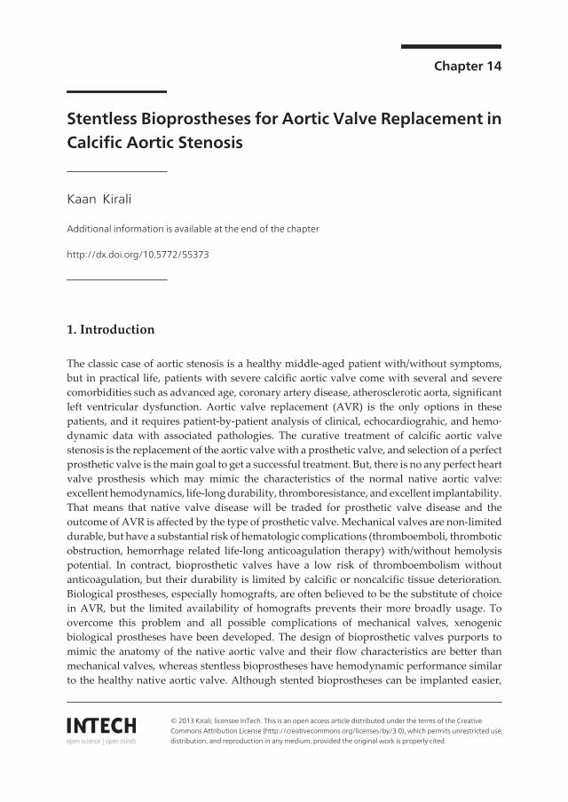
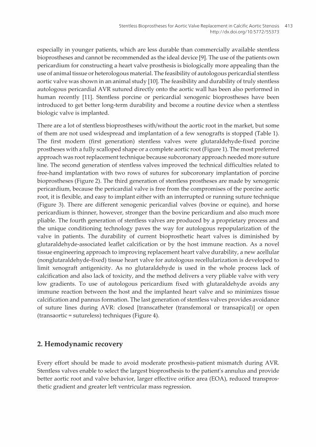
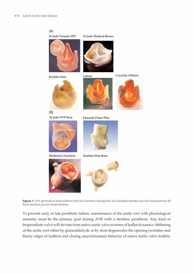
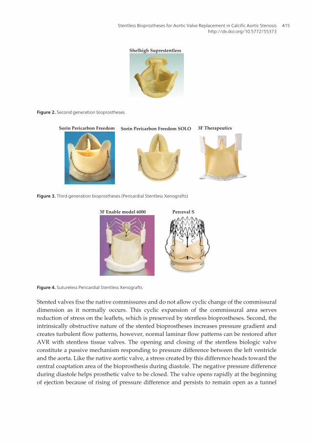
There are a lot of stentless bioprostheses with/without the aortic root in the market, but someof them are not used widespread and implantation of a few xenografts is stopped (Table 1).The first modern (first generation) stentless valves were glutaraldehyde-fixed porcineprostheses with a fully scalloped shape or a complete aortic root (Figure 1). The most preferredapproach was root replacement technique because subcoronary approach needed more sutureline. The second generation of stentless valves improved the technical difficulties related tofree-hand implantation with two rows of sutures for subcoronary implantation of porcinebioprostheses (Figure 2). The third generation of stentless prostheses are made by xenogenicpericardium, because the pericardial valve is free from the compromises of the porcine aorticroot, it is flexible, and easy to implant either with an interrupted or running suture technique(Figure 3). There are different xenogenic pericardial valves (bovine or equine), and horsepericardium is thinner, however, stronger than the bovine pericardium and also much morepliable. The fourth generation of stentless valves are produced by a proprietary process andthe unique conditioning technology paves the way for autologous repopularization of thevalve in patients. The durability of current bioprosthetic heart valves is diminished byglutaraldehyde-associated leaflet calcification or by the host immune reaction. As a noveltissue engineering approach to improving replacement heart valve durability, a new acellular(nonglutaraldehyde-fixed) tissue heart valve for autologous recellularization is developed tolimit xenograft antigenicity. As no glutaraldehyde is used in the whole process lack ofcalcification and also lack of toxicity, and the method delivers a very pliable valve with verylow gradients. To use of autologous pericardium fixed with glutaraldehyde avoids anyimmune reaction between the host and the implanted heart valve and so minimizes tissuecalcification and pannus formation. The last generation of stentless valves provides avoidanceof suture lines during AVR: closed [transcatheter (transfemoral or transapical)] or open(transaortic = sutureless) techniques (Figure 4).
2. Hemodynamic recovery
Every effort should be made to avoid moderate prosthesis-patient mismatch during AVR.Stentless valves enable to select the largest bioprosthesis to the patient's annulus and providebetter aortic root and valve behavior, larger effective orifice area (EOA), reduced transpros‐thetic gradient and greater left ventricular mass regression.
Stentless Bioprostheses for Aortic Valve Replacement in Calcific Aortic Stenosishttp://dx.doi.org/10.5772/55373
413
To prevent early or late prosthetic failure, maintenance of the aortic root with physiologicalanatomy must be the primary goal during AVR with a stentless prosthesis. Any kind ofbioprosthetic valve will deviate from native aortic valve in terms of leaflet dynamics. Stiffeningof the aortic root either by glutaraldehyde or by stent degenerates the opening (wrinkles andblurry edges of leaflets) and closing (asynchronism) behavior of native aortic valve leaflets.
(A)
(B)
St Jude Toronto SPV St Jude Medical-Biocor
Koehler Elan Labcor CryoLife-O'Brien
St Jude SVP Root Edwards Prima Plus
Medtronic Freestyle Koehler Elan Root
Figure 1. First generation bioprostheses (Porcine Stentless Xenografts) A) Scalloped stentless porcine bioprostheses B)Root stentless porcine bioprostheses.
Calcific Aortic Valve Disease414
Stented valves fixe the native commissures and do not allow cyclic change of the commissuraldimension as it normally occurs. This cyclic expansion of the commissural area servesreduction of stress on the leaflets, which is preserved by stentless bioprostheses. Second, theintrinsically obstructive nature of the stented bioprostheses increases pressure gradient andcreates turbulent flow patterns, however, normal laminar flow patterns can be restored afterAVR with stentless tissue valves. The opening and closing of the stentless biologic valveconstitute a passive mechanism responding to pressure difference between the left ventricleand the aorta. Like the native aortic valve, a stress created by this difference heads toward thecentral coaptation area of the bioprosthesis during diastole. The negative pressure differenceduring diastole helps prosthetic valve to be closed. The valve opens rapidly at the beginningof ejection because of rising of pressure difference and persists to remain open as a tunnel
Shelhigh Suprestentless
Figure 2. Second generation bioprostheses.
Sorin Pericarbon Freedom Sorin Pericarbon Freedom SOLO 3F Therapeutics
Figure 3. Third generation bioprostheses (Pericardial Stentless Xenografts)
3F Enable model 6000 Perceval S
Figure 4. Sutureless Pericardial Stentless Xenografts
Stentless Bioprostheses for Aortic Valve Replacement in Calcific Aortic Stenosishttp://dx.doi.org/10.5772/55373
415

A. Autograft
B. Homograft
C. Xenografts
I. First generation (Stentless Porcine Bioprosthesis)
Dacron reinforced inflow tract
Toronto SPV (stentless porcine valve) St Jude Medical, Inc., St Paul, MN, USA
St Jude Medical-Biocor St Jude, Belo Horizonte, MG, Brazil
CryoLife-O'Brien Model 3000 CryoLife International Inc, Atlanta, GA, USA
Toronto SPV Root St Jude Medical, Inc., St Paul, MN, USA
Edwards Prima Plus Edwards Lifesciences, Inc., Irvine, CA,USA
Medtronic Freestyle Medtronic, Inc., Minneapolis, MN, USA
pericardial reinforced inflow tract
Koehler Elan Koehler, Bellshill, Scotland
Koehler Elan Root
tri-composite design (three noncoronary leaflets)
Labcor Labcor, Inc., Belo Horizonte, MG, Brazil
II. Second generation (porcine with single suture line,
No-react treatment)
Shelhigh Suprestentless Shelhigh, Inc, Millburn, NJ, USA
III. Third generation (Stentless Pericardial Bioprosthesis)
porcine pericardium
Sorin Pericarbon Freedom Sorin Biomedica Cardio SpA, Saluggia, Italy
Sorin Pericarbon Freedom SOLO
horse (equine) pericardium
3F Therapeutics 3F Therapeutics, Inc., Lake Forest, CA, USA
IV. Fourth generation (non-gluteraldayhde fixed +
decellularized)
Matrix A
V. Sutureless generation (Sutureless + Stentless
Pericardial Bioprosthesis)
3F Enable model 6000 3F Therapeutics, Inc., Lake Forest, CA, USA
Perceval S Sorin Biomedica Cardio SpA, Saluggia, Italy
D. Autologous pericardium
Table 1. Stentless Bioprostheses.
Calcific Aortic Valve Disease416

during systole, and the aortic root may also expanse at the late diastole to help opening of theleaflets (in native aortic valve, expansion of the aortic root is about 12% and that starts openingthe leaflets to about 20%). At the end of systole, the backward blood flow into the sinuses ofValsalva (behind prosthetic leaflets) and initialization of pressure difference help prostheticleaflets to revert to their original closed position. An in-vivo-study has showed that there isno difference in opening velocities among native, stented and subcoronary stentless valves ina porcine model [12]. However, the closing velocities are significantly higher in the pericardialvalves. The bending deformation increases when implanting a glutaraldehyde-treated valvesubcoronary. Porcine stentless valves display a distinct folding pattern during openingresulting in an altered stress distribution and also tend to fold during opening causingincreased leaflet bending stress [13].
One of the key parameters for stentless xenograft performance is the EOA. In spite of the EOAis significantly higher in stentless bioprostheses it is also dependent on the design and theimplantation technique of the prostheses. The EOA will increase especially during the firstyear and the transvalvular gradient drops dramatically in the first 3 to 6 months after surgery,but some further drop may be seen more later [14]. The reason may be remodeling of the leftventricular outflow tract, diminished aortic root edema, and slight dilatation of the aortic root.Transvalvular gradient is closely related to the EOA: the larger orifice area the lower is thetransvalvular gradient. The second reason to increase transvalvular gradient is usage of a rigidstent. Avoidance of a stent enlarges inner diameter of prosthetic valve and eliminates intralu‐minal obstruction which increases the EOA. Several studies have shown transvalvulargradient across stentless valves is always lower than for their stented valves, especially meanand/or peek gradients [15-1617]. The third possible reason can be excessive tissue of a bio‐prosthesis: the lesser tissue implanted within the recipient aortic root the lesser obstruction.The full root prostheses reduce the intraluminar obstruction because nothing is implantedinside, and they have larger EOA than subcoronary prostheses. The main differences ofstentless biologic tissue valves are the specific gravity of the leaflets which is not equal to thatof blood like native human aortic leaflets and the specific thickness of the leaflets which isthinner in pericardial tissue valves. Both parameters cause transvalvular gradient duringejection which is lesser in fully pericardial stentless valves than porcine. The other reasons maybe small aortic annulus and physically active patients. The change in gradients during exerciseis interesting: when cardiac output increases it also increases the transvalvular flow and raisestransprosthetic gradient, but these gradients under exercise are lower with stentless valvesthan stented bioprostheses, which provide better opening-closing behavior [18].
Left ventricular output is maintained by the development of the left ventricular hypertrophywhich results in a large pressure gradient across the stenotic valve. The left ventricle massincreases and becomes less compliant. Left ventricular hypertrophy and increased mass canbe correlated with sudden death, congestive heart failure, and other cardiovascular events.Left ventricular hypertrophy will regress after AVR regardless of the type of prostheses, andan improved hemodynamic performance of prostheses should result in a faster regression,especially in patients with severe calcific aortic stenosis and left ventricular hypertrophy,because incomplete regression after AVR is related to poor long-term outcome [19]. This
Stentless Bioprostheses for Aortic Valve Replacement in Calcific Aortic Stenosishttp://dx.doi.org/10.5772/55373
417

regression is related to EOA and transvalvular gradient constituted by the prosthetic valve. Asignificant improvement will occur in all type of valves in the first year, but this improvementis greater and faster with the stentless bioprostheses [20]. A lasting benefit beyond the firstyear is possible, especially in severely enlarged ventricles [21]. These improvements includemass regression, wall thickening, fractional shortening, and diastolic relaxation. Patients withsmall aortic annuli or with compromised left ventricular function (EF < 50%) might benefitmore from stentless prostheses [22,23].
3. Structural and nonstructural durability
One of the foremost concern of any tissue valve is its long-term patency, because the limiteddurability represents the main disadvantage of these devices. Tissue valve degenerationcausing stenosis or regurgitation is the primer indication for reoperation.
Durability of any kind of stentless bioprosthesis can be affected adversely by internal (struc‐tural) or external (nonstructural) factors.
Structural valve deterioration (SVD) is a primary tissue failure after biological valve implan‐tation. A major cause of SVD is cusp tear with consequent aortic regurgitation where urgentor emergent reoperation is necessary due to congestive heart failure and hemolytic anemia.The other major reason is prosthetic valve sclerosis and calcification which could permit anelective reoperation in stable condition. An in vivo animal study has shown that native aorticvalves are significantly more distensible at the level of the sinotubular junction, commissuresand ascending aorta when compared with all-valve prosthesis [24]. There is no any study toevaluate how the late scar with/without calcification tissue formation spread and effect thisdistensibility. We can argue that annular calcification developed during follow-up acts similarin native and stentless valves and fixes the aortic annulus. The zero-pressure fixation andantimineralization techniques have improved durability of tissue valves. To avoid from wellknown limited durability of xenogenic bioprostheses owing to structural degeneration andcalcification, the use of autologous pericardium may be an attractive alternative with severaladvantages: no immune reaction, minimum tissue calcification and pannus formation,excellent hemodynamics and dynamics of the aortic root, no complicated reoperation [11].
Nonstructural valve deterioration (NSVD) is independent on the xenograft's tissue. In spite ofleaflets of xenografts work very well, stentless bioprosthesis shows incompetence. There areseveral reasons causing prosthetic stenosis or regurgitation (Table 2).
Technical inadequacy during stentless valve implantation cause hemodynamic problems likeregurgitation, turbulent flow, uncoaptation or stretching of leaflets which aggregate tissuedegeneration. Any increase in mechanical stress causing by surgical implantation techniqueshas a negative impact on durability. Description of all implantation techniques with their tipsis not adequate to avoid iatrogenic valve degeneration, all details of these techniques shouldbe well known. The best way to avoid mechanical stress may be to use the full root replacementtechnique, but most surgeon do not like to replace the aortic root without any pathology
Calcific Aortic Valve Disease418

(dilatation, calcification) because of higher operative risk. Subcoronary implantation techniqueis more acceptable approach for isolated AVR with stentless bioprostheses. Technical errorsrelating to xenograft sizing and failure to achieve appropriate geometry of the xenograft withinthe aortic root are 2 major reasons for early valve failure. The learning curve associated withsubcoronary implantation is the main reason for these technical errors. Suboptimal implanta‐tion resulting in distortion of the valve or bulking of valve tissue into the outflow tract maybe involved in the evolution of higher gradients. Undersizing of xenograft results regurgitationdue to handicapping leaflet coaptation, whereas oversizing may cause higher transvalvulargradient due to making leaflet opening difficult. The other error is to decide and apply thewrong implantation technique, especially in small or dilated aortic root, and subcoronarytechnique might be associated with higher gradient or regurgitation [25].
On the other hand, improvement of the long term patency of an aortic prosthetic valve isdependent on avoidance of paravalvular complications which can be very serious and causereoperation. Paravalvular regurgitation is a dangerous long-term result of insufficientdecalcification, which causes incompetence suturing or suture rupture during follow-up.
Partial dehiscence of the stentless xenograft indeed occurs and that it has a strong predilectionfor the preserved non-coronary sinus after modified subcoronary technique. Supposedly,proteolytic enzymes from captured blood cells in the dead space between native and donoraortas or the potential usefulness of biologic glues might prevent adequate fusion of the wallsand healing of the anastomosis [26].
A. Endocarditis
B. Technically implantation errors
C. Aortic root enlargement
I. Sinotubular junction dilatation
II. Sinus of Valsalva aneurysm (± rupture)
III. Aortic dissection
IV. Left ventricular dilatation
D. Partial dehiscence after preserved non-coronary sinus
E. Insufficiently decalcification
I. Poor decalcification (intra-operatively)
II. Suture rupture or loosening (post-operatively)
III. Calcification on the native aorta (follow-up)
F. Subvalvular fibrous band
G. Hematologic problems (hemolysis, thrombocytopenia)
Table 2. Non-structural Deterioration (regurgitation or stenosis).
Stentless Bioprostheses for Aortic Valve Replacement in Calcific Aortic Stenosishttp://dx.doi.org/10.5772/55373
419

Subvalvular fibrous band is a rare complication resulting significant left ventricular outflowtract obstruction, which can be a derivative of the pannus discovered on the sewing ring ofstented valves. The etiology is unknown, but it may result from thrombus formation orinflammation related to host factors. A chronic inflammatory infiltrate composed of lympho‐cytes and macrophages occurs in equine or porcine stentless valves, which suggests equalimmunogenicity among different various biologic graft materials [27].
4.1. Aortic valve surgery
4.1.1. Cardiopulmonary bypass
Aortic valve surgery can be performed through a median full sternotomy or upper minister‐notomy with conventional or minimal skin incision. The distal ascending aorta cannulation isusually the standard approach in most patients, but the arcus aorta or axiller artery can be alsocannulated when the ascending aorta should be replaced [28]. A single dual-stage venouscannula is inserted through the right atrium appendage. After cardiopulmonary bypass isinitiated the aorta is clamped and cardiac arrest is be achieved with antegrade isothermic bloodcardioplegia administered into the aortic root. Myocardial protection is continued due toretrograde cardioplegic cannula during whole procedure, and retrograde cardioplegia iscontinuously infused whenever clear visulation of the aortic root is not required [29]. Rarelythe retrograde cannula cannot be introduced safely into the coronary sinus, in this situationintermittent antegrade isothermic blood cardioplegia is performed using selective coronaryostial cannulation after transverse aortotomy incision. If both approaches are unsuccessful,bicaval cannulation is performed and the retrograde cannula is placed in the coronary sinusunder direct vision. A vent cannula is inserted into the left atrium through the right upperpulmonary vein after cross clamp to prevent the left ventricle distention. Mild-moderatehypothermia (30-32°C) is achieved and continued during extracorporeal circulation, andrewarming of patients is started before the closure of the aortotomy.
4.1.2. Aortotomy
A small transverse aortotomy incision is made initially at least 15 to 20 mm above the originof the right coronary ostium or the sinotubular junction. The calcific aortic valve and wholeaortic root should be investigated under direct vision and decided which approach will bepreferred. If the aortic root will not be replaced then the transverse aortotomy incision isextended on both sides until 3D view of the aortic root appears. That helps surgeons forexcision of the severe calcific aortic valve, selection of an appropriate stentless bioprosthesisand insertion simple and/or continuous sutures easily and correctly. A transverse aortotomyis also required to image 3D shape of the aortic root which is the main condition for resus‐pention of the prosthetic commissures and to hold a stentless tissue valve in corrected positionfor prevention of the iatrogenic valve degeneration. An oblique or hockey-stick incision ispreferred very seldom, but it could be useful in patients with small aortic root. If the aorticroot is replaced it is excised completely and aortic root implantation technique is performed.Reoperation for severe calcific aortic stenosis is not rare in patients with previously coronary
Calcific Aortic Valve Disease420

artery bypass surgery, and patent proximal anastomoses on the ascending aorta can be aserious problem during aortotomy. I have offered a simple aortotomy incision "Reverse Uaortotomy" to save proximal anastomoses and if it is necessary to apply direct antegradecardioplegia through proximal anastomoses [30].
4.1.3. Excision of the calcific aortic valve
Aortic valve stenosis appears with fusion of one or both commissures, thickening andretraction of the cusps, and restriction of effective orifice area. Calcific involvement of nativeaortic valve is the last step which can be widespread very aggressively: aortic annulus, mitralannulus, aortic root, coronary ostia. The typically pathologic findings of calcific aortic stenosisare discrete, focal lesions on the aortic side of the leaflets. The severe form is characterized bydiffuse calcification of the aortic root and the deposits involve the sinuses of Valsalva and theascending aorta (porcelain aorta). The calcification presents as a cauliflower-like mass withinthe leaflets and often extends deep into the annulus and surrounding tissues. All thesecontiguous anatomical structures can have adverse affects on the surgical techniques.
A surgically complete decalcification of the aortic annulus is an important point. The flexiblecontinuity of the aortic annulus with sub- and supra-annular tissues is indispensable conditionto get better durability and hemodynamics with stentless xenografts, and to avoid from a wholeaortic root replacement technique, which hinders surgeons to perform AVR with a stentlessxenograft. Surgeons must care 1) not to leave any calcific tissue around the aortic annulus, 2)not to allow fragments of calcium to fall into the left ventricle, 3) not to disrupt the annulus aspossible, 4) not to detach the anterior mitral leaflet from the annulus (non-coronary sinus), 5)not to rupture subannular muscular septum (right coronary sinus), 6) not to perforate outsidethe heart (left coronary sinus).
The calcific aortic valve is excised and trimmed with a scissor leaving a 2-3 mm margin at theannulus if the annular margin of the leaflets are healthy. The frequent scenario is converselythat and extensive calcific involvement of the whole aortic annulus is observed very often. Firstof all, complete resection of the calcific aortic valve should be performed without any compli‐cation listed above. Excision of the calcific leaflets with a scissor is usually unsuccessful anddangerous because of breaking of calcification and falling calcium debris into the left ventricle.The best alternative to remove the diseased tissue is excision all of them with a lancet (number15). A folded segment of sponge or tampon is not necessary to place in the left ventricle andit hinders to see the cavity and to remove any calcium particle. The easiest excision with thelancet is to perforate the healthy leaflet near the annulus in partial calcified aortic valve or tobegin excision at the commissure between the non-coronary and right coronary leaflets in en-bloc calcific aortic valve. Cutting of the calcification is begun at the nearest end and the lancetincises the calcified valve from the healthy annular tissue. The sharp edge of the lancet shouldbe headed toward the calcified valve, and cutting is performed just below of calcification. Thewhole calcified valve must be incised as en-block, without fragmentation. If calcification isvery heavy or invaded into the annulus it can be cut with the scissor and then the residualcalcifications will be gently crushed and removed with a rounger. After completion of theaortic valve excision, all residual diseased and/or calcified tissue or particles should be
Stentless Bioprostheses for Aortic Valve Replacement in Calcific Aortic Stenosishttp://dx.doi.org/10.5772/55373
421

removed from around the annulus. Before sizing the prosthesis, the left ventricular cavity isflushed and irrigated with saline solution.
4.1.4. Sizing the stentless aortic bioprosthesis
The stented prostheses must fit snugly in the annulus, because a very loose or tight fit indicatesinadequate effective orifice area (patient-prosthesis mismatch) or oversizing the prosthesis.For the truly measurement of a stented valve, the seizer should be inserted through the aorticannulus and the same (supra-annular) or one number smaller (intra-annular) stented pros‐thesis must be chosen.
Sizing a stentless bioprosthesis is different from stented valves. The most important phase isthe choice of an appropriate stentless bioprosthesis, and measurement of the aortic annulusmust be done with the seizer that corresponds to the specific bioprosthesis. The true seizershould be chosen to implant the appropriate tissue valve with the optimum size. If theprosthetic valve is too small, the inflow end obstructs the EOA which increases transvalvulargradient and the outflow end is stretched out with decreased leaflet coaptation which causesmore regurgitation. On the other hand, oversizing to fit a larger sinotubular junction leads tobuckling of the inflow end which can produce both relative stenosis and regurgitation as wellas harmful turbulent flow. How the stentless valves sized and implanted will influence itsfunction and durability in future. The larger surface area of the cusps allows greater coaptationarea which reduces the risk of bioprosthesis regurgitation. This relatively larger bioprosthesiscan simplify replacement, especially the running sutures for all sinuses. But, it is imperativeto avoid over-sizing of stentless valves with the tubular structure achieved by three tabs onthe commissures, and if sizing is uncertainty the smaller prosthesis should be implanted.
In normal aortic root, the diameter of the aortic annulus is 10-15% larger than those of thesinotubular junction and measurement of the aortic annulus is the correct way to choice anappropriate sized stentless valve. However, most patients with calcific aortic stenosis have anabnormal aortic root and the relationship between both diameters is usually altered. In thissituation, the diameter of the sinotubular junction is more important because the threecommissures of stentless valves are secured at approximately the level of the sinotubularjunction if not the full-root replacement technique will be used.
A cylindrical silicon seizer is more practical to measure the true valve size when both theannulus and the sinotubular junction are measured. The rule is that the sinotubular junctionshould be dominate during measuring and if there is a major difference (> 3 mm) subcoronaryimplantation technique can be not used because the commissures of stentless valves are pulledoutward and cause valvular insufficiency and an alternative technique (root replacement) orstented bioprosthesis must be used. Supra-annular sizing is the best measurement method tochoice an appropriate stentless bioprosthesis, especially during single suture line technique. Iprefer this more practical way and put the appropriate seizer into the aortic root in supra-annular position (not into the annulus) where I put continuous proximal suture line, so I canchoice an acceptable size that is equal to the sinotubular junction size or one number largerstentless prosthesis can be chosen if the seizer fits aortic orifice tightly in patients with aorticroot enlargement. Trans-annular measurement is adequate to get a fit stentless valve for
Calcific Aortic Valve Disease422
subcoronary implantation in patients with normal aortic root, but preferring one size largerprosthesis is better if full-root replacement technique will be performed or a small aortic rootis present. I never suggest to play some traction sutures at the commissures or in the nadir ofthe annulus to open the aortic orifice. It can be useful during the replacement of a stented valve,but it will be better to release the aortic root in its original shape during sizing stentless valves.
5. Implantation techniques
Stentless aortic biologic prostheses can be different in origin: autogenous, homogenous,heterogeneous. Procuring of aortic auto- or homograft is not easy, but production of xenograftsis a sufficiently technical supply of the industry for the treatment of aortic valve diseases. Allstentless biologic valves can be implanted using different techniques: the subcoronary method,the full root implantation technique, and the root inclusion alternative.
The subcoronary technique is the simplest method for implantation, and either a porcine rootcan be adapted intra-operatively or a prefabricated tissue valve can be utilized. The mainadvantages are to avoid the manipulation of coronary ostia and bleeding from suture lines.The disadvantages could be difficulties occurring in the small aortic annulus and calcifiedaortic root, and possibilities of valve insufficiency by changing the shape of the stentless valvein a diseased aortic root [31]. Subcoronary implantation technique can be performed in twomethods: double suture lines (classic) or single suture line (simple) approach.
In classical subcoronary implantation technique, stentless valves are fixed into the host aorticroot using double suture lines. The first suture line attaches the inflow site of the stentlessbioprosthesis in the left ventricular outflow tract: annular suture line. The second suture line,which is constructed using 1 or 3 continuous sutures, connects the outflow site of the prosthesiswith the aortic wall below the coronary ostia: supra-annular suture line. The first suture lineconsists usually of interrupted sutures, but to reduce cross-clamp and cardiopulmonary timesa continuous suture can be preferred [32]. Because the conventional continuous inflow sutureline can increase the postoperative heart block risk, an alternative subcoronary technique hasbeen reported in which the inflow suture line is raised at the level of right-non-coronarycommissure [33].
The single suture line technique is a simple, quick, safe and reliable method to replace thenative aortic valve with a stentless valve. This approach is used for implantation of scallopednew generation tissue valves in supra-annular position and placement of the sutures below orthrough the annulus should be avoided. Running sutures avoid any prosthetic dead spacebetween prosthetic valve and native aortic wall, and selecting a prosthesis a size larger thanthe host annulus minimizes the stress on the suture lines. These new generation pericardialvalve can have manufactured scalloped design [34] or it can be prepared by trimming awayall the extra tissue of the valve inflow side ond scalloping the outflow side [35]. If stentlessprostheses are designed with a tubular structure, the tabs on the commissures should beattached to the aortic wall [36].
Stentless Bioprostheses for Aortic Valve Replacement in Calcific Aortic Stenosishttp://dx.doi.org/10.5772/55373
423

The total root technique requires reimplantation of coronary arteries using the button techni‐que. The main advantages are normal physiological shape of the aortic root and choice of alarger valve in small aortic annulus, and both avoid any patient-prosthesis mismatch. The totalroot technique also prevents torsions of the commissures which avoiding postoperativeprosthetic dysfunction. The main disadvantages are implantation difficulties, requirement ofinterposition a vascular tubular graft between xenograft and native ascending aorta, andxenograft aortic wall calcification making reoperation difficulty. The learning curve seems tobe more pronounced when using the total root technique, whereas single suture line techniquemay be also performed by young surgeons without any problem. Surgeons decide on theirexperience and the patient's anatomy pre- and intra-operatively which approach withappropriate stentless bioprosthesis type they will use for AVR. Isolated AVR using thesubcoronary technique is the best and easiest way in calcific aortic stenosis and using singlesuture line technique increases the success of implantation a stentless xenograft.
The direct suture of autologous pericardium to the aortic wall creating a new aortic valve doesnot need any supporting stent, sewing ring or cuff, allowing to rebuilt 3 symmetrical aorticcusps independent of the geometry of the native aortic valve. Harvesting a circular pericardi‐um about 8-10 cm in diameter, treating with glutaraldehyde, sizing-cutting-shaping (a trefoil)with a specially designed instrument, and suturing the cut pericardium mounted on a tissueholder are the steps of this technique which does not take more time. The important goal is toreconstruct a newly geometrically symmetric valve and to ensure adequate coaptation withno prolapse. the suture technique is similar to the single suture line technique and runningsutures are placed onto supra-annular aortic wall.
5.1. Subcoronary implantation technique
This approach is a simple method to implant a stentless bioprosthesis. In spite of the onlyhandicap is the inexperience in this field, geometric thinking is the key point to perform asuccessful stentless AVR using this approach. A transverse aortotomy helps to image 3Dshape of the aortic root which simplifies sizing and implanting a stentless valve. Theproximal suture line is performed with the simple interrupted suture technique. Thistechnique requires 18-24 sutures (4/0 Ticron or Polypropylene) which are placed in a cir‐cular plane coursing through the aortic annulus (annular suture line) and passed throughthe inflow end of the stentless valve (subannular suture line). All sutures are passedthrough the Dacron skirt of the bioprosthesis just below the lowest aspect of the cusps,but the sutures at the native commissures must be passed through the same level of pros‐thetic commissures to create a geometrical shape without any distortion. It is also impor‐tant not to injury or perforate the prosthetic cusps when the needles are passed throughthe skirt of the stentless valve. If the aortic annulus is weakened or destroyed pledgetedsutures (4/0 Ticron) should be placed in subannular position to hold suture securely,which provides satisfactory buttressing effect and repairs annular ruptures. Because xeno‐grafts are not as pliable as homografts and its inversion into the left ventricle followed bybeing pulled up into the aorta may damage the device, I never use this maneuver. Theprosthesis is lowered into the aortic root and sutured with its annulus to the aortic annu‐
Calcific Aortic Valve Disease424

lus as the baseline line, and all sutures are tied on the skirt. If the prosthesis has threeown sinuses, at least the two sinuses facing the native left and right coronary ostia arescalloped out below the level of those recipient coronaries, leaving a 4-5 mm rim of pros‐thetic tissue behind. To suture sinuses of bioprosthesis to the native aortic sinuses, threecontinuous suture lines (5/0 polypropylene) are started in the nadir of each sinus belowthe native coronary ostia and in the nadir of the non-coronary sinus and progress upwardto the top of three commissures (supra-annular suture line), taking care not to buckle thestentless tissue or distort the positions of the commissural posts. The sutures are takenoutside the aorta, buttressed with a pledget and tied together. The deep bites of continu‐ous sutures on the aortic sinuses can be transverse or horizontal, but they must be full-thickness at the host aortic wall. The broad bites must be taken on the aortic sinuses ofbioprosthesis to avoid any space under device. It is also important to pass the needle wellaway from the margin of the stentless cusp attachment and not to injury the cusps. If thenon-coronary sinus of the stentless valve is kept intact (modified subcoronary technique),it is not necessary to use the third suture, and the distal suture line is completed by run‐ning along the top to join the first two sutures. A stay suture (pledgeted 2/0 Ticron) maybe placed at the top of each commissure to achieve 3D geometric shape of the device. If itis necessary the tops are trimmed down to the level of the native aorta. The aortotomy isclosed with double continuous pledgeted sutures (4/0 polypropylene) beginning fromeach edge.
5.2. Single suture line technique
It is a simple modification of the subcoronary technique and it can be performed according tothe design of stentless valve.
Classical subcoronary stentless valves could be implanted with only supra-annular runningsuture line that places the stentless annulus above and along the native annulus up and aroundeach commissure (Sorin Freedom Solo, CryoLife O'Brien). In this approach, the device shouldfit the supra-annular area because the aortic trimmed wall of stentless valves is sutured andattached only with proximal supra-annular suture line directly to native aortic sinuses insupra-annular position. Three polypropylene sutures are started at the nadir of each sinus andbrought progressively up to each commissural tip with the ends brought outside the aorta fortying (as described above). Because the stentless valve will be placed supra-annular we canchoice a 1 or 2 number larger size than the true annular-size and that prevents any transvalv‐ular gradient.
An alternative approach must be preferred in some stentless prostheses designed as having atubular structure. The outflow orifice is supported by 3 commissural tabs at the distal junctionof the leaflets. Inflow implantation is performed with the same running suture line, but thetops of three commissures are equipped at an appropriate location with stay sutures (pledg‐etless 2/0 Ticron) tied on the outside of the aorta (3f ATS, Shelhigh Superstentless). These tabsare sewn onto the patient’s aortic wall, thereby maintaining the tubular integrity of theprosthesis. It is imperative to achieve true-sizing. Should uncertainty arise, the smallerprosthesis should be implanted because larger prosthesis can block a rapid and unobstructed
Stentless Bioprostheses for Aortic Valve Replacement in Calcific Aortic Stenosishttp://dx.doi.org/10.5772/55373
425

opening, whereas to small prosthesis restricts of fully leaflet-opening. The same problem canalso occur with an excessive or insufficient distal traction on the tabs.
5.3. Root inclusion technique
If the original cylindrical shape of the bioprosthetic root devices wants to be preserved withoutreplacement of the native aortic root to avoid bleeding complication, the root inclusiontechnique can be chosen. A glutaraldehyde-treated porcine aortic root is implanted inside thepatient's aortic root. But this technique is more difficult because both native coronary ostiashould be anastomosed to the prosthesis like in classic Bentall procedure. After transverseaortotomy a proximal suture line is performed like the subcoronary technique in a circularplane coursing below the commissures. Appropriate opening for coronary ostia are made byexcising the sinuses facing the right and left main coronary ostia and then both are suturedcontinuously (5/0 polypropylene). The only difference between the root inclusion andsubcoronary techniques is that the complete sinotubular junction of the stentless valve ispreserved. This method is not used nowadays, and if this technique is preferred it should notbe used unless the root is large enough to place 23-mm or larger prosthetic root.
5.4. Root replacement technique
Complete replacement of the native aortic root is last preference for those devices. Thistechnique is used mostly during auto- or homograft replacement. A part of the patient'sascending aorta with total aortic root is excised and a new glutaraldehyde-treated porcineaorta with total aortic root is inserted using a single proximal and distal suture lines. Onlyindication to prefer this approach is an extended pathology through aortic root (endocarditis,annular abscess, porcelain aorta, dissection) if a stentless valve is used. Since the tubular 3Dgeometry is not altered, its factory-tested performance is not affected by the implantation. Allaortic root is excised and both coronary ostia are separated from the root. The valve seizershould fit in the aortic annulus and 1 or 2 number bigger stentless bioprosthesis is chosen.Depending on the anatomical details of the native right coronary artery, the device may beimplanted anatomically or rotated to put the porcine left in the patient's right sinus. Theproximal suture line is constructed with continuous polypropylene suture or interruptedsutures (4/0). The coronary buttons are re-implanted as the standard fashion (5/0 polypropy‐lene). The distal end of the bioprosthesis is usually smaller than the distal native aorta, but itcan be not a problem during distal anastomosis (4/0 polypropylene).
5.5. Direct suture technique of autologous pericardium
Truly stentless AVR using autologous pericardium sutured directly onto the aortic wallwithout supporting stents is a safe and feasible alternative with excellent hemodynamics ofthe aortic root [11]. With the use of specially designed instruments, the sinotubular junction issized, the pericardium is placed on a base, and a cutting blade of the matching size is placedon top of the pericardium, which cuts it to the required size and shape (a trefoil). The cutpericardium is then mounted on a tissue holder to facilitate suturing it to the aortic wall. Theprepared autologous pericardium is then sutured directly onto the aortic wall close to the
Calcific Aortic Valve Disease426
marked annulus using 4–0 polypropylene sutures. Each running suture starting from the baseof the leaflet cusp ends at each commissure where it passes through to the outside of the aorta,at which point the knot is tied. The commissures are then securely fixed by passing anothermattress suture from inside the commissure to outside the aorta where it is tied. Leafletsymmetry and coaptation are assessed directly at the end of the procedure before closing theaortotomy.
5.6. Sutureless implantation technique
Aortic valve replacement with prosthetic heart valves is the treatment of choice for calcificaortic valve stenosis. Stentless valves are the best option with larger EOA and lower trans‐valvular gradient, but technically the implantations of these valves are more demandingresulting in longer operation times. However, important comorbid conditions in elderlypatients referred for aortic valve replacement require alternative treatment options withpossible reductions of the extracorporeal bypass and cross-clamp times and reliable hemody‐namic features. In order to comply with these requirements, transcatheter (transfemoral ortransapical) valves and sutureless surgical valves have been developed. The transcathetertechniques have the advantage of being performed without circulatory bypass but leaving theaortic calcifications in place, thereby resulting in a high degree of paravalvular insufficiency,atrioventricular block and strokes [37]. The surgical approach has the advantage of removingall calcifications and the valves can be optimally implanted, resulting in minimal paravalvularleak with a low incidence of atrioventricular block and strokes; however, it requires cardio‐pulmonary bypass. The design of sutureless bioprosthesis stems from the intention to offer analternative to traditional flexible stentless prostheses using conventional open-heart surgery.Sutureless new designed bioprosthesis is a trileaflet bovine or equine pericardial valvemounted on an expandable metal frame in nitinol (equiatomic alloy of nickel and titanium).New designed stentless bioprostheses have several advantages: reducing cross-clamp andcardiopulmonary bypass times, reducing related risk by placing of proximal sutures, less riskof tearing the aortic annulus and wall, avoiding damage of the bundle of His, preventingforeign particle embolization. The primary benefit of this aortic bioprosthesis is the potentialfor surgeons to provide the same gold standard outcomes of traditional surgical AVR butwithout the need for sutures, thereby facilitating less invasive or minimally invasive proce‐dures.
The transverse aortic incision is performed 1 cm above the sinotubular junction to preserve asegment of the ascending aorta above the prosthetic valve. Severe calcific aortic valve isremoved and aortic annulus should be decalcified for implantation (it is not necessary acomplete decalcification). To ensure the correct positioning and orientation of the prosthesisguide-suture(s) can be used. Avoidance of proximal suture lines makes the procedure easier.The architectural design of this new kind of bioprosthesis allows perfect function after it adaptsitself to the aortic root. They have two cylindrical ring segments: 'outflow ring' comprisesstraight posts designed to support the valve and 'inflow ring' allows the prosthesis to beanchored to the aortic root in the Valsalva sinuses and reaches a final diameter compatiblewith the aortic root. The configuration of the stentless valve is perfect which allows higher
Stentless Bioprostheses for Aortic Valve Replacement in Calcific Aortic Stenosishttp://dx.doi.org/10.5772/55373
427
hemodynamic performance. There are two types of sutureless aortic bioprostheses in themarket.
The Perceval S Aortic Bioprosthesis (Sorin Biomedia Cardio Srl, Sallugia, Italy) has beenintroduced for minithoracotomy incision [38]. After the device is introduced and parachuteddown into aortic annulus and checking corrected position, a balloon dilatation of the inflowring is performed in the Perceval S valve. If the device is in malpositioning the valve can bequickly removed using the 'χ movement' and repositioned [39]. Because there are only threenumber valves (21, 23, 25), paravalvular leakage can be observed in higher incidence (4.4%postoperatively and 4% during follow-up) which can be a result of either inadequate sizing ordue to inappropriate decalcification of the annulus [40]. For an enlarged aorta with a ratiogreater than 1.3, the predicted diameter according to body surface area represents a contrain‐dication for this device. Early mortality (total 2.4%) and late death (total 2.5%) is acceptablewith lower transvalvular gradient (10.8 mmHg) at the first postoperative year [41].
The 3f Enable Aortic Bioprothesis (Model 6000; Medtronic Inc, Minneapolis, USA) is moredifferent and the implantation is more easier: after insertion the device into aortic root inthe corrected position, only pour warmer saline (> 30°) onto the device to fully deploythe Nitinol frame into its original shape [42]. If malpositioning occurs after complete de‐ployment, rinsing with chilled saline makes the nitinol stent flexible and enhances reposi‐tioning until the valve is correctly placed. Early clinical and hemodynamic performancesof the 3f aortic bioprosthesis are similar to those of the regular stentless aortic valves, butboth parameters could be inconsistent with the established stentless valves during mid-term follow-up: unfavorable mean gradient especially with smaller number (≤ 23 mm), in‐complete left ventricular regression, higher incidence of neurologic complications [43].However, a multi-center study has shown better early and mid-term results: major para‐valvular leakage 2.1%, neurologic events 0.7%, lower mean gradient (10.2 mmHg), lowervalve-related early mortality rate (1.4%; total 3.6%); lower late mortality rate (1.5%; total9.6%), excellent freedom from valve-related mortality at 1-year (96.5%; hazard ratio 1.6%/year), lower paravalvular leakage (0.8%/year) [44].
The analysis of the current outcome of the use of sutureless aortic bioprostheses must take intoconsideration the preliminary nature of these devices and the relevant implantation learningcurve. There are no comparative study analyzing the outcomes of sutureless and stentlessbioprostheses, but it can be said that sutureless bioprostheses have better outcome (mortality,neurologic deficit, renal failure, bleeding) than conventional stentless valves in high-riskpatients with aortic stenosis (such as older, female, left ventricular dysfunction, calcificationin the ascending aorta, previously cardiac operation, pulmonary or renal disease? [45].
5.7. Transcatheter (transfemoral or transapical) aortic valve implantation
The approval of transcatheter aortic valve implantation (AVI) represents a fundamentalchange in the management of calcific aortic stenosis by offering an alternative to traditionalsurgical AVR in carefully selected patients. Patient-selection is very strict nowadays, and AVIis a reasonable alternative to surgical AVR in adults with severe symptomatic calcific aorticstenosis if they have suitable aortic and vascular anatomy for transcatheter AVI and a predicted
Calcific Aortic Valve Disease428

survival > 12 months [46]. Transcatheter AVI can be prefer in patients with severe calcific aorticstenosis if their aortic valve is trileaflet. There are some exclusion criteria in calcific aorticstenosis: en-block calcification (like unicusp), bicuspid aortic valve, severe massive calcifica‐tion closely coronary ostia, small aortic annulus (< 18 mm) or large aortic orifice (> 25 mm),thoraco-abdominal aortic or peripheral arterial pathologies. Transapical AVI is the anotheralternative in patients with calcific aortic stenosis associated thoraco-abdominal aortic orperipheral arterial pathologies.
As experience is gained and technology evolves, new areas will be met with this approaches.The most optional area is bioprosthesis dysfunction requiring reoperation and an attractiveoption is to use a AVI procedure in which the device is deployed within the previously placedbioprosthesis: valve-in-valve. Valve-in-valve procedures require a large enough bioprostheticvalve inserted at the index operation to prevent patient-prosthetic mismatch with the AVIvalve.
6. Special situations
Calcific aortic stenosis is a long-term disease and usually associated with other cardiovascularpathologies. Before AVR, all these situations must be reassessmended and case-specificoperation procedure and its alternatives must be planned. If we do not think preoperativelythat any specific situation needs an intervention intra-operatively, spontaneously home-maderesolutions can be also very helpful in the theater when we decide to correct this pathology.
6.1. Proximal ascending aorta aneurysm
Severe aortic stenosis is usually combined with proximal ascending aorta aneurysm causingby turbulent flow. The gold standard treatment is composite aortic valve and root replacement.Several surgical teams have devised strategies to construct their homemade compositeconduits intra-operatively. It can be a mechanical valved conduit with excellent long-termresults [47]. If any contra-indication for anticoagulation therapy, a composite bioprostheticvalved conduit will be the best alternative. Because severe calcific aortic stenosis is often anelderly disease, improved durability of bioprostheses stimulates also their use in the settingof ascending aorta replacement if proximal ascending aorta requires replacement in thispopulation. The concept of composite bioprosthetic valved conduits has also been taken upby the industry and these conduits are already commercially available in different sizes. Thereare several technical options to allow replacement of the aortic root and ascending aorta usingeither stented or stentless bioprosthesis [48]. There are basically two alternatives to built acomposite graft with a stentless bioprosthesis: the subcoronary technique and the full-roottechnique.
The subcoronary implantation technique requires a tubular graft and double suture lines fordevice implantation is necessary. A stentless valve is placed inside a Dacron tube graft leavinga proximal free margin (3-5 mm) and the proximal suture line of the stentless bioprosthesis isfixed to the graft with a running mattress suture [49]. The free end of the tube graft is then
Stentless Bioprostheses for Aortic Valve Replacement in Calcific Aortic Stenosishttp://dx.doi.org/10.5772/55373
429

sutured to the native annulus with pledgeted interrupted mattress sutures, and following this,the upper circumference of the stentless valve is reimplanted within the tube graft using asecond running mattress suture. To avoid the potential drawbacks of a straight cylindrical tubean aortic graft with pseudo-sinuses can be used [50] or David-V procedure using a stentlessbioprosthesis can be applied to build new sinuses [51]. I implant firstly tubular synthetic graftusing pledgeted interrupted mattress sutures subannularly, and then a stentless valve isimplanted using the single suture line technique as described above. The ready-to-usecomposite biological valved graft is also available in practice currently [The BioValsalvacomposite grafts (Sulzer Vascutek, Renfrewshire, Scotland, UK)] [52].
The full-root technique is preferred in order to reduce distortion risk leading valve regurgita‐tion or deterioration, but the commercially available stentless porcine aortic root devices areusually too short to replace the host ascending aorta. There are also four alternatives to suturea stentless conduit directly to the distal ascending aorta with extended tubular devices:extended version of stentless porcine aortic root bioprosthesis, direct anastomosis afterextensive mobilization of the host aorta, interposition of a Dacron tube graft, and totalxenopericardial valved conduit. The availability of extended root xenograft is extremelylimited, but this approach can achieve an anastomosis between xenograft and the distalascending aorta [53]. Primary end-to-end anastomosis might prevent the need for graftinterposition, but extensive mobilization of the aortic arch and its branches can be dangerousand some tension might be left at the distal anastomosis with a risk of late dehiscence and falseaneurysm development [54]. The most practical technique appears to be the insertion of aDacron tube graft between the xenograft root and the native distal ascending aorta [55]. Thereis a new bioprosthetic conduit, constructed using individual non-coronary porcine cusps,which are fitted on a scalloped shaped tubular bovine pericardium [56]. The 15 cm longpericardial cuff is long enough to facilitate the anastomosis between the conduit and theremaining distal aorta. If mid- or long-term results will confirm excellent results, this optionwill be an attractive alternative to the others techniques.
6.2. Small aortic annulus
Aortic valve replacement with a small stented prosthetic valve is technically straightforwardand frequently performed, but it may result in patient-prosthesis mismatch and a high residualoutflow gradient, which is significant risk factor for early mortality [57]. Patient-prosthesismismatch is associated with an increase in all-cause and cardiac-related mortality over long-term follow-up, and current efforts to prevent prosthesis-patient mismatch should receivemore emphasis and a widespread acceptance to improve long-term survival after AVR [58].When the aortic annulus diameter is less than 20 mm, a relatively high transvalvular velocityhas to be expected after valve replacement. In these cases, a stentless bioprosthesis with/without aortic root enlargement would provide better hemodynamic results than stentedvalves.
For severe small aortic root with small aortic annulus, a xenograft root replacement can be thefirst alternative and this technique avoids the aortic annulus enlargement, but it can beproblematic because of reimplantation of the coronary arteries, calcified aorta and/or coronary
Calcific Aortic Valve Disease430

ostia, or prolonged operation times. The full-root replacement technique is technically moredemanding, but it prevents residual gradient postoperatively, and if one number largerconduit is selected the possible largest orifice area will be gained. Subcoronary techniqueswith/without intact non-coronary sinus can be also used in these patients with excellenthemodynamics in smaller valve sizes and appropriate device can be implanted safely andeasily [59].
Another alternative technique is aortic annulus enlargement to prevent patient-prosthesismismatch and a two-size-larger prosthesis could be inserted. The most commonly usedtechnique is enlargement of the aortic annulus with a biologic or synthetic patch which can beperformed in different approach [60]. A modification of the Manouguian technique has beenintroduced for aortic annulus enlargement without using a patch [61]. A tubular aorticbioprosthesis of one or two sizes larger than the size of the native annulus is prepared formodified subcoronary implantation technique and non-coronary sinus wall be kept intact. Theprosthesis is sutured directly on the enlarged annulus after the aortic incision is extendedthrough the commissure, and the aorta is closed directly with the mural wall of the tubularxenograft.
6.3. Porcelain aorta
The scope of porcelain aorta ranges from isolated plaques to the circumferential calcificationof the ascending aorta. Typically, a heavily calcified ascending aorta with calcific aortic stenosisinvolves aortic annulus, aortic valve, aortic root and ascending aorta (± distal aortic segments).This scenario is associated with higher operative mortality and morbidity than isolated severecalcific aortic stenosis. A more recent study have been demonstrated a link between arterio‐sclerotic changes in aortic valve and ascending aorta [62]. This study compared healthypatients with severe aortic stenosis patients shows that the prevalence of aortic root calcifica‐tion (26% versus 54%; p = 0.008) and of atheroma in the ascending aorta (7% versus 24%; p <0.001) are higher in aortic valve disease patients and patients coexisting coronary artery diseasehave more extensive arteriosclerotic changes in the thoracic aorta compared with those withaortic stenosis alone and control subjects.
The operative management of severe calcific aortic stenosis with porcelain aorta can be difficultand complex because of difficulty of clamping the ascending aorta, aortotomy, supra-annularsutures, or aortic root replacement, and the risk of calcific embolization of major branches(coronary, carotid, or other arteries), aortic dissection. Digital palpation with a loweredsystemic blood pressure or epiaortic sonographic evaluation can be used to confirm that thereis a softer spot in the aortic arch for cannulation intra-operatively. If there is no any healthysite on the distal ascending aorta or aortic arch for regular arterial cannulation (34%), alterna‐tive arterial cannulation should be performed through innominate (8%), axillary (24%) orfemoral (34%) artery [63]. There are several alternatives to perform AVR: standard replace‐ment, endarterectomy for calcified porcelain aorta, no touch technique under circulatory arrest(no cross-clamp, no endarterectomy, no ascending aorta replacement), total replacement of theascending aorta replacement (with/without circulatory arrest), apico-aortic valved conduit,transcatheter AVI. I prefer standard AVR if it is possible, if not I perform David-V total
Stentless Bioprostheses for Aortic Valve Replacement in Calcific Aortic Stenosishttp://dx.doi.org/10.5772/55373
431

ascending aortic replacement with a stentless bioprosthesis [47]. Last decade, ascending aorticreplacement is the most preferred method for the treatment of porcelain aorta, but transfemoral[64] or transapical [65] AVI will replace the first choice of the treatment in this decade. Thesealternatives demonstrate significant advantages (especially very low incidence of neurologicalevents, avoidance of cardiopulmonary bypass and circulatory arrest) in comparison to otherconventional techniques in the setting of severe aortic calcification.
6.4. Concomitant severe coronary artery disease
Many patients with moderate or severe calcific aortic stenosis have significant coronarydisease, suggesting that the degenerative changes of the aortic valve leading to aorticstenosis may be part of a similar arteriosclerotic process. Coronary lesion can be also indifferent coronary arteries or massif calcification involves into coronary ostia. Combinedsurgical treatment is the main modality, but percutaneous coronary intervention is saferin patients undergoing transcatheter AVI, or in patients with high risk (high comorbidi‐ties, reoperation, pericardial adhesion). Because hypercholesterolemia is related to in‐creased risk of aortic valve calcification in patients with aortic stenosis, preventivetreatment of hypercholesterolemia could play an important role to decrease or inhibit de‐velopment of aortic valve calcification [66].
6.5. Concomitant hematologic disease
The best opportunity to improve the treatment of any hematologic disease or to prevent anycomplication aggravating by hematologic pathologies is avoidance from prosthetic foreigndevices. Autologous tissue is the only biologic material preparing prosthetic valve, but thatcan be limited because of pericardial pathologies, inadequate surgical experience or technicalproblems. Mechanical valves have life-long durability with some possible hematologiccomplications such as thrombo-embolism, warfarin related hemorrhage, heparin inducedthrombocytopenia, hemolysis. Prosthetic foreign material can also aggravate hematologicdiseases. To decide which prosthesis can be the acceptable choice for AVR in patients withhematologic pathology is depend on patient's characteristics and patient-by-patient analysisis required. Biomaterials seem better than mechanical prostheses, and stentless aortic biopros‐theses are the best alternatives because of absence of a rigid stent, biodynamic characteristics,larger EOA with lowest transvalvular obstruction, unnecessariness of anticoagulation, whichmight decrease hematologic complications. I prefer stentless xenografts for AVR in patientswith severe hematologic pathologies [67].
Postoperative thrombocytopenia is a transient phenomenon, self–recovering after a few dayswithout any treatment and without any observed recurrence in late follow-up. Microhemo‐dynamic effects of the prosthesis structure or depending on the implantation techniqueand/or specific chemical preparations of biological prosthesis tissue could act as a trigger forthe post-replacement thrombocytopenia. It seems to be possible that transient unspecificactivation of platelets result in diffuse consumption and lower platelet levels. The reason forthis phenomenon is unknown and the use of consistent monitoring is necessary to preventsevere falls in platelet count. It seems unrelated to the type of aortic bioprosthesis and I have
Calcific Aortic Valve Disease432

not observed this phenomenon only in stentless pericardial valves, but also in differentbioprostheses [68]. However, thrombocytopenia after implantation of the stentless pericardialxenografts can develop more common and becomes dangerous for the patient [69,70].
7. Surgical–technical complications
In spite of all implantation techniques of different stentless bioprostheses are demanding andrequire an aortic valve surgical experience, some situations can make trouble AVR intra-operatively or impair operative outcomes in the early postoperative period. Every surgeonmust be aware of these troubles and keep in mind case specific technical solutions in the theater.
7.1. Severe annular calcification
To replace the diseased aortic valve in patients with calcific aortic stenosis is a seriousintervention because of extensive calcification. Debridement of all calcium deposits back tosoft tissue improves seating of stentless prostheses in supra-annular position and providesbetter performance, and may be, protects devices early calcification. I always prefer deepdebridement and decalcification of all around structures. If there is no any damage on theannulus, I implant a stentless valve with the single suture technique (supra-annular implan‐tation); if not, I prefer the classic subcoronary technique and use pledgeted sutures in suban‐nular position to repair defects. Calcification after stentless valve implantation is complicatedif a stentless bioprosthesis is implanted in young patient: faster calcification in homografts hasbeen reported compared with xenografts [71].
7.2. Conduction disturbances
Permanent of transient conduction defects are well-known complications of aortic valvesurgery [72]. Higher degree atrioventricular blocks are often reversible and disappear beforedischarge from the hospital. Approximately 5% of patients undergone isolated AVR requirepermanent pacemaker implantation. Risk factors can be patient-specific: bicuspid aorta,annular calcification, hypertension, preexisting conduction disturbances, coronary arterydisease. Surgeon-specific risk factors cause mostly mechanical injury of the atrioventricularconduction pathways during aortic valve surgery: annular decalcification, deep sutureplacement, suturing techniques, pressure on the conduction tissue. Atrioventricular blockgenerally results from trauma to the atrioventricular node or His bundle in the region ofmembranous septum and right trigone beneath the non-coronary - right coronary cuspscommissure. The continuous inflow suture line is the most common cause for atrioventricularblock because this suture line is placed below each commissure in a horizontal plane based onthe level of the nadir of the attachments of the native aortic valve leaflets to the native aorticvalve annulus. Raising the continuous inflow suture line below non-coronary - right coronarycommissure prevents such conduction complication. Interrupted inflow sutures are also saferthan continuous technique. The best approach is the single suture line technique which doesnot need any inflow suture line.
Stentless Bioprostheses for Aortic Valve Replacement in Calcific Aortic Stenosishttp://dx.doi.org/10.5772/55373
433

7.3. Coronary insufficiency
Coronary flow complications are uncommon after stentless AVR, in spite of calcific aortic valvestenosis appears often with coronary ostia calcification with/without coronary artery disease.Myocardial ischemia developing after AVR can develop due to several reasons. Uniformadequate myocardial preservation during operation is the main preventive strategy. Coronaryartery bypass grafting should be added aortic valve surgery if any coronary artery stenosis isproved angiographically before surgery. Technical or pathologic factors must also keep inmind. Extensive calcific involvement of coronary ostia or any calcific particle embolization canblock antegrade coronary blood flow postoperatively. Endarterectomy or coronary arterybypass grafting should be performed if not any coronary lesion is proved. Decalcification ofthe aortic root may be well without any aggressive manipulation on coronary ostia, but rupturearound coronary ostia can be fatal. Implantation techniques can damage coronary blood flowdue to technical errors. Besides a learning curve for these more complex procedures, otherfactors that could potentially contribute to excess myocardial ischemia or bleeding causingcoronary ostia complications. Technical problems can occur mostly during the aortic rootreplacement with stentless xenografts. This type of coronary insufficiency is uncommon andmore often affects the right coronary artery [73]. Coronary buttons are prepared for suturingto xenografts, but they can be damaged because of extensive cutting, dissection, or aggressivedecalcification of buttons. Severe tension on the button anastomoses can cause bleeding,rupture, kinking or obstruction. Preventive maneuvers are recognition of coronary orientation,routine xenograft rotation, adequate coronary button mobilization, oversizing xenograft. Thesubcoronary implantation is more secure procedure than the root replacement technique andtechnical complication causing coronary problems can occur very seldom if running suturesbite very close to the coronary ostia.
7.4. Dehiscence
Partial or severe dehiscence of aortic prosthetic valves is a serious, but very rare complication.Complete dehiscence occurs with sudden death and it is not seen during practice life. Demandon the severity of dehiscence, the clinic scenario can be variable. Limited dehiscence can besilent and stable, more serious dehiscence shows some signs and unstable. If the aortic rootreplacement technique is preferred dehiscence can be very small at the proximal or distalsuture line which presents bleeding, hematoma or massif hemorrhage. Dehiscence observedafter the subcoronary implantation technique is associated with aortic regurgitation, but usingobliterating sutures prevent usually this complication. In the aortic root inclusion technique,the dead space between native and donor aortas might be prevented adequate fusion of thewalls and healing of the anastomoses, which is observed mostly in non-coronary sinus [74].Any symptomatic dehiscence investigated by echocardiography intra- or early postoperativelyshould be repaired and a reoperation should be performed immediately. In the absence ofvalve dysfunction, progressive dehiscence, or the development of thrombus a reoperation canbe not necessary and conservative management will be safe during early- and long-termfollow-up [75].
Calcific Aortic Valve Disease434

7.5. Progressive sinotubular junction dilatation
This late postoperative complication is observed in some stentless xenografts when they areimplanted with the subcoronary technique. Currently, little is known of the diastolic propertiesof stentless valves that affect stress and strain on leaflets and, hence, their durability. Despitesimilar systolic performances, stentless prostheses behave differently during diastole. Thecommissures of the stentless bioprostheses have to follow the dimensional changes of thenative aortic root not only in a cyclic mode but also the increase of the aortic diameter [76].This change pulls apart the commissures leading to reduction of coaptation area of the cuspsand late aortic insufficiency develops. Aortic regurgitation is often mild or moderate depend‐ing on bioprosthesis type, especially in old generation, but re-operation rate is low. In apressurized aortic root model, a series of in-vitro tests is conducted to determine how stentlessvalves behave in diastole, and how they adapt to different annulus-to-sinotubular junction(STJ) ratios [77]. Pericardial prostheses built to mimic a cylinder (ATS 3F and Sorin Solo)showed the greatest tolerance to STJ dilatation and a larger coaptation surface, but also atendency to roll in on themselves in an italic S-shape if oversized. Valves built to mimic nativeaortic leaflets (porcine Prima Plus and Medtronic Freestyle) showed a reduced tolerance to STJdilatation, resulting in regurgitation and a smaller coaptation surface, but also a reducedtendency to roll if oversized.
A significant difference of tolerance against aortic regurgitation with respect to dilatation ofthe sinotubular junction was found in an in vitro study: fresh porcine aortic root (higher) >fresh porcine pulmonary root > stentless porcine bioprosthesis (lower) [78]. This loss ofadaptability may be related to the glutaraldehyde fixation leading stiffness and shrinkage ofthe bioprosthetic leaflets which leaves inadequate coaptation reserve. An increase of sinotub‐ular junction diameter of more than 32% for the Toronto SPV and 43% for Medtronic Freestylestentless valves results in a distinct loss of leaflet coaptation and causes aortic regurgitation.
New generation of pericardial stentless valves developed for subcoronary implantation havelarger coaptation area than those old generation or porcine stentless valves, which may providebetter adaptability to adverse changes in root dimensions [79]. With massif progressivestepwise dilatation at sinotubular junction level, the free edges of the leaflets are stretchedwider and a triangle-shaped central coaptation defect will occur. For the 3F Aortic valveregurgitation started at approximately 156% of the labeled valve size and 145% for the SorinSolo valve. The increased tolerance of pericardial bioprostheses may improve long-term valveperformance, but durability of these valves may be affected by the redundant leaflet tissueleading increase of leaflet stress and degeneration.
To overcome this disadvantage of stentless valves, a slight oversizing of the devices may resultbetter valve competence and hemodynamic efficiency compared to size-for-size implantation.Sizing with a supra-annular seizer is helpful to find the largest stentless valve number whichis minimum equal to the sinotubular junction diameter in patients with healthy aortic root.The single suture line technique is fixed prosthetic sinuses onto the native aortic wall to preventany leakage or stretching.
Stentless Bioprostheses for Aortic Valve Replacement in Calcific Aortic Stenosishttp://dx.doi.org/10.5772/55373
435

7.6. Reoperation of a stentless aortic bioprosthesis
Stentless aortic valve reoperations may become more common as these bioprostheses reachthe limits of their durability, which are a challenging procedure with an increased risk of death[80].The current generation of stentless valves have been implanted since the early 1990s andare therefore starting to reach the limits of their durability. Reoperation for stentless valves isa complex procedure, especially root inclusion or full-root replacement was preferred. The riskof trauma to the coronary ostia, aortic wall, aortic annulus, anterior mitral valve, and mem‐branous septum can all occur when severe adhesions or calcification are present around thestentless valve. Reoperation after a stentless valve is more complex than after a stented tissueor mechanical valve if root replacement techniques is used in the first operation. However,reoperation of subcoronary implanted stentless bioprosthesis is easier than any stentedprosthesis because cutting only the suture line is enough to remove the degenerated biopros‐thesis. Valve-in-valve replacement with transfemoral [81] or transapical [82] AVI is a moreconservative alternative strategy for re-replacement of degenerated xenograft in high riskpatients.
8. Clinical results
8.1. Survival
Risk of death is highest immediately after AVR in patients with severe aortic stenosis,decreased to its nadir approximately 1 year postoperatively (early hazard period), and thengradually increased (late hazard period) [83]. From approximately 2 years after operation,survival is similar to that of matched population estimates. Early outcome of patients withaortic stenosis after AVR is primarily influenced by severity of the stenosis, left ventricularhypertrophy and dysfunction at operation. Severity of aortic stenosis, severe left ventricularhypertrophy, left ventricular dysfunction, older age and patient-prosthesis mismatch worsenalso long-term survival. Furthermore, stentless AVR requires longer cross-clamp and cardio‐pulmonary bypass times.
Several meta-analysis studies confirm that stentless AVR does not worsen the early and lateoutcome when compared to stented bioprostheses. Also, longer operation times do not haveany adverse effect on the intra-operatively mortal complications and postoperative outcomes.Contrarily, early recovery of hemodynamic malfunctions caused by calcific stenotic nativeaortic valve brings better early and late outcomes.
Hospital mortality rate of stentless bioprostheses is lower than those of stented xenografts[84,85]. Early hospital or 30-day mortality is similar between stentless and stented biopros‐thesis replacement in a meta-analysis (3.2% versus 2.4%; p = 0.39), and further analysis of 30-days mortality is subgroups included predominantly patients with aortic stenosis shows stillno significant difference between two types of aortic bioprosthesis (3.7% versus 2.6%; p = 0.44)[86]. Only one retrospective multicenter study has shown if stentless valves are used only inselected patients (older age, female, full-root replacement) the 30-day mortality is increased
Calcific Aortic Valve Disease436

when compared with stented valves (7.5% versus 3.3%; p = 0.026), but if stentless valves areused widely there is no significant difference in operative mortality between stentless andstented groups [87].Using autologous pericardium does not worse the early hospital outcomeand early mortality is not seen [11].
Several studies showed an improved mid-term (< 10 years) survival after stentless AVRcompared to stented valves [88,89]. A meta-analysis shows that mortality at the first year islower after stentless aortic bioprosthesis replacement than stented bioprosthesis, but notsignificant: 7.5% versus 8.9%; p = 0.73 [15]. Another meta analysis also confirm no significantdifference for valve-related mortality between stentless and stented xenograft replacement inthe first postoperative year [86]. Lehmann and associates [89] showed in a randomized trialthat 8-year survival was 78.1% ± 3.8% stentless versus 66.7% ± 4.9% stented (p = 0.03). Theyconcluded that there was no difference in survival when compared stentless patients with anage-matched German control population.
The long-term results (≥ 10 years) with stentless valves are excellent [90]. The overall 10- and15-year survival rates of Freestyle bioprosthesis are 60.7% and 35%, respectively [91]. The 10-year actuarial survival (44.1% ± 4.3% in subcoronary, 47.3% ± 8.15 in full-root, and 45.4% ±13.7% in root inclusion groups; p = 0.89) and freedom from valve-related death (94.5% ± 2.9%in subcoronary, 92.9% ± 5.8% in full-root, and 87.8% ± 12.5% in root inclusion groups; p = 0.17)are similar between implants techniques with the Freestyle stentless bioprosthesis [92]. Longerfollow-up (> 15 years) of stentless valves is also necessary to compare the excellent results ofstented valves to establish that stentless xenografts are significantly superior than stenteddevices.
8.2. Durability
The rate of structural valve deterioration increases over time, especially after the initial 7 to 8years after implantation. Structural degeneration increases long-term events and the rate offailure is < 1% at 10 years in patients older than 65 years [93]. Pericardial valves might be betterthan porcine valves, but all newer-generation bioprostheses are more durable. In spite of therate of failure of any bioprosthesis decreases with the age of the patient at the time of implan‐tation (< 10% at 10 years in patients older > 70 years), the number of implanted stentlessxenografts has increased due to improved hemodynamic performance and long-term dura‐bility during last decade. Theoretically, xenogenic stentless aortic valves have better durabilitythan stented valves. But in real life, the freedom rate from structural valve deterioration issimilar in stentless and stented bioprostheses (> 90% at 10 years). CryoLife O'Brien and St JudeToronto SPV valves have worst durability compared the other stentless valves (MedtronicFreestyle, Edwards Prima, St Jude Biocor, Sorin Pericarbon and Solo, ATS 3f).
When we focus on the implantation techniques, there are very rare papers in the literature.The overall freedom from reoperation with Freestyle stentless bioprosthesis is 91.0% and 75.0%at 10 and 15 years, whereas freedom from reoperation for structural valve deterioration was95.9% and 82.3%, respectively. At 10 and 15 years, freedom from reoperation for structuralvalve deterioration is 94.0% and 62.6% for patients < 60 years of age and 96.3% and 88.4% forpatients ≥ 60 years of age (p = 0.002) [90]. The actuarial freedom from reoperation (91.7% ± 3.5%
Stentless Bioprostheses for Aortic Valve Replacement in Calcific Aortic Stenosishttp://dx.doi.org/10.5772/55373
437
in subcoronary, 92.3% ± 6.0% in full-root, and 92.0% ± 10.7% in root inclusion groups; p = 0.82)and from structural valve deterioration (97.0% ± 2.2% in subcoronary, 96.0% ± 4.5% in full-root,and 90.9% ± 11.2% in root inclusion groups; p = 0.54) are similar between implants techniqueswith the Freestyle stentless valves [91].
The truly stentless autologous pericardial aortic valve may be better choice in patients whocannot or do not want to take anticoagulation, especially young population, because excellentlong-term durability and easier reoperation, which is technically undemanding compared toother stentless bioprostheses. The use of autologous pericardium avoids any immune reactionbetween the host and the implanted valve, and minimizes tissue calcification and pannusformation, which are important causes of structural valve deterioration. The absence of a stentand sewing ring is also helpful for long-term durability with a freedom from structural valvedeterioration of 100% at 7.5 years [11]. Long-term durability seems better than the otherbioprostheses because it has been reported that there is no calcification, no structural dys‐function on the autologous pericardium used for aortic leaflet extension at 13 years [94].Reoperation must be easier because there is no calcification on the aorta and pericardial aorticvalve.
8.3. Echocardiographic outcomes
The advantage of stentless xenografts is providing a greater EOA index for a given valve size,which results lower transvalvular gradients compared with stented valves. These improve‐ments have been reported in a meta analysis: lower mean aortic valve gradient (-3.57 mmHg;p < 0.01), lower peak gradient (-5.8 mmHg; p< 0.01), but higher EOA index in stentless groupcompared with the stented [15]. It has been shown in an experimental porcine model that theannular cross-sectional area of stentless valves is significantly larger than stented valves [23].The EOA will increase after first postoperative year in stentless valves and significant differ‐ences in mean and/or peak pressure differences between stentless and stented valves willcontinue during long-term follow-up [95,96].
The Freedom SOLO stentless bioprosthesis with all size-number has a lower mean (10.6 ± 3.6mmHg) and peak (15.9 ± 9.1 mmHg) transvalvular gradient at discharged, and below 10 mmHgin all sizes (21-27 mm) at the first postoperative year [97]. The similar results have been shownby other groups: lower mean gradient (6.7 +/- 4.1 mmHg) and a significant regression of leftventricular hypertrophy (23%) at 12 months [98].
3f aortic bioprosthesis has a satisfactory hemodynamic performance with substitutes largerthan 23 mm (< 10 mmHg), but smaller valves have a significant higher mean transvalvulargradient at the 4-postoperative year (18 mmHg for 21 mm and 14 mmHg for 23 mm devices)[36]. The left ventricular mass index decreases during follow-up (showed 18% regression), butcannot reach the normal range, especially with small devices. In another study with a meanvalve size 26.0 ± 1.9 mm has shown that the mean transvalvular gradient of 3f bioprostheseshas increased at 5 years (15.2 ± 5.3 mmHg) [99].
The Edwards Prima Plus stentless valve bioprosthesis is a porcine aortic root cylinder and isassociated with high early peak and mean transprosthetic gradients (37 ± 16 and 18 ±8 mmHg,
Calcific Aortic Valve Disease438

respectively), which regress with significant improvement at 1 year (25 ±7 and 12 ±4 mmHg,respectively) and concomitant regression of left ventricular hypertrophy [100]. The EdwardsPrima Plus stentless xenograft implanted with intact non-coronary sinus technique preventsthe geometry of the device and has excellent long-term result (mean gradient < 10 mmHg) inall sizes (21-29 mm) [101].
There are some studies showing different results regarding transaortic gradient, whichmight be a result of different stentless xenografts or implantation methods. There is noany significantly difference among implantation techniques when the same stentless xeno‐graft is used with different implantation techniques, but full root (4.8 mmHg) or root in‐clusion technique (5.1 mmHg) has lower mean transvalvular gradient than thesubcoronary technique (7.2 mmHg) [102]. Transvalvular gradient and EOA are significant‐ly worse in subcoronary groups in the first postoperative period, but this difference willbe insignificant due to decreasing EOA and root inclusion approach will have the worsthemodynamics at 10th postoperative year [90].
Stentless valves are the best choice in patients with small aortic annuli than large annuli,because the lower transaortic pressure difference of stentless valves has no any significantadvantage over stented bioprostheses if a valve larger than 23 mm will be used [103]. Thedifference will be significantly when a stentless valve sized 23 mm is used (Freestyle in‐clusion 11 mmHg versus Perimount 25 mmHg) [21]. The Freedom SOLO stentless bio‐prosthesis seems to have better hemodynamics even in patients received a small aorticbioprosthesis with a lower mean transvalvular gradient (9 ± 2.9 mmHg for 21 mm, and7.6 ± 5 mmHg for 23 mm) [97].
Physically active patients might benefit from stentless valves. Several studies showed that thegradient difference between different aortic stentless and stented bioprostheses of similar sizewith different implantation techniques increased significantly at each exercise level in favourof stentless valves [104-105106].
9. Conclusion
Aortic valve replacement means that native valve disease is replaced with prosthetic valveinadequacy affected by prosthetic valve hemodynamics, durability, and thrombogenicity.Stentless bioprostheses have better hemodynamic properties because of larger effective orificearea, better coronary flow, lower transvalvular gradient and better left ventricular massregression than stented bioprostheses. They have also better biomechanical properties and thepreserved distensibility may diminish stress considerably. Valve-related morbidity andstructural valve degeneration are not worse than stented valves, but their implantation isdemanding and required experience in this field. Although experienced centers give excellentresults with stentless xenografts, most surgeons also prefer a stented xenograft to keep theprocedure quick, safe, and simple. But, there is a trend to favor stentless valves nowadaysbecause these valves provide larger effective orifice area, lower transvalvular gradients andexcellent hemodynamics which stimulate rapid and effective reduction in left ventricular
Stentless Bioprostheses for Aortic Valve Replacement in Calcific Aortic Stenosishttp://dx.doi.org/10.5772/55373
439
hypertrophy. It seems that the usage of stentless valves has more advantages in patients withimpaired left ventricular function, small aortic, or aortic root abscess or more active patients.In future, using of stentless valves will increase with simpler implantation techniques,increased surgical experience, new design of prostheses, may be, using sutureless valves.
Author details
Kaan Kirali*
Address all correspondence to: [email protected]
Depertment of cardiovascular surgery, kosuyolu heart and research hospital, Istanbul,Turkey
References
[1] Ross DN. Homograft Replacement of the Aortic Valve. Lancet 1962;2:487.
[2] Barrat-Boyes BG. Homograft Aortic Valve Replacement in Aortic Incompetence andStenosis. Thorax 1964;19:131-150.
[3] Binet JP, Duran CCT, Carpentier A, Langlois J. Heterologous Aortic Valve Transplan‐tation. Lancet 1965;2:1275-1276.
[4] Sievers HH, Lange PE, Bernhard A. Implantation of a xenogenic Stentless Aortic Bio‐prosthesis. First Experience. Thoracic Cardiovascular Surgeon 1985;33(4):225-226.
[5] David TE, Pollick C, Bos J. Aortic Valve Replacement with Stentless Porcine AorticBioprosthesis. Journal of Thoracic Cardiovascular Surgery 1990;99(1):113-118.
[6] Pelletier LC, Carrier M. Bioprosthetice Heart Valves: 25 Years of Development andClinical Experience. In: Acar J and Bodnar E (eds). Textbook of Acquired Heart ValveDisease. ICR Publishers: London; 1995. p920-956.
[7] David TE. Stentless Bioprosthetic Valves. In: Acar J and Bodnar E (eds). Textbook ofAcquired Heart Valve Disease. ICR Publishers: London; 1995. p957-964.
[8] Takkenberg JJM, Klieverik LMA, Schoof PH, van Suylen R-J, van Herwerden LA,Zondervan PE, Roos-Hesselink JW, Eijkemans MJ, Yacoub MH, Bogers AJ. The RossProcedure. A Systematic Rewiev and Meta-Analysis. Circulation. 2009;119(2):222-228.
[9] El-Hamamsy I, Clark L, Stevens LM, Sarang Z, Melina G, Takkenberg JJ, YacoubMH. Late Outcomes Following Freestyle versus Homograft Aortic Root Replace‐
Calcific Aortic Valve Disease440

ment. Results from a Prospective Randomized Trial. Journal of American College ofCardiology 2010;55(4):368-376.
[10] Love JW. Autologous Pericardial Reconstruction of Semilunar Valves. Journal ofHeart Valve Disease 1998;7(1):40–47.
[11] Chan KMJ, Rahman-Haley S, Mittal TK, Gavino JA, Dreyfus GD. Truly StentlessAu‐tologous Pericardial Aortic Valve Replacement: An Alternative to Standard AorticValve Replacement. Journal of Thoracic Cardiovascular Surgery 2011;141(1):276-283.
[12] Frost MW, Funderl JA, Klaaborg KE, Wierup P, Sloth E, Nygaard H, Hasenkam JM.Leaflet Opening and Closing Dynamics of Aortic Bioprostheses. Journal of HeartValve Disease 2010;19(4):492-498.
[13] Revanna P, Fisher J, Watterson KG. The Influence of Free Hand Sturing Techniqueand Zero Pressure Fixation on the Hemodynamic Function of Aortic Root and AorticValve Leaflets. European Journal of Cardiovascular Surgery 1997;11(2):280-286.
[14] Cohen G, Christakis GT, Joyner CD, Morgan CD, Tamariz M, Hanayama N, MallidiH, Szalai JP, Katic M, Rao V, Fremes SE, Goldman BS. Are Stentless Valves Hemody‐namically Superior to Stented Valves? A Prospective Randomized Trial. Annals ofThoracic Surgery 2002;73(3):767-775.
[15] Kunadian B, Vijayalakshmi K, Thornley AR, de Belder MA, Hunter S, Kendall S, Gra‐ham R, Stewart M, Thambyrajah J, Dunning J. Meta-analysis of Valve Hemodynam‐ics and Left Ventricular Mass Rregression for Stentless versus Stented Aortic Valves.Annals of Thoracic Surgery 2007;84(1):73-78.
[16] Perez de Arenaza D, Lees B, Flather M, Nugara F, Huseybe T, Jasinski M, Cisow‐skiM, KhanM, HeneinM, GaerJ, GuvendikL, BochenekA, WosS, LieM, VanNootenG,PennellD, PepperJ. Randomized comparison of Stentless versus Stented Valves forAortic Stenosis: Effects on the Left Ventricular Mass. Circulation 2005;112(17):2696-702.
[17] Dunning J, Graham RJ, Thambyrajah J, Stewart MJ, Kendall SW, Hunter S. Stentlessvs Stented Aortic valve Bioprostheses: A Prospective Randomized Controlled Trial.European Heart Journal 2007;28(19):2369-2374.
[18] Gulbins H, Reichenspurner H. Which Patients Benefit from Stentless Aortic ValveReplacement? Annals of Thoracic Surgery 2009;88(6):2061-2068.
[19] Rabus MB, Kirali K, Kayalar N, Tuncer EY, Toker ME, Yakut C. Aortic Valve Re‐placement in Isolated Severe Aortic Stenosis with Left Ventricular Dysfunction:Long-term Survival and ventricular Recovery. AnatolianJournalofCardiolo‐gy2009;9(1):41-46.
[20] Borger MA, Carson SM, Ivanov J, Rao V, Scully HE, Feindell CM, David TE. StentlessAortic Valve are Hemodynamically Superior to Stented Valves During Mid-term Fol‐
Stentless Bioprostheses for Aortic Valve Replacement in Calcific Aortic Stenosishttp://dx.doi.org/10.5772/55373
441

low-up: A Large Retrospective Study. Annals of Thoracic Surgery 2005;80(6):2180-2185.
[21] Williams RJ, Muir DF, Pathi V, MacArthur K, Berg GA. Randomized Controlled Trialof Stented and Stentless Aortic Bioprostheses: Hemodynamic Performance at 3 Years.Seminars Thoracic Cardiovascular Surgery1999;11(4 Suppl 1):93-97.
[22] Narang S, Satsangi DK, Banerjee A, Geelani MA. Stentless Valves versus Stented Bio‐protheses at the Aortic Position: Midterm Results. Journal of Thoracic CardiovascularSurgery 2008;136(4):943-947.
[23] Ali A, Halstead JC, Cafferty F, Sharples L, Rose F, Coulden R, LeeE, DunningJ, Arga‐noV, TsuiS. Are Stentless Valves Superior to Modern Stented valves? A prospectiveRandomized Trial. Circulation 2006;114(1 Suppl):I535-I540.
[24] Funder JA, Ringgaard S, Frost MW, Wierup P, Klaaborg KE, Hjortdal V, NygaardH,HasenkamJM. Aortic Root Distensibility and Cross-sectional Areas in Stented andSubcoronary Stentless Bioprostheses in Pigs. Interactive Cardiovascular ThoracicSurgery 2010;10(6):976-980.
[25] Ennker JAC, Albert AA, Rosendahl UP, Ennker IC, Dalladaku F, Florath I. Ten-YearExperience With Stentless Aortic Valves: Full-Root Versus Subcoronary Implanta‐tion. Annals of Thoracic Surgery 2008;85(2):445-453.
[26] Schoof PH. Stentless Valve Dehiscence. Journal of Thoracic Cardiovascular Surgery2008;136(1):231.
[27] Al Kindi AH, Huu AL, Shum-Tim D. Early Stenosis of Stentless Aortic Valve Pros‐thesis: A Word of Caution. Annals of Thoracic Surgery 2012;
[28] Tuncer A,Tuncer EY,Polat A, Mataracı İ, Keleş C, Aulasaleh S, Boyacıoğlu K, Kara İ,Kırali K. Axillary Artery Cannulation in Ascending Aortic Pathologies. Turkish Jour‐nal of Thoracic Cardiovascular Surgery 2011;19(4):539-544.
[29] Güler M, Akıncı E, Dağlar B, Kırali K, Eren E, Balkanay M, Berki T, Gürbüz A, YakutC. Continuous Retrograde Coronary Sinus Isothermic Blood Cardioplegia with noAntegrade Combination in Aortic Valve Surgery. Turkish Journal of Thoracic Cardi‐ovascular Surgery 1998;6(3):292-300.
[30] Kırali K, Göksedef D, Yakut C. Reverse "U" Aortotomy for Aortic Valve ReplacementAfter Previous Coronary Artery Bypass Grafting. Journal of Cardiac Surgery2005;20(3):269-270.
[31] Kobayashi J. Stentless Aortic Valve Replacement: An Update. Vascular Healt RiskManagement 2011;7:345-351.
[32] Beholz S, Dushe S, Konertz W. Continuous Suture Technique for Freedom StentlessValve: Reduced Crossclamp Time. Asian Cardiovascular Thoracic Annals 2006;14(2):128-133.
Calcific Aortic Valve Disease442
[33] Song Z, lehr EJ, Wang S. An Alternative Subcoronary Implantation Technique De‐creases the Risk of Complete Heart Block After Stentless Aortic Valve Replacement.Journal of Cardiovascular Disease Research 2012;3(1):46-51.
[34] Aymard T, Eckstein F, Englberger L, Stalder M, Kadner A, Carrel T. TheSorinFree‐domSOLOStentlessAorticValve:TechniqueofImplantationandOperativeResult‐sin109Patients.Journal of Thoracic Cardiovascular Surgery 2010;139(3):775-777.
[35] Repossini A, Kotelnikov I, Bouchikhi R, Torre T, Passaretti B, Parodi O, Arena V. Sin‐gle-suture Line Placement of a Pericardial Stentless Valve. JournalofThoracicCardio‐vascularSurgery 2005;130(5):1265-1269.
[36] Pillai R, Ratnatunga C, Soon JL, Kattach H, Khalil A, Jin XY. 3f Prosthesis AorticCusp Replacement: Implantation Technique and Early Results. Asian CardiovascularThoracic Annals 2010;18(1):13-16.
[37] D'OnofrioA, MessinaA, LorussoR, AlfieriOR, FusariM, RubinoP, RinaldiM, DiBarto‐lomeoR, GlauberM, TroiseG, GerosaG. Sutureless Aortic Valve Replacement as anAlternative Treatment for Patients Belonging to the "Gray Zone" Between Transcath‐eter Aortic ValveIimplantation and Conventional Surgery: A Propensity-matched,Multicenter Analysis. JournalofThoracicCardiovascularSurgery 2012 Sep 10. pii:S0022-5223(12)00883-5. doi: 10.1016/j.jtcvs.2012.07.040. [Epub ahead of print]
[38] Shrestha M, KhaladjN, BaraC, HoefflerK, HaglC, HaverichA. A Ataged ApproachTowards Interventional Aortic Valve Implantation with a Sutureless Valve: InitialHuman Implants. ThoracicCardiovascularSurgeon 2008;56(7):398-400.
[39] Santarpino G, Pfeiffer S, Concistrè G, Fischlein T. ASupra-annularMalpositionofthe‐PercevalSSuturelessAorticValve:The'χ-movement'RemovalTechniqueandSubsequen‐tReimplantation.Interactive Cardiovascular Thoracic Surgery 2012;15(2):280-281.
[40] Folliguet TA, Laborde F, Zannis K, Ghorayeb G, haverich A, Shrestha M. SuturelessPercaval Aortic Valve Replacement: Results of Two European Centers. Annals ofThoracic Surgery 2012;93(5):1483-1488.
[41] Santarpino G, Pfeiffer S, Schmidt J, Concistrè G, Fischlein T. SuturelessAorticValveR‐eplacement:First-yearSingle-centerExperience.Annals of Thoracic Surgery2012;94(2):504-508.
[42] Cox JL, Ad N, Myers K, Gharib M, Quijano RC. Tubular Heart Valves: A New TissueProsthesis Design - Preclinical evaluation of the 3f Aortic Bioprosthesis. Journal ofThoracic Cardiovascular Surgery 2005;130(2):520-527.
[43] Risteski P, Adami C, Papadopoulos N, Sirat SA, Moritz A, Doss M. Leaflet Replace‐ment for Aortic Stenosis Using the 3f Stentless Aortic Bioprosthesis: Midterm Results.Annals of Thoracic Surgery 2012;93(4):1134-1140.
Stentless Bioprostheses for Aortic Valve Replacement in Calcific Aortic Stenosishttp://dx.doi.org/10.5772/55373
443

[44] Martens S, Sadowski J, Eckstein FS, Bartus K, Kapelak B, Sievers HH, Schlensak C,Carrel T. ClinicalExperiencewiththeATS3fEnable®SuturelessBioprosthesis.EuropeanJournal of Cardiothoracic Surgery 2011;40(3):749-755.
[45] Sepehripour AH, Harling L, Athanasiou T. What Are the Current Results of Suture‐less Valves In High-risk Aortic Valve Disease Patients? Interactive CardiovascularThoracic Surgery 2012;14(5):615-621.
[46] HolmesDRJr, MackMJ, KaulS, AgnihotriA, AlexanderKP, BaileySR, CalhoonJH, Car‐abelloBA, DesaiMY, EdwardsFH, FrancisGS, GardnerTJ, KappeteinAP, Linderbaum‐JA, MukherjeeC, MukherjeeD, OttoCM, RuizCE, SaccoRL, SmithD, ThomasJD. 2012ACCF/AATS/SCAI/STS Expert Consensus Document on Transcatheter Aortic ValveReplacement: Developed in Collaboration with the American Heart Association,American Society of Echocardiography, European Association for Cardio-ThoracicSurgery, Heart Failure Society of America, Mended Hearts, Society of CardiovascularAnesthesiologists, Society of Cardiovascular Computed Tomography, and Societyfor Cardiovascular Magnetic Resonance. AnnalsofThoracicSurgery 2012;93(4):1340-1395.
[47] Kirali K, Mansuroğlu D, Omeroğlu SN, Erentuğ V, Mataraci I, Ipek G, Akıncı E, IşıkÖ, Yakut C. Five-year Experience in Aortic Root Replacement with the Flanged Com‐posite Graft. Annals of Thoracic Surgery 2002;73(4):1130-1137.
[48] Kirsh MEW, Ooka T, Zannis K, deux JF, Loisance DY. Bioprosthetic Replacement ofThe Ascending Thoracic Aorta: What Are The Options? European Journal of Cardio‐thoracic Surgery 2009;35(1):77-82.
[49] Urbansky PP, Hacker RW. Replacement of the Aortic Valve and Ascending Aortawith a Valved Stentless Composite Graft: Technical Considerations and Early Clini‐cal Results. Annals of Thoracic Surgery 2000;70(1):17-20.
[50] De Paulis R, Nardi P, De Matteis GM, Polisca P, Chiariello L. Bentall Procedure witha Stentless Valve and a New Aortic Root prosthesis. Annals of Thoracic Surgery2001;71(4):1375-1376.
[51] Kırali K, Sarıkaya S, Elibol A, Göçer S, Özer T, Altaş Ö, Ünal ÜS, Şişmanoğlu M.Aortic Root Replacement with the Reimplantation Procedure: Simplifiying the Sizingof Tubular Graft. 20th Annual Meeting for Asian Society for Cardiovascular andThoracic Surgery, 7-11 March 2012; Bali, Indonesia.
[52] Kaya A, Heijmen RH, Kelder JC, Morshuis WJ. First 102 Patients With the Biovalsal‐va Conduit for Aortic Root Replacement. Annals of Thoracic Surgery 2012;94(1):72-77.
[53] Hemmer WB, Botha CA, Böhm JO, Herrmann T, Starck C, Rein JG. replacement ofthe Aortic Valve and Ascending Aorta with an Extended Root Stentless Xenograft.Annals of Thoracic Surgery 2004;78(6):2150-2153.
Calcific Aortic Valve Disease444

[54] Massetti M, Veron S, Neri E, Coffin O, le Page O, Babatasi G, Buklas D, Maiza D, Ger‐ard JL, Khayat A. Long-term Durability of Resection and End-To-End Anastomosisfor Ascending Aortic Aneurysms. Journal of Thoracic Cardiovascular Surgery2003;127(5):1381-1387.
[55] Akpınar B, Güden M, Aytekin S, Sanisoğlu I, Sagbas E, Ozbek U, Caynak B, Bayra‐moglu Z. The Use of Stentless Valves for Root Replacement During Repair of As‐cending Aortic Aneurysms with Aortic Valve Regurgitation. Heart Surgery Forum2002;5(1):52-56.
[56] Carrel TP, Berdat P, Englberger L, Eckstein F, Immer F, Seiler C, Kipfer B, Schmidt J.Aortic Root Replacement with a New Stentless Aortic Valve Xenograft Conduit: Pre‐liminary Hemodynamic and Clinical Results. Journal of Heart Valve Disease2003;12(6):752-757.
[57] Rabus MB, Kirali K, KayalarN, MataraciI, YanartasM, Ulusoy-BozbugaN, YakutC.Effects of Patient-Prosthesis Mismatch on Postoperative Early Mortality in IsolatedAortic Stenosis. Journal of Heart Valve Disease2009;18(1):18-27.
[58] Head SJ, Mokhles MM, Osnabrugge RLJ, Pibarot P, Mack MJ, Takkenberg JJM, Bo‐gers JJCA, Kappetein AP. The Impact of Prosthesis–Patient Mismatch on Long-termSurvival After Aortic Valve Replacement: A Systematic Review and Meta-Analysis of34 Observational Studies Comprising 27 186 Patients with 133 141 Patient-years. Eu‐ropean Heart Journal 2012;33():1518–1529.
[59] Shiono N, Watanabe Y, Kawasaki M, Yokomuro H, Fujii T, Koyama N. Evaluation ofBioprosthetic Valve for Small Aortic Root in Elderly Patients. Asian CardiovascularThoracic Annals 2007;15(1):102-105.
[60] Ardal H, Toker ME, Rabuş MB, Uyar İ, Antal A, Şişmanoğlu M, Mansuroğlu D, Kıra‐li K, Yakut C. Does Aortic Root Enlargement Impair theOoutcome of Patients WithSmall Aortic Root? Journal of Cardiac Surgery 2006;21(5):449-453.
[61] Bical OM, Nutu O, D2eleuze P. A Technique of Aortic Annulus Enlargement with aFreestyle Stentless Bioprosthesis. Annals of Thoracic Surgery 2012;93(3):680-681.
[62] Goland S, Trento A, Czer LSC, Eshaghian S, Tolstrup K, Naqvi TZ, De Robertis MA,Mirocha J, Iida K, Siegel RJ. Thoracic Aortic Arteriosclerosis in Patients With Degen‐erative Aortic Stenosis With and Without Coexisting Coronary Artery Disease. An‐nals of Thoracic Surgery 2008;85(1):113-119.
[63] GillinovAM, LytleBW, HoangV, CosgroveDM, BanburyMK, McCarthyPM, SabikJF,PetterssonGB, SmediraNG, BlackstoneEH. The Atherosclerotic Aorta at Aortic ValveReplacement: Surgical Strategies and Results. Journal of Thoracic CardiovascularSurgery 2000;120(5):957-963.
[64] Rode´s-Cabau J,Webb JG, Cheung A, Ye J, Dumont E, Feindel CM, Osten M, Natara‐jan MK, Velianou JL, Martucci G, DeVarennes B, Chisholm R, Peterson MD, Lichten‐stein SV, Nietlispach F, Doyle D, DeLarochellie`re R, Teoh K, Chu V, Dancea A,
Stentless Bioprostheses for Aortic Valve Replacement in Calcific Aortic Stenosishttp://dx.doi.org/10.5772/55373
445

Lachapelle K, Cheema A, Latter D, Horlick E. Transcatheter Aortic Valve Implanta‐tion for theTtreatment of Severe Symptomatic Aortic Stenosis in Patients at VeryHigh or Prohibitive Surgical Risk: Acute and Late Outcomes of the Multicenter Cana‐dian Experience. Journal of American Collage Cardiology 2010;55(11):1080-1090.
[65] Buz S, Pasic M, Unbehaun A, Drews T, Dreysse S, kukucka M, Mladenow A, HetzerR. Trans-apical Aortic Valve Implantation in Patients with Severe Calcification of theAscending Aorta. European Journal of Cardiothoracic Surgery 2011;40(2):463-468.
[66] RabuşMB, KayalarN, SareyyüpoğluB, ErkinA, Kirali K, YakutC. Hypercholesterole‐mia Association with Aortic Stenosis of Various Etiologies. JournalofCardiacSurgery2009;24(2):146-150.
[67] Taş S, Dönmez AA, Kırali K, Alp M, Yakut C. Aortic Valve Replacement for a Patientwith Glucose-6-Phosphate Dehydrogenase Deficiency and Autoimmune HemolyticAnemia. Jornal of Cardiac Surgery 2005;20(4):380-381.
[68] Kirali K, Aksoy E, Buyukbayrak F, Yanartas M, Elibol A, Ozer T, Boyacioglu K, KisM, Sismanoglu M, Mataraci I. Does Thrombocytopenia Aggravate early Outcome Af‐ter Aortic Valve Replacement with a Bioprosthesis. 16th World Congress on HeartDisease for Intenational Academy of Cardiology, 23-26 July 2011; Vancouver, Cana‐da.
[69] RepossiniA, BlochD, MunerettoC, PiccoliP, BisleriG, BeholzS. Platelet Reduction Af‐ter Stentless Pericardial Aortic Valve Replacement. InteractiveCardiovascularThora‐cicSurgery 2012;14(4):434-438.
[70] GersakB, GartnerU, AntonicM. ThrombocytopeniaFollowingImplantationoftheSsten‐tlessBiologicalSorinFreedomSOLOValve.Journal of Heart Valve Disease 2011;20(4):401-406.
[71] El-Hamamsy I, Zaki M, Stevens LM, ClarkLA, RubensM, MelinaG, YacoubMH. Rateof Progression and Functional Significance of Aortic Root Calcification After homo‐graft versus Freestyle AorticRrootRreplacement. Circulation 2009;120(Suppl 1):S269-S275.
[72] Erdoğan HB, Kayalar N, Ardal H, Ömeroğlu SN, Kırali K, Güler M, Akıncı E, YakutC. Risk Factors for Requirement of Permanent Pacemaker Implantation After AorticValve Replacement. Journal of Cardiac Surgery2006;21(3):211-215.
[73] Kincaid EH, Cordell R, Hammon JW, Adair SM, Kon ND. Coronary Insufficiency Af‐ter Stentless Aortic Root Replacement: Risk Factors and Solutions. Annals of ThoracicSurgery 2007;83(3):964-968.
[74] Schoof P, Baur L, Kappetein A, Hazekamp M, vanRijk-Zwikker G, Huysmans H. De‐hiscence of the Freestyle Stentless Bioprosthesis. Seminars in Thoracic Cardiovascu‐lar Surgery 1999;11(suppl 1):133-138.
[75] Hopkins R, Gitter H, Stave J, Bert A, Atalay M. Stable Partial Dehiscence of AorticHomograft Inserted Freehand by Using the Subcoronary Intra-aortic Root Noncoro‐
Calcific Aortic Valve Disease446

nary Sinus Ross Scallop Inclusion technique. Journal of Thoracic Cardiovascular Sur‐gery 2008;135(1):214-216.
[76] David TE, Ivanov J, eriksson MJ, Feindel CM, Rakowski H. Dilatation of the Sinotub‐ular Junction Causes Aortic Insufficiency After Aortic Valve Replacement with theToronto SPV Bioprosthesis. Journal Thoracic Cardiovascular Surgery 2001;122(5):929-934.
[77] WeltertL, NardellaS, GirolaF, ScaffaR, BellisarioA, MaselliD, DePaulisR. DiastolicProperties of the SorinSolo, ATS 3F, Edwards Prima Plus and Medtronic FreestyleStentless Valves: An Independent in-vitro Comparison. JournalHeartValveDis-ease.2012;21(1):99-105.
[78] Nötzold A, Scharfschwerdt M, Thiede L, Hüppe M, Sievers HH. In-vitro Study onthe Relationship Between Progressive Sinotubular Junction Dilatation and Aortic Re‐gurgitation for Several Stentless Aortic Valve Substitutes. European Journal of Cardi‐othoracic Surgery 2005;27(1):90-93.
[79] Scharfschwerdt M, Sievers HH, Hussein A, Kraatz EG, Misfeld M. Impact of Progres‐sive Sinotubular Junction Dilatation on Valve Competence of the 3F Aortic and SorinSolo Stentless Bioprosthetic Heart Valves. European Journal of Cardiothoracic Sur‐gery 2010;37(3):631-634.
[80] Borger MA, Prasongsukarn K, Armstrong S, Feindel CM, David TE. Stentless AorticValve Reoperations: A Surgical Challenge. Annals of Thoracic Surgery 2007;84(3):737-744.
[81] Finch J, Roussin I, Pepper J. Failing Stentless Aortic Valves: Redo Aortic Root Re‐placement or Valve in a Valve. European Journal of Cardiothoracic Surgery 2012;42():
[82] Ferrari E. Transapical Aortic 'valve-in-valve' Procedure for Degenerated Stented Bio‐prosthesis. European Journal of Cardiothoracic Surgery 2012;41(3):485-490.
[83] Mihaljevic T, Nowicki ER, Rajeswaran J, Blackstone EH, Lagazzi L, Thomas J, LytleBW, Cosgrove DM. Survival After Valve Replacement For Aortic Stenosis: Implica‐tions for Decision Making. Journal of Thoracic Cardiovascular Surgery 2008;135(6):1270-1279.
[84] Luciani GB, Casali G, Auriemma S, SantiniF, MazzuccoA. Survival After Stentlessand Stented Xenograft Aortic Valve Replacement: A Concurrent, Controlled Trial.Annals of Thoracic Surgery 2002;7485):1443-1449.
[85] Dunning J, Graham RJ, Thambyrajah J, StewartMJ, KendallSW, HunterS. Stentless vsStented Aortic Valve Bioprostheses: A Prospective Randomized Controlled Trial. Eu‐ropean Heart Journal 2007;28(19):2369-2374.
[86] Murtaza B, Pepper JR, Jones C, Nihoyannopoulos P, Darzi A, Athanasiou T. DoesStentless Aortic Valve Implantation Increase perioperative Risk? A Critical Appraisal
Stentless Bioprostheses for Aortic Valve Replacement in Calcific Aortic Stenosishttp://dx.doi.org/10.5772/55373
447

of the Literature and Risk of Bias Analysis. European Journal of Cardiothoracic Sur‐gery 2011;39(5):643-652.
[87] Westaby S, JönsonA, PayneN, SaitoS, JinXY, DelRizzoDF, GrunkemeierG. Does TheUse of a Stentless Bioprosthesis Increase Surgical Risk?SeminarsThoracicCardiovas‐cularSurgery 2001;13(4, Suppl 1):143-147.
[88] Borger MA, Carson SM, Ivanov J, RaoV, ScullyHE, FeindelCM, DavidTE. StentlessAortic Valve Are Hemodynamically Superior to Stented valves during Mid-term fol‐low-up: A Large Retrospective Study. Annals of Thoracic Surgery 2005;80(6):2180-2185.
[89] Lehmann S, Walther T, Kempfert J, Leontjev A, Ardawan R, Falk V, Mohr FW. Stent‐less Versus Conventional Xenograft Aortic Valve Replacement: Midterm Results of aProspectively Randomized Trial. Annals of Thoracic Surgery 2007;84(2):467-472.
[90] Bach DS, Kon ND, Dumesnil JG, Sintek CF, Ross DB. Ten-Year Outcome after AorticValve Replacement with the Freestyle Stentless Bioprosthesis. Annals of ThoracicSurgery 2005;80(2):480-487.
[91] Mohammadi S, Tchana-Sato V, Kalavrouziotis D, Voisine P, Doyle D, Baillot R, Spon‐ga S, Metras J, Perron J, Dagenais F. Long-termClinicalandEchocardiographicFollow-upoftheFreestyleStentlessAorticBioprosthesis.Circulation 2012;126(11 Suppl 1):S198-S204.
[92] Bach DS, Kon ND, DumesnilJG, SintekCF, DotyDB. Ten-year Outcome After AorticValve Replacement With the Freestyle Stentless Bioprosthesis. AnnalsofThoracicSur‐gery 2005;80(2):480-486.
[93] Borger MA, Prasongsukarn K, Armstrong S, Feindel CM, David TE. Stentless AorticValve Reoperations: A Surgical Challenge. Annals of Thoracic Surgery 2007;84(3):737-744.
[94] Jeong DS, Kim K-H, Ahn H. Long-term Results of the Leaflet Extension Technique inAortic Regurgitation: Thirteen Years of Experience in a Single Centre. Annals ofThoracic Surgery 2009;88(1):83-89.
[95] Cohen G, Zagorski B, Christakis GT, Joyner CD, Vincent J, Sever J, HarbiS, Feder-ElituvR, MoussaF, GoldmanBS, FremesSE. Are Stentless Valves HemodynamicallySuperior to Stented valves? Long-term Follow-up of a randomized trial ComparingCarpentier-Edwards Pericardial Valve With The Toronto Stentless porcine Valves?Journal of Thoracic Cardiovascular Surgery 2010;139(4):848-859.
[96] Chamber JB, Rimington HM, Hodson F, Rajani R, Blauth CI. The Subcoronary Toron‐to Stentless Versus Supra-annular Perimount Stented Replacement Aortic Valve: Ear‐ly Clinical and Hemodynamic results of a Randomized Comparison in 160 Patients.Journal of Thoracic Cardiovascular Surgery 2006;131(4):878-882.
[97] Iliopoulos DC, Deveja AR, Androutsopoulou V, Filias V, Kastelanos E, SatratzemisV, Khalpey Z, Koudoumas D. Single-centerExperienceUsingtheFreedomSOLOAor‐
Calcific Aortic Valve Disease448

ticBioprosthesis. Journal of Thoracic Cardiovascular Surgery 2012;(:http://dx.doi.org/10.1016/j.jtcvs.2012.06.041)
[98] BeholzS, RepossiniA, LiviU, SchepensM, ElGabryM, MatschkeK, TrivediU, EckelL,DapuntO, ZamoranoJL. The Freedom SOLO Valve for Aortic Valve Replacement:Clinical and Hemodynamic Results from a Prospective Multicenter Trial. Journal‐HeartValveDisease 2010;19(1):115-123.
[99] Linneweber J, Heinbokel T, Christ T, Claus B, Kossagk C, Konertz W. Clinical Experi‐ence with the ATS 3F Stentless Aortic Bioprosthesis: Five Years’ Follow Up. JournalHeart Valve Disease 2010;19(6):772-777.
[100] TanakaK, KinoshitaT, FujinagaK, KanemitsuS, TanakaJ, SuzukiH, TokuiT. Hemody‐namicPerformanceoftheEdwardsPrimaPlusStentlessValveat1Year. GeneralThoracic‐CardiovascularSurgery2008;56(9):441-445.
[101] Luciani GB, Viscardi F, Cresce GD, Faggian G, Mazzucco A.Seven-Year Performanceof the Edwards Prima Plus Stentless Valve with the Intact Non-Coronary Sinus Tech‐nique.Journal of Cardiac Surgery 2008;23(3):221-226.
[102] Kappetein AP, Braun J, Baur LHB, Prat A, Peels K, Hazekamp MG, Schoof PH, Huys‐mans HA. Outcome and Follow-up of Aortic Valve Replacement with The FreestyleStentless Bioprosthesis. Annals of Thoracic Surgery 2001;71(2):601-607.
[103] Van Nooten G, Caes F, Francois K, Van BY, Taeymans Y. Stentless or Stented AorticValve Implants in Elderly Patients? European Journal of Cardiothoracic Surgery1999;15(1):31-36.
[104] Eriksson MJ, Rosfors S, Radegran K, Brodin LA. Effects of Exercise on Doppler-de‐rived Pressure Difference, Valve Resistance, and Effective Orifice Area in DifferentAortic Valve Prostheses of Similar Size. American Journal of Cardiology 1999;83(4):619-622.
[105] Bleiziffer S, Eichinger WB, Wagner I, Guenzinger R, Bauernschmitt R, Lange R. TheToronto Root Stentless Valve in the Subcoronary Position is Hemodynamically Supe‐rior to the Mosaic Stented Completely Supra-annular Bioprosthesis. Journal of HeartValve Disease 2005;14(6):814-821.
[106] Fries R, Wendler O, Schieffer H, Schaefers HJ. Comparative Rest and Exercise Hemo‐dynamics of 23-mm Stentless Versus 23-mm Stented Aortic Bioprostheses. Annals ofThoracic Surgery 2000;69(3):817-822.
Stentless Bioprostheses for Aortic Valve Replacement in Calcific Aortic Stenosishttp://dx.doi.org/10.5772/55373
449
