stelar morphology and the primary vascular system of seed plants
TRANSCRIPT

THE B O T A N I C A L R E V I E W VOL. 48 OCTOBER-DECEMBER, 1982 NO. 4
S T E L A R M O R P H O L O G Y A N D T H E P R I M A R Y V A S C U L A R
S Y S T E M O F S E E D P L A N T S 1
CHARLES B. BECK
Museum of Paleontology and Division of Biological Sciences The University of Michigan
Ann Arbor, Michigan 48109
RUDOLF SCHMID
Department of Botany University of California
Berkeley, California 94720
GAR W . ROTHWELL
Department of Botany Ohio University
Athens, Ohio 45701
I~
II.
III.
IV.
Abstract ....................................................................................................................................................................... 692 Pr6cis ............................................................................................................................................................................. 693 Kurze Obersicht .................................................................................................................................................... 694 Introduction ............................................................................................................................................................. 695 Basic Terminology and Problems of Interpretation in Stelar Morphology ............. 698 A. Basic Terminology of Stelar Morphology ................................................................................. 699 B. Problems in Interpretation and Presentation of Data ..................................................... 708 Evolution of the Stelar Concept ............................................................................................................... 714 A. Formulation of the Stelar Hypothesis ......................................................................................... 714 B. The Concept of Leaf Gap and Jeffrey's Ideas on Stelar Morphology .................. 715 C. A Synopsis of Other Ideas on Stelar Morphology .............................................................. 725 Morphology of the Primary Vascular Systems of Seed Plant Stems ............................ 730 A. Interpretations by Nineteenth Century Botanists ............................................................... 730 B. Vascular Architecture of Progymnosperms and Gymnosperms .............................. 736
1. Progymnospermopsida .................................................................................................................... 736 2. Pteridospermopsida: Lyginopteridaceae, Callistophytaceae and Calamo-
pityaceae ..................................................................................................................................................... 740 3. Pteridospermopsida: Medullosaceae and other "Polystelic" Forms ............. 747 4. Cordaitales ................................................ : ............................................................................................... 750
Reprints of this special issue [48(4)] may be obtained from: Publications Office, The New York Botanical Garden, Bronx, NY 10458, USA. PRICE (includes postage and handling fee): U.S. ORDERS: $16.25; NON-U.S. ORDERS: $17.00. (Payment in U.S. currency drawn on a U.S. bank. Thank you.)
The Botanical Review 48:691-815, October-December, 1982 �9 1982 The New York Botanical Garden

692 THE BOTANICAL REVIEW
5. Coniferales ................................................................................................................................................. 752 6. Taxales ......................................................................................................................................................... 753 7. Cycadales .................................................................................................................................................... 754 8. Ginkgoales ................................................................................................................................................. 756 9. Gnetopsida ................................................................................................................................................ 756
C. Vascular Architecture of Angiosperms ....................................................................................... 759 1. Dicotyledoneae ...................................................................................................................................... 759
a. Open Systems .................................................................................................................................. 760 b. Closed Systems ............................................................................................................................... 771 c. Intermediate Systems ................................................................................................................. 773 d. Other Variations in Vascular Systems .......................................................................... 773
(1) Direction of Trace Divergence and of the Ontogenetic Spiral ......... 773 (2) Number of Traces per Leaf and their Origin ................................................. 775 (3) Number of Internodes Traversed by Leaf Traces ...................................... 779 (4) Nature of Leaf Insertion ................................................................................................ 779 (5) Branch Trace Number and Origin ......................................................................... 781
e. Medullary and Cortical Vascular Systems ................................................................. 781 f. The Problem of Pseudosiphonostely ............................................................................. 783 g. Changes in Vascular Pattern During Ontogeny .................................................... 784
2. Monocotyledoneae .............................................................................................................................. 785 a. The Basic Pattern in Monocotyledons ......................................................................... 785 b. The Morphological Nature of the Monocotyledonous Primary Vascular
System ................................................................................................................................................... 788 V. Nodal Anatomy ..................................................................................................................................................... 792
VI. Evolution in the Seed Plant Eustele ...................................................................................................... 796 A. Origin and Early Evolution of the Eustele--Progymnosperms to Gymno-
sperms .................................................................................................................................................................. 796 B. Evolution in the Eustele of Dicotyledons ................................................................................. 799 C. The Primitive Eustele of Seed Plants ........................................................................................... 805 D. Adaptive Features of the Eustele in Seed Plants ................................................................. 806
VII. Systematic Implications of Studies of Stelar Morphology and of the Primary Vascular System .................................................................................................................................................... 809
VIII. Acknowledgments ................................................................................................................................................ 815 IX. Literature Cited ...................................................................................................................................................... 913
A b s t r a c t
Th i s pape r deals p r i m a r i l y wi th the m o r p h o l o g y , a n a t o m y , a n d e v o -
lu t ion o f the euste le in seed plants . I n t r o d u c t o r y sec t ions t rea t s telar
t e rmino logy , p r o b l e m s o f r e p r e s e n t a t i o n and i n t e r p r e t a t i o n o f s te lar d ia-
grams, and the h i s to ry o f s tudies on the stele. A l so i n c l u d e d is a classi-
f icat ion o f s telar types. A s ignif icant par t o f the pape r consis ts o f descr ip-
t ions and i l lus t ra t ions o f the p r i m a r y va scu l a r sys tems o f the s t ems o f all
m a j o r seed p lan t t axa (and the i r p r o g y m n o s p e r m precursors ) for w h i c h
da ta were ava i lab le . In a cr i t ical analys is o f recen t s tudies, the stele o f
m o n o c o t y l e d o n s is i n t e rp re t ed as a euste le tha t has b e c o m e m o d i f i e d in
re la t ion to the d i s t i nc t ive m o r p h o l o g y a n d m o d e s o f d e v e l o p m e n t o f th is
group. O u r v i e w p o i n t con t ras t s wi th tha t o f Z i m m e r m a n n and T o m l i n s o n
w h o cons ide r the m o n o c o t y l e d o n stele to be f u n d a m e n t a l l y d i f ferent f r o m

STELAR MORPHOLOGY AND THE PRIMARY VASCULAR SYSTEM 693
that of dicotyledons. In a section on nodal anatomy the emphasis by some systematists on characters of nodal structure is decried because, as is demonstrated, taxa with similar nodal anatomy may differ significantly in their internodal structure. An original statistical study, based on char- acters of the primary vascular systems of 102 species of dicotyledons and data from other sources, provides the basis for a model of the primitive eustele in seed plants, for a discussion of the adaptive value of certain characteristics of the eustele, and for recognizing probable trends of spe- cialization in the eustele. The primitive eustele is characterized as an open primary vascular system with helical trace departure, and consisting of five sympodia. It is suggested that during the course of evolution of the eustele there has been an increase in the number of vascular bundles in the system. This, apparently, has been accomplished in the gymnosperms (as reflected in the conifers) by an increase in the number of axial bundles, but in the angiosperms by an increase in the number of traces per leaf and an increase in the number of internodes traversed by leaf traces prior to their entry into leaves. There seems to have been a concomitant es- tablishment of connection between the sympodia in the vascular system. Both the increase in number of vascular bundles and their interconnection seem to be adaptive because they probably enhance the survivability of individuals whose vascular systems are damaged by herbivores or other biotic or physical agents. Because diversity among stelar types is relatively limited, stelar morphology seems to have systematic significance primarily at or above the ordinal level. The paper closes with a set of recommen- dations designed to encourage the future production of comparable, useful data on the stele.
Pr6cis
Ce travail traite surtout de la morphologie, de l'anatomie, et de l'6vo- lution de la strle des plantes h graines. Les parties initiales introduisent la terminologie st61aire, les problrmes de repr6sentation et d'interprrtation des diagrammes st61aires, et l'histoire des 6tudes faites sur la strle. Aussi inclue est une classification des types st61aires. Une trrs grande partie de ce travail est consacr6e aux descriptions et aux illustrations des systrmes vasculaires primaires des tiges de tous les groupes majeurs des plantes graines (aussi bien que les progymnospermes qui les ont prrcrdres) pour lesquels les donres soient disponibles. Dans une analyse critique des 6tudes rrcentes, la strle de monocotyl6dones est considrrre comme une eustrle modifi6e vis-h-vis de la morphologie distinctive et des modes de d6ve- loppement de ce groupe. Notre point de vue difl~re de celui de Zimmer- mann et Tomlinson qui considrrent la strle monocotylrdon6 diffrrente fondamentalement ce celle des dicotyl6dones. Dans une partie sur l'ana- tomie nodale, on drcrie l'emphase mise sur des aspects de structure nodale

694 THE BOTANICAL REVIEW
par quelques taxonomistes car, comme constat6, des taxons d'une ana- tomie nodale similaire peuvent se diff6rer d'une mani~re significative en ce qui concerne leur structure internodale. Une 6rude statistique originale, bas6e sur les caract6res de syst~mes vasculaires primaires de 102 esp~ces de dicotyl6dones, aussi bien que des don6es d'autres sources, fournit la base d'un mod61e de l'eust~le primitive des plantes ~ graines, d'une dis- cussion des valeurs adaptives de certains caract6res de l'eust61e, et d'une reconnaissance des tendances probables de sp6cialisation dans l'eust~le. L'eust~le primitive se caract6rise en syst~me vasculaire primaire ouvert qui comprend un 6cart de trace h61icoidale et cinq sympodies. On sugg6re qu'~t travers l'6volution de l'eust~le il y avait une augmentation de nombre de faisceaux vasculaires dans le syst~me. Ceci a 6t6 apparamment accom- pli dans les gymnospermes (par exemple, parmi les conif~res) par une augmentation du nombre de faisceaux axiaux, maix dans les angiospermes par une augmentation du nombre des traces dans chaque feuille et par une augmentation du nombre d'internodules travers6s par les traces de feuille avant leur entr6e dans les feuilles. I1 semble avoir 6t6 un 6tablis- sement concomitant de liaison entre les sympodies du syst6me vasculaire. Tant l'augmentation du nombre de faisceaux vasculaires que leur liaison semblent adaptives ~t cause de leur capacit6 probable d'am61iorer la ca- pacit6 de survivre chez les individus dont les syst6mes vasculaires sont endommag6s par des herbivores ou par d'autres agents biotiques ou phy- siques. Parce que la diversit6 des types st61aires est relativement limit6e, la morphologie st61aire semble avoir de la signification syst6matique au niveau ou au-dessus de niveau ordinal. On termine le travail en pr6sentant une s6rie de recommandations destin6es ~ encourager la future production des donn6es utile, d'un ordre scientifiquement comparable sur la st61e.
Kurze LTbersicht
Diese Arbeit befasst sich haupts/ichlich mit der Morphologie, Anatomie und Evolution der Eustele in Samenpflanzen. Einleitende Abschnitte be- handeln Stelenterminologie, Probleme der Veranschaulichung und Inter- pretation von Stelendiagrammen, und die Geschichte der Forschung tiber die Stele. Auch enthalten ist eine Klassifizierung von Stelentypen. Ein bedeutender Teil dieser Arbeit besteht aus Beschreibungen und Abbild- ungen der Primiirgef~isssysteme von den St/immen aller wichtigen Sa- menpflanzengruppen (und ihrer Progymnosperm Vorl/iufer) ftir welche Daten vorhanden waren. In einer kritischen Analyse der vor kurzem entstandenen Studien, wird die Stele der Monocotyledonen als eine Eu- stele interpretiert, die in Beziehung zu der eigentiimlichen Morphologie und den Arten der Entwicklung dieser Gruppe modifiziert worden ist.

STELAR MORPHOLOGY AND THE PRIMARY VASCULAR SYSTEM 695
Unser Gesichtspunkt kontrastiert mit dem yon Zimmermann und Tom- linson, die die Stele der Monocotyledonen als grunds/itzlich verschieden yon der Stele der Dicotyledonen ansehen. In einem Abschnitt fiber Kno- tenanatomie wird die yon manchen Systematikern auf Charaktere der Knotenstruktur aufgelegte Betonung abgelehnt, weil, wie gezeigt wird, Gruppen mit /ihnlicher Knotenanatomie in ihrer inwendigen Knoten- struktur wesentlich verschieden sein k6nnen. Eine statistische Original- studie, die sich auf Merkmale der Prim~irgefiisssysteme von 102 Arten Dicotyledonen und auf Daten von anderen Quellen basiert, ergibt die Grundlagen ftir ein Modell der ursprtinglichen Eustele in Samenpflanzen, ftir eine Diskussion tiber den Anpassungswert von bestimmten Merk- malen der Eustele, und ftir das Erkennen yon vermutlichen Tendenzen der Spezialisierung in der Eustele. Die ursprtingliche Eustele wird als ein offenes Prim/irgef~isssystem mit schneckenf6rmigem Spurabgang charak- terisiert, welches aus ftinf Scheinachsen besteht. Es wird behauptet, dass w/ihrend der evolution~iren Entwicklung Eustele eine Erh6hung in der Anzahl der Gef~issbtindel stattgefunden hat. Diese Erh6hung wurde an- scheinend in den Gymnospermen vollzogen (wie bei spielsweise bei den Koniferen), und zwar mit einer Erh6hung in der Anzahl yon achsenf6r- migen Btindeln, in den Angiospermen jedoch mit einer ErhShung in der Anzahl der Spuren pro Blatt und einer Erh6hung in der Anzahl yon inwendigen Knoten, die vor ihrem Eingang in die B1/itter von Blattspuren durchquert worden sind. Es scheint dort eine begleitende, gegenseitige Verbindung der Scheinachsen gegeben zu haben. Sowohl die Erh6hung in der Anzahl der Gef~issbtindel als auch deren gegenseitige Verbindung scheinen anpassungsf~ihig zu sein, weil sie wahrscheinlich die Llberle- bensf~ihigkeit der Individuen, deren Gef~isssysteme von Pflanzenfressern oder anderen biotischen oder physischen Agenten besch/idigt worden sind, steigern. Weil Mannigfaltigkeit unter Stelentypen relativ beschdinkt ist, scheint die systematische Bedeutung der Stelenmorphologie haupts~chlich an oder tiber der Ordnungstufe zu liegen. Die Arbeit schliesst mit einer Reihe von Vorschl/igen, die es beabsichtigen, die kunftige Hervorbringung yon vergleichbaren, ntitzlichen Daten tiber die Stele anzuregen.
I. Introduction
The concept of the stele had its origin over 100 years ago in the studies of van Tieghem (see bibliographies in Bonnier, 1914; Schoute, 1903; and Tansley, 1896), which led to the initial formulation of the stelar concept in 1886 by van Tieghem and Douliot (1886a, 1886b). The stelar concept profoundly influenced subsequent investigations in comparative anatomy and morphology. Since its initial formulation, the stelar concept has been

696 THE BOTANICAL REVIEW
greatly modified and elaborated by many distinguished workers, and, in fact, the original concept and terminology of van Tieghem and Douliot (1886a, 1886b; van Tieghem, 1891a, 1891b, 1896, 1898, 1918)are now chiefly of historical interest (for details see especially the reviews by Belli, 1896; Chauveaud, 1911; Hill, 1906; Meyer, 1917; Schoute, 1903; Scott, 1894; and Tansley, 1896). There was a particularly great burst of activity in stelar morphology around the turn of the century, and a list of the contributors of this era reads like a Who's Who of morphological botany, including such personalities as Boodle, Bower, Brebner, Browne, Chand- ler, Chauveaud, Chrysler, Drabble, Farmer, Gwynne-Vaughan, T. G. Hill, Jeffrey, Kidston, Lulham, Schoute, Scott, Strasburger, Tansley, van Tieghem, Worsdell, and, a decade or two later, P. Bertrand, Campbell, Hayata, Hirmer, Holloway, Lang, McLean Thompson, Meyer, Ogura, Posthumus, Sahni, Wardlaw, and Zimmermann. Of these workers, how- ever, the modifications of the stelar concept by Edward Charles Jeffrey of Harvard University, published mainly between 1898 and 1902, have had the greatest influence on thought concerning the classification and evolution of steles and on the broad classification of vascular plants (see also the article by Schmid following in this issue).
The conception of the primary vascular tissues and certain associated tissues as comprising a unit, the stele, and viewpoints concerning the origin and evolution of stelar patterns have provided important criteria for the establishment of several large taxa. In fact, the groups Lycopsida and Pteropsida (Jeffrey, 1898-99), Sphenopsida (Scott, 1909), and Psi- lopsida (Eames, 1936), especially the first two, were initially established largely on the basis of stelar morphology. In addition, the more recent segregation of ferns from seed plants (see Foster and Gifford, 1974) was based in part on new interpretations of the difference between the stelar morphology of these groups. Finally, some botanists, it might be noted, became so enamored with the stelar concept that they even used it to characterize all vascular plants. For example, Pia (1931) and Lam (1955) independently proposed Stelophyta as a substitute for the well-established Tracheophyta, whereas Pichi-Sermolli (1958) similarly proposed Stelo- phytonta. In contrast, Maekawa's (1952, 1960) division of Tracheophyta into Stelopsida and Phyllopsida was done mainly on the basis of leaf types, not stelar morphology.
Whereas the unity of the vascular system of the entire plant axis was emphasized in the initial formulation of the stelar theory (van Tieghem and Duliot, 1886a; see also Part IIIa), the major stelar concepts have been based on the anatomy of the stem. Although stelar terminology is often applied to roots, we have omitted in this paper any consideration of this organ because it is not clear that similar internal structures of roots and stems are homologous.

STELAR MORPHOLOGY AND THE PRIMARY VASCULAR SYSTEM 697
There are several good reasons for the present review of stelar mor- phology:
(1) The stelar concept, as generally understood and widely accepted, was firmly established by 1910 (see the article by Schmid following in this issue). Consequently, its establishment predated a clear understanding of physiology and development. Partly because of the often teleological and almost mystical or metaphorical framework in which ideas were discussed, the stelar concept soon fell into disrepute. In the last 25 years, however, there has been a resumption of interest in stelar morphology.
(2) Most of the early work on stelar morphology was based on studies of extant plants. It remained for the discovery of the Progymnospermop- sida in 1960 (Beck, 1960) and subsequent detailed studies of the stelar morphology of this group and the peridosperms (see Part IVB) to provide a detailed typological series of steles based primarily on fossil evidence. Only such evidence could provide an alternative to the speculations of the early botanists based largely on morphology of extant plants.
(3) Although van Tieghem and Douliot (1886a, 1886b) initially for- mulated the stelar concept with regard to the seed plants, subsequent workers, largely influenced by the findings of Jeffrey (1898, 1898-99, 1899, 1901, 1902, 1903, 1908, 1910, 1917), quickly focused on stelar mor- phology of, primarily, the vascular cryptogams. This has been the em- phasis ever since, and indeed, recent discussions of nodal anatomy and the primary vascular system of seed plants have often been made largely independently of the terminology and concepts of stelar morphology. In this paper, therefore, we shall re-emphasize the stelar morphology of the seed plants, although due acknowledgment will be paid to the many ter- minological and conceptual contributions derived from studies of the pteridophytes.
(4) There has been no extensive, recent review of stelar morphology, other than the 1938 (revised in 1972) contribution of Ogura on vascular cryptogams, and various speculations of Zimmermann (1930, 1952, 1954, 1956, 1959, 1965), the last being based on the telome theory and presented largely without consideration of the extensive alternative evidence that is available. A detailed review of stelar morphology, especially in the context of the seed plants, is therefore long overdue.
(5) There has been much confusion surrounding the terms of stelar morphology, and a consequent diversity of application of these terms, as well as concomitant difficulties in interpretation and presentation of data. The present paper, therefore, will in the light of previous literature and recent studies attempt to standardize stelar and related terminology (see Part IIA) and to offer interpretive and descriptive guidelines for presen- tation of data (Part IIB), such that various workers and textbook writers can adopt these if they so choose.

698 T H E BOTANICAL REVIEW
(6) Although part of the emphasis of this paper is on various aspects of stelar morphology, we shall also discuss in detail the primary vascular system of the stem of seed plants and attempt to interrelate terminologies and concepts of these two different descriptive points of emphasis.
In view of the foregoing, it seems appropriate again to consider, criti- cally, the stelar concept, not only the widely accepted, essentially Jeffreyian view, but also other stelar hypotheses or modifications of the basic con- cept, as well as the systematic and phylogenetic significance of stelar morphology. We shall consider these topics in the light of recent studies in comparative anatomy and morphology, developmental and experi- mental morphology, and, especially, paleobotany.
Finally, throughout this paper we have referred to "the stelar concept" rather than to "the stelar theory." The latter designation has always been very popular, being used, for example, in the titles of the early works by Belli (1896), Campbell (1921), Hill (1906), McLean Thompson (1920), Meyer (1917), Schoute (1903), Scott (1902b), Tansley (1896), Worsdell (1903), and Ziegenspeck (1925), as well as in influential textbooks such as those by Eames and MacDaniels (1925, but not 1947), Fahn (1974, and the 1967 edition), Foster and Gifford (1959, 1974), Kaussmann (1963), Parihar (1965, and earlier editions), Smith (1955, and the 1938 edition), and Zimmermann (1959, 1965, 1969), to cite just a few. However, "stelar theory" does not have the universality of acceptance of, for example, "cell theory." In view of the various controversies about stelar mor- phology and the fact that some botanists (e.g., Belli, 1896; Bugnon, 1924; Chauveaud, 1911; Chodat, 1908; Haberlandt, 1924; Hasselberg, 1937; Meyer, 1917; Solms, 1903a, 1903b; but not Brebner, 1902, as erroneously indicated in the literature--see Parts IIA and IIB of the article by Schmid following in this issue) prefer not to accept the concept or use its termi- nology, we believe "stelar concept" is a better designation than "stelar theory." Esau (1953, 1960, 1965a, 1977) and Blyth (1958) in her fine review also generally used the former expression. Incidentally, contrary to what is usually seen in the literature, neither expression appears in the initial work on stelar morphology by van Tieghem and Douliot (1886a, 1886b).
II. Basic Terminology and Problems of Interpretation in Stelar Morphology
The past century and a quarter of botanical concern with stelar mor- phology and with the primary vascular system of plants has resulted in a complex terminology and in interpretive disagreements that are, in botany, perhaps rivaled only by those in palynology. Part A following will present a standardized and consistent terminology of aspects of the

STELAR MORPHOLOGY AND THE PRIMARY VASCULAR SYSTEM 699
stele, the primary vascular system, and related structures, whereas Part B will discuss some of the problems involved in representing and inter- preting these structures both in writing and diagrams. It is hoped that these introductory sections will orient and guide the reader through the ensuing terminological and interpretive quagmire of stelar morphology and the primary vascular system of vascular plants. (Although the concern of this review is mainly with the stelar morphology and primary vascular system of shoots of seed plants, this discussion also applies to stelar systems of pteridophytes.)
A. BASIC TERMINOLOGY OF STELAR MORPHOLOGY
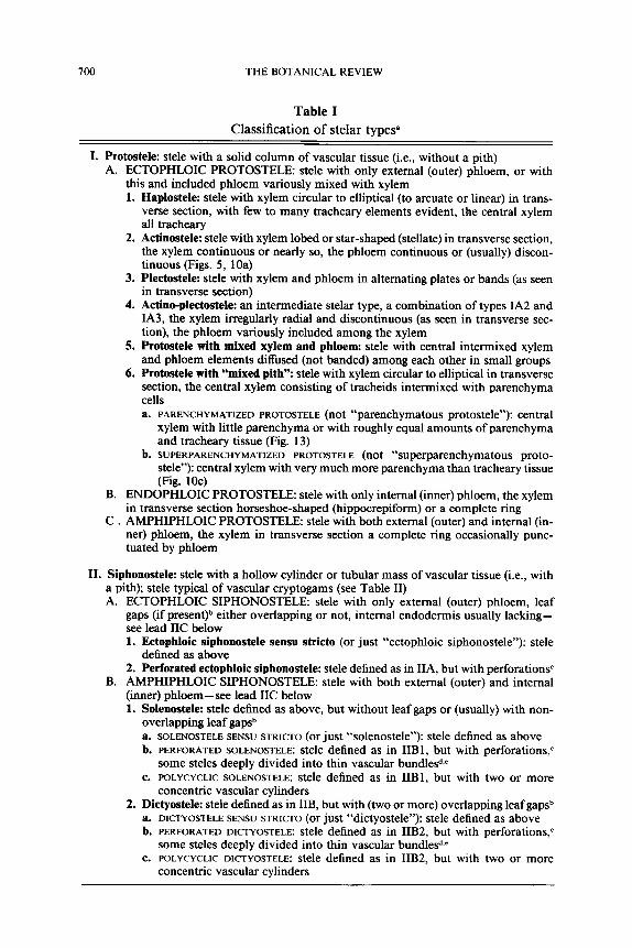
Table I lists and characterizes the types of steles recognized in this paper. Only common types, or stelar types commonly recognized, are defined in Table I. Many other stelar types have been proposed, for example, most recently "haploendoleptostele" and "xestomeristele" by Albergoni et al. (1978). Part III gives the historical development of some of the stelar terms used in Table I. The article by Schmid (especially Table I therein) following in this issue and the reviews by Belli (1896), Chau- veaud (1911), Hill (1906), Meyer (1917), Schoute (1903), Scott (1894), and Tansley (1896), and the glossary by Jackson (1928), should be con- sulted for extended discussions of historical aspects and/or for definitions of the innumerable terms of stelar morphology encountered in the liter- ature. Whenever possible, our definitions follow not only the concepts of the terms as originally introduced, but also such widely used textbooks of morphology as the one by Foster and Gifford (1974, and the 1959 first edition). Some elaborations of terms used in Table I and elsewhere in this paper are necessary (see also the review by Schmid accompanying this article):
Definition of stele.-- By original definition (van Tieghem and Douliot, 1886a, 1886b) and its subsequent general acceptance (e.g., glossaries in Esau, 1960, 1977; and Fahn, 1974), the "stele" (or "central cylinder") includes the primary vascular tissue (xylem and phloem) of axes (stems and roots), plus any associated fundamental or ground tissue ("conjunc- tive tissue" in early works) present, that is, pith, pericycle, interfascicular regions, leaf gaps. The vascular tissue of leaves and of appendages of reproductive structures is generally excluded from the "stele" (see expla- nation in Part III of the article by Schmid following in this issue), although "stelar system" or "stelar region" can be used to refer to their vasculature. In contrast, many works make little or no use of the stelar concept and merely use "stele .... as a convenient abbreviation for the vascular system" (Esau, 1965a, p. 371).
Traditionally, the stele has been regarded as delimited by the endo-
700 THE BOTANICAL REVIEW
Table I
Class i f ica t ion o f s te lar types a
I. Protostele: stele with a solid column of vascular tissue (i.e., without a pith) A. ECTOPHLOIC PROTOSTELE: stele with only external (outer) phloem, or with
this and included phloem variously mixed with xylem 1. Haplostele: stele with xylem circular to elliptical (to arcuate or linear) in trans-
verse section, with few to many tracheary elements evident, the central xylem all tracheary
2. Actinostele: stele with xylem lobed or star-shaped (steUate) in transverse section, the xylem continuous or nearly so, the phloem continuous or (usually) discon- tinuous (Figs. 5, 10a)
3. Plectostele: stele with xylem and phloem in alternating plates or bands (as seen in transverse section)
4. Actino-plectostele: an intermediate stelar type, a combination of types IA2 and IA3, the xylem irregularly radial and discontinuous (as seen in transverse sec- tion), the phloem variously included among the xylem
5. Protostele with mixed xylem and phloem: stele with central intermixed xylem and phloem elements diffused (not banded) among each other in small groups
6. Protostele with "mixed pith": stele with xylem circular to elliptical in transverse section, the central xylem consisting of tracheids intermixed with parenchyma cells a. PARENCHYMATIZED PROTOSTELE (not "parenchymatous protostele"): central
xylem with little parenchyma or with roughly equal amounts of parenchyma and tracheary tissue (Fig. 13)
b. SUPERPARENCHYMATIZED PROTOSTELE (not "superparenchymatous proto- stele"): central xylem with very much more parenchyma than tracheary tissue (Fig. 10c)
B. ENDOPHLOIC PROTOSTELE: stele with only internal (inner) phloem, the xylem in transverse section horseshoe-shaped (hippocrepiform) or a complete ring
C . AMPHIPHLOIC PROTOSTELE: stele with both external (outer) and internal (in- ner) phloem, the xylem in transverse section a complete ring occasionally punc- tuated by phloem
II. Siphonostele: stele with a hollow cylinder or tubular mass of vascular tissue (i.e., with a pith); stele typical of vascular cryptogams (see Table II) A. ECTOPHLOIC SIPHONOSTELE: stele with only external (outer) phloem, leaf
gaps (if present) b either overlapping or not, internal endodermis usually lacking-- see lead IIC below 1. Ectophloic siphonostele sensu stricto (or just "ectophloic siphonostele"): stele
defined as above 2. Perforated eetophloic siphonostele: stele defined as in IIA, but with perforations c
B. AMPHIPHLOIC SIPHONOSTELE: stele with both external (outer) and internal (inner) phloem--see lead IIC below 1. Solenostele: stele defined as above, but without leaf gaps or (usually) with non-
overlapping leaf gaps b a. SOLENOSTELE SENSU STRICTO (or just "solenostele"): stele defined as above b. PERFORATED SOLENOSTELE: stele defined as in IIBI, but with perforations, c
some steles deeply divided into thin vascular bundles ~.e c. POLYCYCL1C SOLENOSTELE: stele defined as in IIB1, but with two or more
concentric vascular cylinders 2. Dictyostele: stele defined as in IIB, but with (two or more) overlapping leaf gaps b
a. DICTYOSTELE SENSU STRICTO (or just "dictyostele"): stele defined as above b. PERFORATED DICTYOSTELE: stele defined as in IIB2, but with perforations, c
some steles deeply divided into thin vascular bundles d,e e. POLYCYCLIC DICTYOSTELE: stele defined as in IIB2, but with two or more
concentric vascular cylinders

STELAR MORPHOLOGY AND THE PRIMARY VASCULAR SYSTEM 701
C. SIPHONOSTELE INCERTAE SEDIS: a provisional category, especially for si- phonosteles of fossil taxa, for use when the nature of the phloem (whether ectophloic or amphiphloic) and/or of the interfascicular regions (whether leaf gaps, branch gaps, root gaps, or perforations--see Part IIA of text) is unknown or not clear f
III. Enstele: stele with a hollow cylinder or tubular mass of vascular tissue (i.e., with or without a definable pith) and with discrete sympodia usually either as a discontinuous cylinder or in a scattered or dispersed arrangement (as seen in transverse section); stele typical of seed plants (see Table II) A. EUSTELE SENSU STRICTO (or just "eustele"): stele defined as above ~
1. Eustele with collateral bundles: stele defined as in III, but with collateral bun- dies--xylem and phloem on the same radius, bundle with only internal (inner) phloem or (usually) with only external (outer) phloem (Figs. 7, 8, 10e, 10f, 12a, 15)
2. Eustele with bicollateral bundles: stele defined as in III, but with bicollateral bundles--xylem and phloem on the same radius, bundle with both external (outer) and internal (inner) phloem
3. Eustele with amphieribral bundles: stele defined as in III, but with amphicribral bundles--bundles concentric, the phloem surrounding the xylem (as seen in transverse section)
4. Eustele with amphivasal bundles: stele defined as in III, but with amphivasal bundles--bundles concentric, the xylem surrounding the phloem (as seen in transverse section)
B. PSEUDOSIPHONOSTELE: stele with vascular cylinder superficially in a contin- uous ring (as seen in transverse section) rather than in discrete bundles (see Part IVC1 f)
C. REDUCED EUSTELE: a eustele that is phyletically reduced in structure so as to appear as a protostele h
D. POLYCYCLIC EUSTELE: stele defined as in III, but with the sympodia as two or more concentric vascular cylinders, or else as a main vascular cylinder with internal or external bundles in a scattered or dispersed arrangement (as seen in transverse section); stele typical especially of those axes with a medullary or cortical vascular system, or both (see Part IVCIe)
Rejected stelar types: "preprotostele" coined by Lemoigne (1967) for bryophytes and "solid protostele" sensu H6bant (1977) used for mosses (see note s to Table I in the article by Schmid following in this issue)
a Table by Rudolf Schmid, derived from his classification of stelar types presented in the paper following in this issue. In that paper Schmid gives etymology, alternate terminology, examples of each stelar type, and detailed notes on rationale and historical aspects. For reasons indicated in Part III of Schmid's paper, this classification of stelar types is based on steles of stems, roots, rhizophores, and reproductive axes.
Rothwell dissents strongly from Schmid's classification and prefers to recognize these major types of steles: protosteles, medullated protosteles, solenosteles, dictyosteles, and eusteles. Beck agrees in general with Schmid, but dissents on some points which may differ from those with which Rothwell takes issue. This dilemma results from our differing con- ceptions of the bases, both philosophical and morphological, upon which a stelar classifi- cation should be erected.
b See Part IIA of text for definition of "leaf gap." c See Part IIA of text for definition of "perforation." d Use of "meristele" for such bundles is deprecated. See Part IIA of text. e The "dissected solenostele" and the "dissected dictyostele" are included in the synonymy
of, respectively, "perforated solenostele" and "perforated dictyostele." For rationale see note i to Table I in the article by Schmid following in this issue.

702 THE BOTANICAL REVIEW
dermis, which then topographically represents the innermost part of the cortex; the pericycle hence is the outermost part of the stele. However, since the endodermis and pericycle are largely lacking from the stems of seed plants (Blyth, 1968; Esau, 1965a; Fahn, 1974), the stele thus essen- tially consists of vascular tissue plus any associated fundamental tissue (pith and interfascicular regions) that is present. The intrastelar versus extrastelar nature of the endodermis was a critical point for the early morphologists. Most of this early discussion, which is well summarized by Belli (1896), Hill (1906), Meyer (1917), Schoute (1903), Scott (1894), and Tansley (1896), is now largely beside the point. Similarly, there has been appreciable discussion about the relationship of the pericycle to the stele. Blyth (1958) cogently reviewed this topic (see also references just cited and Esau, 1965b). Brebner (1902, p. 548) one of the early outstanding contributors to the stelar concept, concluded that due to the "unimpor- tance of the endodermis, pericycle, &c. as morphological criteria .... these layers should be, in many cases, abandoned as morphological criteria" to delimit the stele. This has come to be the prevalent viewpoint (e.g., Esau, 1953, 1960, 1965a, 1965b, 1977; Fahn, 1974; and many other works, but conspicuously not Ogura, 1938, 1972), and it is also the viewpoint adopted here.
By definition, the stele never includes secondary vascular tissue. Con- sequently, terminology such as the "primary stele," "secondary stele," "primary protostele," and "primary siphonostele" of Lemoigne (1967) and of McLean and Ivimey-Cook (1967) is inappropriate. However, it should be emphasized that the inclusion of secondary vascular tissue is a common observational and conceptual error in studies of both stelar morphology and nodal anatomy (see Part IIB). Finally, it should be re- membered that "stele," especially in paleobotanical usage, frequently and of necessity is used to refer only to the primary xylem when other tissues and tissue regions are not preserved or are merely poorly preserved.
Leaf traces.--A leaf trace is a bundle that diverges from an axial bundle (see below), or another leaf trace, and that extends into a leaf. This term applies to that part of a bundle in a stem from the point of its divergence from the stele to the level at which it enters a leaf base. The term "leaf trace bundle" (e.g., Meyer, 1928) is actually contradictory.
f This characterization is deafly not permissible to students for use in quizzes and ex- aminations given in morphology classes!
B Use of"atactostele" and the derivative "atactostely" and "atactostelic" for cases where the vascular bundles are in a scattered or dispersed arrangement (as seen in transverse section) is deprecated. See Part IVC2b of text.
h Typical of stems of Potamogeton (some species), Callitriche, Ceratophyllum, and other aquatic angiosperms, and axes ("thalli") of Lernna and Spirodela (see Sculthorpe, 1967, and especially Arber, 1920).

STELAR MORPHOLOGY AND THE PRIMARY VASCULAR SYSTEM 703
Leaf vascular supply.-- This term is sometimes used for the sum total of traces passing to one leaf.
Branch trace.--A branch trace is defined comparably to a leaf trace, except that the former is related to the vascularization of lateral shoots. A branch trace may also arise from another branch trace.
Gap and interfascicular region.--A gap or interfascicular region is the region of interfascicular (i.e., between vascular bundles) parenchyma in a vascular cylinder. As defined below, there are various types of gaps or interfascicular regions, that is, leaf gaps, branch gaps, root gaps, perfo- rations, dissections, and floral appendage gaps (see comments in brackets at the end of Part IIIB). In this paper, as noted below, we have adopted a restricted definition of interfascicular region. The obsolete terms "med- ullary ray" and "pith ray," which are equivalent to "interfascicular re- gion," have had rather little application in stelar morphology. Gaps or interfascicular regions occur only in siphonosteles and eusteles and are absent from protosteles since the last lack a pith. [In pteridophytes, gaps, if present, may involve (1) both xylem and phloem, (2) only xylem, or (3) only phloem. Examples for leaf gaps include (1) many Filicales, (2) many Osmundaceae, (3) Lepidodendraceae. Descriptions in the literature of "leaf gaps absent" may or may not refer to case (3). See Ogura (1972) for details.]
Leaf gap and interfascicular region.- A leaf gap or interfascicular region is the region of interfascicular parenchyma in the vascular cylinder op- posite a diverging leaf trace or opposite several associated traces of a leaf. This parenchymatous region may extend longitudinally for only a short distance; distally in the stele the "gap" may be closed again by vascular tissue. The conceptual difference between "leaf gap" (or "foliar gap") and "interfascicular region" ("lacuna" is still another term used in the liter- ature) is detailed in Part IIIB. For reasons presented there, we restrict the application of "leaf gap" to the steles of vascular cryptogams (i.e., siphonosteles) and use "interfascicular region" for the steles of the seed plants (i.e., eusteles).
Branch gap.--A branch gap--"ramular gap" or "ramular lacuna" in the older literature--is defined comparably to leaf gap, except that the former is related to the vascularization of lateral shoots.
Root gap.-- Because lateral roots of both seed plants and pterido- phytes originate in the pericycle or in the endodermis, or in both tissue regions (Esau, 1965a; Ogura, 1972), gaps or interfascicular regions related to traces to lateral roots do not occur. However, "root gaps," a term used as early as 1914 (Perry, 1914), may occur in siphonostelic or eustelic rhizomes bearing adventitious roots, as in the perforated (see below) ectophloic siphonostele of Ophioglossum pendulum (Petry, 1914) and the solenostele of Dennstaedtia cicutaria (Stevenson, 1974).

704 THE BOTANICAL REVIEW
Perforation.--A perforation is a region of interfascicular parenchyma in the vascular cylinder not associated with leaf, branch, or root traces. That is, perforations are discontinuities in the stele other than leaf gaps, branch gaps, or root gaps. Steles with perforations are called "perforated steles," specifically "perforated ectophloic siphonosteles," "perforated so- lenosteles," and "perforated dictyosteles" (see Table I). Perforations are common in rhizomes of the solenostelic and especially dictyostelic ferns (see Table I in the article by Schmid following in this issue); they do not occur in eusteles.
Tansley (1907--08, p. 192) originally defined "perforation" as "gaps in the cylinder which are not leaf-gaps." Subsequent definitions of the term have generally been similar, for example, those of Bower (1913, 1923- 28, 1930, 1935), Foster and Gifford (1974), McLean and Ivimey-Cook (1951), Ogura (1938, 1972), Parihar (1965), Sporne (1975), Wardlaw (1952), and others, though specifically not Jeffrey (1908), who defined the term essentially as we do. Though the other works listed defined "per- foration" in relation to gaps other than leaf gaps only, usage of the term in these and other works has been in the more restrictive sense as defined by us above and by Bower.
Synonyms of "perforation" are "incidental gap" (Petry, 1914), "dis- section" (which probably exists in this sense, but which we have not seen in the literature), and "lacuna" (Gwynne-Vaughan, 1903). Modern usage generally restricts the last term to interfascicular regions related to the departure of traces.
For additional aspects of this terminology see Stevenson (1974) and note i to Table I in the article by Schmid following in this issue.
Axial bundle.--An axial bundle is the major vascular bundle o f a sym- podium (see below), and it continues without interruption along the length of a stem segment. Leaf and branch traces may arise from axial bundles. Alternate terms for "axial bundle" include "stem bundle," "cauline bun- dle," "common bundle," "sympodial bundle," "sympodial stem bundle," "sympodial segment," "sympodial strand," and "reparatory strand" (see also Parts IIB and IVA).
Sympodium.--A sympodium consists of an axial bundle and its asso- ciated leaf and branch traces. Figure 25, for example, depicts five sym- podia, each of which consists of an axial bundle and the leaf and branch traces that diverge from it.
Dextrorse versus sinistrorse trace divergence ("direction of trace diver- gence").--These terms denote the divergence of traces from the right side (dextrorse) or the left side (sinistrorse) of an axial bundle when the vascular system is viewed from the pith or inside of the stele. Note that the di- rections of trace divergence would be opposite, that is, mirror images, if the stele were viewed from the cortex.

STELAR MORPHOLOGY AND THE PRIMARY VASCULAR SYSTEM 705
Open, closed, and intermediate vascular systems.- In open systems (Figs. 3; 4; 12b; 14; 16; 17a, b; 18-25; 28; 34; 37; 42, 43) the sympodia are entirely discrete (no fusions between vascular bundles) or essentially dis- crete, that is, with random interconnections consisting of only minor or accessory bundles (see below). In closed systems (Figs. 17c, 31-33, 35, 38--41) anastomoses occur between leaf traces, between leaf traces and axial bundles, or between axial bundles, so that a reticulate vascular pattern results. Intermediate systems (Figs. 9, 26, 27, 29, 30, 36) are partly open and partly closed; such systems may be predominantly open, or predominantly closed.
This terminology is also applicable to the vasculature of leaves and, as proposed by Sporne (1958) and Schmid and Beck (1971), to flowers (for detailed examples see Schmid, 1972a). Various aspects of open, closed, and intermediate vascular systems are discussed in Part IVC1 and also by Dormer ( 1945, 1954, 1972), Esau ( 1965 b), and Philipson and Balfour (1963).
Although Dormer (1945) introduced the terms "open" and "closed" for vascular systems, this distinction, of course, was known by earlier botanists, for example, de Bary (1877, 1884). De Bary (1877, 1884, p. 235) used the terms "sympodial" and "reticulate" for, respectively, "open" and "closed," and the former pair of terms is frequently seen in the older literature. It should also be noted that Frank's (1864, pp. 380-381,410) terms "ungeschlossene" and "geschlossene Gef~tssbtindelsysteme" refer not to vasculature but rather to vascular histology, that is, the appearance of leaf midrib bundles in cross section.
Accessory or bridge bundles.--Accessory bundles (Devadas and Beck, 1971) or bridge bundles (Dormer, 1954) are small strands that intercon- nect two axial bundles and/or other bundles in a largely random fashion. Accessory bundles may be strictly phloic (e.g., see Fig. 21 in Devadas and Beck, 1971), and in such cases have been referred to as "phloem anas- tomoses" (Aloni and Sachs, 1973) or "phloic anastomoses" (Schmid, in preparation).
Ontogenetie spiral or genetic spiral.--This is the single helix, dextrorse or sinistrorse, that can be drawn through the centers of all the leaves in the order of their origin from the shoot apex. [Proper distinction should be made between a "spiral," which circles around from a central point, versus a "helix," which follows the surface of a cylinder. These terms are often confused in anatomy and morphology. For example, "helical cell wall thickening" is the proper designation, not "spiral cell wall thicken- ing."]
Phyllotactic fraction.--This term refers to the fraction denoting the mode by which leaves are arranged on the stem. The fraction expresses

706 THE BOTANICAL REVIEW
the angle of divergence (see below) between two successive leaves and is denoted by fractions in the Fibonacci series (1/2, 1/3, 2/5,3/8, 5/13, etc.), where each series of numbers is formed by successive addition of the last two: 1, 2, 3, 5, 8, 13, 21, 34, etc.). In the fraction the denominator is the number of leaves between two vertically superimposed leaves (e.g., "5" for leaves 9 and 14 in Fig. 12), whereas the numerator is the number of turns around the axis between the two superimposed leaves (e.g., "2" in Fig. 12, for a fraction of ~/n). See the detailed accounts in Dormer (1972) and Esau (1965b) for elaborations.
Angle of divergence.- This term, or the equivalent "angular divergence" or "divergence angle," refers to the smallest fraction of the stem circum- ference separating the points of origin of two successively initiated leaves (i.e., leaves on the same parastichy--see below).
Orthostichy versus parastichy.--These terms denote a vertical line (or- thostichy) or a helix (parastichy) along which is attached a series of leaves or scales on an axis of a shoot or shootlike organ (Esau, 1977). "Orthos- tichy" has been incorrectly applied to a steep helix or parastichy. For elaboration see the detailed accounts in Dormer (1972) and Esau (1965b).
Meristele.--The frequently encountered term "meristele" was coined by van Tieghem (189 lb, p. 284) and, along with its counterpart "schizo- stele" (Strasburger, 1891, pp. 110, 312), was originally applied to the bundle or bundles entering a leaf because these represent "a separated portion or portions of that ["stelar tissue"] of the stem" (Tansley, 1896, p. 149, emphasis his). [Part III of the article by Schmid following in this issue gives van Tieghem's rationale for proposing "meristele." Also, "schizostele" and "schizostely" have other meanings, e.g., Ogura (1972) and van Tieghem (1891 b, 1898, 1918, and his other works-- see Bonnier, 1914).] In 1902, following Jeffrey's (1898-99, pp. 627-628) influence, Brebner (1902, p. 521) formally "modified" the term "from its original meaning as used by Van Tieghem and Strasburger" and applied it to the "individual strands of any vascular system," that is (p. 523), "the vascular bundle in the old sense [i.e., de Bary, 1877, 1884], except that it does not include actino- and haplosteles as formerly." Later, Tansley (1907-08, p. 38), without explanation, restricted "meristele" to "the individual vas- cular strand of a perforated solenostele or of a dictyostele" (see definition of "perforation" above).
The consequence of these modifications has been that following Brebner (1902) "meristele" applies to all types of bundles (collateral, bicollateral, amphicribral, amphivasal, etc.) to all organs, and to all types of vascular plants, whereas following Tansley (1907-08) the term applies only to amphicribral bundles of pteridophytes, especially the ferns. "Meristele," by extension, has also been applied to bundles of flowers (van Tieghem,

STELAR MORPHOLOGY AND THE PRIMARY VASCULAR SYSTEM 707
1896; discussion in Schmid, in preparation). Both the Brebnerian and Tansleyian applications of "meristele" have been commonly used, even in recent anatomy and morphology textbooks and monographs. However, Foster and Gifford (1959, 1974) and the glossaries of such well-known works as Esau (1977, but not in 1953, 1960, or 1965a) and Fahn (1974, and the 1967 edition) define "meristele" as being the amphicribral bundle of dictyosteles. Consequently, the Tansleyian definition of"meristele" is perhaps more commonly encountered today, although equally well-known works as those of Ogura (1972), Parihar (1965), Smith (1955), and Sporne (1975) use "meristele" in a more or less Brebnerian sense [but the latter workers have definitions of"solenostele" and "dictyostele" different from those of the former--see notej to Table I in the article by Schmid following in this issue].
Nevertheless, "meristele," whatever its application, is clearly a poor term. The term refers to only part o fa stelar system ("meri-" means "part of ') , specifically to a vascular bundle, but to the unwary the term implies a type of stele. "Meristele" thus is not comparable to the other stelar types listed in Table I. For this reason, and also because of the diverse applications of the term noted above, we would like to see the term eliminated from textbooks and from discussions of stelar morphology. "Vascular bundle" is a perfectly acceptable, and undeniably clear alter- native to "meristele." But if the latter term must be used, it should be used in the sense of the amphicribral bundles of dictyosteles and perfo- rated solenosteles, as originally defined by Tansley (1907-08).
Vascular segment.--Basinger et al. (1974, p. 1003) coined this term for use in the "polystelic" (see below) Medullosaceae for "a strand of primary xylem that gives rise to leaf traces and is surrounded by a cylinder of secondary vascular tissue." Earlier studies of Medullosaceae have used "meristele" or especially "stele" in place of"vascular segment."
Monostely versus polystely.--These terms were introduced by van Tieghem and Douliot (1886a, 1886b), and especially "polystely," have wide application even today (e.g., Metcalfe, 1979; Stewart, 1976; Part IVB3). "Monostely," or "monostelic" mean having a single stele per organ and have been used for various types ofprotosteles and even siphonosteles and eusteles (see note p to Table I in the following article by Schmid), whereas "polystely" or "polystelic" mean having (as seen in transection) several steles per organ (Scott, 1891; previous references), more explicitly meaning having two or more adjacent complete cylinders of vascular tissue (Fig. 15). We have avoided "monostele" but have followed con- vention in using "polystelic" to refer to the Medullosaceae and other gymnosperms (Part IVB3). "Polystelic" is also used to refer to steles of pteridophytes (e.g., Ogura, 1972).

708 THE BOTANICAL REVIEW
Dictyoxyl ic . - - T h i s adjective refers to siphonosteles in which the xylem ring is interrupted radially by parenchymatous, overlapping leaf gaps to form a net ("dictyo-" means "net," as also in a "dictyostele"), but the phloem and endodermis are continuous (e.g. Chelianthes, O s m u n d a - - s e e Ogura, 1972, and note e to Table I in Schmid's paper in this issue). The confusingly similar "dictyoxylon" or "dictyoxylonic" refer to a scleren- chymatous cortex with a fibrous network, as in Lyginopteridaceae (Jack- son, 1928, and various paleobotanical works).
Coda: The terms defined above and in Table I, as well as the more specialized terms defined elsewhere in this paper, have had, of course, alternative definitions and applications in the literature. For example, Dormer's (1954, 1972) use of "stem bundle" is equivalent to our "axial bundle," but the "cauline bundle" used by older anatomists (e.g., de Bary, 1877, 1884) is different (see also Part IVA). We encourage authors of future papers on stelar morphology and on descriptions of the primary vascular system to cite which worker(s) they are following for terminology and to define carefully any new or revised terms they use. In addition to the references dealing with stelar morphology that are cited in the begin- ning of this section, the following are useful for discussions of non-stelar terminology of the primary vascular system: Barthelmess (1935), de Bary (1877, 1884-- for old usage of terms), Benzing (1967a), Devadas and Beck (1972), Dormer (1954, 1972), Esau (1965a, 1965b, 1977), Kaplan (1937), Meyer (1928), Namboodiri and Beck (1968a), Philipson and Balfour (1963), Schmid (In preparation), Schmid and Beck (1971), and Sporne (1958).
B. PROBLEMS IN INTERPRETATION AND PRESENTATION OF DATA
The study of vascular architecture of the shoot, that is, studies of stelar morphology and the anatomy of the primary vascular system, is attendant with many manipulative and interpretive difficulties. Dormer (1954, 1972) has written on this subject at some length and has, in fact, even presented (Dormer, 1954, p. 303) five "empirical rules" for studying the vasculature of plants with trilacunar nodes. Most of Dormer's points have validity, but we dispute his a posteriori claim (Dormer, 1954, p. 303) that "inter- pretation of vascular systems must be based on the way in which the bundles are connected rather than on their intrinsic properties" since the latter are "inconstant" and "completely unimportant from the standpoint of comparative morphology." Certainly, numerous studies have dealt only with the nature of bundle connections, but the concern of many paleo- botanical works with xylem maturation (see Figs. 5, 7, 13, and 14 and the references in Parts IVB2 and IVB3) as well as of some works on extant plants belies Dormer's claim about the lack of significance of the "intrinsic

STELAR MORPHOLOGY AND THE PRIMARY VASCULAR SYSTEM 709
properties" of bundles. Devadas and Beck (1971), for example, found differences in bundle size and tracheary element number in axial bundles, leaf traces, and branch traces. The following discussion focuses on aspects of interpretation and presentation of vascular architecture largely undis- cussed by Dormer (1954, 1972).
A general problem in studies of the stele and the primary vascular system is that of interpreting the data, which are usually presented as longitudinal diagrammatic representations of the vascular systems (Figs. 3, 4, 9, 12b, 14, 16-18, 20a, 21-43). These diagrams, which can be either very accurate or relatively less accurate depending on the skill and atten- tion to detail of the worker(s), are prepared by plotting the vascular bun- dles of each of a series of transverse sections. The bundles are represented by vertical files of dots, which are then longitudinally connected by lines to form the pattern of the vascular system as if it were split open lengthwise and spread in one plane (see also comments four paragraphs below). Unfortunately, many workers have published diagrams illustrating the generalized pattern of the vascular system. Often, small bridges or acces- sory bundles have been omitted because they "clutter up" the diagram (e.g., see the accessory bundles depicted in Devadas and Beck, 1971, Fig. 21, which were intentionally omitted in Devadas and Beck, 1972, Fig. 1).
The levels at which leaf and branch traces enter lateral appendages is indicated by various symbols, usually circles, triangles, X's, or combi- nations of these (see the figures and their explanatory captions in this paper). Although median traces often enter leaf bases at lower levels than do lateral traces, it is customary to place at the same level in these diagrams the symbols for median and lateral traces that supply one leaf. Although this provides for easy recognition of the number of traces per leaf, it does introduce an inaccuracy in many such diagrams. In addition, as elaborated below, many workers diagram vascular bundles as straight or vertical lines rather than as undulating, helical lines, another inaccuracy usually tolerated for the sake of simplicity.
Since it is acknowledged by most workers that their representations of stelar patterns are, indeed, diagrammatic, some seem to feel free to take unusual liberties, sometimes constructing the diagrams to conform to their preconceived ideas (which may be widely accepted viewpoints, but based on evidence from other sources, especially developmental) about the na- ture of primary vascular systems (see also Dormer, 1972, pp. 155-159). We are referring to the longstanding theoretical debate over the foliar/ cauline (axial) versus foliar (appendicular) nature of the shoot system. Thus, one school of researchers has considered primary vascular systems to consist of sympodia constructed entirely of interconnecting leaf traces (e.g., Balfour and Philipson, 1962; Barthelmess, 1935; de Bary, 1877,

710 THE BOTANICAL REVIEW
1884; Campbell, 1921; Esau, 1965a, 1965b, 1977; Fahn, 1974; Kaplan, 1937; Nast, 1944; Philipson and Balfour, 1963; Stevenson, 1980). For example, Figures 21 and 22 both depict the primary vascular system of the shoot of Iberis amara, but differ conspicuously. Figure 21, published by Carl N~igeli in 1858, is apparently an objective and reasonably accurate representation of the primary vascular system of this plant. However, Figures 22 and 28, produced by Balfour and Philipson (1962) and Figure 24, from Benzing (1976b), clearly reflect the viewpoint that the sympodia consist solely of interconnecting leaf traces. In other features Figures 21 and 22 provide the same information, and both reflect accurately the steeply helical course of the sympodia. On the other hand, workers who adhere to the viewpoint that the sympodia are composed of axial bundles from which leaf traces diverge (e.g., Beck, 1970; Bower, 1923, 1926, 1935; Devadas and Beck, 1972; Dormer, 1954, 1972; Posthumus, 1924; Schoute, 1926, 1938; Wardlaw, 1952; Wetmore, 1943), which is the position we adopt in this paper, often diagram axial bundles as vertical lines (e.g., Figs. 25-27, 29). This also results in inaccurate diagrams in the sense that nearly all axial bundles do indeed form steep helices in their courses through the stem. In addition, these bundles usually follow an undulating rather than a consistently straight course.
The aforementioned debate over the nature of the stelar system is usually also reflected in the terminology adopted by workers to designate the main vascular bundles in a stem, proponents of the foliar, often developmental concept thus generally using "sympodial bundle" or the obsolete "common bundle," proponents of the axial, essentially stelar concept thus generally using "axial bundle" (the term used in this paper), "stem bundle," or "cauline bundle." However, some workers (e.g., Dor- mer, 1945, 1946, 1954, 1972) have used "stem bundle" in the sense of "sympodial bundle." Perhaps as a compromise, Esau (1977) recently has begun using "sympodial stem bundle." In light of these terminological variances, we think that the glossary of terms presented in Part IIA should prove especially useful.
The fact that some students of stelar anatomy determine phyllotactic fractions on the basis of the departure of successive leaf traces along axial bundles, rather than on the basis of the arrangement of leaves in orthos- tichies, leads to inconsistency and possible confusion. This problem is discussed in greater detail in Part IVCla. In addition, the direction of trace divergence will seem to vary depending on whether one constructs the diagrams as if looking from the inside or from the outside of the stele, that is, from the pith or the cortex, respectively. The left-right designations for leaf traces and the direction of trace divergence would thus be mirror images of each other. This problem is also discussed in more detail later in this paper (Part IVC 1 d 1). To avoid such potential for confusion, work-

STELAR M O R P H O L O G Y A N D THE P R I M A R Y VASCULAR SYSTEM 711
ers should clearly indicate the perspective from which their diagrams are drawn.
In some cases it is more convenient (or necessary) to use provascular strands and/or protoxylem strands than mature primary vascular bundles as the basis for determining stelar patterns. For example, Larson (1975) in studying Populus deltoides used provascular strands for his analysis of the primary vascular system, Benzing (1967a, 1967b) used protoxylem strands in his studies of the architecture of the primary vascular systems of some woody ranalean species, and Basinger et al. (1974) analyzed the primary vascular system of Medullosa on the basis of protoxylem strands (for further detail see Part IV).
The justification for these approaches is straightforward. Provascular strands, protoxylem strands and mature vascular bundles are related de- velopmentally. Mature primary vascular bundles develop from provas- cular strands. Metaxylem develops in relation to protoxylem; and there is evidence of a one-to-one relationship between protoxylem strandsand provascular strands in seed plants. This relationship in Populus deltoides has been demonstrated by Larson (1975). It does not necessarily follow, however, that each vascular bundle or trace contains only a single pro- toxylem strand. Indeed, as Larson (1975) showed so clearly, leaf traces in P. deltoides are compound structures consisting of several smaller vas- cular bundles, the number of bundles depending on the level in the trace. Similarly, Benzing (1967a, 1967b) illustrated an increase in the number of protoxylem strands at progressively more distal levels in leaf traces of certain woody ranalean species (Figs. 30, 33-35). It is important to observe that both Larson and Benzing, nevertheless, demonstrated sympodial eustelic vascular patterns based on provascular or protoxylem strands. [Benzing (1967a, 1967b) described the stelar systems of some of the woody species he studied as "lacking sympodia." See Part ICV1 f for an expla- nation and discussion.]
The use of provascular and/or protoxylem strands as the basis for stelar patterns alleviates a serious problem that arises in many woody plants from the tendency of developing metaxylem of adjacent bundles to be- come confluent, or nearly confluent, thus forming pseudosiphonosteles (see Part IVC I f) or compound primary vascular bundles. This phenom- enon is enhanced in dicotyledons by the late development of bridge bun- dles (often consisting solely of primary phloem) that connect with earlier formed bundles (Devadas and Beck, 1971; Dormer, 1945, 1972; Larson, 1975) and, in some species, by the development of radially aligned meta- xylem. Even in those woody species in which the mature primary vascular bundles remain discrete, their recognition and analysis are often hampered by the early development of secondary vascular tissues.
A number of studies have yielded contradictory interpretations of the

712 THE BOTANICAL REVIEW
stele or primary vascular system because workers have not carefully dis- tinguished between primary and secondary vascular tissues. This problem, for example, has involved interpretations of the vegetative shoots of Ephedra (see Part IVB9), the reproductive shoots, that is flowers of Aqui- legia and other angiosperms (see Nast, 1944; Schmid and Beck, 197 l; and Sporne, 1958, 1974), and especially nodal anatomy (see Part V). The obfuscation of primary vasculature by secondary growth perhaps accounts for many interpretive differences in the literature. Stelar morphology and nodal anatomy, needless to say, relate strictly to the primary vascular system. The structure of the secondary vascular system thus is, from the viewpoint ofstelar morphology and nodal anatomy, strictly superimposed on primary structure and is, as a result, irrelevant.
Despite the various problems accompanying diagrammatic represen- tations of steles or primary vascular systems, such diagrams present in readily accessible form a large amount of information that is usually much more comprehensive and comprehensible than it would be if presented solely in written descriptions, which have their own peculiar difficulties (see Part IIA and Boodle, 1903b; and Dormer, 1954, 1972). Furthermore too much morphological and anatomical work in the past, including some that has had great impact, has been remarkably superficial. One merit of preparing such diagrams, tedious as such preparation is, is that it forces the worker to observe hundreds--even thousands--of sections of an axis with the result that the information provided will usually be more detailed and accurate than it might otherwise have been.
Dormer (1954, 1972) has maintained that in studies of vascular ar- chitecture it is better to analyze old stems rather than very young stems, although the preference of most workers has been to study young stems, particularly, of course, if development is the concern. One difficulty of studying very young stems stressed by Dormer (1954, 1972) is that prox- imal connections of bundles may not be recognizable at the time of ob- servation (see also Part IVClf). On the other hand, dealing with older stems requires, as just discussed, distinguishing between primary and secondary vascular tissue. Since vascular patterns change during ontogeny, especially during seedling development (see Part IVClg), analysis of just the most basal part of a shoot should be avoided (Dormer, 1954).
Dormer's viewpoints are based on his utilization of mature primary vascular tissues--largely primary xylem--as the basis for defining the primary vascular system. We believe that the use of provascular bundles and/or protoxylem strands are equally valid approaches-- indeed, possibly the better approaches for the determination of basic stelar patterns (see page 71 l, and Part VII). Structures (i.e., provascular bundles and pro- toxylem strands) developmentally closer to the organizing centers in the plant might reflect more accurately the basic stelar patterns than structures

STELAR MORPHOLOGY AND THE PRIMARY VASCULAR SYSTEM 713
interpreted as vascular bundles. Furthermore, the use ofprovascular bun- dles eliminates the problems of tissue identification associated with sec- ondary growth.
There have been attempts by physiologists to trace interveinal transport and its relationship to phyllotaxy (e.g., Fiscus et al., 1973; Roach, 1939). Work of this sort, of course must be done with full knowledge of the vascular system and phyllotaxy of the plant(s) under study. Open vascular systems and woody habit tend to correlate more strongly than do closed systems and herbaceous habit (see Part VIB). However, as elaborated in Part VIC, from a functional viewpoint the open vascular system of woody plants becomes a closed vascular system with the advent of secondary growth, which often occurs very close to the shoot apex. Precise awareness of anatomical aspects such as these is obviously necessary in physiological studies of transport, but one frequently gets the impression of anatomical ambiguity in physiological work of this sort.
The time-honored method of studying the primary vascular system of shoots is, of course, by analyzing serial transverse sections with the light microscope. This method has its limitations, however, particularly when vascular systems contain numerous bundles. In some cases, especially for rather simple stelar systems, clearings and dissections rather than sections have been used to prepare representations of vascular patterns (e.g., see the figures of clearings in Esau, 1965b; Sands, 1973; and Schmid, 1972a). In a few cases, such as with the inflated inflorescence axis of Caulanthus (Schmid, unpublished), it is possible to determine the vasculature simply by holding the opened axis against a light source. In recent years the study of vascular systems with many bundles has been greatly facilitated by the development by Zimmermann and Tomlinson (1965, 1972, 1974--see also Tomlinson, 1970; and Zimmermann, 1976) of cinematographic anal- ysis of vascular systems using a movie camera, shuttle microscope, and film data analyzer. Cine analysis of vasculature has now had numerous applications, especially from students ofmonocotyledons (see Part IVC2).
Because of the problems and inconsistencies enumerated above, the student of the stele and of the primary vascular system must exercise great care in interpreting diagrams and descriptions of these structures and in using information derived from them (see also Dormer, 1954, 1972). As also stressed by Dormer (1972), regardless of different tech- niques for written description or for diagrammatic presentation, it should be possible, if they were accurately executed, for persons familiar with the conventions employed to translate conflicting accounts to some com- mon ground. This admittedly is a very difficult and often tedious task, but it must be attempted if syntheses of vascular architecture are to be achieved. It would thus be helpful for workers to define precisely the conventions of terminology, description, and diagrammatic representa-

714 THE B O T A N I C A L REVIEW
tion that they are employing. To facilitate this we have attempted in Part IIA a standardized terminology of steles, the primary vascular system, and related structures which various workers and textbook writers can adopt if they so choose. In addition, in Part VII, we have presented a set of recommendations for the study ofstelar morphology which, if followed, will lead to the production of comparable data, the systematic utility of which will, thereby, be greatly enhanced.
Ill. Evolution of the Stelar Concept
A. FORMULATION OF THE STELAR HYPOTHESIS
The primary vascular system of plants has been studied and interpreted in different ways by many workers. The early botanists (e.g., de Bary, 1877, 1884; Geyler, 1867-68; Niigeli, 1858--see elaboration in Part IVA) held that the individual vascular bundle is the fundamental unit in the vascular system of the land plants. Later the continuity of all the vascular tissue in the plant body came to be emphasized. This latter attitude was reflected first in the anatomical-physiological classification of Sachs in 1868 (fourth and last German edition 1874, translations into English in 1875 and 1882), in which plant tissues were histologically divided into three tissue systems, namely, the dermal system, the fundamental or ground system, and the fascicular or vascular system. Although Sach's scheme has been criticized as being structurally and functionally too in- clusive (see Foster, 1949), it nevertheless continues to be adopted in recent anatomy and morphology textbooks (e.g., Esau, 1958, 1960, 1965a, 1977; Foster, 1949; Foster and Gifford, 1959, 1974) because of its considerable didactic value in emphasizing the general topographical anatomy and unity of the higher plant body.
Slightly later, the essential unity of the vascular system came to be emphasized from a different viewpoint, namely a comparative and, even- tually, an evolutionary one. In 1886 van Tieghem and his student Douliot proposed (1886a) and elaborated (1886b--summaries in van Tieghem, 1891a, 1891b, 1896, 1898, 1918) the stelar concept, which formally and emphatically recognized the fundamental unity of the entire vascular region of the plant axis. The fundamental or basic unit was designated the stele, which included all the vascular tissue and, depending on the concepts of various workers (see Part IIA), varying amounts of associated fundamental tissue (pith, pericycle, and gaps or interfascicular regions). Strasburger (1891) in his monumental work on the structure and function of vascular tissue was among the first to adopt and modify van Tieghem's stelar terminology and concepts.
The difference between the stelar concept and the individualistic con- cept of the vascular system can be illustrated by the following analogy.

STELAR MORPHOLOGY AND THE PRIMARY VASCULAR SYSTEM 715
By the individualistic concept, a single vascular bundle of a primary vascular system composed of many bundles, as in the siphonostelic fern Pteridium, was regarded as the morphological equivalent of the entire vascular system of a protostelic fern such as Gleichenia. In contrast, by the stelar concept, all the vascular bundles of Pteridium, collectively, would be considered morphologically equivalent to the single vascular column of Gleichenia.
After its formulation in 1886, the stelar concept was greatly modified and elaborated upon by many distinguished workers, as noted in the introduction (Part I), and much of van Tieghem's stelar concept quickly became discredited. Tansley (1896) divided the "history of the stelar doctrine" into three phases: (1) the idea of the vascular cylinder (1870- 84), (2) polystely and astely (1886-91), and (3) extensions and modifi- cations (1891 to date, "to date," of course, being 1896, for which 1981 can be substituted). The early work on stelar morphology is admirably summarized in the reviews by Belli (1896), Chauveaud (1911), Hill (1906), Jeffrey (1898-99, 1899, 1902), Meyer (1917), Scott (1894), and Tansley (1896), and in the book Die Stelar-Theorie by Schoute (1903). Suffice it to say here that much of this early work, although fascinating, is now largely of historical interest, for example, the undue emphasis placed on the "boundary of the stele" or on the "metamorphoses of the stele" (citing discussion headings of Tansley, 1896). Consequently, only those ideas of early workers that are relevant to modern ideas of systematics and evo- lutionary morphology are appraised in the ensuing discussions.
B. THE CONCEPT OF LEAF GAP AND JEFFREY'S IDEAS ON STELAR MORPHOLOGY
The modifications of the stelar concept by Edward Charles Jeffrey (1866- 1952-- see Wetmore and Barghoorn, 1953) of Harvard University, pub- lished mainly between 1898 and 1902, have had the greatest influence on thought on the classification and evolution of steles and on the broad classification of the vascular plants. Jeffrey also contributed importantly to stelar terminology, and he was among the first to interpret steles from an evolutionary viewpoint (van Tieghem and Douliot, 1886a, 1886b, o f course, were the firs0.
Jeffrey published a number of abstracts and lengthy articles on stelar morphology, all of which are cited in the bibliography (Jeffrey, 1896, 1896-97, 1898, 1898-99, 1899, 1901, 1902, 1903, 1906, 1908, 1910, 1917). The 1896 citation is for a title only, but in the literature it is frequently misleadingly cited as an article. Jeffrey's 1896-97 paper on the gametophyte of Botrychium virginianurn describes and figures the anat- omy of the young sporophyte but other than the use of"foliar gaps" does

716 THE BOTANICAL REVIEW
not use or propose any stelar terminology. Jeffrey's often cited 1898 ab- stract is his first published work on the stelar concept. His 1898-99, 1899, 1901, and 1902 works presented original data, whereas his 1908 and 1910 papers are mainly responses to the largely British criticisms of his views (see Part IIIC and the article by Schmid following in this issue). Jeffrey's 1903 contribution to Coulter and Chamberlain's (1903) book and his own 1917 book The Anatomy of Woody Plants are reviews of his own work, the former being Jeffrey's most cogent statement on stelar morphology. Jeffrey's 1906 publication is only peripherally concerned with stelar mor- phology. In addition, from 1902 through 1917 in The Botanical Gazette Jeffrey further espoused his views in numerous reviews of books and articles. Curiously, Jeffrey did not publish on the stelar concept after 1917, although he was concerned with other aspects of shoot vasculature (e.g., Jeffrey and Torrey, 1921a, 1921b) and published more than 60 papers from 1918 through 1947 (see the bibliography in his obituary by Wetmore and Barghoorn, 1953). [Because many of Jeffrey's works are repetitive in nature, only the most important of Jeffrey's terms, statements, and con- cepts are documented by bibliographic citations since giving a long series of dates would serve only to interrupt the flow of the text.]
In his 1898 abstract and 1898-99 paper Jeffrey proposed the following stelar terminology (see also Table I):
I. Protostele: with a solid column of vascular tissue, that is, without a pith;
II. Siphonostele: with a hollow cylinder or tubular mass of vascular tissue, that is, with a pith A. Ectophloic: with only external (or outer) phloem B. Amphiphloic: with both external and internal (or inner) phloem.
Jeffrey (1898, 1898-99, 1899, 1901, 1902, 1903, 1908)furthermore proposed that siphonosteles can be characterized as:
A. Cladosiphonic: without leaf gaps, but with branch gaps B. Phyllosiphonic: with both leaf gaps and branch gaps.
Jeffrey's stelar terminology became widely accepted, especially the first four terms noted above. Jeffrey himself favored the "cladosiphonic/phyl- losiphonic" terminology over the "ectophloic/amphiphloic" one.
Amphiphloic siphonosteles can be divided into (1) "solenosteles" with non-overlapping leaf gaps and (2) "dictyosteles" with overlapping leaf gaps, which are, respectively, terms ofGwynne-Vaughan (1901) and Breb- ner (1902). Jeffrey (1898, 1898-99, 1901, 1902, 1903) clearly was aware of these distinctions. However, he avoided this British terminology, al- though he did propose (1901, 1902, pp. 144, 141 respectively) "adelo- siphonic" for when the siphonostele "ceases to be obviously tubular"

STELAR MORPHOLOGY AND THE PRIMARY VASCULAR SYSTEM 717
because of the phyletic "overlapping of the foliar gaps." Jeffrey did not have a terminological equivalent of "solenostele."
As is well known, Jeffrey (1898-99, 1901, 1902, 1908, 1910, 1917, but not formally in the 1898 abstract or 1899 paper, as is so commonly indicated), divided the vascular plants ("Vasculares") into the Lycopsida and Pteropsida, which are characterized as follows:
LYCOPSIDA
(lycopods, Psilotales, and horsetails)
(1) Leaf gaps absent (i.e., siphono- stele cladosiphonic)
(2) Leaves microphyllous (i.e., small)
(3) Sporangia adaxial (ventral)
PTEROPSIDA
(ferns and seed plants)
Leaf gaps present (i.e., siphonostele phyllosiphonic)
Leaves megaphyllous (i.e., large) or secondarily microphyllous
Sporangia abaxial (dorsal)
Jeffrey proposed the first distinguishing character involving leaf gaps in his 1898 abstract and reiterated it in almost all his subsequent works on stelar morphology (1898-99, 1899, 190 l, 1902, 1903, 1908, 1910, 1917). Then almost as afterthoughts, Jeffrey added the other two characters, feature (2) in his 1901, 1902, 1903, 1908, 1910, 1917 works, feature (3) in his 1908, 1910, and 1917 works. Branch gaps, it might be noted, were considered to occur in both groups (Jeffrey, 1902), as were perforations, that is, gaps related to neither leaves nor branches (see Part IIA and especially Jeffrey, 1908). In protosteles, of course, neither branch gaps, leaf gaps, nor perforations occur since no pith is present. In addition, gaps are ordinarily not associated with root traces (but see Stevenson, 1974, and Part IIA).
From the above it can be seen that the concept of leaf gap is an integral part not only of Jeffrey's ideas on stelar theory and on the broad classi- fication of vascular plants, but also, concomitantly, of various criticisms of Jeffrey's work (see, e.g., Jeffrey's 1908 response to his critics). A brief consideration of the historical development of the term and concept of leaf gap is thus essential.
The origin of the term foliar gap or leaf gap (German = Blattlticke, French = br6che foliaire, Spanish = espacio foliar) is obscure. The term, however, was used for pteridophytes by de Bary (1877, p. 294; 1884, p. 283) and by earlier German workers (e.g., Mettenius, 1864). Jeffrey's works (especially 1908) seem to have been the most influential factor in promulgating the concept.
There are two prevalent concepts of leaf gap or foliar gap (or lacuna) (see also Namboodiri and Beck, 1968a): (1) According to the older con- cept, the term leaf gap (or equivalents--see Part IIA) has been used in a

718 THE BOTANICAL REVIEW
mainly descriptive sense, without morphological implications, to refer to a region ofinterfascicular parenchyma in a vascular cylinder opposite the diverging trace or traces of a leaf(e.g., de Bary, 1877; Esau, 1965a, 1965b, 1977). (2) Alternatively, and this is the concept widespread in comparative and evolutionary morphology (e.g., Coulter and Chamberlain, 1917, p. 9; Jeffrey, 1908, and his other works), the leaf gap of seed plants and ferns--that is, the so-called pteropsids--represents a dicontinuity of pri- mary vascular tissue in a siphonostele immediately above the point of divergence of the trace or traces to a leaf. The resultant parenchymatous area, the gap, extends longitudinally for only a limited distance since the gap is closed again at a higher (more distal) level in the axis. Only a few botanists (e.g., Coulter and Chamberlain, 1917, p. 9) seem to have ac- cepted Jeffrey's (1908) later and extremely restrictive definition of leaf gap, namely:
The foliar gap may be distinguished from other gaps in the wall of the fibrovascular hollow cylinder by the fact that it occurs immediately above a leaf-trace. A true foliar gap, moreover, is always related to a single leaf-trace [emphasis ours]. If several traces appear in relation to a stelar gap, and especially if they are related to the sides [emphasis Jeffrey's] of the gap, it may be concluded at once that no true foliar gap is present (Jeffrey, 1908, pp. 242-243) . . . . True foliar gaps in the same region of the stem should be nearly of a [similar] size and should occur immediately above a single leaf-trace [emphasis Jeffrey's, p. 246].
As is emphasized below, evolutionary reduction of primary vascular tissue has been assumed (e.g., Eames and MacDaniels, 1925, p. 122; 1947, pp. 148-149) to account for the common occurrence in seed plants of gaps in which the parenchyma occurring above the diverging leaf trace has no definable limits (Barthelmess, 1935; Esau, 1965a, 1965b, 1977; Nast, 1944).
Jeffrey's concepts of stelar morphology are summarized in Figures 1 and 2. Jeffrey regarded the protostele as primitive and the siphonostele as derived. In this respect, Jeffrey was merely echoing the earlier views of van Tieghem (1891 a, 1898; van Tieghem and Douliot, 1886a, 1886b) and others (see Tansley, 1896), that the "monostele" (that is, only one stele, a single column of vascular tissue occurring in an organ) is ancestral to all other types of steles. There has been universal acceptance of this general evolutionary trend. After all, protosteles not only are found in the earliest and presumably most primitive vascular plants, but also are found with much greater frequency in the Devonian and lower Carboniferous than in later geologic periods. Jeffrey's derivation of the siphonostele, however, is controversial.
Jeffrey (1898, and his many later works through 1917) proposed his extrastelar concept of the origin of the pith (Fig. 1) at the turn of the century, and to the end he adhered to this interpretation despite the

STELAR MORPHOLOGY AND THE PRIMARY VASCULAR SYSTEM 719
numerous criticisms levelled against it. The main points of Jeffrey's con- cept are that (1) the pith of the siphonostele is extrastelar in nature, that is, "external tissue [is] included by the stele in the course of evolution" (Jeffrey, 1910, p. 402) (Fig. 2), and (2) the ectophloic siphonostele, in- cluding the eustele, is derived from the amphiphloic siphonostele (Fig. 1). The pith of the siphonostele was thought to be phylogenetically derived from the cortex of a protostelic stem by a process of phyletic "invasion" or "inrolling" of cortical parenchyma (Fig. 2). During this process, and at about point "C" in Figure 2, a leaf gap would begin to be defined. Subsequently, the amphiphloic siphonostele with its extrastelar pith would become reduced and "dissected" (in his works Jeffrey never used the three descriptive terms in quotations) through the production of a greater and greater number of overlapping leaf gaps. A dictyostele thus would result. With further reduction of vascular tissue an ectophloic siphonostele and, eventually, a eustele would result. The eustele of seed plants was thus regarded as representing nothing more than a greatly modified, very re- duced siphonostele, more specifically, a very modified dictyostele (Fig. 1). As explained by later authors (but not explicitly by Jeffrey), for ex- ample, Eames and MacDaniels (1925, p. 122; 1947, pp. 148-149), the fact that many eusteles lack morphologically definable leaf gaps was at- tributed to the extreme dissection and reduction that dictyosteles pre- sumably had undergone during their evolution into eusteles. A compar- ison of the siphonostele and the eustele is presented in Table II.
Jeffrey presented several lines of evidence for his stelar phylesis (Figs. 1, 2), including (1) the presence in many steles of an internal endodermis or both this and an internal phloem, that is, the amphiphloic condition, and (2) ontogenetic recapitulation, Pteridium aquilinum and certain Ra- nunculaceae supposedly exhibiting in some of their early stages (Fig. 2) a passing from a protostelic condition to a siphonostelic one in its de- velopment. Neither this nor other evidence presented by Jeffrey, however, is very compelling, and, significantly, there is no relevant evidence from the fossil record. In fact, the whole process of cortical invasion is rather difficult to visualize, especially in the case of some complicated polycyclic steles (which, however, are also not adequately derived by alternative concepts of stelar evolution). Figure 2, it might be noted, represents our interpretation of Jeffrey's concepts. Jeffrey never presented any summary diagrams, and his low magnification photomicrographs are also unsatis- factory from an interpretative viewpoint.
Jeffrey's concept of Pteropsida is closely linked not only with his concept of stelar evolution, but also with his general belief that the ferns are ancestral to the seed plants. Jeffrey's concept of Pteropsida has two main implications (Fig. 1): (l) The ferns and seed plants are more closely related to each other than either group is to the other vascular plants. (2) The

720 THE BOTANICAL REVIEW
EXTRASTELAR CONCEPT (Jeffrey 1898-1902, etc.)
Protostele
no formation /cortical~ formation of of leaf gaps/ invasion ~ leaf gaps
Amphiphloie 8iphonostele / ~ Solenostele (= a solenostele) / "~ ~ / I
I ~C;L~ / ~i/ I ~ v ellTfp pgiTgs ~ f ~ctophloio sipho=s~el~ 1 7, "~ ~io~o~tele--
/ //loss of internal~
Jeffrey's Lycopsida (leaf gaps absent)
mesarchy replaced by endarchy
reduction of xylem and phloem
Jeffrey's Pteropsida (leaf gaps present) Eustele
INTRASTELAR CONCEPT (Boodle 1901a, etc., and other early British morphologists)
Protostele
parenchymatization (so-called vitali-
zation)
Protostele with mixed pith ~ ontinued~ chymatization
of Ectophloic siphonostele ("medullated ~rotostele")
Eustele formation of internal phloem/endodermis
mesarehy replaced by endarchy formation of
leaf gaps reduction of xylem
and phloem ~ ~Solenostele
~ overlapping of Eustele ~ ~ ] leaf gaps
�9 Ditty stele

STELAR MORPHOLOGY AND THE PRIMARY VASCULAR SYSTEM 721
TEL~41C CONCEPT (Zimmermann 1930-1965)
/,f~plostele~
bundle fusion ( syngenesls of meristeles")
overtopping to ddfere/tiate cauline an~d foliar bundles
centr ipe t aVadial/ent r i f ugal tangent%em maturation
bundle anastomosis ~ r Actinostele-series /bto form leaf gaps Siphonostele-se ie8
Polystele- and Eustele-series
Polystelic
loss of internal phloem/endodermis
mesarchy replaced by endarchy
Eustelic
/ \ formation of/ bundle \ no f .... tion leaf gaps/anastomosis\of leaf gaps
Solenostele \ loss of internal \phloem/endodermis
"~ ~ overlapping of ~ leaf gaps by Siphonostele 8. str.
"~ ~ extreme bundle (= an ectophloic ~ fusion ("syn- siphonostele)
genesis")
Dictyostele
PROTOSTELIC DISSECTION CONCEPT (Posthumus 1924, Namboodiri
and Beck 1968, etc.)
Protostele �9 Eustele �9 Eustele
ribbing change radial to tangential leaf
longitudinal trace divergence dissection to form pith mesarchy replaced
by endarchy formation of discrete sympodia
Fig. 1. Some contrasting views of the evolution of the eustele. (The views of Hayata, Lemoigne, and the pre-1898 concepts of Strasburger and van Tieghem are not shown but are discussed in Part IIIC.) The protostelic dissection concept applies only to seed plants; the other concepts apply also to pteridophytes. Note that all schemes begin with the pro- tostele, but differ widely with respect to subsequent phyletic events. Minor or very specialized phyletic trends are not shown, for example, the derivation by the intrastelar concept of (secondary) ectophloic siphonosteles from a solenostele. In all cases the eustele is of the ectophloic type, with collateral bundles, since bicollateral bundles (amphiphloic condition) are generally considered derived in angiosperms. The stelar terminology used is the integrated terminology in Table I and thus is not necessarily that favored by various authors; for elaboration see Section IIIB and the article by Schmid in this issue. For alternate designations of the extrastelar and intrastelar concepts, and detailed discussion see text, Parts IIIB and IIIC.

722 THE BOTANICAL REVIEW
Fig. 2. Hypothetical diagrammatic representation using transverse sections of pterido- phyte steles to illustrate Jeffrey's extrastelar concept of the evolution of the siphonostele through "cortical invasion." Compare with Figure 1 (also Fig. 10A in Zimmermann, 1956). See text, Part IIIB, for elaboration.
so-called leaf gap in seed plants is homologous or morphologically equiv- alent to the leaf gap of the siphonostelic ferns, the eustele of the seed plants representing a greatly modified, evolutionarily reduced siphono- stele.
These essentially Jeffreyian ideas on stelar evolution and on macro- systematics have, until fairly recently, been generally accepted by bota- nists. The aspects of Jeffrey's conclusions that had been rejected earlier, therefore, were not those involving leaf gaps or the terminal stages of his evolutionary series, but rather his terminology (see accompanying article by Schmid) and the fanciful initial stages involving cortical invasion-- stages that have been largely replaced by the intrastelar concept of Boodle (1901a) and other British morphologists (see Fig. 1 and Part IIIC).
Jeffrey's classification of vascular plants into Lycopsida and Pteropsida was, however, severely criticized, and the former group was quickly bro- ken up into the Lycopsida sensu stricto, the Sphenopsida (Scott, 1909) and the Psilopsida (Eames, 1936). The inclusion of both ferns and seed plants in the Pteropsida received much less criticism (but see below), and until the recent tendency to recognize divisions for the major groups of vascular plants, the concept of Pteropsida was very widely accepted (e.g., Foster and Gifford, 1959, but not 1974). Relatively few workers were concerned with the problem of the leaf gap in seed plants, despite the fact
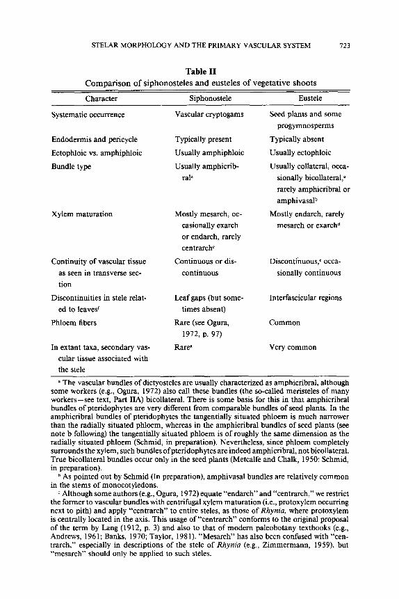
STELAR MORPHOLOGY AND THE PRIMARY VASCULAR SYSTEM 723
Table II
Compar i son of s iphonosteles and eusteles o f vegeta t ive shoots
Character Siphonostele Eustele
Systematic occurrence
Endodermis and pericycle
Ectophloic vs. amphiphloic
Bundle type
Xylem maturation
Continuity of vascular tissue
as seen in transverse sec-
tion
Discontinuities in stele relat-
ed to leaves r
Phloem fibers
In extant taxa, secondary vas-
cular tissue associated with
the stele
Vascular cryptogams
Typically present
Usually amphiphloic
Usually amphicrib-
ral a
Mostly mesarch, oc-
casionally exarch
or endarch, rarely
centrarch c
Continuous or dis-
continuous
Seed plants and some
progymnosperms
Typically absent
Usually ectophloic
Usually collateral, occa-
sionally bicollateral, a
rarely amphicribral or
amphivasal b
Mostly endarch, rarely
mesarch or e x a r c h d
Discontinuous, e occa-
sionally continuous
Leaf gaps (but some- Interfascicular regions
times absent)
Rare (see Ogura, Common
1972, p. 97)
Rareg Very common
a The vascular bundles of dictyosteles are usually characterized as amphicribral, although some workers (e.g., Ogura, 1972) also call these bundles (the so-called meristeles of many workers--see text, Part IIA) bicollateral. There is some basis for this in that amphicribral bundles of pteridophytes are very different from comparable bundles of seed plants. In the amphicribral bundles of pteridophytes the tangentially situated phloem is much narrower than the radially situated phloem, whereas in the amphicribral bundles of seed plants (see note b following) the tangentially situated phloem is of roughly the same dimension as the radially situated phloem (Schmid, in preparation). Nevertheless, since phloem completely surrounds the xylem, such bundles ofpteridophytes are indeed amphicribral, not bicollateral. True bicollateral bundles occur only in the seed plants (Metcalfe and Chalk, 1950; Schmid, in preparation).
b As pointed out by Schmid (In preparation), amphivasal bundles are relatively common in the stems of monocotyledons.
c Although some authors (e.g., Ogura, 1972) equate "endarch" and "centrarch," we restrict the former to vascular bundles with centrifugal xylem maturation (i.e., protoxylem occurring next to pith) and apply "centrarch" to entire steles, as those of Rhynia, where protoxylem is centrally located in the axis. This usage of "centrarch" conforms to the original proposal of the term by Lang (1912, p. 3) and also to that of modern paleobotany textbooks (e.g., Andrews, 1961; Banks, 1970; Taylor, 1981). "Mesarch" has also been confused with "cen- trarch," especially in descriptions of the stele of Rhynia (e.g., Zimmermann, 1959), but "mesarch" should only be applied to such steles.

724 THE BOTANICAL REVIEW
that the idea o f leaf gap is basic to Jeffrey's concept of Pteropsida [Jeffrey's other distinguishing criteria (see above) were attacked long ago--see, for example, Florin (1955) on the significance o f sporangial position].
Recognition, however, must be given to those few workers criticizing the aforenoted Jeffreyian conclusions. As early as 1924, Posthumus, after examining the steles o f many leptosporangiate ferns as well as after re- viewing the literature on the pter idosperms Lyginopteris and Heterangium and other fossil plants, concluded that " the interruption in the vascular tissue in Lyginodendron [=Lyginopteris], resulting from the insertion of the leaf-trace, is quite different f rom the leaf-gap present in the Ferns" (p. 276) and that, consequently, grouping both ferns and seed plants in the Pteropsida is "unjustif iable" (p. 281). Newman (1949), independent ly of Posthumus (1924), arr ived at similar conclusions.
Other workers also quest ioned the homology o f the interfascicular re- gions in steles o f ferns and seed plants, but did not relate this to a criticism of Jeffrey's concept of Pteropsida. For example, Bower (1935, p. 329; also 1930, p. 7 l; and Tansley, 1907-08, p. 3 l) regarded leaf gaps as indicative of merely "size-relat ions" and not phyletic relationships. Barthelmess ( 1935, pp. 246, 251) considered the interfascicular regions (which he called "prim~ire Markstrahlen") in the conifers to be o f an entirely different nature f rom the leaf gaps of ferns. Kaplan (1937, pp. 254-255) expressed agreement with Barthelmess and Pos thumus (1924). More recently, Nast (1944) and Sporne (1958), both independent ly o f each other and o f the aforenoted workers, quest ioned "whether foliar gaps, comparable to those found in siphonostelic ferns, are recognizably present in a primary vas- cular cylinder of the eustelic type" (Nast, 1944, p. 455, emphasis hers). And Sporne (1958, p. 84) would interpret the pr imary vascular cylinders of both reproductive and vegetative shoots in terms o f open and closed
a Mesarchy characterized the eusteles of some Paleozoic seed plants such as Lyginopteris, Calamopitys and Callistophyton poroxyloides (Taylor, 1981), as well as that of the progym- nosperm, Callixylon (Beck, 1979). Exarchy in a eustele is known in Callistophyton boyssetii (Rothwell, 1975).
e In pseudosiphonosteles, characteristic of some woody dicotyledons, the individual vas- cular bundles may be so close together that, superficially, they appear continuous. In some species, vascular tissue is actually continuous in the late mature condition whereas in earlier stages of development it comprised well-defined vascular or provascular bundles (see Parts IVC 1 f and IIB for references and elaboration).
f Other discontinuities such as perforations, branch gaps, and rarely, roots gaps may also occur in the stele. See text for elaboration and also Part IIIA for the distinction between "leaf gap'" and "interfascicular region."
Ogura (1972) lists secondary growth as occurring in Botrychium (see also Stevenson, 1980), Isoetes, which has atypical secondary growth (see also Foster and Gifford, 1974), and several other extant taxa. Some of these interpretations of "relictual" secondary growth are questionable and should be verified by modern techniques and concepts.

STELAR MORPHOLOGY AND THE PRIMARY VASCULAR SYSTEM 725
systems and not "in terms of leaf gaps and leaf traces as in dictyostelic ferns" (see also Sporne, 1974). Finally Govindarajalu (1961, p. 385) noted that "the non-committal term 'lacuna' is preferred to ' g a p ' . . . because doubt is cast upon the relevance of this term in relation to an axis pos- sessing an eustele."
In 1968 and 1970 Namboodiri and Beck in a series of papers (Beck, 1970; Namboodiri and Beck, 1968a, 1968b, 1968c) presented conclusions similar to, but independently of, the preceding workers and, on the basis of detailed studies of living and extinct gymnosperms, may have delivered the coup de grace to Jeffrey's concept of Pteropsida and his homology of the interfascicular regions of ferns and seed plants. The following decade saw many studies of stelar systems of both gymnosperms and angio- sperms. Central to all of these studies, which are discussed in Parts IV and VI, is the conclusion that there is no leaf gap of the filicinean type in the eusteles of seed plants and their progymnospermous ancestors and that, concomitantly, this seriously militates against the common Jeffreyian assumption (e.g., Eames and MacDaniels, 1925, 1947) that the interfas- cicular regions (so-called leaf gaps) in eusteles evolved from a siphonostele with leaf gaps by extreme dissection and reduction. [If leaf gaps of the filicinean type never occurred in eusteles of the vegetative shoots of seed plants, then it is extremely unlikely that appendage gaps of the filicinean type are present in eusteles of the reproductive structures of seed plants. Although Nast (1944), Schmid (In preparation), Schmid and Beck (1971), and Sporne (1958--see quotation above and also his 1974 work) suggested the abandonment of the term "gap" for descriptions of floral vasculature, the term continues to be used by many students of floral anatomy. A detailed consideration of the problem of "floral appendage gaps" in floral anatomy will be presented elsewhere (Schmid, in preparation). Suffice it to say here that the concept of gap was extended from stem to flower by Eames (1931; Eames and MacDaniels, 1925) and his early students (e.g., Bechtel, 1921).]
C. A SYNOPSIS OF OTHER IDEAS ON STELAR MORPHOLOGY
(1) The previous section aired some of the stelar controversies involving Jeffrey and his critics. As Hill (1906, p. 334) so aptly stated, "the majority [of persons] agree as to the facts; it is in the interpretation of the facts that disagreement exists." Thus, at the turn of the century much debate in stelar morphology centered on whether the pith of siphonosteles was phyletically extrastelar in origin, as championed by Jeffrey (1898, 1898- 99, 1899, 1902, 1903), or intrastelar, as championed by Boodle and most British morphologists, for example, Bower, Brebnar, Browne, Gwynne- Vaughan, Hill, Lang, McLean Thompson, Scott, Tansley, Wardlaw, Wors-

726 THE BOTANICAL REVIEW
dell, and many other workers, especially those concerned with pteri- dophytes (for references see Ogura, 1972). Figure 1 contrasts these interpretations of the origin of the pith. A second point of difference between the two interpretations was the presumed ancestral nature of the siphonostele (Fig. 1), amphiphloic according to the extrastelar concept, ectophloic according to the intrastelar concept. [The intrastelar concept as summarized here is usually attributed to Boodle (1901 a, 1901 b, 1903a, 1903b, but not especially 1900), who certainly presented in some detail its main points for various ferns. Thus, Hill (1906), Jeffrey (1902), Parihar (1965), Smith (1955), and other authors (e.g., Chandler, 1905; and Drab- ble, 190.5, cited in Hill, 1906) all pay homage to Boodle. Actually, Scott (1902b, 1909, 1923; Williamson and Scott, 1895) presented somewhat similar ideas for the seed plants. Scott (1902b, p. 211), however, also regarded the intrastelar idea as "Mr. Boodle's view."]
The opposing concepts are partly due to different data bases. Jeffrey, relied on development and tissue "continuity, demarcation and histolog- ical similarity" in drawing homologies, whereas Boodle and his followers relied instead on "corresponding position of the tissues in related forms," including, significantly, fossil representatives (quotations from Scott, 1902b, p. 212).
The main point of the intrastelar concept of the origin of the pith is that it represents tracheary tissue that had been phyletically transformed into parenchyma, that is, the central xylem of a protostele became pro- gressively parenchymatized to form a pith (Fig. 1) as a result of "change of destination of procambial e l e m e n t s . . . [reaching] maturity as paren- chymatous cells rather than as tracheides" (McLean Thompson, 1920, p. 715), or, as Boodle (1903a, p. 533) put it earlier, "assuming an exarch protos te le . . , by incomplete differentiation of the xylem-mass." An ec- tophloic siphonostele ("medullated protostele" in the terminology of Brit- ish morphologists--see following article by Schmid) would thus result. Boodle (1903a, p. 530--see also 1901a, p. 406) reasoned that
to admit of the insertion of a number of large arched [petiolar] bundles, the stele increased its diameter beyond the size required by the exigencies of water-conduction, and [thus] the central part of the xylem of the stele was transformed into parenchyma or other tissues [that is, especially phloem, "the two tissues appearing successively or simultaneously"-- p. 531].
Then, according to McLean Thompson (1920, p. 715),
a further ontogenetic c h a n g e . . , thus established to a solenostele by differentiation of procambial cells as sieve-tubes [sic] and endodermis within the pith i t s e l f . . . [with subsequent] establishment of direct histological connection between the intrastelar pith and the cortex through gaps in the stelar cylinder.
With the phyletic overlapping of leaf gaps a dictyostele would result. Figure 1 summarizes this scheme. Not indicated there is the possibility

STELAR MORPHOLOGY AND THE PRIMARY VASCULAR SYSTEM 727
that particularly in certain specialized ferns loss of internal endodermis and internal phloem resulted in a secondarily derived ectophloic siphono- stele. This interpretation was favored by many workers, for example, Bower (1923-28, 1930, p. 129), Browne (1908-09, p. 15), Gwynne- Vaughan (1903), Hill (1906, p. 333), Ogura (1972, p. 73), Scott (1902b, pp. 209, 212), and Tansley (1907-08, pp. 192-193); Jeffrey (1902), of course, also adopted this view, but his ectophloic siphonostele was pri- mary, not secondary (Fig. 1). A similar interpretation applied to the glei- cheniaceous Platyzoma was accepted by Browne (1908-09, p. 15) and Tansley (1907-08, pp. 142, 262), but denied by Boodle (1901b, p. 739) and Bower (1923, p. 132; 1926, p. 200).
The intrastelar concept may thus be encapsulated as follows, as McLean Thompson (1920) so aptly put it: "Medullation and solenostely [and dictyostely--p. 732] are considered resultants of direct intrastelar read- justments" (p. 715) "in which change of procambial destination is mainly operative" (p. 732).
The critical evidence for this intrastelar interpretation of the origin of the pith is the not infrequent occurrence in both extinct and extant taxa of steles that have in their central parts intermixed tracheids and paren- chyma cells, the "protostele with mixed pith" (Table I). Such steles occur in the fossil lycopods (e.g., Lepidodendron vasculare), extant ferns as Glei- chenia, seed ferns as Heterangium (Fig. 13; Part IVB2), and many other extant and especially extinct taxa, for example, Osmundaceae and zy- gopterid ferns (see Ogura, 1972). This evidence and the fact that the intrastelar concept can be expressed in developmental terms rather than metaphorically plus the corresponding lack of real evidence for Jeffrey's extrastelar concept (see Part IIIB) have provided overwhelming support for the general validity of the former concept.
The many British morphologists favoring the intrastelar concept (see list above) were concerned mainly with stelar morphology of the cryp- togams. However, it is clear that the eustele was considered derived di- rectly from the aforedescribed protostele with mixed pith (Fig. 1). As Scott (1902b, p. 21 l) indicated:
The close agreement in structure between Lyginodendron [=Lyginopteris] and Het- erangium (a genus somewhat summarily dismissed by our author [Jeffrey, 1902]) points clearly to the direct derivation of the medullated structure of the former from the protostele of the latter type.
This idea is clearly stated in Scott's later works (e.g., 1909, p. 646; 1923, p. 6 l) and more vaguely in his earlier works (Williamson and Scott, 1895, pp. 766-767; Scott, 1900, has no definite statement, contrary to indica- tions in Jeffrey, 1902, and Worsdell, 1902). Bower (1930, p. 155), Post- humus (1924, p. 272), and Tansley (1907-08, v. 7, p. 35) had similar ideas and clearly were influenced by Scott.

728 THE BOTANICAL REVIEW
Implicit in the aforenoted interpretation (except that of Posthumus, 1924) is the notion that leaf gaps evolved in the steles of seed plants. Bower (1930) and Scott (1923), however, made little or no mention of leaf gaps occurring in these plants. Nevertheless, in part due to the influ- ence of Jeffrey's concept of Pteropsida (to which the idea of leaf gaps is basic) leaf gaps from both a phyletic and strictly descriptive viewpoint came to be ascribed to steles of the seed plants (see discussion in Part IIIB). Further reduction of the primitive eustele led to the "distinguished stelar structure" (Bower, 1930, p. 155) of the specialized eustele with endarch primary xylem (see also Worsdell, 1902).
(2) In 1930 Walter Max Zimmermann proposed his famous "telome theory" and then and in a series of largely repetitive works through 1965 (see bibliography) he applied his telome concepts to various structures, including the stele, since "the stelar theory is an anatomical component of the telome theory" (Zimmermann, 1952, p. 466). The following syn- opsis (see also Fig. 1) is taken mainly from Zimmermann's 1952 and 1956 English-language accounts:
The starting point is a haplostele as in Rhynia. In more complex pro- tostelic and siphonostelic types there is a "cylinder of strand-initials," that is, "there appears at the beginning of vascular differentiation a cyl- inder of strands in the vicinity of the growing point" (Zimmermann, 1956, p. 403). "The cylinder of strand-initials is to be derived phylogenetically from the protostele of the archae-syntelomes by two elementary [phyletic] processes, viz. syngenesis [fusion] and overtopping" (1956, p. 404). "Pa- renchymatic syngenesis of meristeles," that is, "'basipetal shifting' of stelar bifurcation" occurred repeatedly to establish an axis with a varying number of "meristeles" disposed in an essentially polystelic condition. There also occurred somehow, although "it is not possible to enter into particulars," "differentiation between cauline and foliar bundles by over- topping" of the latter by the former" (Zimmermann, 1956, p. 404). "After [the] formation of the cylinder of strand-initials" xylem maturation in various directions occurred (Fig. l) to result in "the different structure of the different steles" noted below (Zimmermann, 1956, p. 405). Also im- portant phyletically were (A) direction of divergence of leaf traces, that is, radial (centrifugal), tangential, or centripetal, and (B) anastomosis of bundles ("meristeles") to establish leaf gaps (Zimmermann, 1954).
As a result of the aforedescribed "elementary phylogenetic processes of stelar construction" (Zimmermann, 1954, p. 314) three main series of phyletically derived steles are recognizable (Zimmermann, 1959, pp. I 18- 124)--see Figure l: (A) the actinostele-series, (2) the polystele- and eustele- series, with polystele, eustele, and atactostele subtypes, and (3) the si- phonostele-series, with dictyostele, solenostele, and siphonostele sensu

STELAR M O R P H O L O G Y A N D THE P R I M A R Y VASCULAR SYSTEM 729
stricto subtypes. Zimmermann emphasized in 1952 (p. 466) that his mod- ified stelar theory "differs from [established stelar theory] by no longer regarding the siphonostele as an intermediate link between protostele and polystele or eustele." However, this remark pertains only to Jeffrey's extrastelar concept since, as noted above and in Figure 1, the intrastelar concept regarded the eustele as a transformed, that is, medullated, pro- tostele.
A novel aspect of Zimmermann's scheme is that the actinostele was derived by extreme "syngenesis in the stem . . . in a radial direction," that is, by extreme radial fusion of steles in a polystelic axis (Zimmermann, 1952, p. 466). Thus in Asteroxylon the vascular strands extending from its actinostele to the bases of the phylloids ("enations," "leaves") represent vestiges of a polystelic system, that is, phyletic remnants of incomplete radial fusion in the formation of the actinostele. Other stelar concepts, of course, regard the actinostele as directly modified from the protostele.
In addition, syngenesis of"meristeles" tangentially lead to the siphono- stele-series. Whatever the directional mode ofsyngenesis, "only the meta- xylem is regularly involved . . . . whilst the protoxylem still represents the original furcated, open structure of early land-plants" (Zimmermann, 1952, p. 466). The polystelic axis evolved into the eustele by "anatomic trans- formations" of the inner, centripetal metaxylem and phloem to paren- chyma (Zimmermann, 1952, p. 467); in other words, mesarchy changed to endarchy.
The evidence for Zimmermann's telomic stelar interpretation is rather meager, relying mostly on the stelar system of the medullosan pterido- sperms (see Part IVB3). Delevoryas (1955), Stewart (1964), and Stewart and Delevoryas (1956) accepted for the medullosans Zimmermann's te- lomic concept of stelar evolution, and Stewart (1964) also accepted for various taxa the extrastelar and intrastelar concepts. Later, however, in line with the evidence presented in Part IVB3, Stewart (1976) reversed his stand, at least for seed plants, in favor of the protostelic dissection concept of Beck and co-workers (Fig. 1 and especially Part VIA).
(3) Lemoigne's (1971, especially 1967) views on stelar morphology need only brief mention since, like Zimmermann's, they are based largely on hypothetical constructs. Lemoigne (1967) derived the protostele, actually a haplostele, of the pteridophytes from the conducting system of the bryophytes, which he designated a "preprotostele." Then from the pro- tostele there are four phyletic series, a principal one and three secondary ones. The chief series extends from the protostele to the ectophloic si- phonostele with continuous vascular, tissue, thence to a "dialystele," a term of van Tieghem (1891a, p. 1372; 1898, 1918; van Tieghem and Douliot, 1886a, p. 216; 1886b, p. 277) to refer to a form ofpolystely with

730 THE BOTANICAL REVIEW
free, unfused steles ("meristeles"). A stem dialystele with exarch xylem (Sigillaria type) is postulated to have given rise to a root dialystele with exarch xylem and thence back to a stem dialystele, but one with endarch xylem (angiosperm type). Lemoigne's second stelar series involved the medullosan pteridosperms and led from a plectostelic variant to the polystelic dialystele. His third series led from the protostele to the acti- nostele to the plectostele and thence to a dialystele as in Selaginella willdenovii. A fourth series led from the protostele to the polycyclic si- phonostele. In essence, Lemoigne accepted the intrastelar concept of the origin of the pith (see Lemoigne, 1967, p. 98; 1971, pp. 44, 47), and thus it is largely his arrangement of the later stelar stages that is different from the schemes in Figure 1.
(4) Hayata (1929, 193 l) proposed his "wedge theory" ("Keiltheorie") to explain the irregular arrangement of vascular bundles ("meristeles") in the polycyclic steles of Marattiaceae, Psaroniaceae, and other ferns (see Ogura, 1972). According to Hayata, the stelar system in such taxa is not polycyclic but rather is comprised of wedge-like bundles connected to leaf traces, which are arranged around the central axis in a manner comparable to the scales on a pine cone.
(5) Finally, there is the protostelic dissection concept, which was men- tioned previously, and which was proposed independently by Posthumus (1924) and Namboodiri and Beck (1968c; see also Beck, 1970). This concept is summarized in Figure 1 and is discussed in detail in subsequent parts of this paper (see especially Part VIA).
IV. Morphology of the Primary Vascular Systems of Seed Plant Stems
A. INTERPRETATIONS BY NINETEENTH CENTURY BOTANISTS
Relatively detailed, often sophisticated, studies of the architecture of primary vascular systems of seed plants were published in the early part of the 19th century, although the vascular bundles do not seem to have been widely thought of, collectively, as comprising unified systems until after the introduction of the stelar theory of van Tieghem and Douliot (1886a, 1886b) (see Parts I and III.) The earliest of the studies described solely the course or pattern of vascular bundles, whereas later ones dealt with the relationship between vascular architecture and phyllotaxy. These studies were the work largely of German and French botanists such as von Mohl, Lestiboudois, Hanstein, Desfontaines, Frank, Marchand, N~i- geli, Bertrand, and de Bary, among others. It is of interest to note in this connection the following comment by F. O. Bower and D. H. Scott in

STELAR MORPHOLOGY AND THE PRIMARY VASCULAR SYSTEM 731
the preface to their 1884 English translation of de Bary's Comparative Anatomy of the Vegetative Organs of the Phanerogams and Ferns: "In that part of the book which deals with the arrangement of the vascular bundles (pp. 232-315), the introduction of new English terms has been especially necessary, since this part of the science has not hitherto been treated at length in any English text-book."
It is humbling to realize that many of the significant "modern" ideas relating to the development and architecture of the primary vascular system had been expressed and widely accepted long before the turn of the 20th century. Significantly, for example, de Bary (1877, 1884) made clear a distinction between primary and secondary tissues on the basis of their origin from either the apical meristem or a lateral meristem, a distinction which, if followed by some later botanists, might have pre- vented the obfuscation of knowledge about seed plant stelar patterns that resulted in this century, and that exists, even today, especially in North America (see Part IIB). The 19th century botanists also thought that primary vascular bundles develop both basipetally into the stem, and acropetally into the leaf, from sites of initiation in the bases of young leaves (leaf primordia). Indeed, this largely inaccurate observation of basipetal differentiation of leaf traces led directly to the idea in the 19th century that the vascular bundles of the stems of seed plants are essentially leaf traces, a view that has persisted among some developmental mor- phologists (Balfour and Philipson, 1962; Esau, 1977; Kaplan, 1937; Ste- venson, 1980) (see Part IIB). We know today, of course, that, with some exceptions, provascular tissue and primary phloem differentiate acrope- tally. Primary xylem on the other hand, differentiates both basipetally and acropetally from sites in the bases of leaf primordia (Esau, 1965a, 1965b). Significantly, the basic pattern of the primary vascular system is the result of acropetal differentiation of provascular tissue, as re-empha- sized recently by an analysis by Larson (1975) of the system ofprovascular strands in a bud of Populus (Fig. 23).
Early plant anatomists also understood clearly the three-dimensional architecture of primary vascular systems and accurately illustrated both open systems consisting of discontinuous cylinders of discrete sympodia, and closed systems. This comprehension is reflected in de Bary's (1877, the 1884 translation, p. 235) statement that leaf traces "are connected [to bundles in the stem] in a unilateral-sympodial manner, or in a reticulate manner by means of shanks or diverging limbs . . . . " It was also clearly understood in several botanical centers that there is a precise correlation between the architecture of the primary vascular system in the stem and the pattern of leaf arrangement (phyllotaxy) on the stem surface.
The terms "cauline bundle" and "common bundle" were widely used in the 19th century. As defined by de Bary (1877, 1884), a cauline bundle

732 THE BOTANICAL REVIEW
is one that is restricted to the stem, develops acropetally in the stem, and to which leaf traces may or may not be attached. In modem terminology this is a "sympodium" or "sympodial system" (see Part II). A common bundle is one that is common to both stem and leaf, that is, one that traverses a stem longitudinally for some distance (often many internodes) before entering a leaf. In modem terms, a common bundle would be called a leaf trace. In the 19th century, on the other hand, the term leaf trace was used in the collective sense, applying to all of the common bundles that supplied a single leaf.
Although cauline (axial) bundles and their attached common bundles (leaf traces) were described by many early botanists (e.g., de Bary, 1877; Hanstein, 1858; Lestiboudois, 1839, 1848; Niigeli, 1858), the term sym- podium may not have been used to refer to such discrete entities until 1900 when D. H. Scott used it in reference to the primary vascular system of Lyginopteris (see Part IVB2).
Of the early analyses of vascular architecture in seed plants a few stand out as especially significant. These include von Mohl's (1831, 1849) study of palms, Lestiboudois's (1848) study of the relationship between primary vasculature and phyllotaxy in seed plants, Hanstein's (1858) studies of many conifers and dicotyledons, Geyler's (1867) analysis of conifers, Nii- geli's (1858) contributions on numerous dicotyledons and other seed plants, and de Bary's synthesis and original contributions (1877, 1884).
Of the works of 19th century botanists, Carl Niigeli's (1858) detailed and generally accurate analyses made, by far, the greatest contribution to the 19th and early 20th century understanding of the morphology of the primary vascular system of seed plants. The comprehension by van Tieg- hem and Douliot (1886a, 1886b) of the unity of the axial vascular tissue was also a significant contribution, quickly accepted, but some of their more theoretical ideas unfortunately generated controversies and other hypotheses during succeeding decades that have often obscured the sig- nif icance-even the recognition--of the detailed anatomical analysis of workers such as Niigeli. For example, see the focus of the early reviews of the stele by Belli (1896), Chauveaud (1911), Hill (1906), Jeffrey (1898- 99, 1899, 1902), Meyer (1917), Schoute (1903), Scott (1894) and Tansley (1896).
N~igeli's (1858) studies of primary vasculature were made by analyzing serial transverse sections, and recording the patterns of vascular bundles in stem segments, usually terminal segments, in single planes. These pat- terns are characterized by a variable number of vascular bundles and by varying complexity of bundle arrangements. Two features stand out: (1) leaf traces and stem bundles connect unilaterally, or (2) they connect bilaterally. A vascular system of the first type consists of discrete sympodia

STELAR M O R P H O L O G Y A N D THE PRIMARY VASCULAR SYSTEM 733
of bundles (Fig. 21), and the vascular system is considered to be open. In the latter case, leaf traces are the products of the fusion of pairs of bundles (Fig. 32), and the vascular system is, thus, reticulate or closed.
Other important features recognized by Niigeli and some of his con- temporaries included (1) the presence of pairs of traces to axillary organs such as flowers and vegetative buds (Fig. 25), (2) the great longitudinal extent of leaf traces (common bundles), often traversing many internodes, (3) the helical course of vascular bundles in the stem of some plants (Fig. 21), and (4) the relationship between the internal vascular pattern and phyllotaxy, most clearly emphasized in detailed works by Lestiboudois (1839, 1848).
By far the most comprehensive and significant early study of the primary vascular systems of conifers was that of Geyler (1867). He emphasized the correlation between internal vasculature and phyllotaxy, observed the variation in the direction of trace divergence and, most significantly, recognized the discrete nature of continuing stem (axial) vascular bundles and the leaf traces connected to them.
Perhaps the earliest important contribution to our understanding of the course of vascular bundles in a major plant group was yon Mohl's (1831, and the 1849 translation) study of palms. This choice of a plant group compounded the difficulty of his task and makes all the more remarkable the general accuracy of his observations (see Part IVC2). Von Mohl ob- served that as vascular bundles traverse the stem toward the apex they follow an oblique course toward the center of the stem, through many internodes, and then turn outward even more acutely toward the surface of the stem prior to entry into the leaf bases. He observed also that each leaf trace is attached at some basal level to another vascular bundle (con- sidered by him to be another leaf trace), and that the plane of attachment, or divergence (thinking in terms of acropetal development) was radial, tangential or even oblique. Von Mohl initially thought that all of the traces to a leaf penetrate to the same depth toward the center of the stem. This is illustrated in Figure 3, which gives yon Mohl's scheme of the traces to leaves at four nodes (only two traces per leaf shown in this radial plane). De Bary (1877, 1884) pointed out, however, that the median trace and closely approximated bundles on either side penetrate more deeply than the traces farther away from the median, as illustrated in Figure 4. De Bary was quick to note, however, that his figure is inaccurate in the sense that traces do not actually follow courses in vertical planes, but rather, as Niigeli and others had shown, follow helical courses. In De Bary's (1877, p. 263) words, "each radially curved bundle runs also tan- gentially oblique, with a spiral curvature . . . . " De Bary noted that this general pattern of vasculature characterized not only the palms but many

734 THE BOTANICAL REVIEW
t
\
J
3 4
4
3,r~
Figs. 3, 4. Interpretations of the stem vascular patterns of palms. 3. Von Mohl 's (1831) scheme of the primary vascular system of palm stems. Successive nodes are numbered. (From de Bary, 1884.) 4. De Bary's (1887) modification o fvon Mohl 's (1831) scheme (Fig. 3). Successive nodes are numbered; m = median bundle. See text (Part IVA) and Tomlinson and Zimmermann (1966c) for elaboration. (From de Bary, 1884.)
GENERAL EXPLANATION FOR LONGITUDINAL DIAGRAMMATIC REPRESENTATIONS
The longitudinal diagrammatic representations of the stem primary vascular systems commonly represent the shoot vascular system split open lengthwise and spread in a plane (note, diagrams are variously drawn as if viewed from the pith or from the cortex--see Part IIB). The vascular patterns each consist of a number of sympodia comprised of axial bundles and their associated leaf traces and branch traces. In many of the diagrams successively departing leaf traces are numbered. For each dicotyledonous species depicted, Table III should be consulted for details on phyllotaxy, number of sympodia, nodal structure, and open versus closed nature of the vascular system. See Part IIB for additional explanation and caveats regarding interpretation of such diagrams.

STELAR MORPHOLOGY AND THE PRIMARY VASCULAR SYSTEM 735
other monocots as well. For more detailed discussions of early studies of palm vascular patterns see the review by Tomlinson and Zimmermann (1966c).
The studies of the 19th century structural botanists were largely de- scriptive and developmental, and there was little concern with the evo- lutionary significance of the variation in organs, tissue patterns and cells. Our modern comprehension of primitive groups, structures, and trends of evolution is the legacy of paleobotany and comparative morphology, the initial, significant impact of which came in the late 19th and early 20th centuries with the studies by a large number of distinguished British botanists, of whom in these regards, D. H. Scott and F. O. Bower made the most important contributions (see citations in bibliography).
The large fund of information about primary vasculature generated by the 19th century botanists is indeed impressive, but as more recent studies have shown, it cannot be taken at face value. Problems of technique, the restrictions of primitive instruments, and the lack of understanding of evolution during this period imposed severe limitations on this early work. The representations of vascular systems spread out in one plane were often based on relatively few (typically, free hand) sections, and were admittedly "schematic." Many lack detail and some, as shown by sub- sequent studies, are grossly inaccurate. The general lack of understanding of evolut ion--of primitive vs. more advanced or specialized groups-- severely limited the design of research projects and hence the types of conclusions that could be drawn. For example, N/igeli's (1858) study of vascular systems seems to have been approached with the intention solely of describing the vasculature of plants--any plants available--and, con- sequently, many ornamentals, including perennials and vines as well as agriculturally useful species were studied, among them Antirrhinum, Aris- tolochia, Dianthus, Dioscorea, Lathyrus, Lupinus, Parthenocissus, Passi- flora, Phaseolus, Prunus, Ribes, Vinca, Vitis, and others. Any attempt to recognize evolutionary trends on the basis of such a diverse group oftaxa would have been hopeless. The almost total lack of evolutionary theory in the mid-19th century did have one advantage: it afforded the oppor- tunity to interpret vascular patterns in the absence of the biases and prejudices of later evolutionary theorists. It is significant, for example, that we find no reference to leaf (or foliar) gaps in the vascular systems of seed plants in the works of the mid-19th century botanists, although the application of the term to fern vascular systems was widespread at this time. Not until very late in the 19th century, following the impact of paleobotanical discoveries of Carboniferous fern-like foliage, was the term applied to vascular systems of seed plants. The widespread belief that ferns were the precursors of seed plants has persisted (e.g., Eames,

736 THE BOTANICAL REVIEW
1961), even though many of the taxa of supposed fern foliage were dem- onstrated, early in the present century, to belong to primitive gymno- sperms.
In spite of early mistakes and biases, the advent of comparative anatomy and morphology in collaboration with paleobotany provided the only valid basis for interpreting the evolution and interrelationships of major taxa.
B. VASCULAR ARCHITECTURE OF PROGYMNOSPERMS AND GYMNOSPERMS
1. Progymnospermopsida
As the group from which gymnosperms (or at least, some gymnosperms) probably evolved, the Progymnospermopsida will be considered briefly. Recent major papers and reviews (Beck, 1971, 1976a, 1979; Bonamo, 1975; Scheckler, 1976, 1978) will provide more detail and lead the reader to the extensive literature on this group.
The progymnosperms, which lived from Middle Devonian into Lower Mississippian times, are comprised largely of two groups, the more prim- itive Aneurophytales, and the Archaeopteridales. [A third order, Proto- pityales, contains only two species, Protopitys scotica and P. buchiana (see Beck, 1976a).] The aneurophytes, consisting of Aneurophyton, Cai- roa, Proteokalon, Rellimia, Tetraxyloteris and Triloboxylon, are charac- terized by pseudomonopodial branching. Ultimate appendages are di- chotomous and non-laminate. All genera for which internal anatomy is known, are characterized by three or four-ribbed protoste!es which, in several genera, retain their form in successive orders of branching except in the ultimate appendages in which the primary vascular system is un- ribbed and circular in transverse section (Fig. 5). In Proteokalon, however, the stele of first-order branches is four-ribbed, whereas that of second- order branches is three-ribbed (Scheckler, 1976; Scheckler and Banks, 1971). The main axis of Cairoa (Matten, 1973), characterized by a three- ribbed stele is believed to give rise to laterals with four-ribbed steles. The primary xylem of aneurophytes, mesarch in order of maturation, is char- acterized by numerous internal protoxylem strands. Traces depart from the edges of stelar ribs following radial division of the outermost proto- xylem strands. Arrangement of lateral appendages tends to reflect the morphology of the stele--that is, branches and ultimate appendages (leaves) are decussate on axes with four-ribbed steles, and helical on axes with three-ribbed steles, except that in Proteokalon pairs of adaxial leaves alternate with single abaxial leaves. Although characteristics of branching, ultimate appendages, stelar morphology, and fertile structures indicate that the Aneurophytales was a relatively primitive group, it was, never- theless, like the Archaeopteridales, characterized by secondary growth.

STELAR MORPHOLOGY AND THE PRIMARY VASCULAR SYSTEM 737
Z
Z 0
o
I1
I t~ ubr
I i r ~ l
(~1.
U tr@ I OROTEOKALON
CAPROA
p b~ TETRAXYLOPTERIS
TRILOBOXYLON
5 Fig. 5. Transverse sections of various progymnosperms (Aneurophytales) showing
branching and histological patterns. Lined secondary xylem encloses metaxylem (unshaded) containing protoxylem strands (in black). Geologic ranges are indicated by vertical lines. 1. = leaf; 1. tr. = leaf trace; p. br. = penultimate branch; u. hr. = ultimate branch. (From Beck, 1976, by permission.)
In many features, the Archaeopteridales appear advanced by compar- ison with the Aneurophytales. In Archaeopteris; for example, leaves (ul- t imate appendages) are laminate, sporangia are borne adaxially on prob- able leaf homologues rather than terminally, pits in the secondary xylem are restricted largely to the radial walls, and the primary vascular system in the main stems (Callixylon) as well as in the penultimate and ultimate axes of the planated lateral branch systems, is a eustele (Beck, 197 l, 1979; Scheckler, 1978).
The Ca/lixylon eustele (Fig. 8) consists of a variable number of vascular bundles ranging from about 15 to 40 (Beck, 1979). Traces depart in a regular helical sequence in a 2/5 or 3/8 phyllotaxy in axes with the smaller number of bundles and 5/13 in those with the larger number (Beck, 1979). Trace departure in Callixylon is radial (Fig. 8)--a feature unusual in a eustele, and which probably reflects its protostelic origin (see Part VIA).
In penultimate and ultimate axes of Archaeopteris, the primary vascular system is interpreted as a eustele (Scheckler, 1978) on the basis of the distribution ofprotoxylem strands, although these are connected by meta- xylem in more basal and more apical regions and may surround a pith (Fig. 6). In these orders of branching the stele is ribbed, and each rib contains a single protoxylem strand (Beck, 197 l; Carluccio et al., 1966; Scheckler, 1978). The number ofprotoxylem strands, the diameter of the

738 THE BOTANICAL REVIEW
0 . 1 r a m . . o
~ m
7
31 L:~21 �9 32 2O
�9 33 19
34 =8
35 17
37 ~d~ 14
m m
13 20
2 3 I0
4 5 9
7 ~
6 Figs. 6-8. Aspects of the anatomy of Archaeopteris and Callixylon (the main stem axis
of Archaeopteris), an Upper Devonian/Lower Mississippian progymnosperm. 6. Left: trans- verse sections of the primary xylem (black) of an ultimate branch of A. macilenta. Right: longitudinal reconstruction of primary xylem (black) showing positions of the sections. Note that the stele is medullated only in its broader basal part. (From Scheckler, 1978, by per- mission.) 7. Reconstruction of a transverse section of a penultimate axis of A. cf. macilenta with a conspicuous pith. Strands or ribs of primary xylem, essentially triangular in transverse configuration, each contain a single protoxylem strand (black). Secondary xylem is lined. Leaf traces (1. tr.) and branch traces (br. tr.) diverge in helical succession from every other rib (or strand) in the same ontogenetic spiral. Note that leaf and branch traces diverge radially, as from a protostele. Leaf traces dichotomize in the decurrent leaf bases (1. b.). (From Beck, 1971, by permission.) 8. Transverse section of the primary vascular system

STELAR MORPHOLOGY AND THE PRIMARY VASCULAR SYSTEM 739
stele, and the degree of medullation increase from the base toward the mid-region of the axis and diminish from that region toward the tip (Scheckler, 1978) (Fig. 6). These changes are accompanied by changing phyllotactic fractions. It is in the mid-regions of penultimate axes that the stele is most conspicuously eustelic, consisting of separate or only partially connected primary xylem strands (Fig. 7). As in Callixylon (main stems of Archaeopteris) traces depart radially following division of the protoxylem strands (Fig. 7). Traces in lateral branch systems are of two types--smaller leaf traces, and larger branch traces. Both types occur in the same ontogenetic spiral (Fig. 7), indicating that branching in Ar- chaeopteris is pseudomonopodial (i.e., not axillary) (Beck, 1971, 1979; Scheckler, 1978). Traces to ultimate branches are commonly opposite to subopposite or even alternate in one plane (i.e., in two orthostichies on opposite sides of the axis) suggesting that the lateral branch systems were planated (Beck, 1971). Scheckler (1978) has demonstrated, however, that in some parts of some lateral branch systems ofArchaeopteris macilenta there may be three or more orthostichies of branch traces. In regions where this condition occurs, the additional branch traces diverge from the adaxial side of the stele, suggesting that some ultimate lateral branches were pendulous.
Branch traces in a stem segment of Callixylon brownii are regularly arranged in a 5/13 "phyllotaxy" and thus conform to a common Fibonacci pattern (Beck, 1979) (Fig. 9). In the axes of lateral branch systems in which leaf and branch traces develop in the same ontogenetic spiral, however, phyllotactic fractions are difficult to determine and may be highly variable along a segment of axis (Beck, 1971; Scheckler, 1978) because of some irregularity in the arrangement of lateral appendages as well as the determinate nature of the lateral branch systems (Beck, 1971; Scheckler, 1978). The occurrence of phyllotactic fractions such as I/7 and 2//9 which reflect the atypical arrangement of lateral appendages, might be explained, according to Scheckler (1978), by assuming that leaf and branch primordia were of different sizes and were initiated at different plasto- chronic intervals. As noted by Scheckler (1978), this condition is not unexpected in a plant such as Archaeopteris--a Devonian progymnosperm that had not, in all aspects, attained the level of gymnosperm organization. The radial divergence of traces in the Archaeopteris eustele, like that in some calamopityans (see Part IVB2), may, likewise, simply reflect an
(black) of stem of Callixylon brownii. The system is a eustele in which the radial divergence of traces apparently reflects its origin from a protostele. The larger numbers (9-21) indicate, in reverse order, the sequence of trace departure. (From Beck, 1979, by permission.)

740 THE BOTANICAL REVIEW
IO V
Ii
22 ~7
12 ~7
P I ' " ' I ' I I
I I I I I / I I I I I I I I I I I I I I I
I I I I 11 1 I
9
i
I0 20 50 39
25
24 ~7
9 Fig. 9. Longitudinal diagrammatic representation of stem primary vascular system of
Callixylon brownii. Traces observed are indicated by black triangles; those predicted, by unshaded triangles. Traces are numbered in order of development along the ontogenetic spiral. Dashed lines represent regions where the continuity of the bundles was broken by poor preservation. (From Beck, 1979, by permission.)
intermediate evolutionary level (Beck, 1979) or, as suggested by Rothwell (1976a), the possible independent origin of the eustele in the archaeopterid progymnosperms.
2. Pteridospermopsida: Lyginopteridaceae, Callistophytaceae and Calamopityaceae
An early paleobotanical contribution of utmost importance to our un- derstanding of the nature of the stele (primary vascular system) of seed

STELAR MORPHOLOGY AND THE PRIMARY VASCULAR SYSTEM 741
plants was made in 1900 by D. H. Scott (and expanded later in his 1923 work). Scott's analysis of the stele of Lyginopteris, a primitive gymno- sperm (pteridosperm) of the Lower Carboniferous, was largely overlooked, or its significance not fully recognized, by many more recent morpholo- gists. Major exceptions, however, were Posthumus (1924) and Hirmer (1933) whose ideas, based in part on this work were, themselves, largely ignored until recently (see Parts IIIB and VIA). Zimmermann (1930, 1954, 1956, 1959, 1965) also discussed the primary vasculature of Lyginopteris, in fact, both reproducing some of Scott's (1923) diagrams (see our Fig. 12) and presenting similar ones based on new data. However, Zimmer- mann interpreted the stele of Lyginopteris in the context of his telome theory (see Part IIIC and Fig. 1), concluding (Zimmermann, 1956, p. 407) that "the whole stele reveals evidently the original dichotomous organi- zation of the protostele in Psilophyta."
D. H. Scott, who with F. O. Bower had earlier translated de Bary's great Comparative Anatomy of the Vegetative Organs of the Phanerogams and Ferns was, as a result, fully cognizant of the analyses of vascular systems of N~geli and other continental workers. Scott (1900, and especially, 1923), utilizing the same method in analyzing the stelar pattern of Ly- ginopteris, prepared the first detailed diagram of the vascular pattern of a Carboniferous seed plant (Fig. 12b). The essential accuracy of this pat- tern has been corroborated by Hirmer (1933), Zimmermann (1930, 1959, 1965) and, more recently, by Blanc-Louvel (1966). Scott in 1900 dem- onstrated in Lyginopteris a remarkably simple vascular system consisting of a cylinder of five discrete, longitudinal vascular bundles which divide tangentially at regular intervals to produce traces. In his words, a strand which thus divides constitutes "a sympodium composed of the united lower ends of successive adjacent leaf-traces" (Scott, 1900, p. 314). As far as we can determine, this is the first published application of the term "sympodium" to a discrete unit of a seed plant stele. The concept of sympodium in the context of the comparative, evolutionary anatomy of seed plant steles, has been broadened to encompass the continuing axial bundles and the attached leaf traces (see, for example, Beck, 1970; Crafts, 1943; Devadas and Beck, 1972; Esau, 1965a, 1965b; Posthumus, 1924; and Part IIA of this paper). Following divergence from an axial bundle, each leaf trace divides again prior to entry into a leaf base. Scott (1900, 1923) recognized the correlation between internal vascular architecture and leaf arrangement and determined for Lyginopteris a phyllotaxis of 2/5.
An essentially identical pattern occurs in another Paleozoic seed fern, Callistophyton (Rothwell, 1975), and similar patterns have been dem- onstrated in the calamopityacean genera, Bilignea, Calamopitys, and Eristophyton (Beck, 1970; Galtier, 1970, 1973; Scott, 1902a, 1925). [Bi-

742 THE BOTANICAL REVIEW
Figs. 10, 11. Suggested stages in the evolution of the eustele by longitudinal dissection of a protostele. 10. Suggested stages in the evolution of the eustele by longitudinal dissection of a protostele, as illustrated by transverse sections of pteridosperm stems ranging in age from Lower Mississippian (Calamopityaceae, Figs. a-e) to Pennsylvanian (Lyginopteris). This series is a typological one for steles and should not be construed as necessarily indicating phyletic relationships among the taxa. The sequence of letters (A-E) indicates the direction of the ontogenetic spiral and the positions at which leaf traces will originate in succession along it. In each figure the outer line denotes the outer boundary of the secondary xylem whereas primary xylem is shaded. All taxa have z/s phyllotaxy and lack leaf gaps. Compare Figure 10d-f with 1 lc-e, respectively. (a) Protostele 3-flanged, undissected. (b) Protostele

STELAR MORPHOLOGY AND THE PRIMARY VASCULAR SYSTEM 743
lignea and Eristophyton are considered by some to be closely related to the Cordaitales (see e.g. Lacey, 1953).] In a typical t ransverse section of Callistophyton there are 9 to 13 p r imary vascular bundles at the margin of the pith. These consist o f axial bundles and associated leaf traces. The latter are produced by single divisions o f axial bundles, and each trace divides again before entering the base o f the leaf. Lea f traces are produced in a 2/5 helix. As in Lyginopter&, lateral branches are axillary, and the origin o f branch traces conforms to the typical pat tern in woody seed plants (Rothwell, 1976a). Bundles that flank the posit ion of leaf trace emiss ion divide, providing two slender traces that enter the base o f the axillary branch. These vascular bundles divide repeatedly, producing leaf traces and increasing the n u m b e r of axial bundles in the branch. Traces are first produced in a 1/2 helix, followed successively by a 1/3 and then a 2/s pattern.
Another pr imi t ive g y m n o s p e r m o f significance in the evolut ion o f ideas on stelar morpho logy is the protostelic p ter idosperm, Heterangium (Hirmer , 1933; Pos thumus , 1924; Scott, 1917, 1923; Stidd, 1979). Het- erangium shows considerable variat ion in stelar architecture. Some species have a xylem with tracheids interspersed with pa r enchyma (e.g., H. grie- viO. Species o f this type are often assigned to the subgenus Eu-heteran- gium, and are considered to be the mos t p r imi t ive m e m b e r s o f the genus. Other species have distinct strands o f tracheids at the per iphery o f the stele that contr ibute to leaf trace product ion. These species are considered to be a specialized end group, and are placed in the subgenus Polyangiurn (Scott, 1917, 1923). Still other species o f Heterangium have both distinct leaf-trace producing bundles at the per iphery of the xylem, and a great deal o f pa r enchyma at the center o f the stele. These latter steles approach the structure o f seed plants with a true pith, and the species are assignable to the subgenus Lyginangium (Scott, 1923). Specimens of the Lyginan-
longitudinally dissected into three columns separated by parenchyma. (c) Protostele with "mixed pith," i.e., with highly parenchymatous central region containing scattered tracheids; compared to Figure 10b there is more central parenchyma and a clearer definition of five nearly discrete sympodia. (d) Transitional type of stele with distinct pith containing only a very few scattered tracheids; compared to Figure 10c the five sympodia are still more clearly defined (see Fig. 1 lc). (e) Eustele with distinct pith and with five discrete sympodia, each consisting of an axial bundle and its diverging leaf traces and accessory bundles (see Fig. 1 ld). (f) Eustele very. similar to that in Figure 10e but differing chiefly in lacking accessory bundles (see Fig. 1 Ie), see also the representations in Figure 12, in which the ontogenetic spiral is the reverse of that in Figure 10f. (From Beck, 1970, by permission of Cambridge University Press.) 11. Series of diagrams showing possible evolutionary change leading from radial divergence (a--d) to tangential divergence (e) of the leaf trace. Figure 1 lc (Calamopit.vs sp.) and Figure 1 ld (C. foerstet) are intermediate, showing loss of the anastomosis. (a) = C. embergeri; (b) = C. el. embergeri; (e) = Lyginopteris. (From Galtier, 1973, by permission.)

744 T H E B O T A N I C A L R E V I E W
3 8
18 Y
2
10 /
?y 15
/ j
7 4 9
III IV V
11
7
16
/
12 Fig. 12. Lyginopteris oldhamia, a Carboniferous seed fern. (a) Transverse section of
stem. Inner circles = primary vascular tissue of eustele (axial bundles shaded, leaf traces unshaded); outer circles = leaf traces after their divergence from the stele; lined areas = secondary xylem. Note the tangential divergence of the leaf traces and their distal bifurcation. (b) Longitudinal diagrammatic representation of the primary vascular system as illustrated in one plane. The dashed line indicates the level of the section in Figure 12a as unfolded at the wedge at its lower left. Triangles = leaf traces. (Diagrams modified from Scott, 1923, and reproduced from Beck, 1970, by permission of Cambridge University Press.)
gium type are quite similar to Lyginopteris, and the series Eu-heteran- gium-Lyginangium-Lyginopteris has been central to discussions of the evolution of seed plant eusteles for over fifty years (see Scott, 1923; Stidd, 1979). Most of what is known about the three-dimensional architecture of the primary vascular system and the mode of leaf trace production in Heterangium comes from an investigation of H. kukuki by Hirmer (1933).
Transverse sections of the protostele of Heterangiurn kukuki, a species of the subgenus Polyangium, reveal plates of parenchyma interspersed among the tracheids. Some of the plates radiate from the center of the axis, while others are more tangentially disposed (Fig. 13). Protoxylem strands (unshaded regions in Fig. 13) occur peripherally, enclosed by metaxylem. By examining serial sections Hirmer (1933) was able to follow individual leaf trace bundles downward from the level of entry into leaf bases to a point approximately eight internodes below. At more proximal
----'9
Figs. 13-15. Diagrammatic representations of the stem vascular patterns of two Paleo- zoic gymnosperms. 13, 14. Heterangium kukuki, a Carboniferous seed fern. 13. Transverse section of protostelic vascular column of stem. Large numbers (1-8) = the clearly recogniz- able leaf traces (position of leaf trace l only indicated as it has already diverged); small numbers (9-34) = leaf traces departing at higher levels in the stem. Stippled = parenchyma; shaded = metaxylem; unshaded = protoxylem (primary xylem maturation is mesarch). 14.

STELAR MORPHOLOGY AND THE PRIMARY VASCULAR SYSTEM 745
Longitudinal diagrammatic representation of stelar surface showing pattern of leaf trace divergence. Horizontal lines = nodes; dashed lines = presumed downward course of leaf traces. (From Hirmer, 1933, by permission ofE. Schweitzerbart'sche Verlagsbuchhandlung.) 15. Medullosa primaeva, an Upper Carboniferous seed fern. Transverse section of stem. The vascular cylinder is a eustele and not phyletically the polystele of most previous inter- pretations (see text, Part IVB3, for elaboration). Lined areas = secondary xylem; shaded = metaxylem; unshaded = protoxylem (primary xylem maturation is endarch). (From Basinger et al., 1974, by permission.)

746 THE BOTANICAL REVIEW
levels traces could not be identified by structural features, but by mea- suring around the stele in successive increments of 137~ (the limiting angle) from recognizable leaf traces, he plotted the downward courses of the bundles (dashed lines in Fig. 14) for an additional 26 internodes. In Figure 14 no two leaf trace complexes are directly superposed, but parasti- chies are clearly visible. Leaf traces are not shown as interconnected into sympodia because Hirmer was unable to provide conclusive evidence on this matter. A similar, apparent absence of sympodia has been reported by Stidd (1979) in Heterangium lintonii.
At the lowest node where leaf traces of H. kukuki can be identified they consist of a single protoxylem strand and surrounding metaxylem. Dis- tally, the protoxylem dichotomizes and the bundle divides in two. A subsequent dichotomy in each of the resulting bundles produces four strands that enter the base of the leaf (Fig. 14).
In simpler protostelic species such as H. grievii the vascular tissue is represented by a large rod of xylem from the surface of which traces diverge to the leaves. The traces are, therefore, connected to a common core of cauline xylem, as in H. kukukL rather than in sympodia. The transition from a protostelic to a eustelic stem structure in Heterangium is considered by Hirmer (1933) to have been accomplished by the evo- lution of increasing amounts of parenchyma at the center of the stele, and by the progressive organization of sympodia of interconnected leaf traces at the periphery, as reflected, possibly, in H. shorense and H. hoppstaed- teri. The lack of evidence of continuity between protoxylem strands (to form sympodia) in H. kukuki and in H. lintoniL however, tends to weaken Hirmer's proposal (see Stidd, 1979).
The Calamopityaceae have not been proven to be seed plants, but available evidence suggests that they were probably pteridosperms, closely related to the Lyginopteridaceae (Galtier, 1970, 1974; Taylor, 1981). The Calamopityaceae are of particular interest because a series of genera and species seem to provide information significant to a better understanding of the origin of the eustele (Beck, 1970; Galtier, 1973; Figs. 10, 11). Protostelic forms such as Stenomyelon primaevum Long (1964), S. tue- dianum (Fig. 10b), and two unnamed new species of Stenomyelon (Fig. 10a) discussed by Beck (1970) are characterized by a three-ribbed stele, but by the production of leaves in 5 orthostichies and a 2/5 phyllotaxy (Beck, 1970). Other species such as Calamopitys americana Scott and Jeffrey (1914) (Fig. 10a), C. embergeri Galtier (1970), and an unnamed, new species of Calamopitys (Figs. 10d, 1 lc; Beck, 1970) are also char- acterized by a 2/5 phyllotaxy and exhibit characteristics intermediate be- tween a protostele and a true eustele. For example, the steles of C. amer- icana and C. embergeri may be described as protosteles with "mixed piths" in which tracheary elements are scattered throughout the central

STELAR MORPHOLOGY AND THE PRIMARY VASCULAR SYSTEM 747
region of the steles. In all three species the more peripheral parts of the primary vascular system are organized into columns of vascular tissue, interconnected in various degrees, from which traces diverge (Fig. 11). Calamopitysfoerstei Read (1937) is characterized by a true eustele (Beck, 1970). The homogeneous pith, devoid of tracheary tissue, is enclosed by five sympodia (Fig. 10e). Although they are discrete, their probable origin from a stelar system like that of C. americana and similar forms is reflected in the small accessory strands that diverge from the leaf traces just prior to their separation from the axial bundles. Instead of fusing with adjacent bundles as in C. americana, C. embergeri, or the unnamed species of Calamopitys, the accessory strands of C. foerstei terminate a short distance from their points of origin (Figs. 10e, 1 ld; Beck, 1970). The stele of C. foerstei is, therefore, essentially identical to that of Lyginopteris (compare Figs. 10e and I 1 d with 1 Of and I 1 e).
Among these members of the Calamopityaceae, therefore, as (less clear- ly) among species of Heterangium, there is an intergradation of stelar forms between a typical protostele and a typical eustele. These observa- tions will be discussed in relation to the evolution of the eustele in Part VIA.
Scott (1902a--see also 1923) demonstrated a eustele, which he consid- ered to be essentially identical with that of Lyginopteris, in a new species to which he assigned the name, Calamopitysfascicularis. This species was subsequently transferred to Eristophyton by Zalessky (191 l; see also La- cey, 1953).
Bilignea solida (Scott, 1925) has an unusual stele, consisting of a dis- continuous cylinder of primary xylem strands embedded in the periphery of a solid column of cells which are parenchyma-like in form, but tracheid- like in pitting characteristics. The peripheral strands are arranged in a pattern similar to those of Calamopitysfoerstei and Lyginopteris oldham- ia. In contrast to the highly parenchymatous manoxylic secondary xylem of Calamopitys and Lyginopteris, however, Eristophyton and Bilignea are both characterized by sparsely parenchymatous pycnoxylic secondary xy- lem. Lacey (1953) suggested that these, and other similar pycnoxylic forms, might be more closely related to the Cordaitales than to the Calamopit- yaceae, but that in the absence of fructifications no definite taxonomic assignment could be made.
3. Pteridospermopsida: Medullosaceae and other "polystelic" forms
Unique or unusual forms of vascular architecture are found within several major fossil and extant groups. Stelar configurations consisting of two or more cylinders ("polystely") characterize the medullosan seed ferns (Medullosaceae), which extend from Mississippian through Permian time.

748 THE BOTANICAL REVIEW
Incompletely known polystelic taxa are also present in Triassic (Rhex- oxylon) and Jurassic (Pentoxylon) strata, and similar stelar configurations occur in some genera of living cycads (Greguss, 1968; Pant, 1973). While the features of these taxa have been of botanical interest for some time, a paucity of material has hampered most studies and despite often critical observations (e.g., Delevoryas, 1955) has led to highly speculative con- clusions. As a result, two conflicting interpretations of the structure and evolution of polystelic taxa are prevalent in the literature. These are focused primarily on the genus Medullosa and have recently been re- viewed by Stewart (1976).
Some authors interpret Medullosa to be both structurally and evolu- tionarily polystelic (Delevoryas, 1955; Scott, 1899, 1923; Stewart and Delevoryas, 1956; Zimmermann, 1930) while others consider the same stems to be polystelic in only a descriptive sense, i.e., the result of a somewhat unusual appearance of a single stele (Basinger et al., 1974; Namboodiri and Beck, 1968c; Roberts and Barghoorn, 1952; Scott, 1909). Only recently has the vascular architecture of Medullosa been investigated by the same methods as pseudosiphonostelic (see Part IVC1 f) and pro- tostelic forms (e.g., the woody Ranales, Benzing, 1967a, 1967b; Schop- fiastrum, Rothwell and Taylor, 1972; Stidd and Phillips, 1973), and its stelar features more fully understood (Basinger et al., 1974). By concen- trating on the protoxylem architecture and leaf trace emission pattern of Medullosa, one discovers that this genus is only superficially different from more typical monostelic plants. In Medullosa the primary xylem is arranged as a single cylinder of more or less interconnected bundles with several individual bundles or interconnected groups of bundles (Fig. 15) surrounded by cylinders of secondary vascular tissue. The sympodial structure of the primary vascular system can be demonstrated by iden- tifying and characterizing the protoxylem strands. These are arranged at the periphery of the primary xylem zone, and typically number 3, 5, 8 or 13, as do the axial bundles of many other eustelic seed plants (see, for example, Parts IVB5 and IVC1). A large number of traces that supply a single leaf diverge over a considerable longitudinal extent from two or more adjacent "axial bundles" (i.e., protoxylem strands and associated metaxylem and primary phloem) in a consistent and coordinated manner. These "axial bundles" from which traces to a leaf diverge may be com- ponents of a single "stele" or of separate, adjacent "steles" (using the terminology of many earlier interpretations).
The basic similarity of the primary vascular tissues in Medullosa and most other known gymnosperm taxa now eliminates the need to interpret medullosan vascular architecture as fundamentally different from that of typical seed plants. Although the occurrence of a medullosan (Sutcliffia)

STELAR M O R P H O L O G Y A N D THE P R I M A R Y VASCULAR SYSTEM 749
with only one or two groups of interconnected bundles may sugg_ est an independent origin of dissected steles in this group (Basinger et al., 1974), stelar evolution appears to have paralleled that of other known gymno- spermous lines (Beck, 1970).
The vascular architecture of Pentoxylon and Rhexoxylon have also recently received some attention, and preliminary investigations suggest that they too may have eusteles obscured by an unusual disposition of secondary vascular tissue (Bose and Stewart, 1974; Stewart, 1976), a situation, incidentally, which occurs in a number of angiosperms with so- called anomalous secondary growth (Esau, 1965a; Fahn, 1974; Metcalfe and Chalk, 1950). In the Jurassic Pentoxylon (Srivastava, 1945) there are several (usually five) groups of primary xylem strands that are surrounded by secondary vascular tissue. Each of these vascular units contains two independent, cauline, protoxylem strands; and the traces to a single leaf arise by the division of a protoxylem strand in each of two adjacent vascular units (Fig. 11 of Stewart, 1976). Leaves are arranged in a spiral, and leaf trace emission patterns indicate a 2/5 phyllotaxy in many stems.
A similar disposition of vascular tissue and mode of leaf trace emission are possibly present in the Triassic Rhexoxylon (Archangelsky and Brett, 1961; Bancroft, 1913; Stewart 1976), but additional details of the stelar structure in this genus await further investigation.
The above summary suggests that, in seed plants at least, the expression "polystely" has validity only in a descriptive sense since there is appar- ently no fundamental difference between the vascular architecture of monostelic and polystelic seed plants. Consequently, and particularly be- cause of the phyletic implications of the term "polystely," we think that this expression, like "meristele" (see Part IIA), should be avoided in botanical parlance. ["Polystele" is also used in reference to the steles of pteridophytes (e.g., see Ogura, 1972). The problem of polystely here too should also be reconsidered from a terminological, developmental, and comparative anatomical viewpoint.] There is also additional terminolog- ical confusion between "polystelic," "polyxylic," and "polycyclic." Strict- ly speaking, a polystelic condition results from the presence in an axis of two or more adjacent cylinders of vascular tissue (Fig. 15), but the term has also been applied to polycyclic vascular cylinders, that is, concentric cylinders of vascular tissue. The latter, in particular, have been designated "polyxylic," for example, in cycads by Greguss (1968) and Pant (1973), so that "polystelic" and "poiyxylic" are synonyms, as are "monostelic" and "monoxylic." Some of these "polyxylic" steles, however, actually involved only secondary vascular tissue in the accessory steles (e.g., some cycads), which is yet another terminological anomaly since stelar termi- nology properly refers only to primary tissues.

750 THE BOTANICAL REVIEW
4. Cordaitales
The primary vascular structure of Cordaitales is of particular interest because of the close and possibly ancestral relationship of this group to the conifers (Arnold, 1948; Florin, 1951--but see also Beck, 1981 for another viewpoint). Transverse sections of cordaitean stems reveal a ring of primary vascular bundles at the margin of the pith, and leaf traces that typically depart from the stelar region in pairs. The longitudinal course of the bundles, however, has been difficult to interpret and, in vegetative organs, is still unclear. In a recent study of North American and European specimens, Whiteside (1974) has described two distinct patterns of pri- mary vascular bundles. In Mesoxylon pairs of leaf traces diverge from the stelar region as prominent, mesarch strands. They decrease in size basipetally, however, and do not seem to extend very far longitudinally or to connect with other vascular bundles. As a result, there may be no sympodial system of primary vascular strands in Mesoxylon.
Specimens described as Pennsylvanioxylon exhibit a rather large num- ber of endarch bundles at the margin of the pith. In transverse sections these appear to be arranged into several groups. According to Whiteside (1974) individual bundles may divide or fuse from level to level, they may shift from one group to another, or they may extend offas leaf traces. However, in an ongoing investigation of Upper Pennsylvanian cordai- taleans we have encountered several specimens of Pennsylvanioxylon that have a primary vascular architecture that conforms to the pattern found in extant conifers with helical phyllotaxis (Namboodiri and Beck, 1968a; see Fig. 16).
Branching in the cordaites is axillary, and branch traces arise as two bundles that flank the position of the subtending leaf traces (Traverse, 1950). Some specimens have two buds or branches in the axil of a leaf (Baxter, 1959). The possible significance of this variation is not presently understood. In the secondary fertile shoots of the cordaitean microspo- rangiate strobilus, Cordaianthus concinnus Delevoryas (1953) the two branch traces form a single cylinder of vascular tissue at the base of the axis, and then, at successively more distal levels, divide to form axial bundles and leaf traces like those of typical eustelic seed plants. Near the base of the axis there are two sympodia from each of which diverges a single trace to each modified leaf in a 1/2 helix. Distally the number of axial bundles increases to 3, 5, and finally 8. At correspondingly more distal levels the phyllotaxis, as reflected in trace arrangement, changes to 1/3, 2/5 and 3/8 (Rothwell, 1977). The vascular architecture in Cordaianthus concinnus is therefore identical to that found at the base of branches in extant conifers (Rothwell, 1976a; Sterling, 1945) and together with the recently discovered features of some Pennsylvanioxylon specimens dem- onstrates the similarity of vascular architecture in the two orders. At the

STELAR MORPHOLOGY AND THE PRIMARY VASCULAR SYSTEM 751
I
6 0 ,
2o
16 18
6b.
5b
5a 5b 50
3c a i a
I b\ llo / 1
Figs. 16-18.
a b G
17 Longitudinal diagrammatic representations of stem primary vascular pat-
terns of some extant gymnosperms. 16. Abies concolor. Triangles = leaf traces. (From Nam- boodiri and Beck, 1968a, by permission.) 17. Diagrams representing proposed stelar evo- lution in Cupressaceae. Figure 17b is intermediate; it resembles the primitive type in Figure 17a in having an open vascular system with one trace per leaf, but it resembles the derived closed vascular system in Figure 17c in having both sinistrorse and dextrorse divergence of leaf traces. Triangles = leaf traces. (From Namboodiri and Beck, 1968b, by permission.) 18. Ginkgo biloba. The two traces (light lines) entering each leaf are derived from separate adjacent axial bundles (heavy lines). (Redrawn from Gunckel and Wetmore, 1946b, by permission.)

752 THE BOTANICAL REVIEW
present time the significance of the apparently unusual vascular patterns described by Whiteside (1974) in vegetative cordaitean stems is not clear.
5. Coniferales
Several studies of the primary vascular systems of conifers demonstrate convincingly the basic similarity of conifer stelar patterns and those of primitive Paleozoic gymnosperms. The earliest comprehensive survey of the stele in conifers is that of Geyler (1867) although Frank (1864), Han- stein (1858), Lestiboudois (1848), and N~igeli (1858) had made earlier important contributions. Geyler's findings were expanded by Barthelmess (1935) who analyzed a large number of conifers and demonstrated in great detail the architecture of the primary vascular system and its precise correlation with phyllotaxy. Barthelmess interpreted the stelar systems of all taxa with helical leaf arrangement studied to be characterized by open systems consisting of discrete sympodia (Fig. 16). He suggested, further- more, that in the Cupressaceae, with opposite or verticillate leaves, there is a transition between open systems and closed, reticulate systems (Fig. 17a, b, c), a conclusion more recently corroborated by Namboodiri and Beck (1968b) and Pillman (1978). Barthelmess' important study was gen- erally overlooked (perhaps because of the disruptions and distractions of World War II), possibly ignored, or its conclusions were not widely ac- cepted by comparative morphologists and anatomists. Certainly it failed to have the impact it deserved. Barthelmess's interpretations of the vas- cular patterns of conifers did gain some support from the work of Crafts (1943) and Sterling (1945, 1947), but these largely developmental studies failed to support his view ofbasipetal differentiation ofprovascular tissue. Indeed, both Crafts and Sterling demonstrated conclusively that in Se- quoia and Pseudotsuga provascular tissue differentiates solely acropetally.
Not until the 1960s was there an original study (Namboodiri and Beck, 1968a, 1968b, 1968c) designed to gain some insight into the evolution of stelar patterns in conifers by use of the methods of comparative anat- omy in relation to the paleobotanical record. Namboodiri and Beck were also concerned with the reality of the sympodial systems illustrated by Geyler and Barthelmess because of the close association--indeed, the frequent contact--between axial bundles. In other words, they sought to determine whether or not axial bundles maintained their identity through regions of lateral contact with other bundles. By careful observational studies involving cell counts and a statistical analysis, they demonstrated that, although the discreteness of the axial bundles may be obscured by their undulation and lateral contact with adjacent bundles, their identity is maintained. The validity of this interpretation has been supported by an application of similar methodology in a recent comprehensive study

STELAR MORPHOLOGY AND THE PRIMARY VASCULAR SYSTEM 753
of stelar morphology in the Cupressaceae (Pillman, 1978). Namboodiri and Beck also showed that whereas two traces may be apparent at nodal levels, in conifers with helical phyllotaxy the trace supply to a leaf orig- inates as a single bundle. As had others before them, they demonstrated that traces enter leaf bases in patterns that reflect the following fractions of the principal series of Fibonacci numbers: 1/3, 2//5, 3/8, 5/13, and 8/21, or of the secondary series: 2/7, 3/11, and 5/18. They showed, furthermore, as did Camefort (1956), that phyllotaxy may vary within a plant, and that the number of sympodia may either increase or decrease, always correspond- ing to a succession of Fibonacci numbers.
As had Barthelmess (1935) and Camefort (1956), Namboodiri and Beck (1968b) found actual fusion of bundles, or reticulation within the stele (Fig. 17c) only in some members of the Cupressaceae (see also Pillman, 1978). They emphasized the significance of the open morphology of the conifer stele, consisting of discrete sympodia (Figs. 16, 17a, b), the lack of any evidence of siphonostely, and the absence of any morphological feature comparable to the leaf gaps of the filicalean stele (see especially Namboodiri and Beck, 1968c). Because the open sympodial primary vas- cular system of conifers is so prevalent, because it is identical with, or very similar to, the eustelic systems ofcalamopityeans (Figs. 10a-e, 1 la - d), lyginopterids (Figs. 12-14, 10f, 1 le), Archaeopteris (Figs. 39-42), and possible cordaitaleans such as Eristophyton (these taxa are all progym- nosperms or primitive gymnosperms of the late Devonian and/or early Mississippian), and because closed, reticulate systems occur only in the Cupressaceae, a group which on the basis of a large body of evidence is considered to be specialized (Bierhorst, 1971; Chamberlain, 1935; Foster and Gifford, 1974; Sporne, 1965), Namboodiri and Beck concluded that evolution in the conifer stele has proceeded from the open to the closed type.
6. Taxales
Largely because of their distinctive reproductive morphology Taxaceae have been separated from Coniferales as a separate order, Taxales (Florin, 1948, 1951; Sporne, 1965; Zimmermann, 1959). Studies by Barthelmess (1935), Frank (1864), Geyler (1867-68), Hanstein (1858), and Namboo- diri and Beck (1968a) have demonstrated that the stelar system of Tax- aceae consists of discrete sympodia and differs in no significant way from the stelar system of conifers with helical phyllotaxy (Fig. 16). Vascular anatomy, consequently, adds no support to Florin's (1948, 1951) concept of Taxales. [Cephalotaxaceae have been variously allied with Taxaceae or the conifers proper (see Florin, 1948). Analyses of the stelar system of Cephalotaxaceae by Barthelmess (1935), Geyler (1867-68), and Nam-

754 THE BOTANICAL REVIEW
boodiri and Beck (1968a) also revealed an open sympodial system char- acteristic of conifers with helical phyllotaxy.]
7. Cycadales
In transverse view, the cycad stem typically exhibits a ring of cauline bundles that surround the pith. Leaves are vascularized by several traces that extend nearly horizontally around (i.e., girdle) the stem through the cortex before entering the base of a petiole (Chamberlain, 1911, 1935; Dorety, 1908, 1919; Langdon, 1920; Matte, 1904; Mettenius, 1861; Pant, 1973; Thiessen, 1908; Worsdell, 1896). In many species, the large di- ameter of the primary body, the occurrence of so-called medullary bun- dles, and the complicated pattern of leaf traces in the cortex make char- acterization of the primary vascular architecture difficult. For these reasons, and because of the obvious technical difficulties of working with bulky adult stems, the anatomy of only cycad seedlings has been usually inves- tigated. As a result, the cauline vasculature and mode of leaf and branch trace production in cycads are not clearly understood.
In seedlings of many species, four to nine leaf traces diverge from the cauline primary vascular system at approximately the same level and traverse the cortex, some sinistrorsely, others dextrorsely, converging ul- timately in a leaf base. Traces that originate near the leaf base are relatively short, and pass more or less directly into the leaf, whereas those that originate in other parts of the vascular system--some from the opposite side of the system--must extend over a much greater distance (Fig. 19). In Dioon spinulosum, according to Dorety (1919), the bundles from which the five traces originate are separated from each other by five intervening bundles (Fig. 19). Consequently, the sites of origin of the traces are uni- formly distributed around the entire circumference of the primary vas- cular system.
Whether sites of leaf trace origin are uniformly distributed around the stele, and whether all traces diverge at essentially the same transverse level in all cycads is unknown with certainty. Available evidence, however, suggests that these conditions exist at least in the seedlings of many species. The condition in the stems of adult plants has not been investigated.
In species with terminal cones, cone formation terminates activity of the apical meristem. Whereas during vegetative growth the activity of the apical meristem and the subjacent peripheral thickening meristem result in development of a relatively large shoot tip with broad shoulders, upon initiation of cone development the apical meristem alone (Chamberlain, 191 l) functions to produce a relatively slender cone axis, many times smaller than the vegetative axis. The primary vascular bundles that de- velop in relation to the cone axis consequently converge, over a short

STELAR MORPHOLOGY AND THE PRIMARY VASCULAR SYSTEM 755
19 Fig. 19. Dioon spinulosum. Reconstruction of part of the stem primary vascular system
of a seedling, showing the tangential origin and girdling courses of the leaf traces. Each of the two groups of five traces enters a leaf base. (From Dorety, 1919, by permission of the University of Chicago Press.)
longitudinal distance, into a cylinder of small diameter. In the region of convergence (i.e., the transition region between vegetative axis and cone axis) the vascular cylinder thus becomes conspicuously obconical. The vascular bundles in these regions have been inappropriately termed, col- lectively, "cone domes" by Chamberlain (1911).
Vasculature of pollen cone axes of several cycadalean genera has been described (Thibout, 1896), and is simpler than that of vegetative axes. Well-defined sympodia are present, and the pattern of leaf trace divergence and the course of the traces are similar to those in the conifers.
Following cone formation a new apical meristem--in essence, the apical meristem of a lateral bud--develops adjacent to or near the cone (Cham- berlain, 1911, 1935; Smith, 1907). New vegetative growth results from its activity, the apical cone is displaced laterally, and the primary vascular system of the new vegetative stem segment, comparable in diameter to that of the segment that preceded it, literally surrounds (encloses) the vascular tissue that entered the cone axis making it appear to be medullary in origin. This pattern of growth in a cycad plant with terminal cones can be accurately described as sympodial.
Vegetative branching in cycads is infrequent and occurs primarily in response to wounding or cone formation. Lateral buds which are produced along the trunk of some species are regarded by some authors as being of

756 T H E BOTANICAL REVIEW
adventitious origin (Bierhorst, 1971). Neither the developmental nor spa- tial relationship of the primary vascular connections between the main stem and lateral branches is understood.
8. Ginkgoales
Ginkgo, like other extant groups attracted the attention of botanists in the 19th century. N~igeli (1858) who was the first to investigate its stelar anatomy, concluded that the two traces that enter a leaf were derived by division of a single trace which at a lower level had diverged from an axial bundle in the stem. Interestingly, this observation was subsequently supported by Geyler (1867) and Barthelmess (1935), and Florin (1931) used it as support for a telomic interpretation of the primary vascular system of coniferophytes. Unfortunately, N~igeli's original interpretation was incorrect as demonstrated by the studies of Gunckel and Wetmore (1946a, 1946b). This is one of the best examples of the perpetuation of an error, and emphasizes the need for caution in evaluating the accuracy of vascular patterns. The study of Gunckel and Wetmore (1946b) dem- onstrated conclusively that in both long and short shoots each of the two traces that enter a Ginkgo leaf is derived from a separate, adjacent axial bundle (Fig. 18). The two traces originate at different levels and diverge in different directions, one to the right, the other to the left (Fig. 18). Leaf arrangement is helical and the stelar system is characterized by a 5/13 phyllotactic fraction. There are 13 undulating sympodia (axial bundles bearing leaf traces) that follow a steeply helical course through the stem (Gunckel and Wetmore, 1946b), and leaves whose vascular supply is derived from the same axial bundles are separated by 13 internodes (Fig. 18).
9. Gnetopsida
Of the three genera in this group, Gnetum, Welwitschia and Ephedra, the architecture of the primary vascular system of only the last has been intensively studied. The available information on Gnetum and Wel- witschia, which contributes little to an understanding of their stelar anat- omy, has been summarized by Bierhorst (19 71) and especially by Martens (1971).
Since N~igeli (1858) and Geyler (1867) first described and illustrated the pattern of the primary vascular system of Ephedra, it has been a subject of considerable controversy (references below and in Martens, 197 I). Their essentially accurate conception of the system was confirmed and expanded by Marsden and Steeves (1955). Marsden and Steeves studied species with either two or three leaves at each node, and based their analyses on careful observations of serial sections from apical, grow-

STELAR MORPHOLOGY AND THE PRIMARY VASCULAR SYSTEM 757
ing regions as well as from older regions in which development had ceased. They determined that the stele of Ephedra in the nine species studied consists of four (in species with two leaves per node) or six (in species with three leaves per node) longitudinal (axial) bundles from which leaf traces diverge. Pairs of traces supply each leaf, each trace of a pair being derived from an adjacent axial bundle (Fig. 20a). Leaf traces diverge at one node, extend through the internode and enter leaves at the next higher node. Pairs of traces alternate in position at successive nodes. Immediately proximal to each node, the vascular bundles are interconnected by a girdle of primary vascular tissue (Fig. 20a). The stele of species of Ephedra with three leaves per node differs only in having six sympodia and small ac- cessory (or complementing) bundles (three per internode) that extend from girdle to girdle between the two leaf traces of a pair. Complementary bundles also occur infrequently in the stele of two-leaved species (Fig. 20b). Although most species of Ephedra consistently have two traces per leaf(Foster, 1972; Geyler, 1867; Marsden and Bailey, 1955; Marsden and Steeves, 1955; Martens, 1971; N~igeli, 1858; Thompson, 1912; Tiagi, 1966), Foster (1972--see also Foster and Gifford, 1974) recently dem- onstrated that in E. chilensis individual shoots may sporadically have leaves each supplied by three traces.
The major point of controversy in the literature concerns the continuity of the axial bundles and leaf traces through the tissue of the girdle. Some early workers (e.g., Thompson, 1912) described the leaf traces as diverging directly from the girdle. Although not recognizing in Ephedrafoliata what others have described as a girdle of primary vascular tissue, Tiagi (1966) presented diagrams that suggest an interpretation very similar to Thomp- son's. Tiagi's illustrations show leaf traces and axial bundles fused both below and above the nodes. Otherwise, his patterns are comparable to those of Marsden and Steeves (1955).
Marsden and Steeves (1955) emphasized that the continuity of the longitudinal bundles in apical regions is clear and that the girdle forms as development progresses. They could recognize the bundles within the girdle by a difference in the nature of tracheary elements, those in the bundles being longer than those in the added tissue. In fact the primary xylem of the girdle that connects the bundles consists of "nearly isodia- metric" vascular elements similar to those of transfusion tissue (Marsden and Steeves, 1955, p. 253).
Marsden and Steeves (1955, p. 253) concluded that the primary vascular system of Ephedra is a eustele and that the nodal girdle appears to be "a specialized feature superimposed upon the basic eustelic pattern, possibly in relation to some physiological necessity." By contrast, Tiagi (1966, p. 434) interpreted the stele of Ephedrafoliata to be "an ectophloic type of perforated dictyostele." This conclusion, with which we disagree, was

758 THE BOTANICAL REVIEW
eb. a b
20 Fig. 20. Diagrammatic representations of the vascular system of Ephedra. (a) The pri-
mary vascular system of a two-leaved species of Ephedra. The four axial bundles (heavy lines) and leaf traces (light lines) are connected by nodal girdles (lined) that resemble trans- fusion tissue. (b) Part of the vascular system ofEphedra viridis showing a "complementary" (accessory) bundle (cb) between leaf traces (It), stippled = primary xylem; lined = secondary xylem. Leaf and branch traces (bt) and the complementary bundle on the far side of the stele have been omitted. (From Marsden and Steeves, 1955, by permission.)
probably reached in part through a failure to distinguish betwen secondary xylem and primary xylem in the nodal regions. Indeed, Marsden and Steeves (1955) noted that cambial activity begins in nodal regions and extends in both directions into the internodes (Fig. 20b).
If we accept Marsden's and Steeve's suggestion that the girdle is a feature

STELAR MORPHOLOGY AND THE PRIMARY VASCULAR SYSTEM 759
that had been phyletically added, then the basic primary vascular system of Ephedra can be described as an open eustele characterized by unila- cunar, 2-trace nodes. The basic stelar pattern of Ephedra is very similar to that of members of the Cupressaceae with opposite or whorled leaves (compare Fig. 20a with Fig. 17c and also with illustrations in Pillman, 1978) except that the two traces to each leaf in Ephedra do not fuse prior to their entry into the leaf.
C. VASCULAR ARCHITECTURE OF ANGIOSPERMS
Stelar patterns in angiosperms are especially interesting and important because angiosperms are the predominant plant group, because they ex- hibit much more diversity than any other major taxon, because they have been the most extensively studied, and because there are many unan- swered questions about phylogenetic relationships within the angiosperms and about their origin and early evolution (Beck, 1976b).
Some of the many vascular patterns of angiosperms were known in the 19th century, but it has been only within the last two or three decades that the remarkable diversity of vascular architecture among angiosperms has become clearly apparent. This diversity lies not in the fundamental nature of the vascular patterns but rather in such details as direction of trace divergence, direction of the ontogenetic spiral, number of traces per leaf, origin of traces to a single leaf, nature of leaf insertion, number of axial bundles, numer of axial bundles between orthostichies (or parasti- chies), number of internodes traversed by a trace prior to its entry into the leaf base, nature of the vascular supply to axillary appendages, the open or closed nature of the system, the presence or absence of cortical and medullary bundles, etc. Significantly, most--possibly all--vascular patterns thus far determined for angiosperms can be considered variations on a single basic plan. In its elemental form in seed plants, such a vascular system may consist of five discrete sympodia. In other words, there are five axial bundles from which traces diverge unilaterally in regular se- quence to comprise a completely open system, as exemplified by the vascular system of Lyginopteris, a primitive Carboniferous seed plant (Figs. 10f, 1 le, 12) (see Beck, 1970 and Part IVB2). In a later section (Part VI) we shall consider whether this is, indeed, the basic type in seed plants and also consider trends of specialization in stelar evolution. In the present section we shall emphasize some of the diversity of vascular patterns in angiosperms.
1. Dicotyledoneae
In recent years the primary vascular systems in several families, or larger taxa, have been rather intensively investigated, especially Legu- minosae (Devadas and Beck, 1971, 1972; Dormer, 1945, 1946), Che-

760 THE BOTANICAL REVIEW
nopodiaceae (Bisalputra, 1962; Fahn and Arzee, 1959; Fahn and Broido, 1963), Ranunculaceae (Ezelarab and Dormer , 1963), Capparales ("Rhoeadales") (Ezelarab and Dormer, 1966; Sands, 1973), Magnoliales ("woody Ranales") (Benzing, 1967a, 1967b), Crassulaceae (Jensen, 1968), Rosaceae (Devadas and Beck, 1972), Nymphaeaceae (Weidlich, 1976a, 1976b) and Cactaceae (Gibson, 1976). In addition many important con- tributions on the vascular architecture of individual species have appeared (see Table III). From analysis of these studies several facts stand out, as documented in large part by Tables III to X: (1) open systems of five sympodia are very common; (2) such systems characterize most woody species studied, especially those with helical phyllotaxy; (3) most herba- ceous species with open systems produce secondary vascular tissues in relative abundance; (4) the greatest degree of variation on this pattern occurs among predominantly herbaceous taxa or among the herbaceous members of partly woody taxa; and (5) there is a correlation between decussate (as well as distichous and verticillate) phyllotaxy and closed (reticulate) vascular systems composed of an even number of sympodia.
A. OPEN SYSTEMS
Among dicotyledons with helical phyllotaxy, the open system is by far the most common (Figs. 21, 22, 24, 25; Tables III, IV, IX), characterizing 91% of the species in our sample. Open systems of five sympodia char- acterize 67% of the species with helical phyllotaxy (Table IV) and are clearly a common type among dicotyledons. Most of the remaining 33% (all woody ranalean species) are categorized as "non-sympodial" by Ben- zing (1967a, 1967b). Since, however, the evidence thus far presented does not eliminate the possibility that these species have sympodial systems, the actual percentage of species with open systems of five sympodia might be much higher. The problem of supposed non-sympodial systems and the related problem of pseudosiphonostely will be discussed in detail in a later section (Part IVC 10-
The simplest open system of five sympodia consists of five axial bundles from which leaf traces diverge in a regular sequence, each trace supplying a different leaf (Figs. 12b, 21, 22). Plants with such vascular systems are characterized by unilacunar, 1-trace nodal structure. If the sympodia par- allel the longitudinal axis of the stem, the phyllotactic fraction will be 2/5. In other words, one must encircle the stem twice, and pass through five internodes, following the ontogenetic spiral, before coming to the next higher leaf in the same sympodium. In such a system one sympodium intervenes between those from which successive leaf traces diverge (Figs. 12b, 21, 22). As pointed out by Namboodiri and Beck (1968a), in plants with helical phyllotaxy there is a constant relationship between the total

STELAR MORPHOLOGY AND THE PRIMARY VASCULAR SYSTEM 761
% = O
r
0
r
,.....i
0
e~
>
-!11
0
8 O
~~ O
L)
i
~ ~ ~ ~ ~o~o ~
O OO O OO OO OO O O
~ D ~ D D D ~ D D
i
O O O O
"6"
~d~ ~ ~ o ~

7 6 2 T H E B O T A N I C A L R E V I E W
~8
v e~
~ ~'~ ~ 1 ~
0
~ ?i g~gg
E ~
o
5
Z Z
&
0% O~ O%
0
~ } . ~ ~- ~ ~
O0 0000550000
~ad
~u_.
~ ~ ~
~ ~.. ~ ~ . . . . ~6 ~ ~ &, Z Z Z

STELAR MORPHOLOGY AND THE PRIMARY VASCULAR SYSTEM 763
8 0
o o
o
' 0
Z
~ 0
,JD
;z
Z
�9 - ~ ~
o ~ o o o o o o o o o o
t~ r

764 THE BOTANICAL REVIEW
~~o ~ooooo ~ 0
~gg~
~.~ .~
Z
v
Z Z Z
~ ~ ~-~ ~.~ ~:~: .. ~ .~ ~ ~ ~ .~ -~
~ ~ ~ ~

STELAR MORPHOLOGY AND THE PRIMARY VASCULAR SYSTEM 765
0
~ o ~ ~ ~ o o o
Z
*a
r
v
r.,r

7 6 6 T H E B O T A N I C A L R E V I E W
>
0 0 ~,
"0
o ~
e t o
o
a
' 0
Z
' ~
oD~D 0 0 0 0 0
; ~ 0
' 0 i.,
, . 0 .
~o O . : Z
0 O,
~ R
0

STELAR M O R P H O L O G Y A N D THE P R I M A R Y V A S C U L A R SYSTEM 767
~ ' ~ N ~ " ~ " ~ ~ ~ o r'~ '< . ~ .1.-.
o ,. >" r ~ ~ ~ ~ ~ .-~ ~ ~ .., ~ ~, .~, ~ ,~ ~ o I=,,.,o~ ~ ~ ~ ~ - - ~:=O t . ~ ~ , ~ ~ ~ 0 . ~ ,-t q..~ ~ U - ~ ~ - . 0 , " ~
~ o ~ . ~ o ~=~ ~ ~ o ~ - ~ ~ ~
~ : ~ . ~ o ~ ,~ ~ ~ . ,~ r .r:.~ ~ o ~ - ~ ' ~ - ~ �9
, - , .u ~ . ~ . ~ z < , - . ~ * ~ ~ , . r
' ,0 ~J O ' ~ " * " , ~ . ~
' ,~ *-, ~ ~ ~ ~ c~ 0 ~-~ ~ ~ ,.t:: ~ "". ~ ~ ~ ~ ^ ~
C~ ~ =Q.) ~ ~ .~ m C~ r ~ o ~ �9
~ ;~ 0 , ~ =0 0 ~ , . ~ "" , r'~ 0 *-* " ~ ~ 0 ~o ~ . a - , ~ ~= = ~ , ~ ~ o o ' ~ = = -
. ~ , - , ~ = . - . ~ , . , ; . . . . ~ o ~ ~ ~.~
~ : o = : ~ = ~ "~ , -a . ~ ..~ . - ~ = ~ , : ~o, ~ ~=.~ .=~: .~ ~
> u n ~ . . , - = ~ o ~ . , , . . , ~ o o ~
~ ' = --. 0 ~'~ ~ - . ' ~ ~ . ~ ',,~
~._~ ~ ~ ;...~ ~ 0 ~ ~ ~ ' ~ ' 0
o
o
,.(3
E ~

768 THE BOTANICAL REVIEW
[..
0
0
0
e ~
~-~
~.~
0
O . N
e ~
N o
N ,.0
0
Z~ ~ u
0 U
o
0
0 0
v
r ~
,q o
~ ~ ~ o
o ~ o_~
~ ~ .c~ ~I 0 0 . . . . . . ~I~E ~ _ ~ 1 ~ ,~ ~-~ ~ 1 o o
N N

STELAR M O R P H O L O G Y A N D THE PRIMARY VASCULAR SYSTEM 769
number of sympodia in a system and the number of sympodia that sep- arate any two from which traces to successive leaves diverge along the ontogenetic spiral, as follows: In open systems with 3 sympodia no sym- podium intervenes between sympodia from which successive leaf traces diverge (Fig. 28); in a system with 5 sympodia, l sympodium intervenes (Figs. 12b, 21, 22, 24, 25); in a system with 8, 2 intervene; in a system with 13, 4 intervene (Figs. 23, 26); and in a system with 21, 7 intervene. It follows also that plants with vascular systems composed of 3, 5, 8, 13, or 21 sympodia will have phyllotactic fractions of respectively, 1/3, 2/5, a/a, 5/8, or 8/21 if the sympodia are parallel to the orthostichies on the stem surface. When they follow more helical courses, that is, when the sympodia and the orthostichies (imaginary vertical lines along which leaves are attached) do not coincide, the phyllotactic fractions may be considered to vary. For example, in Iberis amara (Figs. 21, 22), leaf trace numbers along each sympodium differ by 5, corresponding to those in many sys- tems with a 2/~ phyllotaxy, but the phyllotactic fraction for Iberis, based on orthostichies, would be 5/13, not 2/5, as can be clearly seen by observing leaf trace numbers along vertical lines of the grid (Fig. 21). If the pitch of the helical sympodia were somewhat less steep so that leaf traces (and leaves) 17, 9 and l, for example, fell along an orthostichy, the phyllotaxy would be 3/8. Similarly, in systems with 8 or 13 sympodia, the phyllotactic fractions would vary from those expected in such systems if sympodia were to follow parastichies (helices along which leaves, or leaf traces, occur) rather than orthostichies, the fraction in such cases depending on the angle and/or direction of the parastichies. Thus, in a system with 8 sympodia, the phyllotactic fraction may be 5/13, as in the conifer, Larix decidua, or 2/5, as in Araucaria bidwillii; or in a system with 13 sympodia, the phyllotactic fraction may be 3/8, as in Podocarpus macrophyllus, or 2/5, as in Tsuga canadensis (see Namboodiri and Beck, 1968a). Although the principle discussed above applies equally to angiosperms, most of the examples are taken from the conifers because vascular patterns with more than five sympodia are rare among the angiosperms studied (Tables III, IX).
It should be clearly noted, however, that the traditional method of determining phyllotaxy has been ignored by some recent workers studying the primary vascular system, and the determination of phyllotactic frac- tions has been related directly to the sympodia. For example, the phyl- lotactic fraction of Drimys winteri (Fig. 24) is listed by Benzing (1967b) as 2/5 although the sympodia parallel steep parastichies, not orthostichies. Applying this approach to Iberis amara (Figs. 21, 22), the fraction would, likewise, be 2/5 not 5/13. This approach has been adopted, perhaps, in recognition of the fact that the rows of leaves considered traditionally to occur in orthostichies parallel to the long axis of the stem actually occur

770 T H E B O T A N I C A L R E V I E W
k,.
22
21 23
-4 -12 o7 -2 -I0 -5 -13 -8 -3 -II -6 -I -9
-13, ~ , l oll
- 9
- 7 "
- 5 '
-:3
3'
5'
9"
11-
1:3 " 9 I 6 II :3 8 1:3 5 I0 2 7 12 4
SYMPODIUM NUMBER
Figs. 21-23. Longitudinal diagrammatic representations of stem primary vascular pat- terns of some angiosperms. 21. Iberis amara. (From N~geli, 1858.) 22. Godetia whitneyi,

STELAR MORPHOLOGY AND THE PRIMARY VASCULAR SYSTEM 771
in very steep parastichies (see Esau, 1965b) and, furthermore, in recog- nition of the significance of sympodia in primary vascular systems. Be- cause of the inconsistency in the method of determining phyllotactic fractions, however, it is not possible to use them as a reliable basis for determining sympodial number. Furthermore, it severely limits their use- fulness as a taxonomic character (see recommendation in Part VII).
B. CLOSED SYSTEMS
Closed vascular systems (Figs. 27, 31-33, 35, 38-41) occur in both woody and herbaceous species, but are more common among herbaceous species (Tables III, VI). Furthermore there is a strong correlation between closed systems and distichous and decussate phyllotaxy (Tables III, IV, V). Closed systems are commonly characterized by an even number of axial bundles, often 4 (Figs. 31, 32, 38), 6 (Fig. 27), or 8 (Figs. 33, 40), but 3 have been reported in several distichous species in the Leguminosae (Dormer, 1945, 1946), and as many as 13 in Hectorella caespitosa (Fig. 41), one of the few species known to have helical phyllotaxy and a closed vascular system (Table V).
Closed systems differ from open systems in being regularly reticulate. In such reticulate systems, the axial bundles of sympodia are connected by the fusion of leaf traces (Figs. 27, 32, 38), or the axial bundles them- selves anastomose (Figs. 31, 33, lower half of 39, 41).
Certain systems, as those ofAscarina lucida (Fig. 37) and Coleus blumei (Balfour and Philipson, 1962), have all of the characteristics of closed systems except that the system of Ascarina is open at the level of entry of leaf traces into the leaf bases and the Coleus system is "closed" prior to leaf base entry by interconnections of the traces by minor bundles only. In Ascarina and Coleus the two leaf traces fuse in the bases of the leaves they supply (Balfour and Philipson, 1962).
In many species with helical phyllotaxy in which the vascular systems have been interpreted as open systems the axial bundles and/or other bundles may be randomly interconnected by small vascular bundles, often lacking xylem, called "bridge bundles" by Dormer (1954), "accessory bundles" by Devadas and Beck (1971), and "phloic anastomoses" by Aloni and Sachs (1973).
Iberis amara, and Suaeda maritima. Circles = leaf traces. (From Balfour and Philipson, 1962, by permission.) 23. Populus deltoides. Pattern based on analysis of predominantly provascular strands. LPI = leaf plastochron index; crosses = median leaf traces; shaded and unshaded triangles = right and left lateral leaf traces, respectively. (From Larson, 1975, by permission.)

772 THE BOTANICAL REVIEW
I LI MI RI 31 ~ T "M4tL,/R4 M3"' R3 L,
~ 6 4 l T l R5 L6 M6 L5 R]~~i ~
' /,OIll Olt l 24 3O
~ 26 27
If i ,

STELAR MORPHOLOGY AND THE PRIMARY VASCULAR SYSTEM 773
C. INTERMEDIATE SYSTEMS
Systems that are part ly closed and part ly open are designated inter- media te systems. They occur rarely among species with helical or decus- sate phyl lotaxy and mos t c o m m o n l y among species with dist ichous phyl- lotaxy (Tables III , IV, V), compris ing about 9% of the 102 species in our sample. Examples among dist ichous species are Trifolium repens (Fig. 29), Annona muricata and Asimina triloba (Fig. 30). Trifolium is unusual in that only one o f the four possible adjacent pairs o f axial bundles is connected and, fur thermore, in that one lateral trace to every leaf in this tr i lacunar species occurs between these connected axial bundles (Fig. 29). In Annona and Asimina two o f the four possible adjacent pairs are con- nected in a regular, al ternating m anne r by pairs o f leaf traces that fuse to fo rm a single trace containing three p ro toxy lem strands (Fig. 30).
Several plants exhibit very interesting systems in which pairs o f axial bundles are connected by the fusion o f leaf traces, as in some typical closed systems, but al ternating pairs are unconnected. Persea americana, which provides an example of such a system, is characterized by ten sympodia , a helical phyllotaxy, and unilacunar, 3-trace nodal structure (Fig. 36). Garrya elliptica, by contrast, has eight sympodia , decussate phyllotaxy, and tr i lacunar nodes (Balfour and Philipson, 1962; Philipson and Balfour, 1963).
D. OTHER VARIATIONS IN VASCULAR SYSTEMS
Aside f rom the n u m b e r and or ientat ion o f sympodia , and their open, closed or in termedia te nature, var ia t ions in vascular systems are related largely to direction of trace divergence, direction o f the ontogenetic spiral, n u m b e r of traces per leaf and their origin, n u m b e r o f internodes t raversed by a leaf trace pr ior to its entering a leaf base, nature o f leaf insertion, and branch trace n u m b e r and origin. We shall now consider each of these.
(1) Direct ion of trace divergence and of the ontogenetic spiral
Divergence o f traces f rom axial bundles or other traces can be either dextrorse, to the right, as in Iberis amara (Fig. 21) and Drimys winteri
Figs. 24-27. Longitudinal diagrammatic representations of stem primary vascular pat- terns of some angiosperms. 24. Drimys winteri. Pattern based on analysis of predominantly mature protoxylem strands (solid lines); dashed lines = incompletely differentiated xylem elements. Large circles = median leaf traces; small circles = lateral leaf traces. (From Ben- zing, 1967b, by permission.) 25. Potentillafruticosa. A = axial bundle; M, L, R = median, left lateral, and right lateral leaf traces, respectively; circles = branch traces. (From Devadas and Beck, 1972, by permission.) 26, 27. Kalanchoe tubiflora. Changes in vascular patterns during ontogeny; 26 = nodes 11-31; 27 = nodes 85-90. Large circles = median leaf traces; small circles = lateral leaf traces. Nodal structure in both figures is mostly trilacunar, but is irregular in places. See elaboration in Part IVClg. (From Jensen, 1968, by permission.)

774 THE B O T A N I C A L REVIEW
28
30 3q
LI MI
" l ~ 3", M3
I'TI L7~, M]
= T" ,1 8 ; f~ ,,
A2 A5
/ ,'...
L2
L4
_6
Ii A4
29
j ~
32

STELAR M O R P H O L O G Y A N D THE PRIMARY VASCULAR SYSTEM 775
(Fig. 24), or sinistrorse, to the left, as in Potentillafruticosa (Fig. 25). The ontogenetic spiral, that is, the helical sequence in which leaves and their traces are produced by the apical meristem, may also vary in direction. Iberis (Fig. 21) and Potentilla (Fig. 25) illustrate counterclockwise onto- genetic spirals whereas Drimys (Fig. 24) is characterized by one that is clockwise. There is, however, no correlation between the direction of the ontogenetic spiral and the direction of trace divergence. For example, the ontogenetic spirals of both Iberis (Fig. 21) and Potentilla (Fig. 25) are counterclockwise, but trace divergence in the former is dextrorse while median trace divergence in the latter is sinistrorse. Within a particular, open system with trilacunar nodal structure, all median traces tend to diverge in the same direction whereas laterals may diverge in the same direction (Fig. 24) or in different directions (Fig. 25). In intermediate systems the direction of median trace divergence may vary, as in Trifolium repens (Fig. 29).
In order to compare trace divergence or direction of the ontogenetic spiral as reported in the literature, one must know, as already pointed out in Part IIB, how the diagrams of vascular patterns were constructed. Since a vascular system observed from the exterior (cortex) will be the mirror image of the same system observed from the interior (pith), apparent differences in direction of trace divergence, or in direction of the onto- genetic spiral, might not reflect actual differences but variation in the manner in which the diagrams were prepared.
(2) Number of traces per leaf and their origin
Most plants are characterized by either one or three traces per leaf (Tables III, IV). When characterized by three traces, all may occur adjacent to each other in a group, forming, thereby, unilacunar, 3-trace nodal structure (Figs. 34-36), or the median trace may be flanked on either side by lateral traces or axial bundles, providing the typical trilacunar nodal
.b---
Figs. 28-32. Longitudinal diagrammatic representations of stem primary vascular pat- terns of some angiosperms. 28. Salix babylonica. Circles = median leaf traces; crosses = lateral leaf traces. (From Balfour and Philipson, 1962, by permission.) 29. Trifoliurn repens. A = axial bundle; M, L, R = median, left lateral, and right lateral leaf traces, respectively; circles = branch traces. (From Devadas and Beck, 1972, by permission.) 30. Asimina triloba and Annona muricata. Pattern based on analysis of predominantly mature protoxylem strands (solid lines); dashed lines = incompletely differentiated xylem elements. Large cir- cles = median leaf traces; small circles = lateral leaf traces. (From Benzing, 1967b, by per- mission.) 31. Haloxylon articulatum. C~, C2, C3, C, = cauline (=axial) bundles. (Diagram modified from Fron, 1899, and reproduced from Bisalputra, 1962, by permission.) 32. Kalanchoe uniflora. Circles = leaf traces. (Slightly modified from Jensen, 1968, by permis- sion.)

776 THE BOTANICAL REVIEW
2
3
5
8
i
1
e 3
2
/ 112 I
IID I
l//I 34
1
i 4
6
11
33
35
t
1 2 ! 5 3 ~
~o ! ,
36

STELAR MORPHOLOGY AND THE PRIMARY VASCULAR SYSTEM 777
structure (Figs. 23-30, 38, upper part Fig. 39). The three traces are usually separated from each other by one or more vascular bundles which may be branch traces, axial bundles or other leaf traces. In regions of transition between different vascular patterns in the same stem, however, the sep- aration of median and laterals may be irregular (Figs. 26, 39), and in some closed systems the traces of a leaf are not separated at all (Fig. 26 lower fight), or only two are separated (bottom of Fig. 26, right side of Fig. 27). In some systems with cortical bundles such as Calycanthus and Chimo- nanthus of the Calycanthaceae, the median bundles diverge from the inner cylinder and the laterals from the outer, cortical cylinder (Fig. 40).
Less commonly leaves are vascularized by two traces, or more than three traces. The two traces supplying a single leaf are never separated by a continuing vascular bundle (Fig. 37). When more than three traces enter a leaf they may be clustered adjacent to one another in a unilacunar node (Fig. 33), or they may be separated by vascular bundles forming a multi- lacunar node, differing from the trilacunar condition only in the larger number of traces (e.g., Magnolia--Benzing, 1967b).
In open systems with unilacunar, 1-trace nodal structure, the trace to a leaf originates as a single strand from an axial bundle (Figs. 21, 22). By contrast, in closed systems, the single trace originates either as two strands which converge from adjacent axial bundles and fuse (Fig. 32) or else originate as a single strand that continues upward from the point of divergence of two axial bundles, fused below that level (Fig. 31). Where nodal structure is unilacunar with two or more traces per leaf, in both open and closed systems, the traces are most commonly derived ultimately from two adjacent axial bundles (Figs. 33, 35-37). In systems with tri- lacunar nodes comprised of five or more sympodia, each trace to a leaf commonly originates in a different sympodium (Figs. 23-25), but two of the three traces may arise from the same sympodium (Fig. 26).
In systems with helical phyllotaxy and only three sympodia (Fig. 28), two of the three traces in a trilacunar node originate in the same sym- podium. In distichous and decussate species with trilacunar nodes, the trace supply to the three leaves may originate nearly equally from two adjacent sympodia with the median traces resulting from the fusion of
Figs. 33-36. Longitudinal diagrammatic representations of stem primary vascular pat- terns of some magnolialean angiosperms, the patterns based on analysis of predominantly mature (solid lines) protoxylem strands (dashed lines = incompletely differentiated xylem elements). The arrows at the lower right of the diagrams indicate the direction of the ontogenetic spiral. Circles = leaf traces (all taxa have unilacunar nodes). (From Benzing, 1967a, by permission.) 33. Xymalos monospora, 34. Lindera benzoin. 35. Sassafras albidum. 36. Persea americana.

778 THE BOTANICAL REVIEW
L
f
\
I I
/ \ /
37 38
�9 . . . . . . . - - 0 ~ �9 . . . .
. . .
"~ !/\
39 40

STELAR MORPHOLOGY AND THE PRIMARY VASCULAR SYSTEM 779
two strands, each o f which diverges f rom one of two adjacent axial bundles (Figs. 30, 38).
Although both median and lateral traces commonly arise f rom axial bundles, they may also arise f rom leaf or branch traces (Figs. 24, 25, 28, 38).
(3) Numbe r of internodes traversed by leaf traces
The total number o f bundles in the vascular system at a given level is the direct result o f the number of axial bundles, the number o f traces per leaf and branch, and the number o f internodes traversed by traces prior to their entry into lateral appendages (that is, the degree o f overlap of traces in the stem). The greater the overlap o f traces, the greater the number o f bundles in the cylinder at any particular transverse level.
In the closed, intermediate, or open systems of plants with decussate, verticillate or distichous phyllotaxy, leaf traces typically traverse approx- imately one internode or less (Figs. 27, 29-32, 37, 38), whereas in the open or intermediate systems o f plants with helical phyllotaxy, traces typically traverse more than one internode as, for example, approximately 3 in Salix babylonica (Fig. 28), 5 in Iberis amara (Fig. 21), 8 in Drimys winteri (Fig. 24), 3-8 in Kalanchoe tubiflora (Figs. 26, 27), and up to 35 in Rubus occidentalis (Devadas and Beck, 1972). In the unusual closed, helical system of Hectorella caespitosa (Fig. 41) each leaf trace traverses nearly eight internodes.
(4) Nature o f leaf insertion
A feature of pr imary vascular systems that has been emphasized par- ticularly by Dormer (1945, 1972) is leaf insertion, that is, the circumfer- ential spacing in the vascular system of the groups o f traces that supply successively formed leaves. This feature is related partly to the number o f traces per leaf, but primari ly to the tangential distance between sites o f origin o f traces in the system. This latter feature is correlated with the
Figs. 37-40. Longitudinal diagrammatic representations of stem primary vascular pat- terns of some angiosperms. 37. Ascarina lucida. The two traces entering each leaf are derived from separate axial bundles. (From Balfour and Philipson, 1962, by permission.) 38. Cer- cidiphyllumjaponicum. Large circles = median leaf traces; small circles = lateral leaf traces. (From Benzing, 1967b, by permission.) 39. Kalanchoe mangini. Large circles = median leaf traces; small circles = lateral leaf traces. (From Jensen, 1968, by permission.) 40. Calycan- thus floridus. Thick lines = central vascular system; thin lines = cortical vascular system. The nodal anatomy is unilacunar; large circles = median leaf traces derived from central vascular system; small circles = lateral leaf traces derived from cortical vascular system. (From Benzing, 1967a, by permission.)

780 THE BOTANICAL REVIEW
41
Fig. 41. Hectorella caespitosa. Three-dimensional representation of primary vascular system of stem. The helix indicates the ontogenetic spiral. The 13 sympodia are numbered at the top of the diagram. Triangles = leaf traces. (From Skipworth, 1962, by permission.)
number of traces per leaf as well as the width of the leaf base. Dormer (1972) developed a terminology for variation in leaf insertion that is particularly well suited to plants with helical phyllotaxy and trilacunar nodes. In plants in which the leaf bases of successively formed leaves overlap, the vasculature to these leaves also overlaps. This condition (Fig. 25) is termed by Dormer (1972, p. 158) as "insertions interlocked." If the vasculature to successive leaves is in contact, that is, if laterals are adjacent (Fig. 28) (or even interlocked, but not separated by an intervening bundle), the condition is described as "insertions in contact." In cases where vasculature to successive leaves is separated by one or more vas- cular bundles (Figs. 24, 26), the term "insertions separated" is applied.
This concept of leaf insertion may very well be of systematic value since it provides a basis for a more detailed characterization of nodal structure than has been usual in the past (see caveats in Part V).

STELAR MORPHOLOGY AND THE PRIMARY VASCULAR SYSTEM 781
(5) Branch trace number and origin
Branch traces commonly arise from axial bundles or other vascular bundles flanking the median leaf trace; or where nodal structure is uni- lacunar, from the single trace, or group of traces, that supply a leaf. Most commonly there are two branch traces, one arising from each of the two flanking bundles (Figs. 25, 29). Unlike leaf traces which commonly tra- verse one to many internodes, branch traces usually originate very close to their level of entry to a lateral appendage, typically traversing a lon- gitudinal distance of less than one internode (Dormer, 1972). There are, however, occasional exceptions (Figs. 25, 29). There are also major de- viations from the typical condition in terms of both origin and number of branch traces. Dormer (1972) provides a detailed discussion of these variations which here will be summarized only briefly:
Variation in the number of traces originating from flanking bundles is fairly common, and may occur in a single individual. In a few families (Umbelliferae, Araliaceae, Plumbaginaceae) the traces supplying a single branch commonly arise from bundles flanking both median and lateral leaf traces. Branch traces may also arise from the leaf traces themselves-- sometimes from only the median trace, but in other cases from both median and lateral traces. In several families (Cruciferae, Papaveraceae) with unilacunar, multi-trace nodes, branch traces may originate from leaf traces as well as from the bundles flanking the leaf-trace group.
E. MEDULLARY AND CORTICAL VASCULAR SYSTEMS
Most dicotyledonous plants are characterized by a system of primary vascular bundles which, in transverse view, are arranged in a single circle or ring, thus, delimiting the pith and cortex. A relatively few families, however, have been described as having species with vascular bundles in the pith or cortex, that is, so-called medullary or cortical bundles, re- spectively. Prominent in this respect are Amaranthaceae, Compositae, Peperomiaceae, Piperaceae, and the most extensively studied in recent times, Chenopodiaceae and Calycanthaceae (Metcalfe and Chalk, 1950; family lists on pp. 1342-1343, and references below).
As early as 1899, Fron observed that some species of Chenopodiaceae are characterized by vascular bundles that follow a radially undulating course through the stem. This observation was corroborated by Wilson (1924) who demonstrated that axial bundles in some species of both Chenopodiaceae and Amaranthaceae undulated into and out of the pith, the greatest degree of intrusion being in nodal regions where, he noted, leaf traces are also medullary. His criterion for determining vascular bun- dles to be medullary was their distance from the extrafascicular vascular cambium. As Dormer (1972) indicated, however, in such forms one could

782 THE B O T A N I C A L REVIEW
equally as validly consider that the leaf traces enter the cortex rather than that the axial bundles become medullary since there is no objective basis for defining the boundaries of the pith and cortex. Neither Fahn and Broido (1963) nor Bisalputra (1962), in fact, made a point of emphasizing the medullary nature of any vascular bundles, or any parts of vascular bundles, in the species of Chenopodiacae they studied.
Medullary bundles, or, perhaps better, bundles that in transverse section appear to be scattered throughout a large parenchymatous region, are fairly common among genera of the Compositae (Metcalfe and Chalk, 1950; Worsdell, 1919). Worsdell (1919) believed this condition indicated an origin of the primary vascular system of the Compositae from a mono- cotyledonous type, but there is no basis for this conclusion. Indeed, almost nothing is known about the course of, or the relationship between, the vascular bundles in these systems.
Another dicotyledon with a monocotyledon-like arrangement of vas- cular bundles is Peperomia (Dormer, 1972; Metcalfe and Chalk, 1950). This genus, as well as Ricinus, is further distinctive in possessing at nodal levels a reticulum of vascular strands within the pith.
Cortical vascular systems, like medullary systems, are conspicuous in Chenopodiaceae and Calycanthaceae. In the highly specialized, articulated Chenopodiaceae (e.g., Anabasis, Arthrocnemum, Haloxylon and Salicor- nia), the assimilatory internodes of young branches are characterized by a reticulate vascular system exterior to the central vascular cylinder (Fahn and Arzee, 1959). This cortical system is derived from leaf traces. Fol- lowing divisions of the traces into three bundles, the median bundles supply the reduced leaves whereas the laterals turn downward and pro- liferate, forming the cortical vascular systems (Fahn and Arzee, 1959). Although earlier workers had considered the fleshy, vascularized cortex of these genera to be of foliar origin, Fahn and Arzee (1959) presented a strong case for considering the region to be a highly specialized stem cortex.
Another well-known type of cortical vascular system, but one of totally different developmental origin, characterizes the two genera of the Cal- ycanthaceae, Calycanthus (Fig. 40) and Chimonanthus (Balfour and Phil- ipson, 1962; Benzing, 1967a; Fahn and Bailey, 1957). Each genus is char- acterized by an inner system of four axial bundles from which median leaf traces diverge, and a cortical system of four axial bundles from which lateral leaf traces diverge. Thus, both sets of axial bundles contribute to the vascular supply of every leaf (Fig. 40). The axial bundles of the inner set are interconnected only by pairs of traces that fuse to form the median trace in the outer cortex (or leaf base). The axial bundles of the outer set are interconnected tangentially by cross connections (Fig. 40). Interest- ingly, the inner and cortical sets of bundles originate within the seedling

STELAR MORPHOLOGY AND THE PRIMARY VASCULAR SYSTEM 783
and maintain their identity throughout the remainder of the stem without any further interconnection betwen the two sets. According to Balfour and Philipson (1962), this is a unique type of vascular system among vascular plants.
The significance of medullary and cortical vascular systems is unclear. Esau (1965a) and Dormer (1972) suggested that they are probably ad- aptations related to specialized habits such as deserts or salt marshes, a view with which we agree. In light of the recent studies of vascular patterns and their development in monocotyledon stems by Zimmermann and Tomlinson (1965, 1967, 1968, 1970, 1972, 1974; see Part IVC2) and others, the parallels in both the primary and secondary vascular systems of Chenopodiaceae and Amaranthaceae and those ofmonocotyledons are most striking. Members of these two dicotyledonous families, like mono- cotyledons, are characterized by undulating axial bundles; and both are characterized by an extrafascicular vascular cambium that produces a secondary tissue of ground parenchyma (or sclerenchyma) containing vas- cular bundles. Zimmermann and Tomlinson (1972) dismiss these simi- larities as merely superficial. And, indeed, they may be. We wish to emphasize, however, that at present the significance of medullary and cortical vascular systems is really unknown and will remain obscure until detailed comparative and developmental studies of the secondary xylem of these groups and of the primary vascular systems of Chenopodiaceae, Amaranthaceae, and possibly other dicotyledonous families such as Com- positae are completed.
F. THE PROBLEM OF PSEUDOSIPHONOSTELY
In many large, woody plants vascular bundles are both numerous and close together. Vascular bundles may, nevertheless, be distinguished as discrete entities in their provascular state in the bud, but additional bun- dles may differentiate between some of these, forming, thereby, a nearly continuous cylinder of primary vascular tissue described by Bailey and Nast (1948) as "pseudosiphonostelic." The difficulty of analyzing the primary vascular pattern is compounded in many such plants by the production of secondary tissues very close to the shoot tip. Benzing (1967a, 1967b) encountered this problem in studying the primary vascular systems of a large number of woody ranalean species, and was forced in some instances to base his diagrams of vascular architecture on protoxylem strands rather than on entire vascular bundles. This has led to some difficulty in equating his diagrams with those of other workers because some bundles contain more than one protoxylem strand (e.g., as in Figs. 30, 34, 35). Nevertheless, we accept this approach of determining vascular patterns on the basis of protoxylem strands as legitimate (see also Part

784 THE BOTANICAL REVIEW
IVB3). Indeed, we believe it may be the most accurate basis for com- parison of vascular systems (for a rationale, see Part IIB).
A more serious problem, however, for Benzing, in studying some species with early formation of secondary xylem, was the developmental oblit- eration of protoxylem. This obliteration, plus the inability to distinguish between metaxylem and secondary xylem, restricted the longitudinal ex- tent over which vascular bundles could be traced, or resulted in difficulty in accurately following the course of vascular bundles and/or protoxylem strands. Benzing's diagrams of the vascular patterns of some of these species contain bundles that do not connect with others (see, e.g., Benzing, 1967a, Figs. 1,14; 1967b, Figs. 5, 6). It is not at all clear whether these patterns are actually non-sympodial or simply appear to be non-sympodial because leaf trace divergence from axial bundles was obscured by the addition of secondary xylem, or because the stem segment studied was too short to show the relationship between leaf traces and axial bundles. At present, we favor the latter alternative. Supporting our view is the work of Devadas and Beck (1971) on several shrubs or trees, Rubus, Physocarpus, Cassia and Prunus which have primary vascular systems reflecting varying degrees of pseudosiphonostely. Ontogenetic studies clearly demonstrated a development of independent vascular bundles as opposed to a continuous cylinder of vascular tissue, and a detailed analysis of the vascular patterns showed a sympodial system in each species. Similar support comes from the work of Balfour and Philipson (1962), who demonstrated a regular sympodial pattern in Godetia whitneyi which has a primary vascular system that in late stages of development becomes pseudosiphonostelic. Recent strong support is provided by Larson's (1975) study of Populus, pseudosiphonostelic even in the provascular condition (see Larson's Fig. 1). Nevertheless, as Larson has emphasized, the bundles can be accurately identified, and his analysis clearly shows a sympodial system (Fig. 23).
The vascular systems in the stems of some Pennsylvanian cordaitaleans, as illustrated by Whiteside (1974), appear quite similar to the "non- sympodial" systems described by Benzing. It seems very possible that the problems of analysis in these plants are comparable to those of living, woody species.
G. CHANGES IN VASCULAR PATTERN DURING ONTOGENY
It is well-known that both the phyllotaxy and the underlying vascular pattern of a plant may change during ontogeny. This fact has been well- documented in the conifers by Camefort (1956), among others, and in the dicotyledons, more recently by Jensen (1968). These changes tend to occur during seedling development, or in Kalanchoe in lateral branches

STELAR M O R P H O L O G Y A N D THE P R I M A R Y VASCULAR SYSTEM 785
that show epidogenetic (increasing in size from the base) and/or apoxo- genetic (decreasing in size toward the apex) development. For example, in K. tubiflora at the stem base, which has a vascular pattern identical to that of K. mangini (Fig. 39), the vasculature is closed by fusion of four axial bundles with decussate phyllotaxy and unilacunar, 1-trace nodal structure. This intergrades distally into a region, still closed, but with less fusion between axial bundles, now totaling eight or more, and trilacunar nodes (Figs. 26, 39). At a higher level the phyllotaxy changes from de- cussate to helical and the system becomes open, with 13 sympodia (Fig. 26). Perhaps most interestingly, at the highest level (by the 85th node), and persisting in the adult plant, the phyllotaxy becomes verticillate and the vascular system closed, with six axial bundles and trilacunar nodal structure (Fig. 27).
2. Monocotyledoneae
A clear understanding of the architecture of the stem vascular system of monocotyledons has only recently begun to develop, largely through the studies of Zimmermann and Tomlinson (1965, 1967, 1968, 1969, 1970, 1972, 1974; French and Tomlinson, 1981; Tomlinson, 1970; Tom- linson and Zimmermann, 1966a, 1966b; Zimmermann, 1976; Zimmer- mann et al., 1974) complemented by several other studies (e.g., Hitch and Sharman, 1971; Majumdar and Saha, 1956; Maze, 1977; Priestley et al., 1935; Sharman, 1942; Simpson and Philipson, 1969).
The morphology of monocotyledonous vascular systems interested bot- anists in the last century (see Tomlinson and Zimmermann, 1966c for a review), and von Mohl (1831, 1849) made significant contributions of remarkable accuracy considering his equipment, the techniques used and the great difficulty of the problem (see Part IVA). Almost no significant additional progress had been made toward an understanding of the vas- cular systems of arborescent monocotyledons, however, until the studies of Zimmermann and Tomlinson, expedited by innovations in equipment and methodology (see Tomlinson, 1970; Zimmermann, 1976; Zimmer- mann and Tomlinson, 1965, 1972, 1974).
A. T H E BASIC P A T T E R N IN M O N O C O T Y L E D O N S
As seen in transverse sections of mature stems, the numerous primary vascular bundles are widely distributed throughout the entire stem. A zonation of larger, more widely spaced, centrally located bundles, and a peripheral region of smaller, more crowded bundles, some to many of which consist largely of fibers, is commonly observed. (In some mono- cotyledons the peripheral bundles are no smaller than the more central

786 THE BOTANICAL REVIEW
ones, and may be even larger. Generally, in such bundles, however, that part consisting o f conducting tissues is smaller than that in the central bundles with a major part o f the peripheral bundles being comprised of fibers.)
In monocoty ledonous seedlings, the vascular system begins as a central cylinder o f relatively few bundles (Tomlinson, 1970; Toml inson and Zim- mermann, 1966b), similar in appearance to that of dicotyledons. Even in the mature stems o f some monocotyledons , there is a central parenchy- matous region (pith) devoid o f bundles (e.g., some grasses such as Avena and Triticum; species of Lilium, Smilax); and in some monocoty ledons that lack sheathing leaves, there is a histologically distinct cortex (e.g., Iris, Potamogeton, Smilax).
The magnitude of the problem of analyzing the architecture of the pr imary vascular system can be grasped when one realizes that the stems of many monocotyledons, especially arborescent forms, contain thou- sands of bundles. The three-dimensional form of the system is initially difficult to comprehend, but becomes understandable when one considers the system in terms of its basic components and follows the course of individual vascular bundles, now possible because o f the methods de- veloped in the enlightening studies of Z i m m e r m a n n and Tomlinson.
Following the divergence of a leaf trace (Fig. 42), an axial bundle follows an oblique (and often hel ica l - -not indicated in Fig. 42) course from a peripheral position toward the center o f the stem, eventually turning sharply outward toward the periphery. [The terms leaf trace and axial bundle as used in this section are consistent with their usage elsewhere in this paper and generally in the field of plant anatomy. They differ f rom the usage o f Z immermann and Toml inson (see their 1974 paper and our discussion in Part IVC2b).] After the divergence o f another leaf trace, the course toward the center of the stem, and then outward, is repeated. Each axial bundle, consequently, follows an undulating, helical course through the stem with successive leaf traces diverging at regular intervals. Those axial bundles characterized by the greatest distances between divergence o f successive leaf traces approach most closely the center of the stem during their longitudinal, undulating course, and may give rise to the
----.4
Fig. 42. Longitudinal diagrammatic representations illustrating, in radial plane, the es- sential features of the stem primary vascular system of monocotyledons. Terminology and concepts largely follow those of Zimmermann and Tomlinson (1972), but see text, Part IVC2, for evaluation. (a) Part of the system to illustrate the pattern of vascular bundles. Eight nodes are shown, and each leaf is supplied by one major, one minor, and one cortical bundle. Upward-pointing branches of leaf traces are bridges and a continuing axial bundle. The uppermost node also shows satellite bundles to an axillary branch. (b) The basic com- ponents of the system shown in Figure 42a, consisting of a major bundle with a leaf-contact

STELAR MORPHOLOGY AND THE PRIMARY VASCULAR SYSTEM 787
,'H
AF ~ LEAF
MAJOR NOR BUNDLE
a b
4 2 distance of six internodes (B-A), a minor bundle with a leaf-contact distance of two inter- nodes (B-C), and a basipetally blindly ending cortical bundle (note, when counting inter- nodes, that only every other node is shown here). (From Zimmermann and Tomlinson, 1972, by permission of The University of Chicago Press. Copyright 1972 by The University of Chicago. All rights reserved.)

788 THE BOTANICAL REVIEW
median and near median traces (see Simpson and Philipson, 1969; Tom- linson, 1970; Zimmermann and Tomlinson, 1965). [Zimmermann and Tomlinson do not draw the conclusion that bundles which approach most closely the stem center are axial bundles from which median or near median traces diverge, but our careful analysis of their data and conclu- sions, with the observations of Simpson and Philipson (1969), suggest strongly that this is a correct interpretation.] Those characterized by short- er distances between points of divergence of successive leaf traces follow a similar course, but do not approach as close to the stem center. These may give rise to lateral traces. In some monocotyledons additional traces comprise a "cortical" or "outer" vascular system (Fig. 42b) (see Zim- mermann and Tomlinson, 1972, 1974).
Adjacent axial bundles are commonly connected by bridge bundles (Fig. 42a), thus providing for lateral transport in the system. Branch traces to lateral appendages (called "satellite bundles" by Zimmermann and Tom- linson) arise from both axial bundles and leaf traces (Fig. 42a).
With a knowledge of the course of the vascular bundles, we are now better able to understand the pattern of zonation as seen in transverse sections. This zonation is related to several factors: (1) the central bundles, of larger diameter, are primarily axial bundles, that is, bundles whose continuity is maintained longitudinally over relatively great distances, and from which leaf and branch traces diverge. (2) In general, axial bundles of monocotyledons exceed in transectional size leaf and branch traces. (3) Peripheral vascular bundles are smaller in diameter and are, in part, leaf and branch traces, often intermixed with strands of fibers and/or bounded by fibers. The remainder are segments of axial bundles distal to points of trace divergence, and reduced in size as a consequence of their division in the formation of traces. (4) Crowding in the peripheral zone is directly related to the fact that the zone contains leaf traces, branch traces, and axial bundles, some parts of all of which are peripheral by virtue of their undulating, longitudinal course through the stem.
B. THE MORPHOLOGICAL NATURE OF THE MONOCOTYLEDONOUS
PRIMARY VASCULAR SYSTEM
The problem of whether the primary vascular system of the monocot- yledons is simply a highly complex variant of a eustele, as advocated below, or whether it is basically different from that of dicotyledons as advocated by Zimmermann and Tomlinson (various works, especially 1972), is both interesting and important. One thing is clear: the textbook concept of the monocotyledonous vascular system as an atactostele, that is, a system of randomly scattered bundles, is misleading and inaccurate. It is now apparent that the system consists of distinctive components

STELAR MORPHOLOGY AND THE PRIMARY VASCULAR SYSTEM 789
whose courses can be accurately predicted. It is also evident that the system, like that of dicotyledons, is basically cylindrical and peripheral. No bundles are characteristic solely of the central region. All bundles that appear in the center undulate into and out of that region, having been initiated in a peripheral position in early developmental stages of the plant.
In this paper we have interpreted the primary vascular system ofmono- cotyledons in the light of our understanding of seed plant vascular systems. Our viewpoints differ in some ways from those of other workers, including Zimmermann and Tomlinson whose interpretations are based largely, if not solely, on their attempts to relate mature structure to development.
Whereas Zimmermann and Tomlinson emphasize the continuity of vascular bundles in the stem, they conclude that the longitudinally con- tinuous bundles are comprised of alternating segments of axial bundles and leaf traces. This seems to be largely the result of their admittedly arbitrary designation (Zimmermann and Tomlinson, 1974, p. 404) of a leaf trace as that part of an axial bundle from the point in the stem where, following it upward and into the center of the axis, it becomes directed toward the stem periphery to its entry into a leaf. This conception of a leaf trace is inconsistent with both of the definitions widely accepted in the field of plant anatomy. Since Hanstein (1858) and de Bary (1877), most workers, as do we, define a leaf trace as a vascular bundle that extends from its connection with another vascular bundle in the stem outward to the base of a leaf. Others consider the entire stem primary vascular system to be comprised solely of interconnected leaf traces. The clearly intermediate concept of Zimmermann and Tomlinson leads to some inconsistency. For example, Zimmermann and Tomlinson (1967, p. 23) state: "Each axially running vascular bundle is linked, at regular intervals, to leaves via a branch, the leaf trace," thus defining leaf trace in a traditional manner. In a more recent paper (Zimmermann and Tom- linson, 1974, p. 40) they note that "One could regard the continuing axial bundles of the Rhapis stem as a single structure from which, at certain intervals, leaf traces branch off, as has been done for dicotyledons (cf. Devadas and Beck, 1972). This makes sense functionally because the continuing bundle consists of metaphloem and metaxylem whereas the leaf trace portion contains only protophloem and protoxylem . . . . "They point out further, however, that "from a developmental point of view, the continuing axial bundle cannot be regarded as a continuous unit with leaf traces attached at intervals . . . . " but their developmental rationale does not seem to us to substantiate their view.
Let us consider their analysis of development in Rhapis (Zimmermann and Tomlinson, 1967, 1972) summarized in Figure 43. Leaf primordia 7 and 1 receive provascular traces from the same provascular axial bundle.

790 THE BOTANICAL REVIEW
As this provascular axial bundle develops, apparently acropetally, the provascular leaf trace 7, developing (according to Zimmermann and Tom- linson) under the influence of leaf primordium 1, connects with the distal end of the provascular axial bundle. Concerning the extent of the leaf traces, and the continuity of axial bundles, the critical question is: Where does subsequent provascular tissue (that will develop into an axial bundle) arise in relation to this point of connection between leaf trace and axial bundle? If the new provascular axial bundle develops from a position equivalent to the distal end of the existing provascular axial bundle, one would conclude that the axial bundle is a continuous structural unit. If, however, the new segment develops from a position on the leaf trace distal to the point of contact between leaf trace and axial bundle, then the continuing bundle would contain short segments of leaf trace as Zim- mermann and Tomlinson propose. This question does not seem to have been answered. Furthermore, we have found no histological documen- tation for the conclusion that the provascular leaf traces within young leaf primordia and provascular axial bundles are discontinuous, and nei- ther has Maze (1977, p. 513). We see no developmental evidence, there- fore, contrary to the viewpoint that, in the primary vascular systems of monocotyledons, leaf traces diverge at intervals from continuous axial bundles.
Zimmermann and Tomlinson have repeatedly emphasized what they consider to be a basic difference between the primary vascular systems of monocotyledons and dicotyledons. They distinguish monocotyledons on the basis of what they describe as a distally open ended or upward branching "inner" vascular system (Zimmermann and Tomlinson, 1972); and although they insist in several papers that the direction of develop- ment of the provascular strands is of no concern to them, their descriptions (see especially their 1967 paper) suggest that the "upward branching" system develops acropetally. They believe that this is basically different from the condition in dicotyledons in which provascular strands in the shoot apex are presumed to develop basipetally. To the contrary, however, the widely accepted viewpoint is that the provascular strand system (as well as primary phloem) in dicotyledons develops upwardly--that is, acropetally within the stem and from the stem into leaf primordia (Esau, 1965a, 1965b; 1977). It is the primary xylem that typically develops from within leaf primordia basipetally into the stem. Furthermore, Larson (1975) recently demonstrated that provascular strands in Populus develop acropetally distal to the position of the most distal leaf primordium con- taining a provascular strand--a condition exactly comparable to that en- visioned by Zimmermann and Tomlinson for large monocotyledons.
Zimmermann and Tomlinson (1972) also attempt to support their con- cept of a distally open ended primary vascular system in monocotyledons

STELAR MORPHOLOGY AND THE PRIMARY VASCULAR SYSTEM 791
• I
6 7
43 Fig. 43. Longitudinal diagrammatic representations illustrating, in radial plane, devel-
opment of the stem primary vascular system ofmonocotyledons. Terminology and concepts follow those of Zimmermann and Tomlinson (1972), but see text, Part IVC2, for evaluation. (a) The basic components of the vascular system in the shoot apex. Bold lines indicate the inner vascular system; the open line indicates the outer vascular system (A-B). The leaf trace at X contacts the meristematic cap (Y), which is regenerated from older leaf traces (Z) in two discrete stages. Strands originating at Z often remain "uncommit ted" (according to Zimmermann and Tomlinson, 1972--see text, Part IVC2b) for a long time at their distal end within the meristematic cap (Y). (b) The pattern of vascular development greatly sim- plified. The seven youngest leafprimordia are numbered 1 to 7. Bold and open lines indicate the inner and outer (cortical) vascular systems, respectively. Only one bundle from the inner system and one from the outer system are shown for each leaf. The bundles from the inner system are only major ones with a leaf-contact distance of six internodes, as shown in Figure 42a. For simplicity minor bundles of the inner system are not shown. (From Zimmermann and Tomlinson, 1972, by permission of The University of Chicago Press. Copyright 1972 by The University of Chicago. All rights reserved.)
by emphasizing the "uncommitted" nature ofprovascular strands, mean- ing that provascular leaf traces develop prior to the development of the leaf primordia they will supply, i.e., precociously. A major difficulty with this interpretation is that the leaf traces of Zimmermann and Tomlinson are, in our view, largely axial bundles. Another is their seeming incon- sistency. In their 1967 paper on development in Rhapis they state (p. 132) that "leaf traces, even those which supply the youngest primordium, are all continuous; below they consist of the vertical bundle, above of the leaf trace." But later in the same paper they state (p. 136): "Leaf traces are 'sent out' by leaves from a period of about 13 plastochrones." And else-

792 THE BOTANICAL REVIEW
where (p. 134), "as a leaf is initiated on the apical meristem its first leaf traces each make contact with the distal extremity of a vertical bundle within the cap." The basis for their contention, therefore, that leaf traces are "uncommitted" to leaf primordia is unclear to us. Finally, however, even if leaf traces develop prior to the primordia they will ultimately supply, this differs in no way from the comparable situation in a rather large number of conifers and dicotyledons as noted by Esau (1965b, pp. 50-52) and, as mentioned above, in Populus (Larson, 1975).
As a corollary to their position that the "inner" vascular system of monocotyledons is unique, Zimmermann and Tomlinson (1972) propose that the "outer" system of monocotyledons is similar in development to that of dicotyledons, both systems developing basipetally, or in their terms, both being open-ended proximally. As we have pointed out above, however, provascular strands develop in a continuous, acropetal direction in many, perhaps most, dicotyledons, and thus are quite different from the pattern of development of the strands in the "outer" system of mono- cotyledons as described by Zimmermann and Tomlinson. Maze (1977) interpreted Zimmermann and Tomlinson's reference to the similarity of monocotyledonous and dicotyledonous systems as being based on the direction of development of primary vascular tissues rather than of pro- vascular tissue, but we have been unable to corroborate this in our careful reading of their papers.
We, like Maze (1977), therefore, cannot accept the view that the primary vascular system of monocotyledons is basically different from that of dicotyledons. Instead, we believe that studies of monocotyledons show a basic similarity between the systems of these two major groups of angio- sperms. The differences that do exist such as the development of some components of the system in relation to a central meristematic "cap" below the apical meristem and others outside the cap, and the solely fibrous cortical bundles of many large monocotyledons are probably ad- aptations to the columnar habit and solely primary growth of many species rather than basic differences that distinguish monocotyledons from di- cotyledons.
On the basis of all available evidence, we conclude that the primary vascular system of monocotyledons is a highly modified eustele.
V. Nodal Anatomy
Nodal anatomy has been an active area of interest and investigation for many years because it is widely regarded as a reliable indicator of natural relationships (Dickison, 1975; Esau, 1977; Howard, 1974). Almost from the inception of the stelar theory the constancy of nodal structure in related taxa has been emphasized (Hasselberg, 1937; Pierre, 1896;

STELAR MORPHOLOGY AND THE PRIMARY VASCULAR SYSTEM 793
Sinnott, 1914). In North America, the systematic significance of nodal anatomy was first elaborated upon by Sinnott (1914), who examined the nodal configurations of numerous flowering plants. Implicit in Sinnott's work was the presumption of Jeffrey (1902) that the siphonostele is char- acteristic of both seed plants and ferns. If true, then the steles of these plants would consist of largely uninterrupted cylinders of vascular tissue except at the nodes. The leaf traces and associated leaf gaps would pre- sumably represent the only significant similarities or dissimilarities in the stelar configurations of closely or more distantly related taxa. Sinnott (1914) proposed that the "evolutionarily conservative" vascular tissue of the node retains primitive features which may have been lost from other parts of the plant. In this work, he examined nearly 400 genera of 164 families and 35 orders, the leaf traces of which were interpreted as de- parting from the stelar cylinder opposite leaf gaps ("lacunae" of Sinnott). Three distinct types of nodal anatomy were identified: the unilacunar, the trilacunar and the multilacunar. Although Sinnott postulated that the earliest flowering plants were probably variable with respect to this feature, he interpreted the trilacunar node as having arisen early and considered it to have evolved first among extant forms. He interpreted unilacunar nodes as having arisen either by coalescence of the three lacunae and traces, or by loss of the lateral lacunae and traces.
Subsequent authors have reconsidered the proposed relationships among the various nodal patterns, and emphasized the potential significance of previously unrecognized types. Ozenda (1949) interpreted multilacunar nodal structure, found commonly among the Magnoliales, as primitive, and considered tri- and unilacunar nodes to have arisen sequentially from multilacunar nodes. Sugiyama (1979) recently concluded the same, but warned that certain multilacunar nodal types, such as in Degeneriaceae and Eupomatiaceae, are specialized types derived from more primitive multilacunar types or even trilacunar types. Canright (1955), Marsden and Bailey (1955), Marsden and Steeves (1955), Bailey (1956) and Carl- quist (1961) recognized the unilacunar node with two traces as distinct from other unilacunar nodes and probably the basic nodal type. They considered this unilacunar, two-trace node to be primitive in gymno- sperms and ferns as well as in flowering plants. Pant and Mehra (1964), however, pointed out that the unilacunar, two-trace node is not as com- mon among gymnosperms and ferns as previously supposed.
Additional proposals include a separate origin of unilacunar and tri- lacunar nodes (Bailey, 1956) and the possible primitiveness of either the trilacunar or unilacunar, one-trace node (Benzing, 1967b) while Howard (1970, 1974) has emphasized the significance o f the split-lateral node. Recently, Takhtajan (1969) has proposed an additional, hypothetical type of nodal anatomy which he considers to be primitive among the angio-

794 THE BOTANICAL REVIEW
sperms. Although this hypothetical trilacunar node with a two-trace cen- tral lacuna is not known to occur among extant forms, it is postulated as ancestral to the more recently evolved nodal configurations. These various nodal patterns are illustrated by Dickison (1975), and their structure and phylesis are evaluated in a comprehensive review by Howard (1974), who rightly emphasized that the node should not be treated in isolation but rather as part of a "stem-node-leaf-continuum." More recently, Kumar (1976) has recognized still another nodal configuration, the bilacunar two- trace condition, in the Geraniales, which he interpreted to be an inter- mediate stage in the derivation of the unilacunar node from the trilacunar node (Kumar, 1977).
Although the presumed systematic importance of nodal anatomy has been widely accepted, and although there have been numerous studies elucidating the possible significance of the various nodal patterns, the resum6 above indicates that there is presently little agreement as to which patterns are primitive and which are derived. In parts VIB and VIC we present evidence that the unilacunar one-trace node is primitive among the seed plants and possibly among the angiosperms. In the present dis- cussion, however, we merely want to caution that over-reliance on nodal anatomy should be avoided in making systematic conclusions.
The current recognition that the steles of seed plants are basically dis- tinct from those of ferns (Devadas and Beck, 1972; Namboodiri and Beck, 1968c; Rothwell, 1976a; Slade, 1971), and a re-emphasis of the nature and significance of internodal stelar features (see Part IV), have posed serious questions about both the philosophical basis for, and the exper- imental approach to, systematic studies of nodal anatomy (Rothwell, 1976b). There appears to have been considerable inconsistency among various authors in the tissues chosen to interpret the steles of different types of plants. In ferns and most other vascular cryptogams, the vascular tissues are generally of primary origin and are, therefore, directly com- parable. Some seed plants, likewise, produce only primary tissues, but most produce both primary and secondary vascular tissues. Some of the latter are characterized by primary vascular tissues that form a nearly continuous cylinder (a pseudosiphonostele--see Part IV), while in others there is a conspicuously discontinuous cylinder of discrete primary vas- cular bundles enclosed by a continuous cylinder of secondary vascular tissues. Sinnott (1914) and numerous other workers (including E. C. Jeffrey and I. W. Bailey--see Marsden and Bailey, 1955) apparently have made little or no distinction between primary and secondary vascular tissues in seed plants in their considerations of stelar and nodal anatomy. And both of these tissues, together, have been compared directly with the entirely primary vascular tissues of the extant ferns. Consequently, the discontinuities in the secondary xylem, associated with diverging leaf

STELAR MORPHOLOGY AND THE PRIMARY VASCULAR SYSTEM 795
traces, have been interpreted as leaf gaps in seed plants. Indeed, the recognition by many botanists of nodal configurations as uni-, tri-, or multilacunar, in seed plants, may rely as heavily on the amount of sec- ondary vascular tissue deposited as on the pattern of primary vascular bundles. Such procedures have contributed to the erroneous conclusion that ferns and seed plants are closely related (see Part IIIB).
Recognition that the siphonostele is not basic to seed plants, and that seed plants do not exhibit leaf gaps homologous with those of ferns (Beck, 1970; Devadas and Beck, 1972; Slade, 1971) has led us to suspect that similar nodal patterns may be produced by plants with basically dissimilar vascular architectures. We believe that the facts prove this to be the case. For example, among the dicotyledons, Salix babylonica (Fig. 28), Poten- tillafruticosa (Fig. 25) and Drimys winteri (Fig. 24) all have a % phyllotaxy and trilacunar nodes, but the internodal vascular architecture of each is distinctly different.
In Salix babylonica (Balfour and Philipson, 1962) (Fig. 28) there are three sympodia. The median trace and one lateral trace to a leaf arise from a single axial bundle, while the other lateral trace diverges from an adjacent axial bundle. The median traces to leaves in the same orthostichy are derived from different axial bundles.
Potentillafruticosa (Devadas and Beck, 1972) (Fig. 25) has five sym- podia and the median traces to leaves in the same orthostichy arise from divisions of the same axial bundle. Median and lateral traces diverge from different axial bundles, and each lateral is separated from the median trace by an axial bundle.
The stele of Drirnys winteri (Fig. 24) forms a nearly continuous cylinder of primary vascular tissue that was interpreted by Benzing (1967b) by comparing the protoxylem strands with entire vascular bundles of other taxa. There are five protoxylem strands in D. winteri that apparently represent axial bundles, and as in Potentilla, the median traces of leaves in an orthostichy diverge from the same strand. There is considerable variation in the mode of lateral trace production in Drirnys winteri, but one lateral typically diverges from the same axial bundles as the median trace. The other lateral may, ultimately, arise from an adjacent axial bundle.
The considerable dissimilarity among the vascular patterns exhibited by the above three taxa with identical phyllotaxy and trilacunar nodal anatomy documents the futility in attempting to characterize stelar struc- ture by nodal anatomy alone. Taxa with unilacunar nodes, because of the simplicity of their primary vascular systems, exhibit less dissimilarity among their internodal vascular patterns. Furthermore, both trilacunar and unilacunar nodes could have been derived by several different path- ways. Indeed, the fact that both trilacunar and unilacunar nodes char-

796 THE B O T A N I C A L REVIEW
acterize so many diverse taxa (Tables III, IV) clearly suggests that nodal anatomy is a character of little or no systematic value and that familial or ordinal relationships based solely on this feature may require re-eval- uation. Consequently, the possession by taxa of given nodal patterns is probably more useful to negate relationships than to establish positive ones.
A related aspect is that the various nodal types are most likely adaptive, so that seemingly similar nodal types may not be morphologically equiv- alent. Conde and Stone (1970) and Stone (1970), for example, recently analyzed the considerable diversity of cotyledonary nodal patterns in Juglandaceae and concluded that nodal evolution in this group was ap- parently correlated with functional demands of the seedling. As stressed by these authors, and also by Dickison (1975), cotyledonary nodal anat- omy should, therefore, be considered independently of the anatomy of mature foliar nodes. The same point could be made concerning the in- dependent evolution of the nodal anatomy of bracts, bracteoles, and floral parts since certainly the functional demands on these organs are different from those on leaves and stems (see Schmid, 1979). Prior to these studies, however, it was customary to use, more or less interchangeably, data from the nodal anatomy of cotyledons, mature shoots, and even reproductive parts (e.g., Bailey, 1956). We shall return to the possible functional aspects of nodal anatomy in Part VID.
VI. Evolution in the Seed Plant Eustele
A. ORIGIN AND EARLY EVOLUTION OF THE EUSTELE--PROGYMNOSPERMS TO GYMNOSPERMS
Until recently, the view ofE. C. Jeffrey that the primary vascular system of the seed plant stem--the eustele--evolved from the fern dictyostele by reduction was widely accepted, essentially as "fact" (see Part IIIB). As early as 1924, however, this hypothesis was roundly denounced as invalid by Posthumus, who hypothesized that the seed plant stele evolved directly from a protostele. This was based on his belief, later supported by Hirmer (1933), that the primary vascular system of the pteridosperm Lyginopteris was a basic seed plant type that had evolved from a protostele such as that characteristic of the apparently closely related Heterangium. Post- humus's analytical and compelling criticism of Jeffrey's views on stelar morphology and evolution as well as his alternate proposal of the origin of the eustele were largely ignored until a similar, more highly documented proposal of the origin of the eustele was independently developed by Namboodiri and Beck (1968c) and Beck (1970).
These workers emphasized the independent evolution of ferns and seed plants, the crowning evidence being the appearance of seed plants (Ar-

STELAR MORPHOLOGY AND THE PRIMARY VASCULAR SYSTEM 797
chaeosperma--Pettitt and Beck, 1968) and plants with eusteles (A rchaeop- teris- Beck, 1970, 1979) in the fossil record (Upper Devonian) long before ferns with siphonosteles are found in the fossil record. In addition Nam- hoodiri and Beck (1968c) and Beck (1970) provided evidence summarized in Figure 10 that supports the view that the eustele in gymnosperms evolved directly from the protostele through longitudinal dissection. Vari- ation in stelar morphology in a series of related genera of Lower Missis- sippian age suggests a gradation from a solid protostele to a eustele, beginning with the protostelic Stenomyelon (Fig. 10a) and extending through transitional stelar forms such as those of Calamopitys americana in which the center of the stele has become medullated, but still contains numerous tracheary elements, and the xylem column has become divided into several peripheral, longitudinal strands (Fig. 10c); a different unnamed Calamopitys species in which a pith and five peripheral strands are well- defined (Fig. 10d); and C. foerstei, characterized by five clearly recogniz- able sympodial systems (Fig. l 0e). The series culminates in Lyginopteris oldhamia (Figs. 5, 10t) with a eustele of five sympodia that is essentially identical to that of other seed plants, and may very well represent a basic stelar type in seed plants. This proposal has been strongly supported recently by Galtier (1973) who, in an analysis of a similar series of spec- imens, has re-emphasized the gradual change from a radial to a tangential plane of divergence of the leaf trace as well as the increasing definition of the sympodia in the calamopityean-lyginopterid line (Fig. 11).
The stele of Lyginopteris (Figs. 10f, 1 l e, 12) has been described as lacking any interconnecting strands, consisting solely of five axial bundles and the leaf traces that diverge from them (Hirmer, 1933; Scott, 1900, 1923). It must be emphasized, however that Blanc-Louvel (1966) reported interconnecting vascular strands (which she called supplementary bun- dles) in Lyginopteris. She noted, however, that they are absent in the young parts of stems that lack secondary xylem, apparently appearing later in development. Whereas the eustele of Lyginopteris, in its partial retention of interconnecting strands may also represent an intermediate stage, it is more highly specialized in its tangential, or circumferential, divergence of traces than that of Calamopitys, and identical in its major features to those of later gymnospermous plants. It should be emphasized, furthermore, that in the stele of Callistophyton, a pteridosperm of similar age, there is no evidence of interconnections between the axial bundles (Rothwell, 1975, 1976a). In the Late Devonian progymnosperm, Callix- ylon, the stem of Archaeopteris, the primary vascular system is also a eustele of similar morphology, but while it can be comprised of as many as thirteen sympodia, its radial divergence of traces (Fig. 8), like that of the Calamopityaceae, reflects an apparently primitive condition (Beck, 1979). This primitive state is also reflected in main axes of lateral branch

798 THE BOTANICAL REVIEW
systems of Archaeopteris in which stelar morphology varies from proto- stelic to eustelic (Rothwell, 1976a; Scheckler, 1978) (Figs. 6, 7).
On the basis of a detailed study of the Pennsylvanian pteridosperm, Heterangium kukuki, and his comparison of this and other species of this genus studied in less detail by others, Hirmer (1933) concluded that the lyginopterid eustele had probably evolved from a protostele by gradual loss of the tracheary tissue in the central core of the protostele. He showed that the peripheral system of xylem strands in H. kukuki (Figs. 13, 14) is organized into a system similar to the primary vascular system of Lyginopteris (see Part IVB2). On the basis of an analysis of leaf trace departure in this and other species, he emphasized the possibility that a eustele of sympodia had evolved in Heterangium, but positive evidence for such a feature was not presented, nor could sympodia be demonstrated by Stidd (1979) in H. lintonii. It should be emphasized that whether Lyginopteris evolved from Heterangium, Stenomyelon or some other pos- sible ancestor is not an issue here. It is important to note, however, that we consider all of the species used to represent different stelar morphol- ogies by Posthumus (1924), Hirmer (1933), and Namboodiri and Beck (1968c) to comprise, in the broad sense, a genetically related group.
The morphology of the stele of the Medullosaceae, a group included, with the Lyginopteridaceae, in the Pteridospermales, has seemd to be inconsistent with the eustelar concept as applied to primitive gymno- sperms. Recently, however, the stele of this group, generally referred to as "polystelic" has been interpreted as a eustele, the basic morphology of which is obscured by an unusual production of secondary xylem that completely encloses two or more groups of stelar components and thus forms separate columns of vascular tissue, constituting the "steles" of previous workers (Basinger et al., 1974; see also Part IVB3 of this paper).
The paleobotanical evidence, therefore, supports the proposal that the gymnosperm eustele, consisting ofsympodial systems (Barthelmess, 1935, Namboodiri and Beck, 1968a, 1968b), evolved directly from the proto- stele and that the former was a common feature in pteridosperms of the Lower Mississippian prior to the appearance of ferns with siphonosteles (Beck, 1970; Hirmer, 1933; Namboodiri and Beck, 1968c; Posthumus, 1924).
The basic phyletic distinction between the dictyostele and the eustele is that the dictyostele apparently evolved through dissection of a sipho- nostele by leaf gaps, and that the eustele evolved directly from the pro- tostele by gradual delimitation of sympodia (Fig. 1). Consequently, leaf traces in the eustele are not related to leaf gaps as they are in the dictyostele (Table II). Indeed, currently available evidence supports the view that there are no leaf gaps in the eustele although the term, leaf gap, continues to be applied as a descriptive term to the interfascicular regions ofeusteles.

STELAR MORPHOLOGY AND THE PRIMARY VASCULAR SYSTEM 799
Table V
Comparison of type of vascular system with phyllotaxy. (For purposes of the chi square test, observed values for intermediate and closed vascular systems have been combined. In this and all other contingency tables, observed values are to the left of the slash, expected values to the right.) Observed values differ from expected by a magnitude which would occur at random less than 1 time out
of 100
Vascular sys tem
Phyllotaxy Open In termedia te Closed
Helical 53/35.25 1 4/22.75
Dis t ichous 4/13.37 6 12/8.63
Decussate 5/13.37 2 15/8.63
(X2~2) = 52.89)
Studies by many workers, discussed in the preceding parts of this paper, have convincingly demonstrated the occurrence and the sympodial nature of the eustele in all major groups of gymnosperms and angiosperms (see also Devadas and Beck, 1972; Slade, 1971). There is furthermore, good reason to believe that the variation and diversity ofstelar patterns reflected in seed plants has resulted from evolution of a basic type, common among both gymnosperms and angiosperms. Since most of the known diversity of eustelar patterns occurs within the dicotyledonous angiosperms, we shall consider in some detail evolutionary trends in the primary vascular systems of this group.
B. EVOLUTION IN THE EUSTELE OF DICOTYLEDONS
Of the many characters utilized by workers in describing angiosperm eusteles, we have selected three for use in postulating trends of evolution in stelar morphology. These are (1) the number of sympodia comprising the system, (2) the nodal structure of the system, that is, whether it is unilacunar, trilacunar or multilacunar, and (3) the morphology of the system as reflected in its open or closed nature. Because these features are common to all primary vascular patterns and less variable than others, we consider them to be characters useful in determining broad trends in taxonomic categories of high rank. Other, more variable characters such as number of traces per leaf, number of internodes traversed by a trace prior to entry into a leaf base, number of branch traces per bud, etc., might be more useful in determining trends within taxa of lower rank once basic, broadly encompassing patterns have been established.
Using these three characters, then, we have prepared contingency tables in which comparisons have been made with two characters used as in- dependent variables, phyllotaxy and habit, the primitive and derived

800 THE BOTANICAL REVIEW
Table VI
Comparison of type of vascular system with habit. Observed values differ from expected by a magnitude which would occur at random only 1 time out of 50
Vascular system
Habit Open Intermediate Closed
Woody 39/34.65 7/5.03 11/17.32
Herbaceous 23/27.35 2/3.97 20/13.68
(X2(2~ = 8.21)
states of which are considered well established. In accordance with the principle of correlation (see Sporne, 1956, 1974), a character state that associates positively with a known primitive character will likewise be primitive. The chi square test for independence has been applied to each table.
Regardless of some possible biases in our sample of 102 species (see, however, footnote a to Table III), we believe that the results of these comparisons offer some significant insights into the evolution of the eu- stele in dicotyledons. The two characters--phyllotaxy and habit--used as independent variables, and as the primary basis of our comparisons have been selected with care in an attempt to limit the potential circularity inherent in such analyses. Helical phyllotaxy is believed to be primitive in angiosperms because it is more common in the fossil record than other leaf arrangements and, according to Sporne (1948, p. 45), "more abundant in fossil families t h a n . . , in the flora of the world today." It is well known, however, that the presumably derived distichous, decussate and verticil- late leaf arrangements evolved very early in the history of vascular plants, all of these having appeared in the fossil record by Upper Devonian time. In addition, when one considers phyllotaxy in the conifers and dicoty- ledons, the major groups of extant vascular plants under consideration here, it is apparent that helical phyllotaxy is more common than other leaf arrangements in both groups, and that distichous, verticillate and, especially, decussate phyllotaxy is characteristic of sub-taxa considered to be advanced. Of course, the circularity in this latter argument is ap- parent; and the correlation is meaningful only if "common equals prim- itive" (see Estabrook, 1977) and because other evidence from many sources, including the fossil record, has been utilized in drawing conclusions about the relative degree of evolutionary advancement of groups of conifers and dicotyledons.
Regarding habit, it is generally accepted that among seed plants in general, and dicotyledons in particular, woody habit is primitive, her- baceous habit derived. Evidence in support of this trend is primarily

STELAR MORPHOLOGY AND THE PRIMARY VASCULAR SYSTEM 801
Table VII Comparison of nodal structure with phyllotaxy. (For purposes of the chi square test, observed values for trilacunar and multilacunar nodes have been combined.) Observed values differ from expected by a magnitude which would occur at ran-
dom less than 1 time out of 100
Nodal structure
Phyllotaxy Unilacunar Trilacunar Multilacunar
Helical 34/29 23 1/29 Distichous 0/11 19 3/11 Decussate 17/11 5 0/11
(X2(2) = 30.27)
indirect. Progymnosperms as well as gymnosperms are predominantly, if not entirely woody (Beck, 1970). Among the gymnosperms, the most likely ancestors of angiosperms, the pteridosperms, were shrubby, often scram- bling, woody plants (Banks, 1970; Stewart and Delevoryas, 1956). Steb- bins (1965, 1974) and Doyle and Hickey (1976) have postulated that the first flowering plants were shrubby (according to Doyle and Hickey, "shrubby, riparian weeds"), a viewpoint supported by the macrofossil leaf record in the Cretaceous which suggests that the early angiosperms were predominantly woody plants. There is good evidence, however, for the evolution in the early Cretaceous of herbaceous types, as well, among both the dicotyledons and especially the monocotyledons (Doyle and Hickey, 1976).
There is little doubt that the prevailing viewpoint, that primitive an- giosperms were woody, is directly related to the resemblance [usually superficial (Hickey and Wolfe, 1975)] of early Cretaceous leaf fossils to the leaves of extant genera. This viewpoint is, indeed, reflected in the generi c names selected, such as Araliaephyllurn, Celastrophyllum, Fico- phyllum, Populites, Proteaephyllum, etc. Lower Cretaceous fossil leaves have been assigned to extant genera such as Crecidiphyllum, Magnolia, Salix, Sapindopsis and Sassafras (Andrews, 1961). Although these iden- tifications and implied taxonomic relationships (as well as others) are highly suspect (Dilcher, 1974), the morphology of such leaf fossils (es- pecially the morphology of their petiole bases) suggests that the plants that bore them were woody. Lacking attachment of such leaves to ana- tomically preserved woody twigs, however, there is no certain method of determining the habit of the source plants. Doyle and Hickey (1976) note that the characteristics of the venation patterns of the angiosperm leaves of the lowermost Potomac group and its correlatives in other parts of the world occur with predominantly monosulcate pollen grains. They note,

802 THE BOTANICAL REVIEW
Table VIII Comparison of nodal structure with habit. (For purposes of the chi square test, observed values for trilacunar and multilacunar nodes have been combined.) Observed values differ from expected by a magnitude which would occur at ran-
dom 1 time out of 5
Nodal structure
Habit Unilacunar Trilacunar Multilacunar
Woody 24/28 28 4/28 Herbaceous 27/23 19 0/23
(X~cI~ = 2.6)
further, that these characteristics are concentrated in the Magnoliales, a group of woody, arborescent habit. Although they emphasize that these primitive characters in the fossils do not necessarily indicate the presence of the Magnoliales in the early Cretaceous or even the presence of other magnolialean characters in plants of that period, the possibility of an early Cretaceous group with features similar to those of Magnoliales is not ruled out.
Of the two characters chosen for use as independent variables, phyl- lotaxy and habit, we consider phyllotaxy to be the more reliable. The several character states of phyllotaxy are fairly clearly delimited whereas the character states of habit are poorly defined and difficult to distinguish. One can, without difficulty, distinguish a very woody plant from a very herbaceous one, but since most plants categorized as herbaceous produce secondary vascular tissues, the characterization of plants as either woody or herbaceous is essentially arbitrary, and it is impossible to define mean- ingful intermediate categories. There is also the problem posed by incon- sistency in the use of the term suffrutescent. Species described as suffru- tescent in taxonomic accounts have been assigned to the woody category in this paper when detailed descriptions have clearly indicated them to be persistent, woody shrubs. Those perennial species with a persistent woody base, but which produce new herbaceous growth annually, have been categorized as herbaceous. When in doubt, we have relied on the advice of several experienced taxonomists at the University of Michigan.
Regardless of our efforts to categorize meaningfully our sample as woody or herbaceous, we suspect that both woody and herbaceous groups contain both primitive and advanced members. This may explain the essentially random nature of two of the contingency tables in which habit has been used as the independent variable (Tables VIII, X). Furthermore, it leads us to suggest that this character should probably not be given much weight in analyses upon which evolutionary conclusions are to be based. Ad-

STELAR MORPHOLOGY AND THE PRIMARY VASCULAR SYSTEM 803
Table IX Comparison of number of sympodia with phyllotaxy. (For purposes of the chi square test, observed values of 3 and 4 sympodia, and 6, 8, 10 and 13 sympodia have been combined.) Observed values differ from expected by a magnitude which
would occur at random only 1 time out of 100
Number of sympodia
PhyUotaxy 3 4 5 6 8 10 13
Helical 2 0/14.7 39/21.9 0 3 2 1/10.4
Distichous 2 7/5.6 1/8.4 8 0 0 0/3.97
Decussate 0 16/6.6 0/9.8 0 5 0 0/4.6
(x2~4) = 62.08)
mittedly, the viewpoint that helical phyllotaxy and woody habit are prim- itive character states is largely derived from circumstantial evidence, but we have no better basis for our comparisons.
Although considered a significant means of determining trends of evo- lution by Sporne (1948, 1956, 1974, 1976), and by many other workers who have utilized it over the years, the principle of correlation should be scrutinized critically, particularly in view of Stebbins' argument (1951, 1974) that character associations may result from adaptive interrelation- ships. This "principle" is primarily a statistical argument. It assumes that the primitive suite of characters, having proven successful, will tend to remain clustered, on the whole, as long as conditions favoring the prim- itive "type" exist. Thus, the greater the number of known primitive char- acters found in a group, the greater the probability that other characters of the group will also be primitive. This is not, however, the working definition adopted in this paper or, to our knowledge, anywhere else. Instead, comparisons are made between pairs of characters of "known" (independent variable) or "unknown" (dependent variable) levels of evo- lutionary advancement, a restriction (and weakness) in the application of the "principle" related to the difficulty in applying statistical tests where combinations of characters are used.
Although the principle of correlation is based on the assumption that primitive characters are associated, many workers have apparently un- critically adopted the corollary conclusion, that advanced characters will also be associated, the assumption here being that rates of evolution of different characters are relatively constant. It seems only reasonable to believe, however, that some characters or some groups of characters have evolved under different levels of selection pressure and, consequently, at different rates. Even when unknown and derived character states do as- sociate, such an association does not necessarily provide a valid basis for

804 THE BOTANICAL REVIEW
Table X
Comparison of number of sympodia with habit. (For purposes of the chi square test, observed values of 3 and 4 sympodia, and 6, 8, 10 and 13 sympodia have
been combined.) The distribution of observed values is essentially random
Number of sympodia
Habit 3 4 5 6 8 10 13
Woody 3 9/14.1 24/20.9 2 4 2 1/9.9
Herbaceous 1 14/12.9 16/19.1 6 4 0 0/9.1
(X2(2) = 1.79)
making evolutionary conclusions. As emphasized by Stebbins (1951, 1974), characters may be associated because as a group they are highly adaptive; and such adaptive relationships can be independent of level of evolu- tionary advancement.
In the following analyses we have been influenced by our belief that the principle of correlation is a valid basis for making evolutionary con- clusions about primitive character states as well as by Stebbins' concept (1951, 1974) of adaptive clustering of characters, for which we provide some support.
In our comparisons of the nature of the vascular system with phyllotaxy (Table V) and habit (Table VI) there is a very strong association of open vascular systems with helical phyllotaxy and a positive, but not nearly as strong, association with woody habit. There is also a strong association of intermediate and closed vascular systems with both distichous and decussate phyllotaxy (Table V). Consequently, we conclude that the prop- osition that open systems are primitive, and intermediate and closed systems derived, is strongly supported. The essentially equal distribution of open and closed vascular systems among herbaceous plants and the general weakness of the associations in Table VI may provide some sup- port for the viewpoint that the herbaceous habit evolved very early among angiosperms (Doyle and Hickey, 1976). At least, they are consistent with it.
Comparisons of nodal structure with phyllotaxy and habit (Tables VII, VIII) are interesting, but may not provide a conclusive basis for making evolutionary statements. Although Table VII demonstrates some strongly significant associations, the interesting positive association of unilacunar nodes and helical phyllotaxy is not very strong. The strongest associations are with the derived character states: trilacunar nodes with distichous phyllotaxy and unilacunar nodes with decussate phyllotaxy. We believe that these associations are best explained as "adaptive peaks" of Stebbins (1951), i.e., that they are linked together through a developmental and/ or functional relationship. Only on such a basis could the exclusive as-

STELAR M O R P H O L O G Y AND THE PRIMARY VASCULAR SYSTEM 805
sociation (in our sample) of trilacunar and multilacunar nodes with dis- tichous phyllotaxy and the association of unilacunar nodes (a probable primitive character state) with decussate phyllotaxy be understood. Con- sequently, the chi square test in this instance may not be a measure of evolutionary advancement as suggested by the principle of correlation, but rather a measure of the adaptiveness of certain character combina- tions. The strong but distinctive associations of distichous and decussate phyllotaxy with nodal structure do, however, suggest that distichous and decussate phyllotaxy may have evolved independently.
Concerning the levels of evolutionary advancement of the character states of nodal structure, our data are probably inconclusive. Table VII suggests that unilacunar nodes are primitive, but since the association of unilacunar nodes with helical phyllotaxy is weak, the opinions of Bailey (1956) and Benzing (1967b) that trilacunar and unilacunar nodes may be equally primitive are, in effect, sustained.
Our comparisons of nodal structure with habit (Table VIII) provides no basis for making conclusions since the variables are essentially inde- pendent. This could be related to a bias toward herbaceous species in our sample, or the result of an imprecise categorization of species as woody or herbaceous.
In our sample, 40 species are characterized by vascular systems con- taining five sympodia, and 23 species by systems containing four sym- podia. Table III provides clear evidence that most open systems are characterized by five sympodia and most closed systems by four sym- podia. Table IX demonstrates a very strong association between vascular systems of five sympodia and helical phyllotaxy. Indeed, among 47 species with helical phyllotaxy, 39 have five sympodia, and all species with four or six sympodia have either distichous or decussate phyllotaxy. These data suggest that five sympodia is the primitive state and that other numbers are derived. The association between systems of four sympodia and decussate phyllotaxy (Table IX) probably reflects both a derived condition and an adaptive relationship. In our comparison of number of sympodia with habit (Table X) the distribution of observed values is essentially independent, again suggesting, as with nodal structure (Table VIII), that habit is not useful as an independent variable, at least not as the character states have been categorized in this paper.
Many of the relationships demonstrated in the contingency tables (Ta- bles V-X) are also emphasized in Table IV in which the data are expressed as both frequency of occurrence and percent.
C. THE PRIMITIVE EUSTELE OF SEED PLANTS
Our analyses of original data and observations summarized herein have led to the following conclusions:

806 THE BOTANICAL REVIEW
(1) Among seed plants, open, sympodial vascular systems are prim- itive, closed systems derived.
(2) Unilacunar nodal structure appears in the fossil record before trilacunar nodal structure, and is the most common type among Paleozoic progymnosperms and primitive seed plants. It is not clear which type is more primitive among angiosperms.
(3) Among steles of vascular plants, fossil and extant, five is the most common number of sympodia and is probably very primitive.
(4) Among pteridosperms (e.g., Callistophyton, Lyginopteris), probable pteridosperms (e.g., Calamopitys, Stenomyelon), and pro- gymnosperms (e.g., Archaeopteris, Tetraxylopteris, Triloboxylon) traces originate near the level of entry into lateral appendages, where- as in derived and more advanced groups traces may (but do not necessarily) traverse many internodes prior to entry into leaves.
(5) There is no evidence that the interfascicular regions in the seed plant eustele, commonly termed "leaf gaps," are homologous with the leaf gaps of the filicinean siphonostele.
(6) The discontinuities in the secondary vascular tissues of many seed plants through which leaf traces pass are not leaf gaps. To con- sider them to be leaf gaps, as is done commonly, is a major conceptual error.
(7) Finally, we support the conclusions of previous workers that helical phyllotaxy is primitive, and other types ofphyllotaxy derived.
On the basis of these conclusions, the preceding analyses, and the fossil record, we propose the following model of an ancestral type of eustele among seed plants (see also Beck, 1970; and Slade, 1971):
Our proposed primitive eustele is an open primary vascular system characterized by five sympodia, unilacunar (one-trace) nodal structure, and a 2/5 arrangement of leaf traces. Leaf traces diverge in succession from every other axial bundle along a helical course, and originate close to the level of their entry into lateral appendages; that is, leaf traces are short, traversing less than one to only a few internodes. Leaf gaps of the fern type are absent. This type of primitive eustele is exemplified by that of Lyginopteris (Fig. 12) and other related genera (Figs. 10, 11).
D. ADAPTIVE FEATURES OF THE EUSTELE IN SEED PLANTS
The evolutionary modifications in such a eustele since its origin in progymnosperms in late Devonian probably have been, in large part, functionally adaptive. Dormer (1945, 1972) emphasized the possibility that open systems containing "accessory" or "bridge" bundles connecting axial bundles, and closed systems evolved in response to selection pressure

STELAR MORPHOLOGY AND THE PRIMARY VASCULAR SYSTEM 807
for increasing tangential transport as the herbaceous habit evolved. He wrote (1972, p. 170):
Typical open systems are found only in plants where development of interfascicular [i.e., secondary] xylem and phloem is copious and reasonably early. In monocotyle- dons, and in those dicotyledons in which the interfascicular tissue is absent, or very late in appearance, or of a purely parenchymatous nature, there is always some pro- vision of vascular connections leading round the stem and permitting some measure of communication between the different vertical strands.
The evolution of intermediate and closed vascular systems from open systems, as a consequence, has occurred in a number of ways: (1) As just noted, "accessory" or "bridge bundles" may interconnect sympodia (Fig. 40; Devadas and Beck, 1971, Fig. 21). (2) Adjacent axial bundles may contact and fuse phyletically (Figs. 31, 39). (3) Leaf traces may contact and fuse phyletically (Figs. 17c, 27, 29, 30, 32, 36, 38, 40). These adap- tations probably occurred many times in the evolutionary history of the stele of seed plants. [Although this commentary clearly emphasizes the derived nature of intermediate and closed vascular systems, the converse is also a phyletic possibility, that is, open systems in a few cases may be reductions from intermediate or closed systems. Indeed, this apparently happened early in the history of the eustele, that is, in the seed ferns, as indicated in Part VIA and Figure 11.]
With the elimination of secondary vascular tissues from long terminal segments, or their complete elimination in very herbaceous plants, lateral transport could very well have become less efficient, and transport more easily disrupted through damage to the primary vascular system by insects or other agents. Indeed, severance of a single axial bundle in an open system of 5 sympodia lacking any bridge bundles could eliminate 20% of the transporting capacity of the system distal to the level of damage. Although this ignores the probability of regeneration of vascular tissue and the consequent reconnection of the axial bundle, it emphasizes the likelihood of a great reduction in transporting efficiency in an extensively damaged vascular system. Bridge bundles in open systems as well as systems that have become closed by reduction of axial bundles and fusion of leaf traces would be highly adaptive and, therefore, selected for. The intensity of selection pressure is very likely to have been related to the leaf area served by individual sympodia, or groups of sympodia, that is, the larger the area relative to amount of transporting tissue, the stronger the selection pressure. It follows, therefore, as suggested by Devadas and Beck (1972) and Carlquist (1975), that in plants with large leaves trilacunar and multilacunar nodes would be functionally more adaptive than uni- lacunar nodes. Among angiosperms, the trilacunar condition may have been derived in some groups from the unilacunar condition, but in other

808 THE BOTANICAL REVIEW
groups the trilacunar condition may have evolved early and have been retained because it was highly adaptive (see Parts V and VIB).
The striking correlation between distichous phyllotaxy and trilacunar nodes (Table VII) strongly supports these hypotheses. In our sample of 22 species with distichous phyllotaxy (Table III), half of which are her- baceous, 20 species are trilacunar, two multilacunar, none unilacunar. In a distichous plant with only two orthostichies of leaves, even more than in plants with a large number of leaf rows and a large number of axial bundles, damage to axial bundles or leaf traces could have dire consequences. Such damage would be even more disruptive in open than in closed systems, and in species with single than multiple strand vascular supplies to leaves. It is not surprising, therefore, that none of the distichous species in our sample is characterized by unilacunar nodes (Table VII) and only four by completely open vascular systems (Table V).
The retention of leaf traces within the eustele through many internodes is another possible way in which the severity of effect of damage to vascular bundles could be minimized. In other words, the larger the number of vascular bundles in the system at any particular level, the smaller the overall effect of bundle damage because of the "insurance factor" of the extra vascular tissue. Such an addition of leaf traces to the small number of axial bundles in the stem would, furthermore, be an efficient method of increasing the volume of the vascular tissue in the eustele with overall increase in diameter of the stem, and might very well account for the fact that five, as the most common number of sympodia, has been a conser- vative character, especially among the angiosperms.
In this regard, it is interesting to observe that in the gymnosperms, where larger numbers of axial bundles are commonly present in a stem, leaf traces typically enter leaves only short distances above their points of divergence from axial bundles. Consequently, the conifers, in particular, may have "adopted" a different evolutionary strategy for increasing the number of vascular bundles in the system, namely, by the addition of axial bundles instead of longitudinal retention of leaf traces within the stem.
As we have seen, decussate phyllotaxy in dicotyledons occurs more commonly among herbaceous species than among woody species (Tables III and IV). Furthermore, most decussate species have closed vascular systems, a derived character state which, on the basis of the preceding analysis, evolved in response to selection pressure favoring vascular in- terconnections in the primary vascular systems of species in which cambial activity had been (or was being) lost or greatly reduced.
Another possible explanation for the common occurrence of closed primary vascular systems in herbaceous plants is suggested by the concept of paedomorphosis, that is, the retention of ancestral juvenile characters

STELAR M O R P H O L O G Y A N D THE P R I M A R Y VASCULAR SYSTEM 809
in later ontogenetic stages of descendents (Carlquist, 1962; Gould, 1977). In many groups, the vascular system is closed in the seedling, but becomes open at a later developmental stage (Bisalputra, 1961; Dormer, 1972; Jensen, 1968; Philipson and Balfour, 1963). One process that results in paedomorphosis is progenesis [often confused with neoteny (see Gould, 1977)], the acceleration of reproductive development that results in the achievement of reproductive capacity prior to completion of vegetative ontogeny as, possibly, in the evolution of some herbaceous annuals. Con- ceivably, in the evolution of some herbaceous forms, vegetative devel- opment began to cease prior to the achievement of conditions that in a larger plant (i.e., one that developed over a longer period) would have influenced the development of an open vascular system. Consequently, such a plant retained in the mature condition the closed system charac- teristic of an early ontogenetic stage in its precursors. Acceptance of this possibility is not inconsistent with the conclusion that we have reached, namely, that open systems (and helical phyllotaxy) are primitive and closed systems (and decussate phyllotaxy) derived.
We agree with Dormer (1972) that it is not valid to compare the mor- phology and anatomy of seedlings with that of mature plants (see also Part V), and that, furthermore, the characteristics of seedlings do not necessarily reflect primitive character states, as advocated by the doctrine of conservative organs or regions (e.g., Jeffrey, 1917--Sporne, 1956, re- jected this concept). It is likely that characteristics of seedlings are func- tionally related primarily to the small size of the individuals (see also Carlquist, 1961; Conde and Stone, 1970). Parts of the genetic potential of the individual are expressed in both the seedling and the mature plant. Since, however, the expressions of genetic potential have different aspects in juvenile and adult stages of the life cycle, equivalent stages must be compared, a point made with regard to nodal anatomy (see Part V). The reservations stated, notwithstanding, we believe that both juvenile and adult stages should be considered whenever possible. One should not decide a priori that only the mature stage is best for comparative purposes. Because, however, juvenile stages are much less commonly preserved as fossils, the bulk of useful information about stelar evolution will probably be derived from comparative analyses of vascular patterns in populations of mature individuals, set in the time frame of the fossil record.
VII. Systematic Implications of Studies of Stelar Morphology and of the Primary Vascular System
Modern systematics is based largely on morphological diversity, i.e., variation from generalized patterns or conditions. Systems or structures are considered to vary in relation to the degree to which they have been

810 THE BOTANICAL REVIEW
exposed to, and reflect, selection pressure. For example, the morphologic diversity among many flowers is widely interpreted as a reflection of their coevolution with particular pollinators. Furthermore, it is well established that the characteristics of leaves reflect the conditions of the environment in which they function and have evolved. It is reasonable to assume, likewise, that primary vascular systems may also vary in form and struc- tural details in ways that have systematic significance. Unlike leaves or flowers that have been exposed to intense selection pressure throughout the evolutionary history of major groups, the diversity among primary vascular systems and other internal systems was probably achieved early in the evolution of land plants. Consequently, the diversity among stelar types is relatively limited and seems to have systematic significance pri- marily at the level of taxa of major rank--largely at or above the ordinal level. For example, the eustele of the progymnosperm-seed plant line of evolution, the siphonostele with leaf gaps of ferns, the undissected si- phonostele (i.e., "medullated protostele") of some arborescent lycopsids, and the stele of the sphenophytes seem to be fundamentally different from each other and to characterize these major groups. Within major taxa additional stelar diversity of systematic importance may be exhibited such as the polycyclic siphonosteles of the Marattiaceae, Cyatheaceae, and Matoniaceae and the steles lacking leaf gaps and characterized by dis- continuous protoxylem strands in the Osmundaceae (Bower, 1923-28; Chau, 1981; Miller, 1967; Posthumus, 1924).
Among seed plants, gymnosperms, dicotyledons and monocotyledons display eusteles of distinctive features (Table II). In many gymnosperms, leaf traces are relatively short and traverse less than one to only a few internodes prior to diverging from the stele into leaf bases. Whereas the proportion of axial bundles to leaf traces in the stele is high, the total number of vascular bundles may be relatively small. In the conifers and taxads, however, the somewhat greater number of bundles in the primary vascular system of many genera results from the larger number of axial bundles in the system (see Part IVB). Leaf traces of dicotyledons typically traverse many internodes (see Part IVCld3). Since several (often 3-7) traces may also supply each leaf, leaf traces commonly comprise the majority of vascular bundles in the stele, and the proportion of axial bundles to leaf traces is correspondingly low. The eustele of the mono- cotyledons differs from that of most dicotyledons in the undulating course of its axial bundles (see Part IVC2a) along which they alternately enter the pith from a peripheral position and then diverge outward toward the periphery of the stem. In addition, the stele is often characterized by a system of cortical bundles. These bundles, the large number of traces that supply each leaf in many genera, and the large number of axial bundles

STELAR M O R P H O L O G Y A N D THE P R I M A R Y VASCULAR SYSTEM 8 ! l
results in a complex system of hundreds--even thousands--of vascular bundles.
Like stelar types, nodal patterns (see Part V) are also of limited diversity and have, we believe, minimal systematic value at the generic level. How- ever, when one considers the entire stelar architecture--that is, the anat- omy (both pattern and histology) of internodal and nodal regions--one finds a somewhat greater wealth of diversity that can be of systematic value at the generic level.
Characters that may be of systematic value at the generic level include the open, intermediate or closed nature of the system, the number of traces per leaf and their origin, the number of internodes traversed by leaf traces, the nature of leaf insertion, branch trace number and origin, presence versus absence of medullary and cortical bundles, and the mor- phology and histological nature of the several categories of vascular bun- dles in the system. Most of these features are discussed above in Part IVC 1. Such features have been used in conjunction with other characters as the basis for making taxonomic decisions at the generic or supra-generic level. Prominent recent studies that illustrate the taxonomic use of stelar characters among angiosperms include Dormer (1946), Ezelarab and Dor- mer (1963), French and Tomlinson (1981) and Jensen (1968).
Among the gymnosperms, the pteridosperms (which with the bennet- titaleans and cycads comprise the cycadophytes) possess eusteles char- acterized by five sympodia (the condition in the bennettitaleans and cy- cads is unknown or poorly understood). In contrast, those groups that comprise the coniferophytes (cordaites, ginkgophytes, and conifers) com- monly have eusteles consisting of a larger number of sympodia (often 13). This is consistent with the prevalent viewpoint that angiosperms probably evolved from some group of pteridosperms (Taylor, 198 l) since the eu- steles of this latter group, as well as those of dicotyledons, are comprised, typically, of five sympodia. The basic number of sympodia in monocot- yledons is unknown.
The validity of Cycadophyta and Coniferophyta as major taxa (i.e., their phylogenetic significance) has been a controversial issue since these groups were first popularized by Coulter and Chamberlain (1917). Al- though the concept of two major evolutionary lines of gymnosperms was supported by Chamberlain (1935), Arnold (1948), Florin (1955), and Foster and Gifford (1974), among others, Harris (1932) argued against grouping the Bennettitales and Cycadales in Cycadophyta because of sig- nificant differences in their reproductive structures and their cuticular structure, the Bennettitales having syndetocheilic stomata, the Cycadales, haplocheilic ones. More recently, Rothwell (1976a, p. 204) noted that "the occurrence of a single basic type of cauline vascular architecture in

812 THE BOTANICAL REVIEW
all known gymnosperm taxa" fails to support the concept of two inde- pendent major lines of gymnosperm evolution. This conclusion gains support from the features of reproductive biology of the Pennsylvanian pteridosperm, Callistophyton, that are strikingly similar to those of extant conifers (Rothwell, 1976a). These similarities include saccate pollen grains, flattened, cardiocarpalean ovules, and male gametophytes with four-celled axial rows and branched pollen tubes (Millay and Eggert, 1974; Rothwell, 1971, 1972, 1980a, 1981; Stidd and Hall, 1970; Taylor, 1981).
Furthermore, the evolution of the eustele from the protostele in the aneurophyte-pteridosperm line (Beck, 1970; Namboodiri and Beck, 1968c; see Part VIA) and the evolution of the seed in the same line (Long, 1966; Matten et al., 1980; Pettitt and Beck, 1968) supports the suggestion that the coniferophytes might have originated within the Pteridospermales (Rothwell, 1980b). In addition, S. V. Meyen has recently suggested that ginkgoaleans are pteridosperms, closely related to the Peltaspermales (see International Organization of Palaeobotany Newsletter #15,1981). A case has also been made, however, for the evolution of the coniferophytes from the archaeopterid progymnosperms (Beck, 1981).
The similarities in vegetative morphology, especially the pinnate plan of leaf architecture among cycadophytes, as well as the degree of paren- chymatization of secondary wood in both cycadophytes and conifero- phytes might be the result of ecological adaptations reflecting evolution under similar environmental conditions. In any event, this latter feature cannot be considered a character of major taxonomic importance since the composition of the wood may vary with the nature of the shoot in which it develops. For example, in Gingko long shoots have pycnoxylic secondary wood (i.e., wood containing little parenchyma) whereas short shoots produce manoxylic wood (i.e., wood containing relatively high proportion of parenchyma) (Chamberlain, 1935). Evidence from several sources, therefore, including stelar morphology, supports the viewpoint that Cycadophyta and Coniferophyta may not be phyletically significant groups.
Whereas the proposal of two major and distinctive lines of gymnosperm evolution gains no support from stelar anatomy, the phyletic distinction between the ferns and the seed plants does (see Part IIIB). The eustele of seed plants, composed of sympodial systems of axial bundles and leaf traces, seems to be fundamentally different, both phyletically and mor- phologically, from the siphonosteles dissected by leaf gaps that charac- terize most ferns. Within the Filicales, the Osmundaceae are characterized by a distinctive type of stele--siphonostelic (except in several protostelic fossil forms), but commonly without true leaf gaps--and with discontin- uous protoxylem strands that enter the stem in leaf traces, but end prior to connection with another protoxylem strand (Chau, 1981; Kidston and Gwynne-Vaughn, 1907-1914; Miller, 1967; Posthumus, 1924). Whereas

STELAR M O R P H O L O G Y A N D THE PRIMARY VASCULAR SYSTEM 813
gaps form in the primary xylem cylinder opposite diverging leaf traces, the primary phloem maintains its continuity as a continuous sheath en- closing the traces. [Kidston and Gwynne-Vaughan (1907--Part I) de- scribed true leaf gaps (i.e., discontinuities in the entire vascular cylinder through which pith and cortex are continuous) in Osmundacaulis (Os- mundites) skidegatensis.] This type of stele may be fundamentally different from the typical dissected siphonostele of the ferns and provides support for the separation of Osmundaceae from the Filicales as the independent order, Osmundales (Pichi-Sermolli, 1958; Sporne, 1975).
In a recent study, Chau (198 l) mapped protoxylem strands in several groups of ferns including Woodwardia virginica, Osmunda cinnamomea, and various Ophioglossaceae. As indicated in Part IIB, there is evidence of a one-to-one relationship between provascular bundles and protoxylem strands in seed plants. This relationship does not exist in W. virginica (Chau, 198 l), thus supporting the view of a fundamental distinction be- tween the seed plant and the fern--or, at least, the filicalean--stele.
It is especially interesting, however, that there does appear to be a one- to-one relationship between provascular bundles and protoxylem strands in members of the Ophioglossaceae (Chau, 1981). On this basis, and because protoxylem strands comprise discrete sympodia similar in ar- chitecture to those of seed plants, Chau has characterized the ophioglos- salean stele as eustelic. The significance of this similarity is unclear at present, but it is interesting to note that Bierhorst (1971) suggested the Ophioglossaceae might be derivatives of the aneurophytalean progym- nosperms. It is certainly possible that the Ophioglossaceae are not closely related to any group of extant pteridophytes.
On the basis of the literature and our own analyses we recognize the following trends of specialization in the eustele of seed plants (see elab- oration in Part VIB):
(1) Helical phyllotaxy is primitive; other types of phyllotaxy are derived.
(2) Open primary vascular systems are primitive; closed systems are derived.
(3) Unilacunar nodes are primitive; multilacunar nodes are derived (except that among dicotyledons, unilacunar and trilacunar nodes may be equally primitive conditions).
(4) Steles consisting of five sympodia are probably primitive; those consisting of more or fewer than five sympodia, derived.
(5) Steles with leaf traces that traverse longitudinally fewer than one to very few internodes are primitive; those with leaf traces that traverse many internodes are derived.

814 THE BOTANICAL REVIEW
Whereas we believe these trends apply generally to seed plants, we accept the possibility that the reverse trends might characterize specific taxa (see Cronquist, 1955; Fahn and Broido, 1963; Jensen, 1968).
For stelar morphology to be of greatest use in either taxonomy or phylogeny, data from different taxa must be comparable (see Part liB). To achieve comparable data we make the following recommendations:
(1) In seed plants stelar patterns should, where possible, be based on provascular bundles and/or protoxylem strands. The use of vas- cular bundles as the basis for stelar patterns has led to varying in- terpretations and, consequently, to confusion. Whereas in some plants vascular bundles may be discrete and easily recognized at an earlier stage of development, later, as the result of development of additional metaxylem and metaphloem and/or accessory bundles, the system becomes pseudosiphonostelic and individual bundles may lose their identity (become obscure) (see especially Part IVC 1 f). Indeed, in some cases, structures interpreted as vascular bundles, e.g., leaf traces in Populus deltoides, are known to be aggregates of smaller bundles (Larson, 1976). All available evidence suggests a one-to-one rela- tionship between provascular bundles and protoxylem strands, and that they do, indeed, reflect the basic stelar architecture (see Part IIB). Their use as the basis for stelar patterns in seed plants would eliminate many problems of interpretation, but often would necessitate use of an optical shuttle and cine analyzer or some more sophisticated sys- tem for analyzing serial sections.
(2) In order that characters such as direction of the ontogenetic spiral and direction of trace divergence will be given identical, ac- curate interpretations, all workers should clearly indicate the per- spective from which one-dimensional diagrams of stelar patterns have been drawn (see Parts IIB and IVC 1 d 1).
(3) Phyllotactic fractions should be based on protoxylem or vas- cular bundle sympodia where possible rather than on arbitrarily se- lected orthostichies (or parastichies) based on external leaf arrange- ment. Although using external features does not pose a problem in plants with relatively few leaves per unit length of stem, variations in interpretation are likely where leaves are densely arranged. One should be especially careful, therefore, in comparing phyllotactic frac- tions since those based on internal sympodia may differ from those based on external leaf arrangement (see Part IVC 1 a). Phyllotaxy may also vary from one part of a plant to another. To be assured of comparable data, therefore, stems of equivalent developmental age should be compared (see Part IVClg).

STELAR M O R P H O L O G Y A N D THE P R I M A R Y VASCULAR SYSTEM 815
(4) Stelar (and nodal) patterns should be based solely on the primary vascular system. The concept of the stele does not include secondary vascular tissue. Consequently, this should be excluded to avoid un- necessary, conflicting interpretations (see Parts IIB and V).
(5) Stelar patterns of adult stages or seedling (juvenile) stages can be compared, but adult and seedling stages should not be compared with each other (see Part VID).
Characters of nodal anatomy (see Part V) have been given undue im- portance by some systematists. Alone, nodal characters have little or no taxonomic utility in establishing positive relationships, although recog- nized trends may be useful as a basis for making phyletic negations. In either capacity nodal characters may be rendered useless or may even become detrimental in cases in which stelar morphology of different taxa is interpreted to be similar because of superficially similar nodal patterns but is in fact different in its internodal structure.
We strongly emphasize that the most effective and accurate use of stelar characters in systematics will follow comprehensive analyses of stelar morphology.
VIII. Acknowledgments
We are especially grateful to Prof. G. F. Estabrook, Dr. W. E. Stein and Mr. D. C. Wight of the University of Michigan for professional advice and assistance in making the statistical analyses in Part VI. Discussions with Dr. Stein on the interpretation of the contingency tables were es- pecially helpful. We also acknowledge with appreciation the expert assis- tance of Prof. R. McVaugh of the University of Michigan in determining the habit of many of the species in our sample, the cooperation of Prof. C. N. Miller of the University of Montana in loaning us sections of Osmundacaulis skidegatensis, and the assistence of Prof. W. R. Farrand and Mrs. Karen Douthit of the University of Michigan in preparing, respectively, the French and German abstracts. We extend our thanks to Mr. F. W. Ewers of the University of California, Berkeley, and Dr. D. W. Stevenson of Barnard College, Columbia University, who read parts of the manuscript and made helpful suggestions. Some parts of this study were supported by NSF grant DEB 78-11165 to CBB.
IX. Literature Cited
For convenience and to conserve space, all references cited are included in the Literature Cited section of the companion piece on terminology and classification of steles by R. Schmid in this issue.