staufen- and fmrp-containing neuronal rnps are ...case.edu/med/coller/barbee et al., 2006.pdf ·...
TRANSCRIPT
Neuron 52, 997–1009, December 21, 2006 ª2006 Elsevier Inc. DOI 10.1016/j.neuron.2006.10.028
Staufen- and FMRP-Containing Neuronal RNPsAre Structurally and Functionally Relatedto Somatic P Bodies
Scott A. Barbee,1,2 Patricia S. Estes,1,2
Anne-Marie Cziko,1,2 Jens Hillebrand,4
Rene A. Luedeman,1,2 Jeff M. Coller,1,3,11
Nick Johnson,1,3 Iris C. Howlett,1,2 Cuiyun Geng,5
Ryu Ueda,10 Andrea H. Brand,6 Sarah F. Newbury,7
James E. Wilhelm,8 Richard B. Levine,2
Akira Nakamura,9,12 Roy Parker,1,3,12,*and Mani Ramaswami1,2,4,12,*1Department of Molecular and Cellular Biology2ARL Division of Neurobiology3Howard Hughes Medical InstituteUniversity of ArizonaTucson, Arizona 857214Smurfit Institute of Genetics and TCINLloyd Building, Trinity College DublinDublin-2Ireland5 Institute for Cellular and Molecular BiologySection of Molecular Cell and Developmental BiologyThe University of Texas at Austin1 University StationAustin, Texas 787126Wellcome/CRC Institute andDepartment of GeneticsUniversity of CambridgeCambridge CB2 IQRUnited Kingdom7Institute of Cell and Molecular BiosciencesUniversity of NewcastleThe Medical SchoolFramlington Place, Newcastle-upon-TyneUnited Kingdom8Section of Cell and Developmental BiologyDivision of Biological SciencesUniversity of California, San DiegoLa Jolla, California 920939Laboratory for Germline DevelopmentRIKEN Center for Developmental Biology2-2-3 MinatojimaminamimachiChuo-ku, Kobe 650-0047Japan10 Invertebrate Genetics LabGenetic Strains Research CenterNational Institute of Genetics (NIG)1111 YataMishima, Shizuoka 411-8540Japan
Summary
Local control of mRNA translation modulates neuro-nal development, synaptic plasticity, and memory for-mation. A poorly understood aspect of this control isthe role and composition of ribonucleoprotein (RNP)particles thatmediate transport and translation of neu-ronal RNAs. Here, we show that staufen- and FMRP-containing RNPs in Drosophila neurons contain pro-teins also present in somatic ‘‘P bodies,’’ including theRNA-degradative enzymes Dcp1p and Xrn1p/Pacmanand crucial components of miRNA (argonaute), NMD(Upf1p), and general translational repression (Dhh1p/Me31B) pathways. Drosophila Me31B is shown toparticipate (1) with an FMRP-associated, P body pro-tein (Scd6p/trailer hitch) in FMRP-driven, argonaute-dependent translational repression in developing eyeimaginal discs; (2) in dendritic elaboration of larvalsensory neurons; and (3) in bantam miRNA-mediatedtranslational repression in wing imaginal discs. Theseresults argue for a conserved mechanism of transla-tional control critical to neuronal function and openup new experimental avenues for understanding theregulation of mRNA function within neurons.
Introduction
Localized translation of mRNAs has emerged as a majormechanism for regulating dynamic intracellular pro-cesses such as those involved in early embryonic devel-opment and synapse plasticity (Johnstone and Lasko,2001; Martin, 2004). In the specific cases of growth-cone guidance and synapse plasticity, temporally andspatially restricted repression of mRNA translationallows subcellular locations within a single neuron totransiently achieve different molecular and functionalproperties. This allows growth-cone turning in specificdirections or, potentially, synapse-specific alterationsrequired during learning and memory (Martin, 2004;Richter and Lorenz, 2002). Similarly, in dendrites, trans-lationally repressed RNAs mobilized by synaptic stimu-lation are translated through control mechanisms thatmay include polyadenylation of mRNAs at stimulatedsynapses (Richter and Lorenz, 2002). It is likely that suchlocally translated mRNAs influence dendritic growth aswell as maintain protein synthesis-dependent forms ofsynaptic plasticity (Ye et al., 2004; Martin, 2004).Translational repression often occurs in cytoplasmic,
ribonucleoprotein (RNP) particles. In the mammaliannervous system, staufen-containingRNPsare thought tomediate translational repression and/ormRNA transportof dendritically localized mRNAs (Kiebler and Bassell,2006). These granules often contain the fragile X mentalretardation protein (FMRP), a translational repressorthat negatively regulates dendritic growth (Nimchinskyet al., 2001), as well as mRNAs translationally regulatedat synapses (Knowles et al., 1996; Kohrmann et al.,1999; Krichevsky and Kosik, 2001; Mallardo et al.,2003; Kanai et al., 2004). However, the compositional
*Correspondence: [email protected] (R.P.), [email protected] (M.R.)11Present address: Center for RNA Molecular Biology, Case West-ern Reserve University, Cleveland, Ohio 44106.12Additional corresponding authors: Akira Nakamura ([email protected]), Roy Parker ([email protected]), and Mani Ramas-wami ([email protected]).

diversity, cellular functions, and underlyingmechanismsof staufen-containing RNPs remain largely unknown.The shared presence of staufen (Stau) and an associ-
ated protein, barentsz (Btz), on maternal and neuronalRNPs suggests a compositional similarity between atleast two classes of RNA storage/transport granules(Kiebler et al., 1999; Macchi et al., 2003; Mallardo et al.,2003). This hypothesis is further supported by roles forStau in both maternal and neuronal mRNA transport(St Johnston et al., 1991; Tang et al., 2001) and forFMRP (dFMR1 in Drosophila) in translational repressionduring Drosophila oocyte development (Costa et al.,2005). While additional shared components may soonbe identified using biochemistry combined with proteo-mics (Elvira et al., 2006; Kanai et al., 2004), there is cur-rently limited information on how far biochemical andfunctional similarities between neuronal and maternalRNPs extend.Recently, a third class of conserved somatic cytoplas-
mic RNPs, termed cytoplasmic RNA processing bodies(‘‘P bodies’’; also termed GW182 or DCP bodies), havebeen described in yeast, C. elegans, and mammaliancells. P bodies contain nontranslating mRNAs and mul-tiple proteins involved in mRNA degradation and trans-lational control (Kiebler and Bassell, 2006). While firstdescribed as sites of mRNA decapping and 50 to 30 exo-nucleolytic degradation (Cougot et al., 2004; Sheth andParker, 2003), P bodies have recently been shown tofunction in conventional and miRNA-mediated transla-tional control as well as mRNA storage (Brengues et al.,2005; Coller and Parker, 2005; Liu et al., 2005a; Pillaiet al., 2005). Indeed, shared features of yeast mRNAturnover and translational pathways are indicated bythe observation that two proteins that accumulate withmRNA in P bodies, Dhh1p and Pat1p, promote bothmRNA decapping and translational repression (Collerand Parker, 2005). Similarities between P bodies andmaternal RNPs are further suggestedby the known func-tions of Dhh1p-orthologous, DEAD-box RNA helicases(Me31B, CGH-1, and Xp54) in maternal RNA granulesofDrosophila,C. elegans, andXenopusoocytes, respec-tively (Coller et al., 2001; Ladomery et al., 1997; Naka-mura et al., 2001; Navarro et al., 2001). Together, theseobservations led us to hypothesize that many RNA gran-ules will share a core composition and function.In this work, we provide experimental support for
a model in which neuronal staufen-containing RNPs(also referred to here as ‘‘staufen RNPs’’ or ‘‘staufengranules’’) share fundamental organization with mater-nal RNA granules and somatic P bodies. Staufen RNPsvisualized in Drosophila are shown to contain not onlymaternal translational control and RNA-transport mole-cules but also components of miRNA, nonsense-medi-ated decay (NMD), and RNA-turnover pathways presenton somatic P bodies. Additionally, we present functionaldata showing that Me31B/Dhh1p, a protein present inneuronal staufen granules, P bodies, and maternal RNAgranules, functions (1) together with another dFMR1-associated,Pbodyprotein (trailerhitch/Scd6p) indFMR1-driven, argonaute-dependent translational repressionin the developing eye disc; (2) dendritic elaboration inlarval sensory neurons, a process previously shown tobe regulated by translational repressor proteins pumilio(Pum), nanos (Nos), and dFMR1; and (3) in bantam
miRNA-mediated translational repression in the devel-opingwing imaginal disc. Thus, in addition to document-ing broadly conserved composition and function ofRNA granules in neuronal, germline, and somatic cells,we identify Me31B as novel component (to our knowl-edge) of thedFMR1pathway,which acts asa critical reg-ulator of dendritic morphogenesis and microRNA func-tion in vivo.
Results
Neuronal Staufen Granules in DrosophilaTo identify and characterize Drosophila RNPs involvedin neuronal translation control, we combined a primarycell-culture system (Kraft et al., 1998) with microscopiclocalization of transgenically expressed Stau, a highlyconserved protein of maternal RNPs and mammalianneuronal granules (Ferrandon et al., 1994; Kiebler et al.,1999). A Stau:GFP fusion protein expressed in Drosoph-ila ventral ganglion neurons is concentrated in punctawithin neurites of 3- to 4-day-old primary cultures of dis-sociated larval ventral ganglia, with large puncta ob-served in the cell body (Figure 1A; see Figure S1 in theSupplemental Data available online). Of 292 granulesanalyzed in nine Stau:GFP-expressing cells, 56.5% ofgranules were within 1 mm of branch points and 33.9%were away from branch points (Figure 1A and inset).This observed localization of staufen granules is consis-tent with the previously proposed role for translationalregulation in controlling dendritic branching inDrosoph-ila (Ye et al., 2004). In vivo, pan-neuronally expressedStau:GFP revealed similar particles within peripheralnerves exiting the larval central nervous system as wellas in cell bodies within the ventral ganglion (Figure 1B).
To determine whether these Stau:GFP particles weresimilar to mammalian RNPs involved in neuronal mRNAregulation, we asked if they contained other establishedcomponents of mammalian neuronal RNPs. As shown inFigure 1, Stau:GFP-containing granules were stronglylabeled by antibodies against dFMR1 (Figures 1C–1E)or Btz (Figures 1F–1H). Stau:GFP and dFMR1 colocal-ized extensively but not completely in wild-type andStau:GFP- or dFMR1-overexpressing neurons (Table 1;Figure 1; Figure S2). These results indicate that dFMR1and Stau exist substantially in the same granules butcan also be observed in separate yet related particles(see Discussion).
For additional evidence that staufen granules couldbe involved in translational repression, we also exam-ined whether a known dendritically transported mRNAwas present in these staufen/dFMR1-positive granules.Recent work has shown that Drosophila CaMKII mRNAis transported along dendrites through a process stimu-lated by neuronal activity (Ashraf et al., 2006). Thisphenomenon is analogous to activity-stimulated move-ment of mammalian CaMKII mRNAs in staufen-positiveneuronal RNPs (Krichevsky and Kosik, 2001). To visual-ize CaMKII mRNA, we cultured neurons coexpressinga GFP-tagged, nuclearly targeted RNA virus capsidprotein (GFP:MCP) and CaMKII mRNA, multiply taggedwith binding sites for MCP (Ashraf et al., 2006). Figures1I–1K show that CaMKII mRNA-containing punctaobserved in neurites overlap with protein markers ofstaufen granules.
Neuron998

The presence of Stau, dFMR1, Btz, and, in at leastsome cases, CaMKII mRNA in overlapping puncta indi-cates that these foci represent Drosophila neuronalRNPs likely to function in the transport and translationalregulation of neuronal mRNAs. Consistent with this hy-pothesis, these staufen/dFMR1-positive granules alsostain with antisera against the RNA-binding protein ypsi-lon schachtel (Yps; Figures S3A–S3C) and the zipcodebinding protein (ZBP/Imp; Figures S3D–S3F), both ofwhich function in transport or regulation of localizedmRNAs (Mansfield et al., 2002;Munro et al., 2006).More-over, these granules also stain positive for (1) the RNA-binding proteins and translational repressors Pum andNos, recently implicated in neuronal translational controland dendrite morphogenesis (Ye et al., 2004; FiguresS3G–S3L); (2) the cap-binding translational-initiationfactor, eIF4E (Sonenberg and Gingras, 1998; Figures
S3M–S3O); together with (3) the eIF4E-inhibitory proteinCup, which represses translation by binding to andblocking eIF4E function (Figures S3P–S3R). The pres-ence of Cup is consistent with translational repressionof particle-associated mRNAs (Lasko et al., 2005). Con-trol experiments (see Supplemental Data) establishedthat the colocalization of various granule proteins de-scribed is observed in neurons of multiple genotypes:(1) wild-type control; (2) UAS-dFMR1; or (3) UAS-Stau:GFP, although images are typically shown from thebright, easily imagedneuronal granules observed in cellsexpressing transgenically encodedStau:GFP or dFMR1.The above results reveal two general properties of
these granules in Drosophila neurons. First, in all cases,a major class of granule exists wherein various proteinscolocalize. For example, in wild-type cells, 77.2% ofdFMR1-containing particles are positive for staufen and
Figure 1. Drosophila Neurons Have Ribonucleoprotein Particles Containing Stau, dFMR1, Btz, and a Dendritically Targeted mRNA
(A) Stau:GFP (green) in cultured Drosophila motor neurons counterstained with a anti-HRP antibody (red). The inset shows Stau:GFP puncta atthe base of small neurite branches (arrows). These puncta show occasional bidirectional movement within neurites (Movies S1 and S2).(B) View of a Drosophila larval ventral ganglion and emerging nerve from an animal expressing Stau:GFP in the nervous system.(C–E) Confocal image pair and merged image of a cultured motor neuron labeled for Stau:GFP (C) and endogenous dFMR1 (D). Dashed boxesshow regions optimized for displaying faint spots: the yellow arrowheads show that particles appearing red on the merged image (E) in factcontain Stau:GFP (green).(F–H) Cell double labeled with Stau:GFP (F) and endogenous Btz (G).(I–K) Drosophila CaMKIImRNA (I) visualized by ms2-tagged CamKIImRNA combined with MCP:GFP detection (Ashraf et al., 2006) is present ondFMR1-positive particles (J).(L) In FRAP experiments in live cultured motor neurons expressing Stau:GFP, images of a staufen granule were recorded ‘‘before,’’ immediatelyafter bleaching (‘‘0 sec’’), and once every 30 s during the course of recovery.(M) For each time point, fluorescence intensity within a small region of interest (ROI) was measured and plotted on the graph after normalizationto a paired ‘‘unbleached’’ spot. From the data set (n = 6 cells; 11 spots), a fluorescence recovery curve was calculated using nonlinear regression.Rectangles frame the bleached particle; ROIs, not shown, were smaller and closer to spot dimensions.Scale Bar, 10 mm.
Neuronal Granules Are Related to Somatic P Bodies999
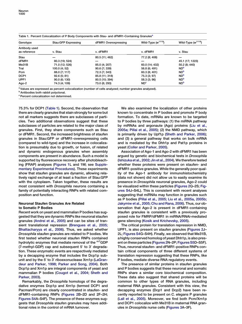
75.3% for DCP1 (Table 1). Second, the observation thatthere are clearly granules that stain strongly for somebutnot all markers suggests there are subclasses of parti-cles. Two additional observations suggest that thesesubclasses of particles are related to the major class ofgranules. First, they share components such as Stauor dFMR1. Second, the increased brightness of staufengranules in Stau:GFP or dFMR1-overexpressing cells(compared to wild-type) and the increase in colocaliza-tion is presumably due to growth, or fusion, of relatedand dynamic endogenous granules when assemblycomponents are present in abundance. Such a model issupported by fluorescence recovery after photobleach-ing (FRAP) analyses (Figures 1L and 1M; see Supple-mentary Experimental Procedures). These experimentsshow that staufen granules are dynamic, allowing rela-tively rapid exchange of at least a fraction of Stau:GFPwith the cytoplasm. Taken together, these results aremost consistent with Drosophila neurons containing afamily of potentially interacting RNPs with related com-position and function.
Neuronal Staufen Granules Are Relatedto Somatic P BodiesRecentwork on yeast andmammalian P bodies has sug-gested that they are dynamic RNPs like neuronal staufengranules (Andrei et al., 2005) and can be sites of tran-sient translational repression (Brengues et al., 2005;Bhattacharyya et al., 2006). Thus, we asked whetherDrosophila staufen granules are related to P bodies. Wefirst tested whether neuronal staufen RNPs containedhydrolytic enzymes that mediate removal of the m7GDP(7-methyl-GDP) cap and subsequent 50 to 30 degrada-tion. These enzymatic events are respectively mediatedby a decapping enzyme that includes the Dcp1p sub-unit and by the 50 to 30 riboexonuclease Xrn1p (LaGran-deur and Parker, 1998; Parker and Song, 2004). BothDcp1p and Xrn1p are integral components of yeast andmammalian P bodies (Cougot et al., 2004; Sheth andParker, 2003).Remarkably, the Drosophila homologs of the degra-
dative enzymes Dcp1p and Xrn1p (termed DCP1 andPacman/Pcm) are clearly concentrated in staufen- anddFMR1-containing RNPs (Figures 2B–2D and 2F–2H;Figures S4A–S4F). The presence of these enzymes sug-gests that Drosophila staufen granules may have addi-tional roles in the control of mRNA turnover.
We also examined the localization of other proteinsknown to concentrate in P bodies and promote P bodyformation. To date, mRNAs are known to be targetedto P bodies by three pathways: (1) the miRNA pathwayby miRNAs and argonaute (Ago) proteins (Liu et al.,2005a; Pillai et al., 2005); (2) the NMD pathway, whichis primarily driven by Upf1p (Sheth and Parker, 2006);and (3) a general pathway that works on bulk mRNAand is mediated by the Dhh1p and Pat1p proteins inyeast (Coller and Parker, 2005).
Association of Ago-1 and Ago-2 with dFMR1 has beenargued by genetic and biochemical tests in Drosophila(Ishizuka et al., 2002; Jin et al., 2004).We therefore testedwhether these proteins were present on staufen- anddFMR1-positive granules.While the generally poor qual-ity of the Ago-1 antibody for immunohistochemistry(data not shown) did not allow us to easily examine itspresence in Drosophila neuronal granules, Ago-2 couldbe visualized within these particles (Figures 2Q–2S; Fig-ures S4J–S4L). This is consistent with recent analysessuggesting that miRNAs may function in granules suchas P bodies (Pillai et al., 2005; Liu et al., 2005a, 2005b;Jakymiw et al., 2005; Chu andRana, 2006). Thus, our ob-servation that Ago-2 is present in dFMR1-containingstaufen granules is consistent with a previously pro-posed role for FMRP/dFMR1 in miRNA/RNAi-mediatedgene silencing (Kosik and Krichevsky, 2005).
The critical protein for translation repression in NMD,UPF1, is also present on staufen granules (Figures 2J–2L; Figures S4G–S4H). Finally, we observed that Me31B,ahighly conservedhomologof yeastDhh1p, is alsopres-ent on theseparticles (Figures 2N–2P; FiguresS5D–S5F).Thus, neuronal staufen- and dFMR1-positive RNPs con-tain critical components of three different systems oftranslation repression suggesting that these RNPs, likeP bodies, mediate diverse RNA regulatory events.
The presence of similar proteins in staufen granulesand P bodies suggests that these neuronal and somaticRNPs share a similar core biochemical composition.These data also suggest that shared proteins will becommon to other types of RNA granules, includingmaternal RNA granules. Consistent with this view, thedecapping enzymes (Dcp1 and Dcp2) have been re-cently reported to be present on C. elegans P granules(Lall et al., 2005). Moreover, we find both Pcm/Xrn1pand DCP1 colocalize with Me31B in maternal RNA gran-ules in Drosophila nurse cells (Figures 3A–3F).
Table 1. Percent Colocalization of P Body Components with Stau- and dFMR1-Containing Granulesa
Genotype Stau:GFP Expressing dFMR1 Overexpressing Wild-Type (w1118) Wild-Type (w1118)
Antibody usedas reference v. Stau v. dFMR1 v. dFMR1 v. Stau
Stau — 80.5 (11; 462) 77.2 (8; 408) —dFMR1 80.3 (19; 524) — — 45.1 (17; 1223)Me31B 71.0 (12; 526) 85.0 (4; 207) 60.0 (14; 432) 50.2 (8; 440)Tral 100.0 (4; 52) 90.6 (7; 339) 56.8 (6; 491) NDb
Pcm 88.0 (7; 117) 72.3 (7; 343) 60.3 (8; 401) NDb
DCP1 92.6 (5; 81) 85.8 (11; 318) 75.3 (3; 97) NDb
UPF1 90.0 (6; 130) 80.5 (10; 394) 58.3 (3; 96) NDb
Ago-2 74.3 (4; 109) 73.0 (8; 293) NDc NDb
aValues are expressed as percent colocalization (number of cells analyzed; number granules analyzed).b Antibodies both rabbit polyclonal.c Percent colocalization not determined.
Neuron1000

Trailer Hitch, a Me31B-Associated MaternalProtein, Is Present on P Bodies and NeuronalStaufen GranulesMe31B functions during oogenesis as a translationalrepressor of oskar mRNA in a well-studied eIF4E-Cup-Bru translational control complex (Lasko et al., 2005).This complex also contains a conserved Sm- and FDF-domain RNA-binding protein, trailer hitch (Tral). In Dro-
sophila ovaries, Tral coimmunoprecipitates with Me31Band colocalizes with Me31B-containing maternal RNAgranules (Boag et al., 2005).Figure 4A shows that a Me31B/Tral/dFMR1 complex
coimmunoprecipitates from Drosophila adult head ex-tracts, consistent with a model in which the three pro-teins function together in neuronal translation control.Me31B, Tral, and dFMR1 all have a similar, ubiquitousexpression pattern in the central nervous system, show-ing a predominantly cytoplasmic, steady-state localiza-tion (Figures S5A–S5C). In cultured Drosophila neurons,Tral also localizes to staufen- and dFMR1-containinggranules (Figures 4B–4D and Figures S5G–S5I). More-over, a GFP fusion to Scd6p, the S. cerevisiae homologof Tral, colocalizes with Dcp2p:RFP under high cell den-sity or nutrient starvation, conditions that enlarge yeastP bodies (Teixeira et al., 2005; Figures 4E–4G). Together,these data indicate that (1) Tral is present on Drosophilaneuronal RNPs in a biochemical complex that containsMe31B and dFMR1; and (2) Scd6p, the yeast homologof Tral, is a component of P bodies. The latter observa-tion further extends similarities between P bodies andstaufen granules.
Me31B and Tral Are Required for dFMR1-MediatedTranslational RepressionThe compositional similarity of P bodies and staufenRNPs suggests that neuronal translational control is
Figure 2. Neuronal Staufen Granules ContainP Body Components Mediating TranslationRepression and RNA Decay
Yeast P-body proteins tagged with GFP inS. cerevisiae cells (left column) with theirDrosophila orthologs localized relative toStau:GFP in cultured Stau:GFP-expressingmotor neurons. (A–D) Dcp1p/DCP1; (E–H)Xrn1p/Pcm; (I–L) Upf1p/UFP1; (M–P) Dhh1p/Me31B; (Q–S) and Ago-2 are also presenton neuronal staufen granules. The inset witha magnified view of small Me31B particlesin a neurite show that these also containStau:GFP. Scale bar, 10 mm for neurons.
Figure 3. RNA Decapping and Degradative Enzymes Are Present onMaternal RNP Granules
(A–C) DCP1 and (D–F) Pcm colocalize with Me31B in cytoplasmicfoci in nurse cells (stage 8 is shown). (C and F) Merged images. Scalebar, 10 mm.
Neuronal Granules Are Related to Somatic P Bodies1001

regulated through proteins andmechanisms associatedwith somatic P bodies. To test this prediction, wefocused on the highly conserved DEAD-box RNA heli-case Me31B, which functions in translational repressionof maternal mRNAs and in the targeting of mRNAs to Pbodies (Coller and Parker, 2005; Nakamura et al.,2001). The presence of Me31B and Tral with Ago-1 ondFMR1-containing complexes suggests that theseproteins may function in neuronal translation control,potentially with dFMR1 in miRNA-mediated processes.To test whether Me31B and Tral function in dFMR1-
mediated translational repression, we asked if defectscaused by dFMR1 overexpression in developing eyeswere modified in genetic backgrounds deficient forMe31B or Tral. Ectopic overexpression of dFMR1 in thecompound eye driven by the sevenless enhancer (sev-dFMR1) results in a ‘‘rough-eye’’ phenotype througha pathway that requires dFMR1 domains essential fortranslational repression as well as Ago-1 function (Fig-ure 5A; Jin et al., 2004; Wan et al., 2000).As shown in Figure 5B, loss of a single copy ofme31B
suppressed sev-dFMR1-induced rough eye pheno-types. Unambiguous suppression was observed witheither me31BD1 or me31BD2 allele (Figure 5B; data notshown). Internally, the disruption of ommatidia causedby dFMR1 overexpression was also suppressed, as ob-served in tangential sections (Figures 5F and 5G). Thissuppression is a direct result of me31B deficiency, be-cause a genomic me31B+ transgene, P[me31BAflII],which is capable of rescuing lethality of me31BD mu-tants (Nakamura et al., 2001) rescues suppression of thesev-dFMR1 rough eye phenotype (Figures 5C and 5H).Results with tral mutations were similar. We isolated
deletion alleles for tral (see Supplementary ExperimentalProcedures) and found them to result in larval lethality.Both tral deletions dominantly suppressed sev-dFMR1-induced rough eyes (Figures 5D and 5I; data not shown).A tral+ genomic transgene (P[tral-10]) containing theentire tral locus was sufficient to rescue the lethalityof tral mutants. This genomic transgene also ‘‘rescued’’dominant suppression of the rough eye phenotype,thereby demonstrating that phenotypic suppressionof sev-dFMR1occurs specifically due to loss of tral (Fig-ures 5E and 5J).
Given that Me31B, Tral, and dFMR1 form a physicalcomplex, the above results suggest that Me31B andTral act, together with dFMR1, as translational regula-tors in neuronal cells. An alternative interpretation is thatsingle-copy deletions of tral or me31B block apoptosisor other developmental errors induced by Sev-dFMR1.However, this is unlikely for three reasons: First, coim-munoprecipitation and colocalization of Me31B, Tral,and dFMR1 are more consistent with a direct mecha-nism. Second, all three proteins have RNA-bindingdomains that predict roles in translational control. Fi-nally, ectopic expression of Me31B in the eye causesrough eyes via a mechanism requiring amino acid resi-dues necessary for translational repression (FiguresS6A–S6C; see below).
Me31B and Tral Regulate Dendrite Morphogenesisin Sensory NeuronsThe observed effect of Me31B (and Tral) induction ondendritic development of sensory neurons (Figure 6 andFigures S6D–S6F) provides further evidence for functionin neuronal translation regulation. Previous studies haveestablished that translational control of gene expressionregulates dendrite morphogenesis in vivo. For example,neurons of human fragile X patients and DrosophiladFMR1 mutants show an increase in dendritic spinenumber and length (Nimchinsky et al., 2001; Lee et al.,2003). Conversely, induction of dFMR1, Pum, or Nos inclass IVDrosophila da sensory neurons greatly perturbs,and can dramatically reduce, higher-order dendriticbranching (Lee et al., 2003; Ye et al., 2004). If Me31Band Tral act in dendritic translational control, we antici-pated that their induction would also have specificeffects on higher-order dendritic branching.
Overexpression of Me31B in class IV neurons sub-stantially reduced high-order dendritic complexity (Fig-ures 6A and 6B). In neurons overexpressing Me31B,the number of higher-order dendrites was significantlyreduced compared with the control, in which only thereporter gene UAS-mCD8:GFP was overexpressed(p % 0.001; Figures 6A, 6B, and 6F). To determine ifthis effect of Me31B induction reflected increased trans-lational repression activity of Me31B, we asked whethersimilar effects would be shown by induction of an
Figure 4. Tral Is an Me31B/dFMR1-Associ-ated Protein Present on Staufen RNPs witha Conserved Homolog, Scd6p, in Yeast PBodies
(A) Western blot of Me31B coimmunoprecipi-tates probed with antibodies against Me31B,Tral, dFMR1, and dynamin.(B–D) Me31B (B) and Tral (C) colocalize inneuronal granules of dFMR1 expressing cul-tured motor neurons (similar results in w1118
cells are shown in Figures S5D–S5I).(E–G) Yeast cells expressing Scd6p:GFP (E)and Dcp2p:RFP (F) showing colocalizationof Scd6p:GFP to P bodies.Scale bar, 10 mm.
Neuron1002

Me31B mutant protein (D207A, E208A) homologous toa yeast Dhh1p mutant incapable of translational repres-sion (Coller and Parker, 2005). Expressed at comparablelevels (data not shown), the mutant transgene had noeffect on dendritic complexity (Figures 6C and 6F), con-sistent with the observed effect being dependent onMe31B-induced translational repression.
Overexpression of Tral in class IV neurons also sub-stantially changed dendrite morphology compared tothe control (Figures S6D–S6F). Interestingly, closerexamination revealed a significant increase in the num-ber of finer dendritic ‘‘tendrils’’ at terminal dendriticbranches compared to control neurons. Differencesbetween effects of Tral and Me31B induction on den-dritic arborization are consistent with a relatively spe-cific role for CAR-1, the C. elegans ortholog of Tral, intranslational control compared to CGH-1 (the Me31Bortholog), suggested by phenotypic differences follow-ing RNAi-mediated inhibition of respective proteins inthe C. elegans germline (Audhya et al., 2005; Navarroet al., 2001).
In class IV sensory neurons, loss of nanos or pumiliocauses abnormal dendritic growth (Ye et al., 2004).This aberrant growth, visible in about 20% of mutantneurons, is most easily apparent as a loss of ‘‘tiling,’’ aterm that refers to the complete, nonoverlapping cover-age of the epidermis by dendrites of wild-type sensoryneurons (Ye et al., 2004; Grueber et al., 2003). We there-fore askedwhether loss ofme31B, achieved by express-ing a transgenic RNAi construct that generates a hairpinMe31B RNA (UAS-Me31Bhpn) would cause similar de-fects. As shown in Figure 6E, UAS-Me31Bhpn sensoryneurons showed frequent defects in terminal dendritemorphology and dendritic tiling highly reminiscent ofnanos and pum phenotypes. Incomplete coverage ofthe epidermis was observed in at least 33% (n = 15 neu-rons) of neurons analyzed. Additionally, Me31Bhpn neu-rons show a modest increase (37%) in high-order den-dritic complexity similar to that observed in dFmr1mutants (Figure 6F; Lee et al., 2003). Parallel analysesof a hairpin construct for Lk6, which encodes the Dro-sophila homolog of the eIF4E-kinase MNK, showed noeffect on dendritic branching of class IV sensory neu-rons (data not shown).
From these data, we conclude that Me31B (and Tral)regulates dendritic arborization of class IV da neurons.This observation, consistent with observations of othertranslational repressors such as dFMR1, Pum, and
Nos provides a second line of evidence suggestingthat Me31B and Tral function as neuronal translationalregulators.
Figure 5. Me31B and Tral Are Required fordFMR1-Induced Defects in the DrosophilaEye
(A–E) SEMs of adult compound eyes withpaired retinal sections (F–J). Magnificationof SEMs is 1503. Tangential sections ofeach genotype are at approximately thesame depth.
Figure 6. Me31B Regulates Dendritic Growth in Sensory Neurons
(A) Control class IV ddaC neuron expressingUAS-mCD8:GFP alone.(B) Class IV ddaC neurons overexpressing Me31B and UAS-mCD8:GFP showing a reduction in higher-order dendrite arborization.(C) The same neurons overexpressing a mutant Me31B incapable oftranslational repression (Me31BD207A, E208A) show normal dendriticbranching.(D) Transgenic RNAi dramatically reduces Me31B protein levels.Anti-Me31B staining of third-instar imaginal discs shows that UAS-Me31Bhpn expressed in the patched domain of wing imaginal discsreducesMe31B levels along the anterior-posterior border (top panel)compared to control wing imaginal discs (lower panel).(E) Class IV ddaC neurons overexpressing a Me31B RNA hairpin(UAS-Me31Bhpn) exhibit abnormal dendrite morphology and in-creased high-order branching.(F) Numbers of dendritic branches in each order, as revealed byreversed Strahler analysis (see Supplemental Data). Number ofneurons analyzed for each genotype are: UAS-mCD8:GFP control(n = 15), UAS-Me31B (n = 10), UAS-Me31BD207A, E208A (n = 11), andUAS-Me31Bhpn (n = 13). Values are mean 6 standard error. A star(*) indicates a significant reduction in fifth-order dendrite branchingfollowing Me31B overexpression compared to the control (p <0.001) and a significant increase in fifth-order dendrite branchingfollowing Me31B RNAi (p < 0.001).Scale bar, 20 mm.
Neuronal Granules Are Related to Somatic P Bodies1003

Me31B Functions in MicroRNA-MediatedTranslational RepressionTwo previous findings led us to the hypothesis that thedFMR1-associated Me31B protein may be required formiRNA/RNAi function. First, FMRP/dFMR1, showingstrong biochemical or genetic interactions with Ago-1and Ago-2, is strongly implicated in microRNA-medi-ated translational repression (Kosik and Krichevsky,2005). Second, miRNA-mediated repression has beenproposed to occur in P bodies of somatic cells (Liuet al., 2005a; Pillai et al., 2005). Thus, we tested whetherMe31B is required in vivo for the function of bantam, anendogenous miRNA that represses hid mRNA transla-tion in wing imaginal discs (Brennecke et al., 2003).We used two transgenically encodedGFP reporters to
assay bantam-mediated translational repression (Bren-necke et al., 2003). The ‘‘hid reporter,’’ which carriesthe 30 UTR of hid fused to the 30 end of EGFP-coding se-quence, closely reports bantam repression of a nativetarget mRNA. This 30 UTR contains four repeats comple-mentary to bantam target recognition sequences, withseveral mismatches typically associated with miRNA-mediated translational repression. The ‘‘bantam re-porter,’’ in which four synthetic repeats 100% comple-mentary to the bantam target recognition element arefused 30 to EGFP coding module, also reports bantamfunction.We used the heat-shock FLP/FRT system to generate
me31b2/2 clones in the wing disc and identified theseclones by loss of b-galactosidase or Me31B stainingwith respective antibodies (Figure 7A–C). We then askedhow a control protein (Dlg), hid reporter, or bantam re-porter expression was affected by loss of Me31B (Fig-ures 7F and 7G). While cells lacking me31B showed nodetectable increase in a control protein (Dlg) expression(Figure 7G), they showed clear increases in both hid re-porter (Figures 7C–7E) and bantam reporter (Figures7H–7J) expression, indicating that bantam-mediated si-lencing does not function in the absence of me31B.These data, from in vivo analyses of an endogenousmiRNA in cells carrying a null mutation for me31B, sup-port a recent study showing a role for RCK (the humanhomolog of Me31B) in Let-7 miRNA-mediated transla-tional repression in cultured mammalian cells (Chu andRana, 2006). In addition, our observations extend thisstudy by demonstrating a role for Me31B in repressionmediated by perfectly base-paired miRNAs. It shouldbe noted that the requirement for Me31B for efficientrepression of the bantam reporter does not necessarily
mean thatMe31B is required formiRNA-mediated endo-nucleolytic cleavage, since it is likely that repression byperfectly base-paired miRNAs can be a combination oftranslation repression, decapping, and/or endonucleo-lytic cleavage of the mRNA (Valencia-Sanchez et al.,2006). Importantly, these data demonstrate that Me31Bis required for repression mediated by an endogenousDrosophila miRNA. An obvious corollary of our analysisin wing imaginal discs is that Me31B plays a similar rolein mediating functions of neuronal miRNAs, althoughassays to directly test this issue are not immediatelyavailable in Drosophila.
Discussion
Neuronal Staufen RNPs Are Related to Somatic PBodiesSeveral observations now indicate that P bodies, mater-nal granules, and a major subclass of neuronal RNP aresimilar in underlying composition and represent a con-served system for the regulation of cytoplasmicmRNAs.As summarized in Table 2, known RNA transport andtranslational repressors shared between maternal andneuronal staufen granules now include, Stau, Btz,dFMR1, Pum, Nos, Yps, Me31B, Tral, Cup, eIF4E, Ago-2, and Imp. Strikingly, in human cells, the Me31B homo-log RCK/p54, the Tral homolog RAP55, the four humanargonaute proteins, eIF4E, and a eIF4E-binding proteinanalogous to Cup, 4E-T, are all found in P bodies (Andreiet al., 2005; Cougot et al., 2004; Kedersha et al., 2005; Liuet al., 2005a; Pillai et al., 2005; Yang et al., 2006). In yeast,homologs of Me31B (Dhh1p) and Tral (Scd6p) are alsoknown to be in P bodies (Sheth and Parker, 2003; Fig-ures 4E–4G), and Dhh1p in particular plays a role inrecruiting RNA-decapping proteins and exonucleasesto these RNPs (Coller and Parker, 2005). Consistentwith the above observations in yeast, the enzymes in-volved in mRNA hydrolysis including the 50 to 30 RNAexonuclease Xrn1p/Pcm and the RNA-decapping en-zyme DCP1 are present on Drosophila neuronal staufenRNPs (Figure 2) and maternal RNA granules (Figure 3).Our data unequivocally demonstrate tight spatial prox-imity of components mediating various RNA regulatoryprocesses in Drosophila neurons.
The large collection of proteins and processes com-mon to P bodies, staufen granules, and likely maternalRNA granules suggests that they share an underlyingcorebiochemicalcompositionandfunction,whichwouldthen be elaborated in different biological contexts. For
Figure 7. Me31B Is Required for the In VivoFunction of an Endogenous MicroRNA
Heat-shock-induced clones in wing imaginaldiscs of hs-FLP1; FRT40A, arm-lacZ/FRT40A, me31BD1; reporter flies show darkme31B2/me31B2 clones revealed either bystaining for LacZ (B) or Me31B (A and C).Anti-GFP staining shows that the hid reporter(D and E) is upregulated in cell clones lackingMe31B (C). Similar analyses (F and G) showthat a control protein Dlg (G) is not upregu-lated in me31B2/me31B2 clones (F). How-ever, consistent with a Me31B requirementin miRNA/RNAi, the bantam reporter expres-sion is upregulated inme31B2/me31B2-lack-ing cells (H–J).
Neuron1004

example, one anticipates that proteins involved inmRNAtransportwill bemoreprevalent inmaternal andneuronalRNPs, which need to be transported for their biologicalfunction.
An interesting aspect of neuronal staufen RNPs de-scribed here is the diversity of translational repressionsystems that are present within them. First, in Me31B,they contain a protein that works in general translationrepression of a wide variety of mRNAs and can alsoaffect miRNA-based repression (Figure 7; Coller andParker, 2005; Chu and Rana, 2006). Second, in Ago-2,they contain a component specific to miRNA/RNAi-dependent repression. Third, neuronal staufen granulesalso contain UPF1, which was originally thought tobe solely involved in mRNA degradation. However, be-cause UPF1 can act as a translation repressor (Muhlradand Parker, 1999; Sheth and Parker, 2006) and physi-cally interacts with Stau (Kim et al., 2005), a reasonablehypothesis is that UPF1 might work in neuronal gran-ules, in conjunction with Stau, to repress the translationof a subset of mRNAs. The presence of multiple mecha-nisms for translation repression colocalizing in granulesin Drosophila neurons may allow for differential transla-tion control of subclasses ofmRNA in response to differ-ent stimuli.
Neuronal Granule Diversity and FunctionEvidence accumulating in the literature suggests thatthere is a potential diversity of RNA granule types in neu-rons. Our observations in Drosophila neurons are mostconsistent with a model in which a major subclass of
neuronal RNP, in which various translational repressorand mRNA turnover proteins colocalize, is related toother compositionally distinct, diverse RNPs. A majorsubclass of staufen-containing RNP is indicated by ourdata showing substantial colocalization among variousproteins we have analyzed (Table 1; data not shown).Diversity is indicated by the lack of 100% colocalization:for instance, 55% of staufen-positive particles in wild-type neurons do not contain detectable dFMR1.Two types of observations suggest that the apparent
subclasses of particles containing Stau or dFMR1, butnot both, are related to the particles in which they coloc-alize. First, these two types of RNPs are clearly compo-sitionally related to particles that contain both proteins.Second, this is supported by the observation that coloc-alization canbesubstantially increasedunder somecon-ditions. Overexpression of either dFMR1 or Stau:GFPincreases colocalization between Stau and dFMR1 from45% inwild-type neurons tomore than 80%. Concurrentwith increased frequency of colocalization, Stau:GFP ordFMR1 induction increases apparent particle size (orbrightness) and reduces the total number of particles.The increase in colocalization and brightness, as wellas reduction in particle number, is most easily explainedby growth and/or fusion of related RNPs. Significantly,similar effects on mammalian neuronal granule size andnumber have been reported following overexpression ofStau or another granule protein, RNG105 (Kiebler et al.,1999; Shiina et al., 2005). Thus, the underlying regulatoryprocesses appear conserved between Drosophila andmammalian neurons.
Table 2. Conserved Components of P Bodies, Maternal Granules, and Neuronal Granules
Protein ClassMammalian andYeast P Bodiesa
DrosophilaMaternal Granulesb
DrosophilaNeuronal Granulesc
MammalianNeuronal Granulesd
RNA transport ? Stau, Btz Stau, Btz Stau, BtzFragile X-like ? dFMR1 dFMR1 FMRP, FXR1, FXR2Zip-code binding ? ? Imp ZBP1Pum domain ? Pum? Pum Puml
CCHC Zn-finger domain ? Nanos Nanosalso f ?DEAD-box RNA helicase RCK/Dhh1p Me31B Me31B ?Sm-like domain RAP55/Scd6pj Tral Tral ?Cap-binding eIF4Ee, k eIF4E eIF4Ealso g under some conditionsEnhancers of decapping Edc3p, Pat1pi ? ? ?eIF4E-binding eIF4E-Tk Cup Cup ?PABP PABPe ? PABPg ?Y-box ? Yps Yps ?50 to 30 RNA decay machinery Lsm, Dcp1p, Dcp2p, Xrn1p DCP1, Pacman DCP1, Pacman
(others not examined)?
NMD machineryh Upf-1 Upf-1 Upf-1 ?miRNA, siRNA machinery mAGO1, mAGO2 ? Ago-2 ?
? Ortholog present but association with RNA granules has not been described.a Sheth and Parker, 2003; Andrei et al., 2005; Liu et al., 2005a; Pillai et al., 2005.b St Johnston et al., 1991; van Eeden et al., 2001; Nakamura et al., 2001; Boag et al., 2005; Nakamura et al., 2004; Wilhelm et al., 2000; this work;Wang et al., 1994; Forbes and Lehmann, 1998.c This work (except PABP).dKiebler et al., 1999; Macchi et al., 2003; Kanai et al., 2004; Antar et al., 2005; Zhang et al., 2001.e eIF4E and PABP can associate with P bodies in yeast under conditions of translational arrest induced by glucose deprivation (Teixeira et al.,2005).f Ye et al., 2004 (describes particles likely to be, but not clearly established as, neuronal RNA granules).g Sigrist et al., 2000 (describes particles likely to be, but not clearly established as, neuronal RNA granules).hUPF1 has also been shown to interact with Stau in a Stau-mediated NMD pathway (Kim et al., 2005).i Pat1p acts as a general repressor of translation in yeast (Coller and Parker, 2005).j Yang et al., 2006; this work.k Andrei et al., 2005.l Vessey et al., 2006.
Neuronal Granules Are Related to Somatic P Bodies1005

While it remains unclear how FMRP, Stau, or RNG105enhance granule growth or fusion, it is conceivable thatindividual mRNAs first form small RNPs whose compo-sitions reflect specific requirements for translationalrepression of the mRNAs they contain. These smallRNPs exist in dynamic equilibrium with larger RNPs inwhich multiple, diverse translational repression com-plexes are sequestered. Induction of factors that pro-mote granule assembly could push the equilibriumtoward mRNP sequestration within large granules. A re-quirement of this dynamic model, which postulates in-teractions among different types of RNP, is that theRNPs themselves can change in composition duringtransport to synaptic domains. This is supported byFRAP analyses showing rapid exchange of Stau:GFPbetween cytosol and granule (Figures 1L and 1M).Additional types of RNPs have also been described in
neurons. For example, polysomes apparently arrestedin translation have been observed near dendritic spines,and these RNPs show no obvious similarity to large,ribosome-containing particles, termed neuronal RNAgranules (Knowles et al., 1996; Greenough et al., 2001;Ostroff et al., 2002). In addition, a potentially distinctRNPcontaining Stau, kinesin, and translationally repressedRNAs, but not ribosomes, has been purified from themammalian brain (Mallardo et al., 2003). More recently,it has been shown that RNPs containing stress-granulemarkers TIA-1 and TIA-R aswell as pumilio2 are inducedby arsenate-treatment of mammalian cultured neurons(Vessey et al., 2006). Interestingly, as previously shownfor somatic cells, these large stress granules appeartightly apposed to domains containing DCP1 and Lsm1,markers of P bodies (Vessey et al., 2006). Determiningthe temporal and compositional relatedness of suchvaried RNPs, their pathways of assembly as well as theirfunctions, is a broad area of future research not only inneuroscience but also in cell biology.These diverse types of biochemical compartments for
individual mRNAs suggest that neural activity or otherdevelopmental signaling events would influence transla-tion in two steps: first, by desequestering mRNPs heldwithin large granules and, then, by derepressing quies-cent mRNAs in individual mRNPs. Thus, RNPs wedescribe here could have a complex precursor-productrelationship with other RNPs, including polysomes dis-covered by now-classical studies at dendritic spines.
Two Functionally Important TranslationalRepressors in dFMR1-Containing Neuronal RNPsDespite the complexity revealed by the diversity of neu-ronal RNPs, the importance and significance of theobserved colocalization of Me31B, Tral, argonaute, anddFMR1 in staufen-positive neuronal RNPs is mostclearly demonstrated by functional analyses revealingbiological pathways in which these proteins functiontogether.Several independent lines of evidence are consistent
with a function for Me31B in neuronal translational re-pression as part of a biochemical complex that includesdFMR1. First, subcellular localization studies indicatethatMe31B and Tral localize to dFMR1-containing RNPsespecially prominent at neurite branch points in culturedDrosophila neurons (Figures S5D–S5I; data not shown).Second, Me31B, Tral, and dFMR1 coimmunoprecipitate
from Drosophila head extract, thus confirming the phys-ical association of three proteins (Figure 4A). Third, loss-of-function alleles of either Me31B or Tral suppressthe rough eye phenotype seen when dFMR1 is over-expressed in the sev-positive photoreceptors (Figure 5).Fourth, overexpression of Me31B in sensory neuronsleads to altered branching of terminal dendrites, a phe-notype also seen with overexpression analyses of Nos,Pum, and dFMR1 (Figures 6B and 6F; Lee et al., 2003;Ye et al., 2004). Finally, reduction of Me31B expressionin sensory neurons by RNAi results in abnormal dendritemorphogenesis and tiling defects, phenotypes similar tothat observed following loss of nanos, pum, or dFmr1function (Figures 6E and 6F; Ye et al., 2004; Lee et al.,2003). Significantly, the effect of Me31B on dendriticgrowth is correlated with its ability to function in transla-tional repression (Figures 6C and 6F). These five inde-pendent lines of evidence provide considerable supportfor Me31B (and Tral) function in neuronal translationcontrol processes. While the site of functional inter-action between dFMR1, Me31B, and Tral (soma or neu-ronal processes) is not identified here, the importance ofthe physical interactions is clearly demonstrated.
Several observations also argue that Me31B acts, atleast in part, within neurons to promote translationrepression and/or mRNA degradation in response tomiRNAs. This possibility was first suggested by thephysical and genetic interactions of Me31B with dFMR1(discussed above; Figure 5; Figures S5D–S5F), a proteinthat has previously been implicated in the miRNA-medi-ated repression (Ishizuka et al., 2002; Jin et al., 2004).Using direct assays for miRNA-mediated functionin vivo (Brennecke et al., 2003), we show that Me31B isrequired for efficient repression by the bantam miRNAin developing wing imaginal discs (Figure 7). This iden-tifies Me31B as a protein required for efficient miRNA-based repression.
Recently, miRNA-based regulation has been shown tobe important for the control of spine growth in hippo-campal neurons (Schratt et al., 2006) and to be a targetof protein-degradative pathways involved in long-termmemory formation in Drosophila (Ashraf et al., 2006).Thus, our data predict that Me31B will be important inmodulating miRNA function pertinent to developmentof functional neuronal plasticity. More generally, be-cause Me31B homologs in yeast and mammals havebeen shown to function in P body formation in somaticcells (Andrei et al., 2005; Coller and Parker, 2005), the re-quirement for Me31B in miRNA function provides evi-dence to support amodel in which formation of P bodiesis required for efficient miRNA-based repression in var-ied cell types and biological contexts.
Implications for Translational Control in NeuronsOur conclusion that staufen- and dFMR1-containingneuronal RNPs are similar in organization and functionto P bodies has several implications for neuronal trans-lational control. First, the presence of diverse transla-tional repression systems on these RNPs suggeststhat, like in P bodies, different classes of mRNAs willbe repressed by different mechanisms. This may allowspecific RNA classes to be released for new translationin response to different stimuli. Such diversity of controlmay allow synapses to remodel themselves differently,
Neuron1006
depending on the frequency and strength of stimulation(e.g., LTD or LTP). Second, FRAP experiments indicatethat both P bodies and staufen granules are dynamicstructures (Andrei et al., 2005; Kedersha et al., 2005; Fig-ures 1K and 1L). This argues that, like P bodies, staufengranules are in a state of dynamic flux, perhaps in activ-ity-regulated equilibrium with the surrounding transla-tional pool. Third, the presence of mRNA-degradativeenzymes on staufen granules suggests regulation ofmRNA turnover may play an important role in local syn-aptic events. For example, if synaptic signaling were toinduce turnover of specific mRNAs at a synapse, thenstimulated synapses could acquire properties differentfrom unstimulated ones that retain a ‘‘naive’’ pool ofstored synaptic mRNAs. Finally, these observationsimply that the proteins known to function in translationrepression within P bodies will play important roles inmodulating translation in neurons. Thus, we anticipatethat proteins of mammalian or yeast P bodies such asEdc3p, Pat1p, the Lsm1-7p complex, GW182, and FASTwill be present on and influence assembly and functionof neuronal granules (Cougot et al., 2004; Eystathioyet al., 2003; Kedersha et al., 2005; Sheth and Parker,2003).
Experimental Procedures
Drosophila StocksFly stocks were raised at 25!C on standard cornmeal and agarmedia. Wild-type (Oregon-R and w1118) were from Ramaswami labstocks. Other strains were obtained from D42-Gal4-chaGal80 (con-structed by S. Sanyal with components from T. Kitamoto andG. Boulianne); C380 (V. Budnik); C155 (C. Goodman); UAS-dFMR1(T. Jongens); sev-dFMR1 (P. Jin); UAS-Stau-GFP (A. Brand); UAS-Me31Bhpn (R. Ueda); and elav, UAS-GFP:MCP:nls and UAS-Cam-KII3
0UTR 2ms2 (S. Ashraf and S. Kunes). Gal4477; UASmCD8-GFP,UAS-flip Act < CD2 < Gal4 was constructed using strains fromW. Grueber and S. Sanyal; hsFLP-1; FRT 40A, armadillo-lacZ(Bloomington); ‘‘hid-reporter’’ and ‘‘bantam reporter’’ lines are de-scribed in Brennecke et al. (2003).Me31B alleles were as previouslydescribed (Nakamura et al., 2001).
Drosophila Neuron Primary Cell CultureCells for culture were obtained from the thoracic-abdominal (ventral)region of the CNS of late third-instar larvae. Tissues were dissectedand placed into a Liberase enzyme (combination of collagenase anddispase) solution and incubated at room temperature for 1 hr.Tissues were then rinsed in culture medium (Schneider’s- or IL15-based medium) and subjected to two mechanical trituration steps.Cells were plated onto coverslips coated with concanavalin A andlaminin in tissue culture dishes and allowed to grow at 25!C for 3–4days prior to immunostaining. We used a composite Gal4/Gal80system (D42-Gal4; chaGal80) to drive expression of a functionalStau:GFP fusion protein (UAS-Stau:GFP) or dFMR1 (UASdFMR1) ina subset of motor neurons. Cells were identified using confocalmicroscopy by the presence of Stau:GFP (or dFMR1-positive) punc-tae allowing for the identification of a discrete population of neuronsin an otherwise heterogeneous neuronal culture.
ImmunohistochemistryPrimary antibodies used for neuronal granule staining are listedin Table 1. Additional primary antibodies used were mouse anti-b-galactosidase (Molecular Probes), rabbit anti-GFP (MolecularProbes), mouse anti-DLG 4F3 (Developmental Studies HybridomaBank), and goat anti-HRP-TRITC (Sigma). Secondary antibodiesused were FITC (Sigma), Alexa 488-, Alexa 555-, Alexa 568-, andAlexa 647-conjugated anti-rat, mouse and -rabbit IgG (MolecularProbes). Cultured cells were fixed and stained as follows. Briefly,3-day old ventral ganglion cell cultures were rinsed in prewarmedPBS buffer (pH = 7.2) and fixed for 10 min in 3.5% paraformaldehyde
in PBS. Cells were blocked for 30min in Block solution (PBS contain-ing 0.1% Triton X-100, 2% BSA, and 5% normal goat serum). Pri-mary and secondary antibodies were diluted in Block solution andincubated with cells for 2 hr and 1 hr, respectively, at RT. After rins-ing, preparations were mounted in Vectashield Mounting Medium(Vector Labs) and imaged on a Nikon PCM2000 laser confocalmicroscope using Simple PCI software. Further discussion ofmethods used to examine colocalization of neuronal granule com-ponents can be found in the Supplemental Data.For larval CNS preparations, wandering third-instar larvae were
processed according to the method of Sanyal et al. (2003), withthe following modification. To permeabilize the sheath surroundingthe ventral ganglion, CNS preps were treated with 50 mg/ml collage-nase diluted in HL-3 saline (+Ca2+) for 3 min prior to fixation.Immunostaining of Drosophila oocytes was done essentially as
described inWilhelm et al. (2003), with the following alterations. Ova-ries were dissected in room temperature PBS + 0.1% Triton X-100and fixed for 10 min in one part 3.7% paraformaldehyde in PBS tosix parts heptane.
Immunoprecipitation of Me31BImmunoprecipitation from head extracts with rat anti-Me31B wascarried out essentially as described (Nakamura et al., 2001). Sam-ples were separated by SDS-PAGE, transferred to PVDF membrane(Millipore), and analyzed by Western blotting. Proteins were de-tected by ECL (Amersham).
Analysis of Drosophila Rough Eye PhenotypesDrosophila genotypes used for SEM analysis and tangential eyesectioning were as follows: dFMR1 overexpression, +/SevdFMR1;Me31B suppression, Me31BD1FRT40A/SevdFMR1 and Me31BD2
FRT40A/SevdFMR1; Me31B ‘‘rescue’’, Me31BD2FRT40A/SevdFMR1;+/P[w+MeAflII]; Tral suppression,+/SevdFMR1; +/Tral D3-FRT2A and +/SevdFMR1; +/ TralD4FRT2A; Tral ‘‘rescue’’,P[Tral-10]/SevdFMR1; +/ Tral D4FRT2A. All indicated stocks (above)were crossed to w1118 to generate heterozygotes for subsequentanalysis.Further analysis of the rough eye phenotype using scanning elec-
tronmicroscopy (SEM) and in tangential eye sections is described inthe Supplemental Experimental Procedures.
Analysis of Dendritic ProcessesExperiments were done in a Gal4477, UAS-mCD8-GFP; UAS-flp,Act < CD2 < Gal4 background in which flp-recombinase targetsequences (‘‘<’’) flankingCD2 stuffer sequence is often excised thor-ough the activity of Gal4477-driven Flp recombinase. Thus, in thisbackground, individual Gal4477-positive da sensory neurons areoccasionally very brightly labeled by Actin-Gal4 mCD8:GFP. Ingenetic backgrounds carrying a Gal4-responsive transgene, thetransgene is also strongly expressed. Further analysis of sensoryneuron dendritic complexity is described in the SupplementalExperimental Procedures.
Generation and Characterization of me31B-Mitotic ClonesMitotic recombination cloneswere induced 486 2 hr after egg laying(AEL) in staged larvae by heat shock at 37!C for 90 min. Larval geno-types used were hs-FLP1; FRT 40A, arm-lacZ/Me31B D1, FRT 40A;bantam-reporter (or hid-reporter). Discs were dissected at 120 62 hr AEL, fixed with 4% formaldehyde, and stained with differentantibodies. The discs were mounted in Vectashield (Vector Labs)and analyzed by confocal microscopy (Zeiss LSM 510) with a 203objective. Clone areas were measured and analyzed using AdobePhotoshop.
Supplemental DataThe Supplemental Data for this article can be found online at http://www.neuron.org/cgi/content/full/52/6/997/DC1/.
Acknowledgments
We thank T. Kitamoto, T. Aigaki, W. Grueber, Y.N. Jan, S. Warren,P. Jin, V. Budnik, G. Boulianne, D. St. Johnston, S. Sanyal, J. Bren-necke, S. Cohen, the National Institute of Genetics (Japan) fly stockcenter, and the Bloomington Drosophila stock center for Drosophila
Neuronal Granules Are Related to Somatic P Bodies1007
stocks; W. Grueber and S. Sanyal for advice; J. Brennecke, E. Holo-han, D. Zarnescu, P. Macchi, V. Rodrigues, and M. Kiebler for usefuldiscussions; G. Hannon, T. Jongens, P. Lasko, D. St. Johnston,C. Temme, J. Raff, and the Developmental Studies HybridomaBank for antibodies; B. Suter for a Drosophila genomic library;S. Zusman (Genetic Services Inc.) for germline transformation ser-vices; C. Boswell of the MCB Imaging Facility for help with micros-copy; D. Bentley, P. Jansma, N. Ingraham, and C. Hedgcock of theArizona Research Labs Divisions of Neurobiology and Biotechnol-ogy for access to and instruction in the SEM, Microscopy (tissuesectioning), Tissue Culture Core, and Photo/Graphics Facilities (re-spectively), as well as their imaging and computing facilities; andG. Bosco for use of the Nikon Eclipse E800 microscope, camera,and software. This work was funded by grants from the NIH/NIDA(DA15495; DA17749) and the Science Foundation of Ireland toM.R., from the Howard Hughes Medical Institute to R.P., from theMinistry of Education, Culture, Sports, Science and Technology,Japan (to A.N.), the UK Biotechnology and Biological SciencesResearch Council (S.F.N.), NIH grant GM54409 to Paul MacDonaldand an NIH grant to R.B.L.
Received: August 9, 2006Revised: September 21, 2006Accepted: October 24, 2006Published: December 20, 2006
References
Andrei, M.A., Ingelfinger, D., Heintzmann, R., Achsel, T., Rivera-Pomar, R., and Luhrmann, R. (2005). A role for eIF4E and eIF4E-transporter in targeting mRNPs to mammalian processing bodies.RNA 11, 717–727.
Antar, L.N., Dictenberg, J.B., Plociniak, M., Afroz, R., and Bassell,G.J. (2005). Localization of FMRP-associated mRNA granules andrequirement of microtubules for activity-dependent trafficking inhippocampal neurons. Genes Brain Behav. 4, 350–359.
Ashraf, S.I., McLoon, A.L., Sclarsic, S.M., and Kunes, S. (2006). Syn-aptic protein synthesis associated with memory is regulated by theRISC pathway in Drosophila. Cell 124, 191–205.
Audhya, A., Hyndman, F., McLeod, I.X., Maddox, A.S., Yates, J.R.,3rd, Desai, A., and Oegema, K. (2005). A complex containing theSm protein CAR-1 and the RNA helicase CGH-1 is required forembryonic cytokinesis in Caenorhabditis elegans. J. Cell Biol. 171,267–279.
Bhattacharyya, S.N., Habermacher, R., Martine, U., Closs, E.I., andFillipowicz, W. (2006). Relief of microRNA-mediated translationalrepression in human cells subjected to stress. Cell 125, 1111–1124.
Boag, P.R., Nakamura, A., and Blackwell, T.K. (2005). A conservedRNA-protein complex component involved in physiological germ-line apoptosis regulation in C. elegans. Development 132, 4975–4986.
Brengues, M., Teixeira, D., and Parker, R. (2005). Movement ofeukaryotic mRNAs between polysomes and cytoplasmic process-ing bodies. Science 310, 486–489.
Brennecke, J., Hipfner, D.R., Stark, A., Russell, R.B., and Cohen,S.M. (2003). bantam encodes a developmentally regulated micro-RNA that controls cell proliferation and regulates the proapoptoticgene hid in Drosophila. Cell 113, 25–36.
Chu, C.Y., and Rana, T.M. (2006). Translation repression in humancells by microRNA-induced gene silencing requires RCK/p54.PLoS Biol. 4, e210. 10.1371/journal.pbio.0040210.
Coller, J., and Parker, R. (2005). General translational repression byactivators of mRNA decapping. Cell 122, 875–886.
Coller, J.M., Tucker, M., Sheth, U., Valencia-Sanchez, M.A., andParker, R. (2001). The DEAD box helicase, Dhh1p, functions inmRNA decapping and interacts with both the decapping and dead-enylase complexes. RNA 7, 1717–1727.
Costa, A., Wang, Y., Dockendorff, T.C., Erdjument-Bromage, H.,Tempst, P., Schedl, P., and Jongens, T.A. (2005). The Drosophilafragile X protein functions as a negative regulator in the orb auto-regulatory pathway. Dev. Cell 8, 331–342.
Cougot, N., Babajko, S., and Seraphin, B. (2004). Cytoplasmic fociare sites of mRNA decay in human cells. J. Cell Biol. 165, 31–40.
Elvira, G., Wasiak, S., Blandford, V., Tong, X.K., Serrano, A., Fan, X.,Del Rayo Sanchez-Carbente, M., Servant, F., Bell, A.W., Boismenu,D., et al. (2006). Characterization of an RNA granule from developingbrain. Mol. Cell. Proteomics 5, 635–651.
Eystathioy, T., Jakymiw, A., Chan, E.K., Seraphin, B., Cougot, N.,and Fritzler, M.J. (2003). The GW182 protein colocalizes withmRNA degradation associated proteins hDcp1 and hLSm4 in cyto-plasmic GW bodies. RNA 9, 1171–1173.
Ferrandon, D., Elphick, L., Nusslein-Volhard, C., and St Johnston, D.(1994). Staufen protein associates with the 30UTR of bicoid mRNA toform particles that move in a microtubule-dependent manner. Cell79, 1221–1232.
Forbes, A., and Lehmann, R. (1998). Nanos and Pumilio have criticalroles in the development and function of Drosophila germline stemcells. Development 125, 679–690.
Greenough, W.T., Klintsova, A.Y., Irwin, S.A., Galvez, R., Bates, K.E.,and Weiler, I.J. (2001). Synaptic regulation of protein synthesis andthe fragile X protein. Proc. Natl. Acad. Sci. USA 98, 7101–7106.
Grueber, W.B., Ye, B., Moore, A.W., Jan, L.Y., and Jan, Y.N. (2003).Dendrites of distinct classes of Drosophila sensory neurons showdifferent capacities for homotypic repulsion. Curr. Biol. 13, 618–626.
Ishizuka, A., Siomi, M.C., and Siomi, H. (2002). A Drosophila fragile Xprotein interacts with components of RNAi and ribosomal proteins.Genes Dev. 16, 2497–2508.
Jakymiw, A., Lian, S., Eystathioy, T., Li, S., Satoh, M., Hamel, J.C.,Fritzler,M.J., andChan, E.K. (2005). Disruption of GWbodies impairsmammalian RNA interference. Nat. Cell Biol. 7, 1267–1274.
Jin, P., Alisch, R.S., and Warren, S.T. (2004). RNA and microRNAs infragile X mental retardation. Nat. Cell Biol. 6, 1048–1053.
Johnstone, O., and Lasko, P. (2001). Translational regulation andRNA localization in Drosophila oocytes and embryos. Annu. Rev.Genet. 35, 365–406.
Kanai, Y., Dohmae, N., and Hirokawa, N. (2004). Kinesin transportsRNA: isolation and characterization of an RNA-transporting granule.Neuron 43, 513–525.
Kedersha, N., Stoecklin, G., Ayodele, M., Yacono, P., Lykke-Ander-sen, J., Fitzler, M.J., Scheuner, D., Kaufman, R.J., Golan, D.E., andAnderson, P. (2005). Stress granules and processing bodies aredynamically linked sites of mRNP remodeling. J. Cell Biol. 169,871–884.
Kiebler, M.A., and Bassell, G.J. (2006). Neuronal RNA granules:movers and makers. Neuron 51, 685–690.
Kiebler, M.A., Hemraj, I., Verkade, P., Kohrmann, M., Fortes, P.,Marion, R.M., Ortin, J., and Dotti, C.G. (1999). The mammalianstaufen protein localizes to the somatodendritic domain of culturedhippocampal neurons: implications for its involvement in mRNAtransport. J. Neurosci. 19, 288–297.
Kim, Y.K., Furic, L., Desgroseillers, L., and Maquat, L.E. (2005).Mammalian Staufen1 recruits Upf1 to specific mRNA 30UTRs so asto elicit mRNA decay. Cell 120, 195–208.
Knowles, R.B., Sabry, J.H., Martone, M.E., Deerinck, T.J., Ellisman,M.H., Bassell, G.J., and Kosik, K.S. (1996). Translocation of RNAgranules in living neurons. J. Neurosci. 16, 7812–7820.
Kohrmann, M., Luo, M., Kaether, C., DesGroseillers, L., Dotti, C.G.,and Kiebler, M.A. (1999). Microtubule-dependent recruitment ofStaufen-green fluorescent protein into large RNA-containing gran-ules and subsequent dendritic transport in living hippocampal neu-rons. Mol. Biol. Cell 10, 2945–2953.
Kosik, K.S., and Krichevsky, A.M. (2005). The elegance of the micro-RNAs: a neuronal perspective. Neuron 47, 779–782.
Kraft, R., Levine, R.B., and Restifo, L.L. (1998). The steroid hormone20-hydroxyecdysone enhances neurite growth of Drosophila mush-room body neurons isolated duringmetamorphosis. J. Neurosci. 18,8886–8899.
Krichevsky, A.M., and Kosik, K.S. (2001). Neuronal RNA granules:a link between RNA localization and stimulation-dependent transla-tion. Neuron 32, 683–696.
Neuron1008

Ladomery, M., Wade, E., and Sommerville, J. (1997). Xp54, theXenopus homologue of human RNA helicase p54, is an integral com-ponent of stored mRNP particles in oocytes. Nucleic Acids Res. 25,965–973.
LaGrandeur, T.E., and Parker, R. (1998). Isolation and characteriza-tion of Dcp1p, the yeast mRNA decapping enzyme. EMBO J. 17,1487–1496.
Lall, S., Piano, F., and Davis, R.E. (2005). Caenorhabditis elegansdecapping proteins: localization and functional analysis of Dcp1,Dcp2, and DcpS during embryogenesis. Mol. Biol. Cell 16, 5880–5890.
Lasko, P., Cho, P., Poulin, F., and Sonenberg, N. (2005). Contrastingmechanisms of regulating translation of specific Drosophila germ-line mRNAs at the level of 50-cap structure binding. Biochem. Soc.Trans. 33, 1544–1546.
Lee, A., Li, W., Xu, K., Bogert, B.A., Su, K., and Gao, F.B. (2003). Con-trol of dendritic development by the Drosophila fragile X-relatedgene involves the small GTPase Rac1. Development 130, 5543–5552.
Liu, J., Valencia-Sanchez, M.A., Hannon, G.J., and Parker, R.(2005a). MicroRNA-dependent localization of targeted mRNAs tomammalian P-bodies. Nat. Cell Biol. 7, 719–723.
Liu, J., Rivas, F.V., Wohlschlegel, J., Yates, J.R., Parker, R., andHannon, G.J. (2005b). A role for the P-body component GW182 inmicroRNA function. Nat. Cell Biol. 2005, 1261–1266.
Macchi, P., Kroening, S., Palacios, I.M., Baldassa, S., Grunewald, B.,Ambrosino, C., Goetze, B., Lupas, A., St Johnston, D., and Kiebler,M. (2003). Barentsz, a new component of the Staufen-containingribonucleoprotein particles in mammalian cells, interacts withStaufen in an RNA-dependent manner. J. Neurosci. 23, 5778–5788.
Mallardo, M., Deitinghoff, A., Muller, J., Goetze, B., Macchi, P.,Peters, C., and Kiebler, M.A. (2003). Isolation and characterizationof Staufen-containing ribonucleoprotein particles from rat brain.Proc. Natl. Acad. Sci. USA 100, 2100–2105.
Mansfield, J.H., Wilhelm, J.E., and Hazelrigg, T. (2002). YpsilonSchachtel, a Drosophila Y-box protein, acts antagonistically to Orbin the oskar mRNA localization and translation pathway. Develop-ment 129, 197–209.
Martin, K.C. (2004). Local protein synthesis during axon guidanceand synaptic plasticity. Curr. Opin. Neurobiol. 14, 305–310.
Muhlrad, D., and Parker, R. (1999). Recognition of yeast mRNAs as‘‘nonsense containing’’ leads to both inhibition of mRNA translationand mRNA degradation: implications for the control of mRNAdecapping. Mol. Biol. Cell 10, 3971–3978.
Munro, T.P., Kwon, S., Schnapp, B.J., and St Johnston, D. (2006). Arepeated IMP-binding motif controls oskar mRNA translation andanchoring independently of Drosophila melanogaster IMP. J. CellBiol. 172, 577–588.
Nakamura, A., Amikura, R., Hanyu, K., and Kobayashi, S. (2001).Me31B silences translation of oocyte-localizing RNAs through theformation of cytoplasmic RNP complex during Drosophila oogene-sis. Development 128, 3233–3242.
Nakamura, A., Sato, K., and Hanyu-Nakamura, K. (2004). Drosophilacup is an eIF4E binding protein that associates with Bruno andregulates oskar mRNA translation in oogenesis. Dev. Cell 6, 69–78.
Navarro, R.E., Shim, E.Y., Kohara, Y., Singson, A., and Blackwell,T.K. (2001). cgh-1, a conserved predicted RNA helicase requiredfor gametogenesis and protection from physiological germline apo-ptosis in C. elegans. Development 128, 3221–3232.
Nimchinsky, E.A., Oberlander, A.M., and Svoboda, K. (2001). Abnor-mal development of dendritic spines in FMR1 knock-out mice.J. Neurosci. 21, 5139–5146.
Ostroff, L.E., Fiala, J.C., Allwardt, B., and Harris, K.M. (2002). Polyri-bosomes redistribute fromdendritic shafts into spines with enlargedsynapses during LTP in developing rat hippocampal slices. Neuron35, 535–545.
Parker, R., and Song, H. (2004). The enzymes and control of eukary-otic mRNA turnover. Nat. Struct. Mol. Biol. 11, 121–127.
Pillai, R.S., Bhattacharyya, S.N., Artus, C.G., Zoller, T., Cougot, N.,Basyuk, E., Bertrand, E., and Filipowicz, W. (2005). Inhibition of
translational initiation by Let-7 MicroRNA in human cells. Science309, 1573–1576.
Richter, J.D., and Lorenz, L.J. (2002). Selective translation of mRNAsat synapses. Curr. Opin. Neurobiol. 12, 300–304.
Sanyal, S., Narayanan, R., Consoulas, C., and Ramaswami, M.(2003). Evidence for cell autonomous AP1 function in regulation ofDrosophila motor-neuron plasticity. BMC Neurosci. 4, 20.
Schratt, G.M., Tuebing, F., Nigh, E.A., Kane, C.G., Sabatini, M.E.,Kiebler, M., and Greenberg, M.E. (2006). A brain-specific microRNAregulates dendritic spine development. Nature 439, 283–289.
Sheth, U., and Parker, R. (2003). Decapping and decay of messengerRNAoccur in cytoplasmic processing bodies. Science 300, 805–808.
Sheth, U., and Parker, R. (2006). Nonsense-mediated decay in yeastinvolves targeting of aberrant mRNAs to cytoplasmic processingbodies. Cell 125, 1095–1109.
Shiina, N., Shinkura, K., and Tokunaga, M. (2005). A novel RNA-bind-ing protein in neuronal RNA granules: regulatory machinery for localtranslation. J. Neurosci. 25, 4420–4434.
Sigrist, S.J., Thiel, P.R., Reiff, D.F., Lachance, P.E., Lasko, P., andSchuster, C.M. (2000). Postsynaptic translation affects the efficacyand morphology of neuromuscular junctions. Nature 405, 1062–1065.
Sonenberg, N., and Gingras, A.C. (1998). The mRNA 50 cap-bindingprotein eIF4E and control of cell growth. Curr. Opin. Cell Biol. 10,268–275.
St Johnston, D., Beuchle, D., and Nusslein-Volhard, C. (1991).Staufen, a gene required to localize maternal RNAs in the Drosophilaegg. Cell 66, 51–63.
Tang, S.J., Meulemans, D., Vazquez, L., Colaco, N., and Schuman, E.(2001). A role for a rat homolog of staufen in the transport of RNA toneuronal dendrites. Neuron 32, 463–475.
Teixeira, D., Sheth, U., Valencia-Sanchez, M.A., Brengues, M., andParker, R. (2005). Processing bodies require RNA for assemblyand contain nontranslating mRNAs. RNA 11, 371–382.
Valencia-Sanchez, M.A., Liu, J., Hannon, G.J., and Parker, R. (2006).Control of translation and mRNA degradation by miRNAs andsiRNAs. Genes Dev. 20, 515–524.
van Eeden, F.J., Palacios, I.M., Petronczki, M., Weston, M.J., andSt Johnston, D. (2001). Barentsz is essential for the posterior local-ization of oskar mRNA and colocalizes with it to the posterior pole.J. Cell Biol. 154, 511–523.
Vessey, J.P., Vaccani, A., Xie, Y., Dahm, R., Karra, D., Kiebler, M.,and Macchi, P. (2006). Dendritic localization of the translationalrepressor Pumilio 2 and its contribution to dendritic stress granules.J. Neurosci. 26, 6496–6508.
Wan, L., Dockendorff, T.C., Jongens, T.A., and Dreyfuss, G. (2000).Characterization of dFMR1, a Drosophila melanogaster homologof the fragile X mental retardation protein. Mol. Cell. Biol. 20,8536–8547.
Wang, C., Dickinson, L.K., and Lehmann, R. (1994). Genetics ofnanos localization in Drosophila. Dev. Dyn. 199, 103–115.
Wilhelm, J.E., Mansfield, J., Hom-Booher, N., Wang, S., Turck, C.W.,Hazelrigg, T., and Vale, R.D. (2000). Isolation of a ribonucleoproteincomplex involved inmRNA localization in Drosophila oocytes. J. CellBiol. 148, 427–440.
Wilhelm, J.E., Hilton,M., Amos, Q., andHenzel,W.J. (2003). Cup is aneIF4Ebindingprotein required forboth the translational repressionofoskar and the recruitment of Barentsz. J. Cell Biol. 163, 1197–1204.
Yang, W.H., Yu, J.H., Gulick, T., Bloch, K.D., and Bloch, D.B. (2006).RNA-associated protein 55 (RAP55) localizes to mRNA processingbodies and stress granules. RNA 12, 547–554.
Ye, B., Petritsch, C., Clark, I.E., Gavis, E.R., Jan, L.Y., and Jan, Y.N.(2004). Nanos and Pumilio are essential for dendrite morphogenesisin Drosophila peripheral neurons. Curr. Biol. 14, 314–321.
Zhang, H.L., Eom, T., Oleynikov, Y., Shenoy, S.M., Liebelt, D.A.,Dictenberg, J.B., Singer, R.H., and Bassell, G.J. (2001). Neurotro-phin-induced transport of a beta-actin mRNP complex increasesbeta-actin levels and stimulates growth cone motility. Neuron 31,261–275.
Neuronal Granules Are Related to Somatic P Bodies1009