static-curis.ku.dkstatic-curis.ku.dk/...et...cellular_microbiology.pdf · international health,...
TRANSCRIPT

u n i ve r s i t y o f co pe n h ag e n
Plasmodium falciparum Plasmodium helical interspersed subtelomeric proteinscontribute to cytoadherence and anchor P. falciparum erythrocyte membrane protein 1to the host cell cytoskeleton
Oberli, Alexander; Zurbrügg, Laura; Rusch, Sebastian; Brand, Françoise; Butler, MadeleineE; Day, Jemma L; Cutts, Erin E; Lavstsen, Thomas; Vakonakis, Ioannis; Beck, Hans-Peter
Published in:Cellular Microbiology
DOI:10.1111/cmi.12583
Publication date:2016
Document versionPublisher's PDF, also known as Version of record
Document license:CC BY
Citation for published version (APA):Oberli, A., Zurbrügg, L., Rusch, S., Brand, F., Butler, M. E., Day, J. L., ... Beck, H-P. (2016). Plasmodiumfalciparum Plasmodium helical interspersed subtelomeric proteins contribute to cytoadherence and anchor P.falciparum erythrocyte membrane protein 1 to the host cell cytoskeleton. Cellular Microbiology, 18(10), 1415-1428. https://doi.org/10.1111/cmi.12583
Download date: 26. jun.. 2020
Cellular Microbiology (2016) 18(10), 1415–1428 doi:10.1111/cmi.12583
Plasmodium falciparum Plasmodium helicalinterspersed subtelomeric proteins contribute tocytoadherence and anchor P. falciparum erythrocytemembrane protein 1 to the host cell cytoskeleton
First published online 26 April 2016
Alexander Oberli,1,2 Laura Zurbrügg,1,2
Sebastian Rusch,1,2 Françoise Brand,1,2
Madeleine E. Butler,3 Jemma L. Day,3 Erin E. Cutts,3
Thomas Lavstsen,4,5 Ioannis Vakonakis3 andHans-Peter Beck1,2*1Swiss Tropical and Public Health Institute, Basel,Switzerland.2University of Basel, Basel, Switzerland.3Department of Biochemistry, University of Oxford,Oxford, UK.4Centre for Medical Parasitology, Department ofInternational Health, Immunology, and Microbiology,University of Copenhagen, Copenhagen, Denmark.5Department of Infectious Diseases, Rigshospitalet,Copenhagen, Denmark.
Summary
Adherence of Plasmodium falciparum-infectederythrocytes to host endothelium is conferredthrough the parasite-derived virulence factor P.falciparum erythrocyte membrane protein 1(PfEMP1), the major contributor to malaria severity.PfEMP1 located at knob structures on the erythro-cyte surface is anchored to the cytoskeleton, andthe Plasmodium helical interspersed subtelomeric(PHIST) gene family plays a role in many host cellmodifications including binding the intracellulardomain of PfEMP1. Here, we show that conditionalreduction of the PHIST protein PFE1605w stronglyreduces adhesion of infected erythrocytes to theendothelial receptor CD36. Adhesion to otherendothelial receptors was less affected or evenunaltered by PFE1605w depletion, suggesting thatPHIST proteins might be optimized for subsets ofPfEMP1 variants. PFE1605w does not play a role inPfEMP1 transport, but it directly interacts with boththe intracellular segment of PfEMP1 and withcytoskeletal components. This is the first report
Received 12 January, 2016; revised 15 February, 2016; accepted 21February, 2016. *For correspondence. E-mail [email protected]; tel. +41-61-284 8116; Fax +41-61-284 8101.
© 2016 The Authors. Cellular Microbiology published by John Wiley & SonThis is an open access article under the terms of the Creative Commons Attrmedium, provided the original work is properly cited.
of a PHIST protein interacting with key moleculesof the cytoadherence complex and the hostcytoskeleton, and this functional role seemsto play an essential role in the pathology ofP. falciparum.
Introduction
After invading the human erythrocyte, the malaria parasitePlasmodium falciparum refurbishes its host cell dramati-cally. The most important changes lead to sequestration ofinfected cells to the microvasculature of human organs –
the sole cause of morbidity and mortality in malariatropica. These changes also allow the malaria parasite togrow in a parasitophorous vacuole inside the erythrocyteand enable nutrient uptake. The parasite invests approx-imately 10% of its proteome to refurbish the host cell inthis way. Hundreds of exported parasite proteins fall intoone of two groups (Spillman et al., 2015). The first group iswell defined and consists of proteins containing apentameric motif, termed PEXEL/HT (Plasmodium exportelement/host targeting signal) (Hiller et al., 2004; Martiet al., 2004), which allows the establishment of a P.falciparum exportome with approximately 400 proteins(Sargeant et al., 2006). A second group of exportedproteins, which do not contain a PEXEL/HT motif or anyother identifiable export motif, has also been observed(PEXEL-negative exported proteins). It is difficult to predictthe true number of PEXEL-negative exported proteins andhence the total number of exported proteins (Heiber et al.,2013). The export of both groups of proteins results inprofound structural and morphological changes in theerythrocyte. For example it causes the formation ofelectron-dense protrusions on the erythrocyte surface,called knobs (Watermeyer et al., 2016), alters red bloodcell (RBC) rigidity (Maier et al., 2008) and increasesmembrane permeability (Nguitragool et al., 2011).
A key molecule and ligand for binding infected red bloodcells (iRBCs) to host cell receptors on the vascularendothelium is the P. falciparum erythrocyte membraneprotein 1 (PfEMP1). This major parasite virulence factor is
s Ltdibution License, which permits use, distribution and reproduction in any
cellular microbiology
1416 A. Oberli et al.
embedded in the knobs through a transmembrane helixand comprises a highly variable ectodomain and asemiconserved intracellular segment, the acidic terminalsegment (ATS) (Lavstsen et al., 2003; Mayer et al., 2012).The extracellular part of PfEMP1 consists of multipleadhesion domains, enabling the infected cell to bind tohost adhesins including CD36, intercellular adhesionmolecule-1 (ICAM-1) and chondroitin sulfate A (CSA).This binding leads to iRBC sequestration within themicrovasculature (Kraemer and Smith, 2006). In contrast,the cytoplasmic domain is relatively conserved and waspreviously thought to interact with the knob-associatedhistidine-rich protein (KAHRP) (Crabb et al., 1997) and,potentially, with the erythrocyte cytoskeleton componentsactin and spectrin (Kilejian et al., 1991; Waller et al., 1999,2002; Oh et al., 2000). Recent data, however, do notsupport a direct ATS–KAHRP interaction but rather anATS interaction with PHIST proteins PFI1780w andPFE1605w (Mayer et al., 2012; Oberli et al., 2014).The proteins encoded by the phist multigene family are
defined by the presence of a 150-amino acid domainconsisting of four consecutive α-helices. Almost all membersinclude a signal sequence and a PEXEL motif (Sargeantet al., 2006). The phist family underwent dramatic lineage-specific proliferation in P. falciparum and is suspected ofplaying a major role in host cell modifications in cytoplasmicprotein associations (Sargeant et al., 2006; Oakley et al.,2007; Frech and Chen, 2013). To date, only a few PHISTproteins have been partially characterized and almost nomolecular functions have been assigned, despite their widedistribution within the iRBC. So far, members of the PHISTprotein family have been implicated in knob formation (Maieret al., 2008), in altered host cell rigidity (Mills et al., 2007;Maier et al., 2008), in trafficking of and interaction withPfEMP1 (Maier et al., 2008; Mayer et al., 2012; Oberli et al.,2014) and in iRBC adhesion to the brain microvasculature(Daily et al., 2005; Claessens et al., 2012). Moreover, PHISTproteins have been shown to localize to the iRBC periphery(Tarr et al., 2014), possibly binding erythrocyte cytoskeletalcomponents (Kilili and LaCount, 2011; Parish et al., 2013;Proellocks et al., 2014). They have also been found indetergent-resistant membrane fractions (Sanders, 2005)and in exosomes mediating cell–cell communication(Regev-Rudzki et al., 2013).Previously, we showed that PFE1605w, another mem-
ber of the PHIST protein family, is exported to knobs andbinds directly to the PfEMP1 ATS domain, displayingsimilar temporal and spatial export as PfEMP1 (Oberliet al., 2014). This finding differs somewhat from those ofProellocks et al. (2014), who suggested an alternativelocalization. Fluorescence polarization experiments usingPFE1605w and a set of ATS domains from differentPfEMP1 molecules showed substantial variation in bind-ing affinity, suggesting that different PHIST proteins might
© 2016 The Authors. Cellular Microbiology publis
have been optimized for different PfEMP1 members(Oberli et al., 2014). Here we present the first functionalanalysis of a PHIST protein by using inducible downreg-ulation of PFE1605w and also a unique controlled systemthat blocks PFE1605w at Maurer’s clefts. Both ap-proaches showed that reduced levels of PFE1605w withinthe knobs lead to strongly reduced adhesion of iRBC toendothelial receptors, but that PFE1605w plays no role intransporting PfEMP1 or its surface exposure. PFE1605wdirectly binds the C-terminus of different ATS domainsin vitro and in iRBC and interacts with components of band3 and junctional complexes at the erythrocyte membrane.This is the first report of a functional role for PFE1605w,which anchors a variety of PfEMP1 variants to thecytoskeleton of the iRBC.
Results
Inducible regulation of PFE1605w
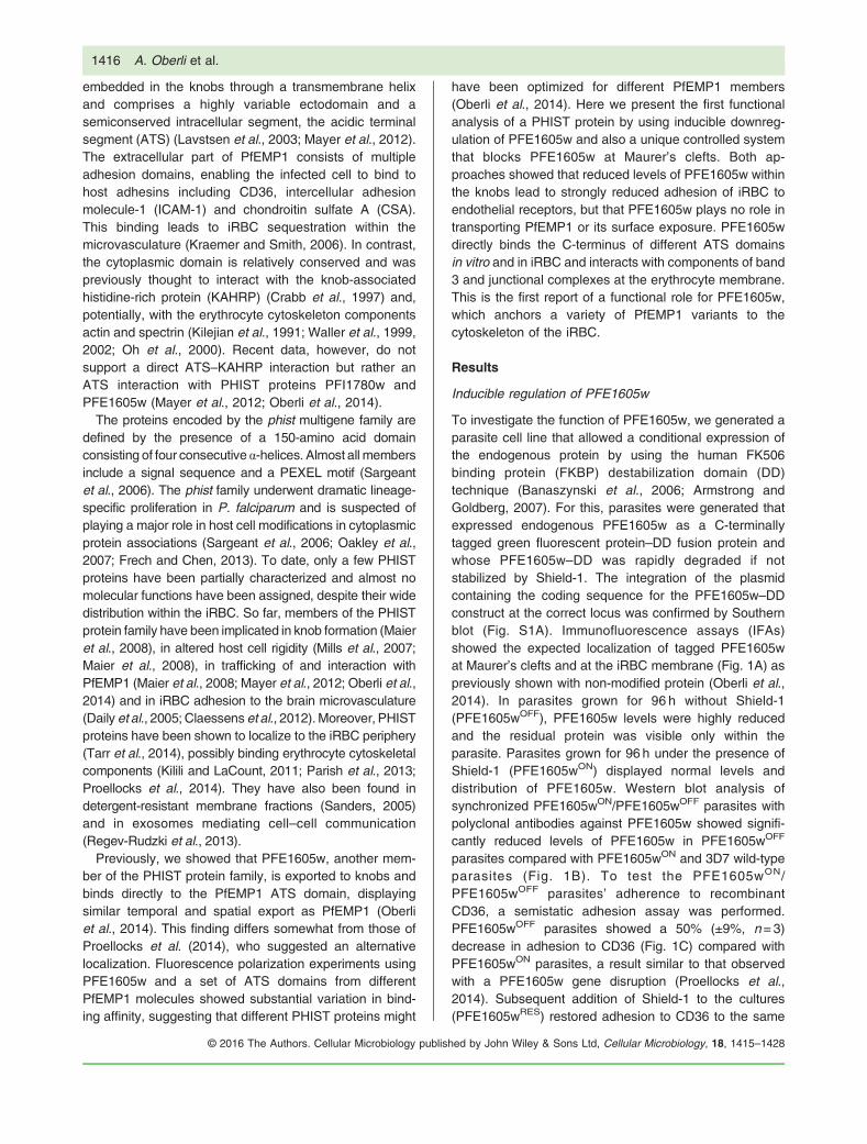
To investigate the function of PFE1605w, we generated aparasite cell line that allowed a conditional expression ofthe endogenous protein by using the human FK506binding protein (FKBP) destabilization domain (DD)technique (Banaszynski et al., 2006; Armstrong andGoldberg, 2007). For this, parasites were generated thatexpressed endogenous PFE1605w as a C-terminallytagged green fluorescent protein–DD fusion protein andwhose PFE1605w–DD was rapidly degraded if notstabilized by Shield-1. The integration of the plasmidcontaining the coding sequence for the PFE1605w–DDconstruct at the correct locus was confirmed by Southernblot (Fig. S1A). Immunofluorescence assays (IFAs)showed the expected localization of tagged PFE1605wat Maurer’s clefts and at the iRBC membrane (Fig. 1A) aspreviously shown with non-modified protein (Oberli et al.,2014). In parasites grown for 96 h without Shield-1(PFE1605wOFF), PFE1605w levels were highly reducedand the residual protein was visible only within theparasite. Parasites grown for 96 h under the presence ofShield-1 (PFE1605wON) displayed normal levels anddistribution of PFE1605w. Western blot analysis ofsynchronized PFE1605wON/PFE1605wOFF parasites withpolyclonal antibodies against PFE1605w showed signifi-cantly reduced levels of PFE1605w in PFE1605wOFF
parasites compared with PFE1605wON and 3D7 wild-typeparasites (Fig. 1B). To test the PFE1605wON/PFE1605wOFF parasites’ adherence to recombinantCD36, a semistatic adhesion assay was performed.PFE1605wOFF parasites showed a 50% (±9%, n=3)decrease in adhesion to CD36 (Fig. 1C) compared withPFE1605wON parasites, a result similar to that observedwith a PFE1605w gene disruption (Proellocks et al.,2014). Subsequent addition of Shield-1 to the cultures(PFE1605wRES) restored adhesion to CD36 to the same
hed by John Wiley & Sons Ltd, Cellular Microbiology, 18, 1415–1428
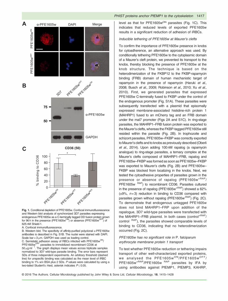
Fig. 1. Conditional depletion of PFE1605w. Confocal immunofluorescenceand Western blot analysis of synchronized 3D7 parasites expressingendogenous PFE1605w as a C-terminally tagged DD fusion protein grownfor 96 h in the presence (PFE1605wON) or absence (PFE1605wOFF) of625 nM Shield-1.A. Confocal immunofluorescence.B. Western blot. The specificity of affinity-purified polyclonal α-PFE1605wantibodies is described in Fig. S1B. The nuclei were stained with DAPI.Scale bar = 3μm. GAPDH was used as loading control.C. Semistatic adhesion assay of RBCs infected with PFE1605wON/PFE1605wOFF parasites to immobilized recombinant CD36 at50 μgml�1. The graph displays mean values across triplicate samplesnormalized to 3D7 wild-type parasite binding. The error bars representSDs of three independent experiments. An arbitrary threshold (dashedline) for unspecific binding was calculated as the mean level of iRBCbinding to 1% w/v BSA plus 2 SDs. P values were calculated by using atwo-tailed Student’s t-test, asterisk indicates P ≤ 0.05.
PHIST proteins anchor PfEMP1 to the cytoskeleton 1417
© 2016 The Authors. Cellular Microbiology published by John Wiley
& Sonslevel as that for PFE1605wON parasites (Fig. 1C). Thisindicates that reduced levels of exported PFE1605wresults in a significant reduction of adhesion of iRBCs.
Inducible tethering of PFE1605w at Maurer’s clefts
To confirm the importance of PFE1605w presence in knobsfor cytoadherence, an alternative approach was used. Byconditionally tethering PFE1605w to the cytoplasmic domainof a Maurer’s cleft protein, we prevented its transport to theknobs, thereby blocking the presence of PFE1605w at theknob structure. The technique is based on theheterodimerization of the FKBP12 to the FKBP-rapamycinbinding (FRB) domain of human mechanistic target ofrapamycin in the presence of rapamycin (Haruki et al.,2008; Busch et al., 2009; Robinson et al., 2010; Xu et al.,2010). First, we generated parasites that expressedPFE1605w C-terminally fused to FKBP under the control ofthe endogenous promoter (Fig. S1A). These parasites weresubsequently transfected with a plasmid that episomallyexpressed membrane-associated histidine-rich protein 1(MAHRP1) fused to an mCherry tag and an FRB domainunder the mal7 promoter (Figs 2A and S1C). In ring-stageparasites, the MAHRP1–FRB fusion protein was exported totheMaurer’s clefts,whereas theFKBP-taggedPFE1605wstillresided within the parasite (Fig. 2B). In trophozoite andschizont parasites, PFE1605w–FKBPwas correctly exportedtoMaurer’s clefts and to knobsaspreviously described (Oberliet al., 2014). Upon adding 100nM rapalog (a rapamycinanalogue) to ring-stage parasites, a ternary complex at theMaurer’s clefts composed of MAHRP1–FRB, rapalog andPFE1605w–FKBPwas formedas soonasPFE1605w–FKBPwas exported to Maurer’s clefts (Fig. 2B) and PFE1605w–FKBP was blocked from localizing in the knobs. Next, wetested the cytoadhesive properties of parasites grown in thepresence or absence of rapalog (PFE1605w+RAP/PFE1605w�RAP) to recombinant CD36. Parasites culturedin the presence of rapalog (PFE1605w+RAP) showed a 62%(±9%, n=3) reduction in binding to CD36 compared withparasites grown without rapalog (PFE1605w-RAP) (Fig. 2C).To demonstrate that endogenous untagged PFE1605wdoes not bind MAHRP1–FRP upon addition of therapalogue, 3D7 wild-type parasites were transfected withthe MAHRP1–FRB plasmid. In both cases (control+RAP/control�RAP), the parasites showed comparable levels ofbinding to CD36, indicating that no heterodimerizationoccurred (Fig. 2C).
PFE1605w has no significant role in P. falciparumerythrocyte membrane protein 1 transport
To test whether PFE1605w reduction or tethering impairstransport of other well-characterized exported proteins,we analysed the PFE1605wON/PFE1605wOFF/PFE1605w+RAP/PFE1605w�RAP parasites by IFA byusing antibodies against PfEMP1, PfEMP3, KAHRP,
Ltd, Cellular Microbiology, 18, 1415–1428

Fig. 2. Controlled tethering of PFE1605w at Maurer’s clefts.A. Schematic representation of controlled PFE1605w tethering. The episomally expressed MAHRP1-FRB fusion protein is exported to Maurer’sclefts (1) prior to the export of C-terminally FKBP-tagged PFE1605w (2). Upon close proximity of the two fusion proteins and addition of rapalog (3),heterodimerization of the FKBP domain and the FRB domain occurs (4) and PFE1605w is immobilized at Maurer’s clefts.B. Confocal immunofluorescence analysis of parasites grown in the absence (PFE1605w�RAP) or presence (PFE1605w+RAP) of 100 nM rapalog.The nuclei were stained with DAPI. Scale bar = 2 μm.C. Semistatic adhesion assay of PFE1605w�RAP/PFE1605w+RAP parasites to immobilized recombinant CD36 protein at 50 μgml�1 concentration.The graph displays mean values across triplicate samples, and the error bars represent the SDs of three independent experiments. An arbitrarythreshold (dashed line) for unspecific binding was calculated as the mean level of iRBC binding to 1% w/v BSA plus two SDs. P values werecalculated by using a two-tailed Student’s t-test; the asterisks indicate P ≤ 0.0001.
1418 A. Oberli et al.
mature parasite-infected erythrocyte surface antigen(MESA), ring-infected erythrocyte surface antigen(RESA), MAHRP1, MAHRP2 and HSP70x. All testedproteins revealed correct subcellular localization inparasites (data shown for PFE1605wON/PFE1605wOFF
parasites; Fig. S2).To test whether the observed reduction inCD36bindingwas
due to a reduction of PfEMP1 surface exposure, we treatediRBCs with trypsin. In all parasite cultures, PfEMP1 wascorrectly displayed on the iRBC surface (Fig. 3A and B) asevident by PfEMP1 proteolysis that yields intact ATS domains.The trypsin cleavage assay also revealed that the size ofPfEMP1 in all parasites were identical, suggesting that thesame PfEMP1 variant was expressed in the parasite linescompared. Scanning electron microscopy (SEM) showed thepresence of knobs in all parasite cell lines (Fig. S3); thus, thereduction in cytoadherence observed in PFE1605wOFF
and PFE1605w+RAP parasites was not due to decreased knobformation.
© 2016 The Authors. Cellular Microbiology publis
Cytoadhesive properties of infected red blood cellsexpressing different P. falciparum erythrocyte membraneprotein 1 in the absence of PFE1605w
Previously, we showed that the recombinant PHISTdomain of PFE1605w interacted with six different ATSvariants with up to 25-fold differences in affinity (Oberliet al., 2014). This suggested that PFE1605w might beoptimized for binding to a subset of PfEMP1 variants;hence, it might be relevant only for iRBC binding to asubset of endothelial receptors. Therefore, we selectedparasites expressing PFE1605w–DD on different hostreceptors, including CD36, ICAM-1 and CSA, in order toisolate parasites expressing different PfEMP1 molecules.After four rounds of pre-selection, we obtained parasitesbinding to CD36, ICAM-1, or CSA (Fig. 4). Expression ofvar genes in all the pre-selected parasite lines was testedby quantitative PCR (qPCR) and showed a cleardifferential expression of var genes, suggesting the
hed by John Wiley & Sons Ltd, Cellular Microbiology, 18, 1415–1428
Fig. 3. PfEMP1 surface exposure isnot impaired upon PFE1605wdepletion in knobs. Determination ofPfEMP1 surface exposure bydetecting trypsin cleavage in 3D7 wildtype, PFE1605wON/PFE1605wOFF
and PFE1605w+RAP/PFE1605w�RAP
parasites.A. PFE1605wON/PFE1605wOFF.B. PFE1605w+RAP/PFE1605w�RAP
parasites. Trophozoites of eachparasite line were treated with trypsin(+) or without trypsin (�), and theextracts were analysed by Westernblot by using antibodies against ATSdomains.
Fig. 4. iRBCs expressing a different PfEMP1 variant show different level of reduction in cytoadherence upon conditional depletion of PFE1605w.A. Preselected iRBCs binding to either recombinant CD36, ICAM-1 or CSA immobilized on tissue-treated glass slides at 50 μgml�1 (CD36, ICAM-1)or 20 μgml�1 (CSA) concentrations. Parasites expressing PFE1605w as a C-terminally tagged DD fusion protein were grown for 96 h in thepresence (PFE1605wON) or absence (PFE1605wOFF) of 625 nM Shield-1. The graphs display overall mean values across triplicate experimentsusing linear regression with a random effect for experiment. The error bars represent the SDs of the triplicate experiments. An arbitrary threshold(dashed line) for unspecific binding was calculated as the mean level of iRBC binding to 1% w/v BSA plus two SDs. P values were calculated byusing a two-tailed Student’s t-test. The asterisks indicate P ≤ 0.0001. ‘ns’ indicates P ≥ 0.05.B. Pie charts show the var transcript distribution in the selected lines. qPCR was performed with specific primers for each var gene as previously reported.
PHIST proteins anchor PfEMP1 to the cytoskeleton 1419
display of a distinct PfEMP1 variant on the iRBC surface(Fig. 4B). Pre-selected parasites were grown with andwithout Shield-1 and after 96 h they were allowed to bind
© 2016 The Authors. Cellular Microbiology published by John Wiley & Sons
to their respective receptor in a semistatic adhesionassay. Parasites grown in the absence of Shield-1showed an approximately 64% reduction in binding to
Ltd, Cellular Microbiology, 18, 1415–1428

1420 A. Oberli et al.
CD36 (Fig. 4A). Binding to ICAM-1 was reduced by 30%and binding to CSA showed no reduction at all (Fig. 4A),indicating that PFE1605w plays no role in CSA-mediatedcytoadherence.
PFE1605w binds the C-terminal part of the acidicterminal segment
Previously, we showed that the recombinant PHISTdomain of PFE1605w binds with low-micromolar affinityto the C-terminal part of the ATS domain (ATS-C) ofPfEMP1 variant PF08_0141 (Oberli et al., 2014). Se-quence conservation among ATS domains suggested thatATS-C provides the PFE1605w binding epitope in mostPfEMP1 variants. To test this, we performed in vitrofluorescence polarization binding experiments by usingthe PFE1605w PHIST domain and fluorescein-labelledrecombinant ATS-C fragments from PfEMP1 variantsdominantly expressed in preselected parasites (Figs 4Band S4A). In almost all cases, we observed direct
Fig. 5. PFE1605w directly binds the ATS C-terminus.A. Fluorescence polarization titrations of 5-FAM-labelled ATS-C constructsdomain. Data points, normalized to the fraction of ATS-C bound at each PFwere derived from four technical replicates. The solid lines correspond to daconstants (Kd) for the PFE1605w–ATS-C interaction are shown. The interacB. Schematic representation of the mini-PfEMP1 construct for Co-IP experiexperiments using parasites expressing mini-PfEMP1 constructs composedOnly peptide hits detected in both of the duplicate experiments are shown.PFE1605w antibodies. IN, input; SN, supernatant; W, wash; E, elution.
© 2016 The Authors. Cellular Microbiology publis
PFE1605w–ATS-C binding with dissociation constants(Kd) in the 4–90μM range (Fig. 5A).
To test whether PFE1605w binds to ATS-C in P.falciparum iRBCs, we designed two mini-PfEMP1 con-structs consisting of an N-terminal part of a PEXEL protein(PF13_0275) including a signal sequence and a PEXELmotif, the ATS-C of two PfEMP1 variants and a 3xHA tag toallow detection (Figs 5B and S4B). The PfEMP1 variantsselected, PF08_0141 and PFF0010w, display approximate-ly 13-fold difference in in vitro affinity (5 and 65μM Kd
respectively) for the PHIST domain of PFE1605w (Oberliet al., 2014). Because the fusion proteins were expressedunder the crt promoter, theywere found early in the life cycle.Due to the lack of a TM domain, the mini-PfEMP1 wassoluble and exported to the erythrocyte cytosol with thepredicted size (Fig. S1D). Potential ATS-C interactionpartners were detected by co-immunoprecipitation (Co-IP)followed by mass spectrometry (MS) for protein identifica-tion. Trophozoite extracts from parasites expressing a mini-PfEMP1 fusion protein were used to isolate potential
from PfEMP1 variants (Fig. S4A) with unlabelled PFE1605w PHISTE1605w concentration, are shown as coloured circles. The error barsta fitted with a single-site association model. Equilibrium dissociationtion of PfEMP1 variant PFD0615c with PFE1605w could not be fitted.ments. C and D. LC-ESI-MS/MS results of two independent Co-IPof the C-terminal part of PF08_0141 (C) or PFF0010w (D).Samples were also analysed by Western blot with α-HA and α-
hed by John Wiley & Sons Ltd, Cellular Microbiology, 18, 1415–1428
PHIST proteins anchor PfEMP1 to the cytoskeleton 1421
interacting proteins through an hemagglutinin (HA)-affinitymatrix. As a negative control, parasite extract with an excessof soluble HA peptide was added during the affinity-matrixbinding of the mini-PfEMP1 fusion proteins. Western blotanalysis confirmed that the mini-PfEMP1 fusion proteinswere successfully purified and that PFE1605wwas coelutedwith both mini-PfEMP1 constructs (Fig. 5C and D). Inaddition, from duplicate Co-IP experiments, the liquidchromatography–mass spectrometry (LC-MS)/MS analysisdetected from both mini-PfEMP1 constructs more than 10peptide hits for PFE1605w (Fig. 5C and D). These resultsdemonstrate a direct protein–protein interaction ofPFE1605w with the C-terminal part of the ATS domain ofdifferent PfEMP1 variants.
Potential PFE1605w interaction partners
To detect other potential PFE1605w interaction partners,we performed Co-IP experiments with parasites express-
© 2016 The Authors. Cellular Microbiology published by John Wiley & Sons
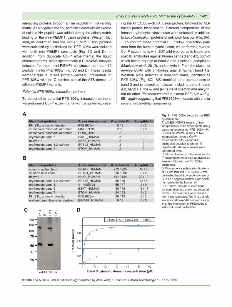
ing the PFE1605w–3xHA fusion protein, followed by MS-based protein identification. Different components of thehuman erythrocyte cytoskeleton were detected, in additionto two Plasmodium proteins of unknown function (Fig. 6A).
To confirm these potential PFE1605w interaction part-ners from the human cytoskeleton, we performed reverseCo-IP experiments with 3D7 wild-type parasite lysate andspecific antibodies against human bands 3 and 4.2, both ofwhich locate equally at band 3 and junctional complexes(Mankelow et al., 2012), and ankyrin 1. From the elution ofreverse Co-IP with antibodies against human band 4.2,Western blots detected a dominant band, identified asPFE1605w (Fig. 6C). MS identified other components ofband 3 and junctional complexes, including band 3, band4.2, band 4.1, the α- and β-chains of spectrin and ankyrin,but no other Plasmodium protein except PFE1605w (Fig.6B), again suggesting that PFE1605w interacts with one orseveral cytoskeletal components.
Fig. 6. PFE1605w binds to the RBCcytoskeleton.A. LC-ESI-MS/MS results of twoindependent Co-IP experiments usingparasites expressing PFE1605w-HA.B. LC-ESI-MS/MS results of twoindependent reverse Co-IPexperiments with α-band 4.2antibodies coupled to protein GDynabeads. All experiments wereperformed twice.C. Elution fractions of the reverse Co-IP experiment were also analysed byWestern blot with α-PFE1605wantibodies.D. Fluorescence polarization titrationsof 5-FAM-labelled PFE1605w-C withunlabelled band 3 cytosolic domain orBSA as a negative control. Data points,normalized to the fraction ofPFE1605w-C bound at each titrantconcentration, are shown as colouredcircles. The error bars were derivedfrom three replicates. The fit to a single-site associationmodel is shownas solidline. The interaction of PFE1605w-Cwith BSA could not be fitted.
Ltd, Cellular Microbiology, 18, 1415–1428
1422 A. Oberli et al.
To further probe the cytoskeletal interactions ofPFE1605w, we produced a fluorescein-labelled recombi-nant PFE1605w fragment (PFE1605w-C) comprising theC-terminal tail of this protein that follows the PHISTdomain. PFE1605w-C was previously shown to bind toinside-out vesicles prepared from uninfected erythrocytes(Proellocks et al., 2014). In the fluorescence–polarizationbinding experiments, PFE1605w-C interacted with recom-binant band 3 with approximately 1.5μM Kd (Fig. 6D). Thisresult demonstrates the direct interaction of PFE1605wwith a specific cytoskeletal protein, although we do notexclude the possibility that PFE1605w partakes in a largermultiprotein complex.
Discussion
The remarkable number of exported PHIST proteinspredicted and the dramatic lineage-specific proliferationof this multigene family in P. falciparum only (Sargeantet al., 2006) suggest an important role for PHIST proteinsin host cell modifications. These modifications lead to thedramatic morbidity and mortality observed with thisparasite. This observation is reflected in the number ofrecent publications showing that PHIST proteins areinvolved in altering host cell rigidity (Mills et al., 2007;Maier et al., 2008), binding erythrocyte components(Silva et al., 2005; Mills et al., 2007; Pei et al., 2007a;Parish et al., 2013; Proellocks et al., 2014), reducingcytoadherence under flow (Maier et al., 2008; Proellockset al., 2014), mediating cell–cell communication(Regev-Rudzki et al., 2013), cytoskeletal association (Tarret al., 2014) and elevated transcript levels of some phistgenes in patients (Daily et al., 2005; Mok et al., 2007;Claessens et al., 2012). Although it has been assumedthat most PHIST proteins contain one or more interactionepitopes (Sargeant et al., 2006), to the best of ourknowledge, no detailed characterization of protein inter-actions directly linked to the functional role of a PHISTprotein has been reported.Here, we have functionally characterized PFE1605w,
which has been shown to bind to the ATS domain ofPfEMP1, comigrates with PfEMP1 in space and time andlocalizes to Maurer’s clefts and knobs (Oberli et al., 2014),although an alternative localization has been suggested(Proellocks et al., 2014). In addition to the well-known andfrequently used conditional post-translational regulationusing an FKBP DD, we applied a ‘knock-sideways’ or‘anchor-away’ system (Haruki et al., 2008; Busch et al.,2009; Robinson et al., 2010; Xu et al., 2010). With thismethod, we took advantage of the rapalog-inducedheterodimerization of the FKBP12 and FRB domains totether PFE1605w at Maurer’s clefts, the transient locationfor a variety of parasite proteins destined to the iRBCmembrane and surface. The tethering technique is a
© 2016 The Authors. Cellular Microbiology publis
powerful way of revealing the function of an exportedprotein in host cell refurbishment and helps to dissect therole of these proteins within the export pathway. Bothmethods, tethering of PFE1605w at Maurer’s cleftsand protein destabilization, confirmed that mislocalizationor depletion of PFE1605w did not result in reducedsurface exposed PfEMP1, suggesting no obvious rolefor PFE1605w in PfEMP1 transport. At the sametime, however, both methods of PFE1605w depletionfrom knobs resulted in large reduction of cytoadherenceto CD36.
The different levels of reduction in parasite cytoadherenceto specific endothelial receptors upon PFE1605w depletionsuggest a highly specialized role for this protein. Previously,we tested six PfEMP1 ATS variants for binding the PHISTdomain of PFE1605w (Oberli et al., 2014) and revealed up to25-fold differences in binding affinities. This suggests thatsequence variation in ATS has optimized PFE1605w forbinding to a PfEMP1 subset and that perhaps otherPHIST proteins might have coevolved with specific ATSdomains to create interaction pairs with maximumbinding strength. In this simplified model, differences inthe PFE1605w–ATS binding affinity might be expected toaccount for differences in the cytoadherence phenotypeupon PFE1605w depletion.
Our assays partly support this model of PFE1605wfunction, as evidenced by the lack of an effect ofPFE1605w depletion on cytoadherence observed forCSA-binding parasites, where the ATS-C fragment of thedominantly expressed PfEMP1 variant (PFL0030c,VAR2CSA) had a very weak binding affinity to thePFE1605w PHIST domain (Kd ~ 90μM). In contrast, theATS-C fragments of PfEMP1 variants most often found inICAM-1-binding parasites, PF07_0050 and PFL0020w,have up to sixfold higher PFE1605w affinity, and ICAM-1parasite cytoadherence is reduced by 30% uponPFE1605w depletion.
The complete picture, however, is more nuanced. Co-IPassays coupled with MS robustly detected the in vivoassociation of PFE1605w with two mini-PfEMP1 con-structs encompassing the ATS-C fragment of two PfEMP1variants. These variants represent the two main subtypesof PfEMP1 ATS, groups A (PF08_0141) and B(PFF0010w). The in vitro affinity of these ATS domainsfor the PFE1605w PHIST, however, varies by more than10-fold. We also observed that the ATS-C fragment of thePfEMP1 variant dominantly expressed in CD36-bindingparasites, PFD0615c, displays essentially no direct affinityfor PFE1605w, despite the large decrease in CD36cytoadherence upon PFE1605w depletion. The resultseems to contradict the simple PFE1605w functionalmodel presented above.
To reconcile these results, we must consider thefollowing: PFE1605w directly binds the majority of ATS
hed by John Wiley & Sons Ltd, Cellular Microbiology, 18, 1415–1428

PHIST proteins anchor PfEMP1 to the cytoskeleton 1423
domains tested here and in previous studies (Oberli et al.,2014). This interaction is present in vivo for both stronglyand weakly associated PFE1605w–ATS pairs. PFE1605wdoes not affect PfEMP1 transport and it colocalizes toknobs with PfEMP1 (Oberli et al., 2014). Indeed, thesignificant reduction in receptor binding upon tetheringPFE1605w to Maurer’s clefts strongly indicates that thisprotein exercises its functional role in knobs. There,PFE1605w is likely to be joined by (and might act togetherwith) other ATS-binding PHIST proteins such asPFI1780w (Mayer et al., 2012; Oberli et al., 2014), therebyaccounting for the partial disruption of cytoadherenceupon PFE1605w depletion. Further, in certain PfEMP1variants, such as the CD36-binding PFD0615c, thePFE1605w–ATS interaction might be mediated or stronglyreinforced by other PHIST proteins. Because most of thePHIST proteins are expressed simultaneously, it seemsthat partnering must occur in the cytosol of the host ordirectly at the periphery (Fig. 7). Transcriptome analysesof all selected cell lines grown with or without shieldexcluded a possible upregulation of certain PHIST (datanot shown).
To date, only a few direct interactions of exportedproteins with cytoskeletal components of erythrocyteshave been described and confirmed, e.g. KAHRP (Pei
© 2016 The Authors. Cellular Microbiology published by John Wiley & Sons
et al., 2005; Weng et al., 2014), PfEMP3 (Pei et al.,2007b), RESA (Pei et al., 2007a) and MESA (Magowanet al., 2000). Both KAHRP and PfEMP3 are required forcorrect trafficking and functional PfEMP1 display on theerythrocyte surface (Crabb et al., 1997; Waterkeyn et al.,2000), while PFE1605w is not. Co-IP experiments with thefull-length PFE1605w–HA fusion protein identified anumber of host integral membrane proteins and compo-nents of the erythrocyte cytoskeleton as putative binders,and fluorescence polarization experiments confirmed thedirect interaction of the PFE1605w C-terminus with thecytosolic domain of band 3. Thus, we have now shown thepresence of two interaction epitopes within PFE1605w, atits PHIST domain and the C-terminus, making it ananchoring molecule between PfEMP1 and the hostcytoskeleton. The findings are consistent with previousassays suggesting an association of PFE1605w C-terminal fragments with erythrocyte-derived inside-outvesicles (Proellocks et al., 2014). Both co-IP and in vitroexperiments suggest that PFE1605w, and thus PfEMP1,targets the host’s band 3 and junctional complexes, while,interestingly, PFE1605w was the only parasite proteindetected. The next step would be to map the exactinteraction epitopes of PFE1605w with band 3 andpossibly other cytoskeletal proteins. In addition to eluci-
Fig. 7. Schematic representation of theproposed functional role of PFE1605w withinthe iRBC knob structure.A. Both Co-IP experiments and in vivoexperiments showed that the PHIST domain ofPFE1605w binds the C-terminal part ofdifferent ATS domains and the C-terminal partof PFE1605w targets the host band 3 andjunctional complexes, thus making it ananchoring molecule between PfEMP1 and thehost cytoskeleton.B. As various PHIST domains interacted withthe same PfEMP1 epitope but with differentaffinities, it is conceivable that another PHISTprotein might take over the function ofPFE1605w depending on the surface exposedPfEMP1 molecule.
Ltd, Cellular Microbiology, 18, 1415–1428
1424 A. Oberli et al.
dating the complex that anchors PfEMP1 to the cytoskel-eton, it would be valuable to study whether other PHISTproteins might bind PfEMP1 variants, in particularVAR2CSA, where CSA cytoadherence was not reducedupon depletion of PFE1605w. The PHIST interactomeinvites further studies to fully understand the remodellingof the host cell leading to pathology.In summary, we show that the PHIST protein
PFE1605w binds not only to PfEMP1 but also to membersof band 3 and junctional complexes of the host cell.PFE1605w, however, plays no role in the transport ofPfEMP1. We also show that various PfEMP1 moleculesinteract differently with PFE1605w and binding to endo-thelial receptors is partially disrupted upon conditionalknock-down or misplacement of PFE1605w. A profoundanalysis of other exported PHIST proteins and theirinteraction partners should help to reveal key componentsof the cytoadherence complex.
Experimental procedures
Parasite culture and transfection
Plasmodium falciparum 3D7 cell culture and transfection were
performed according to standard procedures (Ljungström et al.,
2008). Transfected parasites were grown in the presence of the
indicated combinations of 10 nM WR99210 (Jacobs Pharmaceu-
ticals, Cologne, Germany), 2.5mgml�1 blasticidin (Life Technol-
ogies, Zug, Switzerland), 625 nM Shield-1 and 100 nM A/C
Heterodimerizer (Clontech).
Plasmid constructs
Primers 5′-ATTTGGATCCATGAGGTTTACTAATTCATTATATTCG-
3′ and 5′-ATATGCTAGCATTTTTTTTTTTATTTTCTTTTCCAGA
TTTG-3′ were used to clone full-length PFE1605w into pBcamR–
3xHA (Flueck et al., 2009) via BamHI and NheI restriction sites. To
fuse the FKBP DD to the C-terminus of PFE1605w in 3D7 wild-type
parasites, a 785bp flank of the 3′ end of PFE1605w was cloned into
pARL-DD via BglII and AvrII restriction sites by using the primers 5′-
ATATAGATCTTAACAGCAAATAGATTTT TATGGAG-3′ and 5′-
ATATCCTAGGATTTTTTTTTTTATTTTCTTTTCCAGATTTG-3′.
MAHRP1 was cloned into mal7–mCherry–FRB (kindly provided by
Tobias Spielmann (Grüring et al., 2011)) via XhoI and KpnI restriction
sites by using the primers 5′-ATATCTCGAGATGGCAGAGCAA
GCAGC-3′ and 5′-CAGCGGTA CCATTATCTTTTTTTTCTTGTT
CTAATTTTGC-3′. Mini-PfEMP1 constructs were synthesized
(Fig. S4B) and cloned into pBcamR-3xHA via NcoI and NheI
restriction sites.
Western blot analysis
Parasite proteins were obtained as previously described (Oberli
et al., 2014), and samples were run on 12% w/v polyacrylamide
bis-Tris, 4–12% w/v polyacrylamide bis-Tris or 3–8% w/v
polyacrylamide Tris-acetate NuPAGE gels (Invitrogen). Proteins
were detected by using rabbit antibodies directed against the
© 2016 The Authors. Cellular Microbiology publis
PFE1605w PHIST domain (α-PFE1605w) (Pacific Immunology
Inc.) (Fig. S1B), rabbit α-HA (Roche 1:100), mouse α-glyceralde-
hyde-3-phosphate dehydrogenase (α-GAPDH) (1:20 000), rat α-
mCherry (Life Technologies; 1:1000) and mouse α-ATS (1:500).
PfEMP1 was extracted as described (Van Schravendijk et al.,
1993) and detected with the mouse α-ATS (1:500) antibody.
Southern blot analysis
Genomic DNA of saponin-lysed parasites was isolated as
previously described (Beck, 2002). DNA was digested with AflII
and XhoI restriction enzymes (New England Biolabs), separated
on a 0.8% w/v agarose gel and transferred to a Amersham
Hybond–N+ membrane (GE Healthcare). The blot was probed
with [32P]-dATP-labelled hdhfr PCR fragments.
Fluorescence microscopy
Immunofluorescence assays were performed on acetone-fixed
blood smears of infected parasite cultures (Spielmann et al.,
2003) and blocked with 3% v/w BSA. Primary antibodies included
rabbit α-PFE1605w (1:200), mouse α-KAHRP (1:200), mouse α-
RESA (1:250), rabbit α-MESA (1:250), mouse α-ATS (1:100),
mouse α-PfEMP3 (1:100), rabbit α-MAHRP1 (1:200), rabbit α-
HSP70x (1:500) and rat α-mCherry (Life Technologies; 1:200).
Secondary antibodies (goat α-rabbit Alexa 594, goat α-mouse
Alexa 594, goat α-rabbit Alexa 488, goat α-rat Alexa 594;
Invitrogen) were incubated with 1 μg ml�1 4,6-diamidino-2-
phenylindole (DAPI; Roche) at 1:200 dilution. Images were taken
with a Zeiss LSM 700 confocal microscope (Carl Zeiss GmbH,
Jena, Germany), with ×63 oil-immersion lens (1.4 numerical
aperture) and processed in PHOTOSHOP CS6.
Scanning electron microscopy
Af ter knob select ion and Percol l pur i f icat ion, the
erythrocytes/iRBCs were fixed in 2% v/v glutaraldehyde in
phosphate buffer for 1 h at room temperature. After three washes
in PBS, the samples were transferred to coverslips preliminary
coated with poly-L-lysine (Sigma), dehydrated in increasing
concentration of ethanol (10% v/v, 25% v/v, 50% v/v, 75% v/v,
90% v/v and 2× 100% v/v, 10min each) and dried at the critical
point. Finally, coverslips were mounted onto stubs, sputtered with
5 nm platinum (LEICA EM ACE600) and imaged at 5 kV with a
SEM Versa 3D (FEI). The micrographs were coloured in
PHOTOSHOP CS6.
Trypsin cleavage assay
For trypsin cleavage, Percoll-purified trophozoite stage parasites
were incubated either in L-(tosylamido-2-phenyl) ethyl
chloromethyl ketone-treated trypsin (Sigma, 100 μg ml�1 in
PBS) or in trypsin and 1mgml�1 soybean trypsin inhibitor
(Sigma, 1mgml�1 in PBS) for 15min at 37°C. The digest was
stopped by the addition of soybean trypsin inhibitor to a final
concentration of 1mgml�1. PfEMP1 extraction and subsequent
analysis was done as previously described (Van Schravendijk
et al., 1993; Waterkeyn et al., 2000).
hed by John Wiley & Sons Ltd, Cellular Microbiology, 18, 1415–1428
PHIST proteins anchor PfEMP1 to the cytoskeleton 1425
Selection for receptor binding with recombinant protein
Subpopulations of parasites were selected by panning the
parental parasite cell line (3D7) over purified human recombinant
CD36 (50 μgml�1), CD31 (50 μgml�1), ICAM-1 (50 μgml�1),
Thrombospondin-1 (50 μgml�1), endothelial protein C receptor
(50 μgml�1) and CSA (20 μgml�1) according to Ockenhouse
et al. (1991). Recombinant proteins were dissolved in double-
distilled H2O to the indicated final concentration and absorbed to
a six-well tissue culture plate (Falcon 353045, Corning, NY, USA)
overnight at 4°C. The wells were blocked with 1% w/v BSA in
RPMI medium for 1 h at 37°C. and the parasite culture was added
for 2 h with a gentle shake of the tissue culture plate every 15min.
Unbound parasites were removed by five gentle washes with
RPMI-Hepes and uninfected RBCs (5% haematocrit) were added.
After 24 h of incubation allowing late-stage parasites to release
merozoites to invade new RBCs, the newly invaded RBCs were
transferred into continuous cell culture. The panning procedure
was repeated four times prior to RNA isolation and cytoadhesion
assays.
Cytoadhesion assay
Purified recombinant protein was spotted on wells of an eight-
chamber polystyrene vessel tissue culture-treated glass slide
(Falcon, Big Flats, NY, USA) with concentrations as indicated
and coated overnight at 4°C to allow proteins to absorb to the
surface. The wells were blocked with 1% w/v BSA in RPMI
medium for 1 h at 37°C. Selected parasite cell lines were split
and cultured separately with or without 500 nM Shield-1 for 96 h.
Parasites were washed twice with RPMI-Hepes and spotted
onto immobilized recombinant protein and cultured for 2 h under
continuous and simultaneous shaking (140 r.p.m., proBlot 25
Rocker; Labnet International Inc., NY, USA) (105 r.p.m.,
Lab-Therm LT-W, Kühner, Switzerland) at 37°C. Non-bound
erythrocytes were removed by gently flooding each well with
RPMI-Hepes six times with simultaneous shaking for 2 min.
Bound iRBCs were fixed with 2% v/v glutaraldehyde in
RPMI-Hepes overnight and stained with Giemsa for 1 h and
microscopically quantified. Results are shown as mean
number of parasites bound per square millimetre and normal-
ized to 1% parasitaemia.
Quantitative PCR for P. falciparum erythrocyte membraneprotein 1 expression
Synchronized cultures of PFE1605w–DD expressing parasites
preselected to bind CD36, ICAM-1 or CSA were split and cultured
96 h in the presence (+) or absence (�) of Shield-1, and ring-
stage parasites were used for var transcript profiling, as
previously described (Dahlbäck et al., 2007). Transcript abun-
dance of each 3D7 var gene was determined relative to internal
control transcripts by qPCR by using gene-specific primers and
complementary DNA synthesized from total RNA extracted from
pelleted infected erythrocytes dissolved in TRIzol.
© 2016 The Authors. Cellular Microbiology published by John Wiley & Sons
Recombinant protein expression
Codon-optimized genes encoding the ATS-C fragments of
PfEMP1 variants (Fig. S4A) were cloned in a modified pET-16
vector (Merc Millipore). Gene fragments coding for amino acids
300–528 of PFE1605w (PFE1605w-C) or amino acids 1–379 of
human erythrocytic band 3 were cloned in a pFloat2 vector
(Rogala et al., 2015), which provides an N-terminal His6 tag.
Purification and fluorescent labelling of ATS-C and PFE1605w-
C was performed as previously described (Mayer et al., 2012);
briefly, clones were transformed in Escherichia coli strain BL21
(DE3), grown in Luria–Bertani medium and protein expression
was induced with 0.1mM isopropyl β-D-1-thiogalactopyranoside.
Cells were lysed by sonication, and proteins were purified from
lysate supernatants by using metal-affinity, ion-exchange and
size-exclusion chromatography. Fluorescent labelling was per-
formed by N-(5-fluoresceinyl)maleimide (5-FAM; Invitrogen)
conjugating to a single cysteine residue at the protein N-
terminus that was added during cloning. Labelled ATS-C and
unreacted dye were separated by size-exclusion chromatogra-
phy. Protein identity and 5-FAM labelling was confirmed by
electrospray ionisation (ESI) MS.
Purification of the PFE1605w PHIST domain and the cytosolic
band 3 domain was performed as previously described (Zhang
et al., 2000; Oberli et al., 2014).
Fluorescence polarization binding assays
Fluorescence polarization measurements were recorded at 20°C
by using a CLARIOStar fluorimeter (BMG Labtech; λex = 485 nm,
λem = 520 nm). Five hundred nanomolar 5-FAM-labelled ATS-C
variants in 50mM NaCl, 20mM Na2HPO4 pH 6.5 buffer were
titrated with defined concentrations of PFE1605w PHIST domain
in the same buffer. For the band 3–PFE1605w-C interaction,
0.5 μM 5-FAM-labelled PFE1605w-C in 50mM NaCl, 20mM
Na2HPO4 pH 7.0 buffer was titrated with unlabelled band 3.
Changes in fluorescence polarization were fitted by using a single
binding model in the program ORIGIN (OriginLab).
Coimmunoprecipitation experiments
Three hundred millilitres of cell culture (5% haematocrit, 5-8%
parasitaemia) of 3D7 parasites or 3D7 parasites episomally
expressing PFE1605w–3xHA/mini-PfEMP1 was cross-linked in
1% v/v formaldehyde. The reaction was stopped after 10min by
adding 2.5M glycine. Immunoprecipitation was performed as
previously described (Dietz et al., 2014). For the Co-IP
experiments with the mini-PfEMP1 fusion protein, Pierce α-HA
magnetic beads (Thermo Scientific) were used. For the reverse
Co-IP with α-band 4.2 antibodies, Dynabeads Protein G were
used together with the cross-linking reagent BS3 to avoid
coelution of antibodies, according to the manufacturer’s protocol
(Life Technologies). The eluted fraction was analysed on a 4–
12% w/v polyacrylamide bis-Tris gel (Invitrogen) and fractions of it
or TCA precipitated pellets were sent to the central core facility for
LC-MS/MS analysis.
Ltd, Cellular Microbiology, 18, 1415–1428
1426 A. Oberli et al.
Acknowledgements
The authors are grateful to Tobias Spielmann for sharing themal7–mCherry–FRB vector. We would like to thank the followingcolleagues for sharing antibodies: Brian Cooke (anti-ATS),Claudia Daubenberger (anti-GAPDH), Jude Przyborski (anti-HSP70x), Diane Taylor (anti-KAHRP), Ross Coppel (anti-MESA)and Alex Maier (anti-PfEMP3). We thank Henning Stahlberg andhis team at the C-CINA and the Image Core Facility, Biozentrum,University of Basel, for the access and support for confocal andelectron microscopy work and David Stanton for maintenance ofthe Oxford Biochemistry biophysics facility. We are grateful to DirkReiter for assistance with experiments.
This work was supported by the Swiss National ScienceFoundation (http://www.snf.ch) (Grant 31003A_149297/1 toHPB), the Wellcome Trust (http://www.wellcome.ac.uk) (RCDfellowship 088497/Z/09/Z to IV and PhD studentship to JLD andEEC) as well as by the Lundbeck Foundation (http://www.lundbeckfoundation.com) and the Danish Council for IndependentResearch, Medical Sciences, Sapere Aude program (http://ufm.dk/en/research-and-innovation/councils-and-commissions/the-danish-council-for-independent-research) (DFF–4004-00624B toTL). The funders had no role in the study design, data collectionand analysis, the decision to publish or the preparation of themanuscript.
Conflict of interest
The authors declare no conflict of interest.
Author contributions
AO, LZ and SR performed the cell biological experiments;FB performed the electron microscopy experiments; MEB,JLD and EEC performed the biophysical interactionexperiments; TL analysed the var gene expression andAO, JV and HPB conceived the experiments and wrotethe paper.
References
Armstrong, C.M., and Goldberg, D.E. (2007) An FKBPdestabilization domain modulates protein levels in Plas-modium falciparum. Nat Methods 4: 1007–1009.
Banaszynski, L.A., Chen, L., Maynard-Smith, L.A., Ooi, A.G.L.,and Wandless, T.J. (2006) A rapid, reversible, and tunablemethod to regulate protein function in living cells usingsynthetic small molecules. Cell 126: 995–1004.
Beck, H.-P. (2002) Extraction and purification of plasmodiumpDNA. In Malaria Methods and Protocols. Doolan, D. (ed).Totowa, New Jersey: Humana Press, pp. 159–163.
Busch, A., Kiel, T., and Hübner, S. (2009) Quantification ofnuclear protein transport using induced heterodimerization.Traffic 10: 1221–1227.
Claessens, A., Adams, Y., Ghumra, A., Lindergard, G.,Buchan, C.C., Andisi, C., et al. (2012) A subset of groupA-like var genes encodes the malaria parasite ligands forbinding to human brain endothelial cells. Proc Natl AcadSci 109: E1772–E1781.
Crabb, B.S., Cooke, B.M., Reeder, J.C., Waller, R.F., Caruana,S.R., Davern, K.M., et al. (1997) Targeted gene disruption
© 2016 The Authors. Cellular Microbiology publis
shows that knobs enable malaria-infected red cells tocytoadhereunder physiological shear stress.Cell89: 287–296.
Dahlbäck, M., Lavstsen, T., Salanti, A., Hviid, L., Arnot, D.E.,Theander, T.G., and Nielsen, M.A. (2007) Changes in vargene mRNA levels during erythrocytic development in twophenotypically distinct Plasmodium falciparum parasites.Malar J 6: 78.
Daily, J.P., Le Roch, K.G., Sarr, O., Ndiaye, D., Lukens, A.,Zhou, Y., et al. (2005) In vivo transcriptome of Plasmodiumfalciparum reveals overexpression of transcripts thatencode surface proteins. J Infect Dis 191: 1196–1203.
Dietz, O., Rusch, S., Brand, F., Mundwiler-Pachlatko, E.,Gaida, A., Voss, T., and Beck, H.-P. (2014) Characteriza-tion of the small exported Plasmodium falciparum mem-brane protein SEMP1. PLoS One 9 e103272.
Flueck, C., Bartfai, R., Volz, J., Niederwieser, I., Salcedo-Amaya, A.M., Alako, B.T.F., et al. (2009) Plasmodiumfalciparum heterochromatin protein 1 marks genomic locilinked to phenotypic variation of exported virulence factors.PLoS Pathog 5 e1000569.
Frech, C., and Chen, N. (2013) Variant surface antigens ofmalaria parasites: functional and evolutionary insights fromcomparative gene family classification and analysis. BMCGenomics 14: 427.
Grüring, C., Heiber, A., Kruse, F., Ungefehr, J., Gilberger, T.-W., and Spielmann, T. (2011) Development and host cellmodifications of Plasmodium falciparum blood stages infour dimensions. Nat Commun 2: 165.
Haruki, H., Nishikawa, J., and Laemmli, U.K. (2008) Theanchor-away technique: rapid, conditional establishment ofyeast mutant phenotypes. Mol Cell 31: 925–932.
Heiber, A., Kruse, F., Pick, C., Grüring, C., Flemming, S.,Oberli, A., et al. (2013) Identification of new PNEPsindicates a substantial non-PEXEL exportome and under-pins common features in Plasmodium falciparum proteinexport. PLoS Pathog 9 e1003546.
Hiller, N.L., Bhattacharjee, S., van Ooij, C., Liolios, K.,Harrison, T., Lopez-Estrano, C., and Haldar, K. (2004) Ahost-targeting signal in virulence proteins reveals asecretome in malarial infection. Science 306: 1934–1937.
Ljungström, I., Moll, K., Perlmann, H., Scherf, A., and Wahlgren,M. (2008). Methods in malaria research (MR4/ATCC).
Kilejian, A., Rashid, M.A., Aikawa, M., Aji, T., and Yang, Y.-F.(1991) Selective association of a fragment of the knobprotein with spectrin, actin and the red cell membrane. MolBiochem Parasitol 44: 175–181.
Kilili, G.K., and LaCount, D.J. (2011) An erythrocytecytoskeleton-binding motif in exported Plasmodiumfalciparum proteins. Eukaryot Cell 10: 1439–1447.
Kraemer, S.M., and Smith, J.D. (2006) A family affair: vargenes, PfEMP1 binding, and malaria disease. Curr OpinMicrobiol 9: 374–380.
Lavstsen, T., Salanti, A., Jensen, A.T., Arnot, D.E., andTheander, T.G. (2003) Sub-grouping of Plasmodiumfalciparum 3D7 var genes based on sequence analysis ofcoding and non-coding regions. Malar J 2: 27.
Magowan, C., Nunomura, W., Waller, K.L., Yeung, J., Liang,J., Van Dort, H., et al. (2000) Plasmodium falciparumhistidine-rich protein 1 associates with the band 3 bindingdomain of ankyrin in the infected red cell membrane.Biochim Biophys Acta BBA-Mol Basis Dis 1502: 461–470.
hed by John Wiley & Sons Ltd, Cellular Microbiology, 18, 1415–1428
PHIST proteins anchor PfEMP1 to the cytoskeleton 1427
Maier, A.G., Rug, M., O’Neill, M.T., Brown, M., Chakravorty,S., Szestak, T., et al. (2008) Exported proteins required forvirulence and rigidity of Plasmodium falciparum-infectedhuman erythrocytes. Cell 134: 48–61.
Mankelow, T.J., Satchwell, T.J., and Burton, N.M. (2012)Refined views of multi-protein complexes in the erythrocytemembrane. Blood Cells Mol Dis 49: 1–10.
Marti, M., Good, R.T., Rug, M., Knuepfer, E., and Cowman, A.F.(2004)Targetingmalaria virulence and remodelingproteins tothe host erythrocyte. Science 306: 1930–1933.
Mayer, C., Slater, L., Erat, M.C., Konrat, R., and Vakonakis, I.(2012) Structural analysis of the Plasmodium falciparumerythrocyte membrane protein 1 (PfEMP1) intracellulardomain reveals a conserved interaction epitope. J BiolChem 287: 7182–7189.
Mills, J.P., Diez-Silva, M., Quinn, D.J., Dao, M., Lang, M.J.,Tan, K.S.W., et al. (2007) Effect of plasmodial RESAprotein on deformability of human red blood cells harboringPlasmodium falciparum. Proc Natl Acad Sci 104:9213–9217.
Mok, B.W., Ribacke, U., Winter, G., Yip, B.H., Tan, C.-S.,Fernandez, V., et al. (2007) Comparative transcriptomalanalysis of isogenic Plasmodium falciparum clones ofdistinct antigenic and adhesive phenotypes. Mol BiochemParasitol 151: 184–192.
Nguitragool, W., Bokhari, A.A.B., Pillai, A.D., Rayavara, K.,Sharma, P., Turpin, B., et al. (2011) Malaria parasite clag3genes determine channel-mediated nutrient uptake byinfected red blood cells. Cell 145: 665–677.
Oakley, M.S.M., Kumar, S., Anantharaman, V., Zheng, H.,Mahajan, B., Haynes, J.D., et al. (2007) Molecular factorsand biochemical pathways induced by febrile temperaturein intraerythrocytic Plasmodium falciparum parasites. InfectImmun 75: 2012–2025.
Oberli, A., Slater, L.M., Cutts, E., Brand, F., Mundwiler-Pachlatko, E., Rusch, S., et al. (2014) A Plasmodiumfalciparum PHIST protein binds the virulence factorPfEMP1 and comigrates to knobs on the host cell surface.FASEB J 28: 4420–4433.
Ockenhouse, C.F., Ho, M., Tandon, N.N., Van Seventer, G.A.,Shaw, S., White, N.J., et al. (1991) Molecular basis ofsequestration in severe and uncomplicated Plasmodiumfalciparum malaria: differential adhesion of infected erythro-cytes to CD36 and ICAM-l. J Infect Dis 164: 163–169.
Oh, S.S., Voigt, S., Fisher, D., Yi, S.J., LeRoy, P.J., Derick, L.H.,et al. (2000) Plasmodium falciparum erythrocyte mem-brane protein 1 is anchored to the actin–spectrin junctionand knob-associated histidine-rich protein in the erythro-cyte skeleton. Mol Biochem Parasitol 108: 237–247.
Parish, L.A., Mai, D.W., Jones, M.L., Kitson, E.L., andRayner, J.C. (2013) A member of the Plasmodiumfalciparum PHIST family binds to the erythrocyte cytoskel-eton component band 4.1. Malar J 12: 1–9.
Pei, X., An, X., Guo, X., Tarnawski, M., Coppel, R., andMohandas, N. (2005) Structural and Functional studies ofinteraction between Plasmodium falciparum knob-associated histidine-rich protein (KAHRP) and erythrocytespectrin. J Biol Chem 280: 31166–31171.
Pei, X., Guo, X., Coppel, R., Bhattacharjee, S., Haldar, K.,Gratzer, W., et al. (2007a) The ring-infected erythrocytesurface antigen (RESA) of Plasmodium falciparum stabi-
© 2016 The Authors. Cellular Microbiology published by John Wiley & Sons
lizes spectrin tetramers and suppresses further invasion.Blood 110: 1036–1042.
Pei, X., Guo, X., Coppel, R., Mohandas, N., and An, X.(2007b) Plasmodium falciparum erythrocyte membraneprotein 3 (PfEMP3) destabilizes erythrocyte membraneskeleton. J Biol Chem 282: 26754–26758.
Proellocks, N.I., Herrmann, S., Buckingham, D.W., Hanssen,E., Hodges, E.K., Elsworth, B., et al. (2014) A lysine-richmembrane-associated PHISTb protein involved in alter-ation of the cytoadhesive properties of Plasmodiumfalciparum-infected red blood cells. FASEB J 28:3103–3113.
Regev-Rudzki, N., Wilson, D.W., Carvalho, T.G., Sisquella,X., Coleman, B.M., Rug, M., et al. (2013) Cell–cellcommunication between malaria-infected red blood cellsvia exosome-like vesicles. Cell 153: 1120–1133.
Robinson, M.S., Sahlender, D.A., and Foster, S.D. (2010)Rapid inactivation of proteins by rapamycin-inducedrerouting to mitochondria. Dev Cell 18: 324–331.
Rogala, K.B., Dynes, N.J., Hatzopoulos, G.N., Yan, J., Pong,S.K., Robinson, C.V., et al. (2015) The Caenorhabditiselegans protein SAS-5 forms large oligomeric assembliescritical for centriole formation. eLife 4.
Sanders, P.R. (2005) Distinct protein classes including novelmerozoite surface antigens in raft-like membranes ofPlasmodium falciparum. J Biol Chem 280: 40169–40176.
Sargeant, T.J., Marti, M., Caler, E., Carlton, J.M., Simpson,K., Speed, T.P., and Cowman, A.F. (2006) Lineage-specificexpansion of proteins exported to erythrocytes in malariaparasites. Genome Biol 7: R12.
Van Schravendijk, M., Pasloske, B., Baruch, D., Handunnetti,S., and Howard, R. (1993) Immunochemical characteriza-tion and differentiation of two approximately 300-kDerythrocyte membrane-associated proteins of Plasmodiumfalciparum, PfEMP1 and PfEMP3. Am J Trop Med Hyg 49:552–565.
Silva, M.D., Cooke, B.M., Guillotte, M., Buckingham, D.W.,Sauzet, J.-P., Scanf, C.L., et al. (2005) A role for thePlasmodium falciparum RESA protein in resistance againstheat shock demonstrated using gene disruption: pheno-typing resa-KO Plasmodium falciparum parasites. MolMicrobiol 56: 990–1003.
Spielmann, T., Fergusen, D.J., and Beck, H.-P. (2003)etramps, a new Plasmodium falciparum gene family codingfor developmentally regulated and highly charged mem-brane proteins located at the parasite–host cell interface.Mol Biol Cell 14: 1529–1544.
Spillman, N.J., Beck, J.R., and Goldberg, D.E. (2015) Proteinexport into malaria parasite–infected erythrocytes: mecha-nisms and functional consequences. Annu Rev Biochem84: 813–841.
Tarr, S.J., Moon, R.W., Hardege, I., and Osborne, A.R. (2014)A conserved domain targets exported PHISTb familyproteins to the periphery of Plasmodium infected erythro-cytes. Mol Biochem Parasitol 196: 29–40.
Waller, K.L., Cooke, B.M., Nunomura, W., Mohandas, N., andCoppel, R.L. (1999) Mapping the binding domains involvedin the interaction between the Plasmodium falciparumknob-associated histidine-rich protein (KAHRP) and thecytoadherence ligand P. falciparum erythrocyte membraneprotein 1 (PfEMP1). J Biol Chem 274: 23808–23813.
Ltd, Cellular Microbiology, 18, 1415–1428
1428 A. Oberli et al.
Waller, K.L., Nunomura, W., Cooke, B.M., Mohandas, N., andCoppel, R.L. (2002) Mapping the domains of thecytoadherence ligand Plasmodium falciparum erythrocytemembrane protein 1 (PfEMP1) that bind to the knob-associated histidine-rich protein (KAHRP). Mol BiochemParasitol 119: 125–129.
Waterkeyn, J.G., Wickham, M.E., Davern, K.M., Cooke, B.M.,Coppel, R.L., Reeder, J.C., et al. (2000) Targetedmutagenesis of Plasmodium falciparum erythrocyte mem-brane protein 3 (PfEMP3) disrupts cytoadherence ofmalaria-infected red blood cells. EMBO J 19: 2813–2823.
Watermeyer, J.M., Hale, V.L., Hackett, F., Clare, D.K., Cutts,E.E., Vakonakis, I., et al. (2016) A spiral scaffold underliescytoadherent knobs in Plasmodium falciparum-infectederythrocytes. Blood 127: 343–351.
Weng, H., Guo, X., Papoin, J., Wang, J., Coppel, R.,Mohandas, N., and An, X. (2014) Interaction of Plasmodi-um falciparum knob-associated histidine-rich protein(KAHRP) with erythrocyte ankyrin R is required for itsattachment to the erythrocyte membrane. Biochim BiophysActa Biomembr 1838: 185–192.
© 2016 The Authors. Cellular Microbiology publis
Xu, T., Johnson, C.A., Gestwicki, J.E., and Kumar, A. (2010)Conditionally controlling nuclear trafficking in yeast bychemical-induced protein dimerization. Nat Protoc 5:1831–1843.
Zhang, D., Kiyatkin, A., Bolin, J.T., and Low, P.S. (2000)Crystallographic structure and functional interpretation ofthe cytoplasmic domain of erythrocyte membrane band 3.Blood 96: 2925–2933.
Supporting information
Additional Supporting information may be found in the onlineversion of this article at the publisher's web-site:
Fig. S1. Plasmid maps, Southern blot and Western blots ofextracts from cell lines used in this study.Fig. S2. Localization of well-characterized exported proteinsupon PFE1605w reduction.Fig. S3. Reduced levels of PFE1605w do not alter knobformation.Fig. S4. ATS-C and mini-PfEMP1 constructs.
hed by John Wiley & Sons Ltd, Cellular Microbiology, 18, 1415–1428