stanford cardiovascular ct scanning and injection...
TRANSCRIPT

2
Siemens Sensation 64
Stanford Cardiovascular CT Scanning and Injection Protocols
(v2.04, Nov. 2006)
Department of Radiology Stanford University Medical Center
Stanford, CA
Table of Content
Stanford Cardiovascular CT Scanning and Injection Protocols .................................................. 3
Head and Neck CTA ................................................................................................................... 5
Upper Extremity Runoff ............................................................................................................... 7
Thoracic Outlet ............................................................................................................................ 9
Bilateral Thoracic Outlet ............................................................................................................ 11
Limited Upper Extremity Runoff (Trauma) ................................................................................ 13
Chest/Abdomen/Pelvis CTA...................................................................................................... 15
Abdomen/Pelvis CTA ................................................................................................................ 17
Renal Arteries CTA ................................................................................................................... 19
Living Related Renal Donor ...................................................................................................... 21
Lower Extremity Runoff ............................................................................................................. 23
Popliteal Entrapment CTA......................................................................................................... 25
Limited Lower Extremity Runoff (Trauma) ................................................................................ 27
CaScoring Sequence ................................................................................................................ 29
CaScoring Spiral ....................................................................................................................... 31
CaScoring and Coronary CTA .................................................................................................. 33
CaScoring and Coronary CTA below 50 bpm HR..................................................................... 35
Coronary Bypass Graft CTA ..................................................................................................... 37
Gated Chest .............................................................................................................................. 39
Gated Chest/Abdomen/Pelvis CTA (gated Chest, non-gated Abdomen) ................................ 41
Left Atrial Mapping .................................................................................................................... 43
Coronary Vein Mapping............................................................................................................. 45
Abbreviations............................................................................................................................. 47
4
Stanford Cardiovascular CT Scanning and Injection Protocols
Preface Cardiovascular imaging is one of the most important and visible beneficiaries of the recent and dramatic evolution of comuted tomographic (CT) technology. CT– angiography (CTA) has evolved into a routine minimally-invasive vas and cardiac CT have evolved into widely available routine clinical applications The quality of a CT angiogram or a cardiac CT strudy depends to a great extent on the appropriate selection of CT data acquisition and contrast medium injection parameters. Image quality (section thickness, noise), radiation exposure, and arterial opacification have all to be taken into account when planning a cardiovascular CT study. All user-selectable parameters have to be integrated with patient physiology as well. The following cardiovascular CT scanning and injection protocols have been developed with the goal to derive theoretically sound and at the same time practicable scanning and injections protocols for the Siemens Sensation 64 multi-slice scanner at Stanford. The protocols provided in this text provide complete information regarding (i) patient positioning and scanning ranges, (ii) all scanning parameters for all parts of the respective CT study, and (iii) detailed injection protocols. While much attention to detail may limit easy readability, we feel that this is outweighted by completeness. Both radiologists and technologists benefit more from a detailed reference when facing less common CT examinations. On behalf of the Cardiovascular Imaging Section we would like to thank all the radiological technologists at Stanford who's input and feedback has influenced this text. We are particularly greatful to Dominique Sandner and the support of Siements Medical Solutions, whithout whom this project would not have been possible. Dominik Fleischmann, M.D. Geoffrey D Rubin, M.D. November 2006

5
Head and Neck CTA
Indication: Cerebrovascular disease, intracranial aneurysms
Patient preparation: 20G IV cannula.
Patient positioning: • Head first, supine, arms down by the side of the body; • Remove any metal objects (e.g. ear-rings) out of the scanning range;
Comment: • Instruct patient not to swallow during CT angiogram.
Notes:
↑ ROI in aortic arch 1
2,3 4
6
A: Chronologic Prescription and Scanning Range # Scanning Range Delay BH Dir. 0 Topogram vertex aortic arch insp 1 Head Seq base of skull vertex no 2 premonitoring no N/A 3 monitoring ROI in aortic arch; Trigger level 100 HU 10 s no N/A 4 HN CTA aortic arch vertex. 10 s insp B: Scanning Parameters
# Eff mAs
Ref mAs kV Detector
config. Pitch Rot. time
Scan time Comment
0 Topogram 35 • 120 • • • • LAT, 512 mm 1 Head Seq 410 • 120 64x0.6 18mm 1.0 s • Gantry tilt 2 premonitoring 20 • 120 • • • • 3 monitoring 20 • 120 • • 30 scans, 1.2s cycle time 4 HN CTA • 200 120 64×0.6 variable 0.37 s 10 s C: Reconstruction Parameters
# Type/ orient STh RI Filter Window
WW / WL Field of View /
Comment 1 Head Seq axial 4.8mm 4.8mm H41f 80/40 skull 4a HN CTA axial 1mm 0.7mm B25f 600/80 neck 4b HN CTA axial 3mm 3mm B31f 600/80 same 4c HN CTA MIPcor 3mm 1mm B25f 700/200 same 4d HN CTA MIPsag 3mm 1mm B25f 700/200 same D: Contrast Medium Injection Parameters CM Concentration ≥ 350mgI/ml Scantime 10 s for everyone Injection duration 12 s Bolus Timing Automated bolus trigger (Care-Bolus); 100 HU trigger level, minimum user delay (2 s) Saline flushing 40 mL @ same flow rate as contrast
Body weight
Flow Rate
relaxed Volume
< 121 lbs (<55kg) 4.0 mL/s 48 mL 121 – 143 lbs (<65kg) 4.5 mL/s 54 mL 143 – 187 lbs (~75kg) 5.0 mL/s 60 mL 187 – 209 lbs (>85kg) 5.5 mL/s 66 mL
> 209 lbs (>95kg) 6.0 mL/s 72 mL

Cardiovascular Vascular01^ UPPER_EXTR_RUNOFF Upper Extremity Runoff
7
Upper Extremity Runoff
Indication: upper extremity aneurysmal/embolic/occlusive disease, trauma, AVM, vasculitis, Hemodialysis shunt evaluation, anatomic mapping for free flap graft harvesting, hypothenar-hamate syndrome
Patient preparation: 20G IV cannula at contra lateral arm! Hand and fingers should not be cold !
Patient positioning: • head first, supine or prone (depending on patient’s physical condition), arm to be scanned
above head. • Fingers spread out and taped down (see figure above) • use laser light and adjust table height to align arm and fingers with center of scanner.
Comment: • It is critical to select the scanning range first, and then set the scan-time to 30 seconds in
all patients.
Notes: Blood flow to the upper extremity at rest is generally low. Opacification of small hand/finger arteries may be difficult, notably in a cold environment. This slow acquitision/scanning protocol allows for adequate filling of small peripheral arteries. Consider a one-minute exercising (squeezing a ball/object) before taping down the fingers, or use post-ischemic hyperemia (one minute blood-pressure cuff immediately released before the injection). Scan time can be shorter / scanning range restricted for AVMs.
1 2,3 4
↓ fingers spread out and taped down
↓ ROI in aortic arch
Cardiovascular Vascular01^ UPPER_EXTR_RUNOFF Upper Extremity Runoff
8
A: Chronologic Prescription and Scanning Range # Scanning Range Delay BH Dir. 0 Topogram diaphragm finger tips insp 1 non contrast aortic arch finger tips insp 2 premonitoring no N/A 3 monitoring ROI in aortic arch 10 s no N/A 4 UE Runoff aortic arch. through finger tips 2-3 s insp B: Scanning Parameters
# Eff mAs
Ref mAs kV Detector
config. Pitch Rot. time
Scan time Comment
0 Topogram 35 • 120 • • • • AP, 1500 mm 1 non contrast • 140 120 24×1.2 1.0 0.5 s • 2 premonitoring 20 • 120 • • • • 3 monitoring 20 • 120 • • 30 scans, 1.2s cycle time 4 UE Runoff • 250 120 64×0.6 variable 0.5 s 30s fixed 30s scan time! C: Reconstruction Parameters
# Type/ orient STh RI Filter Window
WW / WL Field of View /
Comment 1 non contrast axial 5mm 5mm B31f 400/40 4 UE Runoff axial 1mm 0.7mm B25f 600/80 Include aortic arch and elbow D: Contrast Medium Injection Parameters (biphasic) CM Concentration ≥ 350mgI/ml Scantime needs to be 30s for all patients ! Injection duration 30s for all patients ! Bolus Timing Automated bolus trigger (Care-Bolus); 100 HU trigger level, minimum user delay (~2s) Saline flushing 40 mL @ same flow rate as Phase II
Body weight
Phase I
Phase II
Total CM Vol.
< 121 lbs (<55kg) 20 mL @ 4.0 mL/s 80 mL @ 3.2 mL/s 100 mL 121 – 143 lbs (<65kg) 23 mL @ 4.5 mL/s 90 mL @ 3.6 mL/s 113 mL 143 – 187 lbs (~75kg) 25 mL @ 5.0 mL/s 100 mL @ 4.0 mL/s 125 mL 187 – 209 lbs (>85kg) 28 mL @ 5.5 mL/s 110 mL @ 4.4 mL/s 138 mL
> 209 lbs (>95kg) 30 mL @ 6.0 mL/s 120 mL @ 4.8 mL/s 150 mL

Cardiovascular Vascular01^THORACIC_OUTLET_SYNDROM Thoracic Outlet
9
Thoracic Outlet
Indication: Thoracic Outlet Syndrome (TOS)
Patient preparation: 20G IV cannula, at contra lateral side Hand and fingers should not be cold
Patient positioning: • Scan 1: head first, supine, arms down; • Scan 2: head first, supine or prone (depending on patients physical condition), arm to be
scanned raised above head with finger tips spread out and taped down; • Head turned away from elevated arm shoulder; • use laser light and adjust table height to align arms and fingers with center of scanner
Comment: • this protocol requires two arterial scans (one scan in a neutral arm position and one
provocation-maneuver scan with elevated arm), • position needs to be changed after first contrast scan and a second Topogram is required; • It is critical to select the scanning range first, and then set the scan-time for the first scan to
10 seconds, for the second scan to 20 seconds in all patients.
Notes: Blood flow to the upper extremity at rest is generally low. Opacification of small hand/finger arteries may be difficult, notably in a cold environment. Consider a one-minute exercising (e.b. squeezing a ball) before taping down the fingers for the provocation-maneuver scan.
1 4 2,3
↑ Scan 1: relaxed position, arms down by the side
↑ fingers taped down
↑ ROI in aortic arch
5,6 7
Scan 2: maneuver or provocative position with affected arm raised above head (prone position also possible) →
Cardiovascular Vascular01^THORACIC_OUTLET_SYNDROM Thoracic Outlet
10
A: Chronologic Prescription and Scanning Range # Scanning Range Delay BH Dir. 0 Topogram neck diaphragm insp 1 non contrast mid chest lower neck insp 2 premonitoring no N/A 3 monitoring ROI aortic arch; 100 HU trigger level; 10 s no N/A 4 relaxed mid chest lower neck 2-3 s insp 0 Topogram diaphragm finger tips insp 5 premonitoring no N/A 6 monitoring ROI aortic arch; 100 HU trigger level; 10 s no N/A 7 maneuver aortic arch through finger tips 2-3 s insp B: Scanning Parameters
# Eff mAs
Ref mAs kV Detector
config. Pitch Rot. time
Scan time Comment
0 Topogram 35 • 120 • • • • AP, 512 mm 1 non contrast • 140 120 24×1.2 1.0 0.5 s • 2 premonitoring 20 • 120 • • • • 3 monitoring 20 • 120 • • 30 scans, 1.2s cycle time 4 relaxed • 250 120 64×0.6 variable 0.5 s 10s fixed 10s scan time! 0 Topogram 35 • 120 • • • • AP, 1500 mm 5 premonitoring 20 • 120 • • • • 6 monitoring 20 • 120 • • 30 scans, 1.2s cycle time 7 maneuver • 250 120 64×0.6 variable 0.5 s 30s fixed 20s scan time! C: Reconstruction Parameters # Type/
orient STh RI Filter Window WW / WL
Field of View / Comment
1 non contrast axial 5mm 5mm B31f 400/40 4 relaxed axial 1mm 0.7mm B25f 600/80 Include both sides 7 maneuver axial 1mm 0.7mm B25f 600/80 Include aortic arch and elbow D: Contrast Medium Injection Parameters (monophasic and biphasic) CM Concentration ≥ 350mgI/ml Scantime Relaxed 10 s, maneuver 20 s for all patients Injection duration Relaxed 10 s; maneuver 20 s for all patients Bolus Timing Autom. bolus trigger (Care-Bolus); 100HU trigger level; 2s and 2s user Delay, respectively Saline flushing 40 mL @ at same flow rate (scan 2: same as phase II)
Body weight
relaxed Flow Rate
relaxed Volume
< 121 lbs (<55kg) 4.0 mL/s 40 mL 121 – 143 lbs (<65kg) 4.5 mL/s 45 mL 143 – 187 lbs (~75kg) 5.0 mL/s 50 mL 187 – 209 lbs (>85kg) 5.5 mL/s 55 mL
> 209 lbs (>95kg) 6.0 mL/s 60 mL
Body weight maneuver
Scan 2: Phase I maneuver
Scan 2: Phase II
Total CM Vol. < 121 lbs (<55kg) 20 mL @ 4.0 mL/s 48 mL @ 3.2 mL/s 108 mL
121 – 143 lbs (<65kg) 23 mL @ 4.5 mL/s 54 mL @ 3.6 mL/s 122 mL 143 – 187 lbs (~75kg) 25 mL @ 5.0 mL/s 60 mL @ 4.0 mL/s 135 mL 187 – 209 lbs (>85kg) 28 mL @ 5.5 mL/s 66 mL @ 4.4 mL/s 149 mL
> 209 lbs (>95kg) 30 mL @ 6.0 mL/s 72 mL @ 4.8 mL/s 162 mL
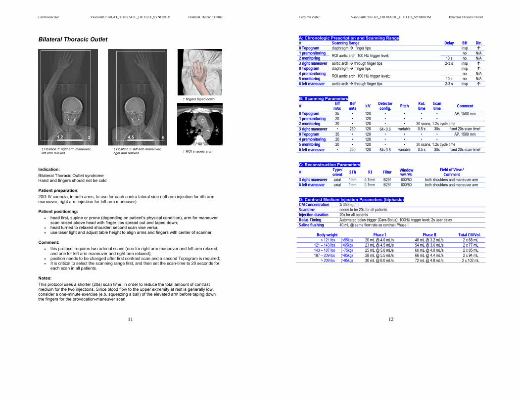
Cardiovascular Vascular01^BILAT_THORACIC_OUTLET_SYNDROM Bilateral Thoracic Outlet
11
Bilateral Thoracic Outlet
Indication: Bilateral Thoracic Outlet syndrome Hand and fingers should not be cold
Patient preparation: 20G IV cannula, in both arms, to use for each contra lateral side (left arm injection for rith arm maneuver, right arm injection for left arm maneuver)
Patient positioning: • head first, supine or prone (depending on patient’s physical condition), arm for maneuver
scan raised above head with finger tips spread out and taped down; • head turned to relaxed shoulder; second scan vise versa; • use laser light and adjust table height to align arms and fingers with center of scanner
Comment: • this protocol requires two arterial scans (one for right arm maneuver and left arm relaxed,
and one for left arm maneuver and right arm relaxed), • position needs to be changed after first contrast scan and a second Topogram is required; • It is critical to select the scanning range first, and then set the scan-time to 20 seconds for
each scan in all patients.
Notes: This protocol uses a shorter (20s) scan time, in order to reduce the total amount of contrast medium for the two injections. Since blood flow to the upper extremity at rest is generally low, consider a one-minute exercise (e.b. squeezing a ball) of the elevated arm before taping down the fingers for the provocation-maneuver scan.
↑ fingers taped down
↑ ROI in aortic arch ↑ Position 2: left arm maneuver, right arm relaxed
4,5 63
↑ Position 1: right arm maneuver, left arm relaxed
1,2 3
Cardiovascular Vascular01^BILAT_THORACIC_OUTLET_SYNDROM Bilateral Thoracic Outlet
12
A: Chronologic Prescription and Scanning Range # Scanning Range Delay BH Dir. 0 Topogram diaphragm finger tips insp 1 premonitoring no N/A 2 monitoring ROI aortic arch; 100 HU trigger level; 10 s no N/A 3 right maneuver aortic arch through finger tips 2-3 s insp 0 Topogram diaphragm finger tips insp 4 premonitoring no N/A 5 monitoring ROI aortic arch; 100 HU trigger level;; 10 s no N/A 6 left maneuver aortic arch through finger tips 2-3 s insp B: Scanning Parameters # Eff
mAs Ref
mAs kV Detector config. Pitch Rot.
time Scan time Comment
0 Topogram 35 • 120 • • • • AP, 1500 mm 1 premonitoring 20 • 120 • • • • 2 monitoring 20 • 120 • • 30 scans, 1.2s cycle time 3 right maneuver • 250 120 64×0.6 variable 0.5 s 30s fixed 20s scan time! 0 Topogram 35 • 120 • • • • AP, 1500 mm 4 premonitoring 20 • 120 • • • • 5 monitoring 20 • 120 • • 30 scans, 1.2s cycle time 6 left maneuver • 250 120 64×0.6 variable 0.5 s 30s fixed 20s scan time! C: Reconstruction Parameters # Type/
orient STh RI Filter Window WW / WL
Field of View / Comment
3 right maneuver axial 1mm 0.7mm B25f 600/80 both shoulders and maneuver arm 6 left maneuver axial 1mm 0.7mm B25f 600/80 both shoulders and maneuver arm D: Contrast Medium Injection Parameters (biphasic) CM Concentration ≥ 350mgI/ml Scantime needs to be 20s for all patients Injection duration 20s for all patients Bolus Timing Automated bolus trigger (Care-Bolus); 100HU trigger level; 2s user delay Saline flushing 40 mL @ same flow rate as contrast Phase II
Body weight
Phase I
Phase II
Total CM Vol.
< 121 lbs (<55kg) 20 mL @ 4.0 mL/s 48 mL @ 3.2 mL/s 2 x 68 mL 121 – 143 lbs (<65kg) 23 mL @ 4.5 mL/s 54 mL @ 3.6 mL/s 2 x 77 mL 143 – 187 lbs (~75kg) 25 mL @ 5.0 mL/s 60 mL @ 4.0 mL/s 2 x 85 mL 187 – 209 lbs (>85kg) 28 mL @ 5.5 mL/s 66 mL @ 4.4 mL/s 2 x 94 mL
> 209 lbs (>95kg) 30 mL @ 6.0 mL/s 72 mL @ 4.8 mL/s 2 x 10² mL

Cardiovascular Vascular01^LTD_UPPER_EXTR_RUNOFF Limited Upper Extremity Runoff
13
Limited Upper Extremity Runoff (Trauma)
Indication: Blunt or penetrating trauma to arm, forearm or hand, suspected vascular injury; hand AVM, hypothenar-hamate syndrome
Patient preparation: 20G IV cannula at contra lateral arm!
Patient positioning: • head first, supine or prone (depending on patient’s physical condition), arm to be scanned
above head. • Fixate arm to the middle of the table; • use laser light and adjust table height to align arm and fingers with center of scanner.
Comment: • It is critical to select the scanning range and then set the scan-time to 15 seconds in all
patients.
Notes:
4
2,3
1
put ROI outside the body and start manually at contrast arrival ↓
Cardiovascular Vascular01^LTD_UPPER_EXTR_RUNOFF Limited Upper Extremity Runoff
14
A: Chronologic Prescription and Scanning Range # Scanning Range Delay BH Dir. 0 Topogram shoulder finger tips insp 1 non contrast shoulder finger tips insp 2 premonitoring no N/A 3 monitoring ROI outside the body at beginning of scan range 10 s no N/A 4 Ltd UE Runoff proximal to elbow through finger tips 2-3 s insp B: Scanning Parameters
# Eff mAs
Ref mAs kV Detector
config. Pitch Rot. time
Scan time Comment
0 Topogram 35 • 120 • • • • AP, 512 mm 1 non contrast • 140 120 24×1.2 1.0 0.5 s • 2 premonitoring 20 • 120 • • • • 3 monitoring 20 • 120 • • 30 scans, 1.2s cycle time 4 Ltd UE Runoff • 250 120 64×0.6 variable 0.5 s 15s fixed 15s scan time! C: Reconstruction Parameters
# Type/ orient STh RI Filter Window
WW / WL Field of View /
Comment 1 non contrast axial 3mm 3mm B31f 400/40 4 Ltd UE Runoff axial 1mm 0.7mm B25f 600/80 Arm, forearm, hand D: Contrast Medium Injection Parameters CM Concentration ≥ 350mgI/ml Scantime needs to be 15s for all patients ! Injection duration 15s for all patients ! Bolus Timing Automated bolus trigger (Care-Bolus); manual start, minimum user delay (~2s) Saline flushing 40 mL @ same flow rate as Contrast
Body weight
Flow Rate
Volume
< 121 lbs (<55kg) 4.0 mL/s 60 mL 121 – 143 lbs (<65kg) 4.5 mL/s 68 mL 143 – 187 lbs (~75kg) 5.0 mL/s 75 mL 187 – 209 lbs (>85kg) 5.5 mL/s 83 mL
> 209 lbs (>95kg) 6.0 mL/s 90 mL
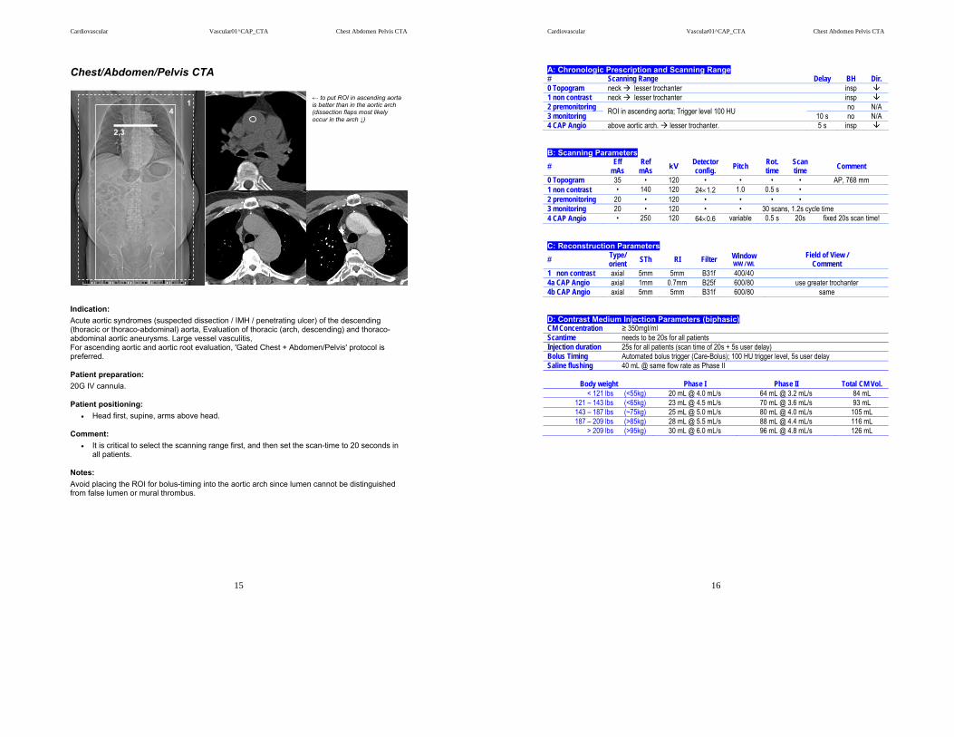
Cardiovascular Vascular01^CAP_CTA Chest Abdomen Pelvis CTA
15
Chest/Abdomen/Pelvis CTA
Indication: Acute aortic syndromes (suspected dissection / IMH / penetrating ulcer) of the descending (thoracic or thoraco-abdominal) aorta, Evaluation of thoracic (arch, descending) and thoraco-abdominal aortic aneurysms. Large vessel vasculitis, For ascending aortic and aortic root evaluation, 'Gated Chest + Abdomen/Pelvis' protocol is preferred.
Patient preparation: 20G IV cannula.
Patient positioning: • Head first, supine, arms above head.
Comment: • It is critical to select the scanning range first, and then set the scan-time to 20 seconds in
all patients.
Notes: Avoid placing the ROI for bolus-timing into the aortic arch since lumen cannot be distinguished from false lumen or mural thrombus.
← to put ROI in ascending aorta is better than in the aortic arch (dissection flaps most likely occur in the arch ↓)
1 4
2,3
Cardiovascular Vascular01^CAP_CTA Chest Abdomen Pelvis CTA
16
A: Chronologic Prescription and Scanning Range # Scanning Range Delay BH Dir. 0 Topogram neck lesser trochanter insp 1 non contrast neck lesser trochanter insp 2 premonitoring no N/A 3 monitoring ROI in ascending aorta; Trigger level 100 HU 10 s no N/A 4 CAP Angio above aortic arch. lesser trochanter. 5 s insp B: Scanning Parameters
# Eff mAs
Ref mAs kV Detector
config. Pitch Rot. time
Scan time Comment
0 Topogram 35 • 120 • • • • AP, 768 mm 1 non contrast • 140 120 24×1.2 1.0 0.5 s • 2 premonitoring 20 • 120 • • • • 3 monitoring 20 • 120 • • 30 scans, 1.2s cycle time 4 CAP Angio • 250 120 64×0.6 variable 0.5 s 20s fixed 20s scan time! C: Reconstruction Parameters
# Type/ orient STh RI Filter Window
WW / WL Field of View /
Comment 1 non contrast axial 5mm 5mm B31f 400/40 4a CAP Angio axial 1mm 0.7mm B25f 600/80 use greater trochanter 4b CAP Angio axial 5mm 5mm B31f 600/80 same D: Contrast Medium Injection Parameters (biphasic) CM Concentration ≥ 350mgI/ml Scantime needs to be 20s for all patients Injection duration 25s for all patients (scan time of 20s + 5s user delay) Bolus Timing Automated bolus trigger (Care-Bolus); 100 HU trigger level, 5s user delay Saline flushing 40 mL @ same flow rate as Phase II
Body weight
Phase I
Phase II
Total CM Vol.
< 121 lbs (<55kg) 20 mL @ 4.0 mL/s 64 mL @ 3.2 mL/s 84 mL 121 – 143 lbs (<65kg) 23 mL @ 4.5 mL/s 70 mL @ 3.6 mL/s 93 mL 143 – 187 lbs (~75kg) 25 mL @ 5.0 mL/s 80 mL @ 4.0 mL/s 105 mL 187 – 209 lbs (>85kg) 28 mL @ 5.5 mL/s 88 mL @ 4.4 mL/s 116 mL
> 209 lbs (>95kg) 30 mL @ 6.0 mL/s 96 mL @ 4.8 mL/s 126 mL

Cardiovascular Vascular01^AP_CTA Abdomen Pelvis CTA
17
Abdomen/Pelvis CTA
Indication: abdominal, iliac or mesenteric aneurysm evaluation and surveillance; atherosclerotic or inflammatory occlusive disease of the aorta and it's branches. Acute and chronic mesenteric ischemia.
Patient preparation: 20G IV cannula.
Patient positioning: • Head first, supine, arms above head.
Comment: • It is critical to select the scanning range first, and then set the scan-time to 10 seconds in
all patients.
Notes: add a portal venous phase for mesenteric ischemia (i.e. to see bowel wall / mesenteric veins)
1
4 2,3
↑ ROI above celiac artery (Th12)
Cardiovascular Vascular01^AP_CTA Abdomen Pelvis CTA
18
A: Chronologic Prescription and Scanning Range # Scanning Range Delay BH Dir. 0 Topogram diaphragm lesser trochanter insp 1 non contrast diaphragm lesser trochanter insp 2 premonitoring no N/A 3 monitoring ROI in aorta above celiac artery (Th12); Trigger level 100 HU 10 s no N/A 4 AP Angio above celiac artery. lesser trochanter. 8 s insp B: Scanning Parameters
# Eff mAs
Ref mAs kV Detector
config. Pitch Rot. time
Scan time Comment
0 Topogram 35 • 120 • • • • AP, 512 mm 1 non contrast • 140 120 24×1.2 1.0 0.5 s • 2 premonitoring 20 • 120 • • • • 3 monitoring 20 • 120 • • 30 scans, 1.2s cycle time 4 AP Angio • 250 120 64×0.6 variable 0.5 s 20s fixed 10s scan time! C: Reconstruction Parameters
# Type/ orient STh RI Filter Window
WW / WL Field of View /
Comment 1 non contrast axial 5mm 5mm B31f 400/40 4a AP Angio axial 1mm 0.7mm B25f 600/80 use greater trochanter 4b AP Angio axial 5mm 5mm B31f 600/80 same D: Contrast Medium Injection Parameters CM Concentration ≥ 350mgI/ml Scantime needs to be 10s for all patients Injection duration 18s for all patients (scan time of 10s + 8s user delay) Bolus Timing Automated bolus trigger (Care-Bolus); 100 HU trigger level, 8s user delay Saline flushing 40 mL @ same flow rate as contrast
Body weight
Flow Rate
Volume
< 121 lbs (<55kg) 4.0 mL/s 72 mL 121 – 143 lbs (<65kg) 4.5 mL/s 81 mL 143 – 187 lbs (~75kg) 5.0 mL/s 90 mL 187 – 209 lbs (>85kg) 5.5 mL/s 99 mL
> 209 lbs (>95kg) 6.0 mL/s 108 mL
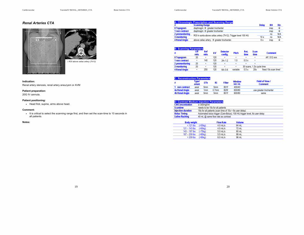
Cardiovascular Vascular01^RENAL_ARTERIES_CTA Renal Arteries CTA
19
Renal Arteries CTA
Indication: Renal artery stenosis, renal artery aneurysm or AVM
Patient preparation: 20G IV cannula.
Patient positioning: • Head first, supine, arms above head.
Comment: • It is critical to select the scanning range first, and then set the scan-time to 10 seconds in
all patients.
Notes:
1
4 2,3
↑ ROI above celiac artery (Th12)
Cardiovascular Vascular01^RENAL_ARTERIES_CTA Renal Arteries CTA
20
A: Chronologic Prescription and Scanning Range # Scanning Range Delay BH Dir. 0 Topogram diaphragm greater trochanter insp 1 non contrast diaphragm greater trochanter insp 2 premonitoring no N/A 3 monitoring ROI in aorta above celiac artery (Th12); Trigger level 100 HU 10 s no N/A 4 Renal Angio above celiac artery. greater trochanter. 6 s insp B: Scanning Parameters
# Eff mAs
Ref mAs kV Detector
config. Pitch Rot. time
Scan time Comment
0 Topogram 35 • 120 • • • • AP, 512 mm 1 non contrast • 140 120 24×1.2 1.0 0.5 s • 2 premonitoring 20 • 120 • • • • 3 monitoring 20 • 120 • • 30 scans, 1.2s cycle time 4 Renal Angio • 250 120 64×0.6 variable 0.5 s 20s fixed 10s scan time! C: Reconstruction Parameters
# Type/ orient STh RI Filter Window
WW / WL Field of View /
Comment 1 non contrast axial 5mm 5mm B31f 400/40 4a Renal Angio axial 1mm 0.7mm B25f 600/80 use greater trochanter 4b Renal Angio axial 5mm 5mm B31f 600/80 same D: Contrast Medium Injection Parameters CM Concentration ≥ 350mgI/ml Scantime needs to be 10s for all patients Injection duration 16s for all patients (scan time of 10s + 6s user delay) Bolus Timing Automated bolus trigger (Care-Bolus); 100 HU trigger level, 6s user delay Saline flushing 40 mL @ same flow rate as contrast
Body weight
Flow Rate
Volume
< 121 lbs (<55kg) 4.0 mL/s 64 mL 121 – 143 lbs (<65kg) 4.5 mL/s 72 mL 143 – 187 lbs (~75kg) 5.0 mL/s 80 mL 187 – 209 lbs (>85kg) 5.5 mL/s 88 mL
> 209 lbs (>95kg) 6.0 mL/s 96 mL
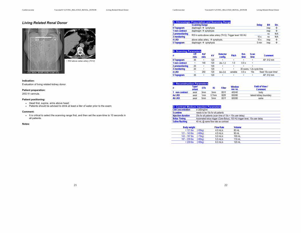
Cardiovascular Vascular01^LIVING_RELATED_RENAL_DONOR Living Related Renal Donor
21
Living Related Renal Donor
Indication: Evaluation of living related kidney donor.
Patient preparation: 20G IV cannula.
Patient positioning: • Head first, supine, arms above head; • Patients should be advised to drink at least a liter of water prior to the exam;
Comment: • It is critical to select the scanning range first, and then set the scan-time to 10 seconds in
all patients.
Notes:
1
4 2,3
↑ ROI above celiac artery (Th12)
Cardiovascular Vascular01^LIVING_RELATED_RENAL_DONOR Living Related Renal Donor
22
A: Chronologic Prescription and Scanning Range # Scanning Range Delay BH Dir. 0 Topogram diaphragm symphysis insp 1 non contrast diaphragm symphysis insp 2 premonitoring no N/A 3 monitoring ROI in aorta above celiac artery (Th12); Trigger level 100 HU 10 s no N/A 4 LRD above celiac artery. symphysis. 10 s insp 5 Topogram diaphragm symphysis 5 min insp B: Scanning Parameters
# Eff mAs
Ref mAs kV Detector
config. Pitch Rot. time
Scan time Comment
0 Topogram 35 • 120 • • • • AP, 512 mm 1 non contrast • 140 120 24×1.2 1.0 0.5 s • 2 premonitoring 20 • 120 • • • • 3 monitoring 20 • 120 • • 30 scans, 1.2s cycle time 4 LRD • 250 120 64×0.6 variable 0.5 s 10s fixed 10s scan time! 5 Topogram 35 • 120 • • • • AP, 512 mm C: Reconstruction Parameters # Type/
orient STh RI Filter Window WW / WL
Field of View / Comment
1 non contrast axial 3mm 3mm B31f 400/40 body 4a LRD axial 1mm 0.7mm B25f 600/80 lateral kidney boundary 4b LRD axial 5mm 5mm B31f 600/80 same D: Contrast Medium Injection Parameters CM Concentration ≥ 350mgI/ml Scantime needs to be 10s for all patients Injection duration 20s for all patients (scan time of 10s + 10s user delay) Bolus Timing Automated bolus trigger (Care-Bolus); 100 HU trigger level, 10s user delay Saline flushing 40 mL @ same flow rate as contrast
Body weight
Flow Rate
Volume
< 121 lbs (<55kg) 4.0 mL/s 80 mL 121 – 143 lbs (<65kg) 4.5 mL/s 90 mL 143 – 187 lbs (~75kg) 5.0 mL/s 100 mL 187 – 209 lbs (>85kg) 5.5 mL/s 110 mL
> 209 lbs (>95kg) 6.0 mL/s 120 mL
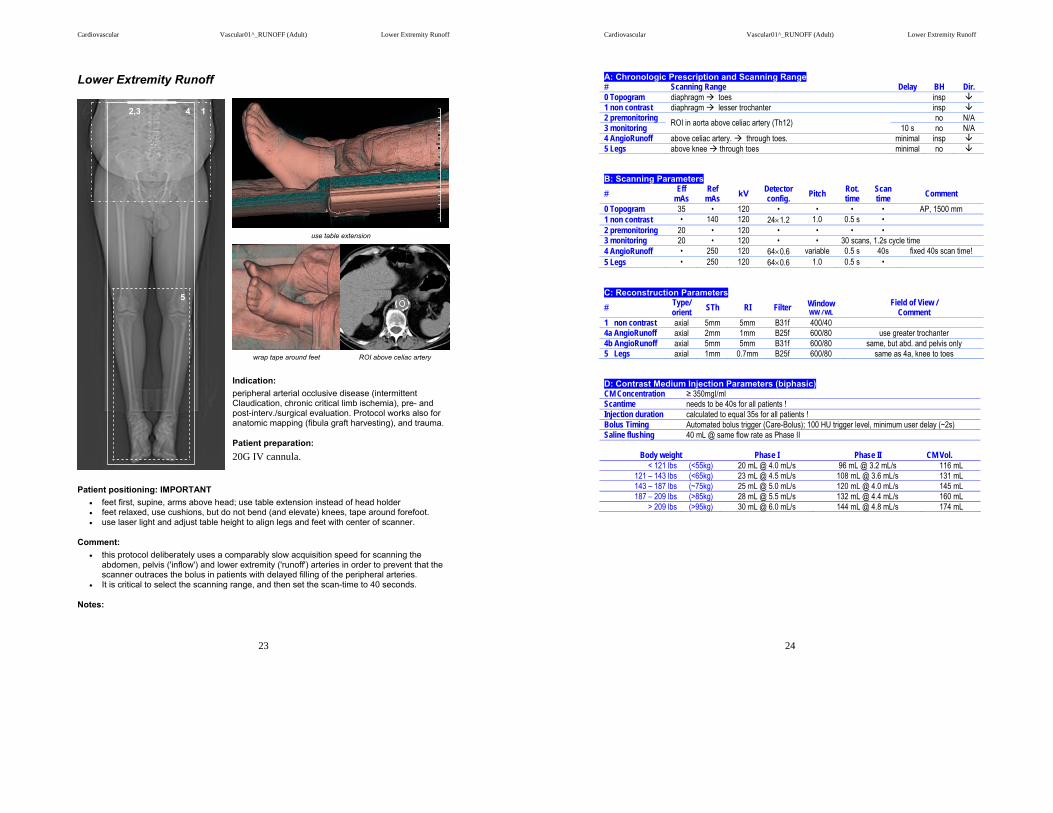
Cardiovascular Vascular01^_RUNOFF (Adult) Lower Extremity Runoff
23
Lower Extremity Runoff
Indication: peripheral arterial occlusive disease (intermittent Claudication, chronic critical limb ischemia), pre- and post-interv./surgical evaluation. Protocol works also for anatomic mapping (fibula graft harvesting), and trauma.
Patient preparation: 20G IV cannula.
Patient positioning: IMPORTANT • feet first, supine, arms above head; use table extension instead of head holder • feet relaxed, use cushions, but do not bend (and elevate) knees, tape around forefoot. • use laser light and adjust table height to align legs and feet with center of scanner.
Comment: • this protocol deliberately uses a comparably slow acquisition speed for scanning the
abdomen, pelvis ('inflow') and lower extremity ('runoff') arteries in order to prevent that the scanner outraces the bolus in patients with delayed filling of the peripheral arteries.
• It is critical to select the scanning range, and then set the scan-time to 40 seconds.
Notes:
wrap tape around feet ROI above celiac artery
use table extension
1 2,3 4
5
Cardiovascular Vascular01^_RUNOFF (Adult) Lower Extremity Runoff
24
A: Chronologic Prescription and Scanning Range # Scanning Range Delay BH Dir. 0 Topogram diaphragm toes insp 1 non contrast diaphragm lesser trochanter insp 2 premonitoring no N/A 3 monitoring ROI in aorta above celiac artery (Th12) 10 s no N/A 4 AngioRunoff above celiac artery. through toes. minimal insp 5 Legs above knee through toes minimal no B: Scanning Parameters
# Eff mAs
Ref mAs kV Detector
config. Pitch Rot. time
Scan time Comment
0 Topogram 35 • 120 • • • • AP, 1500 mm 1 non contrast • 140 120 24×1.2 1.0 0.5 s • 2 premonitoring 20 • 120 • • • • 3 monitoring 20 • 120 • • 30 scans, 1.2s cycle time 4 AngioRunoff • 250 120 64×0.6 variable 0.5 s 40s fixed 40s scan time! 5 Legs • 250 120 64×0.6 1.0 0.5 s • C: Reconstruction Parameters
# Type/ orient STh RI Filter Window
WW / WL Field of View /
Comment 1 non contrast axial 5mm 5mm B31f 400/40 4a AngioRunoff axial 2mm 1mm B25f 600/80 use greater trochanter 4b AngioRunoff axial 5mm 5mm B31f 600/80 same, but abd. and pelvis only 5 Legs axial 1mm 0.7mm B25f 600/80 same as 4a, knee to toes D: Contrast Medium Injection Parameters (biphasic) CM Concentration ≥ 350mgI/ml Scantime needs to be 40s for all patients ! Injection duration calculated to equal 35s for all patients ! Bolus Timing Automated bolus trigger (Care-Bolus); 100 HU trigger level, minimum user delay (~2s) Saline flushing 40 mL @ same flow rate as Phase II
Body weight
Phase I
Phase II
CM Vol.
< 121 lbs (<55kg) 20 mL @ 4.0 mL/s 96 mL @ 3.2 mL/s 116 mL 121 – 143 lbs (<65kg) 23 mL @ 4.5 mL/s 108 mL @ 3.6 mL/s 131 mL 143 – 187 lbs (~75kg) 25 mL @ 5.0 mL/s 120 mL @ 4.0 mL/s 145 mL 187 – 209 lbs (>85kg) 28 mL @ 5.5 mL/s 132 mL @ 4.4 mL/s 160 mL
> 209 lbs (>95kg) 30 mL @ 6.0 mL/s 144 mL @ 4.8 mL/s 174 mL

Cardiovascular Vascular01^POPL_ENTRAPMENT_CTA Popliteal Entrapment CTA
25
Popliteal Entrapment CTA
Indication: Popliteal entrapment syndrome
Patient preparation: 20G IV cannula.
Patient positioning: • feet first, supine, arms at the side holding the ends of a bed-sheet looped under the
forefoot and toes; use table extension; • use laser light and adjust table height to align legs and feet with center of scanner
Comment: • this protocol requires two injections, and three scans. The first injection is for an arterial
and venous phase acquisition in relaxed position, the second injection is acquired in an arterial phase only during the provocation maneuver (gastrognemius muscle contraction); for the provocation maneuver the patient is instructed to push the toes against the resistance of the loop of bed-sheet while pulling back the sheet with his/her hands; this position needs to be hold for the length of the scan (15 s long); It is important to practice the provocation maneuver with the patient.
• the scan range of the maneuver scan needs to be long enough to cover the tipped down toes;
• It is critical to select the scanning range first, and then set the scan-time to 15 seconds.
Notes:
← for the maneuver scan let the patient push against a sheet wrapped around toes
1,2 5,6
3,4,7
↑ relaxed ↑ maneuver
put ROI outside the body and start manually at contrast arrival ↓
Cardiovascular Vascular01^POPL_ENTRAPMENT_CTA Popliteal Entrapment CTA
26
A: Chronologic Prescription and Scanning Range # Scanning Range Delay BH Dir. 0 Topogram mid thigh below toes no 1 premonitoring no N/A 2 monitoring ROI outside the body; start manually at contrast arrival; 10 s no N/A 3 relaxed mid thigh below toes 2 s no 4 venous mid thigh below toes 20 s no 5 premonitoring no N/A 6 monitoring ROI outside the body; start manually at contrast arrival; 10 s no N/A 7 maneuver mid thigh below toes (longer than shown in Topogram) 2 s no B: Scanning Parameters # Eff
mAs Ref
mAs kV Detector config. Pitch Rot.
time Scan time Comment
0 Topogram 35 • 120 • • • • AP, 768 mm 1 premonitoring 20 • 120 • • • • 2 monitoring 20 • 120 • • 30 scans, 1.2s cycle time 3 relaxed • 250 120 64×0.6 variable 0.5 s 15 s fixed 15s scan time! 4 venous • 250 120 64×0.6 variable 0.5 s 15 s fixed 15 s scan time 5 premonitoring 20 • 120 • • • • 6 monitoring 20 • 120 • • 30 scans, 1.2s cycle time 7 maneuver • 250 120 64×0.6 variable 0.5 s 15s fixed 15s scan time! C: Reconstruction Parameters
# Type/ orient STh RI Filter Window
WW / WL Field of View /
Comment 3 relaxed axial 1mm 0.7mm B25f 600/80 use greater trochanter 4 venous axial 1mm 0.7mm B25f 600/80 same 7 maneuver axial 1mm 0.7mm B25f 600/80 same D: Contrast Medium Injection Parameters CM Concentration ≥ 350mgI/ml Scantime needs to be 15s for all patients Injection duration 15s for all patients Bolus Timing Automated bolus trigger (Care-Bolus); start manually; 2s user delay Saline flushing 40 mL @ same flow rate as contrast Phase
Body weight
Flow Rate
Volume
Total CM Vol.
< 121 lbs (<55kg) 4.0 mL/s 60 mL 2x60=120 mL 121 – 143 lbs (<65kg) 4.5 mL/s 68 mL 2x68=136 mL 143 – 187 lbs (~75kg) 5.0 mL/s 75 mL 2x75=150 mL 187 – 209 lbs (>85kg) 5.5 mL/s 83 mL 2x83=166 mL
> 209 lbs (>95kg) 6.0 mL/s 90 mL 2x90=180 mL

Cardiovascular Vascular01^LTD_LOWER_EXTR_RUNOFF Limited Lower Extremity Runoff
27
Limited Lower Extremity Runoff (Trauma)
Indication: Trauma, suspected vascular injury
Patient preparation: 20G IV cannula !
Patient positioning: • head first, supine • position leg(s) to the center of the table; • it is also possible to scan one leg only, with the other leg pulled up. • use laser light and adjust table height to align legs with center of scanner.
Comment: • It is critical to select the scanning range and then set the scan-time to 15 seconds in all
patients.
Notes:
put ROI outside the body and start manually at contrast arrival ↓
2,3 1 4
Cardiovascular Vascular01^LTD_LOWER_EXTR_RUNOFF Limited Lower Extremity Runoff
28
A: Chronologic Prescription and Scanning Range # Scanning Range Delay BH Dir. 0 Topogram mid thigh toes insp 1 non contrast mid thigh toes insp 2 premonitoring no N/A 3 monitoring ROI outside the body at beginning of scan range 10 s no N/A 4 Ltd LE Runoff above knees through toes 2-3 s insp B: Scanning Parameters
# Eff mAs
Ref mAs kV Detector
config. Pitch Rot. time
Scan time Comment
0 Topogram 35 • 120 • • • • AP, 768 mm 1 non contrast • 140 120 24×1.2 1.0 0.5 s • 2 premonitoring 20 • 120 • • • • 3 monitoring 20 • 120 • • 30 scans, 1.2s cycle time 4 Ltd LE Runoff • 250 120 64×0.6 variable 0.5 s 15s fixed 15s scan time! C: Reconstruction Parameters
# Type/ orient STh RI Filter Window
WW / WL Field of View /
Comment 1 non contrast axial 3mm 3mm B31f 400/40 4 Ltd LE Runoff axial 1mm 0.7mm B25f 600/80 Leg of interest D: Contrast Medium Injection Parameters CM Concentration ≥ 350mgI/ml Scantime needs to be 15s for all patients ! Injection duration 15s for all patients ! Bolus Timing Automated bolus trigger (Care-Bolus); manual start, minimum user delay (~2s) Saline flushing 40 mL @ same flow rate as contrast
Body weight
Flow Rate
Volume
< 121 lbs (<55kg) 4.0 mL/s 60 mL 121 – 143 lbs (<65kg) 4.5 mL/s 68 mL 143 – 187 lbs (~75kg) 5.0 mL/s 75 mL 187 – 209 lbs (>85kg) 5.5 mL/s 83 mL
> 209 lbs (>95kg) 6.0 mL/s 90 mL
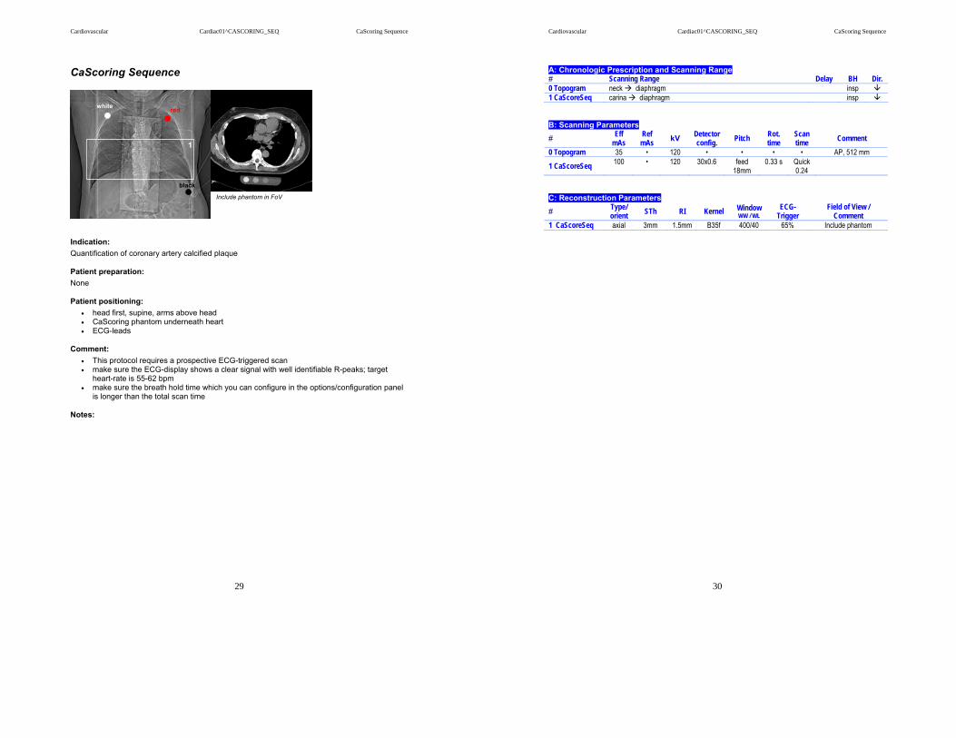
Cardiovascular Cardiac01^CASCORING_SEQ CaScoring Sequence
29
CaScoring Sequence
Indication: Quantification of coronary artery calcified plaque
Patient preparation: None
Patient positioning: • head first, supine, arms above head • CaScoring phantom underneath heart • ECG-leads
Comment: • This protocol requires a prospective ECG-triggered scan • make sure the ECG-display shows a clear signal with well identifiable R-peaks; target
heart-rate is 55-62 bpm • make sure the breath hold time which you can configure in the options/configuration panel
is longer than the total scan time
Notes:
white red
black
1
Include phantom in FoV
Cardiovascular Cardiac01^CASCORING_SEQ CaScoring Sequence
30
A: Chronologic Prescription and Scanning Range # Scanning Range Delay BH Dir. 0 Topogram neck diaphragm insp 1 CaScoreSeq carina diaphragm insp B: Scanning Parameters # Eff
mAs Ref
mAs kV Detector config. Pitch Rot.
time Scan time Comment
0 Topogram 35 • 120 • • • • AP, 512 mm
1 CaScoreSeq 100 • 120 30x0.6 feed 18mm
0.33 s Quick0.24
C: Reconstruction Parameters
# Type/ orient STh RI Kernel Window
WW / WL ECG-
Trigger Field of View /
Comment 1 CaScoreSeq axial 3mm 1.5mm B35f 400/40 65% Include phantom

Cardiovascular Cardiac01^CaScoring Spiral CaScoring Spiral
31
CaScoring Spiral
Indication: Quantification of coronary artery calcified plaque
Patient preparation: none
Patient positioning: • head first, supine, arms above head • CaScoring phantom underneath heart • ECG-leads
Comment: • make sure the ECG-display shows a clear signal with well identifiable R-peaks; target
heart-rate is 55-62 bpm; Use ECG-Pulsing in low and stable heart-rates especially in younger patients;
Notes:
white red
black
1
Include phantom in FoV
Cardiovascular Cardiac01^CaScoring Spiral CaScoring Spiral
32
A: Chronologic Prescription and Scanning Range # Scanning Range Delay BH Dir. 0 Topogram neck diaphragm insp 1 CaScoring carina diaphragm insp B: Scanning Parameters # Eff
mAs Ref
mAs kV Detector config. Pitch Rot.
time Scan time Comment
0 Topogram 35 • 120 • • • • AP, 512 mm 1 CaScoring 220 • 120 24×1.2 0.2 0.33 s • C: Reconstruction Parameters # Type/
orient STh RI Kernel Window WW / WL
ECG-Trigger
Field of View / Comment
1 CaScoring axial 3mm 1.5mm B35f 400/40 65% Include phantom
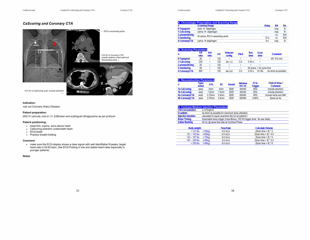
Cardiovascular Cardiac01^CaScoring and Coronary CTA Coronary CTA
33
CaScoring and Coronary CTA
Indication: rule out Coronary Artery Disease
Patient preparation: 20G IV cannula; oral or I.V. β-Blocker and sublingual nitroglycerine as per protocol
Patient positioning: • head first, supine, arms above head • CaScoring phantom underneath heart • ECG-leads • Practice breath-holding
Comment: • make sure the ECG-display shows a clear signal with well identifiable R-peaks; target
heart-rate is 55-62 bpm; Use ECG-Pulsing in low and stable heart-rates especially in younger patients;
Notes:
1 4 2,3
white red
black
← ROI in ascending aorta
FoV for a Coronary CTA; include anterior chest wall and descending aorta ↓
FoV for a CaScoring scan; include phantom →
Cardiovascular Cardiac01^CaScoring and Coronary CTA Coronary CTA
34
A: Chronologic Prescription and Scanning Range # Scanning Range Delay BH Dir. 0 Topogram neck diaphragm insp 1 CaScoring carina diaphragm insp 2 premonitoring no N/A 3 monitoring At carina, ROI in ascending aorta 10 s no N/A 4 CoronaryCTA carina diaphragm 8 s insp B: Scanning Parameters
# Eff mAs
Ref mAs kV Detector
config. Pitch Rot. time
Scan time Comment
0 Topogram 35 • 120 • • • • AP, 512 mm 1 CaScoring 220 • 120 24×1.2 0.2 0.33 s • 2 premonitoring 20 • 120 • • • • 3 monitoring 20 • 120 • • 30 scans, 1.2s cycle time 4 CoronaryCTA 850 • 120 64×0.6 0.2 0.33 s 10-19s As short as possible C: Reconstruction Parameters
# Type/ orient STh RI Kernel Window
WW / WL ECG-
Trigger Field of View /
Comment 1a CaScoring axial 3mm 3mm B35f 400/40 65% Include phantom 1b CaScoring axial 1.5mm 1.5mm B35f 400/40 65% Include phantom 4a CoronaryCTA axial 0.75mm 0.5mm B25f 600/80 65% Include Aorta and IMA 4b CoronaryCTA axial 0.75mm 0.5mm B25f 600/80 0-90% Same as 4a D: Contrast Medium Injection Parameters CM Concentration ≥ 370mgI/ml Scantime As short as possible for maximum dose utilization Injection duration calculated to equal (scantime+8s) for all patients ! Bolus Timing Automated bolus trigger (Care-Bolus); 120 HU trigger level; 8s user delay Saline flushing 40 mL @ same flow rate as Contrast Phase
Body weight
Flow Rate
Calculate Volume
< 121 lbs (<55kg) 4.0 mL/s (Scan time + 8) * 4 121 – 143 lbs (<65kg) 4.5 mL/s (Scan time + 8) * 4.5 143 – 187 lbs (~75kg) 5.0 mL/s (Scan time + 8) * 5 187 – 209 lbs (>85kg) 5.5 mL/s (Scan time + 8) * 5.5
> 209 lbs (>95kg) 6.0 mL/s (Scan time + 8) * 6

Cardiovascular Cardiac01^CaScoring and Coronary CTA below 50bpm Coronary CTA
35
CaScoring and Coronary CTA below 50 bpm HR
Indication: rule out Coronary Artery Disease
Patient preparation: 20G IV cannula; oral or I.V. β-Blocker and sublingual nitroglycerine as per protocol
Patient positioning: • head first, supine, arms above head • CaScoring phantom underneath heart • ECG-leads • Practice breath-holding
Comment: • make sure the ECG-display shows a clear signal with well identifiable R-peaks; this
protocol should be used for heart rates clearly below 50 bpm; Use ECG-Pulsing especially in younger patients;
Notes:
1 4 2,3
white red
black
← ROI in ascending aorta
FoV for a Coronary CTA; include anterior chest wall and descending aorta ↓
FoV for a CaScoring scan; include phantom →
Cardiovascular Cardiac01^CaScoring and Coronary CTA below 50bpm Coronary CTA
36
A: Chronologic Prescription and Scanning Range # Scanning Range Delay BH Dir. 0 Topogram neck diaphragm insp 1 CaScoring carina diaphragm insp 2 premonitoring no N/A 3 monitoring At carina, ROI in ascending aorta 10 s no N/A 4 CoronaryCTA carina diaphragm 8 s insp B: Scanning Parameters
# Eff mAs
Ref mAs kV Detector
config. Pitch Rot. time
Scan time Comment
0 Topogram 35 • 120 • • • • AP, 512 mm 1 CaScoring 220 • 120 24×1.2 0.2 0.33 s • 2 premonitoring 20 • 120 • • • • 3 monitoring 20 • 120 • • 30 scans, 1.2s cycle time 4 CoronaryCTA 850 • 120 64×0.6 0.24 0.37 s 10-19s As short as possible C: Reconstruction Parameters
# Type/ orient STh RI Kernel Window
WW / WL ECG-
Trigger Field of View /
Comment 1a CaScoring axial 3mm 3mm B35f 400/40 65% Include phantom 1b CaScoring axial 1.5mm 1.5mm B35f 400/40 65% Include phantom 4a CoronaryCTA axial 0.75mm 0.5mm B25f 600/80 65% Include Aorta and IMA 4b CoronaryCTA axial 0.75mm 0.5mm B25f 600/80 0-90% Same as 4a D: Contrast Medium Injection Parameters CM Concentration ≥ 370mgI/ml Scantime As short as possible for maximum dose utilization Injection duration calculated to equal (scantime+8s) for all patients ! Bolus Timing Automated bolus trigger (Care-Bolus); 120 HU trigger level; 8s user delay Saline flushing 40 mL @ same flow rate as Contrast Phase
Body weight
Flow Rate
Calculate Volume
< 121 lbs (<55kg) 4.0 mL/s (Scan time + 8) * 4 121 – 143 lbs (<65kg) 4.5 mL/s (Scan time + 8) * 4.5 143 – 187 lbs (~75kg) 5.0 mL/s (Scan time + 8) * 5 187 – 209 lbs (>85kg) 5.5 mL/s (Scan time + 8) * 5.5
> 209 lbs (>95kg) 6.0 mL/s (Scan time + 8) * 6
Cardiovascular Cardiac01^CORONARY GRAFT Coronary Graft CTA
37
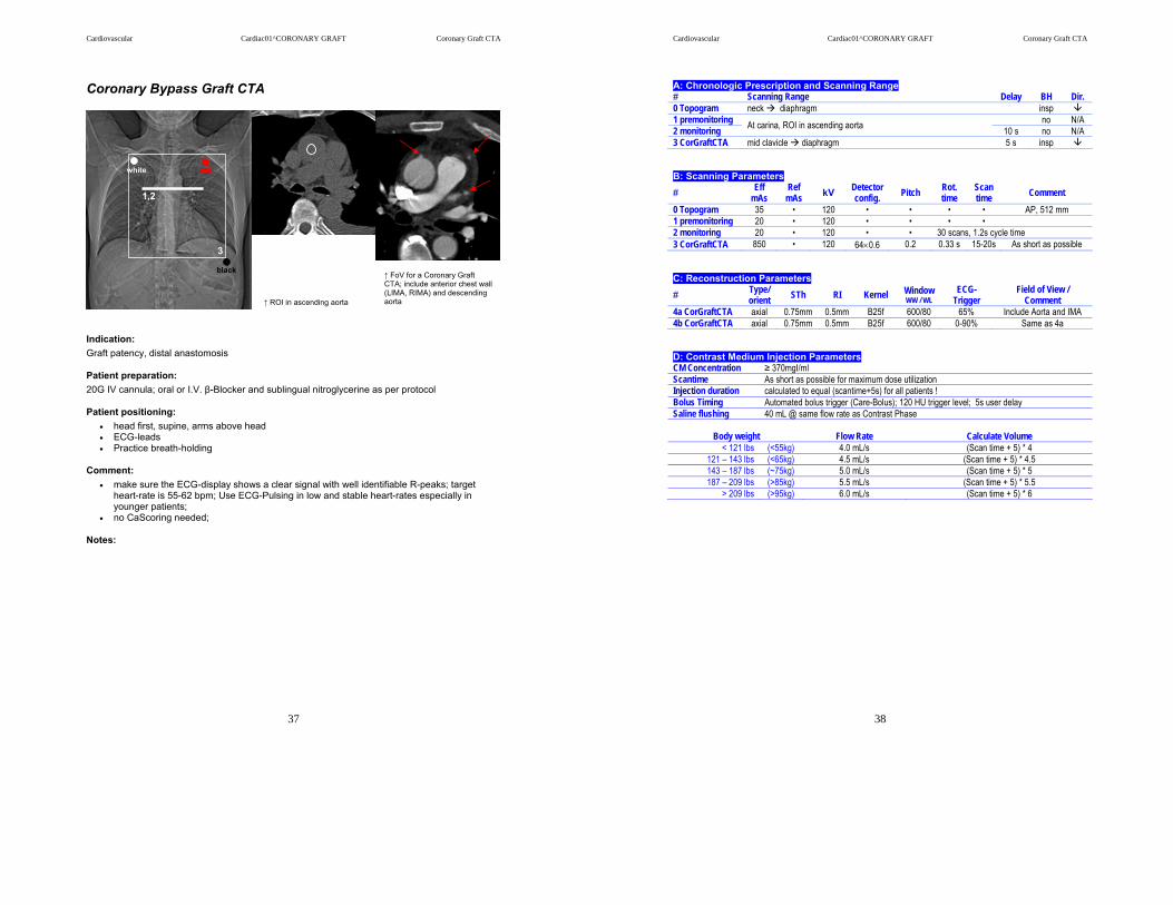
Coronary Bypass Graft CTA
Indication: Graft patency, distal anastomosis
Patient preparation: 20G IV cannula; oral or I.V. β-Blocker and sublingual nitroglycerine as per protocol
Patient positioning: • head first, supine, arms above head • ECG-leads • Practice breath-holding
Comment: • make sure the ECG-display shows a clear signal with well identifiable R-peaks; target
heart-rate is 55-62 bpm; Use ECG-Pulsing in low and stable heart-rates especially in younger patients;
• no CaScoring needed;
Notes:
↑ ROI in ascending aorta
↑ FoV for a Coronary Graft CTA; include anterior chest wall (LIMA, RIMA) and descending aorta
3
1,2
white red
black
Cardiovascular Cardiac01^CORONARY GRAFT Coronary Graft CTA
38
A: Chronologic Prescription and Scanning Range # Scanning Range Delay BH Dir. 0 Topogram neck diaphragm insp 1 premonitoring no N/A 2 monitoring At carina, ROI in ascending aorta 10 s no N/A 3 CorGraftCTA mid clavicle diaphragm 5 s insp B: Scanning Parameters
# Eff mAs
Ref mAs kV Detector
config. Pitch Rot. time
Scan time Comment
0 Topogram 35 • 120 • • • • AP, 512 mm 1 premonitoring 20 • 120 • • • • 2 monitoring 20 • 120 • • 30 scans, 1.2s cycle time 3 CorGraftCTA 850 • 120 64×0.6 0.2 0.33 s 15-20s As short as possible C: Reconstruction Parameters # Type/
orient STh RI Kernel Window WW / WL
ECG-Trigger
Field of View / Comment
4a CorGraftCTA axial 0.75mm 0.5mm B25f 600/80 65% Include Aorta and IMA 4b CorGraftCTA axial 0.75mm 0.5mm B25f 600/80 0-90% Same as 4a D: Contrast Medium Injection Parameters CM Concentration ≥ 370mgI/ml Scantime As short as possible for maximum dose utilization Injection duration calculated to equal (scantime+5s) for all patients ! Bolus Timing Automated bolus trigger (Care-Bolus); 120 HU trigger level; 5s user delay Saline flushing 40 mL @ same flow rate as Contrast Phase
Body weight
Flow Rate
Calculate Volume
< 121 lbs (<55kg) 4.0 mL/s (Scan time + 5) * 4 121 – 143 lbs (<65kg) 4.5 mL/s (Scan time + 5) * 4.5 143 – 187 lbs (~75kg) 5.0 mL/s (Scan time + 5) * 5 187 – 209 lbs (>85kg) 5.5 mL/s (Scan time + 5) * 5.5
> 209 lbs (>95kg) 6.0 mL/s (Scan time + 5) * 6

Cardiovascular Cardiac01^GATED_CHEST Gated Chest
39
Gated Chest
Indication: Aortic root and ascending aortic aneurysm and dissection; acute aortic syndrome with chest pain
Patient preparation: 20G IV cannula; NO β-blocker medication
Patient positioning: • head first, supine, arms above head. • ECG-leads • Practice breath-holding
Comment: • Make sure the ECG-display shows a clear signal with well identifiable R-peaks; Use ECG-
Pulsing in low and stable heart-rates especially in younger patients;
Notes:
1
2,3
white red
black
4
← put ROI in ascending aorta
Cardiovascular Cardiac01^GATED_CHEST Gated Chest
40
A: Chronologic Prescription and Scanning Range # Scanning Range Delay BH Dir. 0 Topogram neck diaphragm insp 1 non-cons neck diaphragm insp 2 premonitoring no N/A 3 monitoring At carina, ROI in ascending aorta 10 s no N/A 4 gated Chest well above arch diaphragm 5 s insp B: Scanning Parameters
# Eff mAs
Ref mAs kV Detector
config. Pitch Rot. time
Scan time Comment
0 Topogram 35 • 120 • • • AP, 512 mm 1 non-cons 220 • 120 24×1.2 1.2 0.5 s 2 premonitoring 20 • 120 • • • 3 monitoring 20 • 120 • • 30 scans, 1.2s cycle time 4 gated Chest 700 • 120 64×0.6 0.2 0.33 s ≤25s As short as possible C: Reconstruction Parameters
# Type/ orient STh RI Kernel Window
WW / WL ECG-
Trigger Field of View /
Comment 1 non-cons axial 5mm 5mm B31f 400/40 4a gated Chest axial 1mm 0.7mm B25f 600/80 65% chest wall 4b gated Chest axial 1mm 0.7mm B25f 600/80 0-90% to heart and aorta D: Contrast Medium Injection Parameters (biphasic) CM Concentration ≥ 370mgI/ml Scantime as short as possible for maximum dose utilization; Injection duration Fixed phase I; calculated second volume to equal scan time -5s for all patients ! Bolus Timing Automated bolus trigger (Care-Bolus); 100 HU trigger level; 5s user delay Saline flushing 40 mL @ same flow rate as Contrast Phase
Body weight
Phase I
Phase II (Calculate Volume)
< 121 lbs (<55kg) 20 mL @ 4.0 mL/s (Scan time - 5) * 3.2 121 – 143 lbs (<65kg) 23 mL @ 4.5 mL/s (Scan time - 5) * 3.6 143 – 187 lbs (~75kg) 25 mL @ 5.0 mL/s (Scan time - 5) * 4.0 187 – 209 lbs (>85kg) 28 mL @ 5.5 mL/s (Scan time - 5) * 4.4
> 209 lbs (>95kg) 30 mL @ 6.0 mL/s (Scan time - 5) * 4.8

Cardiovascular Cardiac01^GATED_CAP_GATED_CHEST Gated CAP
41
Gated Chest/Abdomen/Pelvis CTA (gated Chest, non-gated Abdomen)
Indication: acute dissection, preoperative chronic dissection, thoracic aneurysm, pre-interventional planning
Patient preparation: 20G IV cannula; NO β-blocker medication
Patient positioning: • head first, supine, arms above head. • ECG-leads • Practice breath-holding
Comment: • This protocol consists of two separate scans in auto-range mode with 4s interscandelay.
The Start-button has to be pushed only once. Breathing instruction needs to be given by the technologist.
• The chest part should not exceed 25 s in scan time and the abdomen part should be scanned in 10 s.
• Make sure the ECG-display shows a clear signal with well identifiable R-peaks; Use ECG-Pulsing in low and stable heart-rates especially in younger patients;
• FoV and x/y coordinates should be the same for both contrast scans and merged after reconstruction. The merged dataset should be sent to PACS.
Notes:
← to put ROI in ascending aorta is better than in the aortic arch (dissection flaps most likely occur in the arch ↓)
1 4
2,3
5
white red
black
Cardiovascular Cardiac01^GATED_CAP_GATED_CHEST Gated CAP
42
A: Chronologic Prescription and Scanning Range # Scanning Range Delay BH Dir. 0 Topogram neck lesser trochanter insp 1 non-cons neck lesser trochanter insp 2 premonitoring no N/A 3 monitoring At carina, ROI in ascending aorta 10 s no N/A 4 gated Chest Above arch diaphragm (≤ 25s scan time) 5 s insp 5 Abdomen End of series 4 lesser trochanter (10 s scan time) B: Scanning Parameters
# Eff mAs
Ref mAs kV Detector
config. Pitch Rot. time
Scan time Comment
0 Topogram 35 • 120 • • • AP, 768 mm 1 non-cons 220 • 120 24×1.2 1.2 0.5 s 2 premonitoring 20 • 120 • • • 3 monitoring 20 • 120 • • 30 scans, 1.2s cycle time 4 gated Chest 700 • 120 64×0.6 0.2 0.33 s ≤25s As short as possible 5 Abdomen 250 120 64x0.6 1 0.33 10s C: Reconstruction Parameters # Type/
orient STh RI Kernel Window WW / WL
ECG-Trigger
Field of View / Comment
1a non-cons axial 5mm 5mm B31f 400/40 4a gated Chest axial 1mm 0.7mm B25f 600/80 65% 4b gated Chest axial 1mm 0.7mm B25f 600/80 0-90% Same as abdomen 5a Abdomen axial 1mm 0.7mm B25f 600/80 Same as chest 5b Abdomen axial 5mm 5mm B31f 600/80 Body wall D: Contrast Medium Injection Parameters (biphasic) CM Concentration ≥ 370mgI/ml Scantime Chest: as short as possible for maximum dose utilization; Abdomen: 10s Injection duration calculated to equal 35s scantime for all patients ! Bolus Timing Automated bolus trigger (Care-Bolus); 100 HU trigger level; 5s user delay Saline flushing 40 mL @ same flow rate as Contrast Phase
Body weight
Phase I
Phase II
TotalCM Vol.
< 121 lbs (<55kg) 20 mL @ 4.0 mL/s 96 mL @ 3.2 mL/s 116 mL 121 – 143 lbs (<65kg) 23 mL @ 4.5 mL/s 108 mL @ 3.6 mL/s 131 mL 143 – 187 lbs (~75kg) 25 mL @ 5.0 mL/s 120 mL @ 4.0 mL/s 145 mL 187 – 209 lbs (>85kg) 28 mL @ 5.5 mL/s 132 mL @ 4.4 mL/s 160 mL
> 209 lbs (>95kg) 30 mL @ 6.0 mL/s 144 mL @ 4.8 mL/s 174 mL

Cardiovascular Cardiac01^LAM Left Atrial Mapping
43
Left Atrial Mapping
Indication: Pre and post EP ablation in patients with atrial fibrillation
Patient preparation: 20G IV cannula; NO β-blocker medication
Patient positioning: • head first, supine, arms above head • ECG-leads • Rehears breath-holding
Comment: • This scan protocol utilizes CD4D on a level of 800 ref mAs; spatial resolution is not as
important as in Coronary CTAs since primarily pulmonary veins are to evaluate; therefore the axial slice thickness is increased to 1.0mm
• The scan range should include the aortic arch to allow an evaluation of upper lobe veins. • Make sure the ECG-display shows a clear signal with well identifiable R-peaks and T-
waves; Systole reconstructions are necessary, so ECG-Pulsing is not recommended; • FoV fit to aorta and heart; • If the patient is in atrial fibrillation, T-wave reconstruction may improve image quality;
Notes:
3
1,2
white red
black
ROI in the left atrium
Cardiovascular Cardiac01^LAM Left Atrial Mapping
44
A: Chronologic Prescription and Scanning Range # Scanning Range Delay BH Dir. 0 Topogram neck diaphragm insp 1 premonitoring no N/A 2 monitoring ~ 4cm below carina, ROI in left atrium 10 s no N/A 3 LAM above arch diaphragm (≤ 25s scan time) 5 s mid insp B: Scanning Parameters
# Eff mAs
Ref mAs kV Detector
config. Pitch Rot. time
Scan time Comment
0 Topogram 35 • 120 • • • • AP, 512 mm 1 premonitoring 20 • 120 • • • • 2 monitoring 20 • 120 • • 30 scans, 1.2s cycle time 3 LAM • 800 120 64×0.6 0.2 0.33 s ≤25s As short as possible C: Reconstruction Parameters # Type/
orient STh RI Kernel Window WW / WL
ECG-Trigger
Field of View / Comment
3a LAM axial 1mm 0.7mm B25f 600/80 30% Aorta and heart 3b LAM axial 1mm 0.7mm B25f 600/80 0-90% Aorta and heart D: Contrast Medium Injection Parameters CM Concentration ≥ 350mgI/ml Scantime as short as possible Injection duration scan time + 5 s for all patients ! Bolus Timing Automated bolus trigger (Care-Bolus); 150 HU trigger level; 5 s user delay Saline flushing 40 mL @ same flow rate as Contrast Phase
Body weight
Flow Rate
Calculate Volume
< 121 lbs (<55kg) 3.5 mL/s (Scan time + 5) * 3.5 121 – 143 lbs (<65kg) 4.0 mL/s (Scan time + 5) * 4.0 143 – 187 lbs (~75kg) 4.5 mL/s (Scan time + 5) * 4.5 187 – 209 lbs (>85kg) 5.0 mL/s (Scan time + 5) * 5.0
> 209 lbs (>95kg) 5.5 mL/s (Scan time + 5) * 5.5
Cardiovascular Cardiac01^CVM Coronary Vein Mapping
45
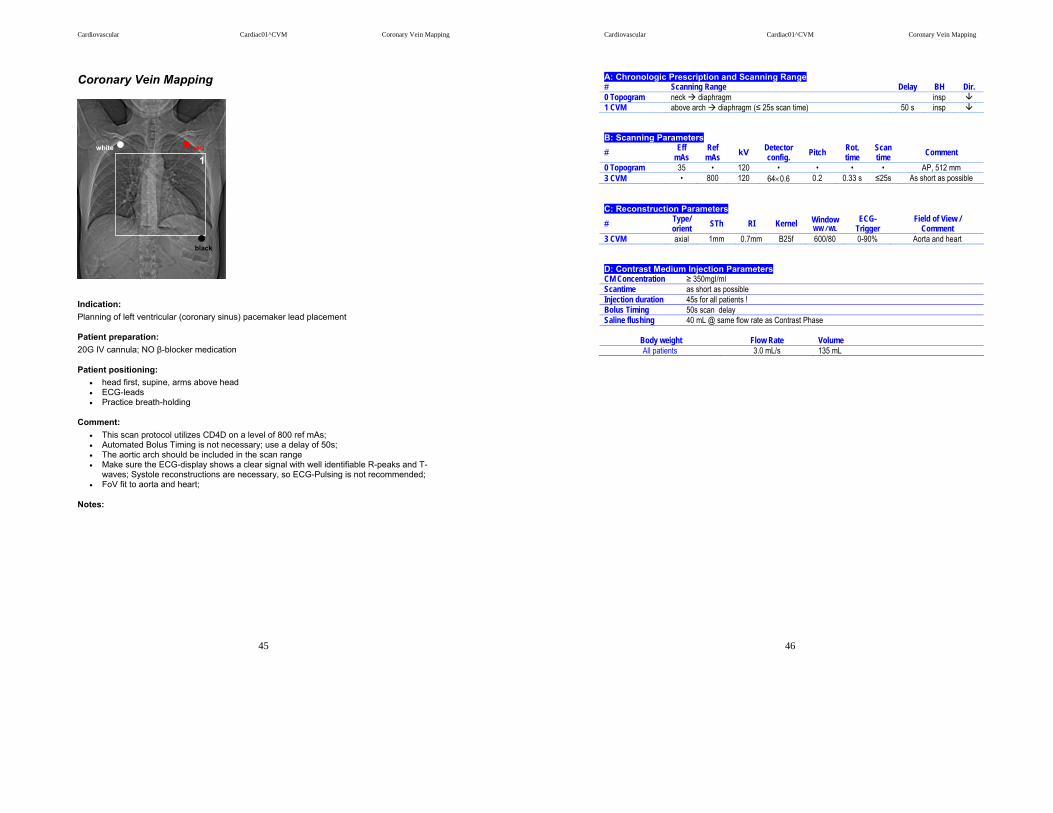
Coronary Vein Mapping
Indication: Planning of left ventricular (coronary sinus) pacemaker lead placement
Patient preparation: 20G IV cannula; NO β-blocker medication
Patient positioning: • head first, supine, arms above head • ECG-leads • Practice breath-holding
Comment: • This scan protocol utilizes CD4D on a level of 800 ref mAs; • Automated Bolus Timing is not necessary; use a delay of 50s; • The aortic arch should be included in the scan range • Make sure the ECG-display shows a clear signal with well identifiable R-peaks and T-
waves; Systole reconstructions are necessary, so ECG-Pulsing is not recommended; • FoV fit to aorta and heart;
Notes:
white red
black
1
Cardiovascular Cardiac01^CVM Coronary Vein Mapping
46
A: Chronologic Prescription and Scanning Range # Scanning Range Delay BH Dir. 0 Topogram neck diaphragm insp 1 CVM above arch diaphragm (≤ 25s scan time) 50 s insp B: Scanning Parameters # Eff
mAs Ref
mAs kV Detector config. Pitch Rot.
time Scan time Comment
0 Topogram 35 • 120 • • • • AP, 512 mm 3 CVM • 800 120 64×0.6 0.2 0.33 s ≤25s As short as possible C: Reconstruction Parameters # Type/
orient STh RI Kernel Window WW / WL
ECG-Trigger
Field of View / Comment
3 CVM axial 1mm 0.7mm B25f 600/80 0-90% Aorta and heart D: Contrast Medium Injection Parameters CM Concentration ≥ 350mgI/ml Scantime as short as possible Injection duration 45s for all patients ! Bolus Timing 50s scan delay Saline flushing 40 mL @ same flow rate as Contrast Phase
Body weight
Flow Rate
Volume
All patients 3.0 mL/s 135 mL

Cardiovascular Abbreviations
47
Abbreviations AP anterior-posterior BH breath-hold Bpm beats per minute CM contrast media config detector configuration cor coronal CTA Computed Tomography Angiography Dir scanning direction ECG electro cardiogram Eff mAs effective milli ampere seconds FoV Field of View HN head and neck HU hounsfield unite IMA internal mammarian artery Insp inspiration IV intra venous Kg kilogram kV kilo voltage LAT lateral Lbs pounds (libra) LE lower extremity Ltd limited MgI milligram Iodine mL milli liter mm milli meter N/A not applicable Orient orientation Ref mAs reference milli ampere seconds RI reconstruction interval ROI region of interest Rot time rotation time s second sag sagittal Seq Sequence STh slice thickness UE upper extremity Vol volume WL window level WW window width