staging of esophageal cancer: role of ct and · pdf filestaging of esophageal cancer: role of...
TRANSCRIPT

STAGING of ESOPHAGEAL CANCER:
ROLE OF CT and PET
R. A. Halvorsen, M.D., FACRMCV Hospitals/VCU Medical Center
Richmond, VirginiaI do not have any relevant financial relationship with any commercial company.

ESOPHAGEAL CANCER
Sixth leading cause of cancer death worldwide
One of the deadliest tumors
Over 50% unresectable at diagnosis

WHAT’S NEW ?Changing tumor types
Staging: CT, PET
Neck nodes: new importance

ESOPHAGEAL CANCER
United States: ↑ incidence: 20% increase over 30 years
Changing histology1975: 75% Squamous-cell Ca2003: More Adeno than SCCa

INCIDENCEDramatic increase of adeno Ca
Last 30 years: ↑ 350-800%

ETIOLOGYSquamous cell carcinoma:• Smoking• AlcoholAdenocarcinoma of esophagus:• Barrett’s esophagus• Reflux, hiatal hernia• Obesity

BARRETT’S ESOPHAGUS
Columnar cell metaplasia
replaces squamous cell lining
5% lifetime risk of Adenocarcinoma
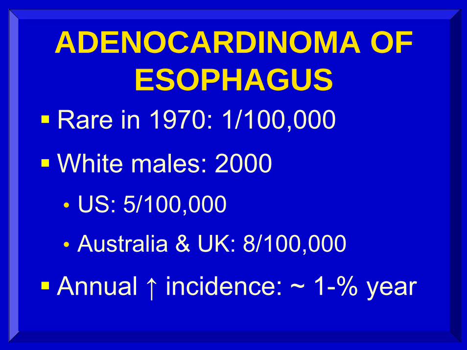
ADENOCARDINOMA OF ESOPHAGUS
Rare in 1970: 1/100,000
White males: 2000 • US: 5/100,000
• Australia & UK: 8/100,000
Annual ↑ incidence: ~ 1-% year

ADENOCARCINOMA OF ESOPHAGUS
Male dominant: 2:1 to 12:1
Age peak: 50 – 60 years old
Middle or upper socioeconomic groups
52% are university graduates

ADENOCARCINOMAGastric adenocarcinoma:
dramatic ↓ incidence
Esophageal adenocarcinoma: dramatic ↑ incidence

ESOPHAGEAL WALLDifferent from rest of GI tract
Lacks outer serosal layer;
therefore, earlier local spread

OFTEN ADVANCED DISEASE
AT TIME OF DIAGNOSIS24% confined to primary site29% lymph nodes or local invasion30% already metastasized17% staging unknown
SEER NCI 2006

NEOADJUVANT CHEMOTHERAPY
Improved survival vs. Surgery alone
Median survival:16.8 vs.13.3 months
2-year survival: 43 vs. 34%
Lancet 2002

LOCAL INVASION
CT: 50 – 70% accuracy
EUS: 70 – 80% accuracy

CT: ESOPHQAGEAL CANCER
Good for local invasion?
Good for lung and liver mets
No good for lymphadenopathy

LYMPHATICSDye injected into esophageal wall
May fill nodes all levelsFrequent long channels ie nodal mets
a long distance from primary tumorFrequently drains into thoracic duct;
therefore, early hematogenous metsRiquet: Surg Rad Anat 1993


SCHRODER ET ALWORLD J SURG 2002
Nodes: long axis
Tumor free 5.1mm
Tumor positive 6.7mm
All nodes 91% < 10mm
No correlation size and nodal mets

LYMPH NODE STAGING
EUS guided fine needle aspiration:
Sensitivity: 93%
Specificity: 100%Gastrointes Endosc 2001

Meta-Analysis: PETSTAGING ESOPHAGEAL
CANCER12 studies met criteriaLoco-regional nodes• Sensitivity: 51%• Specificity: 84%
Distant metastases• Sensitivity: 67%• Specificity: 97%

STAGING ESOPHAGEAL CA
CT first test: if no metastases, then
Positron emission tomographyif no metastases, then
Endoscopic ultrasound:

PET/CT STAGINGLimitations: Nodal staging
Small nodesNodes adjacent to tumor
Variable results in literature

STAGE IV DISEASEPET: Increased detection mets
3 combined studies: 21.6% increase in metastases detected

FDG-PETEsophageal cancer n= 61Pre-op CT, EUS, PETPET: 20% pts avoided
“unnecessary surgery”Improved survival, likely from
avoiding unnecessary surgeryJ Gastrointest Surg 2005

PET DETECTED METSNot seen on CT
CT chest and abdomen
17% of patients

PET DETECTED METSNOT ON CT
Cervical lymph nodes 38%
Bone metastases 23%
Hepatic metastases 15%Imdahl Lang Arch Surg 2004

NECK NODESOne third patients: neck nodes
found during esophagectomyfor “curable” cancerof thoracic esophagus
NYU: J Thorac Cardiovasc Surg 1997

NECK NODESNodal metastases equal frequency:
Neck nodesMediastinal nodes
NYU 1997

NECK NODESThe higher the cancer
The more likely neck nodes:Cervical esophageal ca: 80%Upper 1/3 mediastinum: 52%Middle 1/3 mediastinum: 29%Lower 1/3 mediastinum: 9%

52%
29%
9%
80%NECK NODE
PRIMARY

NECK SONOGRAPHYNodes of interest: 3 cm deep
Use 7.5-10 MHz transducer
“Big node” : over 5 mm
Short / long ratio: S/L: over 50 %

SONOGRAPHYn = 80
Sensitivity: 88%Specificity: 59%Accuracy: 78%
Natsugoe: J Surg Onc 2002

TAKE HOME POINTSDramatic increase in
AdenocarcinomaBarrett’s, middle class, obese
CT first staging testDistant metastasesGuide further testing

TAKE HOME POINTSPET/CT: if CT is indeterminate
or Θ for metastasesEUS with fine needle aspiration
Reserve if PET & CT negativeBest for lymph node stagingBest for degree of wall invasion

CONTROVERSIESNeck US routinely?
Neck CT routinely?