staging - emory universityweb1.sph.emory.edu/gccs/naaccr_webinars/20151203/staging slides.pdfdec 03,...
TRANSCRIPT
Staging 12/3/2015
NAACCR 2015‐2016 Webinar Series 1
1
NAACCR 2015-2016 Webinar Series
AJCC and Summary Stage
Cancer Staging
1
2
Q&A
2
Please submit all questions concerning webinar content through the Q&A panel.
Reminder:• If you have participants watching this webinar at your site, please collect
their names and emails.
• We will be distributing a Q&A document in about one week. This document will fully answer questions asked during the webinar and will contain any corrections that we may discover after the webinar.
Staging 12/3/2015
NAACCR 2015‐2016 Webinar Series 2
3
Fabulous Prizes
4
Prior to assigning stage…
4
• Registrars…• Must have access to their staging manuals
• AJCC 7th edition with errata
• Summary Stage with errata
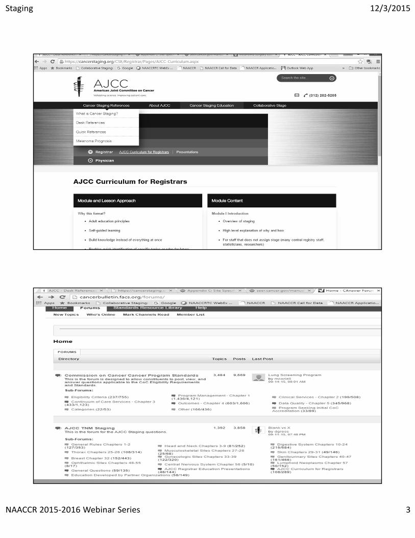
• Are HIGHLY encouraged to view the AJCC Curriculum for Cancer Registrars
• https://cancerstaging.org/CSE/Registrar/Pages/AJCC-Curriculum.aspx
• Must use the CAnswer forum• http://cancerbulletin.facs.org/forums/forum
Staging 12/3/2015
NAACCR 2015‐2016 Webinar Series 3
5
6

Staging 12/3/2015
NAACCR 2015‐2016 Webinar Series 4
7
8
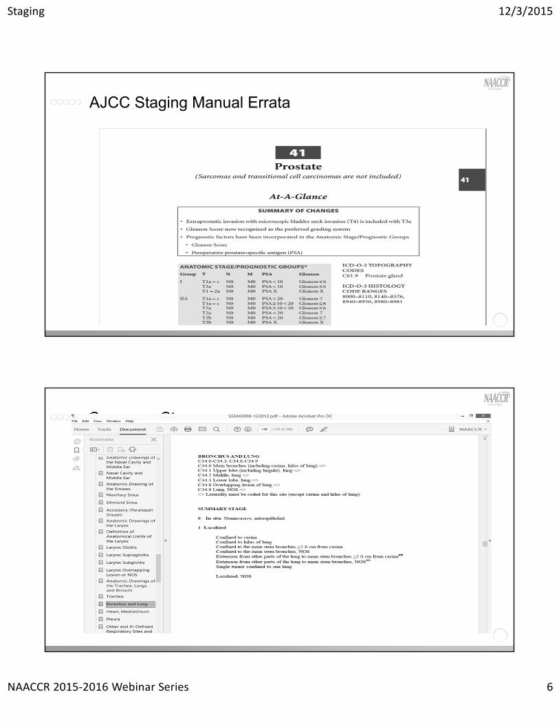
AJCC Staging Manual
Summary Stage
Errata

Staging 12/3/2015
NAACCR 2015‐2016 Webinar Series 5
9
AJCC Staging Manual Errata
10
AJCC Staging Manual Errata
Staging 12/3/2015
NAACCR 2015‐2016 Webinar Series 6
11
AJCC Staging Manual Errata
12
Summary Stage
Staging 12/3/2015
NAACCR 2015‐2016 Webinar Series 7
13
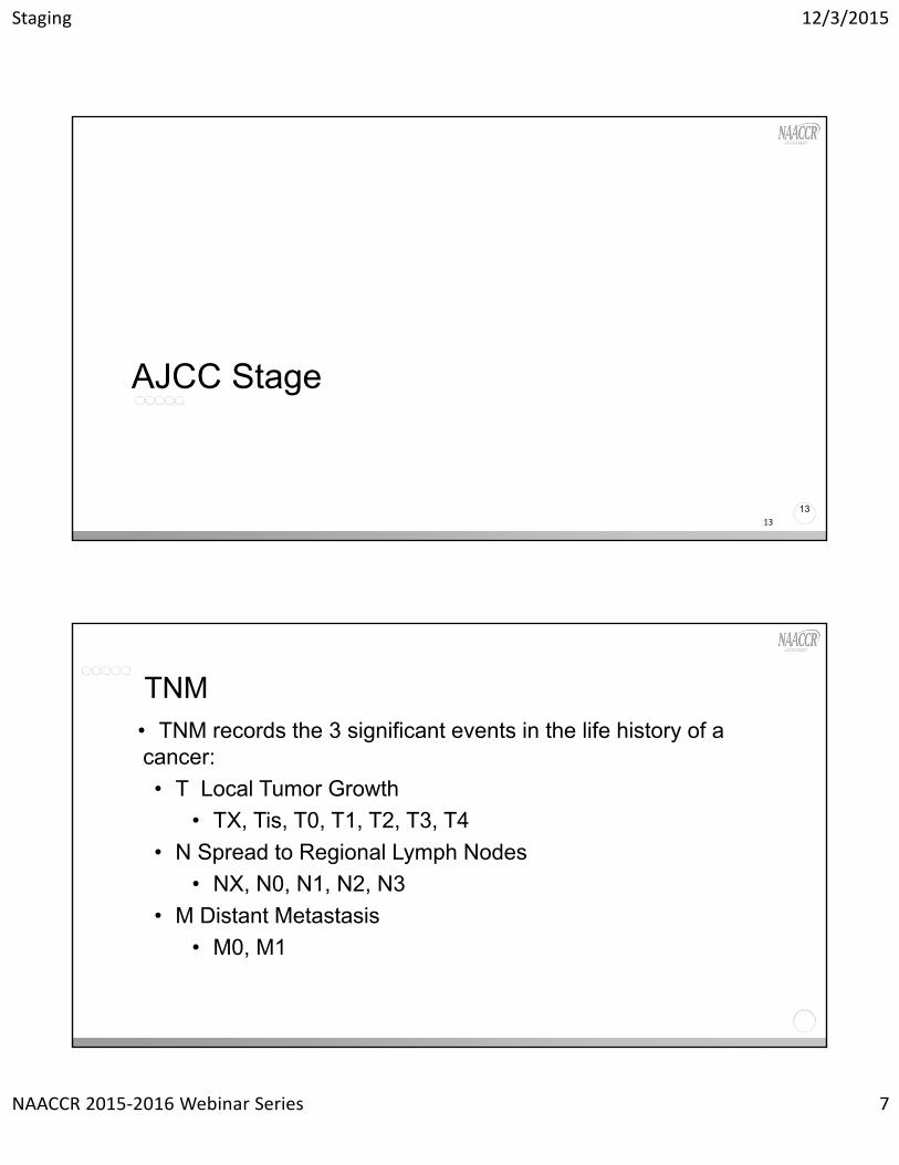
AJCC Stage
13
14
TNM• TNM records the 3 significant events in the life history of a cancer:
• T Local Tumor Growth
• TX, Tis, T0, T1, T2, T3, T4
• N Spread to Regional Lymph Nodes
• NX, N0, N1, N2, N3
• M Distant Metastasis
• M0, M1
Staging 12/3/2015
NAACCR 2015‐2016 Webinar Series 8
15
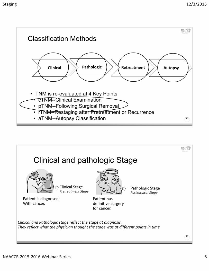
Clinical Pathologic Retreatment Autopsy
Classification Methods
• TNM is re-evaluated at 4 Key Points• cTNM--Clinical Examination• pTNM--Following Surgical Removal • rTNM--Restaging after Pretreatment or Recurrence• aTNM--Autopsy Classification
16
Clinical and pathologic Stage
Clinical StagePretreatment Stage
Pathologic StagePostsurgical Stage
Patient is diagnosedWith cancer.
Patient has definitive surgery for cancer.
Clinical and Pathologic stage reflect the stage at diagnosis. They reflect what the physician thought the stage was at different points in time

Staging 12/3/2015
NAACCR 2015‐2016 Webinar Series 9
17
Summary Stage
17
• Uses both clinical and pathologic information to get the stage
• Regional: potential for spread by more than one lymphatic or vascular supply route
• Surgeon definition vs radiation oncologist definition
18
Scenario
• A patient was found to have a 1 cm tumor in her left breast during routine mammogram. An ultrasound guided biopsy confirmed ductal carcinoma. No indication of enlarged lymph nodes or metastasis.
• The patient went on to have a modified radical mastectomy. Pathology revealed a 1.2 cm ductal carcinoma with negative margins and 3 of 24 lymph nodes positive for metastasis. The largest metastasis measured .5cm.
18
Follow along on page 358 of your AJCC Manual

Staging 12/3/2015
NAACCR 2015‐2016 Webinar Series 10
19
Scenario
• What is the clinical stage (pre-treatment stage)?
• What is the pathologic stage (post surgery stage)?
• See page 358 in your AJCC Manual
• See page 186 of your Summary Stage Manual
19
Data Items as Coded in Current NAACCR Layout
T N M Stage Group
Clin
Path
Summary Stage 3‐Ipsilateral regional nodes only
1b
1a
0 0 IA
IIA1c
20
Entering Data Into your Abstract
20
Staging 12/3/2015
NAACCR 2015‐2016 Webinar Series 11
21
Data Items
• Clinical T• Item Length 4• Upper-case Alphanumeric• Left Justified• NAACCR Item #940• Description
• Detailed site-specific codes for the clinical tumor (T) as defined by AJCC and recorded by the physician
• Rationale• CoC requires that AJCC TNM staging be used in its approved cancer programs.
AJCC developed its staging system for evaluating trends in the treatment and control of cancer. This staging is used by physicians to estimate prognosis, to plan treatment, to evaluate new types of therapy, to analyze outcome, to design follow-up strategies, and to assess early detection results.
21
22
Entering DataTNM Clin T
Valid Codes
• 1
• 1A
• 1A1
• 1A2
• 1B
• 1B1
• 1B2
Implied Values
• c1
• c1A
• c1A1
• c1A2
• c1B
• c1B1
• c1B2
• Pathologic codes cannot be entered into clinical data items

Staging 12/3/2015
NAACCR 2015‐2016 Webinar Series 12
2323
• The assigned stage information is entered in data items• Clinical stage data should only be entered into clinical data fields
• Pathologic stage data into pathologic data fields
• Sometimes clinical data is used to calculate the pathologic stage group
• Sometimes pathologic data is used calculate the clinical stage group
Entering data
Data Items as Coded in Current NAACCR Layout
T N M Stage Group
Clin 1 0 0 I
Path 1 0 IcM0
c cp pp
cT1 + cN0 + cM0 = cStage IpT1 + pN0 + cM0 = pStage I
c c
24
Other Examples of “Phantom Values”
• See table 1.7 on page 11 of your AJCC Manual• Cases with pT and pN may be grouped as pathologic TNM using clinical
M designator (cM0 or cM1)-row 5
• Cases with pM1 may be grouped as clinical and pathologic stage IV –row 6
• In situ• See table 1.8 row 6
• Carcinoma in situ-stage pTis cN0 cM0 as both clinical and pathologic
24

Staging 12/3/2015
NAACCR 2015‐2016 Webinar Series 13
25
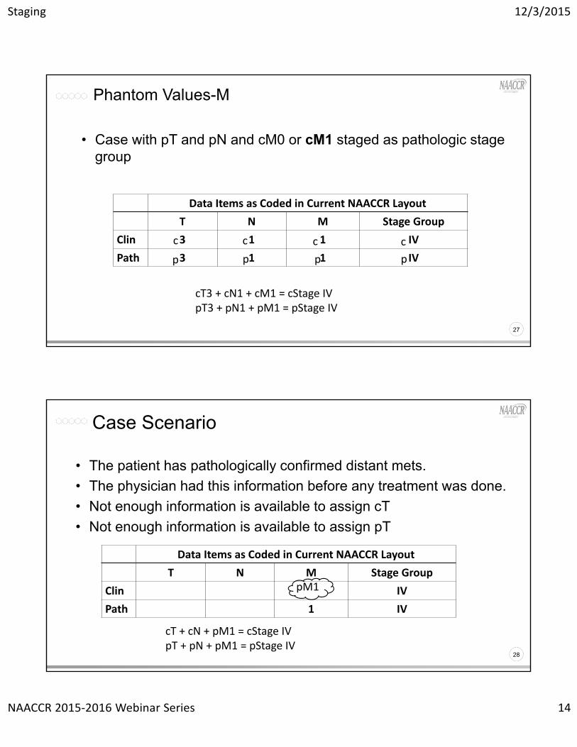
Phantom Values-M
• Case with pT and pN and cM0 or cM1 staged as pathologic stage group
25
Data Items as Coded in Current NAACCR Layout
T N M Stage Group
Clin 3 1 1 IV
Path 3 1 IVcM1
c c c c
p pp
cT3 + cN1 + cM1 = cStage IVpT3 + pN1 + cM1 = pStage IV
26
Phantom Values-M
• Case with pT and pN and cM0 or cM1 staged as pathologic stage group
26
Data Items as Coded in Current NAACCR Layout
T N M Stage Group
Clin 3 1 IV
Path 3 1 1 IV
pM1c c
p
c
p pp
cT3 + cN1 + pM1 = cStage IVpT3 + pN1 + pM1 = pStage IV
Staging 12/3/2015
NAACCR 2015‐2016 Webinar Series 14
27
Phantom Values-M
• Case with pT and pN and cM0 or cM1 staged as pathologic stage group
27
Data Items as Coded in Current NAACCR Layout
T N M Stage Group
Clin 3 1 1 IV
Path 3 1 1 IV
c c
p
c
p pp
cT3 + cN1 + cM1 = cStage IVpT3 + pN1 + pM1 = pStage IV
c
2828
• The patient has pathologically confirmed distant mets.
• The physician had this information before any treatment was done.
• Not enough information is available to assign cT
• Not enough information is available to assign pT
Case Scenario
Data Items as Coded in Current NAACCR Layout
T N M Stage Group
Clin IV
Path 1 IV
pM1
cT + cN + pM1 = cStage IVpT + pN + pM1 = pStage IV
Staging 12/3/2015
NAACCR 2015‐2016 Webinar Series 15
29
Rules for Classification
29
30
Rules for Classification
30

Staging 12/3/2015
NAACCR 2015‐2016 Webinar Series 16
31
Rules for Classification
• If rules for classification have not been met, leave the T, N, and M fields blank (99 for stage group).
• Leave the T and N blank if the rules for classification of the T value have not been met.
• If rules for N have been met, but the rules for T have not been met leave both blank
• If rules for T have been met but rules for N have not been met, assign the appropriate T value and X for N value.
• See fourth row of Table 1.6 on page 10 • Pathologic assessment of the primary tumor (pT) is necessary to assign
pathologic assessment of nodes (pN)….
31
32
Example 1
• A patient with a clinical T1 N0 M0 Stage I supraglottic laryngeal carcinoma (pg 58) has surgery that removed the primary tumor, but no lymph nodes. Tumor was 1cm with negative margins. Per surgeons notes the tumor was confined to a single subsite.
• What do we enter for a our pathologic T, N, M, and Stage Group?
32
Data Items as Coded in Current NAACCR Layout
T N M Stage Group
*Clin 1 0 0 I
Path
*For this example we assume clinical rules for classification have been metFollow along
99X1 cM0
Staging 12/3/2015
NAACCR 2015‐2016 Webinar Series 17
33
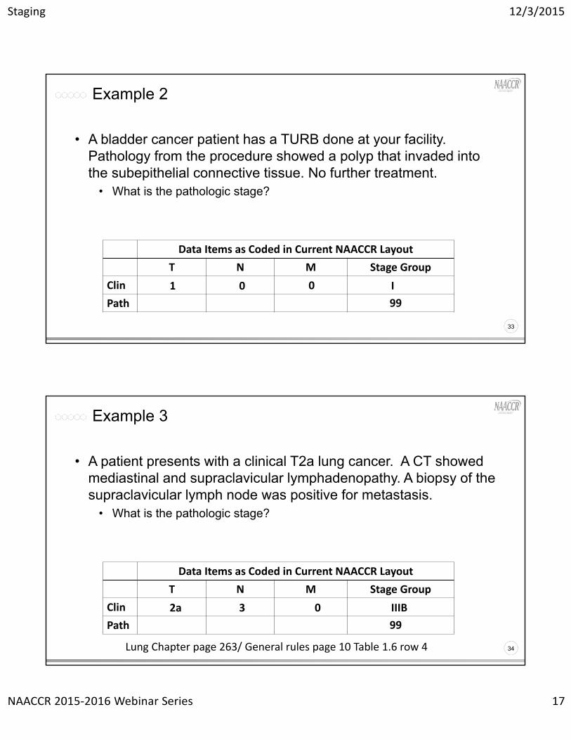
Example 2
• A bladder cancer patient has a TURB done at your facility. Pathology from the procedure showed a polyp that invaded into the subepithelial connective tissue. No further treatment.
• What is the pathologic stage?
33
Data Items as Coded in Current NAACCR Layout
T N M Stage Group
Clin
Path
1 0 0 I
99
34
Example 3
• A patient presents with a clinical T2a lung cancer. A CT showed mediastinal and supraclavicular lymphadenopathy. A biopsy of the supraclavicular lymph node was positive for metastasis.
• What is the pathologic stage?
34
Data Items as Coded in Current NAACCR Layout
T N M Stage Group
Clin
Path
2a 3 0 IIIB
99
Lung Chapter page 263/ General rules page 10 Table 1.6 row 4

Staging 12/3/2015
NAACCR 2015‐2016 Webinar Series 18
35
• A patient presents for a routine colonoscopy and is found to have a large fungating tumor in the sigmoid colon. A biopsy confirmed carcinoma. A CT was negative for metastasis.
• The patient went on to have a segmental resection that showed a tumor that invaded into the submucosa. No lymph nodes were removed
• Physician staged T1 N0 M0 Stage I
Example 4
35
Data Items as Coded in Current NAACCR Layout
T N M Stage Group
Clin
Path
X 0 0 99
1 X 99cM0
3636
• Subcategories may be required to assign a stage group.• For prostate T2 is not sufficient to assign a stage group. Must have T2a
or T2b.
• See the prostate chapter page 461
Subcategories

Staging 12/3/2015
NAACCR 2015‐2016 Webinar Series 19
37
• By definition in situ indicates there is not spread to regional/distant organs or lymph nodes
• In order to call a tumor in situ a pathologist must review the entire tumor under a microscope.
• Results from the pathologic review of the entire tumor is recorded in the pT not cT
• Cannot have a cTis• See page 12 of the AJCC manual
In Situ
37
38
• An exception was made that allows us to use the pTis for both the clinical and pathologic stage and to use the cN0 for both the clinical and pathologic stage.
• However, the criteria for rules for classification have to be met in order to get a pathologic stage.
In situ stage grouping exception
38

Staging 12/3/2015
NAACCR 2015‐2016 Webinar Series 20
39
Data Items as Coded in Current NAACCR Layout
T N M Stage Group
Clin 0 0 0
Path is 0
• A breast cancer patient has lumpectomy and is found to have ductal carcinoma in situ with negative margins. Clinically there is no indication of lymph node involvement or distant mets.
Example 5
c0
Implied value
c0
Implied value
pis
Implied value
pTis + cN0 + cM0 = cStage 0pTis + cN0 + cM0 = pStage 0 39
40
• If patient has a breast biopsy that is positive for ductal carcinoma in situ. There is no clinical evidence of regional or distant mets. She then has a segmental mastectomy that reveals a 1 cm invasive ductal ca, how do I record AJCC clinical T, N, M and stage group?
In Situ Core Biopsy
Data Items as Coded in Current NAACCR Layout
T N M Stage Group
Clin 0 0 0
Path T1b X 99
pTis
pTis + cN0 + cM0 = cStage 0pT1c + pNx + cM0 = pStage 99
40

Staging 12/3/2015
NAACCR 2015‐2016 Webinar Series 21
41
• Rules for Classification-Bladder• Pathologic staging is based on radical or partial cystectomy and removal
of lymph nodes
Pathologic Stage Assumptions
Data Items as Coded in Current NAACCR Layout
T N M Stage Group
Clin 2 0 0 II
Path 2a 1 IV
We know that a radical or partial cystectomy was doneWe know lymph nodes were removed and pT was assigned
41
cM0
Positive nodes or mets
42
• In order for a stage group to be assigned, the rules for classification must be met.
• Example• TURB shows non-invasive TCC
• A patient has a TURB and is found to have a noninvasive transitional cell carcinoma. No further surgery done.
In situ stage grouping exception
42
Staging 12/3/2015
NAACCR 2015‐2016 Webinar Series 22
43
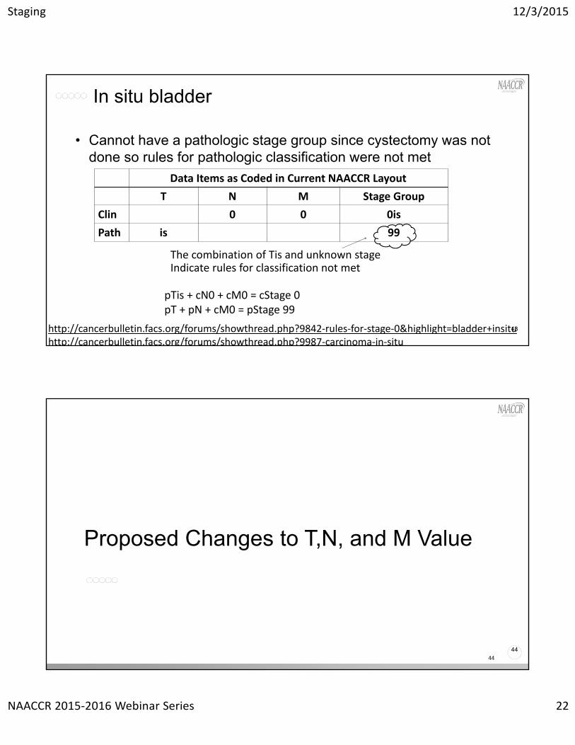
• Cannot have a pathologic stage group since cystectomy was not done so rules for pathologic classification were not met
In situ bladder
Data Items as Coded in Current NAACCR Layout
T N M Stage Group
Clin 0 0 0is
Path is 99
http://cancerbulletin.facs.org/forums/showthread.php?9842‐rules‐for‐stage‐0&highlight=bladder+insituhttp://cancerbulletin.facs.org/forums/showthread.php?9987‐carcinoma‐in‐situ
The combination of Tis and unknown stage Indicate rules for classification not met
pTis + cN0 + cM0 = cStage 0pT + pN + cM0 = pStage 99
43
44
Proposed Changes to T,N, and M Value
44

Staging 12/3/2015
NAACCR 2015‐2016 Webinar Series 23
45
Entering DataTNM Clin T
Current Codes• IS• 1• 1A• 1A1• 1A2• 1B• 1B1• 1B2
Proposed Codes• pTis• c1• c1A• c1A1• c1A2• c1B• c1B1• c1B2
• Pathologic codes cannot be entered into clinical data items
46
Data Items as Coded in Current NAACCR Layout
T N M Stage Group
Clin 0 0 0
Path is 0
• A breast cancer patient has lumpectomy and is found to have ductal carcinoma in situ with negative margins. Clinically there is no indication of lymph node involvement or distant mets.
Current Coding Values
pTis + cN0 + cM0 = cStage 0pTis + cN0 + cM0 = pStage 0
46
Staging 12/3/2015
NAACCR 2015‐2016 Webinar Series 24
47
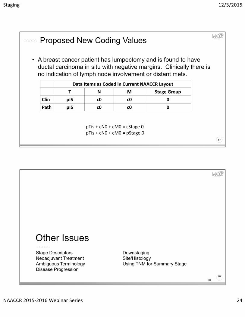
Data Items as Coded in Current NAACCR Layout
T N M Stage Group
Clin pIS c0 c0 0
Path pIS c0 c0 0
• A breast cancer patient has lumpectomy and is found to have ductal carcinoma in situ with negative margins. Clinically there is no indication of lymph node involvement or distant mets.
Proposed New Coding Values
pTis + cN0 + cM0 = cStage 0pTis + cN0 + cM0 = pStage 0
47
48
Stage DescriptorsNeoadjuvant TreatmentAmbiguous TerminologyDisease Progression
DownstagingSite/HistologyUsing TNM for Summary Stage
Other Issues
48
Staging 12/3/2015
NAACCR 2015‐2016 Webinar Series 25
49
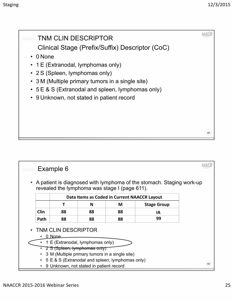
TNM CLIN DESCRIPTOR
Clinical Stage (Prefix/Suffix) Descriptor (CoC)• 0 None
• 1 E (Extranodal, lymphomas only)
• 2 S (Spleen, lymphomas only)
• 3 M (Multiple primary tumors in a single site)
• 5 E & S (Extranodal and spleen, lymphomas only)
• 9 Unknown, not stated in patient record
49
50
Example 6
• A patient is diagnosed with lymphoma of the stomach. Staging work-up revealed the lymphoma was stage I (page 611).
• TNM CLIN DESCRIPTOR• 0 None• 1 E (Extranodal, lymphomas only)• 2 S (Spleen, lymphomas only)• 3 M (Multiple primary tumors in a single site)• 5 E & S (Extranodal and spleen, lymphomas only)• 9 Unknown, not stated in patient record
50
Data Items as Coded in Current NAACCR Layout
T N M Stage Group
Clin 88 88 88
Path 88 88 88
IA99
Staging 12/3/2015
NAACCR 2015‐2016 Webinar Series 26
51
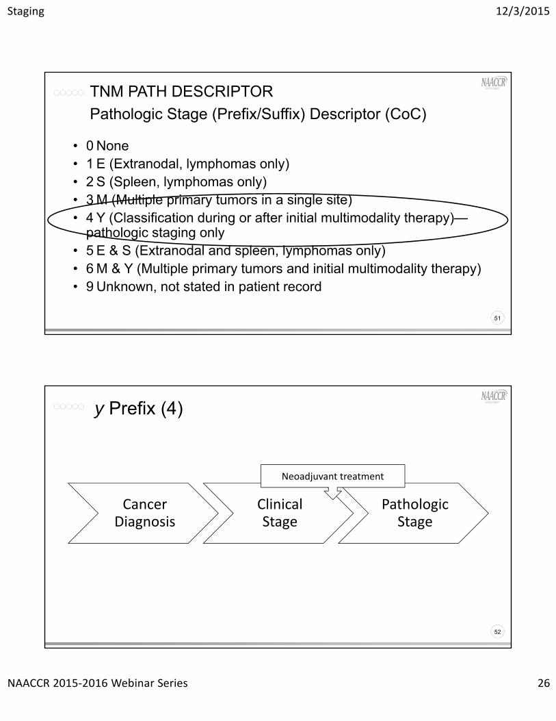
TNM PATH DESCRIPTOR
Pathologic Stage (Prefix/Suffix) Descriptor (CoC)
• 0 None• 1 E (Extranodal, lymphomas only)• 2 S (Spleen, lymphomas only)• 3 M (Multiple primary tumors in a single site)• 4 Y (Classification during or after initial multimodality therapy)—
pathologic staging only• 5 E & S (Extranodal and spleen, lymphomas only)• 6 M & Y (Multiple primary tumors and initial multimodality therapy)• 9 Unknown, not stated in patient record
51
52
Cancer Diagnosis
Clinical Stage
Pathologic Stage
y Prefix (4)
Neoadjuvant treatment
52
Staging 12/3/2015
NAACCR 2015‐2016 Webinar Series 27
53
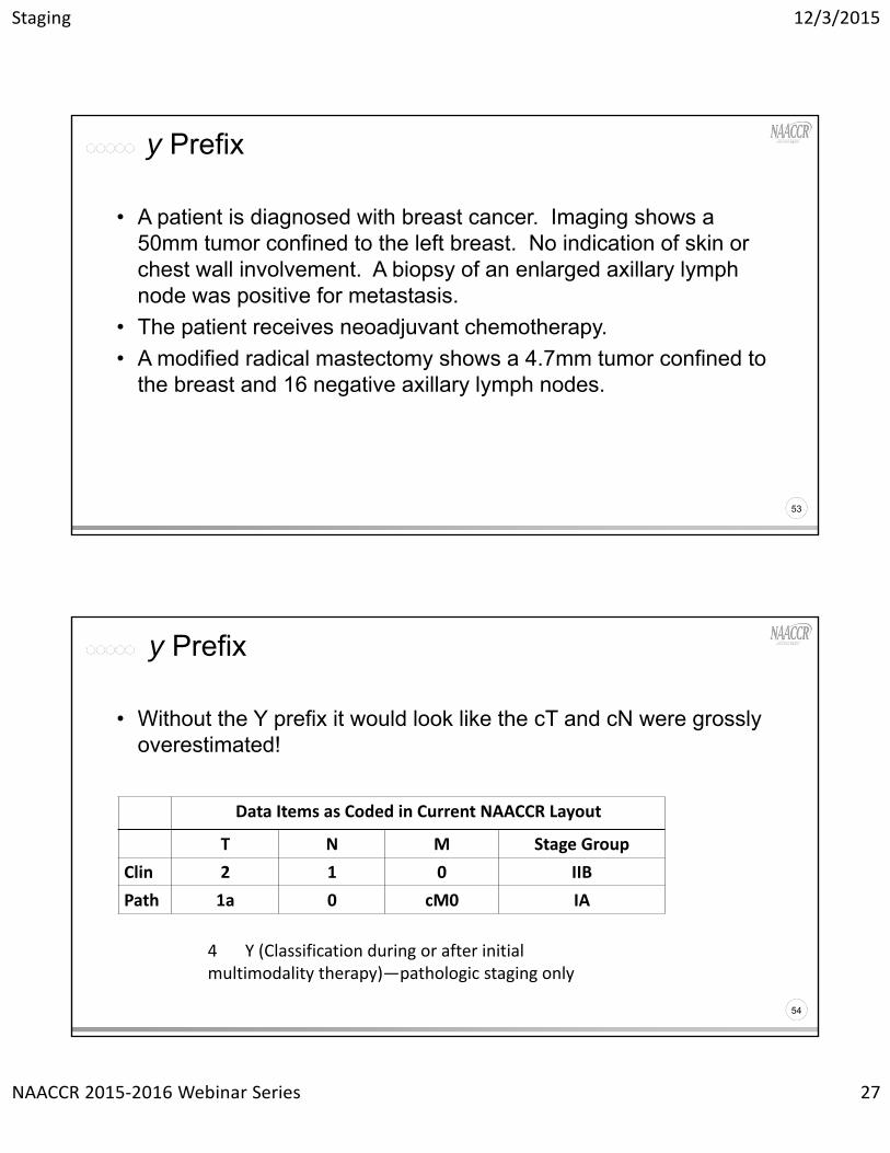
• A patient is diagnosed with breast cancer. Imaging shows a 50mm tumor confined to the left breast. No indication of skin or chest wall involvement. A biopsy of an enlarged axillary lymph node was positive for metastasis.
• The patient receives neoadjuvant chemotherapy.
• A modified radical mastectomy shows a 4.7mm tumor confined to the breast and 16 negative axillary lymph nodes.
y Prefix
53
54
• Without the Y prefix it would look like the cT and cN were grossly overestimated!
y Prefix
Data Items as Coded in Current NAACCR Layout
T N M Stage Group
Clin 2 1 0 IIB
Path 1a 0 cM0 IA
54
4 Y (Classification during or after initial multimodality therapy)—pathologic staging only

Staging 12/3/2015
NAACCR 2015‐2016 Webinar Series 28
55
Example 7
• During a routine colonoscopy a patient is found to have colon cancer. Imaging shows liver mets.
• The patient received neoadjuvant chemotherapy.
• The patient then had a segmental resection of the colon with partial liver resection.
55
Data Items as Coded in Current NAACCR Layout
T N M Stage Group
Clin 3 1 1a IVa
Path 1 0 cM1a IVa
http://cancerbulletin.facs.org/forums/forum/ajcc‐tnm‐staging/general‐rules‐chapters‐1‐2/59967‐m‐classification‐after‐neoadjuvant‐therapy
56
Neoadjuvant Treatment
• Neoadjuvant treatment is usually chemotherapy or radiation
• Not all treatments given prior to surgery should receive a Y descriptor
• Example: Lupron for prostate cancer that is given prior to prostatectomy should not be assigned a Y descriptor unless specified by a physician or as part of a clinical trial
• Example: Synthroid give prior to thyroidectomy for thyroid cancer should not be assigned a Y descriptor unless specified by a physician.
56
Staging 12/3/2015
NAACCR 2015‐2016 Webinar Series 29
57
Ambiguous Terminology
Resource Terms used Comments
Reportability Yes A list of reportable and non‐reportable terms is available
MP/H Rules Yes A list of terms that can be used to describe a histology is available. May not be used to determine multiple primaries.
HematopoieticDB
No Terms should not be used to describe histology
Summary Stage Yes Involvement and non‐involvement terms available in manual
CS Yes Same terms as used for Summary Stage
AJCC No Involvement should be based on physicians interpretation or registrars professional judgement
58
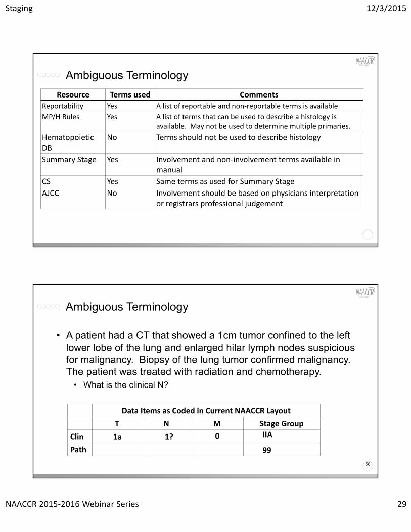
Ambiguous Terminology
• A patient had a CT that showed a 1cm tumor confined to the left lower lobe of the lung and enlarged hilar lymph nodes suspicious for malignancy. Biopsy of the lung tumor confirmed malignancy. The patient was treated with radiation and chemotherapy.
• What is the clinical N?
58
Data Items as Coded in Current NAACCR Layout
T N M Stage Group
Clin
Path
IIA
99
1a 1? 0
Staging 12/3/2015
NAACCR 2015‐2016 Webinar Series 30
59
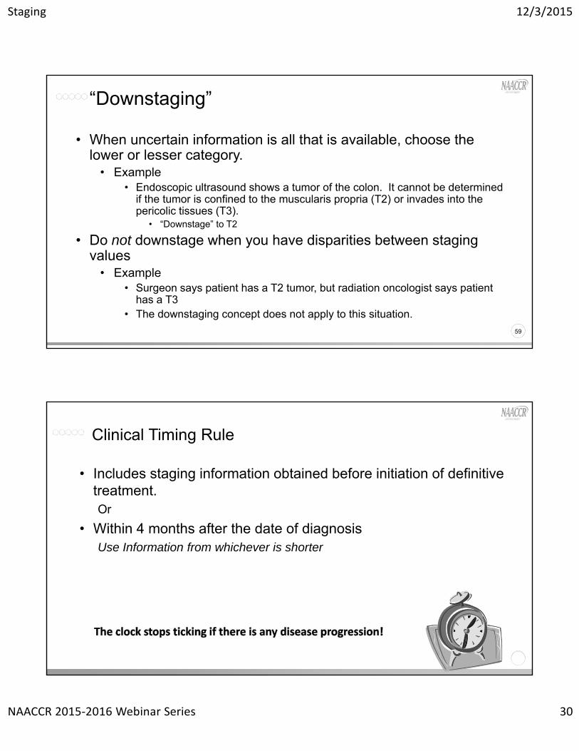
• When uncertain information is all that is available, choose the lower or lesser category.
• Example• Endoscopic ultrasound shows a tumor of the colon. It cannot be determined
if the tumor is confined to the muscularis propria (T2) or invades into the pericolic tissues (T3).
• “Downstage” to T2
• Do not downstage when you have disparities between staging values
• Example• Surgeon says patient has a T2 tumor, but radiation oncologist says patient
has a T3• The downstaging concept does not apply to this situation.
“Downstaging”
59
60
• Includes staging information obtained before initiation of definitive treatment.Or
• Within 4 months after the date of diagnosisUse Information from whichever is shorter
Clinical Timing Rule

Staging 12/3/2015
NAACCR 2015‐2016 Webinar Series 31
61
• Includes staging information obtained through completion of first course treatment Or
• Identified within 4 months after the date of diagnosisWhichever is longer
Pathologic Timing Rule
62
Disease Progression
• Think of disease progression in terms of clinical and pathologic stage.
• Was the disease progression accounted for in the treatment plan?
• Was the disease progression identified before treatment started?
62
http://cancerbulletin.facs.org/forums/forum/ajcc‐tnm‐staging/general‐rules‐chapters‐1‐2/59965‐progression‐of‐disease‐general‐guidelines

Staging 12/3/2015
NAACCR 2015‐2016 Webinar Series 32
63
Summary Stage Time Frame
• All information available through the completion of surgery in the first course of treatment or within four months of dx in the absence of disease progression or whichever is longer.
• Information after treatment with radiation, chemotherapy, hormone or immunotherapy may be included unless it is beyond the time frame specified earlier.
63
64
Subcategories
• Some stage groupings require subcategories• Values can be entered into the T, N, and M categories without
subcategories.
• If the subcategories are required for a stage group and not available, stage group must be 99
64
Staging 12/3/2015
NAACCR 2015‐2016 Webinar Series 33
6565
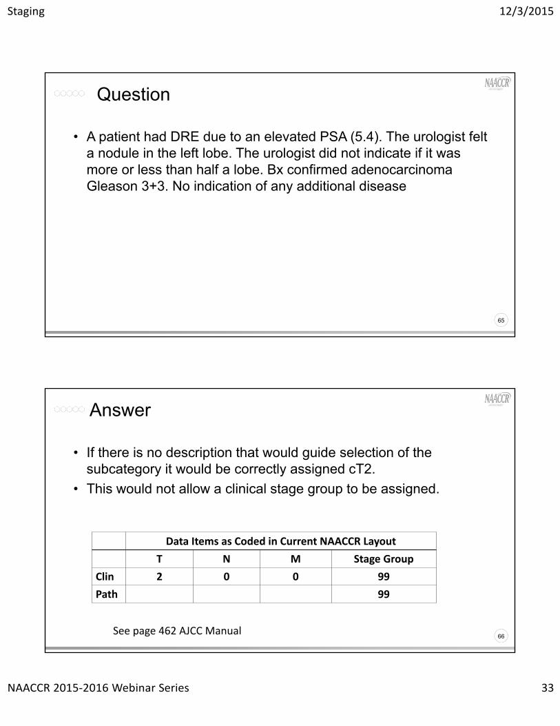
• A patient had DRE due to an elevated PSA (5.4). The urologist felt a nodule in the left lobe. The urologist did not indicate if it was more or less than half a lobe. Bx confirmed adenocarcinoma Gleason 3+3. No indication of any additional disease
Question
6666
• If there is no description that would guide selection of the subcategory it would be correctly assigned cT2.
• This would not allow a clinical stage group to be assigned.
Answer
Data Items as Coded in Current NAACCR Layout
T N M Stage Group
Clin 2 0 0 99
Path 99
See page 462 AJCC Manual

Staging 12/3/2015
NAACCR 2015‐2016 Webinar Series 34
67
Site/Histology
• Every chapter in the AJCC Staging Manual has a list of valid sites and histologies that apply to that chapter
• Not all site/histology combinations can be assigned an AJCC stage
• All sites/histologies can be assigned a Summary Stage• Most are assigned based on primary site
• Some are assigned based on histology• Lymphoma
• Kaposi sarcoma
67
68
Example 7
• A patient is diagnosed with a malignant glioblastoma confined to the occipital lobe of the brain.
68
Data Items as Coded in Current NAACCR Layout
T N M Stage Group
Clin 88 88 88 88
Path 88 88 88 88
Summary Stage 1‐Localized
See page 593 of your AJCC Manual and page 266 of Summary Stage

Staging 12/3/2015
NAACCR 2015‐2016 Webinar Series 35
69
Using TNM with Summary Stage
69
70
Questions?
70
Staging 12/3/2015
NAACCR 2015‐2016 Webinar Series 36
71
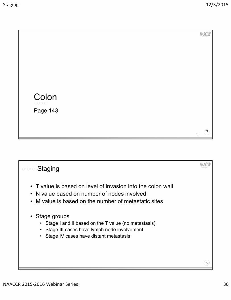
Page 143
Colon
71
72
Staging
• T value is based on level of invasion into the colon wall• N value based on number of nodes involved• M value is based on the number of metastatic sites
• Stage groups• Stage I and II based on the T value (no metastasis)• Stage III cases have lymph node involvement• Stage IV cases have distant metastasis
72

Staging 12/3/2015
NAACCR 2015‐2016 Webinar Series 37
73
Rules for Classification (pg 151)
• Clinical• BE, Endoscopy, virtual colonoscopy/sigmoidoscopy, ultrasound, MRI,
CT, PET scan
• Pathologic• Pathologic exam of the primary tumor and regional nodes
73
74
Colon Example 1
• A patient was diagnosed with colon cancer during a routine screening colonoscopy. The patient went elsewhere for additional work-up and treatment. No further information is available.
74
Data Items as Coded in Current NAACCR Layout
T N M Stage Group
Clin
Path
99
99
X X 0
Staging 12/3/2015
NAACCR 2015‐2016 Webinar Series 38
75
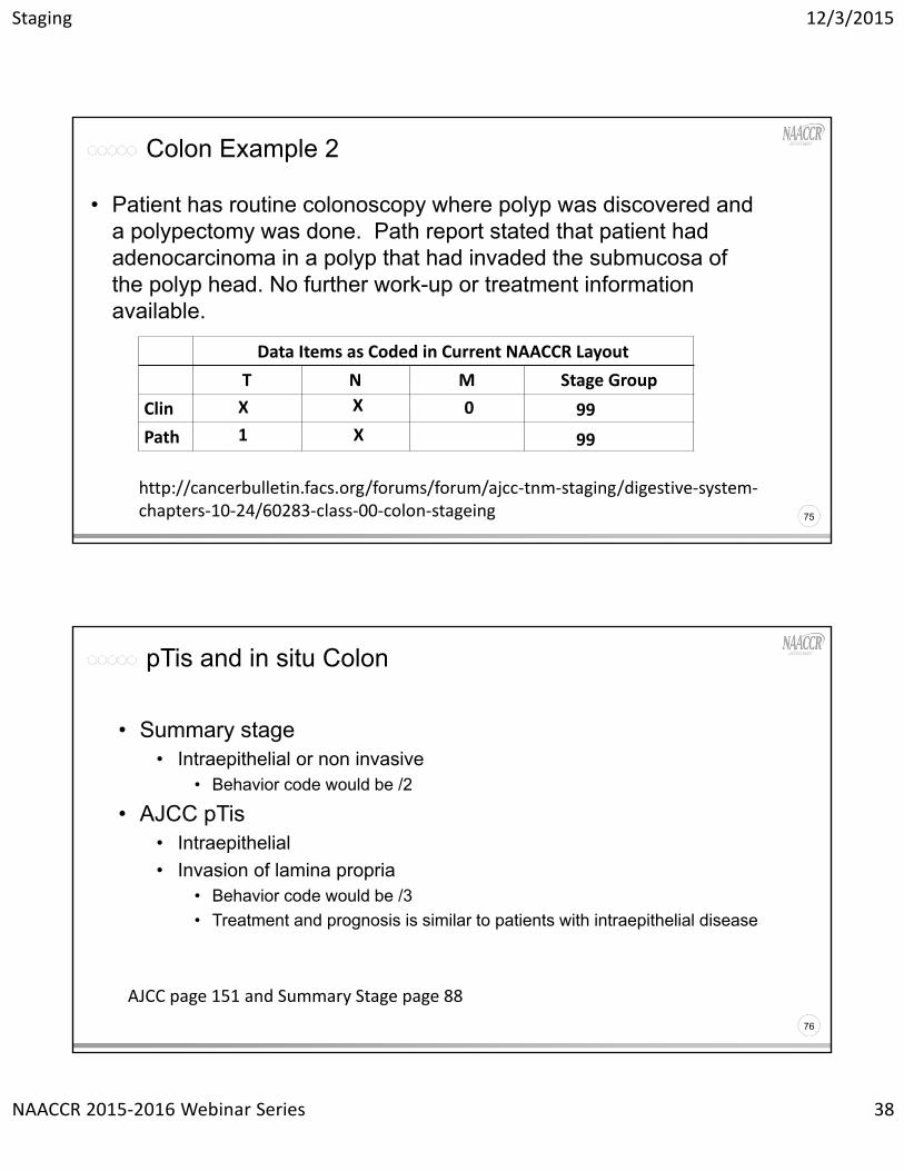
Colon Example 2
75
• Patient has routine colonoscopy where polyp was discovered and a polypectomy was done. Path report stated that patient had adenocarcinoma in a polyp that had invaded the submucosa of the polyp head. No further work-up or treatment information available.
Data Items as Coded in Current NAACCR Layout
T N M Stage Group
Clin
Path
99
99
http://cancerbulletin.facs.org/forums/forum/ajcc‐tnm‐staging/digestive‐system‐chapters‐10‐24/60283‐class‐00‐colon‐stageing
X X 0
1 X
76
pTis and in situ Colon
• Summary stage • Intraepithelial or non invasive
• Behavior code would be /2
• AJCC pTis• Intraepithelial
• Invasion of lamina propria• Behavior code would be /3
• Treatment and prognosis is similar to patients with intraepithelial disease
76
AJCC page 151 and Summary Stage page 88
Staging 12/3/2015
NAACCR 2015‐2016 Webinar Series 39
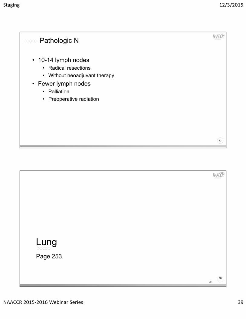
77
Pathologic N
• 10-14 lymph nodes • Radical resections
• Without neoadjuvant therapy
• Fewer lymph nodes• Palliation
• Preoperative radiation
77
78
Page 253
Lung
78

Staging 12/3/2015
NAACCR 2015‐2016 Webinar Series 40
79
• T2• Tumor more than 3 cm but 7 cm or less OR
• Any of the following features• Involves main bronchus 2 cm or more distal to carina
• Invades visceral pleura (PL1 or PL2)
• Associated with atelectasis or obstructive pneumonitis that extends to hilar region but does not involve entire lung
• T2 tumors with above features are T2a if 5 cm or less
• T2a: Tumor more than 3 cm but 5 cm or less
• T2b: Tumor more than 5 cm but 7 cm or less
AJCC Cancer Stage: Lung T Category
79See page 263 AJCC Manual
80
Lung Example 1
• A patient had a CT that showed a tumor in the left upper lobe lung 2.5cm’s from the carina. The tumor measured 2cm in greatest dimension. No adenopathy identified.
80

Staging 12/3/2015
NAACCR 2015‐2016 Webinar Series 41
8181
Figure1El‐Sheriff A H, Lau C T, Wu C C, et al. International Association for the Study of Lung Cancer (IASLC) lymph node map: Radiologic review with CT illustration. RadioGraphics2014;34:1680‐1691
82
Lung Example 1• A patient had a CT that showed a tumor in the left main stem
bronchus 2.5 cm’s from the carina. The tumor measured 2cm in greatest dimension. No adenopathy identified.
82
Data Items as Coded in Current NAACCR Layout
T N M Stage Group
Clin
Path
1B
99
2a 0 0

Staging 12/3/2015
NAACCR 2015‐2016 Webinar Series 42
83
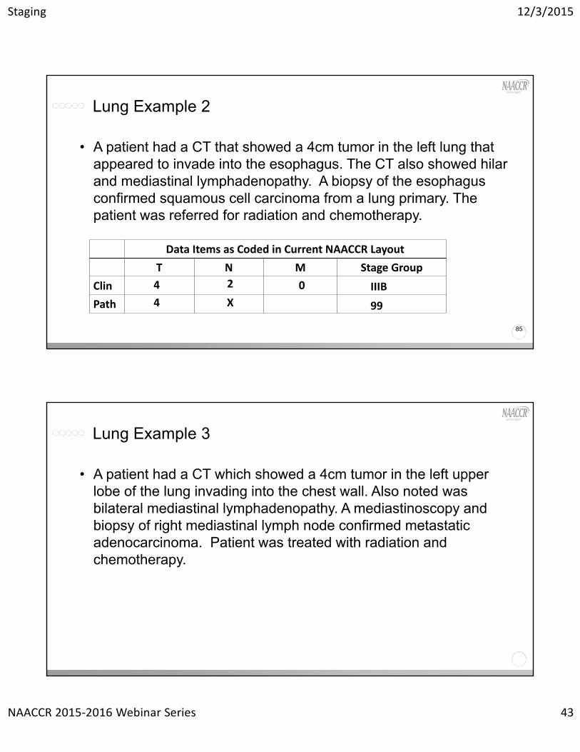
Lung Example 2
83
• A patient had a CT that showed a 4cm tumor in the left lung that appeared to invade into the esophagus. The CT also showed hilar and mediastinal lymphadenopathy. A biopsy of the esophagus confirmed squamous cell carcinoma from a lung primary. The patient was referred for radiation and chemotherapy.
8484
Figure1El‐Sherief A H, Lau C T, Wu C C, et al. International Association for the Study of Lung Cancer (IASLC) lymph node map: Radiologic review with CT illustration. RadioGraphics2014;34:1680‐1691
Staging 12/3/2015
NAACCR 2015‐2016 Webinar Series 43
85
Lung Example 2
85
• A patient had a CT that showed a 4cm tumor in the left lung that appeared to invade into the esophagus. The CT also showed hilar and mediastinal lymphadenopathy. A biopsy of the esophagus confirmed squamous cell carcinoma from a lung primary. The patient was referred for radiation and chemotherapy.
Data Items as Coded in Current NAACCR Layout
T N M Stage Group
Clin
Path
IIIB
99
4 2 0
4 X
86
Lung Example 3
• A patient had a CT which showed a 4cm tumor in the left upper lobe of the lung invading into the chest wall. Also noted was bilateral mediastinal lymphadenopathy. A mediastinoscopy and biopsy of right mediastinal lymph node confirmed metastatic adenocarcinoma. Patient was treated with radiation and chemotherapy.

Staging 12/3/2015
NAACCR 2015‐2016 Webinar Series 44
8787
Figure1El‐Sherief A H, Lau C T, Wu C C, et al. International Association for the Study of Lung Cancer (IASLC) lymph node map: Radiologic review with CT illustration. RadioGraphics2014;34:1680‐1691
88
Lung Example 3
• A patient had a CT which showed a 4cm tumor in the left upper lobe of the lung invading into the chest wall. Also noted was bilateral mediastinal lymphadenopathy. A mediastinoscopy and biopsy of right mediastinal lymph node confirmed metastatic adenocarcinoma. Patient was treated with radiation and chemotherapy.
88
Data Items as Coded in Current NAACCR Layout
T N M Stage Group
Clin
Path
IIIB
99
3 3 0
Staging 12/3/2015
NAACCR 2015‐2016 Webinar Series 45
89
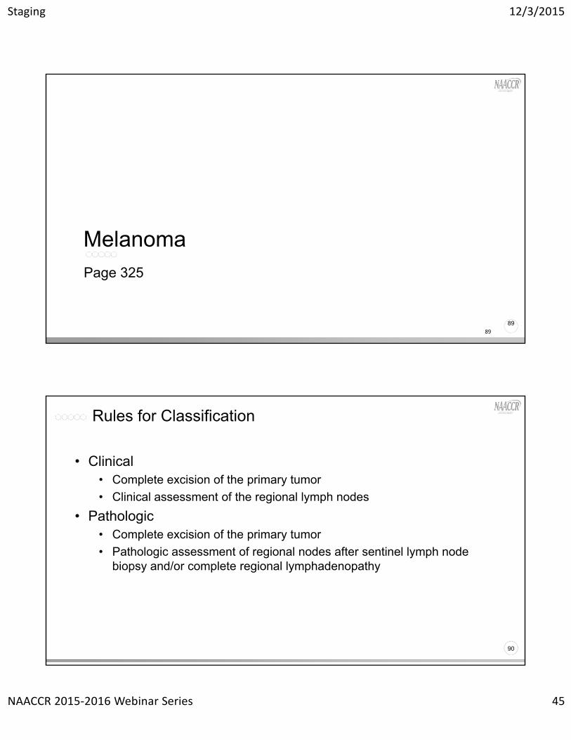
Page 325
Melanoma
89
90
Rules for Classification
• Clinical• Complete excision of the primary tumor
• Clinical assessment of the regional lymph nodes
• Pathologic• Complete excision of the primary tumor
• Pathologic assessment of regional nodes after sentinel lymph node biopsy and/or complete regional lymphadenopathy
90

Staging 12/3/2015
NAACCR 2015‐2016 Webinar Series 46
91
Prognostic Factors Necessary for Stage Grouping
• Ulceration and mitosis• Reflected in the T category (see page 335)
• Microscopic vs macroscopic lymph node metastasis• Reflected in the pN category (see page 336)
• Site of distant metastasis• Reflected in the M category (see page 336)
• LDH• Reflected in the M category (see page 336)
91
92
pStage III
• Stage group IIIA• T1-4a should be interpreted as T(1-4)a, or T1a, T2a, T3a, T4a
• T1-4b should be interpreted as T(1-4)b, or T1b, T2b, T3b, T4b
• The a is without ulceration and all levels of T without ulceration are grouped together
• The b is with ulceration and all levels of T with ulceration are grouped together
92
Page 335
Staging 12/3/2015
NAACCR 2015‐2016 Webinar Series 47
93
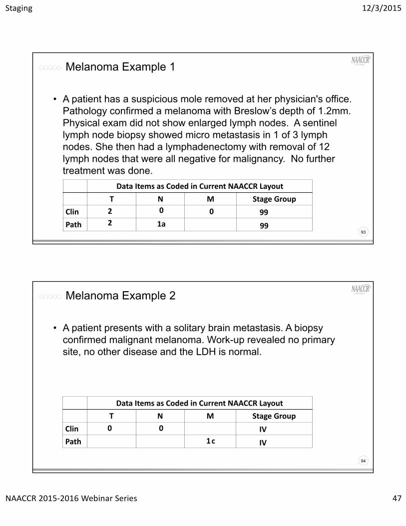
Melanoma Example 1
• A patient has a suspicious mole removed at her physician's office. Pathology confirmed a melanoma with Breslow’s depth of 1.2mm. Physical exam did not show enlarged lymph nodes. A sentinel lymph node biopsy showed micro metastasis in 1 of 3 lymph nodes. She then had a lymphadenectomy with removal of 12 lymph nodes that were all negative for malignancy. No further treatment was done.
93
Data Items as Coded in Current NAACCR Layout
T N M Stage Group
Clin
Path
992 0 0
2 1a 99
94
Melanoma Example 2
• A patient presents with a solitary brain metastasis. A biopsy confirmed malignant melanoma. Work-up revealed no primary site, no other disease and the LDH is normal.
94
Data Items as Coded in Current NAACCR Layout
T N M Stage Group
Clin
Path
IV
IV
0 0
1c

Staging 12/3/2015
NAACCR 2015‐2016 Webinar Series 48
95
Quiz
Case Scenario
Questions?
95
96
Coming Up…
96
• Collecting Cancer Data: Bone and Soft Tissue• 1/7/16
• Collecting Cancer Data: Breast• 2/4/16

Staging 12/3/2015
NAACCR 2015‐2016 Webinar Series 49
97
And the winners are…
98
CE Certificate Quiz/Survey
98
• Phrase
Neoadjuvant
• Link• http://www.surveygizmo.com/s3/2471572/Staging-2015

Staging 12/3/2015
NAACCR 2015‐2016 Webinar Series 50
99
Jim Hofferkamp, [email protected] 698 0800 x 5
Thank you!
99