stable gene silencing in zebra sh with spatiotemporally ... · stable gene silencing in zebrafish...
TRANSCRIPT

INVESTIGATION
Stable Gene Silencing in Zebrafish withSpatiotemporally Targetable RNA Interference
Zhiqiang Dong,1 Jisong Peng,1 and Su Guo2
Department of Bioengineering and Therapeutic Sciences, Programs in Human Genetics and Biological Sciences,University of California, San Francisco, California 94143-2811
ABSTRACT The ability to regulate gene activity in a spatiotemporally controllable manner is vital for biological discovery that willimpact disease diagnosis and treatment. While conditional gene silencing is possible in other genetic model organisms, this technologyis largely unavailable in zebrafish, an important vertebrate model organism for functional gene discovery. Here, using short hairpinRNAs (shRNAs) designed in the microRNA-30 backbone, which have been shown to mimic natural microRNA primary transcripts andbe more effective than simple shRNAs, we report stable RNA interference-mediated gene silencing in zebrafish employing the yeastGal4-UAS system. Using this approach, we reveal at single-cell resolution the role of atypical protein kinase Cl (aPKCl) in regulatingneural progenitor/stem cell division. We also show effective silencing of the one-eyed-pinhead and no-tail/brachyury genes. Further-more, we demonstrate stable integration and germ-line transmission of the UAS-miR-shRNAs for aPKCl, the expressivity of which iscontrollable by the strength and expression of Gal4. This technology shall significantly advance the utility of zebrafish for understand-ing fundamental vertebrate biology and for the identification and evaluation of important therapeutic targets.
STUDIES of genetic model organisms contribute tremen-dously to our understanding of diverse biological pro-
cesses and human disorders at molecular and cellular levels(Davis 2004; Aitman et al. 2011). As a recently establishedanimal model, zebrafish has salient features that promise toprovide new insights into biology and medicine. These in-clude rapid external development, transparent embryonicand larval stages, simpler vertebrate organ systems, andamenability for genetic and chemical screening. The utilityof zebrafish, however, has been hampered by the inabilityto silence genes in a spatiotemporally controllable manner.Although valuable reverse genetic methods are available(Nasevicius and Ekker 2000; Wienholds et al. 2002; Doyonet al. 2008; Meng et al. 2008; Huang et al. 2011; Sanderet al. 2011; Bedell et al. 2012), none offer gene silencing atany desired stages of the life cycle and in any cell types ofinterest.
RNA interference (RNAi) is a powerful approach todissect gene function (Fire 2007; Mello 2007). It was orig-inally discovered in Caenorhabditis elegans, where the obser-vation of gene inactivation by both sense and antisenseRNAs (Guo and Kemphues 1995) led to the finding of double-stranded RNA (dsRNA)-mediated gene silencing (Fire et al.1998). In vertebrates, the effectiveness of RNAi was hinderedby the dsRNA-induced interferon response causing nonspecificeffects (Manche et al. 1992; Stark et al. 1998) until the findingof efficacious small interfering RNAs (siRNAs) (Elbashir et al.2001). However, their delivery into zebrafish remains eitherineffective or has nonspecific effects (Gruber et al. 2005;Zhao et al. 2008). Likewise, simple short hairpin RNAs(shRNAs) driven by RNA polymerase III promoters (Paddi-son et al. 2002) also appears ineffective in zebrafish (Wanget al. 2010).
Micro-RNAs (miRNAs) are endogenous�21- to 23-nucleotideRNAs that can regulate gene expression. Originally discov-ered in C. elegans (Lee et al. 1993; Reinhart et al. 2000), theywere later found conserved across animals and plants (Bartel2009; Ebert and Sharp 2012). The observation that de-signed miRNAs can inhibit cognate mRNA expression inhuman cells (Zeng et al. 2002) has paved the way for recentdevelopments of shRNAs employing the primary miR-30backbones (miR-shRNAs) (Dickins et al. 2005; Silva et al.2005). This natural configuration is reported 12 times more
Copyright © 2013 by the Genetics Society of Americadoi: 10.1534/genetics.112.147892Manuscript received November 21, 2012; accepted for publication January 12, 2013Supporting information is available online at http://www.genetics.org/lookup/suppl/doi:10.1534/genetics.112.147892/-/DC1.1These authors contributed equally to this work.2Corresponding author: Department of Bioengineering and Therapeutic Sciences,Programs in Human Genetics and Biological Sciences, University of California, SanFrancisco, CA 94143-2811. E-mail: [email protected]
Genetics, Vol. 193, 1065–1071 April 2013 1065

efficient than simple hairpin designs. MiR-shRNAs are re-cently shown to be effective in vivo in mice (Dickins et al.2005; Premsrirut et al. 2011; Zuber et al. 2011) and zebra-fish (Dong et al. 2009; De Rienzo et al. 2012). However, thecapability of germ-line transmission is only reported by onestudy in zebrafish (Dong et al. 2009), where functional ef-ficacy and specificity of silencing were not quantitativelydocumented. Therefore, the potential of RNAi for stableand conditional gene silencing in zebrafish remainsuncertain.
With the goal of achieving spatiotemporal and dosagecontrol of gene silencing, we have exploited the miR-shRNAtechnology in combination with the bipartite Gal4/upstreamactivating sequence (UAS) system (Ptashne 1988), whichoffers excellent versatility for spatiotemporal control to-gether with amplification of gene expression level. Usingatypical protein kinase Cl (apkcl), one-eyed pinhead (oep),and no tail/brachyury (ntl) as test loci, we show that RNAi inzebrafish is effective, rapid, dosage controllable, stable withconditional capability, and scalable.
Materials and Methods
Zebrafish strains
Wild-type embryos were obtained from natural spawning ofAB adults. The transgenic line Tg[ubi-GFF] (Asakawa andKawakami 2010) was a gift from K. Kawakami. The apkclmutant heart and soul was kindly provided by D. Stainier.
ShRNA design
ShRNA design was performed using the siRNA design toolfrom the following website: http://www.genscript.com/design_center.html, which ranks target candidates basedon a proprietary algorithm using DE parameters. We furtherrefined the list by removing the candidates that have morethan three G/C in 6–11 nt of the target sequences, based ona thermodynamic study of siRNA (Ui-Tei et al. 2008).
Vector construction
The shRNA precursor structure was designed based on themiR-30e precursor in zebrafish. The miR-30e target andguide sequences were replaced with the predicted shRNAtarget and guide sequences. To best mimic the miR-30eprecursor structure, we kept the original loop region and theflanking sequences of the miR-30e precursor and introducedtwo mismatched “TT” into the shRNA guide sequence be-tween nt 12 and nt 13. We synthesized a pair of comple-mentary oligos that include all the designed shRNAstructures (guide sequence, loop sequence, target sequence,and the flanking sequences) and the annealed oligos wereinserted into zebrafish miR-30e backbone in expression vec-tors kindly provided by the late Ting Xi Liu (Dong et al.2009). The following 5xUAS sequence was used: cggagtactgtcctccgagcggagtactgtcctccgagcggagtactgtcctccgagcggagtactgtcctccgagcggagtactgtcctccg.
DNA and RNA injection
DNA and RNA injections were performed at the one-cell stage. For genetic mosaic analyses, RNAi constructs(UAS-TdTomato-shRNA1apkcl-miniTol2 or UAS-TdTomato-shRNA5apkcl-miniTol2; 75 pg per embryo) were co-injectedwith EF1a-GFF-PT2KXIG (elongation factor 1a regulatoryelement-driven Gal4) (50 pg per embryo) and Tol2 tranpo-sase (10 pg per embryo). Sense strand-capped RNA wassynthesized by SP6 transcription from NotI-linearized plas-mid using the mMESSAGE mMACHINE system (Ambion).RNAi-resistant sense RNAs for apkcl, ntla and οepa, as wellas RNAs encoding the oep-shRNA and ntla-shRNA, wereinjected at 400 pg per embryo. For time-lapse in vivo imag-ing, 3NLS:EGFP sense RNA (200 pg per embryo) was co-injected with EF1a-GFF-PT2KXIG DNA (50 pg per embryo)and 5xUAS-tdTomato-shRNAapkcl plasmid DNA (75 pg perembryo).
Heat shock of zebrafish embryos
Heat shock of zebrafish embryos was performed at the stageof 8 hours postfertilization (hpf) (75% epiboly). Fertilizedembryos were collected from the cross between the UAS-TdTom-miR-shRNA1apkcl founder A and the hsp70-Gal4transgenic animal, and were raised at 28.5� before heatshock. A 500-ml glass beaker filled with 100 ml egg waterwas preheated to 37� in a water bath right before heatshock. Around 100 embryos were transferred to the pre-heated egg water at 8 hpf and incubated in a 37� water bathfor 1 hr. The embryos were then transferred to 28.5� eggwater immediately and raised in a 28.5� incubator untilanalysis.
Phenotypic analyses
Phenotypic analyses were performed manually under a brightfield dissection microscope. For apkcl RNAi, four morphologi-cal phenotypes were analyzed, which are typically observedin the apkcl mutant (heart and soul) (Horne-Badovinac et al.2001): (1) defect in heart tube assembly, (2) patchy retinalpigmented epithelium, (3) failure of the brain ventricle toinflate, and (4) abnormal body curve. The number of em-bryos that are positive for each phenotype as well as the totalnumber of all embryos was counted for both RNAi groups andcontrol groups. Statistic analyses were performed based onpositive percentage for each phenotype. For ntla and oepRNAi, the typical no-tail phenotype and one-eyed pinheadphenotype were analyzed.
Time-lapse in vivo imaging
To observe the division orientation of radial glia progenitorcells, time-lapse in vivo imaging was performed on embryosinjected with 3NLS:EGFP sense RNA, EF1a-GFF-PT2KXIGDNA, and 5xUAS-tdTomato-shRNAapkcl-miniTol2 DNA (seeDNA and RNA injection for detail) at 24 hpf. The imagingwas performed as previously described (Dong et al. 2012).In brief, embryos were mounted in low melting point aga-rose and properly placed on a temperature-controlled stage
1066 Z. Dong, J. Peng, and S. Guo

of the confocal microscope. We used a Nikon C1 spectralconfocal microscope with upright objectives. Fluorescentlylabeled individual neural progenitor cells were imaged for2 hr with a fixed interval of 90 sec under a ·40 water-dippingobjective. The parameters of confocal imaging were deter-mined as sufficient to capture the orientation of cell division,while reducing photobleaching during the imaging period.
Immunohistochemistry
Immunohistochemistry was performed on whole mountembryos as described previously (Guo et al. 1999). Embryoswere fixed with 4% paraformaldehyde. Embryos older than60 hpf were treated with proteinase K before incubation inprimary antibodies (rabbit anti-aPKC 1:100, Santa Cruz sc-216) at 4� overnight. After four washes with phosphate-buffered saline with 0.1% Tween, 0.5% Triton X-100, and1% DMSO, the embryos were incubated with Alexa Fluorsecondary antibody (1:2000, Invitrogen) at 4� overnight.After wash and glycerol sequential treatment, the embryos weremounted and imaged using Nikon C1 confocal microscope.
Quantitative real-time RT–PCR analyses
For the analyses of knockdown of apkcl expression by tran-sient RNAi, the control or RNAi embryos were collected at10 hpf and total RNA from pooled embryos (20 embryos persample for each group) was extracted using Trizol reagent(Invitrogen) followed by treatment with Turbo DNA-freeDNase (Ambion). First-strand cDNA was reverse transcribedusing oligo-dT primers and Superscript reverse transcriptase(Invitrogen). Real-time PCR amplifications of apkcl, oep,ntla, and gapdhs were carried out with SYBR Green PCRMaster Mix (Applied Biosystems). Primers used were:
apkcl F: 59-CCCGCACCAAGTCCGGGTAA-39,apkcl R: 59-GCTGAGAAGAAACGGTGCACGGA-39;oep F: 59-ATGGACTTTTGCATTGCTTCCCACA-39,oep R: 59-AGTGTTCTGAGGGAGCCCGACC-39;ntla F: 59-CCCAGCCATTACTCCCACCGC-39,ntla R: 59-TGGGCCAGGGTTCCCATCCC-39;gapdhs F: 59-ACTCCACTCATGGCCGTTAC-39,gapdhs R: 59-TGAGCTGAGGCCTTCTCAAT-39.
Droplet digital PCR analyses
For the analyses of knockdown of apkcl expression by stableRNAi transgenesis, the sibling control or RNAi transgenicembryos were collected at 24 hpf and total RNA from singleembryos was extracted using Trizol reagent (Invitrogen)followed by treatment with Turbo DNA-free DNase (Ambion).First-strand cDNA was reverse transcribed using oligo-dTprimers and Superscript reverse transcriptase (Invitrogen).Droplet Digital PCR amplifications of apkcl and gapdhs werecarried out with primers and probes carrying fluorophoremodifications. The sequences of the primers are the sameas used in qRT–PCR analyses. Additionally, FAM-modified apkclprobe (59-FAM-ATGTGCTCCATGGACAATGACCAGCT-39) and
HEX-modified gapdhs probe (59-HEX-TTCCAGTGCATGAAGCCTGCTGAGAT-39) were used. Data were analyzed using Quanta-soft version 1.2.10.0.
Stable transgenesis
Stable UAS-miR-shRNAapckcl transgenic lines were generatedusing the Tol2 transposon system as described previously(Suster et al. 2009). RNAi constructs (UAS-TdTomato-MiR-shRNA1apkcl-miniTol2 or UAS-TdTomato-miR-shRNA5apkcl-miniTol2; 50 pg per embryo) together with Tol2 transposase(10 pg per embryo) were injected into one-cell-stage em-bryos of AB wild-type fish. Injected embryos were raisedto adulthood (G0) and screened for germ-line transmissionby crossing with Tg[Ubi-GFF] transgenic fish. The germ-linetransgenic founders would yield embryos with TdTomatoexpression, which could be easily distinguished under theepifluorescent microscope. The embryos with ubiquitousTdTomato expression were raised to adulthood (F1), whichcarry both UAS-apckcl RNAi and Ubi-GFF transgenes. Addi-tionally, UAS-RNAi G0 germ-line founders were crossed withAB wild-type fish. All the progenies were raised to adult-hood and genotyped by PCR using TdTomato-specific pri-mers to screen for stable transgenic F1 fish. Stable transgenicF1 were obtained for UAS-TdTomato-shRNA1apckcl and UAS-TdTomato-shRNA5apckcl.
Results and Discussion
Conditional miR-shRNA expression system
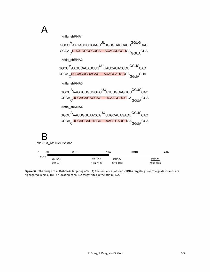
We designed shRNAs employing the primary miR-30 back-bones (hence called miR-shRNAs) (Figure 1A). The fluores-cent reporter TdTomato conveniently marked the cells thatexpress the miR-shRNAs. Three genes were used as the testset: (1) The atypical protein kinase C lambda (aPKCl), forwhich a distinct loss-of-function phenotype has been docu-mented in zebrafish. We chose this gene also because itsdisruption produces a highly specific cellular phenotype,that is, an alteration of spindle rotation in dividing radialglia progenitor cells (Horne-Badovinac et al. 2001). (2) Theno tail a (ntla) and (3) one-eyed pinhead (oep) genes, forwhich distinct loss-of-function phenotypes can be readilyassessed (Schulte-Merker et al. 1994; Zhang et al. 1998).Using the Web-based shRNA design tool (www.genescript.com) together with the filtering criteria based on thermody-namic properties (Ui-Tei et al. 2008), six shRNAs targetingthe apkcl gene (Supporting Information, Figure S1), fourshRNAs each that targeted the ntla (Figure S2), and oep(Figure S3) genes were selected.
Transient in vivo RNAi is highly potent in knockingdown gene activity
For functional validation of shRNAs, we employed a tran-sient in vivo transgenesis method. Although the mosaic ex-pression is an inevitable feature associated with transienttransgenesis, this can be alleviated by coexpression of theTol2 transposase (Li et al. 2010) and selection of high
RNAi in Zebrafish 1067

expressers with the visible reporter TdTomato (Figure S4).DNA plasmids carrying the Pef-1a-Gal4 (elongation factor 1apromoter-driven Gal4) and UAS-miR-shRNA together withthe transposase were co-injected into one-cell-stage zebra-fish embryos. Three of six miR-shRNAs targeting apkcl, andthree of four miR-shRNAs targeting either oep or ntla genes
led to gene-specific phenotypes (with variable penetrance)that could be rescued by delivery of shRNA-resistant wild-type mRNAs for the respective genes (Figure 1B, Table S1,Table S2, and Table S3). Quantitative RT–PCR analysis us-ing total RNAs from 10 hpf shRNA-expressing embryosshowed a gene-specific reduction of endogenous mRNAs
Figure 1 Functional validation of miR-shRNAs by transient in vivo transgenesis.(A) Diagram of the conditional miR-shRNAexpression system. The guide stand (bot-tom) is highlighted in light blue. (B) Mosaicexpression of miR-shRNA5apkcl (left), miR-shRNA4oep (top right), and miR-shRNA4ntla
(bottom right) causes morphological defectssimilar to those observed in respectivemutants, which can be rescued by deliveryof shRNA-resistant wild-type mRNAs. ThemiR-30e vector-injected embryos serve ascontrols. (C) Quantitative RT–PCR analysisshows a gene-specific reduction of endoge-nous mRNA levels by shRNAs cognate totheir target genes. The miR-30e vector-injected embryos serve as controls. (D) Fluo-rescent immunostaining of aPKCl (green)shows knockdown effects at the proteinlevel by different miR-shRNAsapkcl. In a sin-gle miR-shRNAapkcl-expressing cell, as indi-cated by the tdTomato fluorescence (red),miR-shRNA1apkcl (middle panel) and miR-shRNA5apkcl (left panel) lead to significantknockdown of aPKCl, whereas the ineffec-tive miR-shRNA7apkcl does not show obvi-ous knockdown effect (right panel). (E)Mosaic expression of miR-shRNA5apkcl
causes defects in mitotic division orientationof radial glia progenitors in the developingzebrafish forebrain. Left: examples of mitoticdivision orientation in the miR-30e vector-injected (control) and miR-shRNA5apkcl-expressing radial glia progenitors. Right:Quantification of division orientation inmiR-30e vector control and miR-shRNA5apkcl-expressing radial glia progenitors (n = 27 forcontrol, and n = 24 for miR-shRNA5apkcl). Thenuclei of radial glia progenitors were labeledwith 3NLS:EGFP (green) and the individual ra-dial glia progenitors expressing UAS-miR30evector (control) or UAS-miR-shRNA5apkcl werehighlighted by tdTomato (red).
1068 Z. Dong, J. Peng, and S. Guo
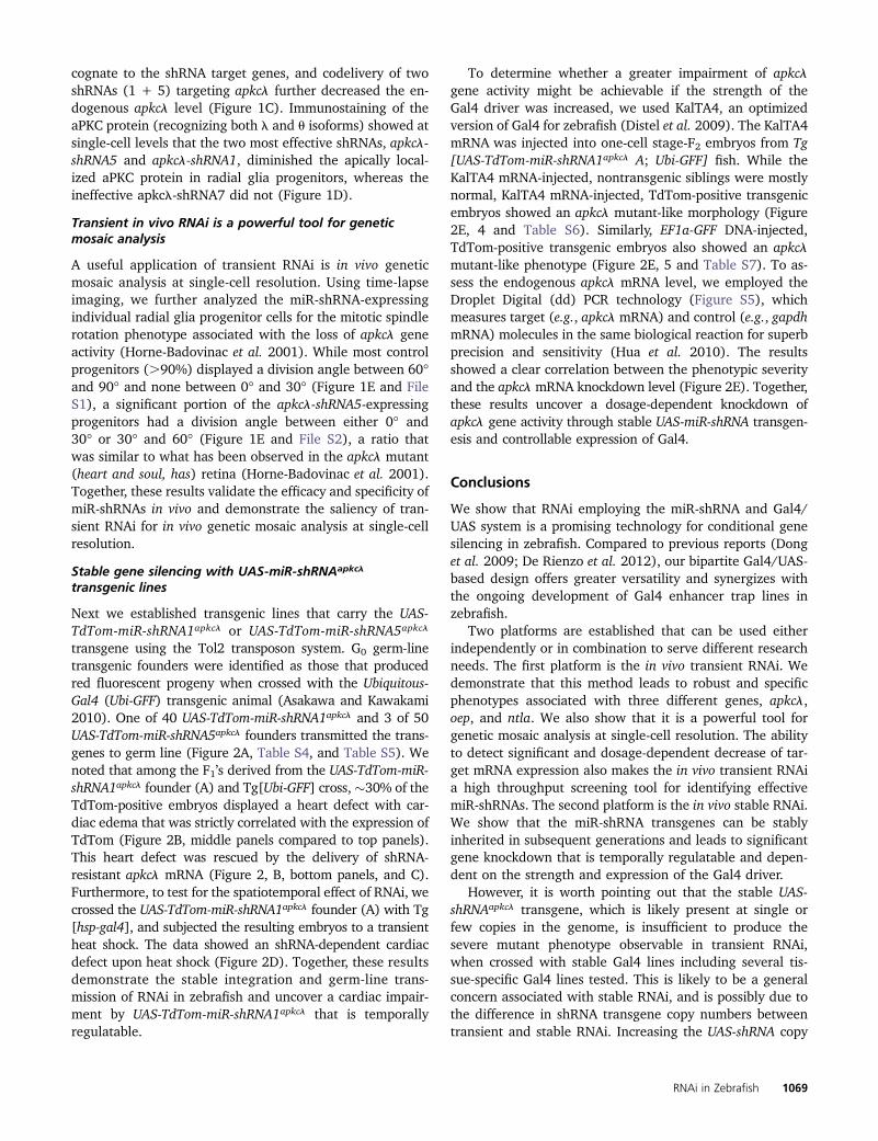
cognate to the shRNA target genes, and codelivery of twoshRNAs (1 + 5) targeting apkcl further decreased the en-dogenous apkcl level (Figure 1C). Immunostaining of theaPKC protein (recognizing both l and u isoforms) showed atsingle-cell levels that the two most effective shRNAs, apkcl-shRNA5 and apkcl-shRNA1, diminished the apically local-ized aPKC protein in radial glia progenitors, whereas theineffective apkcl-shRNA7 did not (Figure 1D).
Transient in vivo RNAi is a powerful tool for geneticmosaic analysis
A useful application of transient RNAi is in vivo geneticmosaic analysis at single-cell resolution. Using time-lapseimaging, we further analyzed the miR-shRNA-expressingindividual radial glia progenitor cells for the mitotic spindlerotation phenotype associated with the loss of apkcl geneactivity (Horne-Badovinac et al. 2001). While most controlprogenitors (.90%) displayed a division angle between 60�and 90� and none between 0� and 30� (Figure 1E and FileS1), a significant portion of the apkcl-shRNA5-expressingprogenitors had a division angle between either 0� and30� or 30� and 60� (Figure 1E and File S2), a ratio thatwas similar to what has been observed in the apkcl mutant(heart and soul, has) retina (Horne-Badovinac et al. 2001).Together, these results validate the efficacy and specificity ofmiR-shRNAs in vivo and demonstrate the saliency of tran-sient RNAi for in vivo genetic mosaic analysis at single-cellresolution.
Stable gene silencing with UAS-miR-shRNAapkcl
transgenic lines
Next we established transgenic lines that carry the UAS-TdTom-miR-shRNA1apkcl or UAS-TdTom-miR-shRNA5apkcl
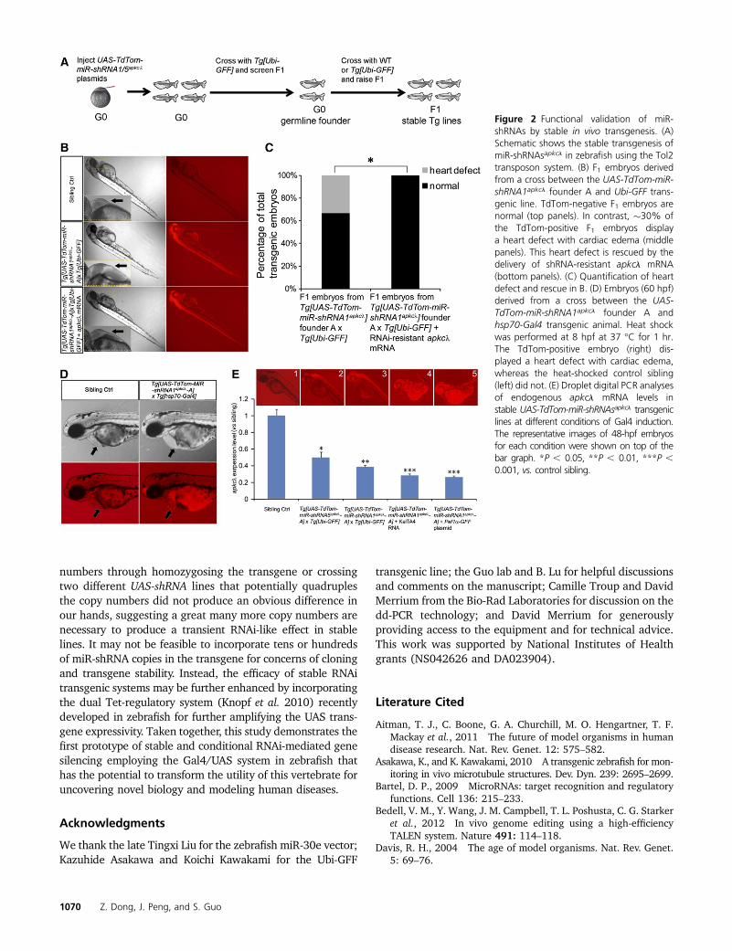
transgene using the Tol2 transposon system. G0 germ-linetransgenic founders were identified as those that producedred fluorescent progeny when crossed with the Ubiquitous-Gal4 (Ubi-GFF) transgenic animal (Asakawa and Kawakami2010). One of 40 UAS-TdTom-miR-shRNA1apkcl and 3 of 50UAS-TdTom-miR-shRNA5apkcl founders transmitted the trans-genes to germ line (Figure 2A, Table S4, and Table S5). Wenoted that among the F1’s derived from the UAS-TdTom-miR-shRNA1apkcl founder (A) and Tg[Ubi-GFF] cross, �30% of theTdTom-positive embryos displayed a heart defect with car-diac edema that was strictly correlated with the expression ofTdTom (Figure 2B, middle panels compared to top panels).This heart defect was rescued by the delivery of shRNA-resistant apkcl mRNA (Figure 2, B, bottom panels, and C).Furthermore, to test for the spatiotemporal effect of RNAi, wecrossed the UAS-TdTom-miR-shRNA1apkcl founder (A) with Tg[hsp-gal4], and subjected the resulting embryos to a transientheat shock. The data showed an shRNA-dependent cardiacdefect upon heat shock (Figure 2D). Together, these resultsdemonstrate the stable integration and germ-line trans-mission of RNAi in zebrafish and uncover a cardiac impair-ment by UAS-TdTom-miR-shRNA1apkcl that is temporallyregulatable.
To determine whether a greater impairment of apkclgene activity might be achievable if the strength of theGal4 driver was increased, we used KalTA4, an optimizedversion of Gal4 for zebrafish (Distel et al. 2009). The KalTA4mRNA was injected into one-cell stage-F2 embryos from Tg[UAS-TdTom-miR-shRNA1apkcl A; Ubi-GFF] fish. While theKalTA4 mRNA-injected, nontransgenic siblings were mostlynormal, KalTA4 mRNA-injected, TdTom-positive transgenicembryos showed an apkcl mutant-like morphology (Figure2E, 4 and Table S6). Similarly, EF1a-GFF DNA-injected,TdTom-positive transgenic embryos also showed an apkclmutant-like phenotype (Figure 2E, 5 and Table S7). To as-sess the endogenous apkcl mRNA level, we employed theDroplet Digital (dd) PCR technology (Figure S5), whichmeasures target (e.g., apkcl mRNA) and control (e.g., gapdhmRNA) molecules in the same biological reaction for superbprecision and sensitivity (Hua et al. 2010). The resultsshowed a clear correlation between the phenotypic severityand the apkcl mRNA knockdown level (Figure 2E). Together,these results uncover a dosage-dependent knockdown ofapkcl gene activity through stable UAS-miR-shRNA transgen-esis and controllable expression of Gal4.
Conclusions
We show that RNAi employing the miR-shRNA and Gal4/UAS system is a promising technology for conditional genesilencing in zebrafish. Compared to previous reports (Donget al. 2009; De Rienzo et al. 2012), our bipartite Gal4/UAS-based design offers greater versatility and synergizes withthe ongoing development of Gal4 enhancer trap lines inzebrafish.
Two platforms are established that can be used eitherindependently or in combination to serve different researchneeds. The first platform is the in vivo transient RNAi. Wedemonstrate that this method leads to robust and specificphenotypes associated with three different genes, apkcl,oep, and ntla. We also show that it is a powerful tool forgenetic mosaic analysis at single-cell resolution. The abilityto detect significant and dosage-dependent decrease of tar-get mRNA expression also makes the in vivo transient RNAia high throughput screening tool for identifying effectivemiR-shRNAs. The second platform is the in vivo stable RNAi.We show that the miR-shRNA transgenes can be stablyinherited in subsequent generations and leads to significantgene knockdown that is temporally regulatable and depen-dent on the strength and expression of the Gal4 driver.
However, it is worth pointing out that the stable UAS-shRNAapkcl transgene, which is likely present at single orfew copies in the genome, is insufficient to produce thesevere mutant phenotype observable in transient RNAi,when crossed with stable Gal4 lines including several tis-sue-specific Gal4 lines tested. This is likely to be a generalconcern associated with stable RNAi, and is possibly due tothe difference in shRNA transgene copy numbers betweentransient and stable RNAi. Increasing the UAS-shRNA copy
RNAi in Zebrafish 1069
numbers through homozygosing the transgene or crossingtwo different UAS-shRNA lines that potentially quadruplesthe copy numbers did not produce an obvious difference inour hands, suggesting a great many more copy numbers arenecessary to produce a transient RNAi-like effect in stablelines. It may not be feasible to incorporate tens or hundredsof miR-shRNA copies in the transgene for concerns of cloningand transgene stability. Instead, the efficacy of stable RNAitransgenic systems may be further enhanced by incorporatingthe dual Tet-regulatory system (Knopf et al. 2010) recentlydeveloped in zebrafish for further amplifying the UAS trans-gene expressivity. Taken together, this study demonstrates thefirst prototype of stable and conditional RNAi-mediated genesilencing employing the Gal4/UAS system in zebrafish thathas the potential to transform the utility of this vertebrate foruncovering novel biology and modeling human diseases.
Acknowledgments
We thank the late Tingxi Liu for the zebrafish miR-30e vector;Kazuhide Asakawa and Koichi Kawakami for the Ubi-GFF
transgenic line; the Guo lab and B. Lu for helpful discussionsand comments on the manuscript; Camille Troup and DavidMerrium from the Bio-Rad Laboratories for discussion on thedd-PCR technology; and David Merrium for generouslyproviding access to the equipment and for technical advice.This work was supported by National Institutes of Healthgrants (NS042626 and DA023904).
Literature Cited
Aitman, T. J., C. Boone, G. A. Churchill, M. O. Hengartner, T. F.Mackay et al., 2011 The future of model organisms in humandisease research. Nat. Rev. Genet. 12: 575–582.
Asakawa, K., and K. Kawakami, 2010 A transgenic zebrafish for mon-itoring in vivo microtubule structures. Dev. Dyn. 239: 2695–2699.
Bartel, D. P., 2009 MicroRNAs: target recognition and regulatoryfunctions. Cell 136: 215–233.
Bedell, V. M., Y. Wang, J. M. Campbell, T. L. Poshusta, C. G. Starkeret al., 2012 In vivo genome editing using a high-efficiencyTALEN system. Nature 491: 114–118.
Davis, R. H., 2004 The age of model organisms. Nat. Rev. Genet.5: 69–76.
Figure 2 Functional validation of miR-shRNAs by stable in vivo transgenesis. (A)Schematic shows the stable transgenesis ofmiR-shRNAsapkcl in zebrafish using the Tol2transposon system. (B) F1 embryos derivedfrom a cross between the UAS-TdTom-miR-shRNA1apkcl founder A and Ubi-GFF trans-genic line. TdTom-negative F1 embryos arenormal (top panels). In contrast, �30% ofthe TdTom-positive F1 embryos displaya heart defect with cardiac edema (middlepanels). This heart defect is rescued by thedelivery of shRNA-resistant apkcl mRNA(bottom panels). (C) Quantification of heartdefect and rescue in B. (D) Embryos (60 hpf)derived from a cross between the UAS-TdTom-miR-shRNA1apkcl founder A andhsp70-Gal4 transgenic animal. Heat shockwas performed at 8 hpf at 37 �C for 1 hr.The TdTom-positive embryo (right) dis-played a heart defect with cardiac edema,whereas the heat-shocked control sibling(left) did not. (E) Droplet digital PCR analysesof endogenous apkcl mRNA levels instable UAS-TdTom-miR-shRNAsapkcl transgeniclines at different conditions of Gal4 induction.The representative images of 48-hpf embryosfor each condition were shown on top of thebar graph. *P , 0.05, **P , 0.01, ***P ,0.001, vs. control sibling.
1070 Z. Dong, J. Peng, and S. Guo
De Rienzo, G., J. H. Gutzman, and H. Sive, 2012 Efficient shRNA-mediated inhibition of gene expression in zebrafish. Zebrafish 9:97–107.
Dickins, R. A., M. T. Hemann, J. T. Zilfou, D. R. Simpson, I. Ibarraet al., 2005 Probing tumor phenotypes using stable and regu-lated synthetic microRNA precursors. Nat. Genet. 37: 1289–1295.
Distel, M., M. F. Wullimann, and R. W. Köster, 2009 OptimizedGal4 genetics for permanent gene expression mapping in zebra-fish. Proc. Natl. Acad. Sci. USA 106: 13365–13370.
Dong, M., Y. F. Fu, T. T. Du, C. B. Jing, C. T. Fu et al., 2009 Heritableand lineage-specific gene knockdown in zebrafish embryo. PLoSONE 4: e6125.
Dong, Z., N. Yang, S. Yeo, A. Chitnis, and S. Guo, 2012 Intra-lineage directional Notch signaling regulates self-renewal anddifferentiation of asymmetrically dividing radial glia. Neuron74: 65–78.
Doyon, Y., J. M. McCammon, J. C. Miller, F. Faraji, C. Ngo et al.,2008 Heritable targeted gene disruption in zebrafish using de-signed zinc-finger nucleases. Nat. Biotechnol. 26: 702–708.
Ebert, M. S., and P. A. Sharp, 2012 Roles for microRNAs inconferring robustness to biological processes. Cell 149:515–524.
Elbashir, S. M., J. Harborth, W. Lendeckel, A. Yalcin, K. Weberet al., 2001 Duplexes of 21-nucleotide RNAs mediate RNA in-terference in cultured mammalian cells. Nature 411: 494–498.
Fire, A., S. Xu, M. K. Montgomery, S. A. Kostas, S. E. Driver et al.,1998 Potent and specific genetic interference by double-stranded RNA in Caenorhabditis elegans. Nature 391: 806–811.
Fire, A. Z., 2007 Gene silencing by double-stranded RNA. CellDeath Differ. 14: 1998–2012.
Gruber, J., H. Manninga, T. Tuschl, M. Osborn, and K. Weber,2005 Specific RNAi mediated gene knockdown in zebrafishcell lines. RNA Biol. 2: 101–105.
Guo, S., and K. J. Kemphues, 1995 par-1, a gene required forestablishing polarity in C. elegans embryos, encodes a putativeser/thr kinase that is asymmetrically distributed. Cell 81:611–620.
Guo, S., S. W. Wilson, S. Cooke, A. B. Chitnis, W. Driever et al.,1999 Mutations in the zebrafish unmask shared regulatorypathways controlling the development of catecholaminergicneurons. Dev. Biol. 208: 473–487.
Horne-Badovinac, S., D. Lin, S. Waldron, M. Schwarz, G. Mbamaluet al., 2001 Positional cloning of heart and soul reveals multi-ple roles for PKC lambda in zebrafish organogenesis. Curr. Biol.11: 1492–1502.
Hua, Z., J. L. Rouse, A. E. Eckhardt, V. Srinivasan, V. K. Pamulaet al., 2010 Multiplexed real-time polymerase chain reactionon a digital microfluidic platform. Anal. Chem. 82: 2310–2316.
Huang, P., A. Xiao, M. Zhou, Z. Zhu, S. Lin et al., 2011 Heritablegene targeting in zebrafish using customized TALENs. Nat. Bio-technol. 29: 699–700.
Knopf, F., K. Schnabel, C. Haase, K. Pfeifer, K. Anastassiadis et al.,2010 Dually inducible TetON systems for tissue-specific con-ditional gene expression in zebrafish. Proc. Natl. Acad. Sci. USA107: 19933–19938.
Lee, R. C., R. L. Feinbaum, and V. Ambros, 1993 The C. elegansheterochronic gene lin-4 encodes small RNAs with antisensecomplementarity to lin-14. Cell 75: 843–854.
Li, Q., D. Ritter, N. Yang, Z. Dong, H. Li et al., 2010 A systematicapproach to identify functional motifs within vertebrate devel-opmental enhancers. Dev. Biol. 337: 484–495.
Manche, L., S. R. Green, C. Schmedt, and M. B. Mathews,1992 Interactions between double-stranded RNA regula-tors and the protein kinase DAI. Mol. Cell. Biol. 12: 5238–5248.
Mello, C. C., 2007 Return to the RNAi world: rethinking geneexpression and evolution. Cell Death Differ. 14: 2013–2020.
Meng, X., M. B. Noyes, L. J. Zhu, N. D. Lawson, and S. A. Wolfe,2008 Targeted gene inactivation in zebrafish using engineeredzinc-finger nucleases. Nat. Biotechnol. 26: 695–701.
Nasevicius, A., and S. C. Ekker, 2000 Effective targeted gene“knockdown” in zebrafish. Nat. Genet. 26: 216–220.
Paddison, P. J., A. A. Caudy, E. Bernstein, G. J. Hannon, and D. S.Conklin, 2002 Short hairpin RNAs (shRNAs) induce se-quence-specific silencing in mammalian cells. Genes Dev.16: 948–958.
Premsrirut, P. K., L. E. Dow, S. Y. Kim, M. Camiolo, C. D. Malone et al.,2011 A rapid and scalable system for studying gene function inmice using conditional RNA interference. Cell 145: 145–158.
Ptashne, M., 1988 How eukaryotic transcriptional activatorswork. Nature 335: 683–689.
Reinhart, B. J., F. J. Slack, M. Basson, A. E. Pasquinelli, J. C. Bet-tinger et al., 2000 The 21-nucleotide let-7 RNA regulates de-velopmental timing in Caenorhabditis elegans. Nature 403:901–906.
Sander, J. D., L. Cade, C. Khayter, D. Reyon, R. T. Peterson et al.,2011 Targeted gene disruption in somatic zebrafish cells usingengineered TALENs. Nat. Biotechnol. 29: 697–698.
Schulte-Merker, S., F. J. van Eeden, M. E. Halpern, C. B. Kimmel,and C. Nüsslein-Volhard, 1994 no tail (ntl) is the zebrafishhomologue of the mouse T (Brachyury) gene. Development120: 1009–1015.
Silva, J. M., M. Z. Li, K. Chang, W. Ge, M. C. Golding et al.,2005 Second-generation shRNA libraries covering the mouseand human genomes. Nat. Genet. 37: 1281–1288.
Stark, G. R., I. M. Kerr, B. R. Williams, R. H. Silverman, and R. D.Schreiber, 1998 How cells respond to interferons. Annu. Rev.Biochem. 67: 227–264.
Suster, M. L., H. Kikuta, A. Urasaki, K. Asakawa, and K. Kawakami,2009 Transgenesis in zebrafish with the tol2 transposon sys-tem. Methods Mol. Biol. 561: 41–63.
Ui-Tei, K., Y. Naito, K. Nishi, A. Juni, and K. Saigo,2008 Thermodynamic stability and Watson-Crick base pairingin the seed duplex are major determinants of the efficiency ofthe siRNA-based off-target effect. Nucleic Acids Res. 36: 7100–7109.
Wang, L., J. Y. Zhou, J. H. Yao, D. R. Lu, X. J. Qiao et al.,2010 U6 promoter-driven siRNA injection has nonspecificeffects in zebrafish. Biochem. Biophys. Res. Commun. 39:1363–1368.
Wienholds, E., S. Schulte-Merker, B. Walderich, and R. H. A. Plas-terk, 2002 Target-selected inactivation of the zebrafish rag1gene. Science 297: 99–102.
Zeng, Y., E. J. Wagner, and B. R. Cullen, 2002 Both natural anddesigned micro RNAs can inhibit the expression of cognatemRNAs when expressed in human cells. Mol. Cell 9:1327–1333.
Zhang, J., W. S. Talbot, and A. F. Schier, 1998 Positional clon-ing identifies zebrafish one-eyed pinhead as a permissiveEGF-related ligand required during gastrulation. Cell 92:241–251.
Zhao, X. F., A. Fjose, N. Larsen, J. V. Helvik, and Ø. Drivenes,2008 Treatment with small interfering RNA affects the micro-RNA pathway and causes unspecific defects in zebrafish em-bryos. FEBS J. 275: 2177–2184.
Zuber, J., K. McJunkin, C. Fellmann, L. E. Dow, M. J. Taylor et al.,2011 Toolkit for evaluating genes required for proliferation and sur-vival using tetracycline-regulated RNAi. Nat. Biotechnol. 29: 79–83.
Communicating editor: B. Goldstein
RNAi in Zebrafish 1071

GENETICSSupporting Information
http://www.genetics.org/lookup/suppl/doi:10.1534/genetics.112.147892/-/DC1
Stable Gene Silencing in Zebrafish withSpatiotemporally Targetable RNA Interference
Zhiqiang Dong, Jisong Peng, and Su Guo
Copyright © 2013 by the Genetics Society of AmericaDOI: 10.1534/genetics.112.147892

2 SI Z. Dong, J. Peng, and S. Guo
Figure S1 The design of miR-shRNAs targeting apkcλ. (A) The sequences of six shRNAs targeting apkcλ. The guide strands are highlighted in pink. (B) The location of shRNA target sites in the apkcλ mRNA.
Z. Dong, J. Peng, and S. Guo 3 SI
Figure S2 The design of miR-shRNAs targeting ntla. (A) The sequences of four shRNAs targeting ntla. The guide strands are highlighted in pink. (B) The location of shRNA target sites in the ntla mRNA.
4 SI Z. Dong, J. Peng, and S. Guo
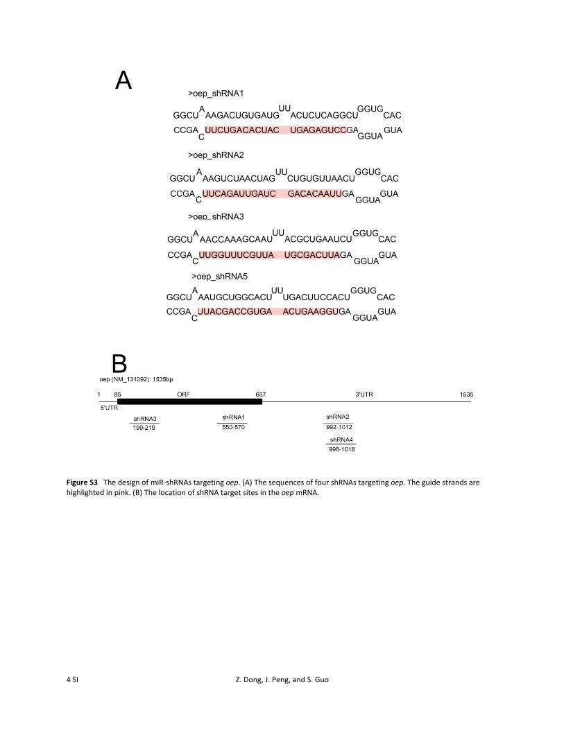
Figure S3 The design of miR-shRNAs targeting oep. (A) The sequences of four shRNAs targeting oep. The guide strands are highlighted in pink. (B) The location of shRNA target sites in the oep mRNA.

Z. Dong, J. Peng, and S. Guo 5 SI
Figure S4 Representative images of TdTom-miR-shRNA expressing embryos in transient transgenesis. (A) The embryo with low expression mosaicism, which is used for our analysis. (B) The embryo with high expression mosaicism, which is not used for our analysis.

6 SI Z. Dong, J. Peng, and S. Guo
Figure S5 Droplet Digital PCR analyses of endogenous apkcλ mRNA levels in stable UAS-TdTom-miR-shRNAapkcλ transgenic lines coupled with the KalTA4 driver. (A-B) FAM intensity plotted against HEX intensity. (A) Sibling control embryos injected with KalTA4 RNA. (B) UAS-TdTom-miR-shRNA1apkcλ-A transgenic embryos injected with KalTA4 RNA. In both (A) and (B), pink lines are thresholds used to assign individual droplets as either positive or negative, which divide the graph into four sections: top left (droplets positive for apkcλ), top right (droplets positive for both apkcλ and gapdhs), bottom right (droplets positive for gapdhs) and bottom left (negative droplets with quenched un-hydrolyzed probes). With similar amount of gapdhs positive droplets, (b) shows less apkcλ positive droplets compared with (A), indicating reduced apkcλ mRNA level. (C) Statistic analysis on Droplet Digital PCR results of (A) and (B). *: P < 0.05, **: P < 0.01, vs sibling.

Z. Dong, J. Peng, and S. Guo 7 SI
File S1
Time-lapse of a single TdTom-miR-30e vector control -expressing radial glia progenitor during mitotic division. The division ends at an angle close to 90 degree. The interval between each frame is 90 seconds.
File S1 is available for download at http://www.genetics.org/lookup/suppl/doi:10.1534/genetics.112.147892/-/DC1.
screenshot from File S1

8 SI Z. Dong, J. Peng, and S. Guo
File S2
Time-lapse of a single TdTom-miR-shRNA5apkcλ -expressing radial glia progenitor during mitotic division. The mitotic spindle undergoes active rotation and the division ends at an angle close to 0 degree. The interval between each frame is 90 seconds.
File S2 is available for download at http://www.genetics.org/lookup/suppl/doi:10.1534/genetics.112.147892/-/DC1.
screenshot from File S2

Z. Dong, J. Peng, and S. Guo 9 SI
Table S1 The efficacy and specificity of different apkcλ-shRNAs based on morphological phenotypes. UAS-TdTom-miR-
shRNAapkcλ -miniTol2 plasmid was co-injected with Pef1α-GFF-PT2KXIG and Tol2 transposase into wild-type embryos at 1-cell stage and phenotypes were analyzed at ~48 hpf. The shRNA-resistant apkcλ mRNA (coding region only) was used for rescue. The miR-30e vector –injected embryos served as controls. *: P < 0.05; **: P < 0.01; ***: P < 0.001 vs Ctrl., ##: P < 0.01; ###: P < 0.001 vs apkcλ-shRNA5, Z-test.

10 SI Z. Dong, J. Peng, and S. Guo
Table S2 The efficacy and specificity of different ntla-shRNAs based on morphological phenotypes. miR-ntla-shRNA (in pCS2) was in vitro transcribed and injected into wild-type embryos at 1-cell stage and phenotypes were analyzed at ~24 hpf. The shRNA-resistant ntla mRNA (coding region only) was used for rescue. The miR-30e vector –injected embryos served as controls. **: P < 0.01; ***: P < 0.001 vs Ctrl., ###: P < 0.001 vs ntla-shRNA4, Z-test.

Z. Dong, J. Peng, and S. Guo 11 SI
Table S3 The efficacy and specificity of different oep-shRNAs based on morphological phenotypes. miR-oep-shRNA RNA (in pCS2) was in vitro transcribed and injected into wild-type embryos at 1-cell stage and phenotypes were analyzed at ~24 hpf. The shRNA-resistant oep mRNA (coding region only) was used for rescue. The miR-30e vector –injected embryos served as controls. **: P < 0.01; ***: P < 0.001 vs Ctrl., ##: P < 0.01 vs oep-shRNA4, Z-test.

12 SI Z. Dong, J. Peng, and S. Guo
Table S4 Summary of stable transgenic lines carrying either UAS-TdTom-miR-shRNA1apkcλ or UAS-TdTom-miR-shRNA5apkcλ.

Z. Dong, J. Peng, and S. Guo 13 SI
Table S5 Summary of morphological phenotypes of UAS-TdTom-miR-shRNA1apkcλ or UAS-TdTom-miR-shRNA5apkcλ transgenic lines when crossed with Ubi-GFF transgenic line.

14 SI Z. Dong, J. Peng, and S. Guo
Table S6 Summary of morphological phenotypes of UAS-TdTom-miR-shRNA1apkcλ-A transgenic lines injected with KalTA4 RNA. ***: P< 0.001, vs Sibling + KalTA4 RNA.

Z. Dong, J. Peng, and S. Guo 15 SI
Table S7 Summary of morphological phenotypes of UAS-TdTom-miR-shRNA1apkcλ-A transgenic lines injected with Pef1α-GFF plasmid. ***: P< 0.001, vs Sibling + Pef1α-GFF plasmid.