stability of cocaine in phosphate buffer and … · stability of cocaine in phosphate buffer and in...
TRANSCRIPT
STABILITY OF COCAINE IN PHOSPHATE BUFFER AND
IN URINE*
Marianna KISZKA, Grzegorz BUSZEWICZ, Roman M¥DRO
Chair and Department of Forensic Medicine, Medical Academy, Lublin
ABSRTACT: The stability of cocaine and benzoylecgonine standard solutions inphosphate buffer and cocaine solutions in three different samples of urine was evalu-ated. The research indicated that cocaine solution is significantly less stable thanbenzoylecgonine solution. Both xenobiotics were isolated from urine by means ofthree-step liquid-liquid extraction. Quantitative analysis was performed using theHPLC method. The effect of time (up to 90 days), temperature (+25°C, +4°C and–20°C), pH (in the range from 5 to 8) and, in the case of urine, sodium fluoride as well,was tested. It turned out that sodium fluoride did not have a noticeable influence onthe degradation process of cocaine and benzoylecgonine. The stability of cocaine andbenzoylecgonine increased with a decrease in samples storage temperature and a de-crease in their pH. But only the freezing of samples, or their acidifying down to pH = 5together with storage at +4°C assured the stability of the investigated cocaine andbenzoylecgonine solutions for 90 days.
KEY WORDS: Cocaine; Benzoylecgonine; Stability; Autopsy material; Urine.
Z Zagadnieñ Nauk S¹dowych, z. XLIV, 2000, 7–23Received 9 July 2000; accepted 10 October 2000
INTRODUCTION
The mean life-time of cocaine in the body amounts to merely 20–90 min-utes [8, 10, 12, 14, 19, 20]. Only 1 to 14% of this xenobiotic is excreted as theunchanged form with urine [8, 14]. The remaining part of cocaine is con-verted mainly to benzoylecgonine and ecgonine methyl ester, which com-prise over 80% of all cocaine metabolites [1, 2, 4, 5]. The above-mentionedtransformations consist mainly in enzymatic (to ecgonine methyl ester) orchemical (to benzoylecgonine) hydrolysis of two ester bonds included in thecocaine molecule [8, 18].
* This article is based on a presentation given at the 11th Meeting of the Polish Society ofForensic Medicine and Criminology, £ódŸ, 1998. The first author, dr Marianna Kiszka,received the Professors Jan Markiewicz and Tadeusz Borkowski of the Institute of ForensicResearch Memorial Award.

The enzymatic and chemical processes that lead to degradation of cocainein the body of living persons are continued after death and in biological ma-terial collected during autopsy and from living persons. The hydrolysis of co-caine to benzoylecgonine depends considerably on pH [9]. This is confirmedby our observations1, indicating that in aqueous medium at pH between8.5 and 9 after 1–6 hours approximately 1/10 to over 1/3 of the initial amountof cocaine is excreted to benzoylecgonine. One should keep in mind the de-pendence of the cocaine degradation rate on pH, because gradual pH changesof biological material take place with time in corpses as well as in vitro [16].
There is no doubt that for diagnostic needs of fatal and other cocaine poi-sonings it is important to find out exactly the transformations which it un-dergoes in vitro. This knowledge is essential to appropriately protect biologi-cal or standard materials against degradation of cocaine and benzoylecgo-nine, to avoid xenobiotics loss during toxicological analysis and correctly in-terpret the results. Cocaine stability was tested in urine because its pH var-ies and also because in the case of living persons it is the only biological ma-terial which can be obtained non-invasively and in sufficient amounts fortoxicological investigations.
MATERIALS AND METHODS
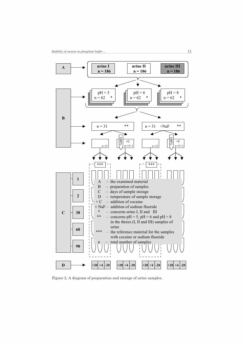
Standard solutions of cocaine and benzoylecgonine were prepared usingthree phosphate buffers with different pH (5.0, 7.4 and 8.0). Phosphatebuffer was applied because it is used as a mobile phase in determination ofthese xenobiotics by means of the HPLC method. Three samples of urinewere collected from three different corpses. Each sample was divided into3 portions and their pH was adjusted to 5.0, 6.0 and 8.0 respectively, withthe use of 0.1 M HCl solution and 0.1 M NaOH solution.
In the next step of urine preparation for analysis, each of the nine sam-ples was divided into 2 parts and sodium fluoride NaF (in the amount re-quired to reach a concentration of 5 mg/ml) was added to one of them.
Next, each buffer and each of the eighteen urine samples were dividedinto two parts. Cocaine was added to one part of each buffer solution andbenzoylecgonine in the amount required to create a concentration of 5 mg/mlwas added to the other one. In the case of urine, cocaine (in the amount re-
8 M. Kiszka, G. Buszewicz, R. M¹dro
1 M¹dro R., Kiszka M., Buszewicz G., Fatal cocaine poisoning – a presentation at the 4th PolishScientific Conference “Poisonings, injuries, alcoholism and drug addiction in forensic andmedical practice”, Bielsko-Bia³a, 27–28 April, 1995.

quired to obtain a concentration of 5 mg/ml) was added to one half of the eigh-teen samples and the other one was left as the background control.
The initial concentration of cocaine and benzoylecgonine, which was as-sumed as 100%, was determined directly after preparation of their solutionsin phosphate buffers and in urine. After determination of initial xenobioticsconcentrations, their solutions were divided and put into tubes which werestored at different temperatures, +25°C, +4°C and –20°C respectively. Thesamples were collected for analysis after 1, 7, 14, 21, 30, 60 and 90 days inthe case of cocaine and benzoylecgonine solutions in buffers and after 1, 7,30, 60 and 90 days in the case of cocaine solutions in urine.
Xenobiotics were isolated from urine by means of three-step liquid-liquidextraction with the use of dichloromethane / isopropanol mixture (3:1), 0.1 NHCl and once again dichloromethane / isopropanol mixture (3:1)2.
Quantitative determinations were carried out by means of the HPLCmethod using a liquid chromatograph manufactured by Gilson equippedwith a spectrophotometric detector with fluent adjustment of wavelength.Chromatographic separation was performed on a Hypersil ODS (250 x4.0 mm, 5 mm) column. A mixture of 80% 0.025 M phosphate buffer, pH = 3(with addition of 0.5% triethylamine) and 20% acetonitryle constituted themobile phase, in a two-pump system. Before each use, the buffer andacetonitryle were filtered on Nylon 66 Membranes 0.45 mm x 47 mm pur-chased by Supelco and vented by constant helium flow. The flow rate of theeluent amounted to 1 ml per minute and the volume of the injected sample –10 ml. The measurements were carried out at l = 233 nm. The detector signalwas processed electronically using Gilson 715 HPLC System ControllerSoftware. Concentrations of cocaine and benzoylecgonine were determinedfrom the appropriate calibration curves using Gilson 715 HPLC System soft-ware. The internal standard method (with use of lignocaine) was applied inthe case of urine investigations and the external standard method was usedin the case of buffer solutions analysis3.
Diagrams of solution preparation and storage are shown in Figures 1 and 2.132 samples of cocaine and benzoylecgonine solutions in phosphate buff-
ers and 558 urine samples were analysed.
Stability of cocaine in phosphate buffer ... 9
2 The procedure was shown in detail at the presentation: Kiszka M., Buszewicz G, M¹dro R.,Determination of cocaine and benzoylecgonine in urine, 14th Szczecin Scientific Symposium,Szczecin, 24–26 September, 1997.
3 The analytical procedures of cocaine and benzoylecgonine determination were presented indetail at the presentation: Kiszka M., M¹dro R., Buszewicz G., Analytical problemsconnected with cocaine determination in tissues, 11th Meeting of the Polish Society ofForensic Medicine and Criminology, £ódŸ, 2–5 September, 1998.

10 M. Kiszka, G. Buszewicz, R. M¹dro
Figure 1. A diagram of preparation and storage of cocaine and benzoylecgonine inbuffer solutions.
Stability of cocaine in phosphate buffer ... 11
Figure 2. A diagram of preparation and storage of urine samples.
RESULTS AND DISCUSSION
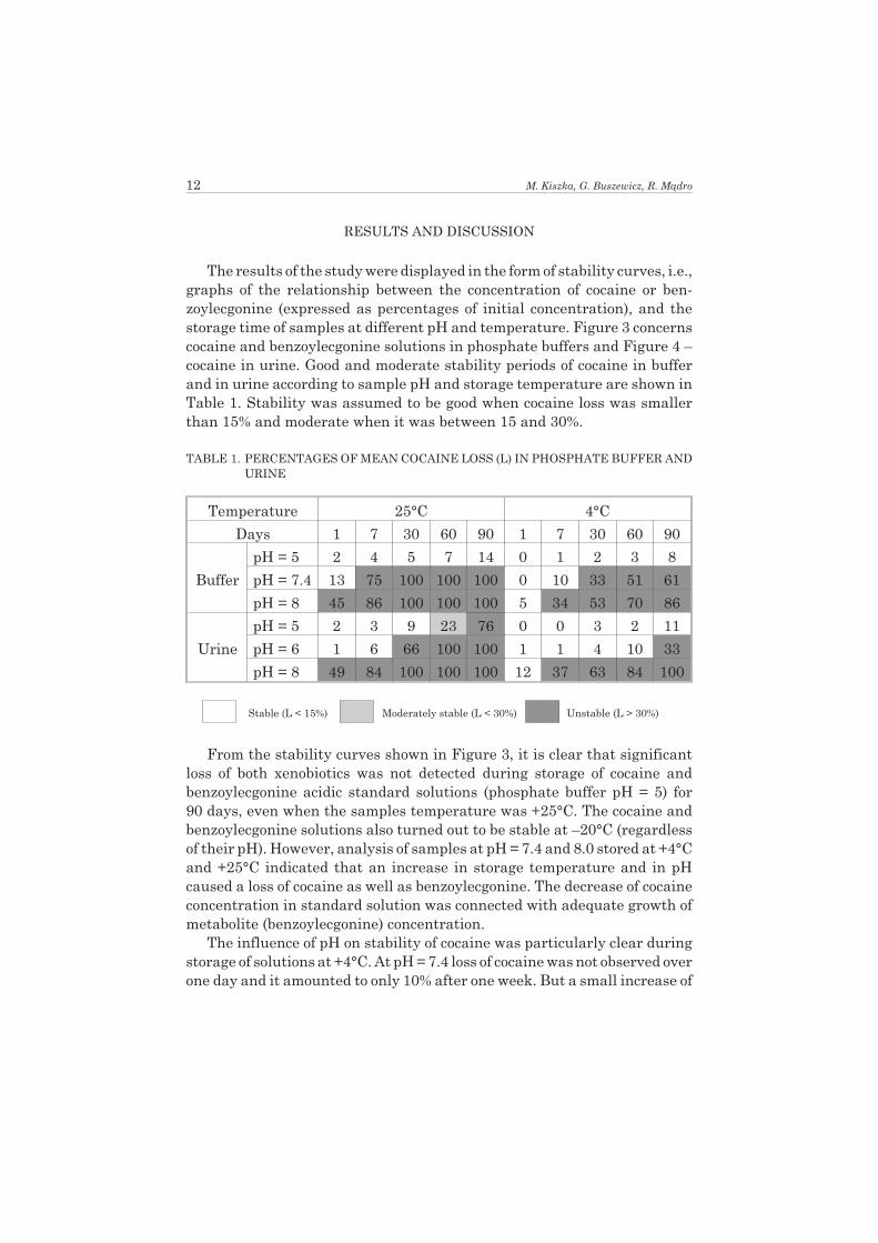
The results of the study were displayed in the form of stability curves, i.e.,graphs of the relationship between the concentration of cocaine or ben-zoylecgonine (expressed as percentages of initial concentration), and thestorage time of samples at different pH and temperature. Figure 3 concernscocaine and benzoylecgonine solutions in phosphate buffers and Figure 4 –cocaine in urine. Good and moderate stability periods of cocaine in bufferand in urine according to sample pH and storage temperature are shown inTable 1. Stability was assumed to be good when cocaine loss was smallerthan 15% and moderate when it was between 15 and 30%.
TABLE 1. PERCENTAGES OF MEAN COCAINE LOSS (L) IN PHOSPHATE BUFFER ANDURINE
Temperature 25°C 4°C
Days 1 7 30 60 90 1 7 30 60 90
Buffer
pH = 5 2 4 5 7 14 0 1 2 3 8
pH = 7.4 13 75 100 100 100 0 10 33 51 61
pH = 8 45 86 100 100 100 5 34 53 70 86
Urine
pH = 5 2 3 9 23 76 0 0 3 2 11
pH = 6 1 6 66 100 100 1 1 4 10 33
pH = 8 49 84 100 100 100 12 37 63 84 100
Stable (L < 15%) Moderately stable (L < 30%) Unstable (L > 30%)
From the stability curves shown in Figure 3, it is clear that significantloss of both xenobiotics was not detected during storage of cocaine andbenzoylecgonine acidic standard solutions (phosphate buffer pH = 5) for90 days, even when the samples temperature was +25°C. The cocaine andbenzoylecgonine solutions also turned out to be stable at –20°C (regardlessof their pH). However, analysis of samples at pH = 7.4 and 8.0 stored at +4°Cand +25°C indicated that an increase in storage temperature and in pHcaused a loss of cocaine as well as benzoylecgonine. The decrease of cocaineconcentration in standard solution was connected with adequate growth ofmetabolite (benzoylecgonine) concentration.
The influence of pH on stability of cocaine was particularly clear duringstorage of solutions at +4°C. At pH = 7.4 loss of cocaine was not observed overone day and it amounted to only 10% after one week. But a small increase of
12 M. Kiszka, G. Buszewicz, R. M¹dro

Stability of cocaine in phosphate buffer ... 13
Figure 3. The stability of cocaine (C) and benzoylecgonine (BE) in standard solutionsaccording to storage periods (days), temperature and pH of phosphate buffers.

the buffer pH (up to 8) caused approximately 2 to 3 times greater loss of co-caine.
Cocaine stability in standard solutions decreased greatly at +25°C. AtpH = 7.4, about 10% of the initial cocaine concentration had alreadydecomposed within one day. The loss level averaged 75% after 7 days and co-caine was totally decomposed after 3 weeks.
14 M. Kiszka, G. Buszewicz, R. M¹dro
Figure 4. The stability of cocaine (C) in urine (expressed in per cents, calculated asthe mean value of 3 urine samples) according to temperature and storage periods(days), pH of urine and NaF addition.

On the other hand, the benzoylecgonine standard solutions were muchmore stable. After 2 months of their storage at +25°C only about 1/5 of theinitial xenobiotic concentration had decomposed both at pH 7.4 and 8.
Other authors [9, 13] have observed a similar influence of temperatureand pH on cocaine hydrolysis in aqueous solutions. There is a lack of data inthe available literature concerning benzoylecgonine stability at different pH.However, in a study by Isenschmid [13], longer stability of benzoylecgonine(the hydrolysis product of cocaine) in alkaline buffers is seen (compared tococaine). Similar trends were observed in (our) described experiment. Co-caine decomposed to benzoylecgonine and over a relatively long period oftime (about 60 days), especially in the weakly alkaline solutions (pH = 7.4)the sum of the concentrations of both substances was comparable to the ini-tial cocaine concentration. However, buffer alkalinity decreased benzoylec-gonine stability – this was particularly clear in samples stored at +25°C.
The freezing of urine samples similar to standard solutions guaranteedthe stability of cocaine over 90 days, regardless of pH. Dugan et al. [6] ascer-tained good stability of drugs in urine samples frozen to –20°C, but even inthese conditions, the cocaine concentration decreased after one year by 37%on average and maximally 87%.
It turned out that the shapes of cocaine stability curves in urine and inbuffers at identical or similar pH’s were similar. But it has to be noted that inroom temperature a change of the pH from 5 to 6, i.e. a small decrease of acid-ity caused a considerable decrease of cocaine stability. Thus the acidity ofthe urine did not stabilise cocaine as effectively as the acidity of the buffer.Satisfactory cocaine stability was ascertained only in urine samples atpH = 5 stored in a refrigerator at +4°C – under these conditions cocaine didnot undergo significant degradation in the space of 90 days. In the case ofurine samples stored at 25°C, acidity (pH = 5) ensured sufficient cocaine sta-bility only for 60 days.
The alkalinity of urine was conducive to faster degradation of cocaine. Insamples of pH = 8 stored at +4°C after just 1 day, cocaine loss averaged 12%and after 7 days – 37%. When samples were stored at +25°C it averaged 49%and 84% respectively. Moreover, an increase of benzoylecgonine concentra-tion, (as was the case in acidic buffers and acidic urine), was observed simul-taneously.
The experimentally demonstrated increased speed of cocaine degrada-tion in urine with increase of pH from acidic to alkaline fully confirmed theresults of Baselt’s research [3]. He ascertained an approximately two-folddecline of cocaine concentration in urine at pH = 8 (after 3 weeks of storageat +4°C), and no significant changes in the cocaine level in urine at pH = 5stored at this temperature. Our observations are also similar to the findingsof Dugan et al. [7]. However, we found a definitely slower cocaine hydrolysis
Stability of cocaine in phosphate buffer ... 15

at +25°C in acidic urine. The observed cocaine loss was only about 10% after30 days, while Dugan ascertained as much as a 10-fold decline of cocaineconcentration after 7 days of urine storage in the same conditions.
Because about 23% of cocaine was lost from urine at pH = 5 stored at+25°C after 60 days, and as much as about 76% after 90 days, the view ofHippenstiel and Gerson [11] should be rejected. These authors deliberate, onthe basis of Isenschmid’s investigation [13], the usefulness of refrigerationof urine at this acidity. It should be noted that Isenschmid performed deter-minations of cocaine in buffers, where it is more stable than in urine. More-over, the research was finished after 4 weeks, while urine storage up to thebeginning of analysis and during the time analysis is performed is very oftenmuch longer.
The findings of this experiment that cocaine degradation in urine at dif-ferent pH was intensified with increase in temperature of sample storageare similar to the observations of Javaid et al. [15] and Matsubara et al. [17].The statement that addition of NaF as a preservative in the amount of5 mg/ml does not significantly affect cocaine stability in urine at pH = 5, 6 and8 during storage from 1 to 90 days at +4°C and at +25°C, was in full agreementwith the observations of other authors [3, 17].
CONCLUSIONS
1. Cocaine is degradation to benzoylecgonine in standard solutions aswell as in urine samples.
2. Because benzoylecgonine is much more stable in biological material, itshould be determined in the case of suspicion of cocaine poisoning.
3. Immediate freezing prevents the degradation of cocaine contained instandard solutions and urine samples.
4. When the investigated material can be stored only at +4°C, satisfac-tory cocaine stability for 90 days is ensured by acidifying of urine sam-ples to pH = 5.
5. Storage of urine at pH = 5 and at +25°C longer than 60 days createsa risk of significant degradation of cocaine present in the sample.
References:
1. A m b r e J . , F i s c h m a n M . , R u o T . I ., Urinary excretion of ecgonine methylester, a major metabolite of cocaine in humans, [in:] Cocaine: determination inhuman body fluids. Reprints of selected articles from the Journal of AnalyticalToxicology, Baselt R. C., Espe E. [ed.], Preston Publications, Niles 1988,pp. 79–81.
16 M. Kiszka, G. Buszewicz, R. M¹dro
2. A m b r e J ., The urinary excretion of cocaine and metabolites in humans: A ki-netic analysis of published date, [in:] Cocaine: determination in human body flu-ids. Reprints of selected articles from the Journal of Analytical Toxicology,Baselt R. C., Espe E. [ed.], Preston Publications, Niles 1988, pp. 99–103.
3. B a s e l t R . C ., Stability of cocaine in biological fluids, Journal of Chromatogra-
phy 1983, vol. 268, pp. 502–505.4. B o g u s z M . , S c h m i d t G ., Cocain-Missbrauch – Neue Bedrohung mit der
alten Substanz, Zeitschrift für Rechtsmedizin 1991, Bd. 35, S. 783–793.5. C l a r k e E . G . C ., Isolation and identification of drugs in pharmaceutical, body
fluids and post-mortem material, The Pharmaceutical Press, London 1986.6. D u g a n S . , B o g e m a S . , S c h w a r t z R . W . [et al.], Stability of drugs of abuse
in urine samples stored at –20°C, Journal of Analytical Toxicology 1994, vol. 18,pp. 391–396.
7. D u g a n S . H . , C o s t a n t i n o A . G . , B o g e m a S . C . [et al.], A study of the sta-bility of cocaine, benzoylecgonine, ecgonine methyl ester, creatinine, and otherchemistries in urine, Journal of Analytical Toxicology 1996, vol. 20, p. 74.
8. F l e m i n g J . A . , B y c k R . , B a r a s h P . G ., Pharmacology and therapeutic ap-plications of cocaine, Anaesthesiology 1990, vol. 73, pp. 518–531.
9. F l e t c h e r S . M . , H a n c o c k V . S ., Potential errors in benzoylecgonine and co-caine analysis, Journal of Chromatography 1981, vol. 206, pp. 193–195.
10. G a w i n F . H . , E l l i n w o o d E . H ., Cocaine and other stimulants. Actions,abuse, and treatment, New England Journal of Medicine 1988, vol. 318,pp. 1173–1182.
11. H i p p e n s t i e l M . J . , G e r s o n B ., Optimization of storage conditions for co-caine and benzoylecgonine in urine: a review, Journal of Analytical Toxicology
1994, vol. 18, pp. 104–109.12. I s e n s c h m i d D . S . , F i s c h m a n M . W . , F o l t i n L . W . [et al.], Concentra-
tion of cocaine and metabolites in plasma of humans following intravenous ad-ministration and smoking of cocaine, Journal of Analytical Toxicology 1992,vol. 16, pp. 311–314.
13. I s e n s c h m i d D . S . , L e v i n e B . S . , C a p l a n Y . H ., A comprehensive studyof the stability of cocaine and metabolites, Journal of Analytical Toxicology 1989,vol. 13, pp. 250–256.
14. I t e n P . X ., Fahren unter Drogen- oder Medikamenteneinfluss. Forensische In-terpretation und Begutachtung, Institut für Rechtsmedizin Forensische Toxiko-logie Universität, Zurich 1994.
15. J a v a i d J . I . , D e k i r m e n i j a n H . , D a v i s J . M . [et al.], Determination of co-caine in human urine, plasma and red blood cell by gas-liquid chromatography,Journal of Chromatography 1978, vol. 152, pp. 105–113.
16. M a r k i e w i c z J ., Swoistoœæ s¹dowych badañ chemiczno-toksykologicznych,Z zagadnieñ kryminalistyki 1971, z. VI, s. 22–29.
17. M a t s u b a r a K . , M a s e d a C . , F u k u i Y ., Quantitation of cocaine, ben-zoylecgonine and ecgonine methyl ester by GC-CI-SIM after Extrelut extraction,Forensic Science International 1984, vol. 26, pp. 181–192.
18. S t e w a r t D . J . , I n a b a T . , L u c a s s e n M . [et al.], Cocaine metabolism: co-caine and norcocaine hydrolysis by liver and serum esterases, Clinical Pharma-
cology and Therapeutics 1979, vol. 25, pp. 464–468.
Stability of cocaine in phosphate buffer ... 17

19. W e i s s R . D . , G a w i n H . F ., Protracted elimination of cocaine metabolites inlong-term, high-dose cocaine abusers, American Journal of Medicine 1988,vol. 85, pp. 879–880.
20. W i l k i n s o n P . , V a n D y k e C . , J a t l o w P . [et al.], Intranasal and oral co-caine kinetics, Clinical Pharmacology and Therapeutics 1980, vol. 27,pp. 386–394.
18 M. Kiszka, G. Buszewicz, R. M¹dro

TRWA£OŒÆ KOKAINY W BUFORZE FOSFORANOWYM
I W MOCZU*
Marianna KISZKA, Grzegorz BUSZEWICZ, Roman M¥DRO
WSTÊP
Œredni czas pó³trwania kokainy (C) w organizmie wynosi zaledwie 20–90 min[8, 10, 12, 14, 19, 20]. Tylko 1–14% tego ksenobiotyku wydalane jest w postaci nie-zmienionej z moczem [8, 14]. Pozosta³a czêœæ C przechodzi w benzoiloekgoninê (BE)i ester metylowy ekgoniny (EME), które stanowi¹ ponad 80% wszystkich metaboli-tów C [1, 2, 4, 5]. Powy¿sze przemiany polegaj¹ g³ównie na hydrolizie enzymatycznej(do EME) lub chemicznej (do BE) dwu wi¹zañ estrowych zawartych w cz¹steczce C[8, 18].
Procesy enzymatyczne i chemiczne, które za¿yciowo prowadz¹ do degradacji C,tocz¹ siê dalej w zw³okach oraz w materiale biologicznym pobieranym podczas sekcjii od osób ¿ywych. Hydroliza C do BE zale¿y przy tym w znacznym stopniu od pH [9],co potwierdzaj¹ spostrze¿enia autorów1, z których wynika, ¿e w wodnym œrodowiskuo pH = 8,5–9 ju¿ po 1–6 godzinach rozk³adowi do BE ulega w przybli¿eniu od 1/10 doponad 1/3 pocz¹tkowej iloœci C. Nale¿y pamiêtaæ o zale¿noœci tempa degradacji C odtego czynnika, bowiem wraz z up³ywem czasu nastêpuj¹ stopniowe zmiany pH mate-ria³u biologicznego zarówno w zw³okach, jak i in vitro [16].
Nie ulega wiêc w¹tpliwoœci, ¿e dla potrzeb diagnostyki œmiertelnych (i innych) za-truæ C istotne znaczenie ma dok³adne poznanie przemian, jakim podlega ona in vitro.Wiedza ta jest niezbêdna po to, by odpowiednio zabezpieczyæ materia³ biologiczny(lub wzorcowy) przed degradacj¹ C i BE oraz po to, by unikn¹æ strat ksenobiotykówpodczas analizy toksykologicznej i prawid³owo zinterpretowaæ jej wyniki. Natomiastrozpoczêcie testowania trwa³oœci C w moczu by³o podyktowane tym, ¿e jego pH wahasiê oraz tym, ¿e w przypadku osób ¿ywych jest to jedyny materia³ biologiczny, którymo¿na uzyskaæ w sposób nieinwazyjny i w odpowiedniej iloœci do badañ toksykolo-gicznych.
* Niniejszy artyku³ opracowany zosta³ na podstawie referatu pt. „Trwa³oœæ kokainy w materia-le biologicznym”, który zosta³ uznany za najlepsz¹ pracê przedstawion¹ podczas XI Krajo-wego Zjazdu Polskiego Towarzystwa Medycyny S¹dowej i Kryminologii w £odzi w 1998 roku,a jej pierwszy autor (dr Marianna Kiszka) otrzyma³a Nagrodê Imienia Profesorów InstytutuEkspertyz S¹dowych – Jana Markiewicza i Tadeusza Borkowskiego.
1 M¹dro R., Kiszka M., Buszewicz G., „Œmiertelne zatrucie kokain¹” – referat wyg³oszony naIV Ogólnopolskiej Konferencji Naukowej „Zatrucia, urazy, alkoholizm i narkomania w prak-tyce s¹dowo-lekarskiej”, Bielsko-Bia³a, 27–28 kwietnia 1995 r.

MATERIA£ I METODY
Roztwory wzorcowe C i BE wykonano z u¿yciem trzech buforów fosforanowycho ró¿nym pH (5,0; 7,4; 8,0), co podyktowane by³o tym, ¿e w³aœnie bufor fosforanowyjest stosowany (jako faza ruchoma) przy oznaczaniu tych ksenobiotyków metod¹HPLC. Trzy próbki moczu pobrano z trzech ró¿nych zw³ok i ka¿d¹ z nich podzielonona trzy porcje, których pH zmodyfikowano do 5,0; 6,0 i 8,0 dodaj¹c odpowiednie iloœci0,1 M roztworu HCl i 0,1 M roztworu NaOH.
W kolejnym etapie przygotowywania moczu do badañ ka¿d¹ z dziewiêciu próbekpodzielono na dwie czêœci i do jednej dodano fluorek sodu NaF (w iloœci potrzebnej douzyskania stê¿enia 5 mg/ml). Nastêpnie ka¿dy bufor i ka¿d¹ z osiemnastu próbekmoczu podzielono na pó³. Do jednej po³owy ka¿dego z buforów dodano C, a do drugiejBE w iloœci odpowiedniej do uzyskania stê¿enia 5 mg/ml. Natomiast w przypadku mo-czu, do jednej po³owy ka¿dej z osiemnastu próbek dodano C (równie¿ w iloœci odpowied-niej do uzyskania stê¿enia 5 mg/ml), a drug¹ po³owê pozostawiono jako kontrolê „t³a”.
Wyjœciowe, przyjmowane nastêpnie za 100%, stê¿enie C i BE oznaczano bezpo-œrednio po przygotowaniu ich roztworów w buforach fosforanowych i w moczu. Pooznaczeniu wyjœciowych stê¿eñ ksenobiotyków ich roztwory rozdzielono do probó-wek, które przechowywano w ró¿nej temperaturze (+25°C, +4°C, –20°C) i pobieranodo badañ po 1, 7, 14, 21, 30, 60 i 90 dniach w przypadku roztworów C i BE w buforachoraz po 1, 7, 30, 60 i 90 dniach w przypadku roztworów C w moczu.
Ksenobiotyki z moczu wyosobniano metod¹ trójetapowej ekstrakcji typuciecz-ciecz przy u¿yciu mieszaniny dichlorometan / izopropanol (3:1), a nastêpnie0,1 NHCl i ponownie mieszanin¹ dichlorometan / izopropanol (3:1)2.
Analizê iloœciow¹ wykonywano metod¹ HPLC przy u¿yciu chromatografu cieczo-wego firmy Gilson z detektorem spektrofotometrycznym o p³ynnej regulacji d³ugoœcifali. Rozdzia³ chromatograficzny przeprowadzono na kolumnie Hypersil ODS (250 x4,0 mm, 5 mm). Fazê ruchom¹ stanowi³a mieszanina: bufor fosforanowy 0,025 M,pH = 3 (z dodatkiem 0,5% trietylaminy) – acetonitryl w proporcjach 80:20 w systemiedwóch pomp. Przed ka¿dym u¿yciem bufor oraz acetonitryl by³y filtrowane (Nylon 66Membranes, 0,45 mm x 47 mm firmy Supelco), a nastêpnie odpowietrzane przezci¹g³y przep³yw helu. Prêdkoœæ przep³ywu eluentu wynosi³a 1 ml/min., a objêtoœæwstrzykiwanej próbki – 10 ml. Pomiary wykonywano przy d³ugoœci fali l = 233 nm.Sygna³ z detektora przetwarzany by³ elektronicznie z zastosowaniem oprogramowa-nia Gilson 715 HPLC System Controller Software. Stê¿enia C i BE wyznaczano me-tod¹ standardu wewnêtrznego (który w trakcie badania moczu stanowi³a lidokaina)lub standardu zewnêtrznego (w przypadku analizy roztworów buforowych) z odpo-wiednich krzywych kalibracji (oprogramowanie Gilson 715 HPLC System3). Schema-
20 M. Kiszka, G. Buszewicz, R. M¹dro
2 Metoda przedstawiona zosta³a szczegó³owo na XIV Szczeciñskim Sympozjum Naukowym,które odby³o siê w dniach 24–26 wrzeœnia 1997 r., w referacie: Kiszka M., Buszewicz G,M¹dro R., „Oznaczanie kokainy i benzoiloekgoniny w moczu”.
3 Procedury analityczne zwi¹zane z oznaczaniem C i BE przedstawione zosta³y szczegó³owow trakcie XIKrajowego Zjazdu Polskiego Towarzystwa Medycyny S¹dowej i Kryminologii(£ódŸ, 2–5 wrzeœnia 1998 r.) w referacie: Kiszka M., M¹dro R., Buszewicz G., „Problemyanalityczneane z oznaczeniem kokainy w tkankach”.

ty, zgodnie z którymi przygotowywano i przechowywano roztwory, przedstawiaj¹ ryci-ny 1 i 2.
Przebadano 132 próbki roztworów C i BE w buforach fosforanowych oraz 558 pró-bek moczu.
WYNIKI I DYSKUSJA
Wyniki badañ przedstawiono w postaci krzywych trwa³oœci (C i BE w buforachfosforanowych – rycina 3 oraz C w moczu – rycina 4), tj. wykresów zale¿noœci stê¿eñ Club BE (wyra¿onych w procentach pocz¹tkowego stê¿enia) od czasu przechowywaniapróbek o ró¿nym pH w ró¿nej temperaturze. Natomiast tabela I zawiera zestawienieokresów dobrej (ubytek C poni¿ej 15%) lub œredniej (ubytek C w zakresie 15–30%)stabilnoœci C w buforze i w moczu w zale¿noœci od pH próbek i temperatury, w jakiejby³y przechowywane.
Z krzywych trwa³oœci, które przedstawia rycina 3, wynika jednoznacznie, ¿eprzez 90 dni przechowywania kwaœnych (bufor fosforanowy pH = 5) roztworów wzor-cowych C i BE nie stwierdzono w nich znacz¹cego ubytku obu ksenobiotyków nawetwówczas, gdy temperatura próbek wynosi³a +25°C. Roztwory C i BE okaza³y siêtrwa³e (niezale¿nie od ich pH) równie¿ w temperaturze –20°C. Natomiast rezultatyanalizy próbek o pH = 7,4 i 8 przechowywanych w temperaturze +4°C i +25°C wy-kaza³y, ¿e wzrost temperatury oraz wzrost pH powodowa³y ubytek zarówno C, jaki BE. W roztworach wzorcowych kokainy jej ubytkowi towarzyszy³o przy tym adek-watne narastanie stê¿enia produktu rozpadu, tj. BE.
Wp³yw pH na stabilnoœæ C by³ szczególnie wyraŸny w trakcie przechowywaniaroztworów w temperaturze +4oC. Przy pH = 7,4 po jednym dniu nie obserwowano bo-wiem strat C, a po jednym tygodniu wynosi³y one zaledwie 10%, ale ju¿ niewielkiwzrost pH buforu (do 8) powodowa³ w przybli¿eniu 2–3-krotnie wiêkszy ubytek C.
Stabilnoœæ C w roztworach wzorcowych obni¿a³a siê najbardziej w temperaturze+25°C. W roztworze o pH = 7,4 ju¿ po jednym dniu rozk³adowi ulega³o bowiem oko³o10% pocz¹tkowego stê¿enia C, po siedmiu dniach poziom strat wynosi³ œrednio 75%,a po trzech tygodniach dochodzi³o do ca³kowitego rozk³adu C.
Natomiast roztwory wzorcowe BE by³y wielokrotnie bardziej stabilne, o czymœwiadczy fakt, ¿e po 2 miesi¹cach ich przechowywania w temperaturze +25°C tylkooko³o 1/5 pocz¹tkowego stê¿enia ksenobiotyku ulega³o degradacji zarówno przypH = 7,4, jak i pH = 8.
Podobny wp³yw temperatury i pH na hydrolizê C w jej wodnych roztworach by³obserwowany przez innych autorów [9, 13]. W dostêpnej literaturze brakuje nato-miast danych na temat stabilnoœci BE w roztworach o ró¿nym pH. W pracy Isen-schmida [13] widoczne jest jednak d³u¿sze (w porównaniu z C) utrzymywanie siê BEbêd¹cej produktem hydrolizy substancji macierzystej w buforach alkalicznych.W opisanym eksperymencie obserwowano podobne tendencje. C rozk³ada³a siê bo-wiem do BE, przy czym przez doœæ d³ugi czas (ok. 60 dni), zw³aszcza w roztworachs³abo alkalicznych (pH = 7,4), suma stê¿eñ obu substancji by³a zbli¿ona do wyjœcio-wego stê¿enia C. Zasadowy odczyn buforu zmniejsza³ jednak trwa³oœæ BE, co by³oszczególnie wyraŸne w próbkach przechowywanych w temperaturze +25°C.
Zamro¿enie próbek moczu, podobnie jak zamro¿enie próbek roztworów wzorco-wych, gwarantowa³o trwa³oœæ C przez 90 dni niezale¿nie od pH. Z publikacji Dugana
Trwa³oœæ kokainy w buforze fosforanowym ... 21

i in. [6], którzy stwierdzali znaczn¹ stabilnoœæ leków w próbkach moczu zamro¿onychdo –20°C, wynika jednak, ¿e nawet w tych warunkach po roku nastêpowa³o obni¿eniestê¿enia C œrednio o 37%, a maksymalnie o 87%.
Okaza³o siê, ¿e krzywe trwa³oœci C w moczu mia³y kszta³t doœæ zbli¿ony do krzy-wych trwa³oœci C w buforach o identycznym lub zbli¿onym pH. Zwraca jednak uwagêfakt, ¿e w temperaturze pokojowej zmiana pH z 5 do 6 (tj. niewielkie obni¿enie od-czynu kwaœnego) powodowa³o znaczne zmniejszenie stabilnoœci C. Zatem kwaœny od-czyn nie stabilizowa³ C w moczu tak skutecznie, jak kwaœny odczyn buforu. W pe³nizadowalaj¹c¹ trwa³oœæ C stwierdzono bowiem tylko w próbkach moczu o pH = 5 prze-chowywanych w lodówce w temperaturze +4°C, gdy¿ wówczas nie ulega³a onaznacz¹cemu rozk³adowi przez 90 dni. Natomiast w przypadku próbek moczu prze-chowywanych w temperaturze +25°C odczyn pH = 5 zapewnia³ wystarczaj¹c¹ sta-bilnoœæ C tylko przez 60 dni.
Zasadowy odczyn moczu sprzyja³ szybkiej degradacji C. W próbkach o pH = 8przechowywanych w temperaturze +4°C ju¿ po 1 dniu straty C wynosi³y œrednio 12%,a po 7 dniach 37%, zaœ w temperaturze +25°C odpowiednio 49% i 84%. Obserwowanoprzy tym (podobnie jak w buforach i w moczu o odczynie kwaœnym) równoczesne na-rastanie stê¿enia BE.
Wykazane eksperymentalnie przyspieszenie degradacji C w moczu w miarêzmiany pH od kwaœnego do zasadowego w pe³ni potwierdza wyniki badañ Baselta[3], który stwierdzi³ oko³o dwukrotne obni¿anie siê stê¿enia C w moczu o pH = 8 (po3 tygodniach przechowywania w temperaturze +4°C) i brak istotnych zmian poziomuC w przechowywanym w tej temperaturze moczu o pH = 5. Obserwacje autorów ni-niejszej publikacji s¹ równie¿ zbie¿ne z ustaleniami Dugana i in. [7]. Autorzy wyka-zali jednak zdecydowanie wolniejsz¹ hydrolizê C w temperaturze +25°C w moczuo kwaœnym odczynie. Po 30 dniach zaobserwowane straty C wynosi³y zaledwie oko³o10%, podczas gdy Dugan po 7 dniach przechowywania moczu w tych samych warun-kach stwierdzi³ a¿ dziesiêciokrotny spadek stê¿enia C.
Ze wzglêdu na to, ¿e w moczu o pH = 5, który przechowywano w temperaturze+25°C, po 60 dniach degradacji ulega³o oko³o 23% C, a po 90 dniach nawet oko³o 76%,odrzuciæ nale¿y pogl¹d Hippenstiela i Gersona [11]. Autorzy ci na podstawie wyni-ków badañ Isenschmida [13] zastanawiaj¹ siê bowiem nad celowoœci¹ sch³adzaniamoczu o tym odczynie. Tymczasem Isenschmid oznacza³ C w buforach, w których jestona bardziej stabilna ni¿ w moczu, a ponadto doœwiadczenie koñczy³o siê po up³ywieczterech tygodni, podczas gdy przechowywanie moczu do chwili rozpoczêcia analizyi w trakcie jej wykonywania niejednokrotnie trwa znacznie d³u¿ej.
Ustalenia niniejszego eksperymentu, z których wynika, ¿e rozk³ad C w moczuo ró¿nym pH nasila³ siê wraz ze wzrostem temperatury przechowywania próbek, s¹w przybli¿eniu zgodne z obserwacjami Javaida i in. [15] oraz Matsubary i in. [17].W pe³ni zgodne z obserwacjami innych autorów [3, 17] okaza³o siê natomiast ustale-nie, ¿e dodatek substancji konserwuj¹cej pod postaci¹ NaF w iloœci 5 mg/ml niewp³ywa w uchwytny sposób na stabilnoœæ C w moczu o pH = 5, 6 i 8 podczas jego prze-chowywania przez okres od 1 do 90 dni zarówno w temperaturze +4°C, jak i +25°C.
22 M. Kiszka, G. Buszewicz, R. M¹dro
WNIOSKI
1. W roztworach wzorcowych i w próbkach moczu kokaina rozk³ada siê do benzo-iloekgoniny.
2. Zdecydowanie wy¿sza trwa³oœæ benzoiloekgoniny w materiale biologicznymstanowi wskazanie do jej oznaczania w przypadku podejrzenia o zatrucie ko-kain¹.
3. Natychmiastowe zamro¿enie zapobiega rozk³adowi kokainy zawartej w roz-tworach wzorcowych i w próbkach moczu.
4. W przypadku, gdy materia³ do badañ mo¿na przechowywaæ jedynie w tempe-raturze +4°C, zadowalaj¹c¹ stabilnoœæ kokainy zapewnia przez 90 dni zakwa-szenie próbek moczu do pH = 5.
5. Przechowywanie moczu o pH = 5 w temperaturze +25°C d³u¿ej ni¿ 60 dni wi¹¿esiê z ryzykiem znacznej degradacji zawartej w nim kokainy.
Trwa³oœæ kokainy w buforze fosforanowym ... 23