stabilisation of scoliosis in two koi (cyprinus...
TRANSCRIPT

The Veterinary Record, July 24, 2004 115
Papers & Articles
Veterinary Record (2004)155, 115-119
P. D. Govett, DVM,G. A. Lewbart, MS, VMD,DACZM,Environmental MedicineConsortium andDepartment of ClinicalSciences,N. J. Olby, VetMB, PhD,DACVIM,D. J. Marcellin-Little,DEDV, DACVS, DECVS,Department of ClinicalSciences,D. S. Rotstein, DVM,MSPVM, DACVP,T. L. Reynolds, DVM,Department ofPopulation Health andPathobiology, College ofVeterinary Medicine,North Carolina StateUniversity, 4700Hillsborough Street,Raleigh, NC 27606, USA
Dr Rotstein’s presentaddress is Department ofPathobiology, Universityof Tennessee, College ofVeterinary Medicine, 2407River Drive, Knoxville,TN 37996, USA
Stabilisation of scoliosis in two koi (Cyprinus carpio)
P. D. Govett, N. J. Olby, D. J. Marcellin-Little, D. S. Rotstein, T. L. Reynolds, G. A. Lewbart
Two koi (Cyprinus carpio) from the same pond developed similar lesions of scoliosis. Radiographicexaminations showed that their spines had become malaligned as a result of vertebral compressionfractures involving T14 to T16. The vertebrae in both fish were stabilised with screws, k-wire andpolymethylmethacrylate. They both appeared to improve after surgery, but they began to decline anddied within three months. A postmortem examination revealed multi-organ inflammation that wasnot associated with the surgical implants.
SCOLIOSIS, or ‘bent-back’, in koi (Cyprinus carpio) is beingreported more often. Many causes have been suggested,including vitamin C deficiency (John and others 1979, Halverand Hardy 1994); electrocution as a result of either lightningstrike (Barlow 1993), faulty submersible pumps (Johnson1997) or electrofishing (Sharber and Carothers 1988); tryp-tophan deficiency (Halver and Shanks 1960, Kloppel and Post1975, Poston and Rumsey 1983, Walton and others 1984, Post1993); trauma; organophosphates (Couch and others 1977,Alam and Maughan 1993, Waddington 1995); and bacterialcold water disease (Noga 1996). Although some fish continueto do well with a noticeable curvature of the spine, othersbecome debilitated and intervention becomes necessary. Thispaper describes the progression of the condition and the sur-gical stabilisation of scoliosis in two koi.
CASE HISTORY 1
In July 2001, an approximately two-year-old, 56 cm, 1314 g,female doitsu sanke koi developed a 3 cm chevron-shapedulcer, dorsal to its right pectoral fin. The fish shared a 56,775litre pond, with a maximum depth of 137 cm, with 30 otherkoi and 10 goldfish (Carassius auratus). The water quality hadbeen good, but in the previous three months some of the fishhad developed ulcerative skin disease, and when this koibegan separating itself from its school the ulcer was firstnoticed. The koi had been treated by the owner with a seriesof three injections of 5·7 mg/kg amikacin sulphate (Ami-glyde-V, 50 mg/ml; Fort Dodge) administered intracoelomi-cally every 48 hours. When the injections were given, the ulcerwas cleansed with 10 per cent povidone-iodine solution(Betadine; Purdue Frederick) and then treated with a topicaltriple antibiotic ointment (Neosporin; Warner Lambert). Allthe fish in the pond were provided with paste food contain-
ing 0·5 mg/g enrofloxacin (Baytril 2·27 per cent; BayerCorporation) at a dose of 10 mg enrofloxacin/kg/day. Whenthere was no improvement, the paste food was insteadimpregnated with 2 mg/g trimethoprim-sulphadiazine(Tribrissen; Schering-Plough) and fed to provide a dose of 30mg/kg/day, upon a veterinarian’s recommendation.
Clinical and radiographic findingsThe ulcer was hyperaemic but healing, and there was mildscoliosis, with a left lateral deviation of the tail at the level ofthe anal fin.
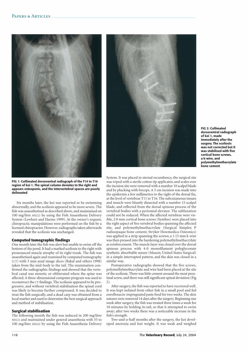
A biopsy taken from the tip of the right pectoral fin had no evidence of microscopic changes. A skin scrape takenfrom the ulcerative lesion contained a few unidentified non-pathogenic protozoal organisms and one free-living nema-tode. The fish was anaesthetised in 200 mg/litre of bufferedtricane methanesulphonate (MS222) (Finquel; ArgentChemical Laboratories) for the purpose of radiographicexamination (Love and Lewbart 1997) and maintained bydelivering the anaesthetic-containing water through a syringeover its gills as needed. Dorsoventral radiographs revealed aright laterodorsal angulation involving the 14th, 15th and16th trunk vertebrae (T14 to T16) at the level of the caudalswimbladder. The vertebrae at this site appeared to beosteopenic and the intervertebral spaces were not well delin-eated (Fig 1). Lateral views revealed a dorsolateral subluxa-tion of the spinal column just caudal to the last rib, involvingT14 and T15. The lateral angulation was considered to be dueto a combination of a fracture and a subluxation. Several cal-lused, mid-body fractures were apparent in the caudal ribs.The koi recovered uneventfully in fresh dechlorinated water;it resumed swimming with the group of fish and the ulcerstarted to heal; it was treated with trimethoprim-sulpha-diazine for two more weeks, but no further treatment wasconsidered necessary.
116 The Veterinary Record, July 24, 2004
Six months later, the koi was reported to be swimmingabnormally, and the scoliosis appeared to be more severe. Thefish was anaesthetised as described above, and maintained on100 mg/litre MS222 by using the Fish Anaesthesia DeliverySystem (Lewbart and Harms 1999). At the owner’s request,chiropractic manipulations were performed on the fish by alicensed chiropractor. However, radiographs taken afterwardsrevealed that the scoliosis was unchanged.
Computed tomographic findingsOne month later the fish was alert but unable to swim off thebottom of the pond. It had a marked scoliosis to the right withpronounced muscle atrophy of its right trunk. The fish wasanaesthetised again and examined by computed tomography(CT) with 3 mm axial image slices (Bakal and others 1998)taken from the mid-body to the tail. The examination con-firmed the radiographic findings and showed that the verte-bral canal was stenotic or obliterated where the spine wasaffected. A three-dimensional computer program was used toreconstruct the CT findings. The scoliosis appeared to be pro-gressive, and without vertebral stabilisation the spinal cordwas likely to become further compressed. It was decided totreat the fish surgically, and a dead carp was obtained from alocal market and used to determine the best surgical approachand method of stabilisation.
Surgical stabilisationThe following month the fish was induced in 200 mg/litreMS222 and maintained under general anaesthesia with 55 to100 mg/litre MS222 by using the Fish Anaesthesia Delivery
System. It was placed in sternal recumbency, the surgical sitewas wiped with a sterile cotton tip applicator, and scales overthe incision site were removed with a number 10 scalpel bladeand by plucking with forceps. A 3 cm incision was made intothe epidermis a few millimetres to the right of the dorsal fin,at the level of vertebrae T11 to T16. The subcutaneous tissuesand muscle were bluntly dissected with a number 15 scalpelblade, and reflected from the dorsal spinous process of thevertebral bodies with a periosteal elevator. The subluxationcould not be reduced. When the affected vertebrae were vis-ible, 2·0 mm cortical bone screws (Synthes) were placed intothe right aspect of five vertebral bodies spanning the affectedsite, and polymethylmethacrylate (Surgical Simplex Pradioopaque bone cement; Stryker Howmedica Osteonics)was applied in a strip spanning the screws; a 1·15 mm k-wirewas then pressed into the hardening polymethylmethacrylateas reinforcement. The muscle layer was closed over the dorsalspinous process with 4-0 monofilament polyglyconate synthetic absorbable suture (Maxon; United States Surgical)in a simple interrupted pattern, and the skin was closed in asimilar way.
Postoperative radiographs showed that the five screws,polymethylmethacrylate and wire had been placed at the siteof the scoliosis. There was little cement around the most prox-imal screw, and there was still significant spinal deviation (Fig2).
After surgery, the fish was reported to have recovered well.It was kept isolated from other fish in a small pool and fedenrofloxacin-impregnated paste food for two weeks. The skinsutures were removed 14 days after the surgery. Beginning oneweek after surgery, the fish was treated three times a week for10 minutes by holding its tail, so that it attempted to swimaway; after two weeks there was a noticeable increase in thefish’s strength.
Two-and-a-half months after the surgery, the koi devel-oped anorexia and lost weight. It was weak and weighed
Papers & Articles
FIG 1: Collimated dorsoventral radiograph of the T14 to T16region of koi 1. The spinal column deviates to the right andappears osteopenic, and the intervertebral spaces are poorlydelineated
FIG 2: Collimateddorsoventral radiographof koi 1, madeimmediately after thesurgery. The scoliosiswas not corrected but itwas stabilised with fivecortical bone screws, a k-wire, andpolymethylmethacrylatebone cement

The Veterinary Record, July 24, 2004 117
only 749 g. It was tube-fed 10 ml of paste food and given 13·4 mg/kg thiamine hydrochloride (Vitamin B ComplexFortified; Phoenix Pharmaceutical) intramuscularly. Threemonths after the surgery it died, and was stored at 7°C untilit was examined 24 hours later.
Pathological findingsThe fish weighed 800 g, both its eyes were sunken and mus-cle atrophy resulted in markedly prominent vertebrae. Thescoliosis involved T12 to T15 and bony changes were evidentfrom T12 to T16, with T12 to T15 being moderately thickenedand roughened bilaterally. The surgical implant was intact.Within the vertebral canal and cranial vault, the cerebrospinalfluid was slightly opaque and thickened; it contained degen-erate granulocytes mixed with melanocytes, macrophages anderythrocytes in a slightly eosinophilic proteinaceous back-ground matrix, consistent with inflammation. The posteriorchamber of the swimbladder appeared mottled red on thetunica externa and contained approximately 2 ml of yellow,opaque, wispy proteinaceous fluid which was mildly cellularwith no evidence of haemodilution; it contained a few gran-ulocytes and fewer macrophages.
The vertebrae showed moderate signs of multifocal peri-osteal proliferation and perivertebral fibrosis, with moder-ate muscle atrophy around the affected site; fibrous tissueextended into and replaced the associated myofibres. Therewere scattered macrophages, haemosiderophages, a few gran-ulocytes and mononuclear cells, and associated haemorrhage.Within the spinal cord, evidence of chronic, diffuse, mildmeningitis was provided by the expansion of the meninges bymacrophages, extravasated erythrocytes, and a few granulo-cytes and lymphocytes. Evidence of spongiosis and neuronalnecrosis was provided by the multifocal swelling of myelinsheaths, and neurons that were swollen and lightly basophilic.The swimbladder showed signs of chronic, moderate, diffuselymphoplasmacytic and histiocytic pneumocystitis andserositis, and it was moderately oedematous. Diffuse super-ficial adherent bacteria were also observed. The gills showedsigns of moderate, multifocal, diffuse granulocytic branchi-tis, and a moderate number of metazoans were identified asmonogeneic trematodes. The fish had chronic, mild lympho-plasmacytic enteritis. At the surgical site, the skin had chronic,moderate, focally extensive lymphoplasmacytic dermatitisand myositis with moderate myofibre atrophy and mild,multifocal epithelial hyperplasia.
CASE HISTORY 2
In August 2002, a one-year-old, 37 cm, 730 g, female gin rinkohaku koi from the same pond, but acquired from a differ-ent breeder, developed scoliosis. The owner reported that twomonths earlier a tree near the pond had been struck by light-ning and that several days later the koi was found strugglingto swim, with an acute curvature to its back. At first the fishappeared to be recovering, but later it had difficulty in swim-ming and was not eating well.
Clinical and radiographic findingsThe koi was bright, alert and in good body condition, apartfrom mild muscle atrophy at the level of the left mid-body,dorsal to the anal fin. It was anaesthetised with 200 mg/litreMS222 to enable phlebotomy and radiographic examination.Its packed-cell volume and total protein concentration werewithin their normal ranges (Groff and Zinkl 1999), and a gillbiopsy was apparently normal. Radiographs revealed a T15vertebral body compression fracture and a narrowing of theT15 to T16 intervertebral space. The spine and the caudalswimbladder curved to the right, beginning at the cranial endof the caudal swimbladder.
Computed tomographic findingsSlices of 3 mm through the spine were acquired by CT andconfirmed the malalignment by showing that there was aslight leftward shift of the entire vertebral column in the mid-spine region as a result of vertebral subluxation, accompaniedby a mild rightward tilt of the dorsal processes.
Surgical stabilisationOne week later the koi weighed 703 g (a 4 per centdecrease). The surgical approach was made left of the dor-sal fin and the vertebrae were stabilised by the same tech-nique as for koi 1, except that six 2·0 mm cortical bonescrews were used instead of five. The fish was given 10mg/kg enrofloxacin as a perioperative antibiotic intramus-cularly just caudal to the dorsal fin, and 0·4 mg/kg butor-phanol tartrate (Torbugesic 3 mg; Fort Dodge) in the sameway to provide analgesia. Postoperative radiographs showedthat the six screws and the accompanying bone cement hadbeen placed along the dorsal aspect of the 13th to the 18thvertebral bodies. Gas consistent with the surgery was pre-sent in the dorsal tissues.
After the surgery the fish did well; it was kept isolated fromother fish in a horse trough containing approximately 750litres of water containing 1 g/litre of salt; it was fed trout chowand enrofloxacin-impregnated paste food for two weeks, and50 per cent of the water was changed every one to three days.From the day after surgery the fish was encouraged to swimdaily for five to 10 minutes by swirling a metal rod behind it.Ten days after the surgery, the fish weighed 726 g, but two ofthe simple interrupted sutures near the centre of the incisionhad pulled through the skin, leaving an approximately 3 cmgap through which the surgical implant was visible. The skinappeared to be pulling away from the suture material twosutures caudal to the dehisced sutures. These sutures wereremoved and a culture was taken from near the surgical site;heavy growths of Escherichia coli and Aeromonas hydrophilawere obtained, both of which were resistant to enrofloxacinbut sensitive to gentamicin. A caseous ribbon of cream-coloured purulent material was entwined in a few of the mus-cle layer sutures. This material was removed and the woundwas flushed with 1 litre of lactated Ringer’s solution (LRS)(Baxter Healthcare Corporation). Before the wound wasclosed, 1·8 mg amikacin sulphate (Amiglyde-V 50 mg/ml;Fort Dodge), diluted to half strength with LRS, was placedinside the surgical cavity, and the skin was closed with a horizontal mattress pattern, using 3-0 Maxon on a cutting needle. The fish was given 5 mg/kg amikacin sulphate intra-muscularly every 48 hours for 14 days. This aminoglycosidewas chosen in preference to gentamicin because it is lessnephrotoxic and if the organisms were sensitive to gentam-icin they were also likely to be sensitive to amikacin. Twoweeks later the sutures were removed and gill, skin and finbiopsies were taken. The fish weighed 698 g, having lost 30 gsince its last visit, and cooked shrimp (14 g protein/day) wasadded to its diet. The incision site had healed well.Ichthyobodo species was found on the gill and skin biopsiesand the animal was treated with a formalin dip, containing 6ml of 37 per cent formaldehyde per 37·85 litres of water, for10 minutes.
Over the next month, the koi’s appetite decreased, itsweight decreased to 684 g and it was still positive for theIchthyobodo organism. In addition, Dactylogyrus species wasfound on skin and fin biopsies. The koi was treated twice,48 hours apart, for 10 minutes with a formalin dip contain-ing 8 ml of 37 per cent formaldehyde per 37·85 litres of water,and by the owner with 1 ml/378·5 litres of 50 mg/ml closan-tel and 75 mg/ml mebendazole (Supaverm; Janssen AnimalHealth). After this treatment, only one Ichthyobodo specieswas found on a skin scrape, and no further treatment wasgiven. The koi continued to lose weight and seven days later
Papers & Articles

118 The Veterinary Record, July 24, 2004
it weighed 659 g. Two months after the surgery the ownernoticed an approximately 4 cm2 swollen area on the fish’sright peduncle and administered a 10 mg/kg intramusculardose of enrofloxacin, following the veterinarian’s advice. Thefish died the following day and was stored at 7°C until it wasexamined 24 hours later.
Pathological findingsThe fish weighed 696 g and had a 5·5 x 6·5 x 2 cm fluctuantswelling over its right caudal abdomen, extending halfwaydown the tail, which contained approximately 7 ml of paleyellow opaque fluid with red streaks; the surrounding musclewas discoloured and necrotic. An aerobic culture of the fluidyielded Aeromonas hydrophila, which was sensitive to cipro-floxacin and gentamicin.
The surgical implant was intact, but there was a small fis-tulous tract starting at the base of the most caudal skin sutureand extending dorsally through the skin. The posterior swim-bladder contained 0·5 ml of dark tan mucinous materialwhich contained mucus cells, white blood cells and numer-ous bacteria in a homogeneous globular eosinophilic back-ground matrix, consistent with inflammation. The skin andthe gills were covered in excess mucus.
The muscle surrounding the abscess contained large num-bers of macrophages and large lymphocytes tracking betweenand among regions of necrotic myofibres (Fig 3). There wasgranulation tissue at the edges of some muscles. The sectionsof the spine and skeletal muscle at the surgical site showedsigns of spinal cord demyelination, and spinal root mononu-clear inflammation, degeneration and necrosis. At the samesite there was bone necrosis and remodelling with granula-tion of the surrounding tissue. Within the swimbladder,the epithelial layer was absent, and the subepithelium wasoedematous and infiltrated with new vessels, macrophages,lymphocytes and polymorphonuclear cells. The adipose tissue, heart, brain and spinal cord also contained a mono-nuclear infiltrate. The gills had severe, diffuse heterohistio-cytic branchitis and there were trematodes within the lesions.The cortical portion of one tail kidney contained multipleectatic tubules, with attenuated epithelium, that were filledwith amorphous, brightly eosinophilic material. The spleenwas diffusely necrotic.
DISCUSSION
In domestic mammals, scoliosis is most commonly a con-genital defect. In dogs and horses it is usually the result of acongenital malformation of the vertebrae (Colter 1993), but
environmental teratogens have been implicated in cows(Leipold and others 1974). Scoliosis has been observed in fishof all ages. Congenital defects have occurred in fingerlings andfry. Organophosphates, vitamin C deficiency and tryptophandeficiency may affect all age groups, and in these circum-stances most of the fish in a pond are usually affected, whereasa spasmodically faulty submersible pump or a lightning strikemay affect only one or a few fish in a pond. Trauma to themusculoskeletal system due, for example, to an intramuscu-lar injection, rough handling, sudden bursts of speed andpond hazards may affect a single fish which often goes un-noticed until it is observed swimming abnormally.
In domestic mammals, the surgical correction of scolio-sis is not usually practicable because the muscular and skele-tal changes associated with the disease are severe, and thecorrection of the initial defect typically does not correct thedeformity. The scoliosis in these two koi was attributable tocompression fractures, and in the rare cases when scoliosis isattributable to vertebral fractures in domestic mammals, theycan be stabilised by a variety of methods, including externalskeletal fixation, spinal stapling, dorsal spine plating, verte-bral body plating, pins and cement, or a combination of thesemethods (Seim 2002).
Internal fixation was chosen for both koi because a correction of the deformity could not be hoped for and thesurgery was aimed at vertebral decompression and stabilisa-tion. In fish, internal fixation has several benefits over exter-nal fixation. First, it uses less hardware and therefore has lesseffect on the fish’s buoyancy. Although closed-cell foam canbe added to improve balance and flotation (Lewbart 1998),it increases the drag on a fish as it swims, and has the poten-tial to exhaust an already compromised animal. Secondly,internal fixation allows the vertebrae to be stabilised with lit-tle disruption to the spinal column, whereas external fixationwould require the vertebral column to be broken to allow forrealignment. Pin tracts from external fixators have the poten-tial to wick bacteria that may cause an infection, and veryclean water would be needed in the fish’s recovery pool todecrease this risk. The size of the fish’s vertebrae was alsotaken into consideration: appropriate screws could beobtained to make the stabilisation rigid with only the affectedand nearby vertebrae being involved; extending the stabili-sation too far down the spine might have affected the fish’sability to swim.
In both cases the site of the lesion was consistent with thatcommonly reported in trauma-related vertebral fractures inkoi (Barlow 1993), cats (Thatcher 1993) and dogs (Hoerlein1978). In the koi, the lesions were between T14 and T16, T14being the last trunk vertebra to have ribs attached. Spinal frac-tures and luxations most commonly occur at the junctionsbetween moveable and stable spinal segments. The ribs havea stabilising effect and, compared with the rest of the spine,the spine caudal to the ribs has the greatest range of motionin flexion and extension. As a result, the vertebrae in thisregion are subject to forces and loads that do not affect theother vertebrae.
Although its death was not necessarily attributable to thesurgery, the first fish survived only three months and,despite more intense aftercare, the second fish survived lessthan two months. Postmortem examinations revealed mul-tiorgan inflammation, including meningitis, pneumocysti-tis, branchitis, enteritis, dermatitis, myositis, myelitis,myocarditis and steatitis that could not be definitively linkedto the surgical implants, but was suggestive of sepsis. Boththe fish lost weight, became parasitised and suffered bacte-rial infections.
By the time the first koi underwent surgery it had hadnoticeable scoliosis for nine months, with appreciable mus-cle atrophy, severe skeletal changes and difficulty in swim-ming. The angulation of the spinal cord was so acute that it
Papers & Articles
FIG 3: Inflammatory cells forming tracks among necroticmyofibres in the muscles of the right trunk of koi 2.Haematoxylin and eosin. x 400

The Veterinary Record, July 24, 2004 119(c) British Veterinary Association. All rights reserved
was under considerable pressure. Decompression was avoidedbecause it could have resulted in the spinal cord being dis-placed outside the vertebral canal.
The second koi was a better candidate for the procedurebecause its scoliosis was less severe and it underwent surgeryonly two months after it was thought to have been injured.However, its smaller size made it difficult to lift the musclecleanly from the dorsal spinous processes of the vertebralbodies, and muscle atrophy had already developed; intensivemanagement and supportive care did not prevent it fromdeteriorating.
The use of antibiotic-impregnated polymethylmethacryl-ate or hydroxyapatite cement (Ethell and others 2000) in placeof the plain polymethylmethacrylate placed over the screwsmight have been beneficial because, according to in vitrostudies, polymethylmethacrylate may compromise localimmunity (Shirtliff and others 2002); local, sustained con-centrations of antimicrobial agents in the compromised areamight have helped. However, the wounds of three of 18 dogstreated with gentamicin-impregnated polymethylmethacryl-ate during vertebral stabilisation still became infected (Blassand Seim 1984). Keeping the fish on oral or injectable anti-biotics for a longer period might also have improved the outcome. It was observed that horizontal mattress suturesprovided more tension relief than simple interrupted sutures,and their use from the beginning might have prevented thesecond koi’s initial infection.
Spinal vertebral stabilisation offers a possible therapeutictreatment for scoliosis due to vertebral fracture in fish.Although the technique and postoperative managementrequire refinement, these cases provide information thatshould guide and benefit future implant surgeries in fish.
ACKNOWLEDGEMENTS
The authors thank Nancy Love, Lenore Mohammadian andSandy Wang for their radiological assistance and interpreta-tions, Talmage Brown for his pathology assistance and inter-pretations, Craig Harms for a review of the manuscript,Christian Osmond for photographic documentation andShane Christian for assistance with case management andanimal husbandry.
ReferencesALAM, M. K. & MAUGHAN, O. E. (1993) Acute toxicity of selected
organophosphorus pesticides to Cyprinus carpio and Barilius vagra. Journalof Environmental Science and Health. Part B: Pesticides, Food Contaminants,and Agricultural Wastes 28, 81-89
BAKAL, R. S., LOVE, N. E., LEWBART, G. A. & BERRY, C. R. (1998) Imaginga spinal fracture in a kohaku koi (Cyprinus carpio): techniques and case history report. Veterinary Radiology and Ultrasound 39, 318-321
BARLOW, A. M. (1993) ‘Broken backs’ in koi carp (Cyprinus carpio) followinglightning strike. Veterinary Record 133, 503
BLASS, C. E. & SEIM, H. B. (1984) Spinal fixation in dogs using Steinman pinsand methylmethacrylate. Veterinary Surgery 13, 203-210
COLTER, S. B. (1993) Congenital anomalies of the spine. In DiseaseMechanisms in Small Animal Surgery. Philadelphia, Lea & Febiger. pp 950-959
COUCH, J. A., WINSTEAD, J. T. & GOODMAN, L. R. (1977) Kepone-inducedscoliosis and its histological consequences in fish. Science 197, 585-587
ETHELL, M. T., BENNETT, R. A., BROWN, M. P., MERRIT, K., DAVIDSON,J. S., & TRAN, T. (2000) In vitro elution of gentamicin, amikacin, and cef-tiofur from polymethylmethacrylate and hydroxyapatite cement. VeterinarySurgery 29, 375-382
GROFF, J. M. & ZINKL, J. G. (1999) Haematology and clinical chemistry ofcyprinid fish: common carp and goldfish. Veterinary Clinics of North America:Exotic Animal Practice 2.3, 741-776
HALVER, J. E. & HARDY, R. W. (1994) L-ascorbyl-2-sulfate alleviates Atlanticsalmon scurvy. Proceedings of the Society for Experimental Biology andMedicine 206, 421-424
HALVER, J. E. & SHANKS, W. E. (1960) Nutrition of salmonid fishes.
Indispensable amino acids for sockeye salmon. Journal of Nutrition 72, 340-346
HOERLEIN, B. F. (1978) Spinal fractures, luxations, and fusion. In CanineNeurology: Diagnosis and Treatment. 3rd edn. Philadelphia, W. B. Saunders.pp 561-594
JOHN, T. M., GEORGE, J. C., HILTON, J. W. & SLINGER, S. J. (1979) Influenceof dietary ascorbic acid on plasma lipid levels in the rainbow trout.International Journal for Vitamin and Nutrition Research 49, 400-405
JOHNSON, E. L. (1997) Koi Health and Disease: Beginner to Advanced Life-Saving Technology. Athens, Reade Printers. pp 20, 25, 87
KLOPPEL, T. M. & POST, G. (1975) Histological alteration in tryptophan-deficient rainbow trout. Journal of Nutrition 105, 861-866
LEIPOLD, H. J., HUSTON, K. & HULBERT, L. C. (1974) Congenital syndromein Hereford calves with kyphoscoliosis, arthrogryposis, and palatoshisis.Cornell Veterinarian 64, 123
LEWBART, G. A. (1998) Self-assessment Color Review of Ornamental Fish.Ames, Iowa State University Press. pp 111-112
LEWBART, G. A. & HARMS, C. (1999) Building a Fish Anesthesia DeliverySystem. Exotic DVM 1.2, 25-28
LOVE, N. E. & LEWBART, G. A. (1997) Pet fish radiography: technique andcase history reports. Veterinary Radiology and Ultrasound 38, 24-29
NOGA, E. J. (1996) Fish Disease Diagnosis and Treatment. St Louis, Mosby.pp 126-127, 221-224
POST, G. W. (1993) Nutrition and nutritional diseases of Salmonids. In FishMedicine. Philadelphia, W. B. Saunders. pp 343-358
POSTON, H. A. & RUMSEY, G. L. (1983) Factors affecting dietary requirementsand deficiency signs of L-tryptophan in rainbow trout. Journal of Nutrition113, 2568-2577
SEIM, H. B., III (2002) Surgery of the thoracolumbar spine. In Small AnimalSurgery. 2nd edn. Philadelphia, Mosby. pp 1269-1301
SHARBER, N. G. & CAROTHERS, S. W. (1988) Influence of electrofishingpulse shape on spinal injuries in adult rainbow trout. American Journal ofFisheries Management 8, 117-122
SHIRTLIFF, M. E., CALHOUN, J. H. & MADER, J. T. (2002) Experimentalosteomyelitis treatment with antibiotic-impregnated hydroxyapatite. ClinicalOrthopaedics 401, 239-247
THATCHER, C. (1993) Biomechanics of cranial fractures, spinal fractures, andluxations. In Disease Mechanisms in Small Animal Surgery. Philadelphia, Lea& Febiger. pp 1006-1008
WADDINGTON, P. (1995) Caring. In Koi Kichi. Liverpool, Kingprint. pp 156-192
WALTON, M. J., COLOSCO, R. M., COWEY, C. B., ADRON, J. W. & KNOX,D. (1984) The effects of dietary tryptophan levels on growth and metabolismof rainbow trout (Salmo gairdneri). British Journal of Nutrition 51, 279-287
Papers & Articles