srsf1-3 contributes to diversification of the...
TRANSCRIPT
SRSF1-3 contributes to diversification of the immunoglobulin variable region gene by
promoting accumulation of AID in the nucleus
Yuka Kawaguchi, Hiroaki Nariki, Naoko Kawamoto, Yuichi Kanehiro1, Satoshi Miyazaki,
Mari Suzuki, Masaki Magari, Hiroshi Tokumitsu, Naoki Kanayama*
Department of Medical Bioengineering, Graduate School of Natural Science and Technology,
Okayama University, 3-1-1 Tsushima-naka, Kita-ku, Okayama 700-8530, Japan.
* Corresponding author. FAX: +81-86-251-8198
E-mail: [email protected] (N. Kanayama)
1 Present address: Department of Microbiology and Immunology, Shimane University School of
Medicine, Izumo, Shimane 693-8501, Japan
Abbreviations: AID, activation-induced cytidine deaminase; AIDDC, C-terminal 10 amino acid-
deleted AID; CSR, class switch recombination; GCV, gene conversion; IgV, immunoglobulin
variable region; NES, nuclear export signal; SHM, somatic hypermutation; SRSF1, serine-
arginine protein splicing factor 1.
ABSTRACT
Activation-induced cytidine deaminase (AID) is essential for diversification of the Ig variable
region (IgV). AID is excluded from the nucleus, where it normally functions. However, the
molecular mechanisms responsible for regulating AID localization remain to be elucidated. The
SR-protein splicing factor SRSF1 is a nucleocytoplasmic shuttling protein, a splicing isoform of
which called SRSF1-3, has previously been shown to contribute to IgV diversification in chicken
DT40 cells. In this study, we examined whether SRSF1-3 functions in IgV diversification by
promoting nuclear localization of AID. AID expressed alone was localized predominantly in the
cytoplasm. In contrast, co-expression of AID with SRSF1-3 led to the nucleus accumulation of
both AID and SRSF1-3 and the formation of a protein complex that contained them both, although
SRSF1-3 was dispensable for nuclear import of AID. Expression of either SRSF1-3, or a C-
terminally-truncated AID mutant increased IgV diversification in DT40 cells. However,
overexpression of exogenous SRSF1-3 was unable to further enhance IgV diversification in DT40
cells expressing the truncated AID mutant, although SRSF1-3 were able to form a protein complex
with the AID mutant. These results suggest that SRSF1-3 promotes nuclear localization of AID
probably by forming a nuclear protein complex, which might stabilize nuclear AID and induce IgV
diversification in an AID C-terminus-dependent manner.
Keywords: AID; DT40; Gene conversion; Ig; Somatic hypermutation; SRSF1
1. Introduction
Germinal center B cells express activation-induced cytidine deaminase (AID), which converts
dC to dU on single-stranded (ss) DNAs to allow somatic hypermutation (SHM) on the Ig variable
region genes (IgV) and class switch recombination (CSR) on Ig constant region genes, during
antibody affinity maturation [1, 2]. The expression and activity of AID are regulated through a
multi-layered process, including regulation of subcellular localization (reviewed in [3] ). AID is
predominantly localized in the cytoplasm by nuclear exclusion and cytoplasmic retention.
However, the molecular mechanisms that shuttle AID between the cytoplasm and the nucleus have
not been fully elucidated.
The chicken B cell line DT40 constitutively expresses AID, and somatic hypermutation (SHM)

and another IgV-diversifying process, gene conversion (GCV), both of which are initiated by a
common intermediate, occur spontaneously in DT40 cells [4, 5]. We previously found an important
role for a splice variant of the serine/arginine (SR)-rich protein splicing factor 1 (SRSF1), referred
to here as SRSF1-3, in SHM and GCV [6]. An engineered DT40 cell line, DT40-ASF, in which
the endogenous SRSF1 gene is disrupted and the human SRSF1 cDNA is expressed [7], was unable
to induce SHM and GCV, whereas introduction of SRSF1-3 into the DT40-ASF cells restored
these IgV diversification events [6]. The prototypical SR protein, SRSF1 (formerly ASF/SF2), is a
nucleocytoplasmic shuttling protein, which is not only essential for RNA splicing but also has a
variety of roles in RNA metabolism [8].
SRSF1-3 is an alternatively spliced product of SRSF1, which shares the N-terminal region with
SRSF1 but contains an alternate C-terminus [9]. Since the alternate C-terminus is not conserved
between species, and does not include any other characteristic protein motifs that indicate its
function, the molecular role of SRSF1-3 in IgV diversification remains unknown. Recently, several
transcription-coupled factors have been shown to associate with AID, thereby forming a protein
complex that induces SHM and CSR (reviewed in [3]). It has also been reported that AID co-
localizes with splicing factors in sub-nuclear domains where SRSF1 is predominantly enriched
[10]. The increase in the number of reports connecting AID and SRSF1 prompted us to explore
whether SRSF1-3, an isoform of SRSF1, contributes to the nuclear localization of AID. In this
study, we investigated a role for SRSF1-3 in the subcellular localization of AID using fluorescently
tagged fusion proteins. The results suggest that SRSF1-3 promotes accumulation of AID in the
nucleus during IgV diversification.
2. Materials and methods
2.1. Cell lines
Wild-type DT40 cells and 293T cells were obtained from the RIKEN Cell Bank. DT40-ASF
cells [7], DT40-SW cells [11], and DT40-SWDC cells [12] were described previously. DT40 cells
were cultured as described previously [11]. 293T cells were cultured in DMEM (Life Technologies,
Gaithersburg, MD, USA) supplemented with 10% fetal bovine serum and 50 µM 2-
mercaptoethanol at 37°C in 5% CO2
2.2. Construction of expression vectors and transfection
The FLAG-tagged chicken SRSF1-3 (FLAG-SRSF1-3) expression vector has been described
previously [6]. An mCherry DNA fragment excised from the pmCherry-C1 vector (Clontech
Laboratories, Mountain View, CA, USA) was inserted in frame just upstream of the FLAG-
SRSF1-3 in the FLAG-SRSF1-3 expression vector. A Myc-tagged chicken AID cDNA fragment
and its C-terminus truncated version were generated from a chicken AID cDNA clone [11] by PCR,
and cloned into the pCI-bsr vector [13]. An AID cDNA fragment generated by PCR was fused in
frame with a DNA fragment encoding EGFP, derived from the pEGFP-N1 vector (Clontech
Laboratories), in the pCI-bsr vector. PCR was carried out using KOD FX neo DNA polymerase
(Toyobo, Osaka, Japan) and primers listed in Supplemental Table 1, and the nucleotide sequences
of all PCR-cloned fragments were confirmed by sequencing analysis.
293T cells were transiently transfected with expression vectors using the X-treme GENE HP
DNA transfection reagent (Roche Diagnostics, Mannheim, Germany). The pCI-bsr vector was
used as a mock vector. DT40 cells were transfected with expression vectors by electroporation
using the Neon Transfection System (Invitrogen/Thermo Fisher Scientific, Waltham, MA, USA),
and selected in a medium containing 20 µg/mL Blasticidin S (Kaken Pharmaceutical, Tokyo,
Japan).
2.3. Microscopic analysis
Transfected 293T cells, which had been cultured on cover slips coated with 0.1% gelatin, and
DT40 cells were fixed with 4% paraformaldehyde for 15 min, permeabilized in 0.1% Triton X-
100/PBS, and mounted on slide glasses with ProLong® Gold Antifade Reagent with DAPI
(Invitrogen). In some experiments, DT40 cells were treated with 10 µM MG132 (Sigma-Aldrich,
St. Louis, MO, USA) and 10 ng/mL Leptomycin B (Cayman Chemical, Ann Arbor, MI, USA) for
6 h and 2 h, respectively.
Cell images were acquired using an FV1000 confocal laser-scanning microscope equipped with
objective lenses, UPlanSApo 60× or UPlanFLN 40× (Olympus) with immersion oil, and FluoView
software (Olympus, Tokyo, Japan). Cells that showed bright GFP and/or mCherry fluorescence
were selected for analysis in each microscopic view under conditions where the gain of each
detector and the power of each laser were adjusted to minimize crosstalk detection between
channels. The mean fluorescence intensity (MFI) of AID-GFP in the nuclear and cytoplasmic
regions of interest in chosen cells was quantified using ImageJ software (NIH, Bethesda, MD,
USA). Regions, which corresponded to AID-GFP or DNA, were considered as representing the
whole cell or the nucleus of each cell, respectively. The MFI of AID-GFP in the cytoplasmic region
of each cell was calculated by subtracting that in the nuclear region from that in the whole cell
region. The subcellular localization of AID-GFP was evaluated by calculating the ratio of the MFI
of AID-GFP in the nuclear region over that in the cytoplasmic region.
2.4. Subcellular fractionation and western blotting
The nuclear and cytoplasmic fractions of transfected 293T cells were separated with the NE-
PER Nuclear and Cytoplasmic Extraction Kit (Thermo Fisher Scientific/Pierce Biotechnology,
Rockford, IL, USA). Whole cell lysates were prepared by sonicating cells in the Laemmli sample
buffer with a Bioruptor UCD-250 (Cosmo Bio, Tokyo, Japan). Samples were subjected to SDS-
PAGE and transfer onto PVDF membranes (Immobilon P; Millipore, Billerica, MA, USA), and
probed with antibodies listed in Supplemental Table 2. Blots were developed using Western
Lightning Plus-ECL (PerkinElmer, Waltham, MA, USA), and chemiluminescence was detected
using the ChemiDoc XRS system (Bio-Rad Laboratories, Hercules, CA, USA). The intensity of
each signal was quantified with Quantity One software (Bio-Rad Laboratories).
2.5 Immunoprecipitation
Cell-free extracts of transfected 293T cells were prepared by disrupting cells with a Bioruptor
sonicator in the presence of a protease inhibitor mixture (Nakalai Tesque, Kyoto, Japan), and by
centrifugation. An aliquot of cell-free extracts was used as an input control. Anti-FLAG mAb (M2,
Sigma-Aldrich) was adsorbed on Dynabeads™ Protein G magnetic beads (Invitrogen) using a 30-
min incubation at room temperature, and the mAb-immobilized magnetic beads were then
incubated with cell-free extracts at 4°C for 1 h. After washing the beads, proteins bound to the
beads were analyzed by western blotting using antibodies (Supplemental Table 2) and Western
Lightning Pro ECL (PerkinElmer) to detect signals at a higher sensitivity.
2.6 Analysis of IgV diversification
The SRSF1-3 expression vector was introduced into DT40-SW and DT40-SWDC cells as
described previously [6]. The transcription level of the SRSF1-3 gene was determined by
quantitative RT-PCR using the GAPDH gene as a control, as described previously [6]. AID
expression in these cell lines can be reversibly switched on or off by 4-hydroxytamoxyfen (4-
OHT) treatment [11]. GCV frequency in these cell lines was evaluated using a G/B construct as an
artificial GCV substrate, which was integrated into the un-rearranged IgVL allele, as described
previously [6, 13]. After 4-OHT treatment, single AID-expressing cells were sorted into culture
plates using a FACS Aria equipped with Auto Cell Deposit Unit (BD Biosciences, San Diego, CA,
USA), and maintained for weeks. The frequency of cells developing a strong green fluorescence
signal was analyzed using FACS Array (BD Biosciences). The nucleotide sequence analysis of the
IgVH gene was carried out after 3-week cultures of DT40-SW cells and DT40-SWDC cells, with
or without overexpression of SRSF1-3, as described previously [6, 12].
2.7 Statistical analyses
Statistical analyses were carried out using GraphPad Prism software (GraphPad Software, La
Jolla, CA, USA). The unpaired two-tailed Student’s t test was performed to evaluate statistically
significant differences between two groups. The Pearson correlation analysis was performed to
calculate correlation coefficients. The chi-square test was performed to evaluate difference in the
mutation frequency of the IgV gene between two groups. Probability (p) values: *, p < 0.05; **, p
< 0.01; ***, p < 0.001; ****, p < 0.0001; ns, not significant.
3. Results
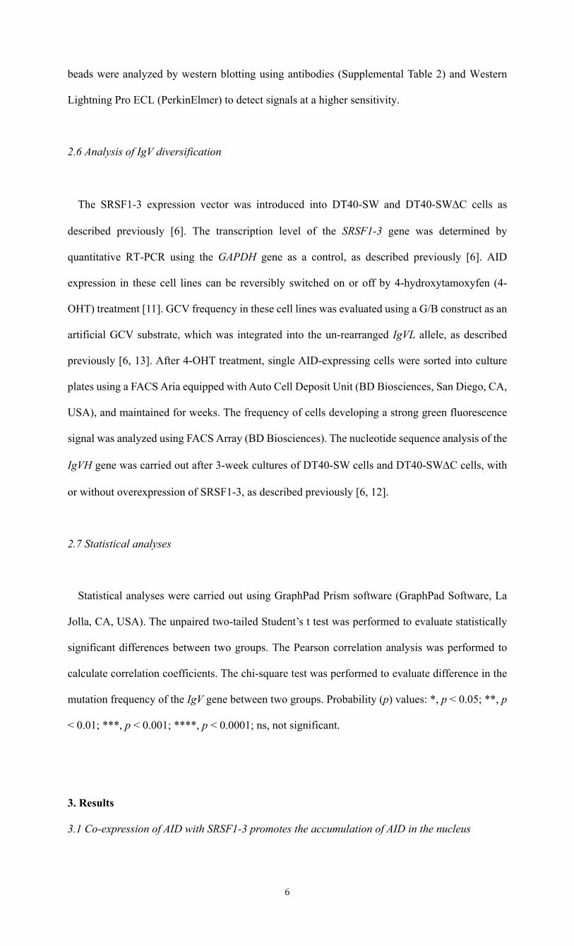
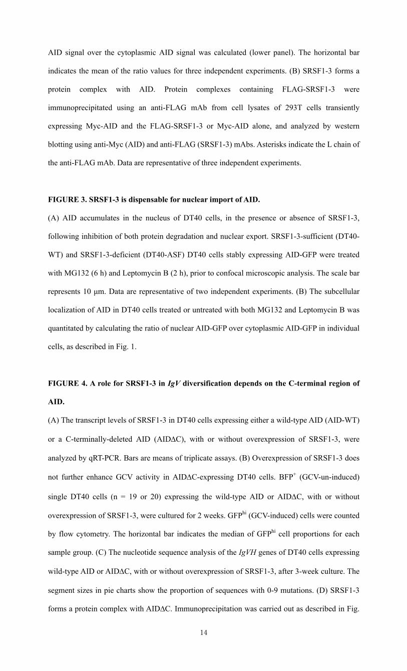
3.1 Co-expression of AID with SRSF1-3 promotes the accumulation of AID in the nucleus
To visualize the subcellular localization of AID and SRSF1-3, chicken AID tagged with GFP at
the C-terminus (AID-GFP) and chicken SRSF1-3 tagged with mCherry at the N-terminus
(mCherry-SRSF1-3) were transiently expressed in human 293T cells. The subcellular localization
of these fluorescent fusion proteins in these cells was then observed by confocal microscopy. When
AID-GFP was expressed alone, i.e., without exogenous expression of SRSF1-3, AID-GFP was
found predominantly in the cytoplasm in 293T cells (Fig. 1A) [14, 15]. Interestingly, when AID-
GFP was co-expressed with mCherry-SRSF1-3 in 293T cells, AID-GFP levels increased in the
nuclear compartment (Fig. 1A). The proportion of the nucleus AID-GFP was significantly higher
in cells co-expressing AID-GFP and mCherry-SRSF1-3 than in cells expressing AID-GFP alone
(Fig. 1B). In addition, the nuclear localization of AID-GFP was positively correlated with the mean
fluorescence intensity of co-expressed mCherry-cSRSF1-3 (Fig. 1C). It should be noted that AID
tended to be co-localized with SRSF1-3 in both the nucleus and the cytoplasm (Fig. 1A). In
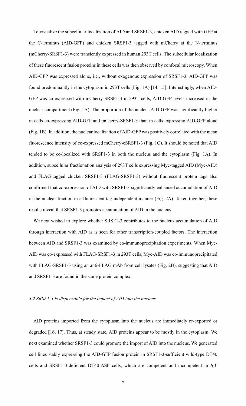
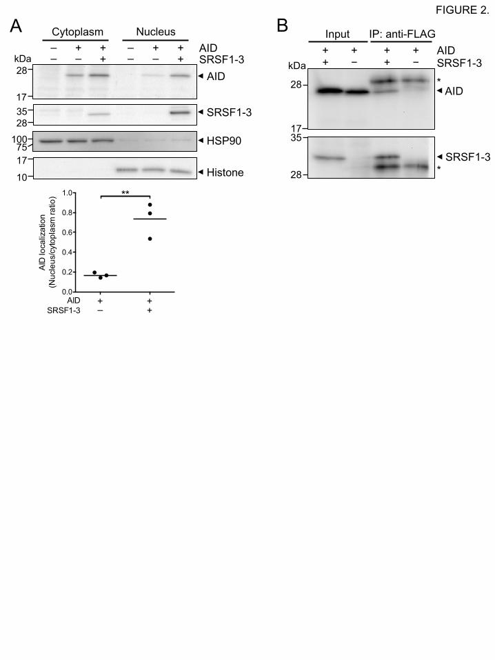
addition, subcellular fractionation analysis of 293T cells expressing Myc-tagged AID (Myc-AID)
and FLAG-tagged chicken SRSF1-3 (FLAG-SRSF1-3) without fluorescent protein tags also
confirmed that co-expression of AID with SRSF1-3 significantly enhanced accumulation of AID
in the nuclear fraction in a fluorescent tag-independent manner (Fig. 2A). Taken together, these
results reveal that SRSF1-3 promotes accumulation of AID in the nucleus.
We next wished to explore whether SRSF1-3 contributes to the nucleus accumulation of AID
through interaction with AID as is seen for other transcription-coupled factors. The interaction
between AID and SRSF1-3 was examined by co-immunoprecipitation experiments. When Myc-
AID was co-expressed with FLAG-SRSF1-3 in 293T cells, Myc-AID was co-immunoprecipitated
with FLAG-SRSF1-3 using an anti-FLAG mAb from cell lysates (Fig. 2B), suggesting that AID
and SRSF1-3 are found in the same protein complex.
3.2 SRSF1-3 is dispensable for the import of AID into the nucleus
AID proteins imported from the cytoplasm into the nucleus are immediately re-exported or
degraded [16, 17]. Thus, at steady state, AID proteins appear to be mostly in the cytoplasm. We
next examined whether SRSF1-3 could promote the import of AID into the nucleus. We generated
cell lines stably expressing the AID-GFP fusion protein in SRSF1-3-sufficient wild-type DT40
cells and SRSF1-3-deficient DT40-ASF cells, which are competent and incompetent in IgV
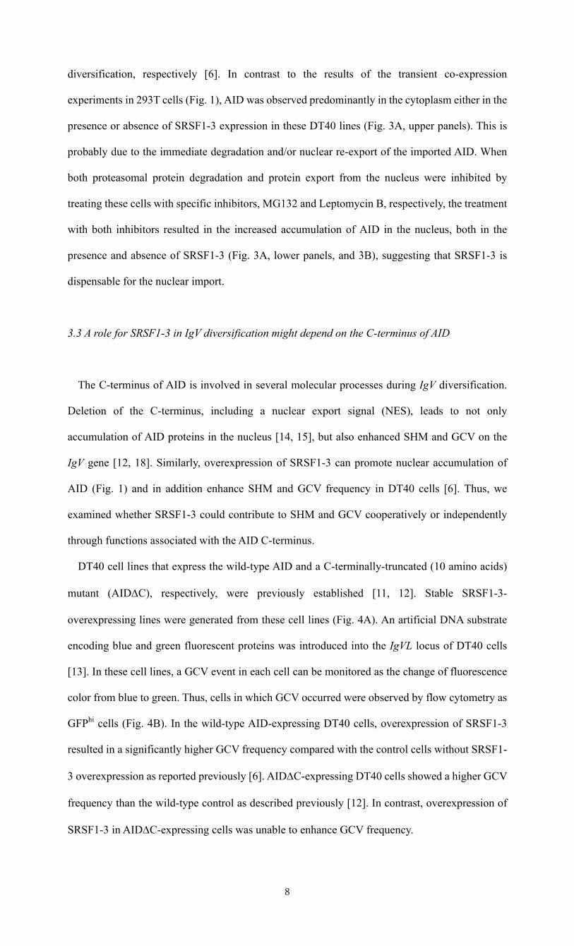
diversification, respectively [6]. In contrast to the results of the transient co-expression
experiments in 293T cells (Fig. 1), AID was observed predominantly in the cytoplasm either in the
presence or absence of SRSF1-3 expression in these DT40 lines (Fig. 3A, upper panels). This is
probably due to the immediate degradation and/or nuclear re-export of the imported AID. When
both proteasomal protein degradation and protein export from the nucleus were inhibited by
treating these cells with specific inhibitors, MG132 and Leptomycin B, respectively, the treatment
with both inhibitors resulted in the increased accumulation of AID in the nucleus, both in the
presence and absence of SRSF1-3 (Fig. 3A, lower panels, and 3B), suggesting that SRSF1-3 is
dispensable for the nuclear import.
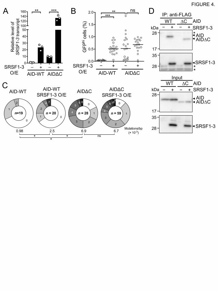
3.3 A role for SRSF1-3 in IgV diversification might depend on the C-terminus of AID
The C-terminus of AID is involved in several molecular processes during IgV diversification.
Deletion of the C-terminus, including a nuclear export signal (NES), leads to not only
accumulation of AID proteins in the nucleus [14, 15], but also enhanced SHM and GCV on the
IgV gene [12, 18]. Similarly, overexpression of SRSF1-3 can promote nuclear accumulation of
AID (Fig. 1) and in addition enhance SHM and GCV frequency in DT40 cells [6]. Thus, we
examined whether SRSF1-3 could contribute to SHM and GCV cooperatively or independently
through functions associated with the AID C-terminus.
DT40 cell lines that express the wild-type AID and a C-terminally-truncated (10 amino acids)
mutant (AIDDC), respectively, were previously established [11, 12]. Stable SRSF1-3-
overexpressing lines were generated from these cell lines (Fig. 4A). An artificial DNA substrate
encoding blue and green fluorescent proteins was introduced into the IgVL locus of DT40 cells
[13]. In these cell lines, a GCV event in each cell can be monitored as the change of fluorescence
color from blue to green. Thus, cells in which GCV occurred were observed by flow cytometry as
GFPhi cells (Fig. 4B). In the wild-type AID-expressing DT40 cells, overexpression of SRSF1-3
resulted in a significantly higher GCV frequency compared with the control cells without SRSF1-
3 overexpression as reported previously [6]. AIDDC-expressing DT40 cells showed a higher GCV
frequency than the wild-type control as described previously [12]. In contrast, overexpression of
SRSF1-3 in AIDDC-expressing cells was unable to enhance GCV frequency.
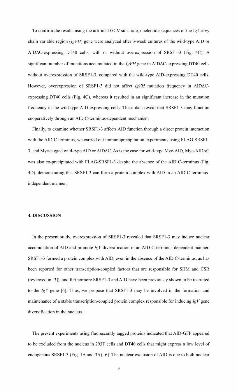
To confirm the results using the artificial GCV substrate, nucleotide sequences of the Ig heavy
chain variable region (IgVH) gene were analyzed after 3-week cultures of the wild-type AID or
AIDDC-expressing DT40 cells, with or without overexpression of SRSF1-3 (Fig. 4C). A
significant number of mutations accumulated in the IgVH gene in AIDDC-expressing DT40 cells
without overexpression of SRSF1-3, compared with the wild-type AID-expressing DT40 cells.
However, overexpression of SRSF1-3 did not affect IgVH mutation frequency in AIDDC-
expressing DT40 cells (Fig. 4C), whereas it resulted in an significant increase in the mutation
frequency in the wild-type AID-expressing cells. These data reveal that SRSF1-3 may function
cooperatively through an AID C-terminus-dependent mechanism
Finally, to examine whether SRSF1-3 affects AID function through a direct protein interaction
with the AID C-terminus, we carried out immunoprecipitation experiments using FLAG-SRSF1-
3, and Myc-tagged wild-type AID or AIDDC. As is the case for wild-type Myc-AID, Myc-AIDDC
was also co-precipitated with FLAG-SRSF1-3 despite the absence of the AID C-terminus (Fig.
4D), demonstrating that SRSF1-3 can form a protein complex with AID in an AID C-terminus-
independent manner.
4. DISCUSSION
In the present study, overexpression of SRSF1-3 revealed that SRSF1-3 may induce nuclear
accumulation of AID and promote IgV diversification in an AID C-terminus-dependent manner.
SRSF1-3 formed a protein complex with AID, even in the absence of the AID C-terminus, as has
been reported for other transcription-coupled factors that are responsible for SHM and CSR
(reviewed in [3]), and furthermore SRSF1-3 and AID have been previously shown to be recruited
to the IgV gene [6]. Thus, we propose that SRSF1-3 may be involved in the formation and
maintenance of a stable transcription-coupled protein complex responsible for inducing IgV gene
diversification in the nucleus.
The present experiments using fluorescently tagged proteins indicated that AID-GFP appeared
to be excluded from the nucleus in 293T cells and DT40 cells that might express a low level of
endogenous SRSF1-3 (Fig. 1A and 3A) [6]. The nuclear exclusion of AID is due to both nuclear
export and protein degradation, since this localization was sensitive to Leptomycin B and MG132
treatment. Although endogenous SRSF1-3 may contribute to the accumulation of AID, the protein
level of AID is kept very low in the nucleus at steady state partly because the level of endogenous
SRSF1-3 is very low in DT40 cells [6]. In addition, particularly in hyper-mutating cells such as
DT40 cells and human Ramos cells, AID is excluded from the nucleus even in the absence of
nuclear export of AID because of accelerated proteasomal degradation [12, 16]. This proteasomal
degradation of AID in these hypermutating cells may be induced in an AID deaminase activity-
dependent manner [19]. Overexpressed SRSF1-3 may manifest a role for endogenous SRSF1-3 in
the nuclear accumulation of AID by competing with intra-nuclear processes, including nuclear
export and proteasomal degradation of AID.
SRSF1 is a nucleocytoplasmic shuttling protein, which moves across the nuclear membrane pore,
along with a number of other proteins and mRNAs, as large ribonucleoprotein complexes [8].
Because the RS domain at the C-terminus of SRSF1 is not required for nuclear import of SRSF1
[20], it could be presumed that SRSF1-3, which lacks the RS domain, might contribute to the
nuclear import of AID as a transportation carrier through an interaction with AID (Fig. 2B).
However, this is unlikely because AID was able to localize to the nucleus even in the absence of
SRSF1-3 (Fig. 3), although a supporting role for SRSF1-3 in nuclear import cannot be excluded.
In addition, the AID molecule has its own nuclear import signal, which enables AID to move into
the nucleus through an importin-mediated mechanism [3].
Whereas expression of either SRSF1-3 or AIDDC was able to increase the nuclear localization
of AID and thus the frequency of SHM and GCV, co-expression of both molecules did not show
any additive effects on SHM and GCV (Fig. 4). Thus, SRSF1-3 might function cooperatively with
AID at the C-terminus of AID. However, since AID∆C can form a protein complex with SRSF1-
3, this cooperation may not be mediated through a direct interaction with the AID C-terminus (Fig.
4). Deletion of the AID C-terminus results in decreased CSR but intact SHM, implying that the C-
terminus of AID might have a role in determining which mechanism, either CSR or SHM, AID is
engaged in [12, 14, 18]. Thus, SRSF1-3 may have a role in the nuclear accumulation of AID or in
the activation of AID by an indirect interaction with the AID C-terminus in a protein complex
containing AID and SRSF1-3, e.g., through regulating other factors that associate with the AID C-
terminus. Further investigations into the roles of SRSF1-3 will reveal whether SRSF1-3
contributes to the regulation of other intra-nuclear processes to induce AID-dependent IgV
diversification.
Acknowledgments: We are grateful to Prof. Hitoshi Ohmori for critical discussion and
encouragement in the early phase of this work. We also acknowledge Dr. Ayano Satoh for
providing helpful advice and support for microscopic analyses. We would like to thank Prof. James
L. Manley for providing the DT40-ASF cells. This study was supported in part by JSPS KAKENHI
Grants (26650126, 15H04196, 16K14783), Takeda Science Foundation, and Naito Foundation to
N. Kanayama.
References
[1] M. Muramatsu, K. Kinoshita, S. Fagarasan, S. Yamada, Y. Shinkai, T. Honjo, Class switch
recombination and hypermutation require activation-induced cytidine deaminase (AID), a
potential RNA editing enzyme, Cell, 102 (2000) 553-563.
[2] J. Chaudhuri, F.W. Alt, Class-switch recombination: interplay of transcription, DNA
deamination and DNA repair., Nat. Rev. Immunol., 4 (2004) 541-552.
[3] S.P. Methot, J.M. Di Noia, Molecular Mechanisms of Somatic Hypermutation and Class Switch
Recombination, Adv. Immunol., (2016) in press.
0663 .7 .21 24/ 5 1 .
[4] H. Arakawa, J. Hauschild, J.-M. Buerstedde, Requirement of the activation-induced deaminase
(AID) gene for immunoglobulin gene conversion., Science, 295 (2002) 1301-1306.
[5] H. Arakawa, H. Saribasak, J.-M. Buerstedde, Activation-induced cytidine deaminase initiates
immunoglobulin gene conversion and hypermutation by a common intermediate., Plos Biol., 2
(2004) E179.
[6] Y. Kanehiro, K. Todo, M. Negishi, J. Fukuoka, W. Gan, T. Hikasa, Y. Kaga, M. Takemoto, M.
Magari, X. Li, J.L. Manley, H. Ohmori, N. Kanayama, Activation-induced cytidine deaminase
(AID)-dependent somatic hypermutation requires a splice isoform of the serine/arginine-rich (SR)
protein SRSF1., Proc. Natl. Acad. Sci. U.S.A., 109 (2012) 1216-1221.
[7] J. Wang, Y. Takagaki, J.L. Manley, Targeted disruption of an essential vertebrate gene:
ASF/SF2 is required for cell viability, Genes Dev., 10 (1996) 2588-2599.
[8] X.-Y. Zhong, P. Wang, J. Han, M.G. Rosenfeld, X.-D. Fu, SR proteins in vertical integration of
gene expression from transcription to RNA processing to translation., Mol. Cell, 35 (2009) 1-10.
[9] H. Ge, P. Zuo, J.L. Manley, Primary Structure of the Human Splicing Factor Asf Reveals
Similarities with Drosophila Regulators, Cell, 66 (1991) 373-382.
[10] Y. Hu, I. Ericsson, B. Doseth, N.B. Liabakk, H.E. Krokan, B. Kavli, Activation-induced
cytidine deaminase (AID) is localized to subnuclear domains enriched in splicing factors., Exp.
Cell Res., 322 (2014) 178-192.
[11] N. Kanayama, K. Todo, M. Reth, H. Ohmori, Reversible switching of immunoglobulin
hypermutation machinery in a chicken B cell line., Biochem. Biophys. Res. Commun., 327 (2005)
70-75.
[12] M. Magari, Y. Kanehiro, K. Todo, M. Ikeda, N. Kanayama, H. Ohmori, Enhancement of
hypermutation frequency in the chicken B cell line DT40 for efficient diversification of the
antibody repertoire., Biochem. Biophys. Res. Commun., 396 (2010) 353-358.
[13] N. Kanayama, K. Todo, S. Takahashi, M. Magari, H. Ohmori, Genetic manipulation of an
exogenous non-immunoglobulin protein by gene conversion machinery in a chicken B cell line.,
Nucleic Acids Res., 34 (2006) e10.
[14] S. Ito, H. Nagaoka, R. Shinkura, N. Begum, M. Muramatsu, M. Nakata, T. Honjo, Activation-
induced cytidine deaminase shuttles between nucleus and cytoplasm like apolipoprotein B mRNA
editing catalytic polypeptide 1., Proc. Natl. Acad. Sci. U.S.A., 101 (2004) 1975-1980.
[15] K.M. Mcbride, V. Barreto, A.R. Ramiro, P. Stavropoulos, M.C. Nussenzweig, Somatic
hypermutation is limited by CRM1-dependent nuclear export of activation-induced deaminase., J.
Exp. Med., 199 (2004) 1235-1244.
[16] S. Aoufouchi, A. Faili, C. Zober, O. D'Orlando, S. Weller, J.-C. Weill, C.-A. Reynaud,
Proteasomal degradation restricts the nuclear lifespan of AID., J. Exp. Med., 205 (2008) 1357-
1368.
[17] A.-M. Patenaude, A. Orthwein, Y. Hu, V.A. Campo, B. Kavli, A. Buschiazzo, J.M. Di Noia,
Active nuclear import and cytoplasmic retention of activation-induced deaminase., Nat. Struct.
Mol. Biol., 16 (2009) 517-527.
[18] V. Barreto, B. Reina-San-Martin, A.R. Ramiro, K.M. Mcbride, M.C. Nussenzweig, C-
terminal deletion of AID uncouples class switch recombination from somatic hypermutation and
gene conversion., Mol. Cell, 12 (2003) 501-508.
[19] Q. Le, N. Maizels, Cell Cycle Regulates Nuclear Stability of AID and Determines the Cellular
Response to AID., PLoS Genet., 11 (2015) e1005411.
[20] J.F. Cáceres, T. Misteli, G.R. Screaton, D.L. Spector, A.R. Krainer, Role of the modular
domains of SR proteins in subnuclear localization and alternative splicing specificity, J. Cell Biol.,
138 (1997) 225-238.
Figure legends
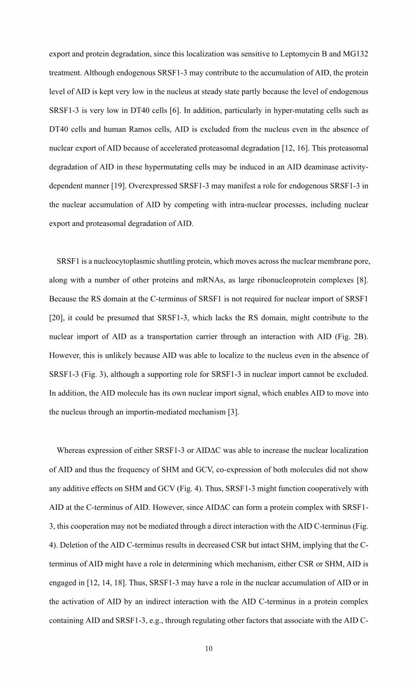
FIGURE 1. Co-expression of AID with SRSF1-3 promotes accumulation of AID in the
nucleus.
(A) AID accumulates in the nucleus in the presence of overexpressed SRSF1-3. The localization
of the fusion proteins in 293T cells was observed by confocal microscopy 36 h after transfection
with the AID-GFP expression vector alone, or with expression vectors encoding AID-GFP and
mCherry-SRSF1-3. The scale bar represents 10 µm. (B) The subcellular localization of AID was
evaluated by calculating the ratio of nuclear AID-GFP over cytoplasmic AID-GFP in individual
cells, as described in Materials and methods. The horizontal bar indicates the median of the ratio
values for each sample group. (C) A correlation analysis for the nuclear localization of AID and
the level of SRSF1-3 expression. The value for AID localization in each cell shown in panel B was
plotted against the mean fluorescent intensity (MFI) of total mCherry-cSRSF1-3 in each cell. The
MFI value was expressed as an arbitrary unit (A.U.).
FIGURE 2. Biochemical analyses of cells co-expressing AID with SRSF1-3.
(A) Co-expression of AID with SRSF1-3 increases AID in the nuclear fraction. The cytoplasmic
and nuclear fractions of non-transfected 293T cells and transiently transfected cells expressing
Myc-AID (AID) and FLAG-SRSF1-3 (SRSF1-3) or Myc-AID alone were analyzed by western
blotting using anti-AID and anti-FLAG (SRSF1-3) mAbs. Hsp90 and acetylhistone H4 were used
as internal controls for the cytoplasmic and nuclear fractions, respectively. The ratio of the nuclear
AID signal over the cytoplasmic AID signal was calculated (lower panel). The horizontal bar
indicates the mean of the ratio values for three independent experiments. (B) SRSF1-3 forms a
protein complex with AID. Protein complexes containing FLAG-SRSF1-3 were
immunoprecipitated using an anti-FLAG mAb from cell lysates of 293T cells transiently
expressing Myc-AID and the FLAG-SRSF1-3 or Myc-AID alone, and analyzed by western
blotting using anti-Myc (AID) and anti-FLAG (SRSF1-3) mAbs. Asterisks indicate the L chain of
the anti-FLAG mAb. Data are representative of three independent experiments.
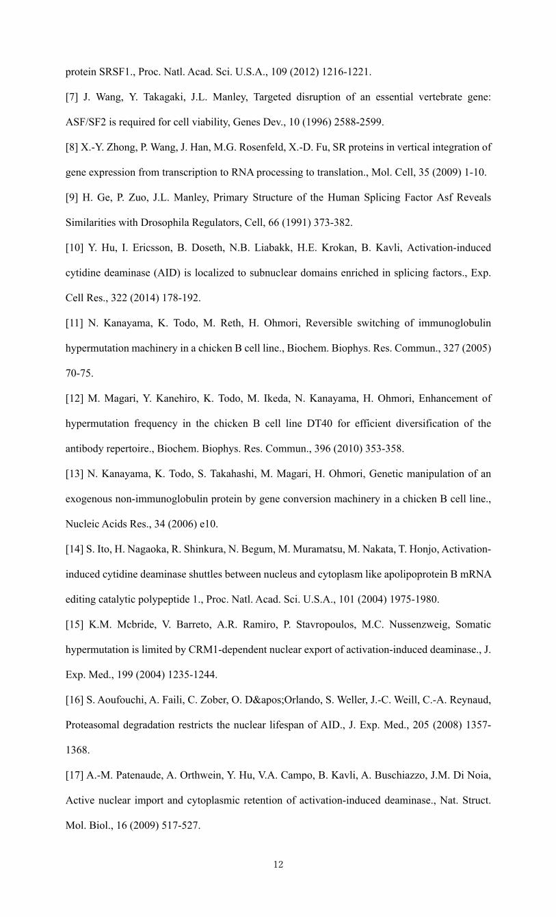
FIGURE 3. SRSF1-3 is dispensable for nuclear import of AID.
(A) AID accumulates in the nucleus of DT40 cells, in the presence or absence of SRSF1-3,
following inhibition of both protein degradation and nuclear export. SRSF1-3-sufficient (DT40-
WT) and SRSF1-3-deficient (DT40-ASF) DT40 cells stably expressing AID-GFP were treated
with MG132 (6 h) and Leptomycin B (2 h), prior to confocal microscopic analysis. The scale bar
represents 10 µm. Data are representative of two independent experiments. (B) The subcellular
localization of AID in DT40 cells treated or untreated with both MG132 and Leptomycin B was
quantitated by calculating the ratio of nuclear AID-GFP over cytoplasmic AID-GFP in individual
cells, as described in Fig. 1.
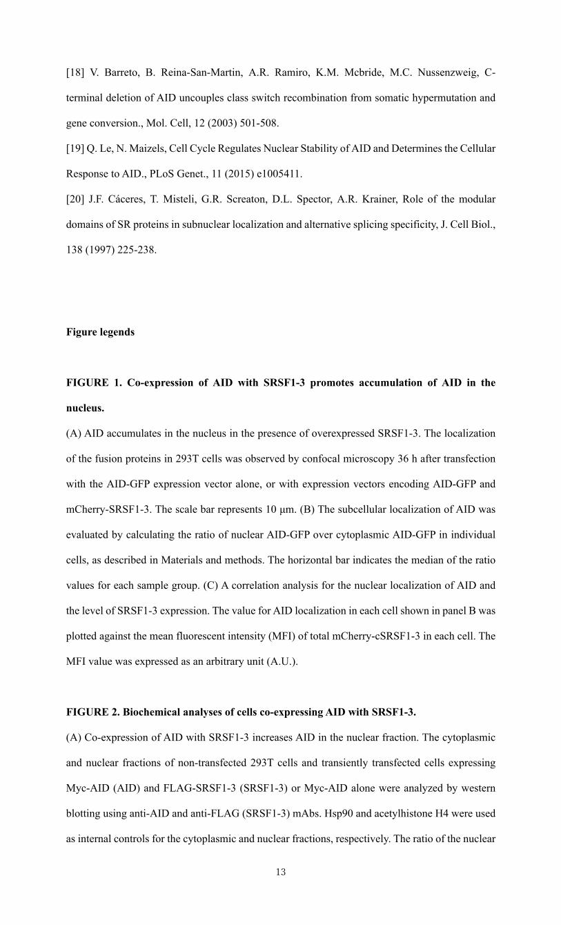
FIGURE 4. A role for SRSF1-3 in IgV diversification depends on the C-terminal region of
AID.
(A) The transcript levels of SRSF1-3 in DT40 cells expressing either a wild-type AID (AID-WT)
or a C-terminally-deleted AID (AIDDC), with or without overexpression of SRSF1-3, were
analyzed by qRT-PCR. Bars are means of triplicate assays. (B) Overexpression of SRSF1-3 does
not further enhance GCV activity in AIDDC-expressing DT40 cells. BFP+ (GCV-un-induced)
single DT40 cells (n = 19 or 20) expressing the wild-type AID or AIDDC, with or without
overexpression of SRSF1-3, were cultured for 2 weeks. GFPhi (GCV-induced) cells were counted
by flow cytometry. The horizontal bar indicates the median of GFPhi cell proportions for each
sample group. (C) The nucleotide sequence analysis of the IgVH genes of DT40 cells expressing
wild-type AID or AIDDC, with or without overexpression of SRSF1-3, after 3-week culture. The
segment sizes in pie charts show the proportion of sequences with 0-9 mutations. (D) SRSF1-3
forms a protein complex with AID∆C. Immunoprecipitation was carried out as described in Fig.
2B. Asterisks indicate the L chain of the anti-FLAG mAb. Data are representative of three
independent experiments.
• SRSF1-3 promotes the nuclear accumulation of AID.
• SRSF1-3 forms a protein complex with AID in an AID C-terminus-independent manner.
• SRSF1-3 contributes to IgV hypermutation in an AID C-terminus-dependent manner.
A
B
FIGURE 1. DAPIGFP MergemCherry
AID-GFP
AID-GFPmCh-SRSF1-3
C
0 20 40 60 80 100 1200.0
0.5
1.0
1.5
MFI of mCh-SRSF1-3 in each cell (A.U.)
AID
loca
lizat
ion
(Nuc
leus
/cyt
opla
sm ra
tio) r = 0.3157
p = 0.0087
0.0
0.5
1.0
1.5
AID
loca
lizat
ion
(Nuc
leus
/cyt
opla
sm ra
tio) ****
AIDonly SRSF1-3
Expressionin 293T cells
AID
A BFIGURE 2.
100
AIDSRSF1-3
AID
SRSF1-3
HSP90
Histone
+– – +– –+– + +– +
Cytoplasm Nucleus
3528
17
10
75
28kDa
17 AID
SRSF1-3
IP: anti-FLAGInput
*
*
28
35
28
kDa
17
AIDSRSF1-3−+
++−+++
0.0
0.2
0.4
0.6
0.8
1.0
AID
loca
lizat
ion
(Nuc
leus
/cyt
opla
sm ra
tio) **
AIDSRSF1-3
+ +– +
A B FIGURE 3.
0
1
2
3
4
AID
loca
lizat
ion
(Nuc
leus
/cyt
opla
sm ra
tio)
+ – +–MG + Lpt
DT40-WT DT40-ASFn 37 24 30 26
**** ****DT40-WT(SRSF1-3 sufficient)
DT40-ASF(SRSF1-3 deficient)
DAPIAID-GFP MergeUntreated
MG132 + Leptomycin B-treated
DT40-WT
DT40-ASF
AIDΔCSRSF1-3 O/E
AIDΔCAID-WTSRSF1-3 O/E
AID-WT
n = 59
0
1
2
3
45
869
7
n = 2001
2
4 7
n=19
0
1
2
6.9 6.7Mutations/bp
(× 10-3)
n = 28
0
1
23
4
586
0.98 2.5
* **
ns
A B
C
FIGURE 4.
AID-WT AIDΔC– + – +SRSF1-3
O/E
0
10
20
30
40
50100120140160
Rel
ativ
e le
vel o
f SRSF1-3
trans
crip
t** ***
AID-WTSRSF1-3
O/E AIDΔC− + − +
0.0
0.5
1.0
1.5
2.0
GFP
hi c
ells
(%)
ns***
**
*SRSF1-3
AID
IP: anti-FLAG
Input
*AID∆C
AIDSRSF1-3
17
kDa28
35
28
∆CWT− + − +
*
17
kDa28
35
28
AIDAID∆C
SRSF1-3
AIDSRSF1-3
∆CWT− + − +
D
Supplemental materials
for
SRSF1-3 contributes to diversification of the immunoglobulin variable region gene by promoting accumulation of AID in the nucleus
Yuka Kawaguchi, Hiroaki Nariki, Naoko Kawamoto, Yuichi Kanehiro, Satoshi Miyazaki, Mari Suzuki, Masaki Magari, Hiroshi Tokumitsu, Naoki Kanayama
Department of Medical Bioengineering, Graduate School of Natural Science and Technology, Okayama University, Okayama 700-8530, Japan.
S-1
Supplemental Table 1.Supplemental Table 2.
S-2
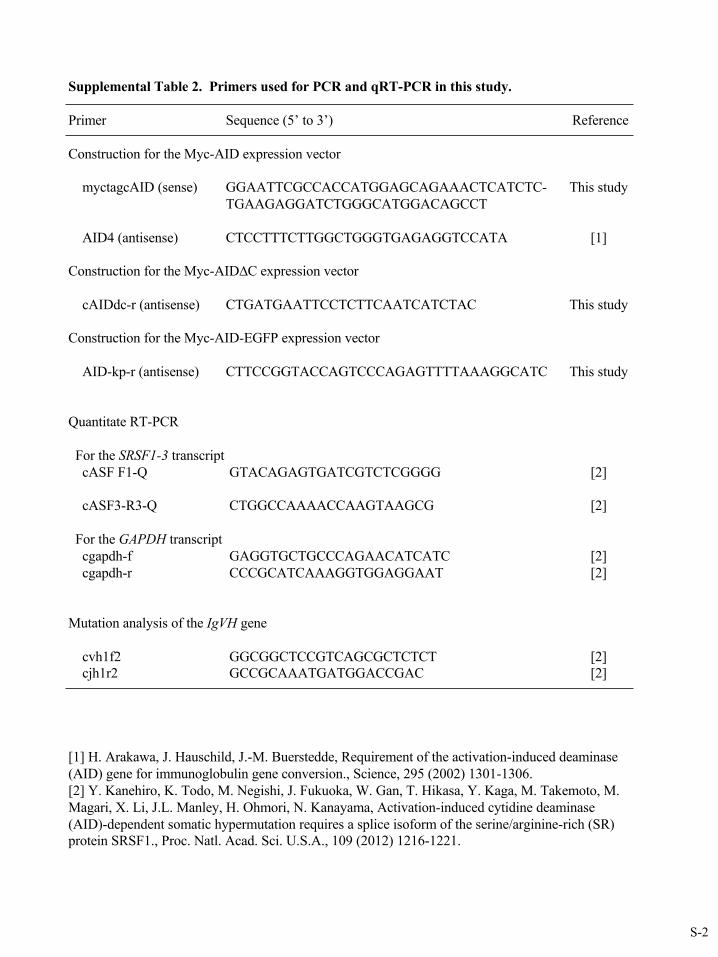
Supplemental Table 2. Primers used for PCR and qRT-PCR in this study.
Primer Sequence (5’ to 3’) Reference
Construction for the Myc-AID expression vector
myctagcAID (sense) GGAATTCGCCACCATGGAGCAGAAACTCATCTC- This studyTGAAGAGGATCTGGGCATGGACAGCCT
AID4 (antisense) CTCCTTTCTTGGCTGGGTGAGAGGTCCATA [1]
Construction for the Myc-AID∆C expression vector
cAIDdc-r (antisense) CTGATGAATTCCTCTTCAATCATCTAC This study
Construction for the Myc-AID-EGFP expression vector
AID-kp-r (antisense) CTTCCGGTACCAGTCCCAGAGTTTTAAAGGCATC This study
Quantitate RT-PCR
For the SRSF1-3 transcriptcASF F1-Q GTACAGAGTGATCGTCTCGGGG [2]
cASF3-R3-Q CTGGCCAAAACCAAGTAAGCG [2]
For the GAPDH transcriptcgapdh-f GAGGTGCTGCCCAGAACATCATC [2]cgapdh-r CCCGCATCAAAGGTGGAGGAAT [2]
Mutation analysis of the IgVH gene
cvh1f2 GGCGGCTCCGTCAGCGCTCTCT [2]cjh1r2 GCCGCAAATGATGGACCGAC [2]
[1] H. Arakawa, J. Hauschild, J.-M. Buerstedde, Requirement of the activation-induced deaminase (AID) gene for immunoglobulin gene conversion., Science, 295 (2002) 1301-1306.[2] Y. Kanehiro, K. Todo, M. Negishi, J. Fukuoka, W. Gan, T. Hikasa, Y. Kaga, M. Takemoto, M. Magari, X. Li, J.L. Manley, H. Ohmori, N. Kanayama, Activation-induced cytidine deaminase (AID)-dependent somatic hypermutation requires a splice isoform of the serine/arginine-rich (SR) protein SRSF1., Proc. Natl. Acad. Sci. U.S.A., 109 (2012) 1216-1221.
S-3
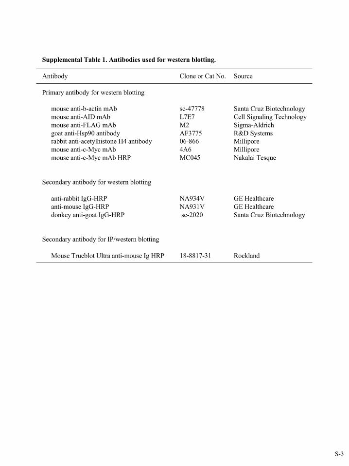
Supplemental Table 1. Antibodies used for western blotting.
Antibody Clone or Cat No. Source
Primary antibody for western blotting
mouse anti-b-actin mAb sc-47778 Santa Cruz Biotechnologymouse anti-AID mAb L7E7 Cell Signaling Technologymouse anti-FLAG mAb M2 Sigma-Aldrichgoat anti-Hsp90 antibody AF3775 R&D Systemsrabbit anti-acetylhistone H4 antibody 06-866 Milliporemouse anti-c-Myc mAb 4A6 Milliporemouse anti-c-Myc mAb HRP MC045 Nakalai Tesque
Secondary antibody for western blotting
anti-rabbit IgG-HRP NA934V GE Healthcareanti-mouse IgG-HRP NA931V GE Healthcaredonkey anti-goat IgG-HRP sc-2020 Santa Cruz Biotechnology
Secondary antibody for IP/western blotting
Mouse Trueblot Ultra anti-mouse Ig HRP 18-8817-31 Rockland