springer-verlag, new york. chapter 10: the genetics of
TRANSCRIPT

Extracted from: Moore, D. & Novak Frazer, L. (2002). Essential Fungal Genetics. Springer-Verlag, New York. ISBN: 0387953671
1Extracted from: Moore, D. & Novak Frazer, L. (2002). Essential Fungal Genetics. Springer-Verlag, New York.
Chapter 10: The genetics of fungal differentiation and morphogenesis 10.1 Differentiation and morphogenesis Growth of the vegetative fungal hypha, showing polarized, invasive extension growth localized at the hyphal apex is the fundamental growth pattern of all members of Kingdom Fungi, and of some members of related groups. If we can borrow a word used in everyday computer terminology, in our view the fungal hypha is the ‘default’ growth condition of the fungal genome. Vegetative hyphal growth requires coordinated expression of the components of the genome so that the whole of the growth process can be supported, located and projected into the extension of the hyphal tip. All of this requires regulation of gene expression. Most fungi also produce a range of cell types differing in cell shape and growth pattern. These require further programs in which gene expression is integrated into developmental routines involving transmission and receipt of signals to organize transitions between different cell types. Some of those signals will be intracellular, some will be extracellular signals relating the nutritional and physical state of the environment, but all will require signal transduction pathways comprising receptor, transmission, and amplification and effector components. This aspect of development, which is cell differentiation, depends on differential management of hyphal functions, part of which relies on genetic regulation leading to synthesis of gene products specific to certain cell types, but part of which can also include epigenetic phenomena including gene silencing as well as phenotype changes in which physical forces establish morphological change by altering cytoskeletal organization, for example. Such regulatory events are sufficiently robust to account for most hyphal differentiation including even that of yeast-like fungi such as Saccharomyces cerevisiae. The yeast-like cells can be interpreted as hyphal cells trapped in a highly differentiated yeast-form morphology in which the normal invasive hyphal apex growth is adapted to the pattern of growth recognized as budding. But even yeasts can be induced to grow as elongated filaments, dedifferentiating to the default fungal invasive growth form. Beyond cell differentiation, but obviously dependent upon it, we place fungal tissue morphogenesis. Even the vegetative fungal mycelium may be considered as a tissue because it grows outwards into new territory and consequently has controlling signals which ensure that hyphae normally grow away from one another to form the typical ‘colony’ with an outwardly-migrating growing front. Tissue development requires that different hyphae cooperate in an organized way. For tissues to be formed the invasive outward growth pattern of the vegetative mycelium must be modified so that independent hyphal apices grow towards each other, allowing their hyphae to branch and differentiate in a cooperative fashion. The structures to which the tissues contribute, spore-forming fruiting bodies, for instance, actually arise on the vegetative mycelium, so these changes in growth pattern must be localized, and must be a response to regulatory processes which are imposed upon the vegetative mycelium. Another aspect is that tissue formation demands that the continuous tube of hypha produced by the growing apex is divided up into cells or compartments by the formation of cross-walls (septa). This enables differentiation to be localized, offering the possibility that adjacent compartments might follow different pathways of differentiation, and even be of different size. Lower fungi (Zygomycotina like Mucor, for instance) have coenocytic hyphae. Although they do not form multicellular structures they do form septa at certain stages during development: the gametangia that eventually fuse and develop into a zygosporangium are separated from the rest of the coenocytic hyphae by septa, so that the zygosporangium develops alongside vegetative hyphae. Fungi that do exhibit complex developmental pathways form septa at regular intervals in mycelial hyphae, but the septa usually have a pore (more or less central), which may be elaborated with the parenthesome apparatus in basidiomycetes, or are associated with Woronin bodies in ascomycetes. Although the septal pore is common feature, it is clearly the case that the movement or migration of cytoplasmic components between adjacent cells is under very effective control. There are instances in which nuclei move freely, but mitochondria do not, and others in which rapid migration of vacuoles is not accompanied by migration of any other organelle. Some biochemical experiments have even demonstrated that different sugars can be translocated in opposite directions in a hypha at the same time. There are also numerous examples available where grossly different pathways of differentiation have been followed on the two sides of what appear (to the electron microscope) to be open septal pores. Clearly, whatever the appearance of the open septa, the hypha can be separated into cells whose interactions are carefully regulated and which can exhibit contrasting patterns of differentiation. The hyphae of Ascomycotina and Basidiomycotina are characteristically divided up into cells by these septa-with-pores, but please don't forget that every fungal cell is just a segment of a tubular hypha. This is very important because the hyphal growth form must influence the characteristics of the controls that regulate fungal tissues. Filamentous hyphal growth can be interpreted on the basis of a regular cell cycle. Hyphal branching, by increasing the number of growing points, is the equivalent of cell division in animals and plants. Although plant morphogenesis depends on placement of the cross-wall, in fungal hyphae cross-walls are formed at right angles

Extracted from: Moore, D. & Novak Frazer, L. (2002). Essential Fungal Genetics. Springer-Verlag, New York. ISBN: 0387953671
2to the long axis of the hypha. Except in cases of injury or in hyphal tips already differentiated to form sporulating structures, hyphal tip cells are not subdivided by oblique cross-walls, nor by longitudinally oriented ones. Even in fission yeast cells forced to produce irregular septation patterns under experimental manipulation, the plane of the septum is always perpendicular to the plane including the longest axis of the cell. In general, then, the characteristic fungal response to the need to convert the 1-dimensional hypha into a 2-dimensional plate or 3-dimensional block cannot depend on a different geometrical arrangement of the septum. The only solution open to the fungal hypha is the formation of branches. The septum in the branch will still be formed at right angles to the long axis of the branch, but its orientation relative to the parent hypha will depend entirely on the positioning of the branch apex, which is established some time prior to septum formation. Consequently, there are two fundamental processes involved in construction of fungal multicellular structures: the first is the origin of the branch (its appropriate placement and orientation on the parent hypha) and the second is the direction of growth of the new hyphal apex that is created by the branching event. The former process seems to be the formal equivalent of determination of morphogenetic growth by orienting the plane of division and the new cross-wall as is seen in plants, and the latter has much in common with the morphogenetic cell migrations that contribute to development of body form and structure in animals. Viewed in this light, therefore, the fungal Kingdom is seen as employing morphogenetic processes that have affinities with both of the other major eukaryote kingdoms. There is no substantial difference in the nature of the questions that need to be answered in studies of development in the three eukaryote Kingdoms. How do genes act to establish basic cell behavior? How do cells become different? How do cells influence one another? How do cells cooperate to form structures? An animal embryologist asks the same questions as a developmental mycologist. The answers may be different in detail, with the details being determined by the life style according to Kingdom-specific adaptations of the organism concerned. But there is likely to be an underlying similarity in strategy because the same basic eukaryotic cell structure is used throughout, and in eukaryotes most gene regulation occurs at the initiation of transcription. 10.2 Genetic approaches for analyzing gene regulation Gene regulation can be imposed at any of the stages in the flow of information from the DNA to the working protein: namely, by controlling which genes are transcribed into RNA, by regulating which RNA products are spliced to make functional messenger RNA, by determining which mRNAs are transported to the cytoplasm, by regulating mRNA translation into protein, and then by regulating the function and lifetime of the protein itself. There is, indeed, evidence for gene regulation at each of these stages. But despite examples of controls at other levels, there is a great deal of evidence to show that transcriptional control is the most critical and widely used level of gene regulation in eukaryotes. Analysis of regulatory factors focuses on mutations that affect gene function without affecting the primary structure (amino acid sequence) of the gene product. A gene responsible for a phenotype that is sensitive to the amount of gene product produced in the cell is the best candidate. After choosing the ‘target gene’, the experimenter searches for mutations that affect expression of that gene; these are regulatory mutations. Regulatory mutants that map within, or in the immediate vicinity of, the target gene can indicate DNA sequences that influence transcription. Such DNA sequences may serve as attachment sites for DNA-binding proteins that regulate transcription. They are called cis-acting elements because they work on the same DNA molecule as the target gene. Promoters are cis-acting elements to which the RNA polymerase binds. Another type of cis-acting element is the enhancer, which is a binding site for proteins that control the level of transcription. Sequences like this can be studied in the laboratory by using reporter constructs. These replace the target gene with the coding region of a heterologous gene that produces an easily identifiable product (the so-called ‘reporter’). Popular reporters because of their colored products are the β-galactosidase gene from the Escherichia coli lac operon (which can be detected through its reaction with chromogenic substrates: a colorless substrate known as X-Gal is turned blue in the presence of β-galactosidase), or the green fluorescent protein (GFP) isolated from the luminescent jellyfish Aequorea victoria, which absorbs blue light and re-emits it as green fluorescence. Color variants have been prepared which provide the opportunity for dual-labeling studies and there is a red fluorescent protein available, isolated from Discosoma spp., an IndoPacific sea anemone. As well as the reporter gene, the whole reporter construct will include the regulatory regions of the target gene so that when the construct is reintroduced into the target genome by transformation, the effect of in vivo regulatory factors can be tested. Systematic mutagenesis (called site-directed mutagenesis) across the presumed regulatory region can then be used to study the influence of each base pair in the regulatory sequence. Regulatory genes located on a different DNA molecule to the target gene are trans-acting elements. They are structural genes for polypeptides, known as trans-acting factors that interact with the cis-acting elements of the target gene. Trans-acting proteins that regulate transcription are generally known as transcription factors. Mutations in trans-acting elements will alter the level of target gene expression (or expression of a reporter construct) but genetic mapping will locate them away from the site of the target gene or reporter. With in vitro techniques it is possible to isolate these proteins that bind to the DNA sequence of the cis-acting element.

Extracted from: Moore, D. & Novak Frazer, L. (2002). Essential Fungal Genetics. Springer-Verlag, New York. ISBN: 0387953671
3
GCbox
GCbox
CAATbox
TATAbox
Initiationcodon Stop
codon
Poly-A additionsite
AATAAPoly-A signal
sequence
Transcriptioninitiation
Upstream control sites Downstrean flankingregionIntron I Intron II
-3'5'- Exon 1 Exon 2 Exon 3
AG5' splice
site
AGGT GT3' splice
site
Promoters Transcribed region Terminator
3' untranslatedregion (3'-UTR)
5' untranslatedregion (5'-UTR)
Direction of transcription of the sense strand
Gene
Fig. 10.1. The basic structure of a typical eukaryotic gene. The schematic diagram indicates the structure of a type II gene, that is a protein-encoding gene transcribed by polymerase II. The diagram is not drawn to scale and the relative sizes of the different sections differ between genes and between the eukaryotic Kingdoms. 10.3 Regulating gene expression: DNA binding proteins The basic structure of a typical eukaryotic protein-coding gene (Fig. 10.1) includes several different components: the protein-coding regions may be in two or more exons separated by introns which are spliced out of the RNA transcript and are untranslated. Regulatory sequences, where gene-controlling transcription factors bind, are mainly just upstream (which means, on the 5'-side) of the transcribed region, although there may be other control regions, lying far outside the gene, which play a role in regulating chromatin structure. A common theme in eukaryote gene regulation is the involvement of DNA-binding proteins, which are involved in all aspects, including deciding which of the genes are to be expressed and for synthesizing the RNA transcripts of genes that are expressed, and a very large proportion of which have been identified from molecular genetic analysis of Saccharomyces cerevisiae. These proteins bind to specific sequences in DNA and then interact with other proteins to activate transcription (Fig. 10.2). They have two structural domains that enable them to do this: a DNA-binding domain and a transcription-activator domain. Some activators have a third domain that reacts to other specific signals, such as hormones or other signaling molecules. When such a molecule binds to these activators they cause an allosteric change that greatly increases the affinity of the protein for its DNA target sequence. This sort of control permits rapid changes in gene expression, enabling the cell to respond to external signals and transient changes in its metabolic circumstances. Rapid control over transcription factor activity of this sort often underlies the ability of extracellular conditions and signaling compound to control events going on within the cell. There may be an indirect activation when the extracellular signal interacts with a cell surface receptor that transduces the message to the cell interior, or a direct activation if the extracellular signaling molecule can enter the cell to interact immediately with a transcription factor or signal transducer. DNA-binding domains in many different transcription factors share particular peptide motifs involved in the DNA helix binding function; these configurations are called the zinc-finger (in which an atom of zinc is conjugated to two cysteines and two histidines in the polypeptide), the helix-loop-helix, and the helix-turn-helix (which orient α-helices of the polypeptide so that they can fit into the major groove of the DNA helix). Zinc finger proteins generally have several ‘fingers’, each of which is able to interact with a specific DNA sequence. There are also some common features in the transcription activation domains, such as being relatively rich in the amino acid asparagine or, alternatively, rich in proline. The shared features are associated with the general function of these molecules as transcription factors. Other, much more subtle, aspects of their primary and secondary structures provide each one with its specificity for its DNA target sequence and the particular part of the transcription machinery it affects. Most activators in eukaryotes must form dimers to function, and the functional proteins may be homomers (multimeric

Extracted from: Moore, D. & Novak Frazer, L. (2002). Essential Fungal Genetics. Springer-Verlag, New York. ISBN: 0387953671
4
Chromatin Chromatin ChromatinTATA
bindingprotein
RNApolymerase II
AF E
H
B
Mediatoracetylase
co-repressor
co-activator
TFTF co-activator
co-repressor
deacetylase Fig. 10.2. Simplified illustration of the transcription machine. The polymerase, basal transcription factors (labeled A, B, E, F and H), TATA-binding protein and mediator (together with other proteins not represented here) occupy the core promoter of a gene. Upstream of this location histone octamers are represented by the tailed octagons labeled ‘chromatin’. A transcription factor (TF) can interact with coactivators that recruit acetylase to acetylate the histone tails and open up the chromatin structure, or with corepressors that recruit a deacetylase, which restores chromatin structure. proteins composed of the same subunit) or heteromers (multimeric proteins composed of different polypeptide subunits). Heterodimerization increases the number of transcription factors that can be assembled from available monomers. Dimer formation depends on yet another characteristic domain of transcription factors, the dimerization domain, which is optimized for very specific interactions between particular polypeptides. The most common primary structure motif in dimerization domains is the leucine zipper. This is a sequence of amino acids that forms into an α-helix with leucine residues extending from the helix at regular intervals. The leucine zipper of one polypeptide can interlock with the leucine zipper of a second polypeptide, like the clothing version. Specificity for the ‘zipping’ depends on the amino acids situated between the leucines. Effectively, there are two stages to transcription: transcriptional initiation and transcriptional elongation. Intrinsic to the initiation step are the specific interactions that determine which gene is expressed and which assemble all the proteins that will copy, or assist in copying, the gene into an RNA transcript. The second stage is the transcription process itself, during which the RNA polymerase translocates along the gene producing the primary RNA transcript as a direct complementary copy of the gene. 10.4 Regulating gene expression: chromatin remodeling One of the defining features of the eukaryotes is the possession of chromosomes, and the DNA packaging in the chromatin that makes up the chromosomes has an enormous influence on gene regulation in eukaryotes. Chromosomes in eukaryotes consist of about one-third genomic DNA, one-third histone proteins and one-third non-histone proteins. ‘Chromatin’ is the name given to the complex between DNA and proteins that makes up the chromosome structure. An important function of chromatin is to reduce basal transcription of all genes to a very low level, and in eukaryotes the normal structure of chromatin is entirely sufficient to maintain transcription at the minimal, basal level. The basic structural unit of chromatin is the nucleosome, which consists of an octamer of histone proteins (two each of H2A, H2B, H3, and H4) around which is wrapped approximately 200 bp of DNA. Histone H1 binds to short stretches of DNA between nucleosomes, and helps maintain chromatin structure. Nucleosomes interact to construct further, higher, levels of chromatin fiber structure: from nucleosomes, to 10 nm fibers, then 30 nm fibers, on to chromosome loops, and ending with fully condensed metaphase chromosomes, which are the most compact form of DNA packaging in eukaryotes. Chromosomes become less compact after completion of nuclear division, but there is a higher order folding (above the level of the 30 nm fiber) in interphase chromosomes. Heterochromatin is in a permanent state of compact folding. So compact, in fact, that proteins needed to activate gene expression cannot access the DNA. Constitutive heterochromatin is the DNA that contains no genes in centromeric and telomeric regions. Facultative heterochromatin is DNA containing genes that are temporarily inactive because of the stage of development or position in the cell cycle. Regions of DNA containing active genes are called euchromatin. Euchromatin consists of loops of 30 nm chromatin fibers, equivalent to lengths of about 40 to 100 kb DNA. AT-rich DNA regions called matrix-associated regions (MARs) or scaffold-attachment regions (SARs) attach the loops to a protein network, called the nuclear matrix that fills the nucleus. Nucleosomes have an over-riding influence on transcription because the DNA packaging within them represses gene expression. Transcription is made possible by specific positive regulatory mechanisms that rearrange nucleosome structure. Then, even when a specific gene is made accessible, the precise positioning of nucleosomes in the immediate vicinity influences transcription of it. This reflects a difference in regulatory strategy between prokaryotes and eukaryotes. Prokaryotes in general use negative regulation, effected by gene-specific repressors acting at structural gene promoters. Arguably, such a mechanism is inadequate for the large genomes of eukaryotes, because such a large number of different repressors would be needed to control gene

Extracted from: Moore, D. & Novak Frazer, L. (2002). Essential Fungal Genetics. Springer-Verlag, New York. ISBN: 0387953671
5expression. Instead, eukaryotes have adopted a mechanism featuring general repression of the genome, and requiring integrated activation of transcription as the basis for cell-type-specific regulation. Nucleosomes repress transcription by covering protein-binding sites of DNA, so interfering with the interaction of the entire collection of DNA binding proteins, regulators, polymerases and transcription factors, required for transcription. Chains of nucleosomes can also become involved in higher-order coiling and thereby repress transcription of large chromosomal regions, and interactions between nucleosomes and other chromosomal proteins produce heterochromatin, in which gene expression is also repressed. The molecular foundation of repression by nucleosomes lies in the configuration of the histone molecules, each of which has a characteristic ‘histone fold’ and an N-terminal ‘tail’. The histone folds keep the DNA in a central core particle, and it is this that prevents access of other DNA-binding proteins. The tail protrudes outside the core particle, taking part in the interactions that produce higher-order coiling, and this is the basis of its involvement in gene activation. Acetyltransferase enzymes acetylate the histone tails, producing a chemical modification characteristic of transcribed chromatin. The acetyltransferases therefore serve as coactivators, stimulating transcription by lifting the repression caused when the core particles take on higher-order structure. Histone deacetylase enzymes do the reverse; they act as corepressors by removing the acetylation of the tail and thereby allowing the chromatin to take on the repressive higher-order structure (Fig. 10.2). However, histone acetylation is not sufficient in itself for transcriptional activation because it does not disrupt the core particle of the nucleosome. Most inactive genes have their promoters occluded by nucleosomes. There are two multiprotein ‘chromatin remodeling complexes’ that rearrange the structure of chromatin in an ATP-dependent manner to remove these promoter-blocking nucleosomes. One, known as ‘switch’ and symbolized SWI/SNF, disturbs the core-particle structure, and the other, ‘imitation switch’ or ISWI shifts the locations of nucleosomes on DNA. Nucleosome positioning is important in modulating gene expression. In yeast, the SWI/SNF complex is the first coactivator to arrive at a gene at which transcription is to be induced. The gene-specific activator proteins, which also recruit an acetyltransferase to acetylate the histone tails, recruit them and the resultant loosening of the chromatin allows general transcription factors to get access to promoter regions. From that point transcription rapidly accelerates. The SWI/SNF complex is one of many that are involved in remodeling chromatin at specific chromosomal locations and in specific cells at particular points of development. Closely related protein complexes able to influence nucleosome position and/or structure are found in human cells, showing that chromatin remodeling machinery has been conserved throughout evolution. 10.5 Regulating gene expression: transcription Regulation of transcription in eukaryotes depends on multi-protein complexes assembled at DNA control sequences immediately adjacent to the start site of transcription, called the promoter. For many protein-coding genes the promoter contains the TATA box, which is a binding site for the constitutively expressed general transcription factor (GTF) called transcription factor TFIID (TF = transcription factor, II = for RNA polymerase II, D = TFII type D). Binding of TFIID to the promoter is critical to the assembly of a basal, stable transcriptional complex, which is able to recognize core promoter elements. This provides low levels of accurate transcription, called basal transcription using a variety of other transcription factors (TFIIA, B, E, F and H, see below) and RNA polymerase II itself. These basal transcription machines are the globally used part of the transcription mechanism. Basal transcription is activated by a highly varied and very large group of transcription factors that assemble at distant enhancer sites. Such transcription activators provide the gene specificity and cell-type specificity of transcription. However, even this transcription machinery, which might be composed of 40 or more polypeptides, is still dependent on a third class of transcription factors called coactivators, which do not have site-specific DNA-binding ability by themselves, but act as intermediaries in the action of transcription activators on the basal transcription machinery (Fig. 10.2). Following chromatin remodeling, the polymerase and accessory factors interact with the promoter (Fig. 10.2). There are three DNA-dependent RNA polymerases in eukaryotes, designated pol I, pol II and pol III, although we know most about pol II, which is responsible for all messenger RNA (mRNA) synthesis. Pol I transcribes the genes for ribosomal RNA (rRNA) and pol III transcribes transfer RNA (tRNA). RNA polymerases are complexes of 12 protein subunits, which require 23 other polypeptides transcription factors to recognize a promoter and initiate transcription. There is a perfect one-to-one correspondence between the components of the yeast and human systems, and components from animal cells function in yeast, indicating a high degree of functional conservation of the transcription apparatus during eukaryotic evolution. However, these polypeptides are insufficient to promote transcription elongation, and an additional coactivator activity is required as an interface between activators and polymerase II, transducing regulatory information from enhancers to promoters. This factor is called ‘Mediator’ and is a 20-subunit complex in yeast; corresponding complexes from mammals vary in subunit composition, but are otherwise functionally the same. There are several recognition sequences in the DNA, which are recognized either by the RNA polymerase itself or by a DNA-binding protein, which enable the transcription initiation complexes to be

Extracted from: Moore, D. & Novak Frazer, L. (2002). Essential Fungal Genetics. Springer-Verlag, New York. ISBN: 0387953671
6constructed at the correct positions on the DNA molecule. Bacterial RNA polymerases bind to promoter sequences, located immediately upstream of the gene to be transcribed. The ‘average’ (or consensus) of all promoter sequences in Escherichia coli shows two six-nucleotide sequences; one is called the -35 box and has the sequence 5'-TTGACA, and the other is the -10 box, with the sequences 5'-TATAAT. The boxes are named for their position relative to the nucleotide base at which transcription begins, which is called +1. There is a stretch of 15 to 17 bases between the two boxes, which brings the two sequence motifs to the same face of the double helix, and ensures both can most effectively interact with the DNA-binding factor component of the RNA polymerase. Eukaryotic promoters are more complex and there may be several sequences that are important in initiation of transcription of a gene. The 5'-TATA box is located about 60 to 120 base pairs upstream from the transcription start nucleotide in yeast (only about 30 base pairs in mammals), and this site directs the polymerase to begin transcribing. It is the binding site for the TATA-box binding subunit (TBP) plus more than 8 trans-acting factors (TAFs), which together make up transcription factor II (TFII), which is one of the general transcription factors for RNA polymerase II. There are several of these, differing in function according to the nature of their components, and they are distinguished by letter-suffixes. TFIID is responsible for promoter recognition, using the TBP subunit to bend DNA in the TATA-box region, it enables interaction with TFIIB, which positions the polymerase on the promoter. TFIIH includes ATP-dependent helicases that unwind the promoter around the start site to trigger the initiation of transcription. They then maintain a ‘bubble’ of unwound DNA around the nucleotide polymerization site, allowing pairing of the RNA product with the template through base pairing of about eight residues immediately adjacent to the polymerization site. Subsequently, the mediator complex interacts with polymerase II to form the ‘holoenzyme’ able to continue elongation. The switch from transcriptional initiation to elongation is associated with the phosphorylation of the carboxy-terminal tail of RNA polymerase II. Several elongation factors have been identified, and some may regulate transcription by interacting with sequences in the RNA transcript. Indeed, there is a very close connection between transcription and mRNA processing. The phosphorylated tail of RNA polymerase II in elongation mode interacts directly with factors involved in mRNA capping, 3'-end processing and even splicing. By so doing, the various RNA-processing components are recruited to the transcription elongation apparatus and the RNA transcript, producing an ‘mRNA factory’ in which synthesis and processing of mRNA are integrated. About 100 base pairs upstream of the transcription start site a 5'-CCAAT box is also involved in promoter activity and a GC-rich sequence (consensus 5'-GGGCGG) about 100 base pairs further upstream may also serve as a promoter element. Further away, enhancer or activating sequences can be found. Enhancers are regulatory sites that can act at a distance and may be located many thousands of nucleotides away from the promoter, and may be able to operate either upstream or downstream from the promoter they control. An enhancer is unable to drive transcription by itself, but it can enhance the activity of the promoter by several orders of magnitude. According to the nature of the transcription factor involved, this enhancement may occur in all cells (if that particular enhancer is bound by constitutively expressed transfer factors) or may occur only in a specific tissue or in response to a specific signal if the enhancer binding site is for factors which are involved in differentiation. In yeast, enhancer elements are usually called upstream activation sequences, or UASs. Operation of enhancers can be tissue-specific and/or specific to environmental conditions, but they need to be intact to ensure maximal rates of transcription. They are the sites to which some other trans-acting (or transcription) factors bind to assist RNA polymerase to construct a preinitiation complex in a manner specific to a particular gene or gene-family, like the yeast GCN4 transcription factor which, in response to amino acid starvation, activates transcription of many genes involved in amino acid synthesis by binding to a common UAS. 10.6 Galactose utilization in yeast: the epitome of eukaryote regulation Six coordinately regulated structural genes encode the proteins needed for hydrolysis and utilization of galactose and the galactose-containing disaccharide, melibiose by Saccharomyces cerevisiae. Both sugars are highly relevant to the natural environment of yeast because they occur in plant exudates, especially in the nectaries. These genes are among the most tightly regulated genes known. This, together with the fact that they control a shift in metabolism from one sugar to another, account for the high level of interest in the system. The GAL genes controlling utilization of galactose as a carbon source include the three structural genes forming a tightly linked cluster, on chromosome II, GAL1, GAL7, and GAL10. GAL1 encodes the enzyme galactokinase, which phosphorylates galactose to galactose 1-phosphate (Fig. 10.3). GAL7 and GAL10 cooperate in the next step, converting galactose 1-phosphate to glucose 1-phosphate. GAL7 encodes galactose 1-phosphate uridyltransferase, which converts galactose 1-phosphate and UDP-glucose into glucose 1-phosphate and UDP-galactose. The GAL10 protein is an epimerase that regenerates UDP-glucose from UDP-galactose. All three enzymes are essential for metabolism of galactose and when galactose is present in the medium each of these proteins can represent up to 1.5% of the total soluble protein in the cytoplasm. Two other important components of the pathway, not physically part of the gene cluster, but certainly part of the regulatory circuit are GAL2, which encodes the galactose transporter, and GAL5, which is responsible for the phosphoglucomutase that
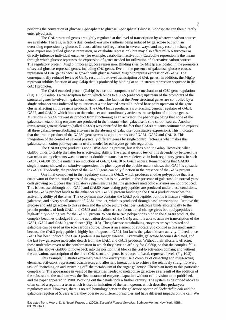
Extracted from: Moore, D. & Novak Frazer, L. (2002). Essential Fungal Genetics. Springer-Verlag, New York. ISBN: 0387953671
7performs the conversion of glucose 1-phosphate to glucose 6-phosphate. Glucose 6-phosphate can then directly enter glycolysis. The GAL structural genes are tightly regulated at the level of transcription by whatever carbon sources are available. There is, in fact, a dual control: enzyme synthesis being induced by galactose but with an overriding repression by glucose. Glucose affects cell regulation in several ways, and may result in changed gene expression (called glucose repression, or catabolite repression), but may also affect mRNA turnover or directly influence individual enzymes (for example, catabolite inactivation). Catabolite repression is the means through which glucose represses the expression of genes needed for utilization of alternative carbon sources. The regulatory protein, Mig1p, imposes glucose repression. Binding sites for Mig1p are located in the promoters of several glucose-repressed genes, including GAL genes. Even in the presence of galactose, glucose causes repression of GAL genes because growth with glucose causes Mig1p to repress expression of GAL4. The consequentially reduced levels of Gal4p result in low-level transcription of GAL genes. In addition, the Mig1p repressor inhibits function of any Gal4p that is produced by binding at an up-stream repression sequence in the GAL1 promoter. The GAL4-encoded protein (Gal4p) is a central component of the mechanism of GAL gene regulation (Fig. 10.3). Gal4p is a transcription factor, which binds to a UAS (enhancer) upstream of the promoters of the structural genes involved in galactose metabolism. The fact that the three structural genes are controlled by a single enhancer was indicated by mutations at a site located several hundred base pairs upstream of the gene cluster affecting all three gene products. The GAL4 locus produces a trans-acting genetic regulator of GAL1, GAL7, and GAL10, which binds to the enhancer and coordinately activates transcription of all three genes. Mutations in GAL4 prevent its product from functioning as an activator, the phenotype being that none of the galactose metabolizing enzymes are produced in the mutants when galactose is sole carbon source. Another trans-acting genetic element (called GAL80) was identified by the fact that GAL80 mutants continued to express all three galactose-metabolizing enzymes in the absence of galactose (constitutive expression). This indicated that the protein product of the GAL80 gene serves as a joint repressor of GAL1, GAL7 and GAL10. This integration of the control of several physically different genes by single control factors is what makes this galactose utilization pathway such a useful model for eukaryote genetic regulation. The GAL80 gene product is not a DNA-binding protein, but it does bind to Gal4p. However, when Gal80p binds to Gal4p the latter loses activating ability. The crucial genetic test of this dependency between the two trans-acting elements was to construct double mutants that were defective in both regulatory genes. In such GAL4-, GAL80- double mutants no induction of GAL7, GAL10 or GAL1 occurs. Remembering that GAL80- single mutants showed constitutive expression, the phenotype of the double mutant shows that GAL4 is epistatic to GAL80. Evidently, the product of the GAL80 gene can only function in the presence of the GAL4 protein. One final component in the regulatory circuit is GAL3, which produces another polypeptide that is a coactivator of the structural genes of the enzymes that is only active in the presence of galactose. In normal yeast cells growing on glucose this regulatory system ensures that the galactose metabolic enzymes are not produced. This is because although both GAL4 and GAL80 trans-acting polypeptides are produced under these conditions, and the GAL4 product binds to the enhancer site, GAL80 protein binding to the GAL4 product quenches the activating ability of the latter. The cytoplasm also contains the GAL3 polypeptide, but this is inactive without galactose, and a very small amount of GAL1 product, which is produced through basal transcription. Remove the glucose and add galactose to this system and the whole picture changes. Galactose binds allosterically to the protein products of both GAL1 and GAL3 and the allosteric conformational change gives both polypeptides a high-affinity-binding site for the GAL80 protein. When these two polypeptides bind to the GAL80 product, the complex becomes dislodged from the activation domain of the Gal4p and it is able to activate transcription of the GAL1, GAL7 and GAL10 gene cluster (Fig.10.3). The galactose metabolizing enzymes are synthesized and galactose can be used as the sole carbon source. There is an element of autocatalytic control in this mechanism because the GAL3 polypeptide is highly homologous to GAL1, but lacks the galactokinase activity. Indeed, once GAL1 has been induced, the GAL3 protein is no longer essential. Eventually, galactose becomes depleted, and the last few galactose molecules detach from the GAL1 and GAL3 products. Without their allosteric effector, these molecules revert to the conformation in which they have no affinity for Gal80p, so that the complex falls apart. This allows Gal80p to move back into the position that blocks the Gal4p activation domain; and without the activation, transcription of the three GAL structural genes is reduced to basal, repressed levels (Fig.10.3). This example illustrates extremely well how eukaryotes use a complex of cis-acting and trans-acting elements, activators, repressors, coactivators and allosteric interactions to achieve the relatively straightforward task of ‘switching on and switching off’ the metabolism of the sugar galactose. There’s an irony to this particular complexity. The appearance in yeast of the enzymes needed to metabolize galactose as a result of the addition of the substrate to the medium was the first instance of enzyme adaptation without cell division to be published, and the paper appeared in 1900. Working out the details took a further century. The system as described above is often called a regulon, a term which is used in imitation of the term operon, which describes prokaryote regulatory units. However, there is no real homology between the galactose operon of Escherichia coli and the galactose regulon of S. cerevisiae; they operate on different principles and have different impacts on the cell. We

Extracted from: Moore, D. & Novak Frazer, L. (2002). Essential Fungal Genetics. Springer-Verlag, New York. ISBN: 0387953671
8
GAL1(galactokinase)
Galactose (out)
Galactose (in)
Externalmedium
Cytoplasm
GAL2 (transporter)
glucose6-phosphate
GAL5(phospho-
glucomutase)
galactose1-phosphate
glucose1-phosphate
GAL7(galactose 1-phosphate
uridyltransferase)
GAL10(UDP-galactose
epimerase)
UDP-glucose
UDP-galactose
GAL7 GAL1GAL10
GAL
4G
AL4
GAL80GAL1
GAL80GAL3
galactosegalactose
GAL
4G
AL4
GAL80GAL1
GAL80GAL3
galactosegalactose
GAL
4G
AL4
GAL80GAL1
GAL80GAL3
galactosegalactose
TRANSCRIBE TRANSCRIBE TRANSCRIBE
GAL7 GAL1GAL10
GAL
4GAL8
0
GAL1
GAL
4
GAL80
GAL3
NO
TRANSCRIPTION
GAL
4GAL8
0
GAL1
GAL
4
GAL80
GAL3
NO
TRANSCRIPTION
GAL
4GAL8
0
GAL1
GAL
4GAL80
GAL3
NO
TRANSCRIPTION
Fig. 10.3. Galactose metabolism and its regulation. The upper panel shows the biochemical reactions involved in uptake of galactose from the medium and its introduction into energy-yielding metabolism. The lower diagrams depict the role of the GAL4p transcription factor in transcription of the genes encoding the main galactose metabolizing enzymes and its interaction to the co-activators GAL1p and GAL3p and the co-repressor GAL80p. think it is dangerous to use a vocabulary that might suggest that there could be some relationship. The most recent analyses of the yeast galactose utilization pathway using DNA microarrays and quantitative proteomics have shown that a thousand mRNAs change in response to changes in the galactose pathway. This indicates the complexity of the eukaryote regulatory and metabolic networks (Fig. 10.4). 10.7 Regulating gene expression: repression and silencing Our emphasis so far has been on positive regulation, that is the activation of gene transcription, but gene expression must also be switched off and transcription factors can have negative effects as well as positive. A transcription factor that suppresses activation of transcription is called a repressor. Repressors may interact with general transcription factors (e.g. TFIIB and TFIID) and so affect assembly of the transcription complex, or they may interact with a corepressor, which recruits a histone deacetylase so that the promoter is silenced by re-establishment of the chromatin structure. Some repressors bind to the same DNA sites as activator proteins and cause repression by competing with activator proteins for binding, so preventing the activators stimulating transcription initiation. In these cases, the inhibitory factor acts by neutralizing the activity of a positively acting factor. Nonetheless, negatively acting factors have an important role in transcriptional regulation. Silencers are cis-acting DNA sequences which, like enhancers, are the recognition sites for transcription factors. But silencers are bound by repressors that inhibit activators and reduce transcription, inhibiting gene expression indirectly. However, as more examples have come to light, it has become evident that many transcription factors can act as either activator or repressor, depending on the gene being regulated and the cell type in which it is expressed.

Extracted from: Moore, D. & Novak Frazer, L. (2002). Essential Fungal Genetics. Springer-Verlag, New York. ISBN: 0387953671
9
Cell cyclecontrol
Proteinmodification
Matingresponse
Proteindegradation
Chromatin & chromosomestructure
Polymerase IItranscription
Membranefusion
Vesicle transport
Cellstructure Cell
polarity
Protein folding
Cytokinesis
Proteintranslocation
Lipid, fatty acidand sterolmetablism
Nuclear-cytoplasmictransport
Cell stress
Carbohydratemetabolism
Signaltransduction
Differentiation
Amino acidmetabolism
Mitosis Meiosis
DNA synthesis
Recombination
DNA repair
Proteinsynthesis
RNAprocessing
RNAturnoverRNA
splicingPolymerase ItranscriptionPolymerase III
transcription
Fig.10.4. A functional group interaction network for some groups of yeast proteins. Each connecting line represents at least 15 interactions between proteins of the connected groups. Indeed, in no case of transcriptional repression studied so far has the possibility been eliminated that repression results from neutralization of an activator. This has led to the tendency to drop the description ‘silencer’ and call all cis-acting elements ‘enhancers’ even though some may sometimes have repressors bound to them. The word ‘silencing’ has taken on other meanings implying longer-term regulation (see below). Transcription factors are polypeptide, and one way of reducing transcription of a gene or genes is to reduce the level of synthesis of the transcription factor. This does not provide rapid control over expression of the target genes, because some time is required to down-regulate the structural gene for the transcription factor and reduce its concentration in the cell. Consequently, this type of control tends to be associated with transcription factors responsible for the longer-term patterns of gene expression related to differentiation and morphogenesis. The specificity of already-synthesized transcription factors can be altered much more rapidly if they are sensitive to modification by other molecules in the cell. We have already referred to an example of this in relation to the yeast mating type α-2 polypeptide, which, in haploid α-cells, acts as a repressor of a-determining genes by binding to their enhancers, but in diploid cells, the same α-2 protein dimerizes with the a-2 gene product to form a repressor of haploid-specific genes. Some repressors function without binding to DNA; rather, they bind to a specific transcription activator and change its function. The process is called quenching, and the regulator protein may quench the DNA-binding activity of an activator (preventing attachment to the enhancer), or may block the activation domain of the activator. We discussed an example of the latter type of repression in the yeast GAL system, above, where Gal80p binding to Gal4p quenches activation by Gal4p. Finally, some eukaryotic repressors bind to DNA sequences very close to the promoter and eliminate transcription by blocking RNA polymerase access to the promoter, in a manner similar to repression in prokaryotes. However, eukaryotic repressors play a different role to their prokaryotic analogues because they mainly modulate the activation caused by transcriptional activators. 10.8 Regulating gene expression: high-level control mechanisms, DNA modification and epigenetics The changes in gene activity discussed so far enable the cell to respond to fairly transient changes in conditions, or make transient changes to its state of differentiation. Development and morphogenesis can involve more permanent changes in genome activity. Implicit in our discussion above about the involvement of nucleosomes and chromatin remodeling in regulating gene expression is the expectation that the DNA sequence will carry appropriate target sites to make these processes gene-specific. However, little is known about this aspect of the involvement of higher-order structure in transcriptional regulation, although it could enable coordinate regulation of multiple genes within large chromosomal domains.

Extracted from: Moore, D. & Novak Frazer, L. (2002). Essential Fungal Genetics. Springer-Verlag, New York. ISBN: 0387953671
10 DNA sequences that are potential candidates for such regulation, which must act over tens to hundreds of kilobases, include enhancers that interact with distant promoters, and may be located upstream or downstream, SAR/MAR sequences, which attach chromatin to the nuclear matrix, and locus control regions (LCRs), which are cis-acting regulatory sequences, which function, like enhancers, by binding to transcription factors with activation domains. They maintain open function across a chromosomal domain comprised of several to many genes in a cluster that are active only during specific developmental stages. A locus control region operates sequentially with other transcription factors at cis-regulatory regions that are directly adjacent to each gene in clusters of related genes at different times during development. A fully assembled LCR-transcription factor complex is called an enhancesome. Absence of any component of the enhancesome prevents expression of the whole sequential programme, and none of the genes in the cluster are activated. The best current example of LCR-based regulation is the control of globin gene clusters in human red blood cells. In the β-globin cluster the LCR is spread over about 10 kbp between 5 and 18 kbp upstream of the first globin gene. It includes four 300 bp regulatory regions. The α-globin LCR is a region of 300 bp lying 40 kb upstream of the embryonic globin gene. The regulatory regions of the LCR contain binding sites for a number of transcription factors and LCRs probably interact with the regulators of individual globin genes to activate, enhance, and developmentally regulate their expression. It is evident that the system provides a mechanism for restricting gene expression to a specific cell lineage. Oddly enough, the LCR is not required for activation of the β-globin cluster in the mouse. So there may be functional (or even experimental) differences between the two mammals. Another example of long term gene silencing is provided by the sequences either side of the mating type locus in yeast. The active chromosomal locus of the mating type gene is called MAT (for mating type, see section 2.6), but there are two additional copies of the mating type gene, called HML and HMR, one each side of MAT and located near the telomeres on each arm of chromosome III. HML and HMR, at least one of which carries a different DNA sequence to the active locus, are storage loci from which active copies are retrieved by intrachromosomal recombination during mating type switching (section 2.6). Under normal circumstances, the storage loci are transcriptionally silent, being kept inactive by external silencer signals flanking the loci. The major genes involved in maintaining this silencing are four SIR loci (silent information regulators), but a gene involved in maintaining telomeric heterochromatin in an inert state is also required as well as an intact histone H4. Mutations that eliminate the activity of any SIR gene, that delete a cis-acting silencing enhancer, or mutate the N-terminal region of histone H4, all abolish silencing. Removing silencing allows simultaneous expression of both mating type idiomorphs, so the cells behave as diploids and do not mate. In such mutants, also, both HML and HMR become targets for mating type switching implying that regulation of recombination and transcription targets involve the same molecules. The SIR polypeptides form a trans-acting complex that acts at the cis-acting sites near HML and HMR, binds to other polypeptides and interact with histones H3 and H4 to form a transcriptionally silent chromosomal domain of heterochromatin. Heterochromatin is highly condensed chromatin that is completely silent, lacking even basal transcription as long as the silencing structure is in place. Eukaryotic chromosomes have heterochromatic regions at centromeres and telomeres, and in higher organisms whole chromosomes can be inactivated this way. The most celebrated example of this is X-chromosome inactivation in female mammals, in which one of the two X-chromosomes is inactivated and perpetuated in a heterochromatic state (the active X-chromosome is part of the euchromatin). This is an example of epigenetic inheritance, this being a heritable change in phenotype, which does not result from a change in genotype. In mammals, this can be expressed as genomic imprinting, which results from selective silencing of the expression of genes inherited from one parent or the other. Genomic imprinting is a violation of the tenets of Mendelian inheritance, which state that the parental origin of an allele has no effect on the phenotype of the progeny. Chromosomal regions silenced by heterochromatin in this way are often associated with DNA methylation. DNA methylation involves the enzymatic addition of methyl (-CH3) groups to DNA, either at position C-5 of cytosine or at position N-6 of adenosine by DNA methyltransferase enzymes (= DNA methylases). In prokaryotes, DNA methylation is used to modify specific DNA sequences to protect against restriction endonucleases, and in directing DNA repair systems to parental DNA strands (which are methylated) rather than newly synthesized strands (which are not methylated) to correct mistakes in replication. In eukaryotes, the major (perhaps the only) modified base is 5-methylcytosine. The highest levels occur in plants, where up to one-third of all cytosines in the genome can be methylated, and vertebrates, where the methylated sequence is CG (that is, methylated cytosine is always followed by G on the 3'-side). The sequence -CG- is self-complementary, and in fully-methylated DNA, methylated cytosines therefore occur in pairs on opposite strands. The most efficient substrate for methyltransferases is a -CG-/-GC- pair in which only one cytosine is methylated, which arises each time methylated DNA is replicated. Consequently, the pattern of methylated cytosines is replicated by a burst of methyltransferase activity after each cell division. Methylated-CG interacts with trans-acting components to alter chromatin structure and prevent transcription. DNA methylation regulates plant development by repressing transcription, and its influence is particularly evident in flower development. In vertebrates also, DNA

Extracted from: Moore, D. & Novak Frazer, L. (2002). Essential Fungal Genetics. Springer-Verlag, New York. ISBN: 0387953671
11methylation is involved in coordinating gene regulation during development. Mice that are genetically deficient in the DNA methyltransferase are unable to complete development. Mutations that affect DNA methylation have very different phenotypes in fungi, plants, and mammals, indicating that DNA methylation serves very different functions in these organisms. DNA methylation seems to be used to impose epigenetic programmes on mammalian embryonic development. The extent of DNA methylation is consistently low in fungi. Indeed, DNA methylation is not detectable in Saccharomyces cerevisiae. Interestingly, the classic model of animal genetics, Drosophila melanogaster, also lacks detectable methylated-CG in the genome. However, DNA methylation does seem to play important roles in at least some filamentous fungi, even though genomic methylation levels are quite low compared to mammals and plants. In filamentous fungi methylation may be used primarily as a ‘genome defense system’ for silencing repeated DNA regions. The rules governing silencing by methylation in fungi are not yet completely clear, but repeated sequences are especially susceptible. The surveillance mechanisms, on which genome defense systems depend, monitor the arrangement and content of the genome by detecting sequence homology. Duplicated genes or genes reorganized in some other way can interfere with proper genetic regulation and their expression must be prevented. For this, filamentous fungi use DNA methylation, point mutations, or both. The only repeats that escape modification are the ribosomal DNA repeats, which seem to be protected by some special feature of their location. Large duplications resulting from chromosomal rearrangements are modified, but the main aim of DNA modification in filamentous fungi is to protect the genome from transposable elements. Several types of transposable elements have been found in these fungi, but all suffer modification unless they are less than about 200 bp. The first such process discovered, in Neurospora crassa, was called RIP (repeat induced point mutation). Subsequently, a related process, named MIP (methylation induced premeiotically), was found in Ascobolus immersus. There is some evidence for similar processes in other filamentous fungi. RIP and MIP search the genome specifically when haploid nuclei of compatible mating types are in a common cytoplasm. Neither of these processes operate after this dikaryotic stage, that is, MIP and RIP detect sequence duplications that are similar enough, and long enough, to allow them to pair in the haploid genomes of nuclei before they undergo karyogamy and meiosis. Mutagenesis by GC to AT transitions is the major consequence of RIP, but remaining cytosine bases in the affected sequences are also frequently methylated after RIP. The mutation process may involve cytosine methylation followed by deamination. Thus, RIP causes both genetic (the mutational) and epigenetic (the methylation) changes. The mutations caused by RIP usually inactivate affected sequences completely, though the extent of mutation is variable. The analogous system in Ascobolus, MIP, causes methylation without mutation. A gene attacked by MIP in Ascobolus produces no mRNA because methylation by MIP interferes with transcription elongation. Silencing by MIP can be reversed if methylation is prevented in growing hyphae of Ascobolus. However, methylation is much more variable in filamentous fungi than it is in mammals. Methylation in fungi is not confined to symmetrical -CG-/-GC- sites, and a methylated sequence can coexist with an identical unmethylated version. It may be that some distinctive feature of the chromatin is more significant than the absolute degree of methylation. Repeat-induced methylation also occurs in the basidiomycete Coprinus cinereus, but the sequences are rarely silenced because the methylation in this case is sparse. Another form of silencing in vegetative hyphae of Neurospora is quelling. Quelling is specifically induced when transforming DNA (also known as transgenes or ectopic DNA sequences) that is homologous to endogenous DNA sequences, is introduced into Neurospora. The transforming DNA inhibits expression of the homologous gene, even when the two sequences are not linked. Very short regions of homology are required to induce quelling; as little as 200 bp will suffice providing it is from the coding region of a gene. Transgenes containing the promoter region only are not effective. Quelling results in reduced levels of mature mRNA but the level of primary transcript is not significantly reduced, so quelling must act post-transcriptionally. It does not involve methylation or direct DNA-DNA interactions but it does involve a trans-acting molecule that is expressed through the cytoplasm. It is probable that the silencing agent is a sense RNA that participates in RNA-RNA or RNA-DNA pairing. Quelling appears to be related to a range of homology-dependent gene silencing (HDGS) processes that occur naturally in eukaryotes. HDGS first became evident when experiments on plant transformation resulted in some transgenes inducing self-silencing as well as silencing homologous transgenes and endogenous sequences. Plants exhibit two forms of HDGS: transcriptional gene silencing (TGS) is caused by suppression of transcription, and post-transcriptional gene silencing (PTGS) is due to mRNA degradation. So far, quelling is the only post-transcriptional gene silencing process that has been found in fungi. These plant and fungal phenomena are related to transvection, which was discovered in the fruit fly, Drosophila melanogaster. Homologous chromosomes are paired in somatic cells of Drosophila, the clearest, and most classic, expression of this being the giant polytene chromosomes of the salivary glands. This pairing of homologues influences gene expression in vivo, and disruption of it can influence development. A gene that exhibits transvection has its function altered by homologue pairing; transvection can lead either to gene silencing or activation. The mechanism can involve direct DNA-DNA contact or pairing through intermediary factors. The test for transvection is to show that disrupting somatic (or meiotic) pairing between, for example, two alleles of a
Extracted from: Moore, D. & Novak Frazer, L. (2002). Essential Fungal Genetics. Springer-Verlag, New York. ISBN: 0387953671
12gene is sufficient to alter the phenotype. The test has proved positive with several genes, and the developing picture is that homologue pairing is a modulator of genome function. However, there is no decisive indication of a mechanism, but rather a collection of likely candidates. These include: (a) pairing allowing enhancers to act on the homologous sequence, (b) propagation of chromatin structure from one homologue to the other, (c) pairing of special sequences leading to the assembly of a silencing chromatin structure, (d) pairing allowing the concentration of a special RNA to trigger silencing, (e) pairing generating a chromosomal topology that augments gene expression. Evidence of transvection has been found in the brief diploid phase of Neurospora. Strains with only one copy of an ascospore maturation gene (asm-1), or those strains with two copies located at non-allelic sites, produced only a token number of mature ascospores, implying that homologous pairing of asm-1 alleles is required for full expression of this gene. Interestingly, paired alleles supported maturation of spores bearing a wild-type allele even if the other allele had a frameshift mutation rendering it non-functional in the vegetative phase. Results with several other Neurospora genes suggest that the fungus might use transvection generally to control expression of development-specific genes. These homology effects force one to recognize that unusual forms of gene regulation involving DNA-DNA, RNA-DNA, and RNA-RNA interactions at the chromosomal level may well prove to be important regulatory processes. Bearing in mind how unsuitable ascomycetes are for studies of dikaryosis, it would be interesting to know whether transvection is important in basidiomycetes, in which stable haploids, heterokaryons, dikaryons, and diploids can be compared at all stages in the life cycle. 10.9 Post-transcriptional regulation: spliceosomes, proteasomes and protein networks Although most regulatory mechanisms control transcription, there are several post-transcriptional events that offer the opportunity for regulation. These include RNA splicing, RNA stability, mRNA editing, trafficking between nucleus and cytoplasm, protein synthesis, and protein stability. As indicated above (section 10.5), most of the pre-mRNA processing reactions occur on the nascent transcripts associated with the transcription machines. Particular sequences in the primary transcript define the borders between introns and exons and are recognized by a range of trans-acting factors that make up the spliceosomes. The spliceosome comprise small nuclear ribonucleoproteins (snRNPs), composed of uridine-rich snRNAs together with a particular collection of RNA-binding proteins, and various nuclear proteins. Many of the latter have a domain rich in serine-arginine dipeptides as well as one or more RNA-binding domains. They are called SR proteins because S and R serve as the single-letter abbreviations of serine and arginine, respectively. The RNA-binding specificity of SR proteins can provide them with the ability to determine alternative splicing patterns. While they are in the nucleus, transcripts synthesized by polymerase II are associated with a group of abundant RNA-binding proteins called heterogeneous nuclear ribonucleoproteins (hnRNPs). These are involved in virtually every aspect of pre-mRNA processing, transport and translation. The tissue-specific expression of SR-proteins and hnRNPs is likely to be critical to the fate and function of the transcription product, but is a largely unexplored aspect of gene regulation. The hnRNP and SR-proteins that shepherd the transcript from first synthesis by the transcription machines also contribute signals for export to the RNPs that translocate mRNA through the nuclear pore complexes (NPCs) that perforate the nuclear membrane. The population of RNAs in a cell changes over time; messengers are synthesized and messengers are degraded. Most eukaryotic mRNAs are modified by the addition of a poly-A tail at their 3'-ends. This is not encoded in the DNA template of the transcript and it can vary in length from less than 20 to more than 200 adenines. Cytoplasmic enzymes gradually remove poly-A tails, and once the tail has been removed the rest of the mRNA is degraded. Other things being equal, mRNAs with many adenines in the tail will have a longer life in the cytoplasm than those with few adenines. Gene functions that have to be rapidly changed require short-lived mRNAs; otherwise the mRNA will remain in the cytoplasm long after transcription of the gene has been repressed. Thus, the working lifetime of the messenger is an important aspect of the regulatory strategy of any gene. It is usually measured as a ‘half-life’, which is the length of time necessary to reduce the population of molecules to half its original value. Yeast mRNAs have half-lives averaging 10 to 20 minutes, though some mRNAs have half-lives of only one minute and others about 35 minutes; in mammals the average can be several hours. Messenger RNAs can include untranslated sequences that influence their translation. Control of translation is most commonly applied at the initiation phase, because this is the rate-limiting step. The 3' poly-A tail is also involved in the initiation of translation, a poly-A-binding protein being one of the important initiation factors needed for binding of the small ribosomal subunit near the 5'-end of the messenger. The messenger circularizes to enable this recruitment of the ribosomal subunit. As it requires a synergistic interaction between 3' poly-A tail and 5'-cap structure, circularization probably ensures that only properly processed mRNA molecules are translated. Regulation of initiation factor function, by phosphorylation or cleavage, can impose global control on translation and thus total protein synthesis. In addition, translation of specific mRNAs can be regulated by cis-acting elements on the mRNA: the untranslated regions or UTR sites. Because circularization of the mRNA is so important for initiating translation, UTRs located at the 3'-end of the mRNA are commonly used as cis-acting elements controlling mRNA translation and/or localization.
Extracted from: Moore, D. & Novak Frazer, L. (2002). Essential Fungal Genetics. Springer-Verlag, New York. ISBN: 0387953671
13 Transcript localization has mostly been studied in animal cells. We have mentioned (section 2.6) the best-known fungal example, which is the localization of Ash1 mRNA to the bud to repress mating type switching, but it would be surprising if it were not used extensively in filamentous fungi because the fungal hypha is an ideal candidate for such compartmentalization. Two recently characterized (animal) examples reveal alternative strategies. Linkage to molecular motors for directional transport on cytoskeletal tracks can localize transcripts. In contrast, transcript localization can be achieved by generalized transcript degradation combined with localized protection. Transcript localization and translational regulation may be intimately connected because for certain messengers only the localized mRNAs are translated, the unlocalized transcripts are translationally repressed. Once the polypeptide has been synthesized, various post-translational modifications subsequently affect protein function. These include binding of substrates, coenzymes, metabolic products, etc. (called ligands), as well as covalent modification reactions, such as oxidation, acetylation or phosphorylation. Such post-translational modification is usually much more rapid than transcriptional control and therefore suits situations that require a rapid response to a stimulus, such as in signalling cascades (section 10.11). The lifetime of the polypeptide product is another consideration and there are many enzyme systems that destroy proteins. Yeast cells, for example, contain more than 40 peptidases, but most are involved in specific protein processing rather than general protein degradation because only seven have been found in lysosomal vacuoles. Many polypeptides that have short half-lives contain one or more regions rich in the amino acids proline (single character code letter = P), glutamic acid (E), serine (S) and threonine (T); obviously, they are called PEST regions. Proteins with long half-lives do not have PEST regions, so they presumably identify the polypeptide as a target for degradation. Such motifs can identify proteins for uptake for degradation by lysosomal proteinases or for binding by peptide recognition protein for disposal by a non-lysosomal mechanism. In general, non-lysosomal mechanisms are used to degrade proteins with relatively short half-lives, while the longer-lived proteins are degraded in lysosomes. One of the non-lysosomal mechanisms attaches chains of ubiquitin as a marker to target other proteins for degradation; the process is called ubiquitination. Ubiquitin is a highly conserved polypeptide, containing 76 amino acids, which serves as a tag for the recognition of proteins for proteolysis by the multicatalytic proteasome in yeast, as well as higher eukaryotic cells. Each proteasome complex contains many subunits and multiple catalytic centers. Cytoplasmic proteins that are old or damaged, or candidates for regulated destruction such as cell cycle proteins or transcription factors, are modified by addition of a chain of several to many ubiquitin molecules attached as a linear or branched polyubiquitin chain, which is recognized by the proteasome. Then, the targeted protein is threaded into the inside of the proteasome and reduced to peptides by the internal proteolytic enzymes. Plasma membrane proteins (receptors and transporters) in yeast, and in animal cells, are also ubiquitinated at the cell surface in response to ligand binding, as a signal for internalization and down regulation. In these cases in yeast, though, ubiquitination triggers degradation in the lysosome rather than proteasome. An example is the ubiquitin-dependent internalization of mating type pheromone receptors in Saccharomyces cerevisiae. When the receptor is activated by pheromone binding, it is phosphorylated and subsequently ubiquitinated. A single ubiquitin molecule is sufficient to promote rapid internalization, followed by lysosomal degradation. This type of ubiquitin modification is different from the polyubiquitination that is required for recognition of proteins targeted for degradation by the proteasome, so there are at least two ways in which interaction with ubiquitin is used to modify other proteins. Interactions between proteins are an essential element in each one of the regulatory phenomena we have described so far, from transcription to protein degradation, and it would probably not be an overestimation to claim that every cellular process depends on polypeptide-polypeptide interaction. This dependence ranges from the need to create structures (cytoskeleton, nuclear scaffold, division spindle, nuclear pores, centrosomes, kinetochores, etc.) through to transient protein-protein interactions that control and regulate so many cellular reactions. As more and more genome-sequencing projects are completed interest in how the genome is reflected in the phenotype has shifted from the particular towards the holistic, in an attempt to approach understanding of the interplay of gene products with other molecules in a cell. One aspect of this is the large-scale identification and display of protein interactions that give rise to protein interaction maps representing the network of interactions between proteins (Fig. 10.4). The technique that has enabled large-scale analysis of protein interactions more than any other is the yeast two-hybrid system. This method allows proteins to be assayed for interaction simply by examining the growth of yeast colonies on a plate. The method uses the fact that many eukaryotic transcription activators have two functional domains, one that directs binding to a promoter DNA sequence and one that activates transcription. The two-hybrid technique exploits the facts that the DNA-binding domain of an activator is incapable of activating transcription unless associated, physically though not necessarily covalently, with an activating domain, and the activation domain of one activator can be associated with the DNA-binding domain of a second to create a functional transcription activator in yeast. In a two-hybrid experiment in practice, the protein of interest is fused to a DNA-binding domain and inserted (transfected) into a yeast cell that has a reporter gene

Extracted from: Moore, D. & Novak Frazer, L. (2002). Essential Fungal Genetics. Springer-Verlag, New York. ISBN: 0387953671
14under the control of the cis-acting element of this same DNA-binding domain. This hybrid protein cannot activate transcription on its own, but it can be used as ‘bait’ to screen a clone library containing cDNA clones fused to an activation domain. Any cDNA clones in the library that encode proteins able to interact with the bait will consequently assemble, as a result of that interaction, a complete transcription activator, and the reporter gene will be expressed. Some proteins are not suited to this approach, and there are several ways in which false-positives can arise, but these problems aside, the two-hybrid approach has generated a wealth of information about potential protein-protein interactions. When compiled into maps depicting the network of interactions between proteins, the maps provide a rough outline of the complexity of protein associations, but they also depict potential signalling pathways, interactive complexes, and clues to the function of previously uncharacterized proteins. Proteins of similar function can be classified together to generate a map that shows the interactions between proteins assigned to functional classes in yeast. Even these ‘simplified’ maps readily indicate the complexity of the 8000 to 12000 protein interactions that are thought to occur in the yeast cell. Protein interaction studies have other functions than simply reminding us of their complexity. The main goal of interaction studies is to learn about individual proteins. What are their likely functions, potential partners, or to which complexes do they contribute? Interaction networks can contribute to this because it has been established that over 70% of all interactions between experimentally characterized proteins in the yeast network occur between two partners of the same functional class. If interactions were randomized, only 12% of all them would belong to the same class. Consequently, the functional category of an unknown protein (that is, a protein of unknown function, identified, perhaps, as an ORF in a genome sequence) can often be assigned by identifying its partners in an interaction network. As one of the groups working in this area has described it: ‘If protein X (uncharacterized) is found to interact with protein Y and protein Z, and both Y and Z are components of the RNA-processing machinery, then it is quite likely that protein X is also involved in RNA processing, perhaps as part of a complex with Y and Z.’ Nobody has yet produced a comprehensive protein interaction map of any cell type, and we are far from being able to display the spatial and temporal expression patterns which must be considered if we are to understand how cellular differentiation and tissue morphogenesis are regulated. Truly holistic studies, incorporating genetical, biochemical, physiological, morphological and temporal information need far more experimental analyses and will generate orders of magnitude more data than we have at the moment. This, in turn, will require improved data-management software and new tools for visualizing complex information; a new approach to integrated science. For the moment, we have to turn away from holistic developmental biology and virtual visualization and return to the real world. Where the fungi grow! 10.10 Shape, form and differential gene expression A general feature of development in eukaryotes is that only a small proportion of the genome is associated with any particular morphogenetic process. The emphasis in morphogenetic gene regulation is on differential expression of activity rather than on large-scale replacement of one set of gene products by another. This is also true for the fungi despite their having a generally smaller genome size than other eukaryotes. Fungal examples of differential gene expression have been revealed in relation to the comparison between homokaryotic and dikaryotic phenotypes in Schizophyllum, the transition from vegetative state to fruit body formation in Coprinus, perithecium formation in Neurospora and Sordaria, sclerotium development in Sclerotinia, and sporulation in Saccharomyces. In the yeast example, only 21 to 75 genes out of the total genome were found, by classical genetic analysis, to be specific to meiosis and ascospore formation. Comparisons between fruiting and non-fruiting cultures like this involve cultures of similar age, which, for environmental or genetic reasons, differ in their ability to undergo a morphogenetic change. This approach is as exclusive as is technically possible and seeks to identify genes on which the morphogenetic change is causally dependent. It shows that there are relatively few of these. A contrasting all-inclusive approach identifies the subset of the genotype that contributes to the morphogenetic change. For example, when the mRNA sequences that accumulated in conidial cultures of Aspergillus nidulans that had germinated for only 16 h (assumed to represent purely vegetative hyphae) were compared with conidiating cultures grown for 40 h, it was found that 11 - 18% of sequences occurring in sporulating cultures were not detectable in vegetative hyphae, and that 6% of the unique sequences were expressed during conidiation. This type of comparison shows the sum total of differences between cultures grown for 16 and 40 h together with differences due to the differentiation associated with conidiation together, possibly, with other age-related differences (e.g. secondary metabolism), which may have no relation to conidiation. Evidently, the gene subsets involved in different morphogenetic events differ by a fairly large minority of the expressed genes (this is what is meant by ‘differential gene expression’), but do have shared components. Even very different pathways of morphogenesis may share aspects of cell differentiation, such as particular parts of primary metabolism, cell inflation, wall thickening, accumulation of metabolites, etc. The problem is to distinguish between the causal and the merely contributory features. In terms of microbial growth kinetics the term ‘balanced growth’ describes the growth that occurs when
Extracted from: Moore, D. & Novak Frazer, L. (2002). Essential Fungal Genetics. Springer-Verlag, New York. ISBN: 0387953671
15all nutrients are available in sufficient quantity and the microbial cells can synthesize all of their components in balance. Unbalanced growth occurs when some limitation, nutritional or environmental, adversely affects synthesis of one or more of the cellular components, yet growth persists and the cells that are formed are abnormal in the sense that they differ from those produced by balanced growth. The relevance to the present discussion is that the growth pattern of a differentiated cell is ‘unbalanced’ in comparison with the growth pattern of an undifferentiated vegetative cell. The direction, progress and extent of the imbalance are precisely what define the state of differentiation. Yet there are numerous ways in which unbalanced growth can be precipitated. By analogy, therefore, perhaps there are numerous ways in which a state of differentiation can be initiated. The master genetic control elements may be involved in defining and providing for the events that could take place, rather than being causally involved in what will take place. Causality may rest with altered temperature, pH, nutrition, etc., which expedite change. The manner of the change may depend on the past history of the cell and the future avenues for change, which that history has made possible. In a crude analogy, bricks may be necessary to build a house, but the manufacture of bricks does not determine the shape of the house, nor even that it should be built. And, in the absence of bricks, a house could be built of timber. 10.11 Yeast-mycelial dimorphism Several fungi have the ability to switch their growth pattern between a cellular yeast form and a filamentous hyphal form in response to environmental cues. This is called dimorphism, and it is observed in plant pathogens like Ustilago maydis and human pathogens like Candida albicans. Non-pathogens also show dimorphism, including species of Mucor and even the ‘classic yeast,’ Saccharomyces cerevisiae. Two different sorts of filamentous growth occur. In the pseudohyphal mode, yeast cells become elongated, and fail to separate after cell division, remaining attached to form chains of elongated cells. Alternatively, a true hyphal filament may be produced. Ability to switch between yeast-like and filamentous forms has been correlated with virulence in pathogens because dimorphic transitions are often required at some stage in the infection process. For example, filaments can be invasive; penetrating solid tissues and beneath the surface to which the infecting cell form is initially attached. Equally, a yeast form is easily distributed in fluid flows and can enable the pathogen to widen its invasion of the host by transport through the circulatory systems in animals and plants alike. The genes that control dimorphism in pathogens have been the focus of many studies because they could offer new, and specific, targets for antifungal agents. The dimorphic switch is triggered by various signals in vitro, and many of the responses can be related to the normal interactions between the fungus and its environment in vivo. In large measure, therefore, the study of dimorphism has become a study of the signalling pathways that connect the external environment with a change in cell differentiation. It has become evident that these pathways are broadly conserved within the fungi. Dimorphism has been studied in two particularly well-known pathogenic fungi: Candida albicans and Ustilago maydis, which infect mammals and maize, respectively. Several signal transduction pathways have been defined in both fungi and they have proved to be similar to pathways involved in the pseudohyphal differentiation that results when some strains of Saccharomyces cerevisiae are starved of nitrogen. However, although different fungi use strikingly similar signalling pathways to respond to environmental cues, the outcome of the signalling events can be very different. All organisms respond to cues from the environment outside the cell. These signals are transduced from the cell surface to the interior of the cell, and ultimately to the nucleus, resulting in altered gene expression. The consequential change in the pattern of protein activities then results in the cellular response to the external environment. The literature on this topic is vast and not entirely genetically relevant, so some sweeping generalizations are called for: (i) any given cell contains multiple signalling pathways, each of which responds to a distinct signal that is transduced to give a specific response; (ii) a given signalling component can be used in more than one pathway to respond to different signals; (iii) different organisms may use the same pathways to respond to the same signal, but some of the components may be used differently; (iv) the environmental signal is perceived at the cell surface by various types of receptor. In eukaryotes, the central component of these signal transduction pathways is the mitogen-activated protein kinase (MAPK) cascade. Three highly conserved protein kinases make up this cascade: MAPK (also known as extracellular signal-regulated kinase, ERK), MAPK kinase (MAPKK, also known as mitogen-activated, ERK-activating kinase, MEK), and MAPK kinase-kinase (MAPKKK, also known as MEK kinase, MEKK). Sequential activation of these kinases by phosphorylation is the most vital part of the transduction and amplification, of the signal through the cascade. MAPK is activated by MAPKK, which is in turn activated by MAPKKK. The latter is activated by the signal receptor. Following activation of the MAPK cascade, activated MAPK generates an output signal, such as a transcription activator. Receptors used in different pathways may be G-protein-coupled (serpentine or seven-trans-membrane) receptors, His-Asp phospho-relay sensors, or integral membrane proteins. G-proteins have essential roles in sexual and pathogenic development. For example, they are part of the mating type pheromone-signaling cascade in both ascomycetes and basidiomycetes. In addition, G-proteins affect a number of developmental and morphogenetic processes.

Extracted from: Moore, D. & Novak Frazer, L. (2002). Essential Fungal Genetics. Springer-Verlag, New York. ISBN: 0387953671
16 In both haploid and diploid strains of Saccharomyces cerevisiae, starvation for nitrogen, and possibly other stresses, activate a MAP kinase cascade, which has the transcription factor Ste12 as its final target. Pseudohyphal growth is the eventual outcome. The genes concerned are also essential for mating pheromone response, emphasizing the involvement of mating type-signaling pathways in the dimorphic transition. A cAMP-dependent pathway operates in parallel, and specifically includes a protein kinase called Tpk2. The two pathways together modulate expression of a gene that encodes a cell-surface protein required for pseudohyphal and invasive growth. The MAP kinase cascade also controls complexes that affect both the budding pattern and cell elongation of yeast cells. Another gene product that stimulates filamentous growth in S. cerevisiae is Phd1; although its mode of action is unknown, it may be regulated by a cAMP-dependent protein kinase. A homologous protein, Efg1, is found in Candida albicans, and is regulated in this way. Efg1 is required for the formation of true hyphae. Dimorphism of Ustilago maydis is governed by the a and b mating type loci (section 2.8). The a factor is necessary for conjugation tube formation and the b locus produces true hyphal filamentous growth. Filamentous growth is dependent on stimulation by the mating pheromone pathway and on a panel of genes whose expression is strictly limited to the filamentous phase and is directly or indirectly regulated by the mating type factors. This model of mating type regulation has encouraged a search for molecular switches of similar sorts in organisms in which dimorphism is not part of the sexual cycle. However, the search is not straightforward because of the wide range of metabolic and environmental factors that influence or govern dimorphism. Even in Ustilago maydis the dimorphic switch responds to environmental and metabolic conditions. An acidic medium is sufficient to induce development of the mycelial form, suggesting that growth at low pH overcomes the control processes governed by the b mating type factor. Metabolically, the cAMP-dependent protein kinase-signalling pathway is involved in controlling morphogenesis in U. maydis. Disruption of the gene encoding adenylate cyclase results in a constitutively filamentous phenotype, budding being restored by growth in the presence of cAMP. Since such relatively unspecific environmental/metabolic effects can be seen in an organism in which the dimorphism is known to be regulated by identifiable master control genes (like the mating type factors), it is not surprising that there is vigorous debate about causality in those organisms which lack an obvious master control genetic element. Unspecific effects are readily demonstrable: dimorphic alterations during temperature shifts in Paracoccidioides brasiliensis are preceded by significant changes in protein synthesis during the yeast to mycelium transition (temperature downshift 36° to 26° C), though there are few changes during the mycelium to yeast differentiation (26° to 36° C). More specific differences can also be found, such as promotion of differentiation of the dimorphic yeast Yarrowia lipolytica from yeast to mycelium by a gene product, which stimulates Golgi secretory function. But these are all associations rather than causes. Even in more extensively studied fungi, there is considerable debate over the relative importance of differential gene expression and the pattern of metabolism. Another example is dimorphism in the zygomycete Mucor. The chemistry of the wall is similar in both phases, but what distinguishes them is the way in which the wall is synthesized: isodiametric in the yeast form, apical and vectorial in the hyphae. Various enzyme activities and physiological processes alter during the dimorphic change, but none seem to be strictly causal. Cyclic AMP and other signalling molecules, and enzymes governing their intracellular concentrations, also show consistent dimorphism-related patterns of change. Similarly, cytoskeletal components and their protein kinase regulators are involved in apical growth, but again a causal link is lacking. A similar story can be told for dimorphism of Candida albicans in which gene expression has been extensively studied. However, the studies reveal complex alterations in gene expression during the dimorphic transition with most genes examined showing transient or persistent increases or decreases in mRNA levels. Further complication is added by strain- or medium-dependence of morphogenesis-specific gene expression of two chitin synthase genes (CHS2 and CHS3) and three aspartyl proteinase genes that, in the affected strain and/or effective medium, are transcribed preferentially in the hyphal form. Generally speaking, yeast-like cell morphology results from a particularly patterned interaction of polarized growth and cytokinesis, combined with subsequent division of the cell wall to separate daughter cells. Imbalances of the equilibrium of these two central processes lead to formation of morphological variants. There are numerous physiological examples that could be cited. For example, when osmotic stress is applied to filamentous water moulds their growth can become disorganized with weak and malformed hyphal walls, the filaments taking on shapes like budding yeasts. Some chemostat-grown fission yeasts cultured at low dilution rate (that is, under nutrient limitation) divide asymmetrically, yielding daughter cells of unequal volume. Fission yeast shaped like round-bottom-flasks were induced by treatment with aculeacin A, which inhibits β-glucan synthesis, though α-glucan synthesis continues apparently normally. This suggests that imbalanced glucan synthesis gives rise to the aberrant cell shapes, everything else being a consequence of the cell coping with the deformed cell shape caused by that unbalanced wall assembly. This idea of an altered equilibrium, or imbalance, causing morphological change(s) could be a valuable generalization as a mechanism that could drive a wide range of fungal cell differentiation processes.

Extracted from: Moore, D. & Novak Frazer, L. (2002). Essential Fungal Genetics. Springer-Verlag, New York. ISBN: 0387953671
1710.12 Conidiation: translational triggering and feedback fixation Conidia are asexual spores of ascomycetes that can survive in a dormant state for longer periods than vegetative hyphae. They arise on specialized hyphae called conidiophores and the spores themselves are essentially rounded off hyphal segments, often more or less spherical in shape, which detach for dispersal. The formation of conidia by surface cultures of Aspergillus nidulans occurs after about 16 h hyphal growth, this period of vegetative growth being required to make the cells competent to respond to the induction process. Induction requires exposure to air and is probably a reaction to cell-surface changes at air-water interfaces. After induction, some mycelial hyphae produce aerial branches that become conidiophore stalks. The cell from which the branch emerges is the conidiophore foot cell (Fig. 10.5), which is distinguishable from other vegetative cells by having a brown pigmented secondary wall thickening on the inside of its original wall. The stalk grows apically until it reaches a length of about 100 µm when the apex swells to form the conidiophore vesicle, which has a diameter of about 10 µm. A single tier of numerous primary sterigmata, called metulae, then bud from the vesicle and secondary sterigmata, the phialides, bud from the exposed apices of the metulae. The phialides are the stem cells, which then undergo repeated asymmetric divisions to form the long chains of conidia that are approximately 3 µm in diameter (Fig. 10.5). Classical genetic analysis, by isolation and analysis of mutants, has been used to establish the basic genetic outline of this process. By comparing mutation frequencies at loci affecting conidiation with those for other functions it was estimated that between 300 and 1000 loci are concerned with conidiation. Analysis of mRNA species indicates that approximately 6000 are expressed in vegetative mycelium, and an additional 1200 are found in cultures that include conidiophores and conidia, 200 of these additional mRNAs being found in the conidia themselves. As mentioned above, though, this method does not distinguish conidiation-specific mRNAs from those coincidentally associated with conidiation. Only about 2% of mutants of A. nidulans that lacked conidia (aconidial mutants) have defects in stages concerned with conidiophore growth and development. By far the majority (83%) is defective in the preconidiophore stage, and 15% are affected in conidium germination or pigmentation. 85% of conidiation mutants are also defective for vegetative hyphal growth. Assuming all mutations have an equal chance of isolation, these proportions suggest that attaining competence involves the largest number of gene functions. Of the few genes that seem to determine conspicuous developmental events in conidiophore morphogenesis, two in particular play key roles. These are the ‘bristle’ (brlA) gene, which has defects in vesicle and metula formation, and ‘abacus’ (abaA) in which conidia are replaced by beaded lengths of hypha. These two loci are each represented by thirty or more mutant alleles, and no other mutations have been identified which affect these stages of conidiophore morphogenesis.
PhialideMetulaVesicle
Stalk
Foot cell
Fig. 10.5. Diagrammatic structure of the conidiophore of Aspergillus nidulans. Studies of temperature-sensitive mutants, combined with epistatic interaction and RNA transcript detection studies, have indicated that brlA is required during vesicle, metula and phialide stages, and abaA during conidial budding from the phialide. A third gene with regulatory properties is wetA, which is defective at an early stage of spore maturation. Conidia of wetA lack pigment and hydrophobicity; they fail to express a range of spore-specific mRNAs, and autolyse after a few hours. The wetA gene transcript is lacking in brlA and abaA mutants. Gene expression patterns and epistasis between the genes in double mutants suggests that these three genes function in the order brlA → abaA → wetA. There are many other A. nidulans mutations that affect a variety of specific functions in sporulation, but these three genes brlA, abaA and wetA, seem to be the key

Extracted from: Moore, D. & Novak Frazer, L. (2002). Essential Fungal Genetics. Springer-Verlag, New York. ISBN: 0387953671
18control elements. A striking feature of the mutational analysis of conidiophore development in A. nidulans is that each phenotype is represented by mutation in just one locus. This may suggest that the genes that are isolated in mutation analysis are regulators, which integrate the expression of other genes that are not themselves specific to conidiation. Molecular analyses support this interpretation. The amino acid sequence of the brlA product contains zinc fingers near the carboxy-terminus, indicating that brlA encodes a transcription factor, which is required for activation of transcription of developmentally regulated target genes. The regulatory network, which has emerged from the studies we describe, is illustrated in Fig. 10.6. Phenotypes of some brlA mutants which have only partially lost function, and in which target genes show varied effects out of proportion to the loss of brlA function, suggest the brlA product has different affinities for different target genes. The brlA locus consists of overlapping transcription units, the downstream unit being designated brlAα and the upstream unit brlAβ. The two share the same reading frame for most of their length but brlAβ has an additional 23 amino acid residues at the amino-terminal end of that reading frame, and its transcript also possesses an ATG-initiated reading frame of 41 amino acid residues (called µORF) near its 5' terminus. The two transcription units are needed for normal conidiophore development but the two BrlA peptides they encode can substitute for each other. Their functional difference seems to be in the very earliest stages of the initiation of development. The brlAβ transcript can be detected in vegetative hyphae but the BrlA peptide is not translated from the transcript because translation initiation at µORF represses translation from the downstream (BrlA) reading frame. Thus, the competent hypha is primed to undertake conidiophore development, only this translational repression maintaining vegetative growth and preventing irreversible activation of the conidiation pathway.
Chromosome VIIIcentromere
+2085 base pairsfrom start codon
+1-99-851 +293
brlAβ mRNAAAA
BrlAbrlAα mRNAAAA
µORF BrlAintron Primary transcript
During vegetative growthtranslation of ORF blockstranslation of A message
µβbrl
Activates
Developmental inductionA message translatedbrl β
Early genes
Early genes
Late genes
Middle genes
Middle genes
Activates Activates
Activates
abaA wetA
brlA locus
brlA transcriptβ
brlA transcriptα
brlA transcriptionα
Fig. 10.6. Summary of the genetic regulatory circuit for conidiophore development in Aspergillus nidulans (upper panel). The lower panel shows the structure of the brlA locus of Aspergillus nidulans. The brlAα mRNA is shown at the top, alongside a clear box that represents the BrlA segment of chromosome VIII. The brlAβ primary transcript and mRNA are shown in the lower part of the figure. The brlAα sequence is a single exon encoding a Cys2-His2 zinc finger polypeptide (location of the zinc fingers shown as a shaded box within BrlA). The brlAβ sequence contains one intron. The polypeptide encoded by brlAβ contains an additional 23 amino-terminal residues (corresponding sequence shown as a black box) and the transcript has a short upstream Open Reading Frame (µORF), which regulates translation of brlAβ.

Extracted from: Moore, D. & Novak Frazer, L. (2002). Essential Fungal Genetics. Springer-Verlag, New York. ISBN: 0387953671
19 Activation of the conidiation pathway in this way has been called translational triggering, because if the repression caused by µORF can be overcome the brlAβ transcript will be translated and BrlA will activate conidiation. The translational trigger may be a way of making development sensitive to the nutritional status of the hypha, as nitrogen limitation (a common environmental signal for initiation of sporulation) reduces aminoacyl-tRNA pools, which could disturb translational regulation by µORF. Activation of brlA depends on a gene called flbA that encodes a mRNA that is expressed throughout the A. nidulans asexual life cycle. The sequence encodes a polypeptide with some similarity to a Saccharomyces cerevisiae protein which is required by yeast cells to resume growth following prolonged exposure to yeast mating pheromone a. The flbA protein is thought to contribute to the signaling pathway in Aspergillus that distinguishes between continued vegetative growth and conidiophore development. Activation of brlA is therefore seen as the first step in conidiophore development, and its product in turn activates a panel of conidiation-specific genes among which are rodA (encodes a hydrophobic component of the conidium wall), yA (encodes a p-diphenol oxidase (= laccase) responsible for conversion of yellow spore pigment to green), and, directly or jointly with medA, the next regulator, abaA. The abaA product is also a transcription factor that enhances expression of brlA-induced structural genes. The brlA and abaA genes are reciprocal activators, because abaA also activates brlA. Of course brlA expression must occur before abaA can be expressed, but the consequential abaA-activation of brlA reinforces the latter's expression and effectively makes progress of the pathway independent of outside events. The abaA product also activates additional structural genes and the final regulatory gene, wetA, which activates spore-specific structural genes. Since brlA and abaA are not expressed in differentiating conidia, wetA is probably involved in inactivating their expression in the spores. Expression of wetA is initially activated in the phialide by sequential action of brlA and abaA. There is, however, evidence that wetA is autoregulatory. Positive autoregulation of wetA maintains its expression after the conidium has been separated (physically or cytologically) from the phialide. Spatial organization of gene expression of this sort is also imposed upon the core regulators by the genes stuA and medA. Mutants in stuA form diminutive (stunted) conidiophores with unthickened walls. This locus is classified as an auxiliary regulator as a number of conidiophore-expressed transcripts are missing in stuA mutants. Medusa (medA) mutants form conidia on top of multiple layers of metulae; these mutants are also sterile and unable to form cleistothecia. The stuA gene is complex: two transcripts are produced from distinct transcription start signals, both having short open reading frames (mini-ORFs) in their leader sequences. There is also some evidence for translational regulation of stuA expression. Both stuA transcripts increase in concentration by a factor of about 50 when cells become developmentally competent, and there is an additional 15-fold increase in stuA expression (which requires brlA activity) following developmental induction. The medA gene interacts with brlA but it is not yet clear how. However, the medA transcript level declines following developmental induction. Non-regulatory development-specific genes have been categorized into four classes on the basis of transcript accumulation in strains carrying mutations of the regulators. Class A genes are involved in early development and are activated by brlA or abaA or both, but independently of wetA. Class B genes are involved in late (spore-related) functions and are activated by wetA independently of brlA or abaA. Genes put into classes C and D are thought to encode phialide-specific functions and their activation requires the combined activity of all three regulators. The genetic structure revealed in this analysis is significant because it demonstrates that the conidiophore developmental process is naturally divided into sequential steps. Translational triggering exposes a mechanism that can relate a developmental pathway to the development of competence on the one hand, and to initiation in response to environmental cue(s) on the other hand. Further, the reciprocal activation, feedback activation and autoregulation seen in the core regulatory sequence reinforce expression of the whole pathway, making it independent of the external environmental cues which initiated it. This has been called feedback fixation, and it results in developmental determination in the classic embryological sense. Many of the Aspergillus conidiation mutants are also defective in sexual reproduction. Thus, another conclusion to be drawn from these A. nidulans mutants is that there is some economy of usage of morphogenetic genes in different developmental processes. Presumably, different developmental modes employ structural genes that are not uniquely developmental, but function in numerous pathways, having their developmental-specificity bestowed upon them by the regulators to which they respond. This is epitomized in the idea that the key to eukaryote development is the ability to use relatively few regulatory genes to integrate the activities of many others. Neurospora crassa forms two types of conidium, microconidia and macroconidia. Microconidia are small uninucleate spores, which are essentially fragmented hyphae. They are not well adapted to dispersal and are thought to serve primarily as ‘male gametes’ in sexual reproduction. Macroconidia are more common and more abundant; they are large multinucleate, multicellular spores produced from aerial conidiophores. Conidiation (and sexual reproduction, too) in N. crassa seems to respond more to environmental signals than to complex genetic controls like those operating in Aspergillus. Macroconidia are formed in response to nutritional limitation, desiccation, change in atmospheric CO2, and light exposure (blue light is most effective, and though

Extracted from: Moore, D. & Novak Frazer, L. (2002). Essential Fungal Genetics. Springer-Verlag, New York. ISBN: 0387953671
20light exposure is not essential, conidia develop faster and in greater numbers in illuminated cultures). In addition, a circadian rhythm provides a burst of sporulation each morning. When induced to form conidia, the Neurospora mycelium forms aerial branches, which grow away from the substratum and form many lateral branches that become conidiophores, which undergo apical budding to produce conidial chains. The genetics of conidiation has been studied by means of mutation and molecular analysis. There are some parallels in terms of types of mutants obtained with N. crassa and A. nidulans, and a particular example would be the hydrophobic outer rodlet layer which is missing in the N. crassa ‘easily-wettable’ (eas) and A. nidulans rodA mutants. Despite such functional analogies, there is no underlying similarity between the genetic architectures used by these two organisms to control conidiation. Importantly, there is no evidence for regulatory genes in N. crassa similar to the brlA-abaA-wetA regulators of A. nidulans. Nevertheless, a large number of mutants have been isolated which have defects in particular stages of conidiation though there is a general absence of analysis at the molecular level. Several conidiation (con) genes are known which encode transcripts that become more abundant at specific stages during conidiation. At least four of these genes are expressed in all three sporulation pathways in Neurospora (macroconidia, microconidia and ascospores) but others have specific localization to macroconidia. However, many of the con genes can be disrupted without affecting sporulation; so despite being highly expressed during sporulation, they presumably encode redundant or non-essential functions. 10.13 Sexual reproductive structures in ascomycetes and basidiomycetes The conidiation mutants of Aspergillus and Neurospora make it clear that mycelium has a number of alternative developmental pathways open to it: continuation of hyphal growth, production of asexual spores, and progress into the sexual cycle. Sexual reproduction predominates over conidiation in many strains of A. nidulans collected from the wild when grown on normal media; laboratory strains carry a mutation (veA = velvet), which shifts the balance towards conidiation. However, mutations to increased sexual reproduction at the expense of conidiation were frequent amongst induced A. nidulans conidiation mutants. In contrast, some of the Aspergillus conidiation mutants (including medA, stuA, yB, and acoA) also exhibit defects in sexual reproduction, suggesting shared functions in the different morphogenetic pathways. Unfortunately, far less attention has been given to sexual reproduction in these ascomycetes than to conidiation. In Ascomycotina, the sexually produced asci are enclosed in an aggregation of hyphae termed an ascoma. Ascomata are not formed from hyphae that have taken part in the meiotic cycle, instead they arise from non-dikaryotic sterile hyphae which surrounding the ascogonial hyphae of the centrum. A variety of ascomata exist, including the open cup-like ‘discocarps’ of Peziza, the flask-like perithecium (found, for example in Neurospora and Sordaria) and the completely closed cleistothecium formed by, for example, Aspergillus. Classical genetic approaches, namely identification of variant strains, application of complementation tests to establish functional cistrons, construction of heterokaryons to determine dominance/recessive and epistatic relationships and to indicate the sequence of gene expression, were used many years ago to establish a ‘developmental pathway’ for perithecium formation in Sordaria (Fig. 10.7). Unfortunately, this remains the closest approach we have to a complete genetic pathway for ascome development.
mycelium
spore
ascogonium
protoperithecium
karyogamyascus formation
meiosissporulation
sporematuration
sporedischarge
Fig. 10.7. Life cycle diagram and perithecium developmental pathway of Sordaria macrospora. A variety of mutants are known which block the pathway at each of the stages represented by arrows, so the whole pathway is interpreted as being essentially a single sequence. Contrast this with the multiple parallel ‘subroutines’ that characterize basidiomycete fruit body development (Fig.10.8).

Extracted from: Moore, D. & Novak Frazer, L. (2002). Essential Fungal Genetics. Springer-Verlag, New York. ISBN: 0387953671
21 Almost all the recent research on sexual development in Neurospora has been aimed at understanding mating type structure and function. Heterokaryons in which one nucleus carried a recessive colour mutant have been used as genetic mosaics to show that perithecia of Neurospora arise from an initiating population of 100 to 300 nuclei, and that the perithecium wall is composed of three developmentally distinct layers. Twenty-nine complementation groups (equivalent to functional genes) have been identified as being involved specifically in perithecium development. The Escherichia coli β-glucuronidase reporter gene has been used to study development of ascomata in Pyrenopeziza brassicae and this work also revealed three tissue layers, but it also showed differential expression of the mating types. Both mating types are expressed in one of the layers, but the two mating types are expressed separately in each of the other two layers. The significance of extensive tissue layers in which only one mating type is expressed is unknown but may be analogous to differential expression of genes in dikaryotic hyphae of the basidiomycete Schizophyllum commune, which is thought to depend on change in proximity of nuclei carrying the mating-type factors. Cleistothecium development in Aspergillus nidulans has been fully described but the developmental observations have not been accompanied by extensive genetic analysis as yet. Apart from the involvement of conidiation mutants coincidentally noted above, a β-tubulin gene has been shown to be essential for sexual reproduction, and laccase activity is specifically located in cleistothecium primordia. Laccase enzymes have been associated with several asexual and sexual reproductive processes in ascomycetes and basidiomycetes. Increased phenoloxidase activity accompanies the initiation of fruit bodies in various basidiomycete species. The cohesiveless mutant of Schizophyllum commune has lost the ability to form hyphal aggregates and has no phenoloxidase activity in monokaryons. Dikaryons of cohesiveless could not form hyphal aggregates or fruiting bodies but regained phenoloxidase activity. Again, phenoloxidase is necessary but not sufficient for the formation of fruit body initials. When enzyme activity is measured in extracts of whole mycelium of Coprinus congregatus there is a simple correlation between the levels of laccase activity and the development of fruit body primordia, but localization studies reveal the true correlation to be with sensitivity to photo-induction. Light is able to induce primordia only in those regions of the mycelium that have high laccase activity. Perhaps laccase has a role in attaining developmental competence. Several mutations have been described which can suppress or modify incompatibility reactions in Podospora anserina, and nearly all these modifying (mod) mutants affect protoperithecial formation and fertility. Several mod mutants have altered proteinase enzymes; others are thought to be affected in ribosomal structure and translation or to suffer from plasma membrane defects. Some of the mutants are defective in protoperithecial production and/or ascospore germination, but in others protoperithecial production is increased and occurs earlier than in the wild type. These observations suggest a model in which the key feature of fungal development is seen as the achievement of a quiescent state (= competence?), which is prerequisite for protoperithecial development. The quiescent state may have evolved as a survival mechanism under conditions of nutrient limitation. This interpretation equates production of the protoperithecium with production of a vegetative survival structure and is interesting in view of the non-perithecial multicellular structures observed in Sordaria and the close genetic relationship that has been demonstrated between sclerotium production and the fruit body initiation pathway in the basidiomycete Coprinus cinereus (see below). For the fruit bodies of basidiomycetes, which include mushrooms, toadstools, bracket fungi, puffballs, stinkhorns and bird's nest fungi, the picture revealed by classical genetic approaches is less clear. One reason for this is that fruit bodies in basidiomycetes are normally formed by secondary mycelia, which are heterokaryotic. The co-existence of two (or more) nuclei, and, therefore, two or more genotypes, makes it difficult to study the genetics of development by conventional means. On the other hand, fruiting by monokaryotic mycelia has been recorded in many basidiomycetes and these strains have allowed a start to be made on the genetic control of fruit body development. The frequency of monokaryons able to fruit differs drastically between genera: 27% of Sistotrema isolates form monokaryotic fruit bodies, 7% of Schizophyllum strains do so, but only one of 16 monokaryons of Coprinus cinereus. Most ‘monokaryotic fruits’ are abnormal structures, usually incomplete, sterile or both. This raises the question of whether genes that influence fruiting in monokaryons are relevant to the normal process of dikaryotic fruiting. Several monokaryotic fruiting strains have been identified in the collection of Polyporus ciliatus isolates. Conventional genetic crosses made between them revealed three unlinked genes involved in monokaryotic fruiting: fi+, which was thought to initiate monokaryotic fruiting, fb+, which is seen as being responsible for ‘moulding’ the structure of the fruit initiated by fi+ into a fruit body. The third gene, mod+, appeared to direct development into a futile pathway leading to formation of non-fruiting mycelial masses called stromata. In the dikaryon mod+ inhibited fruiting, but neither fi+ nor fb+ showed any expression even when homozygous. It is not known how these genes function. A very similar genetic system was found in analogous experiments with the agaric Agrocybe aegerita. Again, one gene, fi+, was identified as being responsible for initiation of monokaryotic fruiting, and a second, fb+, was considered to be responsible for modeling the initiated structures into fruit bodies. A contrast with the genes found in the polypore, Polyporus was that the Agrocybe genes were found to influence fruiting in the dikaryon as well. Fertile fruit bodies were produced only by dikaryons carrying at least one allele of both fi+ and fb+.

Extracted from: Moore, D. & Novak Frazer, L. (2002). Essential Fungal Genetics. Springer-Verlag, New York. ISBN: 0387953671
22 Monokaryotic fruiting strains of Schizophyllum commune (called hap) show there is no correlation between monokaryotic and dikaryotic fruiting, and that monokaryotic fruiting is probably under polygenic control. Four genes of S. commune which control monokaryotic fruiting include two ‘fruiting initiation genes’ (fi-1+ and fi-2+, either of which alone allow differentiation into fruit body initials of about 2 to 3 mm in size; when both are present, fruit body stems 6 to 8 mm long are formed. A third gene (fb+) determines formation of complete monokaryotic fruit bodies. The fourth gene (st+) prevents expression of the others. A monokaryon carrying st+ produces only stromata and a homozygous st+/st+ dikaryon is also unable to fruit. The other three genes have no affect on differentiation of fruit bodies in the dikaryon but do influence how quickly the dikaryon fruits. Dikaryons homozygous for all three monokaryotic fruiter genes fruit most rapidly. Dikaryons which do not carry any of the monokaryotic fruiter alleles fruit most slowly, but they do form fruiting bodies and that clearly implies a major difference in the impact that these genes have on the fruit body development pathway in the two types of mycelium. Increased frequency of fruiting in dikaryons made from monokaryotic fruiters has also been reported in Lenzites trabea. There are also several genes which enable monokaryons of Schizophyllum commune to initiate fruiting bodies in response to mechanical and chemical treatments; a total of eight genes have been identified, involved in four distinct pathways. These genes operate at a stage prior to the formation of aggregations of cells without defined shape and may be distinct from those described earlier in this paragraph that produce structures with a recognizable stem-like shape. The wide range of genetic factors involved in monokaryotic fruiting mirrors the range of physiological conditions that are able to promote such fruiting. Some of the genes identified in monokaryons do show expression in the dikaryon but the role they might play is obscure. Another peculiarity is the induction of dikaryotic fruiting bodies on originally monokaryotic cultures of Coprinus cinereus when the latter are subjected to nutritional stress for several weeks to several months. In this case, nutritional stress may trigger a mating type switch, which results in a conventional dikaryon being established. Spontaneous mating type switching enabling homokaryotic mycelia to become fruiting dikaryons has occasionally been observed in Agrocybe aegerita and Agaricus bitorquis, but the molecular processes involved are unknown. The only molecular observation made on fruit body induction is a DNA sequence that induced monokaryotic fruiting in strains of Schizophyllum commune into which it was introduced by transformation. The gene is called FRT1. Disruption of FRT1 in the homokaryon results in a large increase in the expression of genes normally associated with enhanced growth of aerial mycelia, and with expression in the dikaryon. However, disruption of FRT1 has no effect on either dikaryon growth or on the development of fruit bodies by the dikaryon. It is likely that FRT1 normally acts as a repressor of dikaryon-expressed genes. The predicted sequence of the FRT1 protein suggests that it could be part of a signal transduction pathway involved in the regulation of genes that must be expressed in the dikaryon. The pcc1 gene in Coprinus cinereus represents what might be a similar phenomenon. This recessive mutation arose in a homokaryotic strain that produced fertile fruit bodies after prolonged culture. Vegetative hyphae of the pcc1 strain formed incomplete clamp connections (pseudoclamps, see Fig. 2.3), but pcc1 is distinct from the A and B mating type factors. Cloning and sequencing established that pcc1 encodes a transcription factor that functions as part of the A mating type factor pathway, and that the mutation in pcc1 results in fruit body formation being released from repression in the homokaryon. Homologues of the pcc1 sequence occur in the mating type pathways of Podospora anserina, Schizosaccharomyces pombe, and Ustilago maydis. The only organism in which any concerted attempt has been made to study the genetic control of fruit body formation by the dikaryon is C. cinereus. Dikaryons of C. cinereus can form sclerotia and basidiomata; monokaryons may also form sclerotia but normally do not form basidiomata. Initial steps in the development of both sclerotia and fruit bodies have been described separately and the descriptions are remarkably similar. In the formation of both structures, development from the mycelium involves similar patterns of hyphal aggregation so the likeness observed may indicate a shared initial pathway of development or coincidentally analogous separate, but parallel, pathways. These possibilities were distinguished with the aid of monokaryons unable to form sclerotia, a phenotype which segregated in crosses as though controlled by a single major gene. Four scl (sclerotium- negative) genes were found; one, scl-4, caused abortion of developing fruit body primordia even when paired in the dikaryon with a wild type nucleus but the other scl genes behaved as recessive alleles in such heteroallelic dikaryons and were mapped to existing linkage groups. Homoallelic dikaryons (dikaryons in which both nuclei carried the same scl allele) were unable to form either sclerotia or fruit bodies. Since these single genetic defects blocked development of both dikaryon structures it was concluded that in the initial stages sclerotia and basidiomata share a common developmental pathway governed by the scl genes (Fig. 10.8). When they mutate they are usually recessive so the pathway can proceed only in the heteroallelic dikaryon where the missing scl function is provided by the nucleus from the other parent. The basic genetic control of dikaryon fruit body development in Coprinus cinereus has been examined by searching for developmental abnormalities among the survivors of mutagen-treated fragments of dikaryotic mycelium. Including spontaneous mutations, a total of 1594 were identified out of 10641 dikaryotic survivors tested, and were classified into categories on the basis of the phenotype of the fruit body produced. The

Extracted from: Moore, D. & Novak Frazer, L. (2002). Essential Fungal Genetics. Springer-Verlag, New York. ISBN: 0387953671
23
Cap expansion
Hymenophoredifferentiation
Hymeniumdifferentiation
Meiosis andsporulation
Capdemarcation
Sclerotiuminitial
Initiation ofaggregated
growth
Fruit bodyinitial
Hyphalgrowth
Rind pigmentationMedulla differentiation
Sclerotiummaturation
Stemdifferentiation
Stemexpansion
Stemdemarcation
fruit bodyinitials
maturefruit body
monokaryoticfruiting
hyphalfusion
dikaryotic fruiting
genef i
incompatibilityfactors
genef b
fruit bodyinitials
maturefruit body
fruit bodyinitials
maturefruit body
Fig. 10.8. Models for the genetic control of fruit body development in basidiomycetes. The top panel shows a proposed model for the action of major genes controlling mushroom formation in Agrocybe aegerita. The bottom panel shows the genetically distinct pathways involved in sclerotium and fruit body development in Coprinus cinereus. categories were: (i) knotless, no hyphal aggregations are formed; (ii) primordiumless, aggregations are formed but they do not develop further; (iii) maturationless, primordia are produced which fail to mature; (iv) elongationless, stem fails to elongate but cap development of the mushroom is normal (v) expansionless, stem elongation is normal but the cap fails to open; (vi) sporeless, few or no spores are formed in what may otherwise be a normal fruit body. Since dikaryotic mutagen survivors were isolated, the genetic defects identified are all dominant. Elongationless mutants have been used to study stem elongation, and sporeless mutants have been used to study sporulation. These mutants suggest that fruit body development is organized into different pathways, which are genetically separate. Prevention of meiosis still permits the fruit body to develop normally, demonstrating, as do monokaryotic fruit bodies, that meiosis and spore formation are entirely separate from construction of the spore-bearing structure. It is also very significant that mutants were obtained with defects in either cap expansion or stem elongation. Both processes depend on enormous cell inflation in Coprinus, and the fact that they can be separated by mutation indicates that the same result (increase in cell volume) is achieved by different means. There is a problem in accounting for the induction of dominant mutations at the high frequency observed in this study and the peculiarity that over 72% of the mutants belong to just two phenotypes; there being 595 maturationless and 582 sporeless isolates out of the total of 1582 induced mutants. These frequencies might suggest that genes involved in development may be easy to mutate, but an alternative interpretation is that the genes which were being caused to mutate were not those involved directly in development, but rather genes which modify the dominance of pre-existing developmental variants. Dependence of dominance (or penetrance)

Extracted from: Moore, D. & Novak Frazer, L. (2002). Essential Fungal Genetics. Springer-Verlag, New York. ISBN: 0387953671
24on the modifying action of other genes is a well-established idea in genetical theory, and could have considerable selective advantage in a system that imposes recessiveness on variants in genes concerned with development. The penetrance of scl genes in heteroallelic dikaryons of C. cinereus depends on the segregation of modifiers, and dominance modification has also been invoked to explain segregation patterns of a gene conferring resistance to p-fluorophenylalanine in C. cinereus. As differentiation in basidiomycetes involves extensive protein processing, modifiers might be involved in processing signal sequences of structural proteins. In the presence of particular modifier alleles (those which cause the change in penetrance), signal processing might lead to normal structural proteins failing to reach their correct destination, or abnormal proteins being partially corrected so that they do reach the target site, despite being defective. Isolation of strains of C. cinereus with mutations in both mating type factors (Amut Bmut strains) has opened up new possibilities for genetic analysis of morphogenesis in this organism. Amut Bmut strains are homokaryotic phenocopies of the dikaryon; that is, they emulate the dikaryon in that their hyphae have binucleate compartments and extend by conjugate nuclear division with the formation of clamp connections. Also, the cultures can produce apparently normal fruit bodies. On the other hand they are homokaryons, and are able to produce asexual spores (usually called oidia) and, most importantly, containing only one (haploid) genetic complement. This last feature allows expression of recessive developmental mutations and these strains have been used in this way to study mutants in meiosis and spore formation, and in the formation of fruit body primordial. However, no overall fruit body developmental pathway has yet emerged, nor has any information about major regulators. Genetic analysis of the sort discussed so far gives no guidance about the way in which genes causing developmental variants exercise their effects. Among the first enzymes identified as having an important role in morphogenesis were glucanases involved in the degradation of fungal cell walls. The concept that cell wall materials are re-utilized during morphogenesis originated with studies on Schizophyllum commune, and has received support from work with fruit bodies of Flammulina velutipes and Coprinus congregatus among basidiomycetes, as well as Aspergillus nidulans cleistothecia. The latter example is important because a mutant of A. nidulans which lacked α-1→3 glucan is unable to form cleistothecia, and mutants deficient in either cleistothecial formation or conidiation, or both, confirm there is at least a correlation between the presence of α-1→3 glucan, depletion of glucose, synthesis of α-1→3 glucanase and cleistothecial formation. Another important aspect of the sequence of studies on A. nidulans cleistothecium development is that it emphasizes the flexibility of the developmental process by showing that if glucan reserves are low, proteins may be utilized for cleistothecium formation. The exact nature of the nutrient limitation conditions determine whether glucans or proteins are used during morphogenesis, but when circumstances demand, specific glucanase activity is replaced by specific proteinase action. This sort of flexible integration of enzyme activities to suit the prevailing conditions goes some way to explaining why only a small fraction of the genome is specific to morphogenesis, and correspondingly few morphogenesis-specific polypeptides have been identified. A development specific protein has been identified in sclerotia of Sclerotinia sclerotiorum, and a polypeptide specific to fruit body (ascomatal) development has been detected in Neurospora tetrasperma and localized to the mucilaginous matrix surrounding the asci and paraphyses. In Sordaria brevicollis, 17 out of over 200 polypeptides detected after pulse labeling were found in perithecia. Only 15 polypeptides were found to be specifically expressed in fruit body primordia of Schizophyllum commune. Analysis of specifically transcribed RNA also suggest that expression of only a small proportion of the genome is devoted to morphogenesis in both S. commune and Coprinus cinereus. In the latter organism, only four so-called ‘cap proteins’ have been found which were abundant in cap cells but rare in the stem. Another example of differentially expressed proteins in C. cinereus is the appearance of lectin proteins, which specifically bind β-galactosides. Two of these galectins are expressed differently. The galectin known as cgl1 is expressed in primordia and mature fruit bodies, whereas cgl2 appears in the very earliest stages of fruit body initiation and is maintained until maturation. The function of these proteins is unknown, but they are excreted into the extracellular matrix and may be involved in cell-cell aggregation. In situ hybridization has been used to demonstrate the reallocation of ribosomal-RNA between fruit bodies and their parental vegetative mycelium in S. commune; accumulation of fruiting-specific RNAs in the fruit body has also been demonstrated. Sequences cloned from among the fruiting-specific genes belong to a family that encode hydrophobins. These are cysteine-rich polypeptides that are excreted into the culture medium but polymerize on the wall of aerial hyphae as they emerge into the air (to form fruit body initials, for example) and invest them with a hydrophobic coating. In S. commune, some hydrophobin genes are under control of the mating-type genes, and sequences coding for hydrophobins have been found in Agaricus bisporus, one of which specifically accumulates in the outer layers of mushroom caps (the ‘peel’ tissue) during fruit body development. However, hydrophobins have been very widely encountered in fungi; about 20 have been recognized by gene sequence homology. They are small, secreted proteins comprised of 75 to 125 amino acids, with a high proportion of non-polar amino acids, and 8 cysteine residues spaced in a specific pattern (X2-38-C-X5-9-C-C-X11-
39-C-X8-23-C-X5-9-C-C-X6-18-C-X2-13, in which C = cysteine and X = any amino acid). Hydrophobin proteins have two domains, one hydrophilic and the other hydrophobic, and are capable of self-assembly at

Extracted from: Moore, D. & Novak Frazer, L. (2002). Essential Fungal Genetics. Springer-Verlag, New York. ISBN: 0387953671
25hydrophilic-hydrophobic interfaces (= interfacial self-assembly). They form amphipathic films that may be very insoluble. Protein films formed by S. commune SC3 are insoluble in most aqueous and organic solvents. The hydrophobins are a large and diverse family of proteins, which contribute to the non-specific interactions that assist microorganisms to attach to surfaces. As such, they have been suggested to have roles in spore dispersal and adhesion (particularly in pathogens) as well as during morphogenesis. In the morphogenetic context it is important to remember that there are numerous hydrophobins that may function differently and at different times. The S. commune SC3 hydrophobin coats aerial hyphae and hyphae at the surface of fruit bodies, the SC4 hydrophobin coats voids (possibly air channels) within solid fruit body tissues and prevents them filling with fluid. Both confer hydrophobicity to these surfaces, but since hydrophobins form amphipathic layers, they can also make hydrophobic surfaces wettable. Teflon sheets immersed in SC3 hydrophobin become coated with a strongly adhering protein film that makes the plastic surface completely wettable. The hydrophobins alone suggest mechanisms that may be responsible for adherence of hyphae to each other and to other surfaces. More generally, they indicate that the surface properties of the hypha can be controlled and manipulated to serve particular morphogenetic purposes as a result of specific gene expression. Genes that encode hydrophobins expressed during the formation of emergent structures like aerial hyphae and fruit body initials are potential downstream targets of control genes involved in regulating fruit body development. Unfortunately, apart from the hydrophobins, relatively few such genes have yet been identified. Some of the genes mentioned in the early paragraphs of this section are obvious candidates: genes which, when defective, affect fruit body initiation, the form and structure of the fruit body, meiosis, spore formation and dispersal. Unfortunately, none have yet been cloned for molecular analysis, so the dynamics of their expression remain unknown. An exception might be the ichijiku (ich1) mutant of Coprinus cinereus. In the wild type of C. cinereus, a rudimentary fruit body cap can be clearly seen on the top of primordia, even those that are only about 1 mm in height. This rudimentary cap is missing in the ich1 mutant, which arose as a spontaneous mutation in the progeny of a normal fruit body collected in the field. Because the cap is missing, there is no hymenophore, so the fruit body is sterile. The mutant was called ichijiku because this means ‘fig’ in Japanese and the Chinese characters mean ‘a fruit without flowering’. The ich1 gene product seems to be essential for cap formation, and in normal fruit body development the ich1 transcript is specifically expressed in the cap, and its abundance decreases as basidiospores are produced. The ich1 gene encodes a large protein (1353 amino acids), the sequence of which contains nuclear targeting signals. This suggests that the Ich1 protein functions in the nucleus and may be a transcription regulator, although the sequence does not contain known DNA binding motifs. It is likely that Ich1 regulates the expression of other genes required for cap and hymenophore development. The ich1 mutant lacks the promoter region of the gene and no ich1 mRNA can be detected in the mutant. Other hymenophoreless mutants of C. cinereus have been isolated and have proved to be different from each other and different from ich1. As might be expected, lack of a hymenophore is a phenotype that can result from several genetic defects. Whatever genes are directly involved in morphogenesis, they are presumably ultimately controlled in some way by the transcriptional regulators produced by the mating type factors (section 2.9). Certainly, most of the recognizable developmental-specific genes seem to be transcriptionally regulated. However, the translational regulation observed in Aspergillus conidiation is a powerful means of relating entry into a developmental pathway to the nutritional status of the supporting mycelium. We will explain below how translational triggering might be more widely used as a regulatory strategy in higher fungi. Another message, which comes clearly from these studies, is that recessive mutations can lead both to loss and gain of the ability to form multicellular structures. As examples we can cite the scl mutants of C. cinereus, which are involved in fruit body initiation and which have lost the ability to form sclerotia. Contrast these with the fis mutants, some of which cause monokaryotic fruiting, the roc gene, which causes stromatic proliferations of C. cinereus, and the hap, fi and fb genes in Schizophyllum which confer on the monokaryon the ability to form a fruit body, a phenotype which is normally a character of the dikaryon. Attempts have been made to simplify many of these observations into a single developmental pathway (Fig. 10.8), yet much of the evidence points to there being a number of discrete partial pathways which can run in parallel. This appears to be reflected in the fact that variation in fruit body morphology is common in higher fungi and can span generic and even wider taxonomic boundaries. Consideration of these fruit body polymorphisms has led to the suggestion that normal morphogenesis may be an assemblage of distinct developmental subroutines. This concept views the genetic control of overall morphogenesis as being compartmentalized into distinct segments, which can be put into operation independently of one another. Thus, this model postulates subroutines for hymenophore, hymenium, stem, cap, etc., which in normal development appear to be under separate genetic control (Fig. 10.8). In any one species they are thought to be invoked in a specific sequence which generates the particular ontogeny and morphology of that species but the same subroutines may be invoked in a different sequence as an abnormality in that same species or as the norm in a morphologically different species. The model provides a unifying theme for categorizing fruit body ontogeny and for clarifying phylogenetic and taxonomic relationships. Using what is known about the few systems that are reasonably well understood (mating type factors

Extracted from: Moore, D. & Novak Frazer, L. (2002). Essential Fungal Genetics. Springer-Verlag, New York. ISBN: 0387953671
26and conidiation regulators), it is tempting to speculate on the genetic architecture that might underpin such a model. A word of warning is necessary, though, because although there is a good catalogue of major similarities between fungi and other eukaryotes, there seem to be some major differences in gene regulation between the different groups of fungi. Remarkably, efforts to express genes of filamentous fungi introduced into yeast have failed, and expression of ascomycetous genes in basidiomycetes has resulted, in most cases, in partial or total loss of regulation. Such observations imply that gene regulation mechanisms may be specific at a high taxonomic level, certainly beyond the family level and perhaps at phylum or sub-phylum level. Whilst of great interest from the point of view of evolution, where does this leave attempts to use observations made in one group for prediction and speculation in other groups? It probably has little effect on such speculations, providing they do not attempt to explain the unknown in too much detail. The strategy of the regulation may be more similar than the tactics employed. Although the genetic structure may be different, there are many similarities at the level of functional expression. For example, mating type factors seem to serve the same function in much the same way in most fungi in which they occur, despite their different gene structures. Similarly, there is an over-riding impression that the membrane and hyphal surfaces are crucial players in morphogenesis. Hydrophobins are now known to be an extremely common feature throughout the fungi and represent the sorts of proteins that can manipulate the surface properties of hyphae. There must be many more such proteins awaiting discovery. 10.14 Genetic control of morphogenesis of fungal fruit bodies The keys to form and structure in fungi can be interpreted from the discussion so far and presented as a set of plausible mechanisms for the control of fungal morphogenesis. Key words at each stage of development in fungi seem to be competence, induction and change. Competence is repeatedly encountered. Hyphae must be able to initiate the next step, but the next step is not inevitable. Competence may be genetic (for example mating types) but is primarily a physiological state. Induction is the process by which the competent tissue is exposed to conditions that overcome some block to progress and allow the next stage to proceed. Change occurs when the competent tissue is induced. The next stage always involves change in hyphal behavior and physiology, usually quite drastic and representing an additional property to those already expressed. That is, each developmental step takes the tissue to a higher order of differentiation. Differentiated hyphal cells require reinforcement of their differentiation ‘instructions’. This reinforcement is part of the context within which they normally develop (that is, it is part of their network), but when removed from their normal environment most differentiated hyphae revert to the mode of differentiation that characterizes vegetative hyphae. Hyphal differentiation is consequently an unbalanced process in comparison with vegetative hyphal growth. In most hyphal differentiation pathways the balance must be tipped in the direction of ‘differentiation’ by the local microenvironment, which is, presumably, mainly defined by the local population of hyphae. Another common feature is that morphogenesis is compartmentalized into a collection of distinct developmental processes (called ‘subroutines’). These separate (or parallel) subroutines can be recognized at the levels of organs (for example cap, stem, and veil), tissues (for example hymenophore, context, and pileipellis), cells (for example basidium, paraphysis, and cystidium) and cellular components (for example uniform wall growth, growth in girth, growth in length, growth in wall thickness). They are distinct genetically and physiologically and may run in parallel or in sequence. When played out in their correct arrangement the morphology that is normal to the organism under consideration results. If some of the subroutines are disabled (genetically or through physiological stress), the rest may still proceed. This partial execution of developmental subroutines produces an abnormal morphology. Homologous subroutines can be recognized in different fungi, and gross differences in morphology can then be related to the different ways in which homologous subroutines are executed. The flow chart in Fig. 10.9 summarizes these notions. The flexibility in the expression of developmental subroutines allows the fruit body to react to adverse conditions and still produce a crop of spores. It also illustrates that tolerance of imprecision is an important attribute of fungal morphogenesis. The ultimate flexibility, of course, is that the differentiation process can be abandoned in favor of vegetative hyphal growth and a reversion to the invasive mycelium. A lesser level of flexibility may be that an incompletely adapted cell type carries out a particular function. When it comes to searching for mechanisms that might control fungal morphogenesis there is no shortage of candidates. Homologues and analogues of all of the mechanisms known in animals and plants can be found in fungi. For control at the genetic level the mating type factors (chapter 2) provide prime examples of transcriptional control elements able to regulate specific morphogenetic subroutines. The regulation involves transcriptional activation and repression and further ‘complication’ can be introduced, if necessary, by using intrachromosomal recombination to interchange regulatory cassettes. Given the prevalence of data which indicate that hyphal systems (a) need to develop a state of competence before they are able to undertake a developmental pathway, and (b) can be precipitated into embarking upon a particular morphogenesis by a variety of environmental signals, it is difficult to believe that translational triggering and feedback fixation are not widely used as regulators throughout the higher fungi.

Extracted from: Moore, D. & Novak Frazer, L. (2002). Essential Fungal Genetics. Springer-Verlag, New York. ISBN: 0387953671
27
revertto
revertto
Mating typeswitchingmay occur
Vegetativegrowth
Make fruit bodies
Physiological competence fordifferentiation and
multicellular development
Vegetative hyphal growthaccumulates nutrients and
establishes mycelial structure
Asexualperennating
organs
Asexualspores
Some kind of disturbance causes a checkto hyphal growth - light exposure, temperatureshock, nutritional crisis, injury, edge encounter
sharedsteps?
revertto
revertto
germinateto
No genetic competencerequired in homothallicsor heterothallics with 'on'
mutations in mating type factors
Genetic competencerequired in
heterothallics andsecondary homothallics
either/or
continue
Fig. 10.9. Flow chart showing a simplified view of the processes involved in development of fruit bodies and other multicellular structures in fungi. Translational triggering is a mechanism that can relate a morphogenetic pathway to the development of competence on the one hand, and to initiation in response to environmental cue(s) on the other hand. There are indications from a wide range of physiological studies that nitrogen metabolism may be crucial in regulating morphogenesis. There would certainly be scope for associating particular differentiation pathways with particular aspects of metabolism, so that supply of specific aminoacyl-tRNA molecules might regulate entry into differentiation pathways by affecting translation of a controlling reading frame (trigger-ORF in Fig. 10.10). The mechanism envisaged is in many ways similar to the attenuation mechanism that regulates several biosynthetic operons in bacteria. Since translation and transcription are so closely coupled in prokaryotes, attenuation regulates transcription. In an operon subject to attenuation, translation of mRNA commences soon after transcription begins. The RNA encodes a short (approx 15 amino acid) leader peptide, which contains several adjacent codons for the amino acid product of the operon. When product levels are low, the
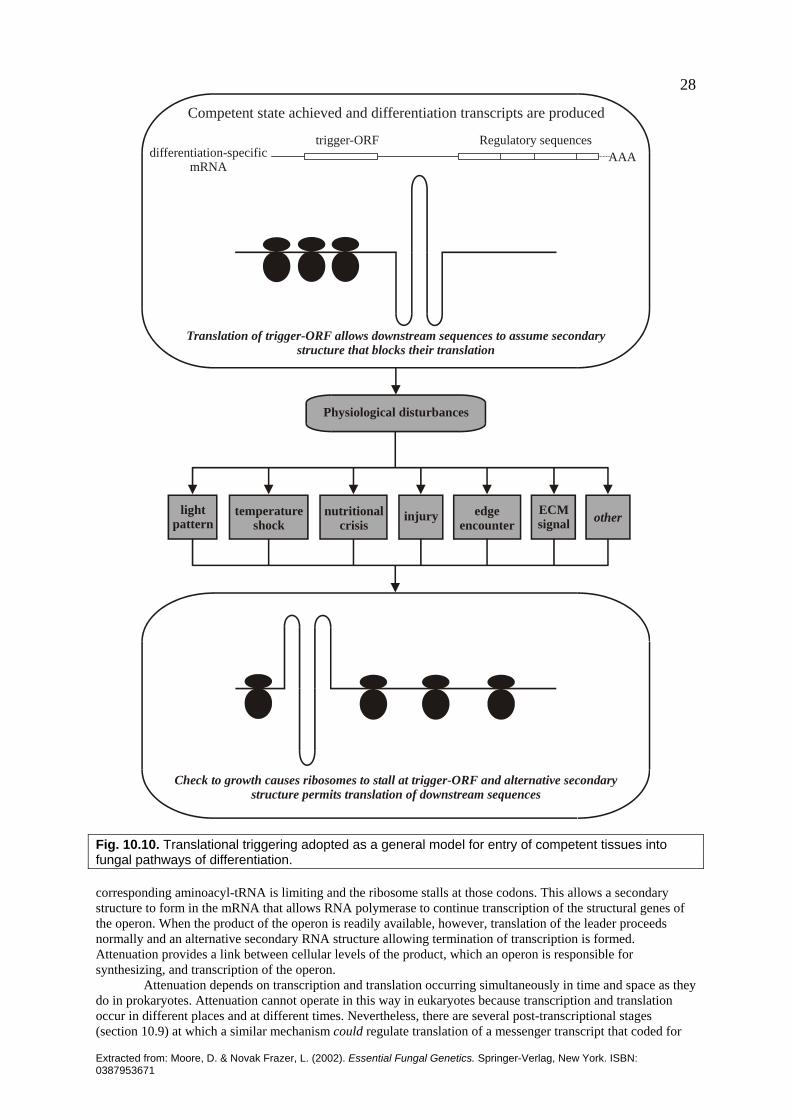
Extracted from: Moore, D. & Novak Frazer, L. (2002). Essential Fungal Genetics. Springer-Verlag, New York. ISBN: 0387953671
28
Regulatory sequencesdifferentiation-specific
mRNAAAA
trigger-ORF
Competent state achieved and differentiation transcripts are produced
Translation of trigger-ORF allows downstream sequences to assume secondarystructure that blocks their translation
Check to growth causes ribosomes to stall at trigger-ORF and alternative secondarystructure permits translation of downstream sequences
Physiological disturbances
lightpattern
temperatureshock
nutritionalcrisis
injury edgeencounter
ECMsignal other
Fig. 10.10. Translational triggering adopted as a general model for entry of competent tissues into fungal pathways of differentiation. corresponding aminoacyl-tRNA is limiting and the ribosome stalls at those codons. This allows a secondary structure to form in the mRNA that allows RNA polymerase to continue transcription of the structural genes of the operon. When the product of the operon is readily available, however, translation of the leader proceeds normally and an alternative secondary RNA structure allowing termination of transcription is formed. Attenuation provides a link between cellular levels of the product, which an operon is responsible for synthesizing, and transcription of the operon. Attenuation depends on transcription and translation occurring simultaneously in time and space as they do in prokaryotes. Attenuation cannot operate in this way in eukaryotes because transcription and translation occur in different places and at different times. Nevertheless, there are several post-transcriptional stages (section 10.9) at which a similar mechanism could regulate translation of a messenger transcript that coded for

Extracted from: Moore, D. & Novak Frazer, L. (2002). Essential Fungal Genetics. Springer-Verlag, New York. ISBN: 0387953671
29several reading frames. If the trigger-ORF contained adjacent attenuating codons for aminoacyl-tRNAs subject to variation in supply, stalling/non-stalling of translation of the trigger-ORF might determine whether the messenger transcript forms secondary structures which permit/do not permit translation of down-stream reading frames. Note that either one or both components of the aminoacyl-tRNA may be the limiting factor and the limitation may be imposed by compartmentalization. That is, amino acid or a specific tRNA (or, presumably, an aminoacyl-tRNA synthetase) may be compartmentalized, regulated in local concentration, or both. The interpretation offers a way by which a competent tissue can be released to undertake differentiation by a range of physiological events. Competence is interpreted to mean that messenger transcripts for the necessary regulators (and perhaps some key structural genes) are produced but not fully translated because an upstream sequence (trigger-ORF) prevents translation. There may be a number of different such transcripts with regulators corresponding to the different pathways upon which the competent cell can embark, their trigger-ORFs responding to separate physiological events (Fig. 10.10). On the other hand, there may be a number of similar transcripts in different cellular compartments so that the translational trigger can be released by the particular activities of those compartments with the result that one differentiation process may be triggered by different physiological events. It could also be that such a transcript is limited to one compartment, even one type of vesicle, perhaps, from which the trigger molecule can be excluded until some highly specific and/or localized physiological change occurs. Unfortunately, there is no direct evidence for any of these speculations, though a variety of physiological signals and stresses cause translation-level controls to direct competent fungal tissues to undertake specific differentiation processes. A comparison with the operation of mating type factors makes it reasonable to suggest that the translational trigger could immediately lead to translation of components of highly specific transcription activators and inhibitors, which then regulate gene sequences required for the differentiation that has been initiated. These, or their eventual products, may be involved in feedback fixation of the differentiation pathway. Feedback fixation is the outcome of feedback activation and autoregulation that together reinforce expression of the whole regulatory pathway to make it independent of the external environmental cues that initiated it. Feedback fixation results in developmental determination in the classic embryological sense. The epigenetic aspects of the network governing fungal morphogenesis start with feedback fixation, but also include signals from outside the cell (Fig. 10.11). The fungal extracellular matrix is extensive and complex. Its reaction to, and interaction with the environment can be communicated to the intracellular environment to modify cytoplasmic activity. Since neighboring cells are components of the external environment, it must be the case that the activity of one hyphal cell is modulated by changes made to the extracellular matrix by a neighboring hyphal cell. On this interpretation, therefore, continued progress in differentiation for most fungal cells requires continued reinforcement from their local microenvironment. This may involve production of location- and/or time-specific extracellular matrix molecules, or any of a range of smaller molecules, which might be classed as hormones or growth factors. Smaller molecules might exert their effects by being taken up into the cell. But uptake is not necessary. Any of these molecules may also affect relations between integrins and the existing extracellular matrix. As a result there could be direct effects on the cytoskeleton, which are able to cause immediate metabolic changes in one or more cellular compartments, or directly influence gene transcription. Connections to the extracellular matrix may also be involved in that other great enigma: the control of hyphal branching. By varying extracellular matrix/membrane or wall/membrane connections external signals may be able to specify branch initiation sites. Similarly, internal cytoskeletal architecture could also arrange specific membrane/wall connections to become branch initiation sites. Branch initiation sites specified in these ways may then become gathering sites for the molecules that create a new hyphal tip. The branch would consequently emerge in a position precisely defined by the stimulation of generalized cytoskeleton/membrane/wall connections by a positional stimulus. The focus of these hypothetical regulatory activities is, obviously, the hyphal wall, its surface and the immediate extracellular environment. These are features about which we are very ignorant and urgent and extensive research on these topics is necessary. The key to fungal morphogenesis lies in understanding how that which is outside a hypha can influence that which goes on inside the hypha in a time- and place-dependent manner. We are still a long way from reaching that understanding. Recent publications and websites worth a visit Banuett, F. (1998). Signalling in the yeasts: an informational cascade with links to the filamentous fungi.
Microbiology and Molecular Biology Reviews 62, 249-274. Belmont, A. S., Dietzel, S., Nye, A. C., Strukov, Y. G. & Tumbar, T. (1999). Large-scale chromatin structure
and function. Current Opinion in Cell Biology 11, 307-311. Biological Procedures Online, a library of step-by-step recipes at http://www.sciencemedcentral.com. Bölker, M. (1998). Sex and crime: heterotrimeric G proteins in fungal mating and pathogenesis. Fungal Genetics
and Biology 25, 143-156.

Extracted from: Moore, D. & Novak Frazer, L. (2002). Essential Fungal Genetics. Springer-Verlag, New York. ISBN: 0387953671
30Control of expression reinforced by external signals
Activates transcription
Activates transcription
Coactivator+ external signal
Regulatory sequencesdifferentiation-specific
mRNA AAAtrigger-ORF
differentiation-specificDNA
trigger-ORF
differentiatingmRNA
+ external signalCoactivator
AAA
Control of expression in committed cells is independentof external signals
Activator
Activator
Regulatory sequencesdifferentiation-specific
mRNA AAAtrigger-ORF
Activates transcription
Activates transcriptiondifferentiation-specific
DNA
trigger-ORF
differentiatingmRNA AAA
Epigenetic
and
other
extracellular
signals
execute subroutineanother
execute meiocyte subroutine
execute initiation subroutine
AND
execute fruit body subroutine
AND
execute subroutineanother
AND
AND
execute dikaryosis subroutine
Fig. 10.11. Feedback fixation adopted as a general model for maintaining progress through fungal pathways of differentiation. In this flow chart the box at the top shows the type of feedback fixation process envisaged to apply to most developmental subroutines in which epigenetic reinforcement from the local microenvironment is needed to interact with co-activators in order to maintain the feedback activation loop. In the bottom panel, the alternative of direct feedback fixation independently of other signals is shown as being applicable to cell types that show developmental commitment; only meiocytes are known to be committed, but there may be other committed cell types. Chiu, S. W. & Moore, D. (1990). A mechanism for gill pattern formation in Coprinus cinereus. Mycological
Research 94, 320-326. Cogoni, C. & Macino, G. (2000). Post-transcriptional gene silencing across kingdoms. Current Opinion in
Genetics & Development 10, 638-643.
Extracted from: Moore, D. & Novak Frazer, L. (2002). Essential Fungal Genetics. Springer-Verlag, New York. ISBN: 0387953671
31De Groot, P. W. J., Schaap, P. J., Sonnenberg, A. S. M., Visser, J. & Van Griensven, L. J. L. D. (1996). The
Agaricus bisporus hypA gene encodes a hydrophobin and specifically accumulates in peel tissue of mushroom caps during fruit body development. Journal of Molecular Biology 257, 1008-1018.
De Groot, P. W. J., Schaap, P. J., Van Griensven, L. J. L. D. & Visser, J. (1997). Isolation of developmentally regulated genes from the edible mushroom Agaricus bisporus. Microbiology 143, 1993-2001.
Developmental Biology Virtual Library is at http://sdb.bio.purdue.edu/other/vl_db.html. Developmental Biology Cinema is at http://sdb.bio.purdue.edu/dbcinema. Dreyfuss, G. & Struhl, K. (1999). Nucleus and gene expression. Multiprotein complexes, mechanistic
connections and nuclear organization. Current Opinion in Cell Biology 11, 303-306. Finnegan, E. J., Peacock, W. J. & Dennis, E. S. (2000). DNA methylation, a key regulator of plant development
and other processes. Current Opinion in Genetics & Development 10, 217-223. Gancedo, J. M. (2001). Control of pseudohyphae formation in Saccharomyces cerevisiae. FEMS Microbiology
Reviews 25, 107-123. Grosveld, F. (1999). Activation by locus control regions? Current Opinion in Genetics & Development 9, 152-
157. Habu, Y., Kakutani, T. & Paszkowski, J. (2001). Epigenetic developmental mechanisms in plants: molecules
and targets of plant epigenetic regulation. Current Opinion in Genetics & Development 11, 215–220. Hicke, L. (1999). Gettin’ down with ubiquitin: turning off cell-surface receptors, transporters and channels.
Trends in Cell Biology 9, 107-112. Horton, J. S., Palmer, G. E. & Smith, W. J. (1999). Regulation of dikaryon-expressed genes by FRT1 in the
basidiomycete Schizophyllum commune. Fungal Genetics and Biology 26, 33–47. Hsieh, C.-L. (2000). Dynamics of DNA methylation pattern. Current Opinion in Genetics & Development 10,
224-228. Idekar, T., Thorsson, V., Ranish, J. A., Christmas, R., Buhler, J., Eng, J. K., Bumgarner, R., Goodlett, D. R.
Aebersold, R. & Hood, L. (2001). Integrated genomic and proteomic analyses of a systematically perturbed metabolic network. Science 292, 929-934.
Johnston, M. (1999). Feasting, fasting and fermenting: glucose sensing in yeast and other cells. Trends in Genetics 15, 29-33.
Jones, P. A. (1999). The DNA methylation paradox. Trends in Genetics 15, 34-37. Kass, S. U., Pruss, D. & Wolffe, A. P. (1997). How does DNA methylation repress transcription? Trends in
Genetics 13, 444-449. Kornberg, R. D. (1999). Eukaryotic transcription control. Trends in Genetics 15, M46-M49. Kron, S. J. & Gow, N. A. R. (1995). Budding yeast morphogenesis: signalling, cytoskeleton and cell cycle.
Current Opinion in Cell Biology 7, 845-855. Kües, U. (2000). Life history and developmental processes in the basidiomycete Coprinus cinereus.
Microbiology and Molecular Biology Reviews 64, 316-353. Kües, U. & Liu, Y. (2000). Fruiting body production in basidiomycetes. Applied Microbiology & Biotechnology
54, 141-152. Legrain, P., Wojcik, J. & Gauthier, J.-M. (2001). Protein–protein interaction maps: a lead towards cellular
functions. Trends in Genetics 17, 346-352. Lipshitz, H. D. & Smibert, C. A. (2000). Mechanisms of RNA localization and translational regulation. Current
Opinion in Genetics & Development 10, 476-488. Madhani, H. D. & Fink, G. R. (1998). The control of filamentous differentiation and virulence in fungi. Trends
in Cell Biology 8, 348-353. Malik, S. & Roeder, R. G. (2000). Transcriptional regulation through Mediator-like coactivators in yeast and
metazoan cells. Trends in Biochemical Sciences 25, 277-283. Martienssen, R. A. & Richards, E. J. (1995). DNA methylation in eukaryotes. Current Opinion in Genetics &
Development 5, 234-242. Matzke, M. A., Matzke, A. J. M., Pruss, G. J. & Vance, V. B. (2001). RNA-based silencing strategies in plants.
Current Opinion in Genetics & Development 11, 221-227. Melcher, K. (1997). Galactose metabolism in Saccharomyces cerevisiae: a paradigm for eukaryotic gene
regulation. In: Yeast Sugar Metabolism (ed. F. K. Zimmermann & K. –D. Entian), pp. 235-269. Technomic Publishing Co. Inc.: Lancaster, Pennsylvania.
Miller, B. L. (1993). Brushing-up on bristles - complex genes and morphogenesis in molds. Trends in Genetics 9, 293-295.
Moore, D. (1998). Fungal Morphogenesis. Cambridge University Press: New York. Muraguchi, H. & Kamada, T. (1998). The ich1 gene of the mushroom Coprinus cinereus is essential for pileus
formation in fruiting. Development 125, 3133-3141. Murata, Y., Fujii, M., Zolan, M. E. & Kamada, T. (1998). Molecular analysis of pcc1, a gene that leads to A-
regulated sexual morphogenesis in Coprinus cinereus. Genetics 149, 1753-1761. Pérez-Martín, J. (1999). Chromatin and transcription in Saccharomyces cerevisiae. FEMS Microbiology Reviews

Extracted from: Moore, D. & Novak Frazer, L. (2002). Essential Fungal Genetics. Springer-Verlag, New York. ISBN: 0387953671
3223, 503-523.
Preiss, T. & Hentze, M. W. (1999). From factors to mechanisms: translation and translational control in eukaryotes. Current Opinion in Genetics & Development 9, 515-521.
Sánchez-Martínez, C. & Pérez-Martín, J. (2001). Dimorphism in fungal pathogens: Candida albicans and Ustilago maydis - similar inputs, different outputs. Current Opinion in Microbiology 4, 214-221.
Selker, E. U. (1997). Epigenetic phenomena in filamentous fungi: useful paradigms or repeat-induced confusion? Trends in Genetics 13, 296-301.
Timberlake, W. E. (1993). Translational triggering and feedback fixation in the control of fungal development. Plant Cell 5, 1453-1460.
Tucker, C. L., Gera, J. F. & Uetz, P. (2001). Towards an understanding of complex protein networks. Trends in Cell Biology 11, 102-106.
Van Griensven, L. J. L. D. (2000). Science and Cultivation of Edible Fungi, Mushroom Science XV, vols 1 & 2. Balkema: Rotterdam.
Vaucheret, H. & Fagard, M. (2001). Transcriptional gene silencing in plants: targets, inducers and regulators. Trends in Genetics 17, 29-35.
Wessels, J. G. H. (2000). Hydrophobins, unique fungal proteins. Mycologist 14, 153-159. Wu, C.-T. & Morris, J. R. (1999). Transvection and other homology effects. Current Opinion in Genetics &
Development 9, 237-246. Historical publications worth knowing about Buller, A. H. R. (1909). Researches on Fungi, vol. 1. Longman, Green & Co.: London. Buller, A. H. R. (1922). Researches on Fungi, vol. 2. Longman Green & Co: London. Buller, A. H. R. (1931). Researches on Fungi, vol. 4. Longmans, Green & Co.: London. Chang, S. T. & Hayes, W. A. (1978). Biology and Cultivation of Edible Mushrooms. Academic Press: New
York & London. Chiu, S. W., Moore, D. & Chang, S. T. (1989). Basidiome polymorphism in Volvariella bombycina.
Mycological Research 92, 69-77. Esser, K. & Meinhardt, F. (1977). A common genetic control of dikaryotic and monokaryotic fruiting in the
basidiomycete Agrocybe aegerita. Molecular & General Genetics 155, 113-115. Esser, K., Saleh, F. & Meinhardt, F. (1979). Genetics of fruit body production in higher basidiomycetes. II.
Monokaryotic and dikaryotic fruiting in Schizophyllum commune. Current Genetics 1, 85-88. Esser, K. & Straub, J. (1958) Genetische Untersuchungen an Sordaria macrospora Auersw., Kompensation und
induktion bei genbedingten Entwicklungsdefekten. Zeitschrift für Vererbungslehre 89, 729-746. Flegg, P. B., Spencer D. M. & Wood, D. A. (1985). The Biology and Technology of the Cultivated Mushroom.
John Wiley & Sons: New York. Hoge, J. H. C., Springer, J. & Wessels, J. G. H. (1982). Changes in complex RNA during fruit-body initiation in
the fungus Schizophyllum commune. Experimental Mycology 6, 233-243. Leslie, J. F. & Leonard, T. J. (1979). Three independent genetic systems that control initiation of a fungal
fruiting body. Molecular & General Genetics 171, 257-260. Moore, D. (1981). Developmental genetics of Coprinus cinereus: genetic evidence that carpophores and
sclerotia share a common pathway of initiation. Current Genetics 3, 145-150. Moore, D., Casselton, L. A., Wood, D. A. & Frankland, J. C. (1985) Developmental Biology of Higher Fungi.
Cambridge University Press: Cambridge, UK. Moore, D., Elhiti, M. M. Y. & Butler, R. D. (1979). Morphogenesis of the carpophore of Coprinus cinereus.
New Phytologist 83, 695-722. Mulder, G. H. & Wessels, J. G. H. (1986). Molecular cloning of RNAs differentially expressed in monokaryons
and dikaryons of Schizophyllum commune in relation to fruiting. Experimental Mycology 10, 214-227. Watling, R. (1971). Polymorphism in Psilocybe merdaria. New Phytologist 70, 307-326. Yashar, B. M. & Pukkila, P. J. (1985). Changes in polyadenylated RNA sequences associated with fruiting body
morphogenesis in Coprinus cinereus. Transactions of the British Mycological Society 84, 215-226. Ye, X. S., Wu, L. & Osmani, S. A. (2001). Signal transduction in filamentous fungi: regulation of protein
kinases. In Molecular and Cellular Biology of Filamentous Fungi (ed. N. J. Talbot), pp. 157-174. Oxford University Press: Oxford, UK.
Illustration credits Fig.10.4. based on Fig. 2 in Tucker, C. L., Gera, J. F. & Uetz, P. (2001). Towards an understanding of complex protein networks. Trends in Cell Biology 11, 102-106. Figs 10.5 to 10.8 based on Figs 5.11 to 5.16, and Figs 10.9 to 10.11 based on Figs 7.1 to 7.3 in Moore, D. (1998). Fungal Morphogenesis. Cambridge University Press: New York.