spontaneous adult-onset pulmonary arterial hypertension ... atvb 30... · spontaneous adult-onset...
TRANSCRIPT
ISSN: 1524-4636 Copyright © 2010 American Heart Association. All rights reserved. Print ISSN: 1079-5642. Online
7272 Greenville Avenue, Dallas, TX 72514Arteriosclerosis, Thrombosis, and Vascular Biology is published by the American Heart Association.
DOI: 10.1161/ATVBAHA.109.200121 2009;
2010;30;509-517; originally published online Dec 30,Arterioscler Thromb Vasc BiolBelik, Mansoor Husain, Mark Henkelman and Michelle Letarte
Brendan A.S. McIntyre, Adrienne Davis, Yu Jing Wang, Duncan J. Stewart, Jaques Mourad Toporsian, Mirjana Jerkic, Yu-Qing Zhou, Mohammed G. Kabir, Lisa X. Yu,
Hemorrhagic TelangiectasiaIncreased Endothelial Oxidative Stress in a Murine Model of Hereditary
Spontaneous Adult-Onset Pulmonary Arterial Hypertension Attributable to
http://atvb.ahajournals.org/cgi/content/full/ATVBAHA.109.200121/DC1Data Supplement (unedited) at:
http://atvb.ahajournals.org/cgi/content/full/30/3/509
located on the World Wide Web at: The online version of this article, along with updated information and services, is
http://www.lww.com/reprintsReprints: Information about reprints can be found online at
[email protected]. E-mail:
Fax:Kluwer Health, 351 West Camden Street, Baltimore, MD 21202-2436. Phone: 410-528-4050. Permissions: Permissions & Rights Desk, Lippincott Williams & Wilkins, a division of Wolters
http://atvb.ahajournals.org/subscriptions/Biology is online at Subscriptions: Information about subscribing to Arteriosclerosis, Thrombosis, and Vascular
at University of Toronto on March 16, 2010 atvb.ahajournals.orgDownloaded from
Spontaneous Adult-Onset Pulmonary Arterial HypertensionAttributable to Increased Endothelial Oxidative Stress in aMurine Model of Hereditary Hemorrhagic Telangiectasia
Mourad Toporsian, Mirjana Jerkic, Yu-Qing Zhou, Mohammed G. Kabir, Lisa X. Yu,Brendan A.S. McIntyre, Adrienne Davis, Yu Jing Wang, Duncan J. Stewart, Jaques Belik,
Mansoor Husain, Mark Henkelman, Michelle Letarte
Objective—Loss-of-function mutations in genes coding for transforming growth factor-�/bone morphogenetic proteinreceptors and changes in nitric oxide• (NO•) bioavailability are associated with hereditary hemorrhagic telangiectasiaand some forms of pulmonary arterial hypertension. How these abnormalities lead to seemingly disparate pulmonarypathologies remains unknown. Endoglin (Eng), a transforming growth factor-� coreceptor, is mutated in hereditaryhemorrhagic telangiectasia and involved in regulating endothelial NO• synthase (eNOS)-derived NO• production andoxidative stress. Because some patients with pulmonary arterial hypertension harbor ENG mutations leading to haploinsufficiency, we investigated the pulmonary vasculature of Eng�/� mice and the potential contribution of abnormaleNOS activation to pulmonary arterial hypertension.
Methods and Results—Hemodynamic, histological, and biochemical assessments and x-ray micro-CT imaging of adult Eng�/�
mice indicated signs of pulmonary arterial hypertension including increased right ventricular systolic pressure, degenerationof the distal pulmonary vasculature, and muscularization of small arteries. These findings were absent in 3-week-old Eng�/�
mice and were attributable to constitutively uncoupled eNOS activity in the pulmonary circulation, as evidenced by reducedeNOS/heat shock protein 90 association and increased eNOS-derived superoxide (•O2
�) production in a BH4-independentmanner. These changes render eNOS unresponsive to regulation by transforming growth factor-�/bone morphogenetic proteinand underlie the signs of pulmonary arterial hypertension that were prevented by Tempol.
Conclusion—Adult Eng�/� mice acquire signs of pulmonary arterial hypertension that are attributable to uncoupled eNOS activityand increased •O2
� production, which can be prevented by antioxidant treatment. Eng links transforming growth factor/bonemorphogenetic protein receptors to the eNOS activation complex, and its reduction in the pulmonary vasculature leads to increasedoxidative stress and pulmonary arterial hypertension. (Arterioscler Thromb Vasc Biol. 2010;30:509-517.)
Key Words: Alk-1 � endoglin � free radicals � nitric oxide � pulmonary arterial hypertension� transforming growth factor
Endoglin (Eng; CD105) is a an ancillary receptor for severaltransforming growth factor (TGF)-� superfamily ligands,
including bone morphogenetic proteins (BMP).1 It is predomi-nantly expressed on vascular endothelial cells2 and found in bothTGF-� and BMP receptor complexes,1,3 where it modulatesTGF-�1/�34 and BMP9/10 effects,5 respectively, via its physicalassociation with the activin-like kinase receptor-1 (ACVLR1)gene product, ALK1. Eng-null mice die at mid-gestation withimpaired angiogenesis and severe cardiac defects.6,7 WhereasEng�/� mice have a normal lifespan, they display abnormal
systemic vascular autoregulatory functions related to endothelialnitric oxide synthase (eNOS) activity.8
Mutations in the endoglin (ENG) and activin-like kinase 1receptor (ACVLR1) genes lead to haploinsufficiency and are theunderlying cause of hereditary hemorrhagic telangiectasia type 1(HHT1)9 and type 2 (HHT2),10 respectively. This disease ischaracterized by multiple focal telangiectases and arteriovenousmalformations (AVMs) in the pulmonary, hepatic, and cerebralmicrocirculations.11 These fragile structures are low-pressureconduits that can affect local tissue blood flow, and their
Received July 3, 2009; revision accepted December 8, 2009.From the Molecular Structure and Function Program (M.T., M.J., A.D., Y.J.W., M.L.), Mouse Imaging Centre (Y.-Q.Z., L.X.Y., M.H.), Department
of Pediatrics (B.A.S.M., J.B.), The Hospital for Sick Children, Toronto, Canada; Heart and Stroke Richard Lewar Center of Excellence (M.T., M.J.,M.G.K., M.H., M.L.), Department of Immunology (M.T., M.L.), Department of Medical Biophysics (M.H., M.L.), University of Toronto, Toronto,Canada; Division of Pulmonary, Critical Care and Sleep Medicine (M.T.), Department of Medicine, Beth Israel Deaconess Medical Center, HarvardMedical School, Boston, Mass. Ottawa Health Research Institute (D.J.S.), University of Ottawa, Canada; Division of Cellular & Molecular Biology(M.G.K., M.H.), Toronto General Hospital, Toronto, Canada.
M.T. and M.J. have equally contributed data to this work.Correspondence to Dr Mourad Toporsian, Beth Israel Deaconess Medical Center, Department of Medicine, Division of Pulmonary, Critical Care and
Sleep Medicine, Center for Vascular Biology Research, 99 Brookline Avenue, RN-233, Boston, MA 02115. E-mail [email protected]© 2010 American Heart Association, Inc.
Arterioscler Thromb Vasc Biol is available at http://atvb.ahajournals.org DOI: 10.1161/ATVBAHA.109.200121
509 at University of Toronto on March 16, 2010 atvb.ahajournals.orgDownloaded from

potential rupture in vital organs may lead to internal hemorrhage,anemia, and death. Patients with HHT1 and HHT2 displayphenotypically similar vascular lesions but diverge with respectto organ involvement with a higher prevalence of pulmonaryarteriovenous malformations (PAVMs) in HHT1.12
Mutations in ACVRL1 have also been reported in patientspresenting with pulmonary arterial hypertension (PAH),13–15
suggesting that HHT and PAH may share defects in relatedsignaling pathways. PAH is characterized by a sustained eleva-tion in mean pulmonary arterial pressure causing progressiveright ventricular hypertrophy and leading to heart failure anddeath.16 The increase in pulmonary arterial pressure is attribut-able to increased pulmonary vascular resistance (PVR) causedby progressive loss/pruning of the peripheral lung vasculatureand muscularization of small arterioles. Similar to HHT,8 pa-tients and animal models with pulmonary hypertension showendothelial dysfunction from changes in NO• bioavailabilityattributable to increased oxidative stress in the pulmonaryvasculature.16–18 eNOS activity is reduced in hypoxia-inducedpulmonary hypertension as a result of impaired association ofeNOS with its allosteric regulator, heat shock protein 90(Hsp90).19 Such changes in eNOS activation were shown to leadto increased eNOS-derived reactive oxygen species (ROS)generation instead of NO• in endothelial cells,20 and the result-ing oxidant stress contributes to an animal model of persistentpulmonary hypertension of the newborn.21,22 Whereas loss-of-function mutations in TGF-�/BMP receptors have been associ-ated with familial forms of PAH, the contributions of abnormaleNOS biology and endothelial oxidative stress in these forms ofPAH remain to be elucidated.
We report that adult Eng�/� mice spontaneously developsigns of PAH that are attributable to uncoupled eNOS activityand ROS production causing progressive loss in pulmonaryvascularity and increased muscularization of arterioles. Englinks TGF-�/BMP receptors to the eNOS activation complex,where its reduction leads to constitutive endothelial eNOS-derived oxidative stress, rendering the enzyme unresponsive toregulation by TGF-�/BMP signaling. These changes underliePAH in Eng�/� mice, which can be prevented by treatment withTempol.
Materials and MethodsAnimal StudiesAll experimental protocols were performed in accordance with theCanadian Council on Animal Care and approved by the Animal CareCommittee of the Hospital for Sick Children. N17-N19 Eng�/� andEng�/� C57BL/6 mice were generated by Dr Letarte at the Hospitalfor Sick Children by successive backcrosses. Mice ranging from 3 to18 weeks old were used; 8- to 12-week-old mice were exposed tohypoxia (12% O2) for 3 weeks in a Plexiglas chamber or to room air(21% O2). In separate experiments, 3-week-old Eng�/� and Eng�/�
mice were exposed to 1 mmol/L Tempol (4-hydroxy-2,2,6,6-tetramethypiperidine-1-oxyl; Fluka) in the drinking water for 6weeks. Mice were anesthetized with 1.5% isoflurane for hemody-namic measurements or euthanized for lung histology, x-ray micro-CT, and biochemical studies.
Cardiac MeasurementsPeak right ventricular systolic pressure (RVSP) was measured in miceby Millar Mikro-tip pressure transducer catheterization of the rightventricle (RV) via the external jugular vein (AD Instruments). The RV
was dissected from the left ventricle and septum, and the Fulton index(RV/left ventricle�septum weight ratio) was calculated.
Ultrasound BiomicroscopyUsing a Vevo 770 ultrasound biomicroscope (VisualSonics), observa-tions of the mouse heart and great vessels were conducted as de-scribed.23 Dynamic changes in chamber/lumen dimensions were rec-orded using M-mode. RV stroke volume was calculated by multiplyingthe velocity–time integral by the main pulmonary artery cross-sectionalarea at peak systole. The isovolumetric contraction, relaxation, andejection times were measured to calculate the Tei index�(isovolumetriccontraction � isovolumetric relaxation IVRT)/ejection time.
Lung X-Ray Micro-CTAnesthetized mice were intubated by tracheotomy and breathing wasmonitored using a pressure-controlled ventilator. Mice were perfusedat 20 mm Hg via the RV with warm heparinized phosphate-bufferedsaline, followed by Microfil (Flow Tech) at 40 mm Hg using apressure Servo System PS/200 (Living Systems Instrumentation).For 3-week-old mice, lungs were perfused at 10 mm Hg and with3-fold diluted Microfil at 25 mm Hg. Specimens were scanned at29 �m using a Micro-CT scanner (GE Healthcare). Three-dimensional volume data were reconstructed using the Feldkampalgorithm for cone-beam CT geometry. Three-dimensional iso-surface rendering of the pulmonary vasculature was accomplishedusing MicroView software (GE Healthcare).
Morphometric Analysis of Small Arteries in LungsParaffin-embedded transverse lung sections of 8- to 12-week-old and18-week-old mice were stained with Movat’s pentachrome, and 5independent fields were quantified for the number of small arteriesper 100 alveoli. Average wall areas of arteries were determined fromthe difference between outer and inner wall areas for 20 vessels persection and averaged for each mouse.
Lung ROS MeasurementsLungs were homogenized in phosphate-buffered Krebs containing1 mmol/L NaN3, and ROS levels were assessed using 10 �mol/L5-(and6-)-caboxy-2-,7-dichloro dihydrofluorescein diacetate (carboxy-H2DCFDA; Molecular Probes) at 37°C. Fluorescence was quantified ona SpectraMax spectrofluorometer (Molecular Devices) using 488 nmexcitation and 525 nm emission wavelengths and was normalized forprotein content.
Lung NO• MeasurementsExcised lung tissue was incubated in Krebs-Henseleit buffer for 2hours at 37°C in the presence and absence of L-NAME. NO•
production was quantified using an NO• microsensor ISO-NOP700attached to an Apollo 4000 Free Radical Analyzer. The microsensorwas calibrated using S-Nitroso-N-acetylpenicillamine in the pres-ence of copper sulfate.
eNOS/Hsp90 Association and ImmunoblottingLung segments were stimulated with vehicle or 1 �mol/L ionomycinfor 15 minutes. Mouse Eng�/� and Eng�/� endothelial cells derivedfrom the yolk sac of E8.5 embryos8,24 were stimulated with increas-ing concentrations of TGF-�1. Extracts were prepared in 10 mmol/LTris-HCl (pH�7.4; 1% Triton X-100 with protease/phosphataseinhibitors) and precleared with protein A/G mixture. eNOS ho-modimer formation in lung extracts were performed by low-temperature SDS-PAGE. Proteins were immunoprecipitated or im-munoblotted with antibodies to phospho-eNOS Thr495, eNOS,Hsp90, b-actin (BD Biosciences), and Eng (MJ7/18, SouthernBiotech) or Alk1 (Santa Cruz Biotech). Bands were visualized bychemiluminescence and quantified.
NOS-Derived O2� Measurements
eNOS-derived ROS levels were assessed by dihydroethidium(DHE; Molecular Probes) staining in isolated main pulmonary
510 Arterioscler Thromb Vasc Biol March 2010
at University of Toronto on March 16, 2010 atvb.ahajournals.orgDownloaded from
arteries. After preincubation with and without L-NAME (10�3 mol/L),vessels were incubated with acetylcholine (ACh, 10�5 mol/L) and2 �mol/L DHE at 37°C. Vessels were washed, mounted en face on glassslides exposing the endothelial layer, and analyzed by fluorescencemicrocopy.
Eng�/� and Eng�/� embryonic endothelial cells were grown inOptilux 96-well black clear-bottom plates and serum-starved for 3hours. Cells were incubated with 5 �mol/L DHE in the presence andabsence of L-NAME and stimulated with 1 �mol/L ionomycin. Livecells were observed through the CY3 fluorescence channel andphotographed using a TE2000 inverted microscope (Nikon)equipped with an environmental chamber set to 37°C and 5% CO2.The number of positive nuclei/field was quantified based on size andintensity using 3-dimensional imaging software (Volocity).
Lung BH4 and BH2 MeasurementsLungs were homogenized in 50 mmol/L phosphate buffer (pH 2.6)containing 0.1 mmol/L DTPA and 1 mmol/L DTE. Samples wereloaded onto a high-performance liquid chromatography system (Cou-lArray system, Model 582 and 542; ESA Biosciences) with a SynergiPolar-RP column (4 �; 250�4.6 mm; Phenomenex) and eluted withargon-saturated 50 mmol/L phosphate buffer (pH 2.6). Calibrationcurves were made by summing up the peak areas collected at 0 and 150mV for BH4 and 280 and 365 mV for BH2. Intracellular concentrationswere calculated using authentic BH4 and BH2 (10 to 100 nmol/L) asstandards and normalized to protein content.
eNOS ActivityCultured Eng�/� and Eng�/� endothelial cells were preincubatedwith and without L-NAME and NOS activity was assayed bymonitoring the conversion of 3H-L-arginine to 3H-L-citrulline inresponse to TGF-�1 (250 pmol/L), as previously described.8
Statistical AnalysisComparisons were performed by 1- or 2-way ANOVA, and signif-icant overall differences were evaluated posthoc using the Bonfer-roni procedure. Results are expressed as the mean�SEM, withP�0.05 representing significance.
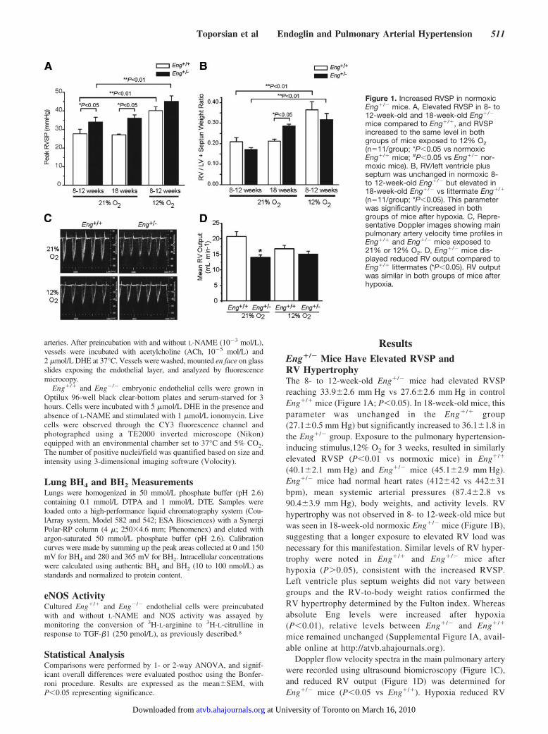
ResultsEng�/� Mice Have Elevated RVSP andRV HypertrophyThe 8- to 12-week-old Eng�/� mice had elevated RVSPreaching 33.9�2.6 mm Hg vs 27.6�2.6 mm Hg in controlEng�/� mice (Figure 1A; P�0.05). In 18-week-old mice, thisparameter was unchanged in the Eng�/� group(27.1�0.5 mm Hg) but significantly increased to 36.1�1.8 inthe Eng�/� group. Exposure to the pulmonary hypertension-inducing stimulus,12% O2 for 3 weeks, resulted in similarlyelevated RVSP (P�0.01 vs normoxic mice) in Eng�/�
(40.1�2.1 mm Hg) and Eng�/� mice (45.1�2.9 mm Hg).Eng�/� mice had normal heart rates (412�42 vs 442�31bpm), mean systemic arterial pressures (87.4�2.8 vs90.4�3.9 mm Hg), body weights, and activity levels. RVhypertrophy was not observed in 8- to 12-week-old mice butwas seen in 18-week-old normoxic Eng�/� mice (Figure 1B),suggesting that a longer exposure to elevated RV load wasnecessary for this manifestation. Similar levels of RV hyper-trophy were noted in Eng�/� and Eng�/� mice afterhypoxia (P�0.05), consistent with the increased RVSP.Left ventricle plus septum weights did not vary betweengroups and the RV-to-body weight ratios confirmed theRV hypertrophy determined by the Fulton index. Whereasabsolute Eng levels were increased after hypoxia(P�0.01), relative levels between Eng�/� and Eng�/�
mice remained unchanged (Supplemental Figure IA, avail-able online at http://atvb.ahajournals.org).
Doppler flow velocity spectra in the main pulmonary arterywere recorded using ultrasound biomicroscopy (Figure 1C),and reduced RV output (Figure 1D) was determined forEng�/� mice (P�0.05 vs Eng�/�). Hypoxia reduced RV
Figure 1. Increased RVSP in normoxicEng�/� mice. A, Elevated RVSP in 8- to12-week-old and 18-week-old Eng�/�
mice compared to Eng�/�, and RVSPincreased to the same level in bothgroups of mice exposed to 12% O2(n�11/group; *P�0.05 vs normoxicEng�/� mice; #P�0.05 vs Eng�/� nor-moxic mice). B, RV/left ventricle plusseptum was unchanged in normoxic 8-to 12-week-old Eng�/� but elevated in18-week-old Eng�/� vs littermate Eng�/�
(n�11/group; *P�0.05). This parameterwas significantly increased in bothgroups of mice after hypoxia. C, Repre-sentative Doppler images showing mainpulmonary artery velocity time profiles inEng�/� and Eng�/� mice exposed to21% or 12% O2. D, Eng�/� mice dis-played reduced RV output compared toEng�/� littermates (*P�0.05). RV outputwas similar in both groups of mice afterhypoxia.
Toporsian et al Endoglin and Pulmonary Arterial Hypertension 511
at University of Toronto on March 16, 2010 atvb.ahajournals.orgDownloaded from
output in Eng�/� mice but did not further affect this param-eter in Eng�/� mice. The calculated RV Tei index and RVfractional shortening were unchanged in normoxic Eng�/� mice,suggesting normal RV function (Supplemental Figure IIA, IIB,available online at http://atvb.ahajournals.org). Moreover, leftventricle function was normal in Eng�/� mice, as evidenced byunchanged percent fractional shortening (27.9�1.3 vs25.9�1.9), isovolumetric contraction time-to-ejection time ratio(0.22�0.01 vs 0.22�0.02), and isovolumetric relaxation time-to-filling time ratio (0.28�0.01 vs 0.27�0.01).
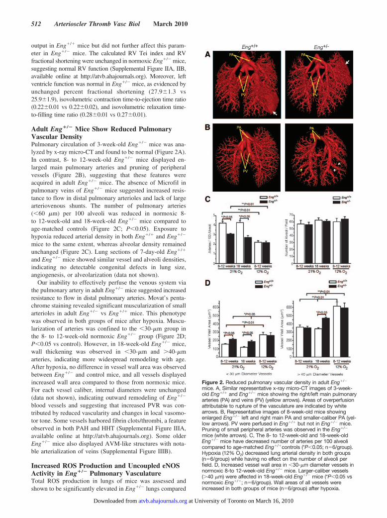
Adult Eng�/� Mice Show Reduced PulmonaryVascular DensityPulmonary circulation of 3-week-old Eng�/� mice was ana-lyzed by x-ray micro-CT and found to be normal (Figure 2A).In contrast, 8- to 12-week-old Eng�/� mice displayed en-larged main pulmonary arteries and pruning of peripheralvessels (Figure 2B), suggesting that these features wereacquired in adult Eng�/� mice. The absence of Microfil inpulmonary veins of Eng�/� mice suggested increased resis-tance to flow in distal pulmonary arterioles and lack of largearteriovenous shunts. The number of pulmonary arteries(�60 �m) per 100 alveoli was reduced in normoxic 8-to 12-week-old and 18-week-old Eng�/� mice compared toage-matched controls (Figure 2C; P�0.05). Exposure tohypoxia reduced arterial density in both Eng�/� and Eng�/�
mice to the same extent, whereas alveolar density remainedunchanged (Figure 2C). Lung sections of 7-day-old Eng�/�
and Eng�/� mice showed similar vessel and alveoli densities,indicating no detectable congenital defects in lung size,angiogenesis, or alveolarization (data not shown).
Our inability to effectively perfuse the venous system viathe pulmonary artery in adult Eng�/� mice suggested increasedresistance to flow in distal pulmonary arteries. Movat’s penta-chrome staining revealed significant muscularization of smallarterioles in adult Eng�/� vs Eng�/� mice. This phenotypewas observed in both groups of mice after hypoxia. Muscu-larization of arteries was confined to the �30-�m group inthe 8- to 12-week-old normoxic Eng�/� group (Figure 2D;P�0.05 vs control). However, in 18-week-old Eng�/� mice,wall thickening was observed in �30-�m and �40-�marteries, indicating more widespread remodeling with age.After hypoxia, no difference in vessel wall area was observedbetween Eng�/� and control mice, and all vessels displayedincreased wall area compared to those from normoxic mice.For each vessel caliber, internal diameters were unchanged(data not shown), indicating outward remodeling of Eng�/�
blood vessels and suggesting that increased PVR was con-tributed by reduced vascularity and changes in local vasomo-tor tone. Some vessels harbored fibrin clots/thrombi, a featureobserved in both PAH and HHT (Supplemental Figure IIIA,available online at http://atvb.ahajournals.org). Some olderEng�/� mice also displayed AVM-like structures with nota-ble arterialization of veins (Supplemental Figure IIIB).
Increased ROS Production and Uncoupled eNOSActivity in Eng�/� Pulmonary VasculatureTotal ROS production in lungs of mice was assessed andshown to be significantly elevated in Eng�/� lungs compared
Figure 2. Reduced pulmonary vascular density in adult Eng�/�
mice. A, Similar representative x-ray micro-CT images of 3-week-old Eng�/� and Eng�/� mice showing the right/left main pulmonaryarteries (PA) and veins (PV) (yellow arrows). Areas of overperfusionattributable to rupture of the vasculature are indicated by whitearrows. B, Representative images of 8-week-old mice showingenlarged Eng�/� left and right main PA and smaller-caliber PA (yel-low arrows). PV were perfused in Eng�/� but not in Eng�/� mice.Pruning of small peripheral arteries was observed in the Eng�/�
mice (white arrows). C, The 8- to 12-week-old and 18-week-oldEng�/� mice have decreased number of arteries per 100 alveolicompared to age-matched Eng�/�controls (*P�0.05; n�6/group).Hypoxia (12% O2) decreased lung arterial density in both groups(n�6/group) while having no effect on the number of alveoli perfield. D, Increased vessel wall area in �30-�m diameter vessels innormoxic 8-to 12-week-old Eng�/� mice. Larger-caliber vessels(�40 �m) were affected in 18-week-old Eng�/� mice (*P�0.05 vsnormoxic Eng�/�; n�6/group). Wall areas of all vessels wereincreased in both groups of mice (n�6/group) after hypoxia.
512 Arterioscler Thromb Vasc Biol March 2010
at University of Toronto on March 16, 2010 atvb.ahajournals.orgDownloaded from
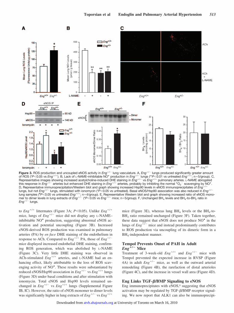
to Eng�/� littermates (Figure 3A; P�0.05). Unlike Eng�/�
mice, lungs of Eng�/� mice did not display any L-NAME–inhibitable NO• production, suggesting abnormal eNOS ac-tivation and potential uncoupling (Figure 3B). IncreasedeNOS-derived ROS production was examined in pulmonaryarteries (PA) by en face DHE staining of the endothelium inresponse to ACh. Compared to Eng�/� PA, those of Eng�/�
mice displayed increased endothelial DHE staining, confirm-ing ROS generation, which was abolished by L-NAME(Figure 3C). Very little DHE staining was observed inACh-stimulated Eng�/� arteries, and L-NAME had an en-hancing effect, likely attributable to the loss of ROS scav-enging activity of NO•. These results were substantiated byreduced eNOS/Hsp90 association in Eng�/� vs Eng�/� lungs(Figure 3D) under basal conditions and after stimulation withionomycin. Total eNOS and Hsp90 levels remained un-changed in Eng�/� vs Eng�/� lungs (Supplemental FigureIB, IC). However, the ratio of eNOS monomer to dimer levelswas significantly higher in lung extracts of Eng�/� vs Eng�/�
mice (Figure 3E), whereas lung BH4 levels or the BH4-to-BH2 ratio remained unchanged (Figure 3F). Taken together,these data suggest that eNOS does not produce NO• in thelungs of Eng�/� mice and instead predominantly contributesto ROS production via uncoupling of its dimeric form in aBH4-independent manner.
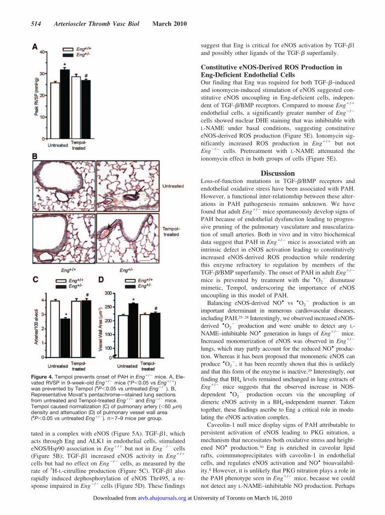
Tempol Prevents Onset of PAH in AdultEng�/� MiceTreatment of 3-week-old Eng�/� and Eng�/� mice withTempol prevented the expected increase in RVSP (Figure4A) in adult Eng�/� mice, as well as the outward arterialremodeling (Figure 4B), the rarefaction of distal arterioles(Figure 4C), and the increase in vessel wall area (Figure 4D).
Eng Links TGF-�/BMP Signaling to eNOSEng immunoprecipitates with eNOS,8 suggesting that eNOSactivation may be regulated by TGF-�/BMP receptor signal-ing. We now report that ALK1 can also be immunoprecipi-
Figure 3. ROS production and uncoupled eNOS activity in Eng�/� lung vasculature. A, Eng�/� lungs produced significantly greater amountof ROS (*P�0.05 vs Eng�/�). B, Lack of L-NAME–inhibitable NO• production in Eng�/� lungs (**P�0.01 vs untreated Eng�/�; n�5/group). C,Representative images showing increased acetylcholine-induced DHE staining in Eng�/� vs Eng�/� pulmonary arteries. L-NAME abrogatedthis response in Eng�/� arteries but enhanced DHE staining in Eng�/� arteries, probably by inhibiting the normal •O2
� scavenging by NO•.D, Representative immunoprecipitation/Western blot and graph showing increased Hsp90 levels in eNOS immunoprecipitates of Eng�/�
lungs, but not Eng�/� lungs, stimulated with ionomycin (*P�0.05 vs untreated). Basal eNOS/Hsp90 association was also reduced in Eng�/�
lung samples (#P�0.05 vs untreated Eng�/�; n�4/group). E, Representative Western blot and graph showing increased ratio of eNOS mono-mer to dimer levels in lung extracts of Eng�/� (*P�0.05 vs Eng�/� mice; n�5/group). F, Unchanged BH4 levels and BH4-to-BH2 ratio inEng�/� lungs.
Toporsian et al Endoglin and Pulmonary Arterial Hypertension 513
at University of Toronto on March 16, 2010 atvb.ahajournals.orgDownloaded from
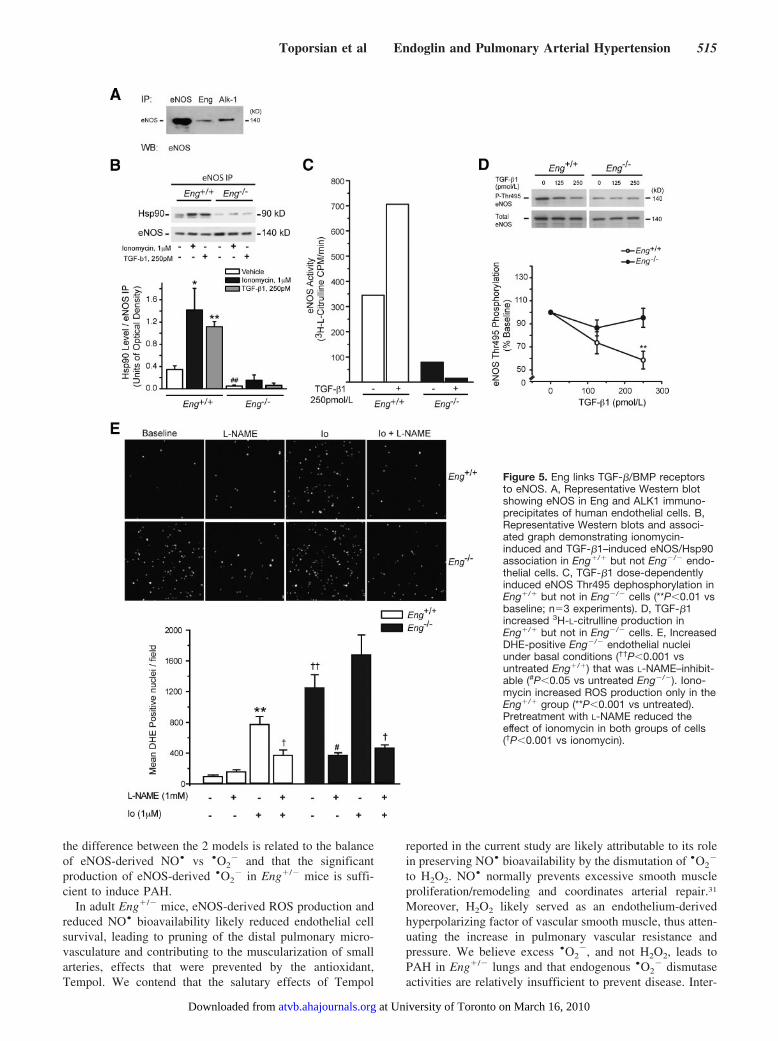
tated in a complex with eNOS (Figure 5A). TGF-�1, whichacts through Eng and ALK1 in endothelial cells, stimulatedeNOS/Hsp90 association in Eng�/� but not in Eng�/� cells(Figure 5B); TGF-�1 increased eNOS activity in Eng�/�
cells but had no effect on Eng�/� cells, as measured by therate of 3H-L-citrulline production (Figure 5C). TGF-�1 alsorapidly induced dephosphorylation of eNOS Thr495, a re-sponse impaired in Eng�/� cells (Figure 5D). These findings
suggest that Eng is critical for eNOS activation by TGF-�1and possibly other ligands of the TGF-� superfamily.
Constitutive eNOS-Derived ROS Production inEng-Deficient Endothelial CellsOur finding that Eng was required for both TGF-�–inducedand ionomycin-induced stimulation of eNOS suggested con-stitutive eNOS uncoupling in Eng-deficient cells, indepen-dent of TGF-�/BMP receptors. Compared to mouse Eng�/�
endothelial cells, a significantly greater number of Eng�/�
cells showed nuclear DHE staining that was inhibitable withL-NAME under basal conditions, suggesting constitutiveeNOS-derived ROS production (Figure 5E). Ionomycin sig-nificantly increased ROS production in Eng�/� but notEng�/� cells. Pretreatment with L-NAME attenuated theionomycin effect in both groups of cells (Figure 5E).
DiscussionLoss-of-function mutations in TGF-�/BMP receptors andendothelial oxidative stress have been associated with PAH.However, a functional inter-relationship between these alter-ations in PAH pathogenesis remains unknown. We havefound that adult Eng�/� mice spontaneously develop signs ofPAH because of endothelial dysfunction leading to progres-sive pruning of the pulmonary vasculature and musculariza-tion of small arteries. Both in vivo and in vitro biochemicaldata suggest that PAH in Eng�/� mice is associated with anintrinsic defect in eNOS activation leading to constitutivelyincreased eNOS-derived ROS production while renderingthis enzyme refractory to regulation by members of theTGF-�/BMP superfamily. The onset of PAH in adult Eng�/�
mice is prevented by treatment with the •O2� dismutase
mimetic, Tempol, underscoring the importance of eNOSuncoupling in this model of PAH.
Balancing eNOS-derived NO• vs •O2� production is an
important determinant in numerous cardiovascular diseases,including PAH.25–28 Interestingly, we observed increased eNOS-derived •O2
� production and were unable to detect any L-NAME–inhibitable NO• generation in lungs of Eng�/� mice.Increased monomerization of eNOS was observed in Eng�/�
lungs, which may partly account for the reduced NO• produc-tion. Whereas it has been proposed that monomeric eNOS canproduce •O2
�, it has been recently shown that this is unlikelyand that this form of the enzyme is inactive.29 Interestingly, ourfinding that BH4 levels remained unchanged in lung extracts ofEng�/� mice suggests that the observed increase in NOS-dependent •O2
� production occurs via the uncoupling ofdimeric eNOS activity in a BH4-independent manner. Takentogether, these findings ascribe to Eng a critical role in modu-lating the eNOS activation complex.
Caveolin-1 null mice display signs of PAH attributable topersistent activation of eNOS leading to PKG nitration, amechanism that necessitates both oxidative stress and height-ened NO• production.30 Eng is enriched in caveolar lipidrafts, coimmunoprecipitates with caveolin-1 in endothelialcells, and regulates eNOS activation and NO• bioavailabil-ity.8 However, it is unlikely that PKG nitration plays a role inthe PAH phenotype seen in Eng�/� mice, because we couldnot detect any L-NAME–inhibitable NO production. Perhaps
Figure 4. Tempol prevents onset of PAH in Eng�/� mice. A, Ele-vated RVSP in 9-week-old Eng�/� mice (*P�0.05 vs Eng�/�)was prevented by Tempol (#P�0.05 vs untreated Eng�/�). B,Representative Movat’s pentachrome—stained lung sectionsfrom untreated and Tempol-treated Eng�/� and Eng�/� mice.Tempol caused normalization (C) of pulmonary artery (�60 �m)density and attenuation (D) of pulmonary vessel wall area(#P�0.05 vs untreated Eng�/�). n�7–9 mice per group.
514 Arterioscler Thromb Vasc Biol March 2010
at University of Toronto on March 16, 2010 atvb.ahajournals.orgDownloaded from
the difference between the 2 models is related to the balanceof eNOS-derived NO• vs •O2
� and that the significantproduction of eNOS-derived •O2
� in Eng�/� mice is suffi-cient to induce PAH.
In adult Eng�/� mice, eNOS-derived ROS production andreduced NO• bioavailability likely reduced endothelial cellsurvival, leading to pruning of the distal pulmonary micro-vasculature and contributing to the muscularization of smallarteries, effects that were prevented by the antioxidant,Tempol. We contend that the salutary effects of Tempol
reported in the current study are likely attributable to its rolein preserving NO• bioavailability by the dismutation of •O2
�
to H2O2. NO• normally prevents excessive smooth muscleproliferation/remodeling and coordinates arterial repair.31
Moreover, H2O2 likely served as an endothelium-derivedhyperpolarizing factor of vascular smooth muscle, thus atten-uating the increase in pulmonary vascular resistance andpressure. We believe excess •O2
�, and not H2O2, leads toPAH in Eng�/� lungs and that endogenous •O2
� dismutaseactivities are relatively insufficient to prevent disease. Inter-
Figure 5. Eng links TGF-�/BMP receptorsto eNOS. A, Representative Western blotshowing eNOS in Eng and ALK1 immuno-precipitates of human endothelial cells. B,Representative Western blots and associ-ated graph demonstrating ionomycin-induced and TGF-�1–induced eNOS/Hsp90association in Eng�/� but not Eng�/� endo-thelial cells. C, TGF-�1 dose-dependentlyinduced eNOS Thr495 dephosphorylation inEng�/� but not in Eng�/� cells (**P�0.01 vsbaseline; n�3 experiments). D, TGF-�1increased 3H-L-citrulline production inEng�/� but not in Eng�/� cells. E, IncreasedDHE-positive Eng�/� endothelial nucleiunder basal conditions (††P�0.001 vsuntreated Eng�/�) that was L-NAME–inhibit-able (#P�0.05 vs untreated Eng�/�). Iono-mycin increased ROS production only in theEng�/� group (**P�0.001 vs untreated).Pretreatment with L-NAME reduced theeffect of ionomycin in both groups of cells(†P�0.001 vs ionomycin).
Toporsian et al Endoglin and Pulmonary Arterial Hypertension 515
at University of Toronto on March 16, 2010 atvb.ahajournals.orgDownloaded from
estingly, PAH is acquired in adult Eng�/� mice, suggestingan increased predisposition of these mice to age-relateduncoupling of eNOS, a developmentally regulated phenom-enon recently reported in piglets.32
Our finding that eNOS co-immunoprecipitates with bothEng and ALK1 further supports a central role for Eng inlinking TGF-�/BMP receptors to eNOS. TGF-�1 causedincreased eNOS/Hsp90 association, eNOS Thr495 dephos-phorylation, and 3H-L-arginine to 3H-L-citrulline conversionin Eng�/�, but not in Eng�/� endothelial cells. However,failure of the receptor-independent eNOS agonist, ionomycin,and of TGF-�1 to induce eNOS/Hsp90 association in Eng-deficient cells suggests an intrinsic defect in eNOS activation.This is supported by our findings of significantly elevatedeNOS-derived ROS production in unstimulated Eng�/� cells,indicating constitutive uncoupling and thus rendering eNOSrefractory to regulation by TGF-�1/BMP and other ligands inthis superfamily.
Our study supports clinical observations in which HHTpatients manifest PAVMs and varying degrees of pulmonaryhypertension,15,33,34 suggesting a common defective pathwayinvolving Eng/ALK1 that predisposes to lesions associatedwith these diseases. We have found abnormal vascularstructures suggestive of PAVMs in Eng�/� mice that alsodisplay PAH (Supplemental Figure III). We have previouslyreported that Eng�/� systemic arteries have a reduced abilityto contract in response to elevations in transmural pressure,thus failing to effectively reduce vessel wall tension. Elevatedparietal wall tension attributable to increased pulmonaryperfusion pressure would increase the probability of PAVMsand their subsequent risk of rupture. Perhaps the observedenlargement of large feeding pulmonary arteries in Eng�/�
may be a prelude to abnormal vascular structures reminiscentof HHT. We propose that small, low-resistance AVM-likestructures may arise from elevated pulmonary vascular pres-sure and account for the relatively mild increase in RVSP andPAH progression in adult Eng�/� mice. Manifestations ofPAH or HHT may be influenced by genetic and environmen-tal modifying factors that specifically affect the integrity ofthe Eng�/� pulmonary vasculature and its ability for normalremodeling/repair undergoing elevated intravascular pres-sure. Interestingly, shRNA-mediated silencing of BMPreceptor-2 in mice resulted in signs of HHT instead of theexpected PAH.35 More recently, conditional ablation of BMPreceptor-2 in the pulmonary endothelium was sufficient toelicit signs of PAH only in a subset of mice.36 It remains tobe determined if the appearance of small PAVM-like struc-tures may have counteracted the increase in RVSP andprogression of PAH in the seemingly asymptomatic group ofBMP receptor-2–deficient mice.
PAVMs are more prevalent in HHT1 than in HHT2 patients(48% vs 5%).12 These low-resistance structures could alleviateoverall PVR, providing a partial explanation for the higherprevalence of PAH in patients with ACVRL1 vs ENG muta-tions.15 Whereas a current study suggests that embolization ofPAVMs does not lead to a sustained elevation in pulmonaryarterial pressure in HHT patients,34 this does not necessarilypreclude an increased PVR because embolization can also beassociated with reduced cardiac output. Patients with ENG
mutations presenting with PAH typically have lower PVR thanthose with ACVRL1 mutations.4 Moreover, in some of thesecases, the increased pulmonary arterial pressure is eventuallynormalized with the appearance of PAVMs.13,37 Our findingssuggest that ENG mutations leading to haploinsufficiency are apredisposing factor to PAH, and that PAVMs in HHT1 mayresult from abnormal vascular remodeling under high localpressure conditions that may, in turn, serve to alleviate PVR.
Our study ascribes to Eng a critical role in the maintenanceof the mature lung vasculature. In adult Eng�/� mice,alterations in eNOS activation leading to uncoupling rendersthe enzyme refractory to regulation by TGF-�/BMP signalingand constitutes a critical event leading to excessive oxidativestress and PAH pathogenesis. The absence of disease inyounger mice may be related to the prolonged time courserequired for PAH manifestations, or mechanisms surroundingeNOS uncoupling are developmentally regulated. Our studysuggests a close association between PAH and HHT1, andour experimental system may provide a means to define thespecific determinants in the pathogenesis of both diseases.Future genetic screening of families with PAH may unravelmore individuals with ENG mutations.
AcknowledgmentsThe authors thank Jennifer Whitsett (Medical College of Wisconsin)for the BH4/BH2 measurements by high-performance liquid chroma-tography, and Aditi M. Jhaveri from the Beth Israel DeaconessMedical Center for technical support.
Sources of FundingThis work was supported by grants from the CIHR (MOP-6247),Heart & Stroke Foundation of Ontario (T5598), and the HHTInternational Foundation (contract #5) to M. Letarte; the KlarmanFamily Foundation and BIDMC/Department of Medicine Commit-ment Fund to M. Toporsian; CFI grant to M. Henkelman (2555); andCIHR grant to M. Husain (MOP-64352).
DisclosuresNone.
References1. Barbara NP, Wrana JL, Letarte M. Endoglin is an accessory protein that
interacts with the signaling receptor complex of multiple members of thetransforming growth factor-beta superfamily. J Biol Chem. 1999;274:584–594.
2. Gougos A, Letarte M. Primary structure of endoglin, an RGD-containingglycoprotein of human endothelial cells. J Biol Chem. 1990;265:8361–8364.
3. Blanco FJ, Santibanez JF, Guerrero-Esteo M, Langa C, Vary CP,Bernabeu C. Interaction and functional interplay between endoglin andALK-1, two components of the endothelial transforming growthfactor-beta receptor complex. J Cell Physiol. 2005;204:574–584.
4. Lastres P, Letamendia A, Zhang H, Rius C, Almendro N, Raab U, LopezLA, Langa C, Fabra A, Letarte M, Bernabeu C. Endoglin modulatescellular responses to TGF-beta 1. J Cell Biol. 1996;133:1109–1121.
5. David L, Mallet C, Mazerbourg S, Feige JJ, Bailly S. Identification of BMP9and BMP10 as functional activators of the orphan activin receptor-like kinase1 (ALK1) in endothelial cells. Blood. 2007;109:1953–1961.
6. Bourdeau A, Dumont DJ, Letarte M. A murine model of hereditaryhemorrhagic telangiectasia. J Clin Invest. 1999;104:1343–1351.
7. Li DY, Sorensen LK, Brooke BS, Urness LD, Davis EC, Taylor DG,Boak BB, Wendel DP. Defective angiogenesis in mice lacking endoglin.Science. 1999;284:1534–1537.
8. Toporsian M, Gros R, Kabir MG, Vera S, Govindaraju K, Eidelman DH,Husain M, Letarte M. A role for endoglin in coupling eNOS activity and
516 Arterioscler Thromb Vasc Biol March 2010
at University of Toronto on March 16, 2010 atvb.ahajournals.orgDownloaded from
regulating vascular tone revealed in hereditary hemorrhagic telangi-ectasia. Circ Res. 2005;96:684–692.
9. McAllister KA, Grogg KM, Johnson DW, Gallione CJ, Baldwin MA,Jackson CE, Helmbold EA, Markel DS, McKinnon WC, Murrell J, et al.Endoglin, a TGF-beta binding protein of endothelial cells, is the gene forhereditary haemorrhagic telangiectasia type 1. Nat Genet. 1994;8:345–351.
10. Berg JN, Gallione CJ, Stenzel TT, Johnson DW, Allen WP, Schwartz CE,Jackson CE, Porteous ME, Marchuk DA. The activin receptor-like kinase1 gene: genomic structure and mutations in hereditary hemorrhagic tel-angiectasia type 2. Am J Hum Genet. 1997;61:60–67.
11. Guttmacher AE, Marchuk DA, White RI Jr. Hereditary hemorrhagictelangiectasia. N Engl J Med. 1995;333:918–924.
12. Letteboer TG, Mager JJ, Snijder RJ, Koeleman BP, Lindhout D, Ploosvan Amstel JK, Westermann CJ. Genotype-phenotype relationship inhereditary haemorrhagic telangiectasia. J Med Genet. 2006;43:371–377.
13. Harrison RE, Berger R, Haworth SG, Tulloh R, Mache CJ, Morrell NW,Aldred MA, Trembath RC. Transforming growth factor-beta receptormutations and pulmonary arterial hypertension in childhood. Circulation.2005;111:435–441.
14. Harrison RE, Flanagan JA, Sankelo M, Abdalla SA, Rowell J, MachadoRD, Elliott CG, Robbins IM, Olschewski H, McLaughlin V, Gruenig E,Kermeen F, Halme M, Raisanen-Sokolowski A, Laitinen T, Morrell NW,Trembath RC. Molecular and functional analysis identifies ALK-1 as thepredominant cause of pulmonary hypertension related to hereditary hae-morrhagic telangiectasia. J Med Genet. 2003;40:865–871.
15. Trembath RC, Thomson JR, Machado RD, Morgan NV, Atkinson C,Winship I, Simonneau G, Galie N, Loyd JE, Humbert M, Nichols WC,Morrell NW, Berg J, Manes A, McGaughran J, Pauciulo M, Wheeler L.Clinical and molecular genetic features of pulmonary hypertension inpatients with hereditary hemorrhagic telangiectasia. N Engl J Med. 2001;345:325–334.
16. Farber HW, Loscalzo J. Pulmonary arterial hypertension. N Engl J Med.2004;351:1655–1665.
17. Grobe AC, Wells SM, Benavidez E, Oishi P, Azakie A, Fineman JR,Black SM. Increased oxidative stress in lambs with increased pulmonaryblood flow and pulmonary hypertension: role of NADPH oxidase andendothelial NO synthase. Am J Physiol Lung Cell Mol Physiol. 2006;290:L1069–L1077.
18. Wunderlich C, Schmeisser A, Heerwagen C, Ebner B, Schober K, Braun-Dullaeus RC, Schwencke C, Kasper M, Morawietz H, Strasser RH.Chronic NOS inhibition prevents adverse lung remodeling and pulmonaryarterial hypertension in caveolin-1 knockout mice. Pulm Pharmacol Ther.2008;21:507–515.
19. Murata T, Yamawaki H, Hori M, Sato K, Ozaki H, Karaki H. Hypoxiaimpairs endothelium-dependent relaxation in organ cultured pulmonaryartery. Eur J Pharmacol. 2001;421:45–53.
20. Ou J, Fontana JT, Ou Z, Jones DW, Ackerman AW, Oldham KT, Yu J,Sessa WC, Pritchard KA Jr. Heat shock protein 90 and tyrosine kinaseregulate eNOS NO* generation but not NO* bioactivity. Am J PhysiolHeart Circ Physiol. 2004;286:H561–H569.
21. Konduri GG, Bakhutashvili I, Eis A, Pritchard K Jr. Oxidant stress fromuncoupled nitric oxide synthase impairs vasodilation in fetal lambs withpersistent pulmonary hypertension. Am J Physiol Heart Circ Physiol.2007;292:H1812–H1820.
22. Konduri GG, Ou J, Shi Y, Pritchard KA Jr. Decreased association ofHSP90 impairs endothelial nitric oxide synthase in fetal lambs withpersistent pulmonary hypertension. Am J Physiol Heart Circ Physiol.2003;285:H204–H211.
23. Zhou YQ, Foster FS, Nieman BJ, Davidson L, Chen XJ, Henkelman RM.Comprehensive transthoracic cardiac imaging in mice using ultrasoundbiomicroscopy with anatomical confirmation by magnetic resonanceimaging. Physiol Genomics. 2004;18:232–244.
24. Pece-Barbara N, Vera S, Kathirkamathamby K, Liebner S, Di GuglielmoGM, Dejana E, Wrana JL, Letarte M. Endoglin null endothelial cellsproliferate faster and are more responsive to transforming growth factorbeta1 with higher affinity receptors and an activated Alk1 pathway. J BiolChem. 2005;280:27800–27808.
25. Khoo JP, Zhao L, Alp NJ, Bendall JK, Nicoli T, Rockett K, Wilkins MR,Channon KM. Pivotal role for endothelial tetrahydrobiopterin in pulmo-nary hypertension. Circulation. 2005;111:2126–2133.
26. Nandi M, Miller A, Stidwill R, Jacques TS, Lam AA, Haworth S, HealesS, Vallance P. Pulmonary hypertension in a GTP-cyclohydrolase1-deficient mouse. Circulation. 2005;111:2086–2090.
27. Tang JR, Markham NE, Lin YJ, McMurtry IF, Maxey A, Kinsella JP,Abman SH. Inhaled nitric oxide attenuates pulmonary hypertension andimproves lung growth in infant rats after neonatal treatment with a VEGFreceptor inhibitor. Am J Physiol Lung Cell Mol Physiol. 2004;287:L344–L351.
28. Wunderlich C, Schober K, Schmeisser A, Heerwagen C, Tausche AK,Steinbronn N, Brandt A, Kasper M, Schwencke C, Braun-Dullaeus RC,Strasser RH. The adverse cardiopulmonary phenotype of caveolin-1deficient mice is mediated by a dysfunctional endothelium. J Mol CellCardiol. 2008;44:938–947.
29. Whitsett J, Martasek P, Zhao H, Schauer DW, Hatakeyama K, Kaly-anaraman B, Vasquez-Vivar J. Endothelial cell superoxide anion radicalgeneration is not dependent on endothelial nitric oxide synthase-serine1179 phosphorylation and endothelial nitric oxide synthase dimer/monomer distribution. Free Radic Biol Med. 2006;40:2056–2068.
30. Zhao YY, Zhao YD, Mirza MK, Huang JH, Potula HH, Vogel SM,Brovkovych V, Yuan JX, Wharton J, Malik AB. Persistent eNOS acti-vation secondary to caveolin-1 deficiency induces pulmonary hyper-tension in mice and humans through PKG nitration. J Clin Invest. 2009;119:2009–2018.
31. Luo JD, Wang YY, Fu WL, Wu J, Chen AF. Gene therapy of endothelialnitric oxide synthase and manganese superoxide dismutase restores delayedwound healing in type 1 diabetic mice. Circulation. 2004;110:2484–2493.
32. Aschner JL, Foster SL, Kaplowitz M, Zhang Y, Zeng H, Fike CD. Heatshock protein 90 modulates endothelial nitric oxide synthase activity andvascular reactivity in the newborn piglet pulmonary circulation. Am JPhysiol Lung Cell Mol Physiol. 2007;292:L1515–L1525.
33. Montani DP, LC, Girerd B, Chinet T, Lacombe P, Simonneau G,Humbert M. Fatal rupture of pulmonary arteriovenous malformation inhereditary hemorrhagic telangiectasia and severe PAH. Eur Respir Rev.2009:42–46.
34. Shovlin CL, Tighe HC, Davies RJ, Gibbs JS, Jackson JE. Embolization ofpulmonary AVMs: no consistent effect on pulmonary artery pressure. EurRespir J. 2008.
35. Liu D, Wang J, Kinzel B, Mueller M, Mao X, Valdez R, Liu Y, Li E.Dosage-dependent requirement of BMP type II receptor for maintenanceof vascular integrity. Blood. 2007;110:1502–1510.
36. Hong KH, Lee YJ, Lee E, Park SO, Han C, Beppu H, Li E, Raizada MK,Bloch KD, Oh SP. Genetic ablation of the BMPR2 gene in pulmonaryendothelium is sufficient to predispose to pulmonary arterial hyper-tension. Circulation. 2008;118:722–730.
37. Mache CJ, Gamillscheg A, Popper HH, Haworth SG. Early-life pulmo-nary arterial hypertension with subsequent development of diffuse pul-monary arteriovenous malformations in hereditary haemorrhagic telangi-ectasia type 1. Thorax. 2008;63:85–86.
Toporsian et al Endoglin and Pulmonary Arterial Hypertension 517
at University of Toronto on March 16, 2010 atvb.ahajournals.orgDownloaded from

For the best experience, open this PDF portfolio inAcrobat 9 or Adobe Reader 9, or later.
Get Adobe Reader Now! at University of Toronto on March 16, 2010 atvb.ahajournals.orgDownloaded from