sponsored by & training 1 cet point anterior segment imaging
TRANSCRIPT
25/0
1/13
CET
39
CET CONTINUING EDUCATION & TRAINING
1 CET POINT
Visit www.optical.org for all the information about Enhanced CET requirements
The anterior segment of the eye encompasses all structures from the front surface of the cornea to the front surface of the vitreous. Since the introduction of the slit lamp biomicroscope in 1913, there have been many developments to enhance imaging of each individual ocular structure. There are now numerous techniques and instruments available and each have various applications including pathological diagnosis, refractive surgery and contact lenses. This article discusses some of the principles behind currently available instruments and demonstrates their clinical usefulness.
Anterior segment imagingSundeep Vaswani, BSc (Hons), MCOptom
About the authorSundeep Vaswani is a self employed optometrist. His most recent position was a senior laser and IOL optometrist at Optical Express in Harley Street, London. He carried out laser vision consultations, post-operative assessments, IOL consultations and complex IOL aftercare management. He has previously authored CET articles on posterior eye imaging, laser vision correction, and intraocular lens implantation.
My AcademyA unique online resource, offering personalised education to meet individual needs and interests.
Sponsored by
Course code C-30079 | Deadline: February 22, 2013
Find out when CET points will be uploaded to the GOC at http://www.optometry.co.uk/cet/upload-dates | For the latest CET visit www.optometry.co.uk/cet
Learning objectives (Group 3, 3.1.1) Be able to understand and interpret corneal topography, specular microscopy and anterior segment OCT outputs in order to identify abnormalities in corneal shape and regularity, with particular with reference to laser refractive surgery
(Group 3, 3.2.1) Be able to understand and interpret corneal topography, specular microscopy and anterior segment OCT outputs in order to identify abnormalities in corneal shape and regularity, with particular with reference to laser refractive surgery microscopy
Learning objectives (Group 3, 3.1.1) Be able to understand and interpret corneal topography, specular microscopy and anterior segment OCT outputs in order to identify abnormalities in corneal shape and regularity, with particular with reference to laser refractive surgery
25/0
1/13
CET
40
CET CONTINUING EDUCATION & TRAINING
1 CET POINT
Corneal topography and tomographyStandard automated keratometry assesses corneal curvature by measuring four data points over the central 3-4mm of the cornea. Corneal topography can, in contrast, capture more than 8000 points on the cornea over a wider area including the periphery.1 This gives a more superior overview of corneal shape, which has many clinical advantages.
Interpreting topography mapsMost corneal topographers produce an array of maps and displays. Some of the most common maps include:•���Sagittal�map�–�this�shows�variations�in�
corneal curvature. Cooler colours such as blue and green indicate flatter areas of corneal curvature, while warmer colours such as orange and red indicate steeper areas of corneal curvature•��Tangential�map�–�this�can�recognise�sharp�
variations in curvature and can sometimes yield locations of smaller defects that are not detected on sagittal maps•��Elevation�map�–�this�measures�the�height�
by which the corneal curvature varies from a designated reference plane. Cool colours indicate lower areas and warm colours depict more elevated areas•���Refractive�map�–�shows�the�refractive�power�
distribution across the cornea.1
There are a variety of topographers available, which employ different techniques in the way data is collected. One of the most common methods is Placido disc imaging. This uses concentric circles that are projected onto the cornea and the reflected image is recorded. Deviations in shape are then measured against a perfect sphere. Along with a detailed assessment of the anterior corneal surface, some topographers can also produce a wavefront map, which allows quantitative visualisation of the corneal higher order aberrations. This is exceptionally useful for refractive surgery
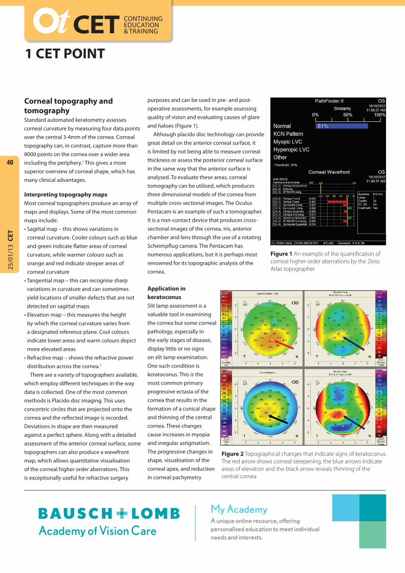
Figure 1 An example of the quantification of corneal higher order aberrations by the Zeiss Atlas topographer
Figure 2 Topographical changes that indicate signs of keratoconus. The red arrow shows corneal steepening, the blue arrows indicate areas of elevation and the black arrow reveals thinning of the central cornea
My AcademyA unique online resource, offering personalised education to meet individual needs and interests.
purposes and can be used in pre- and post-operative assessments, for example assessing quality of vision and evaluating causes of glare and haloes (Figure 1).
Although placido disc technology can provide great detail on the anterior corneal surface, it is limited by not being able to measure corneal thickness or assess the posterior corneal surface in the same way that the anterior surface is analysed. To evaluate these areas, corneal tomography can be utilised, which produces three dimensional models of the cornea from multiple cross-sectional images. The Oculus Pentacam is an example of such a tomographer. It is a non-contact device that produces cross-sectional images of the cornea, iris, anterior chamber and lens through the use of a rotating Scheimpflug camera. The Pentacam has numerous applications, but it is perhaps most renowned for its topographic analysis of the cornea.
Application in keratoconusSlit lamp assessment is a valuable tool in examining the cornea but some corneal pathology, especially in the early stages of disease, display little or no signs on slit lamp examination. One such condition is keratoconus. This is the most common primary progressive ectasia of the cornea that results in the formation of a conical shape and thinning of the central cornea. These changes cause increases in myopia and irregular astigmatism. The progressive changes in shape, visualisation of the corneal apex, and reduction in corneal pachymetry
25/0
1/13
CET
41
are well noted in topographic examinations (Figure 2). The indicators typically seen are: (1) steepening of the inferior cornea, displayed on the sagittal curvature map (due to the conical apex being formed), (2) an isolated elevation on both the anterior and posterior surfaces (which indicate the point of weakness on the cornea where protrusion is occurring), and (3) thinning of the central corneal thickness, as shown on the pachymetry map. Asymmetry in the corneal curvature provides a clue to corneal pathology such as keratoconus and this can be identified from the sagittal curve map. The amount of asymmetry is calculated as the difference in dioptric values directly above and below the central value. In normal eyes, these values should be similar, leaving only a small difference, but in keratoconus, the inferior dioptric value is much greater, thereby giving a large difference (Figure 2).
One of the advantages of performing corneal tomography over Placido disc topography alone is that sub-clinical cases of keratoconus (known as forme fruste keratoconus) can be identified. At this stage, patients are asymptomatic and no other clinical changes can be observed other than tomographic abnormalities, which include abnormal posterior elevation or abnormal corneal pachymetric progression, but completely normal anterior elevation and curvature.2 Early diagnosis of keratoconus is very important as patients can then be counselled and referred for potential corneal collagen cross-linking treatment in order to halt progression. This is much more favourable as a form of management in comparison to late stage treatments, which involve corneal transplantation.3
Application in laser refractive surgeryCorneal topography is an essential technique used in the pre-operative suitability assessment for patients presenting for laser vision correction (LVC) as well as post-operative management. Keratoconus and other diseases associated with abnormal corneal shape must be ruled out, as this would contraindicate LVC. There are several forms of LVC available and analysis of the corneal steepness and shape allows recommendation of the most appropriate treatment, for example intralase LASIK (which uses a femtosecond laser instead of a mechanical microkeratome to create a flap) is advised for patients with a steep cornea, while LASEK (laser in situ epithelial keratomileusis) is advised for those with a thin cornea.4 Furthermore, pachymetry is also required to ensure sufficient corneal thickness is present for full correction of refractive error. It is well known that a minimum residual stromal bed of 250µm is required after LVC to ensure maintenance of corneal biomechanical strength and minimise risk of post-operative ectasia.5 Tomographic analysis also allows for quantification of the anterior chamber depth and volume, which are important for the evaluation of suitability for intraocular lens (IOL) procedures.
Application in contact lensesContact lens software is included in some topographers, which simulates fluoroscein patterns for RGP contact lens fitting (Figure 3). It allows extensive customisation of lens parameters to create optimum fitting lenses, without having to physically insert and trial multiple lenses with patients. In particular, this is very useful for matching the peripheral curves of RGP lenses to the peripheral corneal shape, which can be particularly troublesome if only the central keratometry readings are available. This tool can prove to be especially valuable for patients with unusual corneal shapes, including keratoconus and post-corneal graft surgery.
As well as general RGP lens fitting, topography is tremendously useful in the field of orthokeratology, which involves the fitting of reverse geometry rigid contact lenses to allow temporary correction of myopia and astigmatism. This process requires the contact lenses to be worn throughout the night, as they induce flattening of the central cornea lasting for approximately 12 hours, allowing patients to
be spectacle-free throughout the day.6 Corneal topography is needed in order to determine parameters for the contact lenses required, as well assessing the fit and centration. Furthermore, post lens wear topography is the only way to measure how much corneal flattening is being produced from the contact lenses, which will directly correlate to the visual performance achieved.
Specular microscopyThe corneal endothelium comprises a rich density of regularly arranged hexagonal cells that maintains corneal deturgescence. There is an average of 4000 cells/mm2 in the first decade of life and this number reduces at an average rate of 0.6% every year.7 Specular microscopy allows for a high resolution magnified view of the corneal endothelium and most specular microscopes allow assessment of three main areas (Figure 4).
Endothelial cell density (CD)The endothelium requires a minimum number of cells in order to maintain its metabolic pump function as well as creating a barrier to restrict the amount of aqueous entering the corneal stroma.8 Certain pathological conditions create an accelerated loss of endothelial cells and, as these cells do not possess regenerative capabilities, this can ultimately lead to endothelial decompensation and permanent
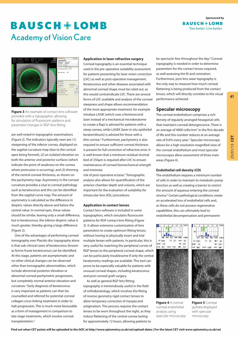
Figure 3 An example of contact lens software provided with a topographer, allowing for simulation of fluorescein patterns and parameter changes in RGP lens fitting
Find out when CET points will be uploaded to the GOC at http://www.optometry.co.uk/cet/upload-dates | For the latest CET visit www.optometry.co.uk/cet
Sponsored by
Figure 4 A normal corneal endothelial analysis using specular microscopy
Figure 5 Corneal guttata displayed with specular microscopy
25/0
1/13
CET
42
corneal opacification. An average minimum cell density of approximately 300-500 cells/mm2 is required to maintain the barrier function of the endothelium, in order to prevent pathological loss of corneal transparency.8
Hexagonal cell density (HEX)If the majority of corneal endothelial cells
are of similar shape and size, a more effective barrier between the cornea and aqueous humour is formed. Physiological stress on the endothelium from ocular pathology or contact lens wear can result in changes in the shape of endothelial cells, deviating away from the normal hexagonal morphometry. This is known as pleomorphism.
The proportion of endothelial cells that are hexagonal can be quantified by specular microscopy, with clinically significant pleomorphism arising if the hexagonal cell density is less than 50%.9 A normal endothelium will usually have more than 60% of cells that are hexagonal in shape.9 Long-term wear of contact lenses with low oxygen permeability can result in pleomorphism due to stress on the cornea created from hypoxia and acidosis.10 This can lead to reduced contact lens tolerance as well as symptoms of blurred vision and photophobia. In such cases specular microscopy is useful as endothelial changes can be monitored and patients can be advised to cease contact lens wear or to change to lenses that have greater oxygen permeability.
CET CONTINUING EDUCATION & TRAINING
1 CET POINT
Coefficient of variation (CV)Cells that are naturally lost with age from the corneal endothelium disperse into the anterior chamber. The gaps left in the endothelium are filled by adjacent cells, which either stretch or move to fill the void.9 This creates variation in the cell size, referred to as polymegathism, the degree of which is measured by the coefficient of variation (CV). As this is a direct result of the wound repair mechanism, all corneae will have some degree of polymegathism. Values up to 0.31 indicate average levels of polymegathism and values of 0.40 or more are considered to be abnormal.9 Increased polymegathism can also arise from contact lens wear as well as ocular pathologies such as glaucoma and uveitis.
Fuch’s endothelial dystrophyAs well as maintaining appropriate levels of corneal hydration, the endothelium also releases collagen, which forms Descemet’s membrane.11 As age advances, this physiological function can become impaired causing the endothelium to release an abnormal basement membrane material which forms nodules on Descemet’s membrane that are known as guttata. This can be seen as dark areas on specular microscopy, giving the appearance of holes in the endothelium (Figure 5). This appearance arises from the fact that guttata lie on a different plane of focus to that of the endothelial cells, as seen on specular reflection.12 Corneal guttata can
progress from a small darkened area within the centre of an endothelial cell, eventually enlarging to fill the entire cell and finally spreading to other cells, leading to confluence of multiple guttata.9 Fuch’s endothelial dystrophy can arise from an increase in corneal guttata, which leads to endothelial decompensation. This then causes stromal oedema and eventually epithelial oedema, resulting in bullous keratopathy. Before stromal and epithelial oedema occur, specular microscopy can be extremely useful in aiding referral of a patient for treatment such as Descemet’s stripping endothelial keratoplasty.
Iatrogenic endothelial cell lossThis is cell loss caused by a surgical procedure, most commonly IOL implantation as performed in cataract surgery or phakic IOL implantation for refractive correction. Patients who have had the latter require regular monitoring of their endothelial CD, as cell loss can be accelerated by IOL movement, IOL contact with the corneal endothelium from eye rubbing, or post-operative inflammation.13
Glaucoma-induced endothelial cell lossExposure to high levels of intraocular pressure (IOP) over a long period of time can result in an irregular reduction of endothelial CD.14 Therefore regular measuring of cell density can be used as a parameter to evaluate the effect of glaucomatous damage, as well as a means of monitoring the effectiveness of topical therapy in glaucoma.15
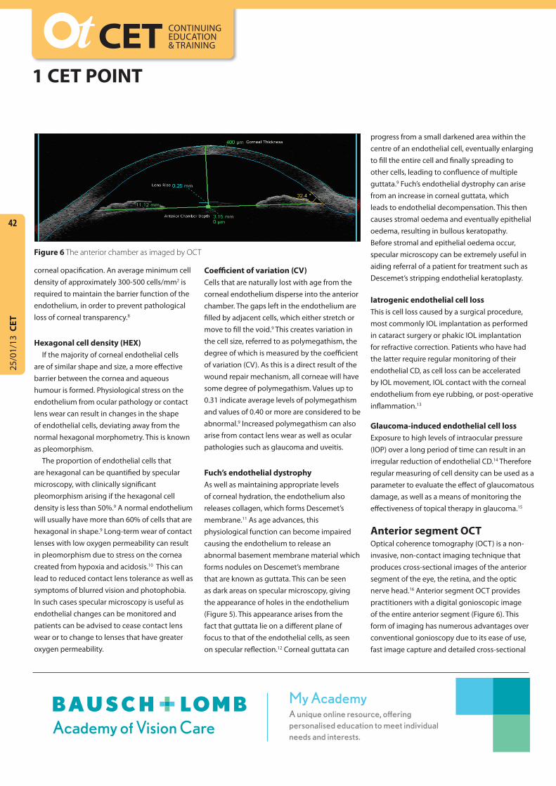
Anterior segment OCTOptical coherence tomography (OCT) is a non-invasive, non-contact imaging technique that produces cross-sectional images of the anterior segment of the eye, the retina, and the optic nerve head.16 Anterior segment OCT provides practitioners with a digital gonioscopic image of the entire anterior segment (Figure 6). This form of imaging has numerous advantages over conventional gonioscopy due to its ease of use, fast image capture and detailed cross-sectional
My AcademyA unique online resource, offering personalised education to meet individual needs and interests.
Figure 6 The anterior chamber as imaged by OCT
visualisation of anterior segment structures. Fundamentally this technique has primary applications for a wide range of pathology encompassing corneal, iris and angle abnormalities. Perhaps of greatest use however, is in patients with, or at risk of developing, glaucoma as well as for anterior eye surgery such as phakic IOL implantation.
Glaucoma screeningIn angle closure glaucoma, IOP increases due to obstruction of aqueous outflow through the trabecular meshwork, either through a pupil block mechanism or by plateau iris syndrome.17 Those with normal IOPs, but very shallow angles, can be at risk of angle closure glaucoma and therefore can subsequently be referred for potential prophylactic laser iridotomy treatment. Plateau iris displays as an abnormal peripheral iris configuration whereby there is increased anterior positioning of the ciliary body.17 The latter is more common in younger patients and is extremely difficult to diagnose clinically without the aid of OCT. Preventative treatment for such individuals include laser iridotomy or argon laser gonioplasty.
Intraocular lens (IOL) surgeryAlthough anterior segment OCT can be utilised
within cataract surgery and refractive lens exchange, it has more benefit in phakic IOL surgery. Precise measurements of anterior chamber depth, crystalline lens rise, corneal diameter, sulcus diameters and distance between iridocorneal angles are all parameters used to assess the suitability of prospective patients, as well as determining the size of IOL required. Suitability for phakic IOLs requires a minimum anterior chamber depth of 2.8mm (specified by phakic IOL manufacturers Ophtec BV and Staar Surgical Company), which is measured from the corneal endothelium to the anterior lens surface. This creates a safe distance between the cornea and the phakic IOL, to minimise endothelial touch. Furthermore, a maximum lens rise of 600µm is required to prevent interaction between the phakic IOL and the natural lens.18
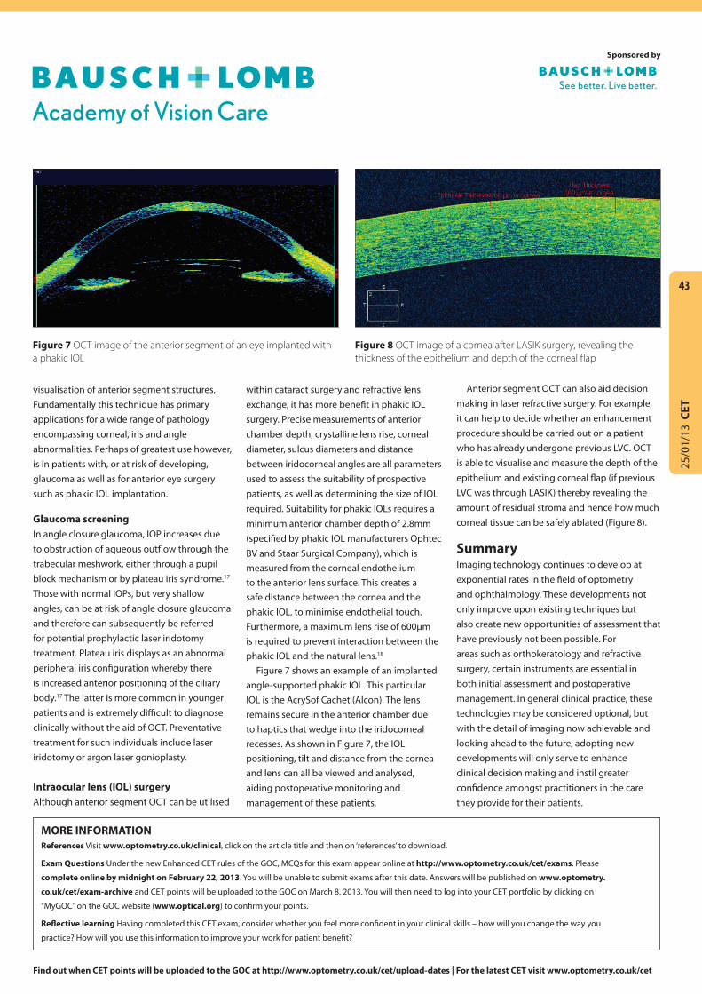
Figure 7 shows an example of an implanted angle-supported phakic IOL. This particular IOL is the AcrySof Cachet (Alcon). The lens remains secure in the anterior chamber due to haptics that wedge into the iridocorneal recesses. As shown in Figure 7, the IOL positioning, tilt and distance from the cornea and lens can all be viewed and analysed, aiding postoperative monitoring and management of these patients.
Anterior segment OCT can also aid decision making in laser refractive surgery. For example, it can help to decide whether an enhancement procedure should be carried out on a patient who has already undergone previous LVC. OCT is able to visualise and measure the depth of the epithelium and existing corneal flap (if previous LVC was through LASIK) thereby revealing the amount of residual stroma and hence how much corneal tissue can be safely ablated (Figure 8).
SummaryImaging technology continues to develop at exponential rates in the field of optometry and ophthalmology. These developments not only improve upon existing techniques but also create new opportunities of assessment that have previously not been possible. For areas such as orthokeratology and refractive surgery, certain instruments are essential in both initial assessment and postoperative management. In general clinical practice, these technologies may be considered optional, but with the detail of imaging now achievable and looking ahead to the future, adopting new developments will only serve to enhance clinical decision making and instil greater confidence amongst practitioners in the care they provide for their patients.
25/0
1/13
CET
43
Find out when CET points will be uploaded to the GOC at http://www.optometry.co.uk/cet/upload-dates | For the latest CET visit www.optometry.co.uk/cet
MORE INFORMATION References Visit www.optometry.co.uk/clinical, click on the article title and then on ‘references’ to download.
Exam Questions Under the new Enhanced CET rules of the GOC, MCQs for this exam appear online at http://www.optometry.co.uk/cet/exams. Please
complete online by midnight on February 22, 2013. You will be unable to submit exams after this date. Answers will be published on www.optometry.
co.uk/cet/exam-archive and CET points will be uploaded to the GOC on March 8, 2013. You will then need to log into your CET portfolio by clicking on
“MyGOC” on the GOC website (www.optical.org) to confirm your points.
Reflective learning�Having�completed�this�CET�exam,�consider�whether�you�feel�more�confident�in�your�clinical�skills�–�how�will�you�change�the�way�you
practice? How will you use this information to improve your work for patient benefit?
Figure 7 OCT image of the anterior segment of an eye implanted with a phakic IOL
Figure 8 OCT image of a cornea after LASIK surgery, revealing the thickness of the epithelium and depth of the corneal flap
Sponsored by