splenic hamartoma: a report of two cases - snu open...
TRANSCRIPT

Tlrc S(~olr1 Ji~~irrrnl of A.lcdic-it/(, VoI. 30, No. 3: 279-287, Llc~rc~r~rhcr 1989
Splenic Hamartoma: A Report of Two Cases
Jung Ran Kim, Eui U Park, Je G. Chi and Yong II Kim
= Abstract = Two cases of splenic hamartoma are described. Both cases occurred in women in their fifties and presented as vague abdominal symptoms. Case 1 was a large solitary hamartoma, 6.5 cm in size and hypervascular mass angiographically. No white pulp with germinal centers or central arterioles were seen in the mass. Instead, a mass of red pulp with well-formed sinuses, often exhibiting active endothelial cells, were the predominent feature, scattered with lymphoid nodules. Extramedullary hemopoieses was meager. Case 2 was an example of multiple (3) hamartomas that showed extraordinary extramedullay hemo- poiesis particularly in the mass itself. The proliferative megakayocytes were quite atypical, and in areas they mimicked the neoplastic process. There was evidence of compression of normal parenchyma by the hamartomatous masses both grossly and microscopically.
It seems true that the histological features of splenic hamartoma can v a y from case to case and that one should pay particular attention to these lesions so that they are not misinter- preted as true neoplastic processes.
Key words: Spleen, Hamartoma, Splenoma
INTRODUCTION
Hamartomas are rare tumors of the spleen. Most splenic hamartomas are discovered in- cidentally during splenectomy or at autopsy (Coe 1952, Jaffe 1985, Rappaport 1966, Silverman 1978, Teates et a/., 1972, Wolf and Neiman 1989). They rarely attain a size sufficient to pro- duce a palpable enlargement of the spleen (Bhagwat et a/., 1975). Associated hematologic abnormalities are rare (Rappaport 1966, Wolf and Neiman 1989). They present as a solitary mass in most cases. However, some of these lesions are multiple (Steinberg et a/., 1985) and can be associated with clinical symptoms and foci of ex- tramedullary hematopoiesis in the spleen (Jaffe 1985, Rappaport 1966).
We report two cases of splenic hamartoma,
Kece~ved 30/10/89, revised 30/11/89, accepted 1 / 12/89 'Present address Ilcpartmcnt of Pathology, C'ollcge of Med~c ine . Dong- k u k U n ~ v e r s ~ t y , Kyung Joo. Korea
one solitary and one multiple, seen in one 54-year-old and one 55-year-old female. The rarity of this entity and presence of massive ex- tramedullary hemopoieses in the hamartoma nodule in Case 2 prompted this report.
CASE REPORT
Case 1 A 54-year-old woman was transferred to
Seoul National University Hospital on January 15, 1987 for the management of a large cystic mass in the spleen found at a local hospital. She had suffered from abdominal discomfort in the left upper quadrant and periumbilical region for the last three years. No other related symptoms were present. She had no history of trauma in this region and enjoyed relatively good health, except for an operation she had undergone 10 years before for a herniated intervertebral disc.
The physical examination was essentially nor- mal except for a suspicious left upper quadrant mass. The liver was not palpated. Laboratory data also remained within normal limits except for a mild anemia (Hgb 14.0 g/dl and hematocrit
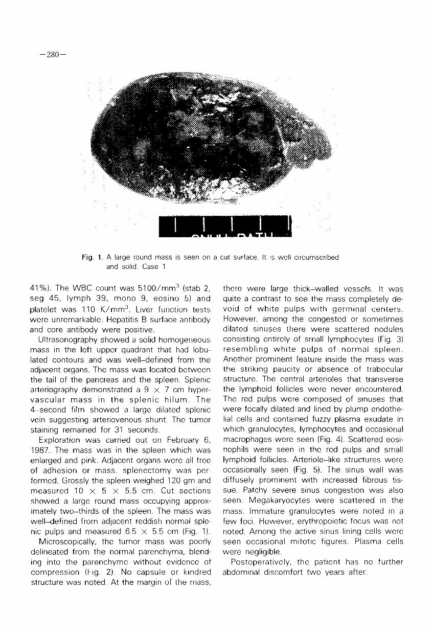
Fig. 1. A large round mass is seen on a cut surface. It IS well circumscribed and solid. Case 1
41 %). The WBC count was 5100/mm3 (stab 2, seg 45, lymph 39, mono 9, eosino 5) and platelet was 110 K/mm3. Liver function tests were unremarkable. Hepatitis B surface antibody and core antibody were positive.
Ultrasonography showed a solid homogeneous mass in the left upper quadrant that had lobu- lated contours and was well-defined from the adjacent organs. The mass was located between the tail of the pancreas and the spleen. Splenic arteriography demonstrated a 9 x 7 cm hyper- vascular mass in the splenic h i lum. The 4-second film showed a large dilated splenic vein suggesting arteriovenous shunt. The tumor staining remained for 31 seconds.
Exploration was carried out on February 6, 1987. The mass was in the spleen which was enlarged and pink. Adjacent organs were all free of adhesion or mass. splenectomy was per- formed. Grossly the spleen weighed 120 gm and measured 10 x 5 x 5.5 cm. Cut sections showed a large round mass occupying approx- imately two-thirds of the spleen. The mass was well-defined from adjacent reddish normal sple- nic pulps and measured 6.5 x 5.5 cm (Fig. 1 ).
Microscopically, the tumor mass was poorly delineated from the normal parenchyma, blend- ing into the parenchyme without evidence of compression (Fig. 2). No capsule or kindred structure was noted. At the margin of the mass,
there were large thick-walled vessels. It was quite a contrast to see the mass completely de- void of white pulps with germinal centers. However, among the congested or sometimes dilated sinuses there were scattered nodules consisting entirely of small lymphocytes (Fig. 3) resembling whi te pulps of normal spleen. Another prominent feature inside the mass was the striking paucity or absence of trabecular structure. The central arterioles that transverse the lymphoid follicles were never encountered. The red pulps were composed of sinuses that were focally dilated and lined by plump endothe- lial cells and contained fuzzy plasma exudate in which granulocytes, lymphocytes and occasional macrophages were seen (Fig. 4). Scattered eosi- nophils were seen in the red pulps and small lymphoid follicles. Arteriole-like structures were occasionally seen (Fig. 5). The sinus wall was diffusely prominent with increased fibrous tis- sue. Patchy severe sinus congestion was also seen. Megakaryocytes were scattered in the mass. Immature granulocytes were noted in a few foci. However, erythropoietic focus was not noted. Among the active sinus lining cells were seen occasional mitotic figures. Plasma cells were negligible.
Postoperatively, the patient has no further abdominal discomfort two years after.

Fig. 2. Low power picture of the margin of the mass (arrows) showing a vascular trabecula. The right side represents normal spleen. Case 1
Fig. 3. Photomicrograph of lymphoid nodules in the hamartoma, showing no germinal center. Case 1 H & E x 100

Fig. 4. Dilated sinuses ~nside the mass, that are lined by plump endothelial cells. Case 1, H & E x 300
Fig. 5. In the red pulp arteriole-like structure is seen. Case 1, H & E x 300

Fig. 6. Gross appearance of the removal spleen. Two discrete masses are seen on cut surface. Case 2
Case 2 A 55-year-old female presented in August
1987 with a one-month history of fatigue and abdominal distention. Physical examination re- vealed a massive splenomegaly. The spleen was palpable, 6 cm below the left costal margin. Laboratory data included a hemoglobin of 7.6 gm /dl and a leukocyte count of 6,000/mm3 with a normal differential count; other routine blood chemistries were normal. However, an extensive hematologic work-up, except for a bone marrow study, failed to disclose a reason for the anemia. Ultrasonography of the spleen demonstrated a diffuse enlargement and a relatively echolucent mass within the substance of the spleen. Splenectomy was performed.
The spleen was enlarged and weighed 900 gm and measured 17 x 15 x 9 cm. The splenic outline was retained and the capsule was smooth. On sectioning, three firm well-circums- cribed nodules were found in the splenic paren- chyma. Each measured 5, 4 and 2 cm in dia- meter. They bulged slightly from the cut surface (Fig. 6). The nodules were well-defined from the adjacent splenic parenchyme and appeared dark
red (Fig. 7). The remaining white pulps were not prominent.
Histologically, the masses were composed of disorganized, solid-appearing stromal cells, which did not present the distinct sinus pattern charac- teristic of normal splenic tissue. The mass could be delineated by condensation of reticulin fiber from the normal splenic tissue (Fig. 8). Inside the masses were infiltration of all bone marrow elements. The non-hemopoietic cells had plump and ovoid nuclei with peripheral condensed chro- matin arranged in a disorganized pattern. The sinuses were occasionally lined by endothelial cells and markedly congested. Vascular channels of different size coursed through the masses. Reticulin frameworks were condensed along the periphery of the nodules. Hemopoietic precur- sors were present inside and outside the nodules. Numerous atypical megakaryocytes and cells of the erythroid and myeloid series were seen (Fig. 9 & 10). Hematopoietic precursors were also seen in both the cords of Billroth and in the sinuses of the red pulps of the masses.

Fig. 7. A close-up view of one of the 3 masses. They compress the surrounding normal parenchyma. Case 2
Fig. 8. Low power photomicrograph showing the demarcation of mass from the normal spleen (arrows). Case 2. Retic. x 40
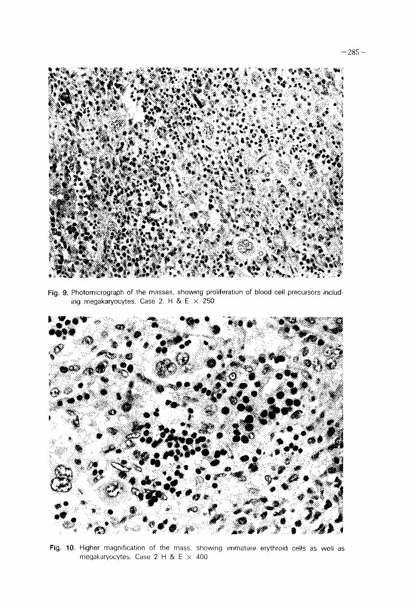
Fig. 9. Photomicrograph of the masses, showing proliferation of blood cell precursors includ- ing megakaryocytes. Case 2. H & E x 250
Fig. 10. Higher magnification of the mass, showing immature erythroid cells as well as megakaryocytes. Case 2 H & E x 400

DISCUSSION
Case 1 appears to be a classic case of splenic hamartoma. It is still an enigma why this hamar- tomatous mass was so hypervascular and even showed evidence of arteriovenous shunt on 4-second film. Although we have seen vessels distributing the hamartoma itself, the numbers and sizes of them were not unusually larger or more prominent than normal parenchyme. Furth- ermore, na arterialized vein suggesting arteriove- nous shunt could be seer: in the mass. It was, however, explicable by the fact that focal sinus widening and collections of plasma fluid that were scattered in the mass. The early return of the dye into the dilated splenic vein might have been due to the direct drainage of venous blood from the sinuses of the mass without going through postcapillary venules, which appeared to be deficient or minimal in the mass histologi- cally.
Case 2 is unique in the sense that active hemopoiesis was associated in the hamartoma- tous masses in the spleen. The hamartomas were multiple, and three similar masses were found in the spleen. There is a picture that is almost identical to this one in the AFlP fascicle (Rappaport, 1966). It is not known whether there were similar histological features in their case as was seen in ours. However, it would be of in- terest in that increased hemopoietic activity was there based on the gross appearance of the le- sion. Unfortunately, complete hematological stu- dies have not been performed in our case,
although clinically the patient had no evidence of other sitgmata of hematological malignancy. And even if it were of hematologic origin, it would be more usual to find such nodular lesions in the spleen as well as the proliferation of all three cell lines of hemopoiesis, including atypical megakaryocytic cells.
REFERENCES
Bhagwat AG, D'Alta DV, Mitras, et al.,. Splenoma with portal hypertension. Br. Med. J., 1975, 1 : 520
Coe JI, Von Drasheks. Hamartoma of the spleen. A report of four cases. Am. J. Pathol. 1962, 28: 663
Garvin DF, King FM. Cysts and nonlymphomatous tumors of the spleen. In Sommers SC, and Rosen, P.P.(eds.). Pathology Annual, part 1, New York, Ap- pleton-Century-Crofts, Vol. 16, 1981, pp. 71-80
Jaffe ES. Surgical pathology of the lymph nodes and related organ, Vol. 16, in the series major problems In pathol, SB Saunders, Philadelphia, 1985, pp. 274-279
Rappaport H. Tumors of the hematopoietic system. In atlas of tumor pathology. section 3, fascicle 8. Washington. DC. Armed Forced Institute of Patholo- gy, 1966, pp. 380-388
Silverman ML, Livolsi VA. Splenic hamartoma. Am. J. Clln. Pathol. 1978, 70: 224-229
Steinberg JJ, Suhrland MJ, Quentin JV. The associa- tion of splenoma with d~sease. Lab. Invest. 1985, 52: 65A
Teates CD, Seale DL, Allen MS. Hamartoma of the spleen. Am. J Roentgenol. 1972, 116: 419
Wolf BC, Neiman RS. Disorders of the spleen Vol. 20 in the series major problems In pathol. SB Saunders Co. Philadelphia, 1989. pp. 102-1 93
