spine tango report international 2012 - the spine society of europe
TRANSCRIPT

SPINE TANGO ReportInternational 2012
The International Spine RegistryEuroSpine
C. Röder, M. Neukamp, E. Aghayev, T. Zweig, T. Ambrose, E. Munting, T. Pigott

2
Contents
Contact:
University of BernInstitute for Evaluative Research in Orthopaedic SurgeryChristoph Röder, MD MPHStauffacherstr. 78CH-3014 [email protected]
This annual report is digitally available in the literature section of the Spine Tango web page under www.eurospine.org
Introduction T. Pigott 3
Profile C. Röder, T. Zweig 4
Registries vs randomized trials C. Röder 5
New developments C. Röder 6
Application C. Röder, T. Zweig 10
Data entry 12
A complete case 13
Documentation workflow 14
Statistics and comments C. Röder 15
Part I: Descriptive analysis form version 2011 and 2005/2006 M. Neukamp, C. Röder, E. Aghayev 16
Part II: Analysis of failed surgeries E. Aghayev, C. Röder 38
Participants/ module analysis C. Röder, M. Neukamp 54
Security concept T. Ambrose, P. Abt 56
Implant capture P. Abt 58
Available questionnaires in the Spine Tango E. Röösli 60
Publications M. Neukamp 61

3
IntroduCtIon
Since the year 2000 EuroSpine – The Spine Society of Europe has been developing and enhancing
a documentation system for spinal surgery and also for non-surgical spinal treatments in form of a
registry. With Spine Tango we are meeting the growing demand to assess the safety and comparative
effectiveness of surgical and non-surgical interventions and therapies of the spine. Only few other
fields in medicine are under comparable scrutiny. Reacting to these tendencies, endeavors of pioneer
clinicians and the Spine Tango committee, in collaboration with the Institute for Evaluative Research in
Orthopedic Surgery of the University of Bern, have led to the implementation of the only international
spinal registry to date. The idea for Spine Tango was proposed a decade ago by Dieter Grob and Max
Aebi, under the auspices of the SSE. Developments and participation have constantly progressed
since those days. Now, having reached a recognized status we would like to encourage national
societies and individual partners to join the registry. The German Spine Society DWG, the largest
spine society in Europe, is successfully conducting its 2-year pilot of a national spine registry adopting
the Spine Tango technology and content, and in fall 2013 a new Polish Spine Tango module will be
launched. Health and reimbursement authorities are already limiting the accessibility of some spinal
treatment modalities since evidence is lacking in many aspects. Therefore Spine Tango as a registry
with routine data resulting from the hospitals` day-to-day work is offered as a common language to
make our services visible and transparent. Conclusions from the registry have an admittedly lower
internal, i.e. methodological validity compared with higher evidence studies like RCTs, but the external
validity and therefore generalizability of our findings is what makes the dataset and its clinical and
scientific findings so valuable for health service and outcome research. With a constantly increasing
activity in the registry we would like to inform you about its history, its objectives and its current status.
T. Pigott
Chair, on behalf of the Spine Tango committee

4
1. Aebi M, Grob D (2004)SSE Spine Tango: a European Spine Registry promoted by the Spine Society of Europe (SSE)Eur Spine J. 2004 December; 13(8): 661–662. 2. Kessler J, Melloh M, Zweig T, Aghayev E, Röder C (2011)Development of a Documentation Instrument for the Conservative Treatment of Spinal Disorders in the Internatio-nal Spine Registry Spine Tango. Eur Spine J. 2011 March; 20(3): 369–379.
ProFILe
Spine Tango enables you to document the whole spectrum of spinal pathologies and the possible
surgical and non-surgical treatment options. The generic approach of the Spine Tango documentation
system is a must to reach the maximum number of participants using a common web based
technology. This, in turn, reduces the potential for customizing the Tango in order to meet the individual
expectations of specific users. There are, nevertheless, still a number of possibilities to parameterize
the data collection processes according to the various hospital workflows in the user community. To
give you the opportunity to document not only the surgical treatments, we have developed Spine
Tango Conservative, which is now available in its first version. Spine Tango is an international, non-
commercial system under the auspices of EuroSpine, the Spine Society of Europe aiming at enabling
national societies to organize and control their own part of the registry. For that a technology called
“national module concept” has been implemented to enhance participation options and to provide the
hardware structure for appropriate security measures for patient and user privacy protection. The new
software release 2012 does further improve these aspects. In conclusion, Spine Tango is a unique
applied medical and scientific documentation and technology solution. It is to the benefit of patients,
physicians and therapists whilst generating evidence based findings to improve spinal care (1,2).

5
*unclear terminology, Cochrane called it “efficiency”, better always specify what you mean (evidence derived from controlled experiment versus evidence derived from routine clinical practice)
reGIstrIes Versus rAndoMIZed ControLLed trIALs (rCt)
Cochrane AL, British Epidemiologist, 1909-88. The father of Evidence Based Medicine. Effectiveness and Efficiency.
Random Reflections on Health Services.London: Nuffield Provincial Hospitals Trust, 1972
RCT RegistryType of evidence Efficacy Effectiveness*, safetyPrincipal question Can it work?
The first step of evidence generationDoes it work?Verification in daily clinical practice
Internal validity (methodologicalquality)
+++ + - ++ (expandable with eg. monitoring, audits orcomparison with secondary data etc.)
External validity (transferability/ generalizability)
- +++
Levels of evidence 1a, 1b 2b-4, depending on methodologyHypothesis-based approach, Yes Usually noDuration of observation period Predefined Open-ended or predefinedFocus of research/measurement Sharp, narrow (see hypothesis) BroadQuality assessment Not intended (strictly defined indications, process
quality at least derivable, outcome quality dependson effectiveness, a given indication and process)
Indication, process, outcome
Early warning system Not possible FeasibleLong-term follow-up Feasible Feasible, depending on registry set-up maybe only
for a representative sampleCoverage Only among participants From individual center/surgeon over representative
clinic sample to full national / regional coverageBenchmarking Only benchmarking of group Depending on the final composition of participants
regional to nationally representative benchmarkType of quality assurance Internal, external vs. benchmark of participants Internal, external vs. representative regional or
national benchmarkEffort Very high for a few participants Low for many participantsCost High to very high Low cost basis, costs increase depending on the
stage of development and number of participantsUse of generated data Only in the framework of the scientific
goal/hypothesisOpen hypothesis generation possible
Comparator Given per definition Ranges between none to numerous comparators,depending on registry set-up

6
neW deVeLoPMents
The new follow-up calendar function allows predefinition of follow-up intervals and related forms
within a project. These intervals become part of an overview and planning tool which allows visualizing
the performed, pending, and missed as well as “outlier” follow-ups for each case and related forms as
well as planning upcoming follow-ups by defining a time interval in the near or far future and viewing
all related follow-ups, the dates they should be performed and the respective forms that need to be
administered. What type of (different) forms belongs to what follow-up, and what the related “anchor”
or “index” form is, can all be specified by the study administrator. Follow-up rules are proposed
“downwards” from the module administrator to the participants, but each lower level of organization
(hospital – department - physician) can alter the proposed rules and adjust them to local processes
and needs. Additional forms can always be created, completed and submitted outside these intervals
and will be regarded and listed as “outliers” to the predefined intervals in the various calendar views.
ASIA score: the IEFO team is proud to introduce a “smart” ASIA documentation form for spinal
trauma. It automatically calculates the respective scores and has some intelligent functionalities to
avoid the very cumbersome completion of sensory and motor functions of all spinal levels. Optionally
you can document the Functional Independence Measure (FIM) within this setting.

7
The case ID is a seemingly harmless but truly powerful new function to help users establish a clearly
defined relation between all their forms and make later online and offline statistical analyses less error
prone. This is especially helpful in cases with several interventions on various levels and a multitude
of related follow-ups at different dates and hence different intervals. Given a certain number of cases
with such complexity in the registry, and no clearly defined case history, many statistical analyses
become probabilistic. Therefore, the new case ID function allows users to clearly link forms with each
other that belong to one case, e.g. cervical disc protrusion and related follow-ups and outcomes, but a
lumbar stenosis surgery with a new and different set of follow-ups and outcome forms at a later point
in time. Such a patient consequently has two “cases” in his chart and all related forms are clearly and
intentionally linked with each other by the user. If the time and location relationship of an intervention
form and a follow-up or outcome form does not match while the user is adding it to a case, the system
displays warnings or suggests creating a new “case”. Hence overall across-form data quality and
analyzability become significantly improved with the registry. In addition, form selection becomes
more comfortable, since a new “Plus” icon next to an intervention form allows the user to directly
link a follow-up/outcome form to an intervention form. The user is only offered those forms that can
theoretically be linked based on location (e.g. no ODI displayed to a cervical intervention form) or
diagnosis (e.g. no SRS-30 offered for a degenerative intervention form). With the Plus sign, follow-up
intervals are controlled by the system, based on surgery and follow-up dates. If the user chooses a
grossly incorrect interval, warnings are displayed and a better matching interval is proposed.
Spine Tango adolescent scoliosis add-on: long awaited and a project that was initiated by our
Spine Tango fellow Dr. T. Zweig, the first generation of adolescent scoliosis add-on forms is available.
Thanks to an international effort of experts from Eurospine, DWG and the Hospital for Joint Diseases
at New York University, a carefully developed and comprehensive “add-on” form in conjunction with
the surgery form allows specialists to document these types of interventions in a more detailed mode.
In addition to the already available SRS-30 outcome form, the SAQ Spinal Appearance Questionnaire
will also be uploaded in those language versions that are validated.
Spine Tango adult deformity add-on: following along the lines of the adolescent scoliosis form, a
similar yet distinct add-on form for adult degenerative deformity surgery was also developed

8
)) II postop / followupperiop
Directions
IIIIII
other.......................postsurgicalposttraumaticneuromuscularcongenitalidiopathic
Type of scoliosis
- Lenke ClassificationCurve type
Lumbar modifierIII
III
Type 6, thoracolumbar/lumbar-main thoracic (TL/L -MT)Type 5, thoracolumbar/lumbar (TL/L)Type 4, triple major (TM)
Type 3, double major (DM)Type 2, double thoracic (DT)Type 1, main thoracic (MT)
III
CBA
Thoracic sagittal modifier (Th 5-Th12) III + (Hyper)N (Normal)- (Hypo)
Imbalance
JJ
JJJ
hereditary motor sensory neuropathyspinocerebellar dysfunction
mixed upper and lower motor neuronlower motor neuronupper motor neuron
JJJ
congenital myopathyarthrogryposismuscular dystrophy
specify:
)I Completely fill in boxes to record answers.
Use a #2 soft pencil for marking.Text answers must be entered with the web interface.All questions must be answered unless otherwise indicated.
I only 1 answer allowed J multiple answers allowedQuestion types mandatory question
Pulmonary function testsVC (Vital Capacity)
FEV1 (Forced ExpiratoryVolume in one second) FVC (Forced Vital Capacity)
TLC (Total Lung Capacity)J not applicable
II
% of pred. valueliter
..................................
periop only
Truncal appearanceII
noyesPhotographs Please upload photographs!
IIIIIIIIII0 1 2 3 4 5 6 7 8 9IIIIIIIIII0 1 2 3 4 5 6 7 8 9
Scoliometer
IIIIIIIIII0 1 2 3 4 5 6 7 8 9IIIIIIIIII0 1 2 3 4 5 6 7 8 9
IIIIIIIIII0 1 2 3 4 5 6 7 8 9IIIIIIIIII0 1 2 3 4 5 6 7 8 9
Prox. thoracic (PT) Main thoracic (MT) Thoracolumbar/lumbar (TL/L)
Radiological skeletal maturationBone age determination
IIIIII 543210
Bone ageyearsmonths
IIIIIIIIIIIIIIIIIII0 1 2 3 4 5 6 7 8 9 10 11 12 13 14 15 16 17 18IIIIIIIIIIIII0 1 2 3 4 5 6 7 8 9 10 11 12
II
closedopen
Risser sign / score Triradiate cartilage (TRC)
J not applicable J not applicable J not applicable
Osteotomy Classification (Schwab)
Technique/ screw insertion
Material/ Rod specification
Perioperative management of bleeding
Details of Classification are provided in the Dictionary of terms.
III
alternatingeach level/VB unilaterallyeach level/VB bilaterally
Operation/ additional surgical measures
JJJ
JJJJ
other................................thrombinfibrin glue
tranexamic acidfibrinogenFFPnone
JJ neuropathic conditionsmyopathic conditions
Examination date
Abbreviations: VB = vertebral body CoCr = cobalt-chrome FPP = fresh frozen plasma
JJJJJJJJJJJJJJJJJ L5L4L3L2L1Th12Th11Th10Th9Th8Th7Th6Th5Th4Th3Th2Th1
JJJJJJJJJJJJJJJJJ L5L4L3L2L1Th12Th11Th10Th9Th8Th7Th6Th5Th4Th3Th2Th1
JJJJJJJJJJJJJJJJJ L5L4L3L2L1Th12Th11Th10Th9Th8Th7Th6Th5Th4Th3Th2Th1
JJJJJJJJJJJJJJJJJ L5L4L3L2L1Th12Th11Th10Th9Th8Th7Th6Th5Th4Th3Th2Th1
JJJJJJJJJJJJJJJJJ L5L4L3L2L1Th12Th11Th10Th9Th8Th7Th6Th5Th4Th3Th2Th1
JJJJJJJJJJJJJJJJJ L5L4L3L2L1Th12Th11Th10Th9Th8Th7Th6Th5Th4Th3Th2Th1
Neuromonitoring
JJJ
by neurophysiologistby surgeonnone
JJ leftrightJJ leftright
Form to be completed with SSE surgery or followup.Iperiop
IIIIIIIIIIIIIIIIIIIIIIIIIIIIIII1 2 3 4 5 6 7 8 9 10 11 12 13 14 15 16 17 18 19 20 21 22 23 24 25 26 27 28 29 30 31IIIIIIIIIIII1 2 3 4 5 6 7 8 9 10 11 12 IIIIIIIIIIIIIIIII10 11 12 13 14 15 16 17 18 19 20 21 22 23 24 25 26
DayMonth Year
PER
IOP
ON
LY
Please specify
.................................. ..................................
..................................
Please specify
JJJJJJ
other concave................other convex..................Titanium concaveTitanium convexCoCr concaveCoCr convex
Please specify
JJ leftrightPlease specify
-> Specify levels / VBs
-> Specify levels / VBs
partial facet joint resection
complete facet joint resection
pedicle and vertebral wedge
monosegmental vertebrectomy
Grade III plus resection
multisegmental vertebrectomy
-> Specify levels / VBs
-> Specify levels / VBs
-> Specify levels / VBs
-> Specify levels / VBs
(Smith-Petersen)
resection (PSO)
endplate and disc
(including adj. discs)PER
IOP
ON
LY
J
J
J
J
J
JJ
Grade VI:
Grade V:
Grade IV:
Grade III:
Grade II:
Grade I:none
periop only
GradeIII
French/European grading systemUS grading systemnot evaluated I
II other method....................
Tanner & White-houseGreulich & Pyle
Pulmonary function
III adolescentjuvenileinfantileIII combination of bothfailure of segmentationfailure of formationspecify:
specify:
if myopathic conditions
Diagnosis
if neuropathic conditions
ev. bothRightLeft
If Ileum screws - no ofIII 321III 321
Sacro-iliacal fixation
JJJJ
Ileum-screwS2-ala-ileum-screwS2-ala-screwnone
SCOLIOSISadolescent add-on
Last name
Street
Country code
Social security number
Zip code
GenderFirst name
City
M.R.N.
Inte
rnal
Use
Onl
yN
ot re
ad b
y sc
anne
r
Birthdate (DD.MM.YYYY)
Copyright MEMdoc, 2013 All rights reserved15.07.2013
JJJ
coronalsagittalnone
(*in degrees) *Example: 37° = mark 3 in the 1st row and 7 in the 2nd row.
SPINE TANGO
Adolescent scoliosis add on formFront side
The back side with the radiographic measurements is not displayed.

9
Adult deformity add on formFront side
ADULT DEFORMITYadd-on
)I Completely fill in boxes to record answers.
Use a #2 soft pencil for marking.Text answers must be entered with the web interface.All questions must be answered unless otherwise indicated.
I only 1 answer allowed J multiple answers allowedQuestion types
Directions
JJJJJJ
other.........................neuromuscular/ neuro degenerativeposttraumaticpost-surgicaldegenerativeAIS in adults
Deformity type SRS-Schwab ClassificationCurve pattern
Lumbar modifier
IIII
primary sagittallumbar onlythoracic and lumbarthoracic only
III CBAThoracic sagittal modifier (Th 5-Th12) III + (hyper)N (normal)- (hypo)
Global balance modifierIII VP (SVA >95 mm)P (SVA 40 - 95mm)N (SVA <40mm)
Abbreviations: PT = Pelvic Tilt (if not declared); PI = Pelvic Incidence; LL = Lumbar Lordosis; SVA = Sagittal Vertical Axis; LBP = low back pain; DEXA = Dual-Energy X-ray Absorptiometry, DXA/DEXA; VB = Vertebral Body; CoCr = cobalt-chrome; FPP = fresh frozen plasma
mandatory question
Primary deformity pattern
III
combinedkyphosis/flatback/sagittal planescoliosis/coronal plane
Primary surgical indication
JJJJJJ
other.........................concerns over deformity progressiongeneral disability/functional lossloss of ambulatory enduranceleg pain/neuro findingslow back pain
PI-LL modifier
III
high (PT >30 °)medium (PT 20° - 30°)low (PT < 20°)
Pelvic tilt modifier
III
C (PI-LL >20 °)B (PI-LL 10° - 20°)A (PI-LL < 10°)
Lenke Classification
III
III
Type 6, thoracolumbar/lumbar-main thoracic (TL/L -MT)Type 5, thoracolumbar/lumbar (TL/L)Type 4, triple major (TM)
Type 3, double major (DM)Type 2, double thoracic (DT)Type 1, main thoracic (MT)
Curve type
Risk factors - Comorbidities
IIIIIIIIIIIIIIIIIIIIIIIIIIIIIII1 2 3 4 5 6 7 8 9 10 11 12 13 14 15 16 17 18 19 20 21 22 23 24 25 26 27 28 29 30 31IIIIIIIIIIII1 2 3 4 5 6 7 8 9 10 11 12 IIIIIIIIIIIIIIIII10 11 12 13 14 15 16 17 18 19 20 21 22 23 24 25 26
DayMonth Year
Form to be completed with SSE surgery or followup.Examination date
DEXA/ osteoporosis signsDEXA Prior vertebral fracturesJ not
performed
Z-scoreT-scoreLocation
IIII
<= -2,5 andfractures
<= -2,5-1 - -2,5>=-1
III
other................hipL1
Fx Location
IIII
>2.............21none
JJ
lumbarthoracic
IIII
<= -2,5 andfractures
<= -2,5-1 - -2,5>=-1
Medication for osteoporosis Medication for spinal surgery/pathology
JJJ
JJJ
JJJ
other ....................antibioticsvitamin B complex
antidepressivessteroidsstrong opiates (WHO III)
weak opiates (WHO II)NSAID,Paracetamol (WHO I)none
Please specifyJJJ
JJJJ
other.........................denosumab (=Polia)bisphosphonate
teriparatide (Forsteo)vitamine Dcalciumnone
Medication
Further details are provided in the dictionary of terms.
AIS = adolescentidiopathic scoliosis
Further details are provided in the dictionary of terms.
perioperative only
Osteotomy Classification (Schwab)
Technique/ screw insertion
Material/ Rod specification
JJJJJJ
other concave................other convex..................Titanium concaveTitanium convexCoCr concaveCoCr convex
Perioperative management of bleeding
J
J
J
J
J
JJ
Grade VI:
Grade V:
Grade IV:
Grade III:
Grade II:
Grade I:none Details of Classification are provided in the Dictionary of terms.
III
alternatingeach level/VB unilaterallyeach level/VB bilaterally
JJJ
JJJJ
other..................................thrombinfibrin glue
tranexamic acidfibrinogenFFPnone
JJJJJJJJJJJJJJJJJ L5L4L3L2L1Th12Th11Th10Th9Th8Th7Th6Th5Th4Th3Th2Th1
JJJJJJJJJJJJJJJJJ L5L4L3L2L1Th12Th11Th10Th9Th8Th7Th6Th5Th4Th3Th2Th1
JJJJJJJJJJJJJJJJJ L5L4L3L2L1Th12Th11Th10Th9Th8Th7Th6Th5Th4Th3Th2Th1
JJJJJJJJJJJJJJJJJ L5L4L3L2L1Th12Th11Th10Th9Th8Th7Th6Th5Th4Th3Th2Th1
JJJJJJJJJJJJJJJJJ L5L4L3L2L1Th12Th11Th10Th9Th8Th7Th6Th5Th4Th3Th2Th1
JJJJJJJJJJJJJJJJJ L5L4L3L2L1Th12Th11Th10Th9Th8Th7Th6Th5Th4Th3Th2Th1
(Smith-Petersen)
resection (PSO)
endplate and disc
(including adj. discs)
Neuromonitoring
JJJ
by neurophysiologistby surgeonnone
JJ leftrightJJ leftright
Please specify
Please specify
Please specify
Please specify
Please specify
Please specify
Please specify
Please specify
)) II postop / followupperiopIperiop
Diagnosis periop only
PER
IOP
ON
LY
periop onlyOperation/ additional surgical measures
PER
IOP
ON
LY
JJ leftright
Sacro-iliacal fixation
III 321JJJJ
Ileum-screwS2-ala-ileum-screwS2-ala-screwnone
RightLeft
If Ileum screws - no. of
ev. both III 321
-> Specify levels / VBs
-> Specify levels / VBs
partial facet joint resection
complete facet joint resection
pedicle and vertebral wedge
monosegmental vertebrectomy
Grade III plus resection
multisegmental vertebrectomy
-> Specify levels / VBs
-> Specify levels / VBs
-> Specify levels / VBs
-> Specify levels / VBs
Last name
Street
Country code
Social security number
Zip code
GenderFirst name
City
M.R.N.
Inte
rnal
Use
Onl
yN
ot re
ad b
y sc
anne
r
Birthdate (DD.MM.YYYY)
Copyright MEMdoc, 2013 All rights reserved15.07.2013
JJ
JJJ
JJJ
JJJ
other........................rheumatoid arthritis
musculoskeletal comorbiditiesdiabetes mellitusanticoagulation
hypertensionthrombembolic historygastroenterological
pulmonarycardiovascularnone
SPINE TANGO
The back side with the radiographic measurements is not displayed.
10
APPLICAtIon
Quality control, health service, comparative effectiveness and outcomes research, postmarket
surveillance of implants, national and international study network
Internal quality control: assuming that you have a complete data collection Spine Tango enables you
to monitor all types of surgery during a specific period, observing the date and duration of operation,
patient characteristics and outcomes (patient- and physician-based). The comprehensive annual
report that users currently receive will soon be available as online quarter annual reports that await the
user in his download section.
External quality control: Benchmarking, the comparison of own performance with that of the national
or international results in the Tango data pool is a powerful management tool because it overcomes
“paradigm blindness.” Paradigm blindness can be summed up as the mode of thinking, “The way we
do it is the best because this is the way we’ve always done it.” Benchmarking opens organizations to
new methods, ideas and tools to improve their effectiveness. It helps overcome resistance to change
by presenting successful methods of problem solving that are different to the ones currently employed.
Enabling benchmarking possibilities is one of the fundamental goals of the Spine Tango venture.
Similar to the annual report, a benchmarking report comparing the user`s accumulated data with the
accumulated pool data will be available in the download section once a year.
Code of Conduct: the underlying principles for participation in the Spine Tango registry have been
written up by the ST committee and will be distributed in the near future. The Code of Conduct shall
serve as a common agreement between all registry stakeholders for ensuring that the collected data
itself is of an acceptable quality which does no compromise the overall goals of the project. The Code
of Conduct can be read in the appendix of this annual report.
11
Health service research: as a subdiscipline of health systems research, this young science is an
interdisciplinary field that describes and causally explains the provision of health services to the
diseased and the healthy, contributes to the development of new concepts for delivery of health
services and scientifically accompanies their implementation, and evaluates the effectiveness of
structures and processes of healthcare delivery under routine day-to-day conditions. The focus of
health service research is the “last mile” of the health care system, where the concrete and decisive
delivery of care takes place in hospitals, practices and other institutions.
Outcomes research: this aspect is actually just taking a different view for the same basic activity, i.e.
the systematic and prospective collection of key data regarding interventions and outcomes for and
of spinal pathologies. While quality assurance is rather used for the purposes of improving ones` own
standards of care, outcomes research wants to generate new medical and scientific knowledge and
make it available in the peer-reviewed literature.
Postmarket surveillance of implants: implants play a major role in modern spine surgery and
just like in the domains of total joint arthroplasty their true performance can only be evaluated by
systematically following the devices after implantation and documenting their outcomes in large clinical
databases like the Tango.
National and international study network: the Tango is a technology backbone and currently
networks about 60 active hospitals in Europe, North and South America, Australia and Asia. This
provides a great opportunity for national and international multicenter studies that piggyback on
the ongoing routine data collection, add some hypothesis based questions and collect this extra
information for the time of primary and follow-up data collection as specified in the joint study protocol.

12
Online data entry Online-scanner-assisted entry Regular mail
Email/Internet
TelephoneInhouse/Hospital
Mail/Invitation
Web service
Clinic informationsystem
Data collection
Data
Online implant capture
dAtA entrY
There are 5 possible ways data can be transferred to the database (figure1)
1. Online data entry via the web-interface using stationary computers or wireless tablet devices (no
software to be installed)
2. OMR (Optical Mark Reader) i.e. scanner-assisted entry of paper forms.
3. Paper based data capture with mailing to the IEFO or other partner institutions for OMR scanner-
assisted entry of paper forms.
4. Hybrid method of online data entry and OMR scanner-assisted entry of paper forms (not pictured).
In the rectangles multiple methods of gathering patient and physician generated data are shown (by
mail, inhouse, outpatient clinics, telephone and new electronic media).
5. A handheld barcode scanner with USB (cable) or bluetooth (wireless) interface can be used to enter
the exact implant information into the surgery form. Alternatively the online supplier catalogues or a
section for manual entry of implant data is available.
The goal to generate a comprehensive database is achieved by collecting data of the patient layer
and the clinic/physician layer. Having created a consistent data set the options of analyses are almost
unlimited. Outcome evaluation can now be done in particular.
Figure 1: Spine Tango methods of data entry

13
A CoMPLete CAse
Following Ernest Codman’s “end result system” the result of a surgical intervention should be
recorded if the outcome can be considered as definitive (3). In most cases of spinal surgery, this
can be done after a minimum of 3 months after surgery as demonstrated by Mannion et al (4). In
accordance with figure 02. EuroSpine encourages one physician and patient based follow-up in the
first year after surgery, ideally later than 3 months postop, and further, at least patient based follow-
ups around year one and two after surgery. The registration of complications at any time during the
postoperative period is self understood. Patient based outcome documentation with the COMI (Core
Outcome Measure Index) questionnaires for neck and back pain has become an essential part of the
Spine Tango documentation (5). Figure 03 on the next page illustrates the ideal case of a completely
documented treatment (6).
3. Codman, Ernest A. (1916). A Study in Hospital Efficiency. Boston, Mass., privately printed4. Mannion AF, Porchet F; Kleinstück FS, Lattig F, Jeszenszky D, bartanusz V, Dvorak J, Grob D. (2009)The quality of spine surgery from the patient`s perspective. Part 1: the Core Outcome Measures Index in clinical practice. Eur Spine J. 18 Suppl 3:367-73 5. Mannion AF, Elfering A, Staerkle R, Junge A, Grob D, Semmer NK, Jacobshagen N, Dvorak J, Boos N (2005) Outcome assessment in low back pain: how low can you go? Eur Spine J 14:1014-10266. Zweig T, Mannion AF, Grob D, Melloh M, Munting E, Aebi M, Tuschel A, Röder C. (2009) How to Tango – a manual for implementing Spine Tango. Eur Spine J 18 Suppl 3:312-2
Figure 2: Patient based outcome documentation with the COMI (Core Outcome Measure Index)questionnaires, AF Mannion et al. (2009)(4)

14
Pre-& PostoPerAtIVe doCuMentAtIon WorkFLoW oF A CAse
Figure 3: Timetable of data collection
Apart from the preoperative assessment of patients` quality of life and the recording of the surgical
intervention, the Spine Tango code of conduct recommends one physician and patient based follow-
up around the 3 months postoperative time interval. In accordance with international standards in the
medical literature, an additional and at least patient based follow-up for the follow-up intervals 1 year
and 2 years is highly desirable. If a surgeon based follow-up can also be achieved, a perfect outcome
documentation is in place.

15
stAtIstICs And CoMMents
A study of the weighting and frequency of statistical reports was published by Windish in JAMA in
2007 (7). This work comprises the study of 239 original articles in 6 journals (American Journal of
Medicine, Annals of Internal Medicine, BMJ, JAMA, Lancet, New England Journal of Medicine) with
regard to statistical evaluation. 91.6% of the articles included descriptive statistics and 50.2% were
compiled from simple statistical methods. Multivariate analyses were used for 68.6% of the cases. All
the above mentioned methodologies can be used in Spine Tango. The Spine Tango international pool
offers currently close to 65’000 cases. The number of entries increases constantly. Below you will find
a short summary of all the documented surgeries in Spine Tango followed by a detailed assessment of
the patient subgroup with various types of spondylolisthesis.
7. Windish D, Huot SJ, Green ML (2007).Medicine Residents’ Understanding of the Biostatistics and Results in the Medical Literature; JAMA. 2007;298(9):1010-1022.
0
10000
20000
30000
40000
50000
60000
70000
2002 2003 2004 2005 2006 2007 2008 2009 2010 2011 2012
Spine TangoPrimary surgeriesStaged surgeriesFollowupsCOMI low backCOMI neck
Figure 4: Growth curves of implemented forms (primary and staged surgery and follow-up) as well as COMI low back and neck over the years.

16
PART I: DESCRIPTIVE ANALYSIS FORM VERSION 2011 AND 2005/2006Population description
Since January 2012 the newly developed Spine Tango form version 2011 were exclusively used for
data collection. Consequently, the information gained during the year 2012 is based on these new
forms while the previous annual report covered the complete data pool based on the SSE forms
versions 2005 and 2006.
This year we would like to highlight the new variables and new possibilities in information retrieval with
the form version 2011.
In order to point out the differences to the former forms the distribution of some parameters in the
2005/2006 patient sample is also shown.
In total the form version sample 2005 and 2006 counts 41`735 surgeries.
Until the end of 2012 8`946 new surgeries could be registered with the form version 2011.
Figure 5 and figure 6 show the age and gender distribution for both samples.
0
5
10
15
20
25
0-10 10-20 20-30 30-40 40-50 50-60 60-70 70-80 >80 yrs
Percent
Age
Distribution of age by gender (at surgery) (surgery form version 2011)
female male
43235
323
848
1523
1774 18211706
673
Figure 5: Distribution of age by gender (at surgery), all cases based on surgery form version 2011 (N=8‘946)
A similar age and gender distribution can be seen for both form versions.

17
Demographic dataLengths of stay (LOS)
0
5
10
15
20
25
0-10 10-20 20-30 30-40 40-50 50-60 60-70 70-80 >80 yrs
Percent
Age
Distribution of age by gender (at surgery) (surgery form version 2005/ 2006)
female male
155740
1363
3921
74768406 8551 8191
2932
Figure 6: Distribution of age by gender (at surgery), all patients with surgery form version 2005/2006 (N=41‘735)
The hospitalization times (length of stay (LOS)) reveal some differences. The form version 2011
displays a slightly higher percentage of LOS between 0-2 and 3-5 days and lower percentage of
longer LOS compared to the 2005/2006 forms. This may reflect the trend of shorter hospitalizations
over the past years.
0
5
10
15
20
25
30
35
0-2 3-5 6-8 9-11 12-14 15-17 18-20 21-23 24-26 27-28 29-31 >31
Percent
Days
Distribution of lengths of stay (in days)
Form version 2011 (N=8'745) Form version 2005/2006 (N=37'938)
Figure 7: Length of stay for the surgery form version 2011 (N=8`745) and version 2005/2006 (N=37‘938)

18
SURGERY FORMNew parameters: BMI / smoker
Further description of the patient sample can be made with new risk parameters such as body mass
index (BMI) and smoking status which are newly evaluated with the 2011 forms.
0 5 10 15 20 25 30 35
Unknown
>35
31-35
26-30
20-25
<20
Percent
BMI (surgery form version 2011)
Figure 8: Distribution of body mass index (BMI), surgery form version 2011 (N=8`770)
For BMI the classification underweight: < 20, normal weight: 20 – 25, overweight: 26-30, moderately
obese: 31-35 and severely obese: >35 was used for categorization. A total of 42.3% of cases have a
BMI over 25 which means they are at least overweight or even obese (15.2%).
38.7 % of patients receiving spinal surgery were labeled as currently smoking, in 13.8% of cases the
smoking status was unknown.
0 10 20 30 40 50 60
Unknown
No
Yes
Percent
Smoker(surgery form version 2011)
Figure 9: Distribution of current smoking status, surgery form version 2011 (N=8`770)

19
0 10 20 30 40 50 60 70
Not assessable/applicable
Black
Blue
Orange
Yellow
Red
None
Percent
Risk factors - Flags (surgery form version 2011)
Figure 10: Distribution of risk factors - flags, all patients with surgery form version 2011 (N=8’940)
The flags are an additional new parameter. It is a classification/ assessment for the treatment of low
back pain (LBP) patients considering psychosocial risk factors. The psychosocial flag system can help
e.g. occupational health practitioners to create suitable rehabilitation plans for employees.
A brief legend of the meanings of the different colors is given in table 1.
Table 1: Description of flag types
Flag Short descriptionRed: Biomedical Factors; serious spinal pathology
Yellow: Psychosocial or behavioral factors
Orange: Abnormal psychological processes indicatingpsychatric disorders
Blue: Socioeconomic/ work factors
Black: Occupational and societal factors
SURGERY FORMNew parameters: risk factors - flags

20
SURGERY FORMDistribution of main pathology
Degenerative disease remains the most frequent main pathology in the form version 2011 with 76.5%
(74.9% in the versions 2005/2006).
Spondylolisthesis seemed to be slightly more frequent in the 2005/2006 versions. This can be
explained by the fact that the degenerative type of spondylolisthesis was previously included, which is
different in the version 2011. Now the degenerative spondylolisthesis and degenerative deformity are
both part of the degenerative diseases.
Another trend is seen in the higher proportion of the “repeat surgery” in version 2011 (7.4%) compared
to the percentage of the “failed surgery” in the former forms (4.0%). This might be due to renaming of
the question from “failed surgery” to “repeat surgery” with additional answer options like e.g. adjacent
segment pathology, hardware removal or failure to reach therapeutic goals.
0 20 40 60 80
Other
Repeat / failed surgery
Tumor
Infection
Inflammation
Spondylolisthesis(V 2011: non deg. only)
Pathological fracture
Fracture/trauma
Deformity(V 2011: non-deg. only)
Degenerative disease
Percent
Main pathology
Form version 2011 (N=8'947) Form version 2005/2006 (N=41'733)
Figure 11: Distribution of main pathology for the surgery form version 2011 (N=8’947) and the form version 2005/2006 (N=41’733)

21
SURGERY FORMSpecification of degenerative disease
Fig. 12 shows the distribution of the old and new answer categories of degenerative disease. Spinal
stenosis was replaced and can now be further specified in central, lateral and foraminal stenosis.
Degenerative deformity, degenerative spondylolisthesis, other instability and myelopathy are new cat-
egories. Adjacent segment degeneration was transferred to the section “repeat surgery” in the version
2011. A direct comparison is difficult due to the different categories.
0 10 20 30 40 50 60
Other
Adjacent segmentdegeneration
Myelopathy
Other instability
Degenerativespondylolisthesis
Degenerativedeformity
Spondylosis
Facet joint arthrosis/spondylarthrosis
Spinal stenosis
Foraminal stenosis
Lateral stenosis
Central stenosis
Degen. disc disease/ Disc degeneration
Black disc
Disc herniation(/protrusion)
Percent
Specification of degenerative disease
Form version 2011 (N=6'844) Form version 2005/2006 (N=31'251)
Figure 12: Specification of degenerative disease for the surgery form version 2011 (N= 6’844) and the form versi-on 2006/2005 (N=31’251)
22
SURGERY FORM Type of spondylolisthesis
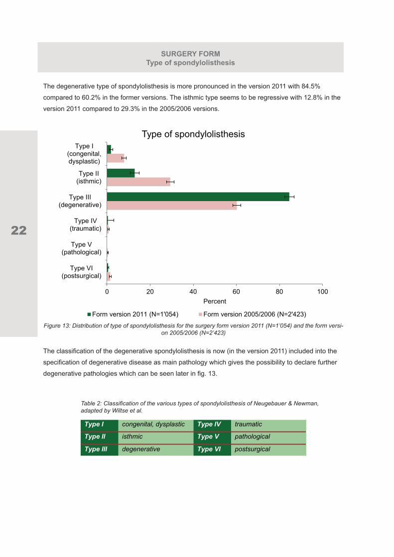
The degenerative type of spondylolisthesis is more pronounced in the version 2011 with 84.5%
compared to 60.2% in the former versions. The isthmic type seems to be regressive with 12.8% in the
version 2011 compared to 29.3% in the 2005/2006 versions.
0 20 40 60 80 100
Type VI(postsurgical)
Type V(pathological)
Type IV(traumatic)
Type III(degenerative)
Type II(isthmic)
Type I(congenital,dysplastic)
Percent
Type of spondylolisthesis
Form version 2011 (N=1'054) Form version 2005/2006 (N=2'423)
Figure 13: Distribution of type of spondylolisthesis for the surgery form version 2011 (N=1’054) and the form versi-on 2005/2006 (N=2’423)
The classification of the degenerative spondylolisthesis is now (in the version 2011) included into the
specification of degenerative disease as main pathology which gives the possibility to declare further
degenerative pathologies which can be seen later in fig. 13.
Table 2: Classification of the various types of spondylolisthesis of Neugebauer & Newman, adapted by Wiltse et al.
Type I congenital, dysplastic Type IV traumatic
Type II isthmic Type V pathological
Type III degenerative Type VI postsurgical

23
The changes in the distribution of type of spondylolisthesis are similar to the distribution of the
aetiology of deformities. Idiopathic is the most frequent predominant aetiology for deformity in the
2005/2006 form versions (39.6%) whereby in the newer form version 2011 degenerative is the most
frequent aetiology. This is probably due to transferal into the section “specification of degenerative
disease” which might lead to a more frequent selection of this pathology.
0 20 40 60 80
Other
M. Scheuermann
Posttraumatic
Degenerative
Neuromuscular
Congenital
Idiopathic
Percent
Predominant aetiology of deformity
Form version 2011 (N=750) Form version 2005/2006 (N=1'419)
Figure 14: Distribution of predominant aetiology of deformity for the surgery forms version 2011 (N=750) and the forms version 2005/2006 (N=1’419)
SURGERY FORMAetiology of deformity

24
SURGERY FORMAdditional deg. diseases for patients with deg. spondylolisthesis or deformity
Fig. 15 demonstrates the new possibilities of the description of additional degenerative pathologies in
patients with deg. spondylolisthesis and deg. deformity. The degenerative disc disease and facet joint
arthrosis seem to occur more often in patients with deg. deformity with 60.0% and 44.6% compared
to 33.7% and 30.0% in patients with deg. spondylolisthesis.
0 10 20 30 40 50 60 70
Other
Degenerativedeformity
Other instability
Degenerativespondylolisthesis
Facet joint arthrosis
Myelopathy
Foraminal stenosis
Lateral stenosis
Central stenosis
Degenerative discdisease
Disc herniation/protrusion
None
Percent
Type of other/ additional degenerative diseases for patients with degenerative spondylolisthesis and
degenerative deformity (form version 2011)
Degenerative deformity (N=448) Degenerative spondylolisthesis (N=891)
Figure 15: Distribution of type of other/ additional degenerative diseases for patients with degenerative spondylolisthesis (N=891) and degenerative deformity (N=448), surgery form version 2011
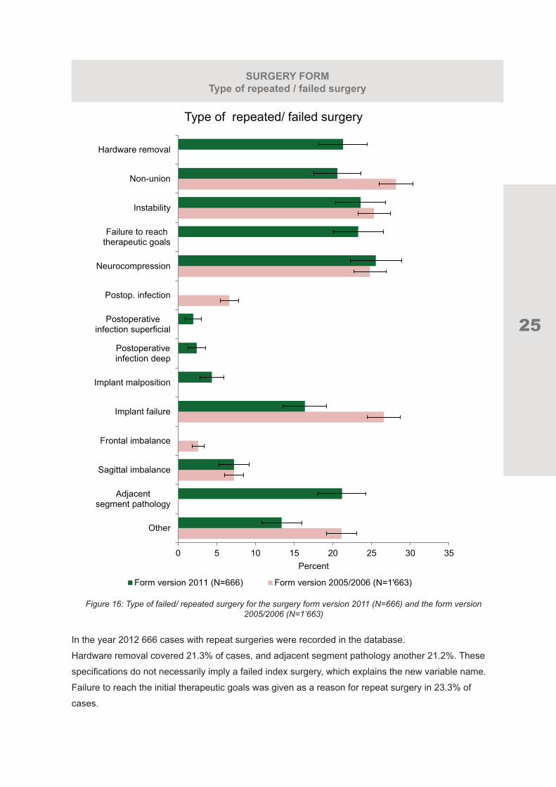
The parameter failed surgery was renamed to repeat surgery and some more specifications were
added in the version 2011. The postoperative infection can now be further specified into superficial
or deep infection. Other new specifications are hardware removal, failure to reach therapeutic goals,
implant malposition and adjacent segment pathology.
25
0 5 10 15 20 25 30 35
Other
Adjacentsegment pathology
Sagittal imbalance
Frontal imbalance
Implant failure
Implant malposition
Postoperativeinfection deep
Postoperativeinfection superficial
Postop. infection
Neurocompression
Failure to reachtherapeutic goals
Instability
Non-union
Hardware removal
Percent
Type of repeated/ failed surgery
Form version 2011 (N=666) Form version 2005/2006 (N=1'663)
Figure 16: Type of failed/ repeated surgery for the surgery form version 2011 (N=666) and the form version 2005/2006 (N=1’663)
In the year 2012 666 cases with repeat surgeries were recorded in the database.
Hardware removal covered 21.3% of cases, and adjacent segment pathology another 21.2%. These
specifications do not necessarily imply a failed index surgery, which explains the new variable name.
Failure to reach the initial therapeutic goals was given as a reason for repeat surgery in 23.3% of
cases.
SURGERY FORMType of repeated / failed surgery

26
SURGERY FORMInfection / tumor
The parameters “Affected structures of infection” and “Localization of tumor” were newly defined. The
former forms considered only the disc and vertebra as affected structure of infection. The new answer
possibilities in the version 2011 also take the epidural space and the paravertebral soft tissue into
account.
0 20 40 60 80
Other
Paravertebralinfection
Epiduralspace
Discitis
Spondylitis
Percent
Affected structures of infection (surgery form version 2011)
Figure 17: Distribution of affected structures of infection, surgery form version 2011 (N=101)
0 20 40 60 80
Extraosseous(intradural)
Extrasosseou(extradural)
Intraosseous(deep)
Intraosseous(superficial)
Extraosseoussoft tissues
Percent
Localization of tumor(surgery form version 2011)
Figure 18a/ b: Distribution of localization of tumor for a: the surgery form version 2011 (N=296) and b: the form version 2005/2006 (N=1’171)
0 20 40 60 80
Vertebral body
Posterior bonyelements
Extradural
Intraduralextramedullary
Intraduralintramedullary
Other
Percent
Localization of tumor(surgery form version 2005/2006)

27
SURGERY FORMTherapeutic goals / goals of surgery
0 20 40 60 80 100
Other
Diagnostic measures
Cosmeticimprovement
Prophylacticdecompression
Stop deformityprogression
Spinal stabilization
Bladder/sex functionimpr.
Neurologicalimprovement
Sensory improvement
Motor improvement
Functionalimprovement
Pain relief
Peripheral pain relief
Axial pain relief
Percent
Therapeutic goals/ goals of surgery
Form version 2011 (N=8'946) Form version 2005/2006 (N=41'735)
Figure 19: Distribution of therapeutic goals/ goals of surgery for the surgery form version 2011 (N=8’946) and the form version 2005/2006 (N=41’735)
The therapeutic goals can be more precisely defined in the form version 2011. Pain relief was split-
ted into axial and peripheral pain relief to consider back/ neck and leg/ arm pain. The neurological
improvement can now be specified as sensory, motor and bladder/sex function improvement. Further
new answer options are spinal stabilization, stop deformity progression and prophylactic decompres-
sion.

28
SURGERY FORMProphylaxis / fusion promoting measures
0 20 40 60 80 100
Other
Ossification
Thrombembolism
Infection
None
Percent
Prophylaxis (surgery form version 2011)
Figure 20: Distribution of type of prophylaxis, surgery form version 2011 (N=8’769)
Prophylaxis is a new question in the form version 2011. Infection prophylaxis was performed in 87.3%
of cases, thromboembolic prophylaxis in 79.6% of cases.
0 5 10 15 20 25 30
Other
Posterior fusion
Posterolateralfusion
Other interbodyfusion
Interbody fusion(XLIF)
Interbody fusion(TLIF)
Interbody fusion(PLIF)
Interbody fusion(A-IF)
Percent
Specification of fusion promoting measures (surgery form version 2011)
Figure 21: Specification of fusion promoting measures, surgery form version 2011 (N=3’920)

29
The fusion promoting measures can now be more precisely specified in terms of the different types of
interbody fusion. The most frequently performed interbody fusions are A-IF and PLIF with 20.6% and
19.4%. A TLIF was performed in 16.3% and a XLIF in 6.1% of cases where a fusion was performed.
The posterolateral fusion was the most frequently performed fusion promoting measure in total with
nearly 25%.
0 10 20 30 40 50
Other
BMP or similar
Cement
Bone substitute
Allogenic bone
Autologous bonelocally procured
Autologous boneharvested
None
Percent
Fusion material (surgery form version 2011)
Figure 22: Specification of fusion material, surgery form version 2011 (N=3’920)
The specification of the fusion material was also redesigned. Especially for autologous bone it can
now be distinguished whether the bone was locally produced e.g. during decompression or whether
the bone was harvested e.g. via beck crest biopsy.
SURGERY FORMFusion material

30
SURGERY FORMIntraoperative complications
The surgical complications are now divided into intraoperative complications and complications
occurring during hospitalization before discharge.
For intraoperative complications which are shown in fig. 23 the dura lesion was the most frequent
complication with 4.3%. This rate is similar to the distribution of the complications of the form version
2005/2006 (see figure 25). No intraoperative surgical complications occurred in 93.4% of the cases, in
1.3% of the cases they were not documented.
0 1 2 3 4 5
Not documented
Other
Fx vertebral structures
Vascular injury
Dura lesion
Spinal cord damage
Nerve root damage
Percent
Intraoperative surgical complications(surgery form version 2011)
Figure 23: Distribution of intraoperative surgical complications, excluded was the answer “none”, surgery form version 2011 (N=8’947)
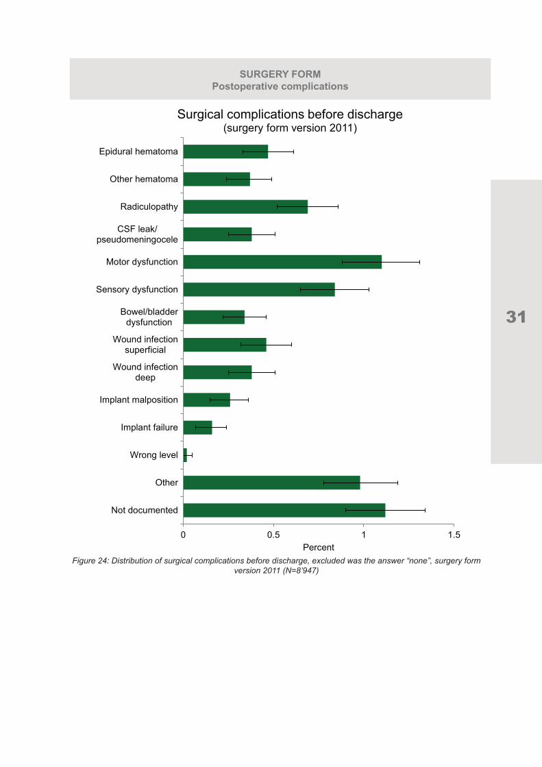
Postoperative complications which occurred during hospitalization are shown in figure 24.
The most frequent complications were motor dysfunction with 1.1%, sensory dysfunction with 0.8%
and radiculopathy with 0.7%. Even though a dura lesion was the most frequent complication during
surgery, a CSF leak/ pseudomeningocele occurred in only 0.4% of cases. In 1.1% of cases the
complications before discharge were not documented, in 94.2% of cases no complications occurred.
31
SURGERY FORMPostoperative complications
0 0.5 1 1.5
Not documented
Other
Wrong level
Implant failure
Implant malposition
Wound infectiondeep
Wound infectionsuperficial
Bowel/bladderdysfunction
Sensory dysfunction
Motor dysfunction
CSF leak/pseudomeningocele
Radiculopathy
Other hematoma
Epidural hematoma
Percent
Surgical complications before discharge (surgery form version 2011)
Figure 24: Distribution of surgical complications before discharge, excluded was the answer “none”, surgery form version 2011 (N=8’947)

32
SURGERY FORMComplications
The distribution of surgical postoperative complications in the former form version 2005/2006 is given
in figure 25. The dura lesion rate was 2.8%. Bleeding in and outside of the spinal canal occurred in
0.4% of cases. The wound infection rate was 0.5% like the implant failure rate.
In 94.4% of these 39’721 surgeries documented with the form version 2005/2006 no surgical
complications appeared.
0 1 2 3 4
Other
Implant failure
Woundinfection
Duralesion
Malpositionof implant
Bleeding out-side spinal canal
Bleedingin spinal canal
Spinal corddamage
Cauda equinadamage
Nerve rootdamage
Wrong level
Percent
Distribution of surgical complications(surgery form version 2005/2006)
Figure 25: Distribution of surgical complications, excluded was the answer “none”, surgery form version 2005/2006 (N=39’721)

33
The status of complications at discharge refers to all cases with an intra and/ or postoperative complication at hospitalization. For the sample based on the form version 2011 812 cases with complications were documented. In 61.6% of those cases the complications were resolved at discharge, in 9.0% they were persisting. The sample based on the form version 2005/2006 had a higher rate of resolved complications at discharge with 77.7%. Here in only 5.7% of cases the complications were persisting at discharge.
0 20 40 60 80 100
Persisting
Improved
Resolved
Percent
Status of complications at discharge
Form version 2011 (N=812) Form version 2005/2006 (N=1'675)
Figure 26: Status of complications at discharge for patients with an intraoperative complication and/ or a compli-cation after discharge, for surgery form version 2011 (N=812) and form version 2005/2006 (N=1’675)
A new question is the forseen follow-up. This parameter was added to have the opportunity to calculate realistic follow-up rates. In 96.1% of cases a follow-up was foreseen.
0 20 40 60 80 100
No
Yes
Percent
Follow-up forseen at discharge(surgical form version 2011)
Figure 27: Follow-up forseen, surgery form version 2011 (N=8’749)
SURGERY FORMStatus of complications / FU forseen

34
FOLLOW-UP FORMDistribution of follow-up interval
In the following section we refer to the Spine Tango follow-up form.
The majority of documented follow-ups in the routine clinical setting are captured at 6 weeks and 3
months after surgery. The literature suggests that at least the mid-term outcomes at three months can
basically be considered as the final outcomes (Mannion et al (2009) (4); Swespine: the Swedish spine
register: The 2012 report (8)). 6-month, 1-year and longer follow-ups are strongly desired, but remain
a major challenge of the registry.
0 10 20 30 40 50
Other
2 years
1 year
6 months
3 months
6 weeks
Percent
Distribution of follow-up intervalall available follow-up forms for patients with failed surgery
Form version 2011 (N=3'823) Form version 2005/2006 (N=13'020)Figure 28: Distribution of follow-up interval for the follow-up form version 2011 (5’905 forms for 3’823 patients) and
the form version 2005/2006 (38’466 forms for 13’020 patients)
Form version 2011: 5’905 FUs / 3’823 patients: current mean FU at 101.3 days, if last available FU is
considered. Due to the young age of the form version, the mean FU times will most likely increase in
the future.
Form version 2005/2006: 38’466 FUs / 13’020 patients: Mean FU 348.9 days, if last available FU per
patient is considered.
4. Mannion AF, Porchet F; Kleinstück FS, Lattig F, Jeszenszky D, Bartanusz V, Dvorak J, Grob D. (2009)The quality of spine surgery from the patient`s perspective. Part 1: the Core Outcome Measures Index in clinical practice. Eur Spine J. 18 Suppl 3:367-738. Strömqvist B, Fritzell P, Hägg O, Jönsson B, Sandén B; Swedish Society of Spinal Surgeons (2013)Swespine: the Swedish spine register : The 2012 report. Eur Spine J. 2013 Apr;22(4):953-74.

35
0
10
20
30
40
50
60
70
excellent good fair poor
Percent Physician based overall outcomelast available follow-up
Form version 2011 (N=3'935) Form version 2005/2006 (N=13'577)
Figure 29: Physician based overall outcome (surgeon) for the follow-up form version 2011 (N=3’935) and the form version 2005/2006 (N=13’577)
Converting the surgeon based outcome rating into a binary format, about 4 out of 5 cases have a
desired outcome, and 1 out of 5 cases has an undesired outcome.
0
10
20
30
40
50
60
goal achieved goal part. achieved goal not achieved goals not inagreement
Percent Surgical goal: pain relief (axial and peripheral)last available follow-up
Form version 2011 - axial pain relief (N=2'292)Form version 2011 - peripheral pain relief (N=3'020)Form version 2005/2006 - pain relief (N=12'710)
Figure 30: Achievement of the surgical goal pain relief (axial and peripheral) for the follow-up form version 2011 and 2005/2006
Peripheral pain relief seems the easier to achieve surgical goal compared with axial pain relief.
FOLLOW-UP FORM Overall outcome / surgical goal - pain relief

36
FOLLOW-UP FORMSurgical goal – neurological improvement
0
10
20
30
40
50
60
goal achieved goal part. achieved goal not achieved goals not inagreement
Percent Surgical goal: neurological improvement last available follow-up
Form version 2011 - motor improvement (N=743)Form version 2011 - sensory improvement (N=735)Form version 2011 - bladder/ sex functional improvement (N=28)Form version 2005/2006 - neurological improvement (N=4'146)
Figure 31: Achievement of the surgical goal neurological improvement (motor, sensory, bladder and sex function) for the follow-up form version 2011 and 2005/2006
The main focus of neurological improvement lies on motor and sensory function, which is equally likely
achieved. Improvement of bladder and sexual function is a less frequently desired goal, but also one
that is more difficult to achieve.

37
FOLLOW-UP FORMSurgical goals – functional improvement / additional goals
0
10
20
30
40
50
60
goal achieved goal part. achieved goal not achieved goals not inagreement
Percent Surgical Goal: functional improvementlast available follow-up
Form version 2011 - functional improvement (N=1'724)
Form version 2005/2006 - functional improvement (N=9'124) Figure 32: Achievement of the surgical goal functional improvement for the follow-up form version 2011 and
2005/2006
In only about 1 out of 10 patients, a functional improvement could not be achieved.
0
10
20
30
40
50
60
70
80
goal achieved goal part. achieved goal not achieved goals not inagreement
Percent Additional surgical goals version 2011last available follow-up
Form version 2011 - spinal stabilization (N=722)Form version 2011 - stop deformity progression (N=307)Form version 2011 - prophylactic decompression (N=75)
Figure 33: Achievement of additional surgical goals (spinal stabilization (N=722), stop deformity progression (N=307) and prophylactic decompression (N=75)) for the follow-up forms version 2011
Spinal stabilization and stopping deformity progression are equally likely to achieve as surgical goals,
and in the majority of patients they are completely or partially achieved. A prophylactic decompression
is not a frequent goal, but it seems one of the easiest goals to achieve or partially achieve.

38
PART II:FAILED SURGERY ANALYSIS
For this year`s annual report an analysis of failed surgeries in the Spine Tango pool was performed.
After transition to the 2011 version, only the surgery forms 2005/06 were considered. The definition
of failed surgery, revision and reoperation procedures is not an easy one, since elective procedures
like metal removal, repeat procedures concerning an implant (revision) or repeat procedures leaving
all implanted material in place (reoperation) all need to be accommodated with one question title.
Therefore, in the new 2011 form generation, the term “repeat surgery” was introduced, in order to
cover more scenarios of a repeat intervention for various reasons.
PreFACeAdmission / PathologyIIIIIIIIIIIIIIIIIIIIIIIIIIIIIII1 2 3 4 5 6 7 8 9 10 11 12 13 14 15 16 17 18 19 20 21 22 23 24 25 26 27 28 29 30 31IIIIIIIIIIII1 2 3 4 5 6 7 8 9 10 11 12 IIIIIIIIII11 12 13 14 15 16 17 18 19 20
DayMonth Year
)I Completely fill in boxes to record answers.
Use a #2 soft pencil for marking.Text answers must be entered with the web interface.All questions must be answered unless otherwise indicated.
Directions
SPINE TANGO2011
SURGERY
III
II
II
II
II coccyx
sacrallumbo-sacral
lumbarthoraco-lumbo-sacral
thoracolumbarthoracic
cervico-thoraco-lumbarcervicothoracic
mid lower cervicalupper cervical
Extent of lesion (segments/vertebral bodies)
Level of intervention
II
II
II
II
II
other: specify ..........................repeat surgery
tumorinfection
inflammationspondylolisthesis (non degen.)
pathological fracturefracture/trauma
non degen. deformitydegenerative disease
Type of degenerationSpecification of Main Pathology
JJJJJ
JJJJJJ
other ...........................facet joint arthrosismyelopathyother instabilitydegen. spondylolisthesis
degen. deformitydegen. disc diseaseforaminal stenosislateral stenosiscentral stenosisdisc herniat./protrusion
Type of deformity
Predominant etiology
III
III
other ..........M. Scheuermannposttraumatic
neuromuscularcongenitalidiopathic
Type of (pathological) fracture/trauma
IIIII
IIIII
other ..........sacrum fracturefracture C3-L5/S1soft tissue injury neckC2 other fracture
C2 dens fractureC1/2 instabilityC1 fractureC0/1 dissociationcondylar (C0)
Dens fracture type
III
IIIIII
C3-L5/S1 AO fracture typeIII CBA
Type of inflammation
IIII
other ..........ankylosing spondylitis (M. Bechterew)seronegative arthritisinflammatory arthritis (seropos)
Localization
JJJJJJ
other ..........extraosseous (intradural)extrasosseous (extradural)intraosseous (deep)intraosseous (superficial)extraosseous soft tissues
Type of tumor
IIIII
other ..........tumor like lesionsecondary malignantprimary benignprimary malignant
Specify type of tumor
Deg
en. d
isea
se
Only answer questions related to Main Pathology (Main Pathology "other" requires no specification.).
Type of spondylolisthesis
III
II
Type VI (postsurgical)Type V (pathologic)Type IV (traumatic)
Type II (isthmic)Type I (congenital, dysplastic)
Grade ofspondylolisthesis
IIIIII
Spondyloptosis (V)Grade IVGrade IIIGrade IIGrade IGrade 0
Spon
dylo
listh
esis
Infla
mm
atio
n
Infection specification
II
III
otherfungal
tuberculoticparasiticpyogenic
Affected structure(s)
J
J
JJJ
other ...............
paravertebralinfection
epidural spacediscitisspondylitis
Infe
ctio
n
Type or reason of repeat surgery
J
JJJ
JJ
JJ
JJJJ
other ..........
adjac. segmentpathology
sagittal imbalanceimplant failure
implant malpositionpostop. infect. deep
postop. infectionsuperficial
neurocompression
failure to reachtherapeutic goals
instabilitynon-unionhardware removal
Rep
eat s
urg.
.......................................................
Fracture age
II
old fracturefresh fracture
Additional fractures w/different treatments require separate forms
Pathologicalfracture due to ...
III
other ..........tumorosteoporosis
Type of scoliosis
Tum
or
(Pat
holo
gica
l) Fr
actu
re/T
raum
aD
efor
mity
Main pathology
Comments regarding main pathology: .....................................................................................................................................................................
IIIIIII0 1 2 3 4 5 >5Number of previous spine surgeries
Answer "0" excludes both "Previous surgery"questions ("at same level" and "at same hospital".)
In case of tumor, answer questions "Type oftumor" and "Localization" in section "TUMOR"
Last name
Street
GenderFirst name
City
Birthdate (DD.MM.YYYY)
M.R.N.
Internal Use Only / Not read by scanner
Social security number
Country code Zip code
Type III see type of degeneration
Also specify type of degenerative deformity
Specify grade of spondyl.
Specify type of deformity below
Most severely affected
IIIIIIIIIIIIIIIIIIIIIIIIIIIICOSAS1L5L4L3L2L1T12T11T10T9T8T7T6T5T4T3T2T1C7C6C5C4C3C2C1C0
II vertebral bodysegment
SA = sacrum / CO = coccyx
Format
IIminimal
complete
IIIIIIIIIIIIIIIIIIIIIIIII
>24242322212019181716151413121110987654321
JJJ
JJ
JJ
JJ
JJ other:
specify ..................repeat surgerytumor
infectioninflammation
spondylolisthesis (non-degen.)pathological fracture
fracture/traumanon-degen. deformity
degen. diseasenone
Additional pathology (Answer to question "Main pathology" is excluded.)
Previous surgeries at same level
Prev. surg. same hospital or surgeonIII partiallyyesno
Previous treatment for main pathology (by specialist)
JJJ
JJJ
> 12 mon. conservative6-12 mon. conservative3-6 mon. conservative
< 3 mon. conservativesurgicalnone
III partiallyyesno
Ris
k fa
ctor
s
III
III
unknown>3531-35
26-3020-25< 20
BMICurrentsmoker
III
unknownnoyes
TypeIII 321Group
Subgroup III 321
In segments, mark cranial VB
...................
SA = sacrum (S2-5) / CO = coccyx
J
JJJ
JJJ not
assessable/applicableblack
blueorange
yellowrednone
Presence of flags - low back painRed:Yellow:Orange:
Biomedical Factors; serious spinal pathologyPsychosocial or behavioral factorsAbnormal psychological processes indicatingpsychatric disorders
Blue:Black:
Socioeconomic/work factorsOccupational and societal factors
Question typesI only 1 answer allowedJ multiple answers allowed
mandatory questionsplease specify......
II
II
other ............combined
kyphosisscoliosis
II double curvesingle curve
Copyright MEMdoc, 2011 All rights reserved31.12.2011 / Version v1
Admission / PathologyIIIIIIIIIIIIIIIIIIIIIIIIIIIIIII1 2 3 4 5 6 7 8 9 10 11 12 13 14 15 16 17 18 19 20 21 22 23 24 25 26 27 28 29 30 31IIIIIIIIIIII1 2 3 4 5 6 7 8 9 10 11 12 IIIIIIIIIIIIIIIII00 01 02 03 04 05 06 07 08 09 10 11 12 13 14 15 16
DayMonth Year
)I Completely fill in boxes to record answers.
Use a #2 soft pencil for marking.Text answers must be entered with the web interface.All questions must be answered unless otherwise indicated.
I only 1 answer allowed J multiple answers allowedQuestion types
Last name
Street
Country code
Occupation
Zip code
Gender
Telephone
First name
City
Birthdate (DD.MM.YYYY)
Directions
M.R.N.
SSE SPINE TANGO SURGERY2006
Inte
rnal
Use
Onl
yN
ot re
ad b
y sc
anne
rI
II
II
II
II
II coccyx
sacrallumbo-sacral
lumbarthoraco-lumbo-sacral
thoracolumbarthoracic
cervico-thoraco-lumbarcervicothoracic
mid lower cervicalupper cervical
Copyright MEMdoc, 2006 All rights reserved01.10.06
Most severely affected segment/vertebral body
I
IIIIII
IIIIIIII
IIIIIIII
IIIIIIII
IIIIIIII
IIIIIIII
IIIIIIII
coccyx
sacrum(S2-5)
S1L5 / S1L5L4 / 5L4
L3 / 4L3L2 / 3L2L1 / 2L1Th12 / L1Th12
Th11 / 12Th11Th10 / 11Th10Th9 / 10Th9Th8 / 9Th8
Th7 / 8Th7Th6 / 7Th6Th5 / 6Th5Th4 / 5Th4
Th3 / 4Th3Th2 / 3Th2Th1 / 2Th1C7 / Th1C7
C6 / 7C6C5 / 6C5C4 / 5C4C3 / 4C3
C2 / 3C2C1 / 2C1C0 / 1C0unknownnot applicable/assessable
Extent of lesionIIII >5 segments/vertebral bodies4-5 segments/vertebral bodies2-3 segments/vertebral bodies1 segment/vertebral body
Level of procedure
II
II
II
II
II
other: specify .........................................failed surgery
tumorinfection
inflammationspondylolisthesis
pathological fracturefracture/trauma
deformitydegenerative disease
Type of degeneration
Specification of Main Pathology
JJJJ
JJJJ
other ..........adjacent segment degen.spinal stenosisspondylarthrosis
spondylosisdisc herniationdisc degenerationblack disc
Type of deformity
II
II
other ..........combined
kyphosisscoliosis
Predominant etiology
III
IIII
other ..........M. Scheuermannposttraumatic
degenerativeneuromuscularcongenitalidiopathic
Type of (pathological) fracture/trauma
IIIII
IIIII
other ..........sacrum fracturefracture C3-L5/S1soft tissue injury neckC2 other fracture
C2 dens fractureC1/2 instabilityC1 fractureC0/1 dissoziationcondylar (C0)
Dens fracture type
III
IIIIII
C3-L5/S1 AO fracture type
III
III
III
C3C2C1
B3B2B1
A3A2A1
Type of inflammation
IIII
other ..........ankylosing spondylitis (M. Bechterew)seronegative arthritisinflammatory arthritis (seropos)
Localization
JJJJJJ
other ..........intradural intramedullaryintradural extramedullaryextraduralposterior bony elementsvertebral body
Type of tumor
IIIII
other ..........tumor like lesionsecondary malignantprimary benignprimary malignant
Specify type of tumor
Deg
ener
ativ
eD
isea
seD
efor
mity
(Pat
holo
gica
l) Fr
actu
re/T
raum
a
Only answer questions related to Main Pathology (Main Pathology "other" requires no specification.).
Type of spondylolisthesis
IIIIII
Type VI (postsurgical)Type V (pathologic)Type IV (traumatic)Type III (degenerative)Type II (isthmic)Type I (congenital, dysplastic)
Grade of spondylolisthesis
IIIIII
Spondyloptosis (V)Grade IVGrade IIIGrade IIGrade IGrade 0
Spon
dylo
listh
esis
Infla
mm
atio
n
Infection specification
II
III
other ..........fungal
tuberculoticparasiticpyogenic
Affected structure(s)
III
spondylodiscitisdiscitisspondylitis
Infe
ctio
nTu
mor
Type of failed surgery
JJ
JJJ
JJJ
other ..........frontal imbalance
sagittal imbalanceimplant failurepostop. infection
neurocompressioninstabilitynon-union
Faile
d su
rg.
.......................................................
Fracture age
II
old fracturefresh fracture
Additional fractures w/different treatments require separate forms.
Pathologicalfracture due to ...
III
other ..........tumorosteoporosis
Type of scoliosisII double curvesingle curve
Main pathology
Comments regarding main pathology: .............................................................................................................................................................
JJJ
JJ
JJ
JJ
JJ
other: specify ...........................................................failed surgerytumor
infectioninflammation
spondylolisthesispathological fracture
fracture/traumadeformity
degenerative diseasenone
Additional pathology (Answer to question "Main pathology" is excluded.)
IIIIIII0 1 2 3 4 5 >5Number of previous spine surgeries Previous surgeries at same level
Previous surgeries at same hospitalIII partiallyyesno
Previous treatment for main pathology
JJJ
JJJ
> 12 mon. conservative6-12 mon. conservative3-6 mon. conservative
< 3 mon. conservativesurgicalnoneIIIIIII0 1 2 3 4 5 >5
Answer "0" excludes both "Previous surgery" questions("at same level" and "at same hospital".) III partiallyyesno
(In case of tumor, answer questions "Type of tumor"and "Localization" in section "TUMOR"
Form version 2011
Form version 2006
39
FAILED SURGERY Demographic data
To generate a more homogeneous group the following inclusion criteria were set to define the
analyzed sample of cases.
- Main pathology: failed surgery
- Number of previous surgeries: 1, at the same or partially same level.
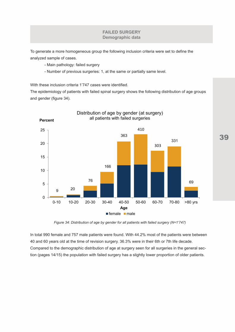
With these inclusion criteria 1’747 cases were identified.
The epidemiology of patients with failed spinal surgery shows the following distribution of age groups
and gender (figure 34).
0
5
10
15
20
25
0-10 10-20 20-30 30-40 40-50 50-60 60-70 70-80 >80 yrs
Percent
Age
Distribution of age by gender (at surgery) all patients with failed surgeries
female male
9 20
76
166
363410
303331
69
Figure 34: Distribution of age by gender for all patients with failed surgery (N=1‘747)
In total 990 female and 757 male patients were found. With 44.2% most of the patients were between
40 and 60 years old at the time of revision surgery. 36.3% were in their 6th or 7th life decade.
Compared to the demographic distribution of age at surgery seen for all surgeries in the general sec-
tion (pages 14/15) the population with failed surgery has a slightly lower proportion of older patients.

40
FAILED SURGERYType of failed surgery
The specification of the type of failed surgery of the sample is given in figure 35.
0 10 20 30 40
other
postop.infection
instability
non-union
frontalimbalance
neuro-compression
implant failure
sagittalimbalance
Percent
Type of failed surgery
Figure 35: Distribution of type of failed surgeries (N=1‘749)
The multiple choice format of the question “Type of failed surgery” needs to be considered as often
more than one type of failure was defined. The most frequent failure modes were non-union, implant
failure, neurocompression and instability with 24.3 to 27.8%.
A cluster analysis helped us to define the most frequent failure groups around the 5 following key
events:
- implant failure
- instability
- neurocompression
- non-union
- postoperative infection.
We allowed some combinations of failed surgery types but only included combinations with certain
homogeneity.

41
FAILED SURGERYDemographics of the groups
The Implant Failure group (N= 330) consists of patients with a sole implant failure and of patients who
had a combination of Implant Failure with “Other” failure reasons or with Instability or Neurocompressi-
on.
For the Instability group we allowed a combination with Neurocompression or “Other” failure reasons.
175 cases were found.
For the Neurocompression (N= 311) and Postoperative Infection group (N= 77) only a combination
with “Other” failure reasons was accepted.
Combinations of Non-Union with Neurcompression, Instability or “Other” reasons as well as a sole
Non-Union were included into the Non-Union group which counted 350 cases.
The distribution of age and gender for each group is given in the following graphs.
0
5
10
15
20
25
30
35
0-10 10-20 20-30 30-40 40-50 50-60 60-70 70-80 >80
Percent
Age
Distribution of age by gender (at surgery) patients with implant failure
female male
1722
80
97
142 6
48 44
Figure 36: Distribution of age by gender for patients with implant failure as type of failed surgery (N=330)
0
5
10
15
20
25
30
0-10 10-20 20-30 30-40 40-50 50-60 60-70 70-80 >80
Percent
Age
Distribution of age by gender (at surgery)patients with instability
female male
2 0
8 11
25
38
46
34
11
Figure 37: Distribution of age by gender for patients with instability as type of failed surgery (N=175)

42
FAILED SURGERYDemographics of the groups
0
5
10
15
20
25
0-10 10-20 20-30 30-40 40-50 50-60 60-70 70-80 >80
Percent
Age
Distribution of age by gender (at surgery)patients with neurocompression
female male
2 2
20
38
58 6054
67
10
Figure 38: Distribution of age by gender for patients with neurocompression as type of failed surgery (N=311)
0
5
10
15
20
25
30
35
0-10 10-20 20-30 30-40 40-50 50-60 60-70 70-80 >80
Percent
Age
Distribution of age by gender (at surgery)patients with non-union
female male
1 1 4
44
94102
48 50
6
Figure 39: Distribution of age by gender for patients with non-union as type of failed surgery (N=350)
For patients with implant failure (Fig. 36) and non-union (Fig. 39) over 50% of the sample was bet-
ween 40 and 60 years old at the time of revision surgery.

43
FAILED SURGERYDemographics, location
05
10152025303540
0-10 10-20 20-30 30-40 40-50 50-60 60-70 70-80 >80
Percent
Age
Distribution of age by gender (at surgery)patients with postoperative infection
female male
0 2
9
2
6
14 13
27
4
Figure 40: Distribution of age by gender for patients with postoperative infection as type of failed surgery (N=77)
In the smallest of all groups, the one with postoperative infections, 35.1% of patients were in their 7th
life decade at the time of surgery.
0
20
40
60
80
100
cervical thoracic and/ or lumbar
Percent Location of revision surgery
Implant failure (N=330) Instability (N=175) Neurocompression (N=313)Non-union (N=350) Postop infection (N=77)
Figure 41: Location of revision surgery for all five groups.
As shown in figure 41 most of the revision surgeries were performed in the thoracic and or lumbar
spine. The highest proportion of cervical revision surgeries is found for the Non-Union and the Implant
Failure group.

44
FAILED SURGERYIndex surgery, interval
In about 600 cases, the index surgery was also found in the Spine Tango data pool. The interval bet-
ween failed index surgery and the revision surgery is given in fig. 42 for each group.
0
20
40
60
80
100
120
0 - 0.5y 0.5y - 1y 1 - 1.5y 1.5 - 2y >2y
Percent Primary surgery - revision interval
Implant failure (N=145) Instability (N=55) Neurocompression (N=109)Non-union (N=74) Postop infection (N=50)
Figure 42: Distribution of interval between failed index surgery and revision surgery for all five groups.
For all groups most of the failures seem to become evident in the first months after surgery and ac-
cordingly the revision surgeries are performed within the first 6 months after the index surgery. This is
most pronounced in the postoperative infection group where 96% of cases are revised within this time
period. For the Neurocompression group 71.6% of cases fall within this range.
For patients with instability 34.5% are only revised between 6-12 months after the index surgery. The
Non-Union group shows a relative high proportion (16.2%) of patients which are only revised 2 years
after the index surgery or later.

45
FAILED SURGERYDiagnosis at index surgery
0 20 40 60 80 100
Others
Tumor
Infection
Spondylo-listhesis
Pathologicalfracture
Fracture /trauma
Deformity
Degenerativedisease
Percent
Diagnosis at index surgerypatients with failed surgery
Implant failure (N=145) Instability (N=55) Neurocompression (N=109)Non-union (N=74) Postop infection (N=50)
Figure 43: Distribution of diagnosis at failed index surgery for all 5 groups.
The diagnoses at index surgery are given in fig 43 for the 5 groups. Degenerative disease is the most
frequent diagnosis, whereby the distribution is similar to the general distribution of main pathologies in
the Spine Tango pool (see fig 16). Implant failure, Instability and Non-union show a higher proportion of
spondylolisthesis (14-16%) as main pathology at index surgery.
The surgical measures which were performed during the index surgery and the revision surgery are
listed in the following figures.

46
0 10 20 30 40 50 60 70 80
Other
Deco+Fusion+Stabrigid+Percutaneous
measures
Percutaneousmeasures
Deco+Fusion+Stabrigid+motionpreserving
Stab rigid+Fusion
Deco+Stab motionpreserving
Deco+Stab rigid
Deco+Fusion+Stabrigid
Deco+Fusion
Deco only
Percent
Surgical measures at index surgerypatients with failed surgery
Implant failure (N=145) Instability (N=55) Neurocompression (N=109)Non-union (N=74) Postop infection (N=50)
Figure 44: Surgical measures performed during failed index surgery for all five groups
The most frequently performed surgical measures for all groups were decompression alone and
decompression in combination with fusion and rigid stabilization. Decompression only had the highest
percentage of Neurocompression as type of failed surgery (58.7%). If decompression was performed
in combination with fusion and stabilization rigid the most frequent types of failed surgeries were Non-
Union with 55.4% and Implant Failure with 49.0%.
FAILED SURGERYSurgical measures at index surgery

47
0 20 40 60 80 100
Other
Deco+Fusion+Stabrigid+Percutaneous
measures
Percutaneousmeasures
Deco+Fusion+Stabrigid+motionpreserving
Stab rigid+Fusion
Deco+Stab motionpreserving
Deco+Stab rigid
Deco+Fusion+Stabrigid
Deco+Fusion
Deco only
Percent
Surgical measures at revision surgerypatients with failed surgery
Implant failure (N=204) Instability (N=100) Neurokompression (N=172)Non-union (N=122) Postop Infection (N=75)
Figure 45. Surgical measures performed during revision surgery for all five groups
The most frequent surgical measures performed during revision surgery were (further) decompression
alone for Neurocompression and Other measures for postoperative infection. In cases of Implant failure,
Instability or Non-Union, fusion with stabilization rigid with or without decompression were the principal
measures.
FAILED SURGERYSurgical measures at revision surgery

48
FAILED SURGERYComplications at index surgery
The complications documented for the failed index surgery are shown in fig 46.
0 5 10 15 20
Other
Implant failure
Wound infection
Duralesion
Malposition ofimplant
Bleeding outsidespinal canal
Bleeding in spinalcanal
Nerve root damage
Percent
Surgical complications at failed (index) surgerypatients with failed surgery
Implant failure (N=138) Instability (N=45) Neurocompression (N=91)Non-union (N=69) Postop infection (N=45)
Figure 46: Surgical complications at failed index surgery for all five groups.
During index surgery the neurocompression group showed the highest complication rates with 6.6%
bleeding in the spinal canal, 4.4% dura lesion and 2.2% malposition of implant, bleeding outside the
spinal canal and nerve root damage each. In patients who were revised due to postoperative infection
8.9% suffered from a dura lesion during index surgery; in 4.4% a wound infection occurred. In revisions
due to Instability a malposition of the implant was documented at index surgery in 4.4% of cases.

49
FAILED SURGERYOutcome lumbar
To analyze the outcomes all cases with available pre- and postoperative COMI Back forms were consi-
dered (N=246). For this subsample the outcome of back/leg pain as well as COMI score improvement
is shown. The demographic details of this lumbar group are given in figure 47. The cervical sample
resulted in too few cases for making meaningful analyses.
0
5
10
15
20
25
30
0-10 10-20 20-30 30-40 40-50 50-60 60-70 70-80 >80 yrs
Percent
Age
Distribution of age by gender (at surgery) patients with existing pre- and postop lumbar COMI
female male
0 16
15
48
72
5157
4
Figure 47: Distribution of age by gender for patients with failed surgery and existing pre- and postoperative COMI back (N=246)
The last available COMI Back form was taken into account. The mean FU time for all patients was
177.7 days after revision surgery.
0
2
4
6
8
10
Back painprerevision
Back painpostrevision
Back pain relief
VAS points Back pain (VAS)pre-, post- revision surgery and pain relief
Implant failure (N=47) Instability (N=33) Neurocompression (N=81)Non-union (N=77) Postop Infection (N=16)Figure 48: Back pain pre- and post- revision surgery and back pain relief for all five groups
Back pain reduction was similar for all groups with about 1.2-2.3 VAS points after revision surgery.

50
FAILED SURGERYOutcome lumbar
0
2
4
6
8
10
Leg painprerevision
Leg painpostrevision
Leg pain relief
VAS points Leg pain (VAS)pre-, post- revision surgery and pain relief
Implant failure (N=47) Instability (N=33) Neurocompression (N=81)Non-union (N=77) Postop Infection (N=16)
Figure 49: Leg pain pre- and post- revision surgery and leg pain relief for all five groups
Leg pain improved more in the Neurocompression and Postoperative Infection group, nearly no impro-
vement was seen in the Implant Failure group.
0
2
4
6
8
10
COMI scoreprerevision
COMI scorepostrevision
COMI scoreimprovement
COMI score COMI (back) scorepre-, post- revision surgery and improvement
Implant failure (N=47) Instability (N=33) Neurocompression (N=81)Non-union (N=77) Postop Infection (N=16)
Figure 50: COMI (back) score pre- and post- revision surgery and COMI score improvement for all five groups
The COMI score improved on average 1.78. The biggest functional improvement was seen in patients
with Infection (mean difference preop- postop 2.7).

51
FAILED SURGERYOutcome lumbar
A perfectly documented case should carry outcome information about all important time points, which
are pre-index-surgery, post-index-surgery, pre-revision-surgery, post-revision-surgery. However, this
complete outcome documentation was only available for a small patient group (N=19). Due to the
relative small sample size no further subgroups regarding type of failed surgery were built.
These 19 cases were comprised of 11 female and 8 male patients; their mean age at surgery was 63.2
years.
The mean follow-up interval from the index surgery to the post-index COMI was 177 days (~6 months).
The mean follow-up interval after revision surgery was 239 days (~8 months). The mean interval index
surgery - revision surgery was 437 days (~14 months).
0
1
2
3
4
5
6
7
8
9
10
Pre-Index OP Post-Index OP Pre-Revision OP Post-Revision OP
VASback pain
Distribution of back painpre- and post index and revision surgery
Mean MedianFigure 51: Distribution of back pain pre- and post- index and revision surgery (N=19)
Despite the small sample size one can hypothesize that pain levels after failed index surgery return to
those bevor the index intervention. Despite clear pain alleviation after revision surgery, pain levels may
not go back to those after the index intervention. More data is needed to confirm these observations.

52
FAILED SURGERYOutcome lumbar
0
1
2
3
4
5
6
7
8
9
10
Pre-Index OP Post-Index OP Pre-Revision OP Post-Revision OP
VASleg pain
Distribution of leg painpre- and post index and revision surgery
Mean MedianFigure 52: Distribution of leg pain pre- and post- index and revision surgery (N=19)
Despite the extremely large box-plots a similar dynamic of pain intensification and alleviation can be
seen. Leg pain levels after revision surgery, may, however, be lowered to a larger extent than back pain
levels.
0
1
2
3
4
5
6
7
8
9
10
Pre-Index OP Post-Index OP Pre-Revision OP Post-Revision OP
COMIscore
Distribution of COMI scorepre- and post index and revision surgery
Mean Median
Figure 53: Distribution of COMI score pre- and post- index and revision surgery (N=19)
COMI scores, reflecting back specific function, are also lower after revision surgery compared with the
failed index surgery.

53
EuropeanSpine Journal
Affiliated societiesAustrian Spine SocietyBrazilian Spine SocietyCervical Spine Research Society (European Section)Czech Spine Surgery SocietyDeutsche WirbelsäulengesellschaftDutch Spine SocietyEuroSpine SocietyFrench Society of Spine SurgeryGEER (Grupo de Estudio de Enfermedades del Raquis)G.I.S. (Italian Spine Society)Hellenic Spine SocietyPolish Society of Spinal SurgerySILACO (Sociedad IberoLatinoamericana de Columna)South African Spine SocietyTurkish Spine Society
Open Operating TheatreVideos available online at:www.springer.com/586
123
Case ReportsAccepted between 04 May 2012 and 04 May 2013
Publication of Case ReportsCase Reports will be published electronicallyonly in a yearly online supplement to the journal.
ELECTRONIC ONLYVolume 22 · Supplement 3 · May 2013
Indexed in PubMed/Medline and Science Citation Index Expanded/Journal Citation Report22 (Suppl 3) S297–S525 May 2013
New!
Videos o
nline
www.sprin
ger.c
om/58
6
Check o
ut
the additio
nal Spine T
ango an
nual
report
2012
in th
e
ESJ Live
rpool S
upplemen
tum
ESJ exemplary front page

54
0
5000
10000
15000
20000
25000
till2005
2005 2006 2007 2008 2009 2010 2011 2012
years
Australian
Austrian
British
German
Italian
Pan American
Swiss/ International
Growth rates of the various Spine Tango modules
PArtICIPAnts/ ModuLe AnALYsIs
Figure 54 displays the cumulative growth curves of the various national modules. The different starting
dates of the modules need to be considered (Swiss/International 2005, Austria 2005; Germany 2006;
North America 2007; Brazil/South America 2008; Italy 2008; Mexico 2008; Great Britain 2010; Australia
2010). During 2012 the North American, South American and Mexican modules have been combined
to the Pan American Module. The Swiss/ International module was divided into one Swiss and one
International module.
The Australian and British modules are both not available via www.eurospine.org because of national
data privacy regulations, but the contact persons for these modules are displayed on the Spine Tango
web page.
The hospitals classification of all participating 63 Spine Tango clinics can be seen in figure 55. The
highest proportion is made up by university or teaching hospitals with 40%.
Figure 56 shows an overview of the Spine Tango participating hospitals and their country of origin
until the end of 2012. We divided their total case load into primary forms and follow-up forms.
Figure 54: Growth curves (number of cases of the single Spine Tango modules over the years)

55
Figu
re 5
6: O
verv
iew
of t
he S
pine
Tan
go p
artic
ipat
ing
hosp
itals
acc
ordi
ng to
thei
r cou
ntry
of o
rigin
with
cas
e lo
ad d
ivid
ed in
to p
rimar
y fo
rms
and
follo
w-u
p fo
rms
unt
il th
e en
d of
201
2.
050
0010
000
1500
020
000
2500
030
000
3500
040
000
US
A (2
hos
pita
ls)
UK
(3 h
ospi
tals
)
Sw
itzer
land
(15
hosp
itals
)
Spa
in (1
hos
pita
l)
Slo
veni
a (1
hos
pita
l)
Sin
gapo
re (1
hos
pita
l)
Pol
and
(1 h
ospi
tal)
Net
herla
nds
(1 h
ospi
tal)
Mex
ico
(1 h
ospi
tal)
Italy
(4 h
ospi
tals
)
Ger
man
y (1
9 h
ospi
tals
)
Finl
and
(2 h
ospi
tals
)
Bra
zil (
1 ho
spita
l)
Bel
gium
(5 h
ospi
tals
)
Aus
tria
(4 h
ospi
tals
)
Aus
tralia
(2 h
ospi
tals
)
Sur
gery
form
sFo
llow
-up
form
s
Spin
eTa
ngo
form
s pe
r par
ticip
atin
g co
untr
y (a
ctiv
e ho
spita
ls)
by th
e en
d of
201
240%
20%
27%
13%
Hospital classification63 Spine Tango clinics
Universityhospital
Specialized spinecenter
General hoapital
Privat hospital
Figure 55: Hospital classification for the 63 Spine Tango clinics
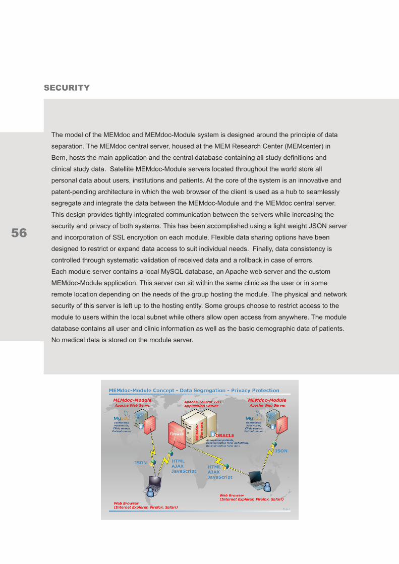
56
seCurItY
The model of the MEMdoc and MEMdoc-Module system is designed around the principle of data
separation. The MEMdoc central server, housed at the MEM Research Center (MEMcenter) in
Bern, hosts the main application and the central database containing all study definitions and
clinical study data. Satellite MEMdoc-Module servers located throughout the world store all
personal data about users, institutions and patients. At the core of the system is an innovative and
patent-pending architecture in which the web browser of the client is used as a hub to seamlessly
segregate and integrate the data between the MEMdoc-Module and the MEMdoc central server.
This design provides tightly integrated communication between the servers while increasing the
security and privacy of both systems. This has been accomplished using a light weight JSON server
and incorporation of SSL encryption on each module. Flexible data sharing options have been
designed to restrict or expand data access to suit individual needs. Finally, data consistency is
controlled through systematic validation of received data and a rollback in case of errors.
Each module server contains a local MySQL database, an Apache web server and the custom
MEMdoc-Module application. This server can sit within the same clinic as the user or in some
remote location depending on the needs of the group hosting the module. The physical and network
security of this server is left up to the hosting entity. Some groups choose to restrict access to the
module to users within the local subnet while others allow open access from anywhere. The module
database contains all user and clinic information as well as the basic demographic data of patients.
No medical data is stored on the module server.

57
All users from every MEMdoc-Module make their initial connection to the MEMdoc central server that
houses the core MEMdoc application as well as all clinical study definitions. The MEMdoc application
then recognizes the URL of the connection to determine which MEMdoc-Module to utilize and delivers
the appropriate custom module application to the user’s web browser. Each time a user requests
data the application contacts both the local MEMdoc-Module and MEMdoc central database (Oracle)
to seamlessly integrate the data from each for display. Newly entered data is likewise split so that
only internal numeric identifiers for the user, patient, clinic, department and module are stored on the
MEMdoc central database. All medical data is retrieved from and stored directly to the MEMdoc central
server and linked to the module by these internal identifiers. Medical data never passes through the
MEMdoc-Module server and is never stored on the MEMdoc-Module server. The birth year and gender
of each patient are the only pieces of personal information stored on the MEMdoc central database for
performing pooled statistics.
The physical and network security of all the MEMdoc servers is maintained by IEFO (Institute
for Evaluative Research in Orthopedic Surgery) at the MEM Research Center. This includes the
MEMdoc central (web) server, the MEMdoc database server and the MEMdoc statistics (SAS) server.
All servers are physically housed at the MEMcenter in a dedicated, locked, climate controlled and
monitored server room. The network is protected by a Sonicwall NSA 3500 firewall with real-time
gateway anti-virus, anti-spyware, anti-spam and intrusion prevention. The firewall only allows access
to the servers from the outside via port 443. Additional access is restricted to connections from within
the MEMcenter. Web security is controlled by a DigiCert certified SSL web server certificate with 256-
bit encryption on the MEMdoc central server and on each satellite module. Each server is continuously
monitored to log all connections and to detect any suspicious activity. Additionally, any modules that
are hosted at the MEMcenter fall within the same security parameters.
The following hardware is recommended for a MEMdoc-Module:
Processor (1 CPU) Intel Xeon 3500 / AMD Opteron
Memory 4 GB RAM
Hard drive (2 drives) 250 GB, Sata or SAS
RAID-Controller with battery backup unit (Raid 1)
Debian 6
or a virtual machine with comparable performance

58
Application flow and features, although much appreciated by users, are secondary to easy and flex-
ible data collection methods. With this in mind the MEMdoc application has expanded data collection
facilities for all forms of medical data including image and implant data. OMR (optical mark recogni-
tion), a proven technology for data collection, has long been a staple of the MEMdoc application and
has been expanded and simplified in Release 4 with support for both Windows and Mac users from
all popular web browsers. Image capture has been extended with a redesigned image upload func-
tion that allows multiple uploads for a single case as well as simplified image retrieval for subsequent
analysis and presentations. The SEDICO (secure data integration concept) implant capture tool, previ-
ously only available to users documenting on the MEMdoc central server, is now available to MEMdoc-
Module users. Users who do not use the SEDICO interface can now use an inexpensive hand-held
barcode reader to scan implant barcodes directly into the recorded questionnaires. Finally, access to
the ever growing list of online suppliers and products has been enhanced with a faster search engine
and the ability to segregate products by category (e.g. cups, heads, stems, liners, etc.) and anatomical
location of use.
IMPLAnt CAPture

59
While one of the major advantages of the MEMdoc system lies with its web-based access that allows
contributions from users around the world to create a global data pool, IEFO is keenly aware that
MEMdoc is not the only documentation solution available. Users of other systems, however, may still
want to contribute to a nationally endorsed data pool like Spine Tango housed exclusively on MEMdoc.
To this end IEFO developed the MEMdoc Web Service. This tool provides an interface that can be
integrated into existing hospital information systems (HIS) and virtually any third-party data collection
system to facilitate data entry, reduce physician workload and decrease data entry errors. Through
our web service, applications can request the latest version of the definition of our data collection
forms along with the complete set of validation rules. It is then up to the third-party application to col-
lect the data and apply the rules. Once the data is collected it can then be contributed to the MEM-
doc central repository. We developed such a system to ensure that the data received from external
sources was of the same quality as that recorded directly on MEMdoc. Hence, redundant data entry
is reduced while the value of the collected information is increased via silent synchronization of local
data with a central instance. Such an interface between commercial systems and an academic docu-
mentation portal is unique and the combination of the advantages of both approaches goes beyond
the current state of art of medical documentation.

60
AVAILABLe QuestIonnAIres
Table 3: Available questionnaires in the SS
E S
pine Tango registry (01.01.2013)
onlin
e available
OM
R p
aper form
s available
Forms u
sed in
Sp
ine Tan
go R
egistry -
01
.01
.20
13
multilingual
english
german
french
italian
spanish
english
german
french
italian
spanish
portuguese
turkish
polish
greek
hungarian
norwegian
dutch
russian
Reg
istry Forms
Spine Tango
Surgery 2006
Spine Tango
Staged 2006
Spine Tango
Follow-up 2006
Spine Tango
Surgery 2011
IP
Spine Tango
Staged 2011
IP
Spine Tango
Follow-up 2011
IP
Spine Tango
Conservative treatm
ent 2011
Patien
t Forms
Spine Tango
Core O
utcome M
easures Index: CO
MI N
eck
IP
IPIP
Spine Tango
Core O
utcome M
easures Index: CO
MI B
ack
IP
IPIP
Spine Tango
Core O
utcome M
easures Index: CO
MI N
eck Conservative
Spine Tango
Core O
utcome M
easures Index: CO
MI B
ack Conservative
Spine Tango
Osw
estry Disability Index O
DI 2.1
Spine Tango
Neck D
isability Index ND
I
IP
IP
Spine Tango
Scoliosis R
esearch Society: S
RS 30
Spine Tango
EuroQol™
: EQ-5D
™
Spine Tango
RM
Q -
Low B
ack and Disability Q
uestionnaire
IPIP
IPIP
IPIP
IPIP
IPIP
IP
Spine Tango
SAQ
Scoliosis appearance Q
uesitonnaire
IPIP
IPIP
IP = in process
OM
R =
Optical M
ark Reader

61
PAPERS IN PEER REVIEWED JOURNALS
Aghayev E, Henning J, Munting E, Diel P, Moulin P, Röder C; SWISSspine and Spine Tango Registry groupComparative effectiveness research across two spine registries.Eur Spine J. 2012 Aug;21(8):1640-7. Epub 2012 Mar 14.
Fankhauser C, Mutter U, Aghayev E, Mannion AValidity and responsiveness of the Core Outcome Measures Index (COMI) forthe neck.Eur Spine J 2012 Jan;21(1):101-14.
Lattig F, Fülöp Fekete T, Kleinstück FS, Porchet F, Jeszenszky D, Mannion AFLumbar Facet Joint Effusion on MRI as a Sign of Unstable Degenerative Spondylolisthesis: Should it Influence the Treatment Decision?J Spinal Disord Tech. 2012 Jul 24. [Epub ahead of print]
Röder C, Errico TJ, Spivak JM, Murray M, Protopsaltis T, Lis A, Nordin M,Bendo JHospital for Joint Diseases participates in international spine registry SpineTango after successful pilot study.Bull NYU Hosp Jt Dis. 2012;70(4):254-8.
Sobottke R, Aghayev E, Röder C, Eysel P, Delank S, Zweig TPredictors for surgical, general and follow-up complications in lumbar spinalstenosis relative to patient age.Eur Spine J 2012 Mar;21(3):411-7.
PuBLICAtIons

62
ORAL PRESENTATIONS
May 2012Eysel P, Röder C, Aghayev E, Sobottke RThe influence of age on outcome after surgical decompression of degenerativelumbar spinal stenosis.13th EFORT Congress 2012, Berlin, Germany.23rd - 25th May 2012
October 2012Sobottke R, Munting E, Röder C, Aghayev ELumbar spinal stenosis: Are there objective reasons for fusion or instrumentedsurgery? A Spine Tango registry study.German Conference for Orthopedics and Traumatology (DKOU: DeutscherKongress für Orthopädie und Unfallchirurgie), Berlin, Germany.23rd – 26th October 2012
Röder CPotentials of analyzing an international spine register - from assessing standardsof care to creating benchmarks.Annual meeting of the Norwegian Orthopaedic Society, Spine Symposium,Oslo, Norway.25th October 2012
December 2012Sobottke R, Siewe J, Eysel P, Röder C, Aghayev EInfluence of number of reoperations and reoperation cause on the post-revisionpatient results7th Annual conference of the German Spine Society (DWG), Stuttgart, Germany.6th-8th December
POSTER PRESENTATIONS
December 2012Röder C, Aghayev E, Munting EThe Spine Tango Benchmarking Project: Comparison of instrumented fusionand total disc arthroplasty in patients with cervical disc protrusion with radiculopathy.7th Annual conference of the German Spine Society (DWG), Stuttgart, Germany.6th-8th December 2012

63
Christoph Röder, MD MPHDirector a.I.
Institute for Evaluative Research in Orthopaedic SurgeryUniversity of Berne, Switzerland
Michal Neukamp, MD MScSpine Tango Support & Research
Institute for Evaluative Research in Orthopaedic SurgeryUniversity of Berne, Switzerland
Emin Aghayev, MD MScStatistics
Institute for Evaluative Research in Orthopaedic SurgeryUniversity of Berne, Switzerland
Markus Melloh, MD PhD MPH MBAOrthopaedic surgeon, EuroSpine (Past Fellow)
Thomas Zweig, MDOrthopaedic surgeon, EuroSpine (Past Fellow)
Everard Munting, MDForner Chair Spine Tango Committee, EuroSpine
Clinique Saint PierreOttignies, Belgium
Tim Pigott, MDNew Chair Spine Tango Committee, EuroSpineWalton Centre for Neurology and Neurosurgery
Liverpool, United Kingdom
