spindle cell sarcoma of bone arising from a non-ossifying ... report.pdf · classification was...
TRANSCRIPT
ww.sciencedirect.com
j o u r n a l o f c l i n i c a l o r t h o p a e d i c s a n d t r a uma 4 ( 2 0 1 3 ) 8 0e8 4
Available online at w
journal homepage: www.elsevier .com/locate/ jcot
Case Report
Spindle cell sarcoma of bone arising from a non-ossifyingfibroma: A case report
Alessio Biazzo MDa,*, Costantino Errani MDb, Marco Gambarotti MDc,Massimiliano De Paolis MDa, Davide Maria Donati PhDa, Sandro Giannini PhDa,d
a IV Orthopaedic Clinic, Rizzoli Orthopaedic Institute, via G. C. Pupilli 1, 40136 Bologna, ItalybOrthopaedic Clinic Villa Santa Teresa, Bagheria, ItalycPathology Department, Rizzoli Orthopaedic Institute, via di Barbiano 10/1, 40136 Bologna, Italyd II Orthopaedic Clinic, Rizzoli Orthopaedic Institute, via G.C. Pupilli 1, 40136 Bologna, Italy
a r t i c l e i n f o
Article history:
Received 21 April 2013
Accepted 23 April 2013
Available online 29 April 2013
Keywords:
Spindle cell sarcoma
Secondary osteosarcoma
Malignant transformation
Non-ossifying fibroma
* Corresponding author.E-mail address: [email protected] (A. B
0976-5662/$ e see front matter Copyright ªhttp://dx.doi.org/10.1016/j.jcot.2013.04.002
1. Introduction 2. Case history
The majority of malignant bone lesions, and spindle cell sar-
coma of bone is no exception, arise spontaneously. Less
frequently, they may be secondary to other conditions like
radiation therapy, bone pathologies like Paget’s disease, bone
infarct, aseptic necrosis and many benign tumors or tumor
like bone conditions like metaphyseal fibrous defect (MFD),
giant cell tumor (GCT), osteochondroma, chondroma and
others.1
This work describes a very rare case of sarcoma of bone
arisen in the same area of a benign lesion, diagnosed occa-
sionally ten years ago, which has not undergone previously
medical, radiotherapic or surgical treatment.
iazzo).2013, Delhi Orthopaedic
We report a case of a 45-year old man with a pathologic
fracture of the distal left femur because of a spindle cell sar-
coma of bone arising in a pre-existent pseudotumoral lesion
diagnosed like non-ossifying fibroma (NOF) ten years before.
We show that a sarcoma of bone can arise in the same area of
a tumor-like lesion.
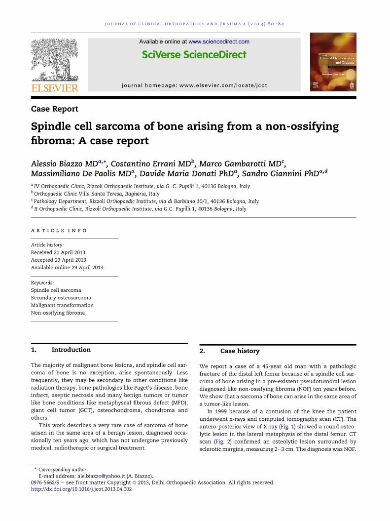
In 1999 because of a contusion of the knee the patient
underwent x-rays and computed tomography scan (CT). The
antero-posterior view of X-ray (Fig. 1) showed a round osteo-
lytic lesion in the lateral metaphysis of the distal femur. CT
scan (Fig. 2) confirmed an osteolytic lesion surrounded by
sclerotic margins, measuring 2e3 cm. The diagnosis was NOF.
Association. All rights reserved.
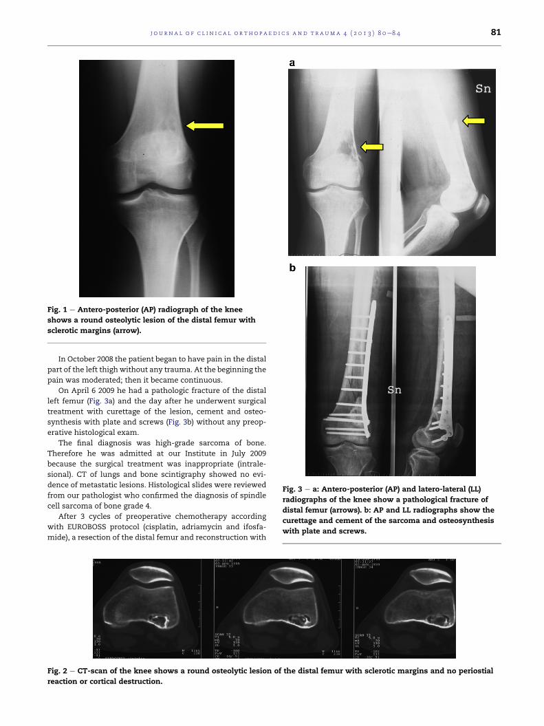
Fig. 1 e Antero-posterior (AP) radiograph of the knee
shows a round osteolytic lesion of the distal femur with
sclerotic margins (arrow).
Fig. 3 e a: Antero-posterior (AP) and latero-lateral (LL)
radiographs of the knee show a pathological fracture of
distal femur (arrows). b: AP and LL radiographs show the
curettage and cement of the sarcoma and osteosynthesis
with plate and screws.
j o u rn a l o f c l i n i c a l o r t h o p a e d i c s a n d t r a uma 4 ( 2 0 1 3 ) 8 0e8 4 81
In October 2008 the patient began to have pain in the distal
part of the left thigh without any trauma. At the beginning the
pain was moderated; then it became continuous.
On April 6 2009 he had a pathologic fracture of the distal
left femur (Fig. 3a) and the day after he underwent surgical
treatment with curettage of the lesion, cement and osteo-
synthesis with plate and screws (Fig. 3b) without any preop-
erative histological exam.
The final diagnosis was high-grade sarcoma of bone.
Therefore he was admitted at our Institute in July 2009
because the surgical treatment was inappropriate (intrale-
sional). CT of lungs and bone scintigraphy showed no evi-
dence of metastatic lesions. Histological slides were reviewed
from our pathologist who confirmed the diagnosis of spindle
cell sarcoma of bone grade 4.
After 3 cycles of preoperative chemotherapy according
with EUROBOSS protocol (cisplatin, adriamycin and ifosfa-
mide), a resection of the distal femur and reconstruction with
Fig. 2 e CT-scan of the knee shows a round osteolytic lesion of the distal femur with sclerotic margins and no periostial
reaction or cortical destruction.

Fig. 4 e AP radiograph shows reconstruction of the distal
femur with modular prosthesis.
Fig. 5 e a: On gross section of the entire specimen a greyish lesio
in the metaphysis and epiphysis around the areas of the previ
around the bone. b, c: Low-power (b) and high-power (c). Prolife
diffusely infiltrating the bony trabecule. A histological diagnosi
classification was made. Tumoral necrosis after neo-adjuvant c
classification) (HematoxylineEosin, 3).
j o u r n a l o f c l i n i c a l o r t h o p a e d i c s a n d t r a uma 4 ( 2 0 1 3 ) 8 0e8 482
modular prosthesis (GMRS) was performed (Fig. 4). Then he
underwent postoperative chemotherapy with the same
protocol.
The analysis of the specimens confirmed the diagnosis of
spindle cell sarcoma of bone grade 4 (Fig. 5 aec).
During the follow-up the patient showed pulmonary nod-
ules and he underwent metastasectomy on September 2010.
At last follow-up on September 2012 the patientwas alivewith
no evidence of disease.
3. Discussion
The first descriptions of malignant transformation of a pseu-
dotumoral lesion were attributed to Hastrup2 and Bhag-
wandeen,3 who reported a case of osteogenic sarcoma of bone
arisen in a NOF, previously treated with radiation therapy and
curettage and packing with bone allograft respectively.
Then, Braddock et al4 and Rockwell et al5 described respec-
tively a case of osteosarcoma developing in a patient with
multiple enchondromatosis and in a solitary enchondroma.
Later, several authors6e8 reported coexistence between
sarcoma and MFDs, like fibrous cortical defect (FCD) and NOF,
which occur in 35e40% of growing children studied with x-
rays. These studies don’t have evidence that a sarcoma can
arise from a MFD: both FCD and osteosarcoma occur in the
same age (second decade of life) and in the same site (distal
femur and proximal tibia). Therefore, it’s to be expected that
n measuring about 9 cm in the longitudinal axis is present
ous curettage. The lesion also infiltrates the soft tissue
ration of malignant spindle cells in a storiform pattern
s of spindle cells sarcoma grade 4 according to Broder’s
hemotherapy was 57% (grade 2 according to Huvos
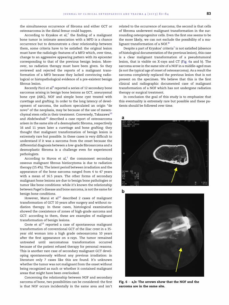
Fig. 6 e a,b: The arrows show that the NOF and the
sarcoma are in the same site.
j o u rn a l o f c l i n i c a l o r t h o p a e d i c s a n d t r a uma 4 ( 2 0 1 3 ) 8 0e8 4 83
the simultaneous occurrence of fibroma and either GCT or
osteosarcoma in the distal femur could happen.
According to Kryakos et al,7 the finding of a malignant
bone tumor in intimate association with a MFD is a chance
occurrence but to demonstrate a clear relationship between
them, some criteria have to be satisfied: the original lesion
must have the radiologic features of a MFD which, over time,
change to an aggressive appearing pattern with its epicenter
corresponding to that of the previous benign lesion. More-
over, no radiation therapy must have been given. So they
reviewed and rejected the reports of a malignant trans-
formation of a MFD because they lacked convincing radio-
logical or histopathological evidence of a pre-existent benign
fibrous lesion.
Recently Picci et al9 reported a series of 12 secondary bone
sarcomas arising in benign bone lesions as GCT, aneurysmal
bone cyst (ABC), NOF and simple bone cyst treated with
curettage and grafting. In order to the long latency of devel-
opment of sarcoma, the authors speculated an origin “de
novo” of the neoplasia, may be because of the use of mesen-
chymal stem cells in their treatment. Conversely, Takazawa10
and Abdelwahab11 described a case report of osteosarcoma
arisen in the same site of a desmoplastic fibroma, respectively
16 and 11 years later a curettage and bone grafting: they
thought that malignant transformation of benign lesion is
extremely rare but possible. In these cases is very difficult to
understand if it was a sarcoma from the onset because the
differential diagnosis between a low-grade fibrosarcoma and a
desmoplastic fibroma is a challenge even for experienced
pathologists.
According to Huvos et al,1 the commonest secondary
osseous malignant fibrous histiocytoma is due to radiation
therapy (15.4%). The latent period between irradiation and the
appearance of the bone sarcoma ranged from 4 to 47 years
with a mean of 16.5 years. The other forms of secondary
malignant bone lesions are due to benign bone pathologies or
tumor like bone conditions: while it’s known the relationship
between Paget’s disease and bone sarcoma, is not the same for
benign bone conditions.
However, Marui et al12 described 2 cases of malignant
transformation of GCT 10 years after surgery and without ra-
diation therapy. In these cases, histological examination
showed the coexistence of zones of high-grade sarcoma and
GCT: according to them, these are examples of malignant
transformation of benign lesions.
Grote et al13 reported a case of spontaneous malignant
transformation of conventional GCT of the iliac crest in a 35-
year old woman into a high grade osteosarcoma 10 years
after the first appearance on x-rays. The tumor remained
untreated until sarcomatous transformation occurred
because of the patient refused therapy for personal reasons.
This is another rare case of secondary malignant GCT devel-
oping spontaneously without any previous irradiation: in
literature only 7 cases like this are found. It’s unknown
whether the tumor was not malignant from the onset without
being recognized as such or whether it contained malignant
areas that might have been overlooked.
Concerning the relationship between NOF and secondary
sarcoma of bone, two possibilities can be considered: the first
is that NOF occurs incidentally in the same area and isn’t
related to the occurrence of sarcoma; the second is that cells
of fibroma underwent malignant transformation in the sur-
rounding osteoprogenitor cells. Even the first one seems to be
the more likely, we can not exclude the possibility of a ma-
lignant transformation of a NOF.8
Despite a part of Kryakos’ criteria7 is not satisfied (absence
of histological documentation of the previous lesion), this case
is a clear malignant transformation of a pseudotumoral
lesion, that is visible on X-rays and CT (Fig. 6a and b). The
sarcoma arose in the same site of a NOF in amiddle-agedman
(is not the typical age of onset of osteosarcoma). As a result the
sarcoma completely replaced the previous lesion that is not
present on the specimen. We believe that this is the first
clinical and radiographic documented case of malignant
transformation of a NOF which has not undergone radiation
therapy or surgical treatment.
In conclusion the goal of this study is to emphasize that
this eventuality is extremely rare but possible and these pa-
tients should be followed over time.

j o u r n a l o f c l i n i c a l o r t h o p a e d i c s a n d t r a uma 4 ( 2 0 1 3 ) 8 0e8 484
Conflicts of interest
No benefits in any form have been received or will be received
from a commercial party related directly or indirectly to the
subject of this article.
r e f e r e n c e s
1. Huvos AG, Woodard HQ, Heilweil M. Postradiation malignantfibrous histiocytoma of bone. Am J Surg Pathol. 1986;10(1):9e18.
2. Hastrup J, Jensen TS. Osteogenic sarcoma arising in a non-osteogenic fibroma of bone. Acta Pathol Microbiol Scand.1965;63:493e499.
3. Bhagwandeen SB. Malignant transformation of a non-osteogenic fibroma of bone. J Pathol Bacteriol. 1966;92:562e564.
4. Braddock GTF, HadlowVD. Osteosarcoma in enchondromatosis(Ollier’s disease). Report of a case. J Bone Joint Surg. 1966;48-B:145e149.
5. Rockwell MA, EnnekingWF. Osteosarcoma developing insolitary enchondroma of the tibia. J Bone Joint Surg.1971;53(2):341e344.
6. Abdelwahab IF, Klein MJ, Kenan S, et al. Coexistence ofprimary bone tumors: report of 4 cases of collision tumors.Can Assoc Radiol J. 2002;53(5):296e302.
7. Kryakos M, Murphy WA. Concurrence of metaphysealfibrous defect and osteosarcoma. Skeletal Radiol.1981;6:179e186.
8. Suginoshita T, Kusuzaki K, Nagaoka T, et al. Case report:natural development of osteosarcoma from precancerouslesion. Anticancer Res. 2000;20:511e514.
9. Picci P, Sieberova G, Alberghini M, et al. Late sarcomadevelopment after curettage and bone grafting of benign bonetumors. Eur J Radiol. 2011;77:19e25.
10. Takazawa K, Tsuchiya H, Yamamoto N, et al. Osteosarcomaarising from desmoplastic fibroma treated 16 years earlier: acase report. J Orthop Sci. 2003;8(6):864e868.
11. Abdelwahab IF, Klein MJ, Hermann G, Steiner GC,Yang DC. Osteosarcoma arising in a desmoplasticfibroma of the proximal tibia. Am J Roentgenol. 2002;178(3):613e615.
12. Marui T, Yamamoto T, Yoshihara H, Kurosaka M, Mizuno K,Akamatsu T. De novo malignant transformation of giant celltumor of bone. Skeletal Radiol. 2001;30:104e108.
13. Grote HJ, Braun M, Kalinski N, et al. Spontaneous malignanttransformation of conventional giant cell tumor. SkeletalRadiol. 2004;33:169e175.