spinal tumors- imaging
TRANSCRIPT

SPINAL TUMORS

Tumors of the spine are important due to
their potentially devastating clinical
effects and challenging radiographic
appearance.
In establishing the differential diagnosis
for a spinal lesion, location is the most
important feature, but the clinical
presentation and the patient‟s age and
gender are also important.

CLASSIFICATION OF LESIONS
Spinal tumors are subdivided according to
their point of origin:
Intramedullary,
Extramedullary – Intradural,
Extradural.

Intramedullary Extramedullary
Intradural, Extradural

Intramedullary Tumors
Single: Multiple:
Ependymoma, Hemangioblastomas,
Myxopapillary ependymoma, Metastases
Astrocytoma, Lymphoma
Ganglioglioma,
Hemangioblastoma,
Subependymoma,
Paraganglioma

Intradural-extramedullary Tumors
Single: Multiple:
Meningiomas, All except
Nerve sheath tumors, paraganglioma
Intradural metastases,
Lymphoma/leukemia,
Paraganglioma

Extradural Tumors
Single: Multiple:
Aneurysmal bone cyst, Metastatic disease
Giant cell tumor, Hemangiomas
Osteoblastoma, Multiple myeloma
Osteochondromas, Lymphoma.
Chordoma,
Chondrosarcoma,
Chondroblastoma,
Metastasis,
Hemangioma,
Solitary Plasmacytoma,
Lymphoma.

Epidural Lesions:
Angiolipoma
Angiomyolipoma,
Epidural lipomatosis,
Lymphoma

Intramedullary tumors
Intramedullary spinal cord neoplasms are
rare, accounting for about 4-10 percent of
all central nervous system tumors.
Intramedullary tumors include
1. Gliomas (ependymomas, astrocytomas
and gangliogliomas) and
2. Nonglial tumors (such as
hemangioblastomas, lymphoma and
metastases).
Ependymomas
The most common intramedullary
neoplasm in adults.
Usually occurs in the cervical region.
Slightly more common in women of 40
to 50 years of age.
Increased incidence of these tumors in
patients with NF-2.

The symptoms are chronic and consist
mainly of pain, myelopathy and
radiculopathies.
Occasionally, these ependymomas may
bleed and produce subarachnoid
hemorrhage or hematomyelia and hence
acute symptoms.

Ependymomas are characterized by slow growth and compress rather than infiltrate adjacent spinal cord tissue, generally yielding a cleavage plane that aids in surgical resection.
These lesions arise from ependymal cells that line the central canal and therefore tend to be central in location with respect to the spinal cord. Almost all spinal cord ependymomas are low grade.
Malignant ependymomas are quite rare.

Imaging
On MRI, iso- to hypointense on T1WI and
hyperintense on T2WI.
Ependymomas tend to produce symmetric spinal
cord expansion and usually have solid and cystic
components.
Cysts can be intratumoral, located within the solid
tumor, or peritumoral (polar), occurring at the cranial
or caudal aspects of the tumor.
These cysts are not specific for ependymomas and
can be seen with astrocytomas, hemangioblastomas
and gangliogliomas.

Intratumoral cysts should be resected with the solid tumor, as they may contain tumor cells within them. However, polar cysts do not contain malignant cells and, therefore, need not be resected.
The solid components of ependymomas usually enhance avidly, although the degree of enhancement may vary considerably.
In addition, ependymomas can hemorrhage, resulting in the “cap sign”, a hypointense rim at the periphery of the tumor on T2-weighted imaging that is related to hemosiderin deposition from prior hemorrhage

Clear tumor margins, more uniform
enhancement and central locations can
help differentiate ependymomas from
other intramedullary spinal cord tumors.
Spinal cord ependymomas may result in
metastases in the subarachnoid space.


Myxopapillary Ependymoma
Myxopapillary ependymomas represent the most frequent type of ependymomas found at the conus medullaris-cauda equina- filum terminale level.
Neuroectodermal tumors.
Mainly observed during the fourth decade of life.

Pediatric cases have been rarely described at an age range of 10 to 13 years.
Manifests with lower back or sacral pain and weakness or sphincter dysfunction.
Thought to arise from the ependymal glia of the filum terminale.

Imaging
Myxopapillary ependymomas are lobulated,
sausage-shaped masses that are often
encapsulated.
Isointense relative to the spinal cord on T1WI
and hyperintense on T2WI
Hyperintensity on both T1 and T2WI may be
noted occasionally, a finding that reflects
mucin content or hemorrhage.
Superficial siderosis may be seen but is not
specific, as it has been noted in association
with other highly vascular tumors.


The differential diagnoses of a mass arising in the filum terminale are:
Ependymoma,
Astrocytoma,
Nerve sheath tumor,
Metastases,
Paraganglioma,
Hemangioblastoma.

Subependymoma
Represent a variant of CNS ependymomas
that may also occur in the spinal cord.
Believed to have their origin from tanycytes
cells that bridge the pial and ependymal
layers.

Similar to ependymomas, these tumors
produce a slowly progressive clinical
course with pain as the most common
symptom.
Sensory and motor dysfunctions are
reported less frequently.

Imaging.
At MR imaging, they manifest with fusiform
dilatation of the spinal cord with well-defined
borders.
Unlike other ependymomas, they are
eccentrically located.
Enhancement has sharply defined margins (50
% of cases), whereas those that do not
enhance have diffuse symmetric spinal cord
enlargement.

Edema may or may not accompany the main
lesion.
Spinal subependymoma may manifest as an
extramedullary lesion within the subarachnoid
space, perhaps secondary to leptomeningeal
heterotopic glial cells.


Astrocytomas
They are the most common childhood
intramedullary neoplasms of the spinal cord and
are second only to ependymomas in adults.
Clinical presentation varies from nonspecific
backpain to sensory and motor deficits, according
to the size and location.
In contradiction to ependymomas, astrocytomas
are located eccentrically within the spinal cord.

The vast majority of spinal cord astrocytomas
in adults are of low malignancy.
However, spinal cord astrocytomas tend to
infiltrate the cord and are, therefore, difficult to
resect completely and have worse prognosis.

Imaging.
The cervicomedullary junction and the cervico-
thoracic cord.
On MR imaging, pilocytic astrocytomas are
characterized by enlargement of the spinal
cord within a widened spinal canal.
They frequently involve a large portion of the
cord, spanning multiple vertebral levels in
length.

Tumors can show areas of necrotic-cystic
degeneration, can have a „„cyst with mural
nodule‟‟ appearance, or can be structurally
solid.
The solid components are iso- to
hypointense on T1WIs and hyperintense
on T2WIs.

The pattern of enhancement can be focal nodular, patchy or inhomogeneous, diffuse enhancement and does not define tumor margins.
Nonenhancing intramedullary astrocytomas are not uncommon.
Like ependymomas, they can have intratumoral or polar cysts but do not tend to hemorrhage and, therefore, do not usually display a cap sign.


Rarely, astrocytomas arise following
radiation therapy, either for a primary
central nervous system lesion or for a
lesion occurring outside the spine.
Radiation-induced astrocytomas tend to
be of a higher grade than idiopathic
astrocytomas.

An acute cord lesion in patients with multiple
sclerosis (MS) may be virtually
indistinguishable from an astrocytoma. MS
lesions, however, generally appear more
homogeneous than astrocytomas and typically
demonstrate a surrounding rim of normal cord
intensity, which is less common in
astrocytomas.

Gangliogliomas
Gangliogliomas are composed of a mixture of ganglion cells and neoplastic glial elements; the majority of neoplastic glial cell types are of astrocytic subtype.
Gangliogliomas are the second most common intramedullary tumor in the pediatric age group and mostly affect children between 1 and 5 years of age, as do pilocytic astrocytomas.
Cervical spine > thoracic region.

These tumors tend to have a low malignant
potential, slow growth, but they have a
significant propensity for local recurrence.
Gangliogliomas tend to be extensive on
presentation, occupying an average length of
8 vertebral segments, compared with
ependymomas and astrocytomas, which
average 4 vertebral segments in length.

Imaging
Calcification is probably the single most suggestive feature of gangliogliomas.
In the absence of gross calcification, the MR imaging appearance of gangliogliomas is nonspecific and does not allow differentiation from astrocytomas.
Solid portions have mixed iso-hypointensity on T1WI and heterogeneous iso- hyperintensity on T2WI.

Like astrocytomas, gangliogliomas tend to be
eccentrically located within the spinal cord.
Tumoral cysts are more common in
gangliogliomas than in either astrocytomas or
ependymomas.

Chronic bony changes, including scoliosis and
erosions, are often seen with gangliogliomas
due to their relatively slow growth; these are
rarely seen with ependymomas or
astrocytomas.
T1 signal characteristics of gangliogliomas are
most often mixed, possibly secondary to the
fact that gangliogliomas have a dual cell
population composed of ganglion cells and
glial elements.

T2 signal characteristics of
gangliogliomas are generally
hyperintense, although surrounding
edema is not as commonly seen as
with ependymomas or astrocytomas.
The majority of gangliogliomas show
patchy enhancement.


Hemangioblastomas
Hemangioblastomas are nonglial, highly vascular neoplasms of unknown cell origin.
Although most of these tumors (75%) are intramedullary, they may involve the intradural space or even be extradural.
Thoracic spinal cord > cervical spinal cord.
Most spinal hemangioblastomas occur sporadically, but approximately one-third of cases occur in association with Von Hippel-Lindau disease.

Imaging.
MR features of spinal hemangioblastoma
depend on the size of the tumor.
Small (<10 mm)- isointense on T1WI
hyperintense on T2WI
homogeneous enhancement,
Large (>10mm) - hypo or mixed onT1WI
heterogeneous on T2WI
heterogeneous enhancement

Small hemangioblastomas are located at the
surface of the spinal cord, most frequently at the
posterior aspect and show well-demarcated,
intense enhancement.
A hemangioblastoma larger than 24 mm is usually
accompanied by vascular flow-voids.
A tumor is not likely to be a hemangioblastoma
if it is 25 mm or larger and is not associated
with vascular flow voids on MR images.

Spinal hemangioblastomas may be associated
with syrinx that are usually more extensive
than those seen with ependymomas or
astrocytomas.
In patients with von Hippel- Lindau disease,
hemangioblastomas are often multiple and this
necessitates screening of the entire spine and
brain.


Paraganglioma
Although spinal paragangliomas are rare, they
are the third most common primary tumor to
arise in the filum terminale (after ependymoma
and astrocytoma).
MR imaging studies of these lesions typically
reveal a well-circumscribed mass that is
isointense relative to the spinal cord on T1WI
and iso- to hyperintense on T2WI

Hemorrhage is common (third most common after ependymoma and hemangioblastoma) and a low signal- intensity rim (cap sign) may be seen on T2WI.
Heterogeneous and intense enhancement is virtually always seen.
Multiple punctate and serpiginous structures of signal void due to high-velocity flow may be seen around and within the tumors on all sequences.


Intramedullary Lymphoma
Primary intramedullary spinal lymphomas are extremely rare.
These tumors are of the non-Hodgkin variety and can occur in both the immunocompromised and immunocompetent patients.
The majority of these tumors occur in the cervical or thoracic regions of the spinal cord.

They are solid tumors without necrosis.
Marked T2 hyperintensity and enhance following gadolinium administration.
There is no associated syringomyelia.
Clinically, these patients initially respond to steroid treatment for a short time but usually recur after treatment.

Intramedullary Metastases
Intramedullary spinal cord metastases are rare.
Usually involve the cervical cord.
The most common primary tumors that metastasize to the spinal cord include lung, breast, colon, lymphoma and kidney.

On MRI, intramedullary spinal cord
metastases are T1- hypointense, T2-
hyperintense and demonstrate homogeneous
enhancement.
The amount of surrounding edema is out of
proportion to the size of the lesion.


INTRADURAL EXTRAMEDULLARY
TUMORS
Since the arachnoid is essentially
continuous with the dura in the spine,
intradural lesions are located in the
subarachnoid space.

Meningiomas
Spinal meningiomas have a strong female predominance with a peak occurrence in the fifth and sixth decades.
Multiples spinal meningiomas are seen in patients with NF-2.
Most spinal meningiomas are found in the thoracic spine, followed by the craniocervical junction and the lumbar region.
Although most thoracic and lumbar meningiomas are based on the posterior dura, craniocervical ones may be anterior or posterior in location.

Typically, these lesions demonstrate T1 and T2 signal that is isointense with the spinal cord and display intense homogeneous enhancement.
A dural tail may be seen, reflecting tumor spread or reactive changes in the dura adjacent to the tumor.
CT may show intratumoral calcifications and this finding may aid in distinguishing between meningiomas and nerve sheath tumors, which do not contain calcifications.
Occasionally, spinal meningiomas have a plaque-like configuration and may encircle the cord.


Nerve Sheath Tumors
Schwannomas and Neurofibromas.
Schwannomas are most common, while
neurofibromas generally occur in
association with neurofibromatosis
(especially NF-1).

Approximately 50 percent of nerve sheath tumors are intradural-extradural (dumbbell-shaped) in location and 50 percent are purely extradural.
Malignant degeneration of neurofibromas may occur in patients with NF-1, but schwannomas rarely undergo malignant transformation.
Both masses are slow growing and cause bone remodeling (e.g. expansion of neural formina) and both show low T1 and high T2.

Both may be slightly T2 hypointense
secondary to fibrous tissue proliferation
in the mass.
Cystic spaces and hemorrhage,
however, are more common in
schwannomas than in neurofibromas.

Both may show homogeneous or
inhomogeneous enhancement, but
schwannomas may have typical ring or
target type of enhancement in which the
central portion of the mass remains
relatively hypointense after contrast
administration.


Intradural Metastases
Metastasis to the dura may arise from a
variety of primary malignancies; most
commonly breast cancer, lung cancer
and melanoma.
Tumors of the central nervous system
(glioblastoma multiforme and posterior
fossa ependymomas) may produce
“drop metastases.”
These metastatic lesions usually appear
as small, round, multifocal lesions that
enhance and stud the surface of the
cord.

EXTRADURAL TUMORS
METASTASES.
Spinal metastasis is the most common tumor of the spine.
Multiple in 90 % of cases.
In adults, the most common primary tumors are adenocarcinomas of lung, prostate and breast.
In children, most vertebral metastases arise from neuroblastoma and Ewing‟s sarcoma.
Thoracic > lumbar > cervical spine.

The metastatic foci mostly involve the
posterior elements.
Most spinal metastases are lytic.
Densely sclerotic metastases are typical
for prostrate and rare cancers such as
carcinoid tumors.

Metastases to spine generally present as
T1-hypointense and T2- hyperintense
lesions that replace normal marrow.
Most metastases enhance.
MRI may be helpful to differentiate
between an osteoporotic and a neoplastic
compression fracture.

The latter tends to show complete
replacement of the fatty marrow in the
vertebral body and a possible soft tissue
component that extends beyond the
bone.
Osteoporotic compression fractures may
only demonstrate a band of marrow
replacement representing edema.

Gradual return to the normal fatty
marrow on follow-up.
Diffusion weighted imaging may be
helpful in differentiating benign
osteoporotic.


Multiple Myeloma
Multiple myeloma (MM) is a malignancy characterized by monoclonal proliferation of malignant plasma cells.
Nearly always, the disease is systemic, but occasionally it may be isolated (plasmacytoma).
In most patients, plasmacytoma is the initial manifestation of the disease and MM develops in most of the cases 5 to 10 years after the initial diagnosis.
MM is most common primary neoplasm of spine with the majority occurring in the thoracic and lumbar spine.

Most patients are men, 60 years of age or older.
Plasmacytomas are expansile lytic masses that may extend into the epidural space; as with other tumors of the spine, they may undergo pathologic fracture.
On plain film or CT, they usually appear as focal lytic lesions, but often the disease may present innocuously, appearing only as diffuse osteopenia.

In general, abnormalities are identified as hypointensities on T1WI, hyperintensities on STIR images and enhancement on gadolinium-enhanced images.
These imaging features are not pathognomonic for MM and may also be seen in other diseases that affect the marrow.


Lymphoma
Spinal lymphoma is found most often
between the 5th and 7th decades of life.
Most patients have underlying non-
Hodgkin‟s lymphoma.
Spinal involvement develops in
approximately 2 % of these patients and
affects mostly epidural space.
Bone lesions also occur, most frequently
in the dorsal and lumbar spine

Osteolysis is the rule, but patchy
sclerosis and “ivory vertebrae” as well
as mixed lytic and blastic lesions are
frequently seen.
Vertebral collapse is also common.
On MRI, lymphoma appears as a
nonspecific mass that is hyperintense on
T1 images and hypointense or bright on
T2 images.

Lymphomas demonstrate intense
enhancement and may narrow the
spinal canal, resulting in compression of
the spinal cord.
Paravertebral soft-tissue masses occur
consistently.
Gouge defects of the anterior border of
the vertebrae are frequently the result of
erosion by lymph nodes.


Hemangioma
Vertebral body hemangiomas are the most common primary bone tumor and are found in over 10 percent of population.
They are composed of thin-walled vessels lined by endothelial cells infiltrating the medullary cavity between bone trabeculae.
They are most commonly found in the fourth to sixth decades with slight female predominance.

They may be solitary (70%) or multiple (30%).
The most common locations are the thoracic, lumbar and cervical regions.
Most of the hemangiomas arise in the body of the vertebra, but may also involve the pedicles.
The majority of hemangiomas that involve bone are discovered incidentally in asymptomatic patients.
Some hemangiomas enlarge and become symptomatic during pregnancy.

At radiography, vertebral hemangiomas classically have a coarse, vertical, trabecular pattern, with osseous reinforcement (trabecular thickening) adjacent to the vascular channels that have caused bone resorption.
At CT, the thickened trabeculae are seen in cross section as small punctate areas of sclerosis, often called the „polka-dot‟ appearance.

The presence of high signal intensity on T1 and T2WI is related to the amount of adipocytes or vessels and interstitial edema, respectively.
Fatty vertebral hemangiomas may represent inactive forms of this lesion, whereas low signal intensity at MR imaging may indicate a more active lesion with the potential to compress the spinal cord.


Solitary Lesions
Aneurysmal Bone Cyst.
Aneurysmal bone cyst (ABC) represents fewer than 1 percent of all primary bone tumors.
Approximately 20 percent of all ABCs are located in the spine, particularly in the cervical and thoracic regions, where the posterior elements are typically involved.

The peak incidence is in the second decade of life with a slight female predominance.
Patients complain of back pain and neurologic symptoms resulting from encroachment on the spinal canal.
Pathologically, ABC often has a characteristic appearance consisting of multiloculated blood-filled spaces, which are not lined by endothelium and, therefore, do not represent vascular channels

Solid components are usually in septations and are composed of fibrous tissue, reactive bone and giant cells.
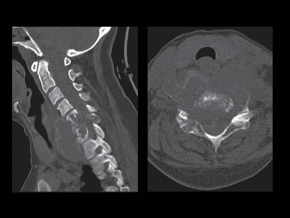
Radiographs of spinal ABCs generally show marked expansile remodeling of bone centered in the posterior elements, although extension into the vertebral body is frequently seen.
Spinal ABC, similar to GCT and chordoma, may extend into adjacent vertebral bodies, intervertebral disks, posterior ribs and paravertebral soft tissues.

CT and MR imaging may reveal multiple fluid-fluid levels reflecting hemorrhage with sedimentation, a characteristic feature of this tumor.
These lesions often have a soft-tissue-attenuation or low-signal-intensity rim on CT and MR images (all pulse sequences), respectively, that corresponds to an intact, thickened periosteal membrane.

Gadolinium enhancement of these lesions on MR images is usually seen within the rim and septations, rather than the cystic spaces.
The presence of fluid-fluid or hematocrit levels is suggestive, but not pathognomic of ABC and have also been reported in giant cell tumors, chondroblastoma, fractured simple cyst, fibrous dysplasia and malignant fibrous histiocytosis.


Giant Cell Tumor
Giant cell tumors (GCT) of the spine are
uncommon.
More frequent in women and affect patients in
the 2nd to 4th decades of life.
Sacrum.
Compared with chordomas, which are central
lesions, sacral GCT are frequently eccentric
and abut or extend across the sacroiliac joint.

When GCT occurs in the spine above the sacrum, it is usually located in the vertebral body with or without extension to the posterior elements.
Involvement of the adjacent intervertebral disks and vertebrae is not uncommon.
Pathologically, GCT is composed of abundant osteoclastic giant cells intermixed throughout a spindle cell stroma.
Cystic areas(similar to those seen in
ABC), hemorrhage with hemosiderin
and prominent areas of fibrous tissue
that are high in collagen content are a
frequent finding.
Despite benign pathology, they may
rarely metastasize and they recur
without complete resection.

Radiography typically shows a lytic lesion with cortical expansion.
On CT scans, the tumor has soft-tissue attenuation with well-defined margins that may show a thin rim of sclerosis.
These very vascular neoplasms show heterogeneous signal intensity on both T1 and T2WI because of the presence of necrosis, hemorrhage, or cystic spaces.

Low signal intensity is frequently noted on
T2WI and is related to the hemorrhagic and
fibrotic content of this tumor.
There is usually no marrow edema in the
absence of a pathologic fracture.
The lesion enhances after intravenous
injection of contrast material.

Chordoma
Chordoma is the most common non lymphoproliferative primary malignant tumor of the spine and accounts for 2-4 percent of malignant osseous neoplasms.
These arise from notochordal rests and therefore, almost always occur in a midline or paramedian location in relation to the spine.
Nearly 50 percent of all chordomas originate in the sacrococcygeal region, particularly in the fourth and fifth sacral segments.

Men are affected twice as frequently as women; the mean age of patients is 50 years.
Chordomas are slow-growing tumors that are commonly discovered as large masses. As they enlarge, they tend to involve adjacent vertebral bodies and extend into the adjacent paraspinal tissues and epidural space; they may even grow into and expand neural foramina, potentially mimicking nerve sheath tumors.

The most suggestive manifestation is a destructive lesion of a vertebral body associated with a soft-tissue mass with a “collar button” or “mushroom” appearance and a “dumbbell” shape, spanning several segments and sparing the disks.
Areas of amorphous calcifications are noted in 40 percent of chordomas of the mobile spine and in up to 90 percent of sacrococcygeal lesions.

Most chordomas are iso-or hypointense relative to muscle on T1WI.
The focal areas of hemorrhage and high protein content of the myxoid and mucinous collections may account for the high signal intensity on T1WI.
On T2WI, most chordomas have a high signal intensity due to the presence of their signature physaliphorous cells (clear cells with intracytoplasmic vacuoles and abundant mucin).
The fibrous septa that divide the
gelatinous components of the tumor are
seen as areas of low signal intensity on
T2WI.
The presence of hemosiderin also
accounts for the low signal intensity
seen on T2WI.
.

Moderate heterogeneous enhancement,
but ring and arc enhancement and
peripheral enhancement have also been
described.
Chordomas generally have a poor
prognosis due to local recurrence
following resection.


EPIDURAL LESIONS
Angiolipoma
Spinal angiolipomas are rare lesions usually found in the epidural space of the thoracic spine.
Mean age of occurrence is 9 years most patients presenting with slowly progressive symptoms of spinal cord compression.
Most of these lesions are found in adults and in the thoracic region.

Spinal angiolipomas are typically located
in the posterior and lateral aspects of
the epidural space. However, infiltrating
forms of tumor are generally in the
anterior epidural space.
On MRI, angiolipomas are
predominantly hyperintense on T1WI
and inhomogeneous owing to
interspersed vascular elements.
A high vascular content is correlated
with the presence of large hypointense
regions on T1WI.
These masses are hyperintense on
T2WI.
The larger tumors may result in
compression of the spinal cord.
Intramedullary angiolipomas have been
rarely described.

QUIZ
1. Cap sign is seen in which tumors?

1. Cap sign?
Ependymoma
Hemangioblastoma
Paraganglioma.

2. Long segment involvement in which
of the following?
A. Ependymoma.
B. Astrocytoma.
C. Ganglioglioma.

2. Long segment involvement?
A. Ependymoma.
B. Astrocytoma.
C. Ganglioglioma.

3. Most common intramedullary tumor in
adults and children?

3. Most common intramedullary tumor in
adults and children?
Ependymoma - adults.
Astrocytoma- children.

4. Tumors showing calcification?
Intramedullary.
Intradural extramedullary.
Extradural.

4. Tumors showing calcification?
Intramedullary. Ganglioglioma.
Intradural extramedullary. Meningioma.
Extradural. Cordoma

5. Which of these are eccenteric
tumors?
A. Subependymoma.
B. Ependymoma.
C. Astrocytoma.
D. Ganglioglioma.

5. Eccenteric tumors?
A. Subependymoma.
B. Ependymoma.
C. Astrocytoma.
D. Ganglioglioma.

6. Spinal tumors associated with.
A. NF 1.
B. NF 2.
C. VHL.

6. Spinal tumors associated with.
A. NF 1. Neurofibromas.
B. NF 2. Ependymoma and Meningioma.
C. VHL. Hemangioblastoma.

THANK YOU…..