sperm-associated antigen 9 as a candidate diagnostic and
TRANSCRIPT

Original Article
Middle East Journal of Cancer; July 2015 6(3):
Sperm-Associated Antigen 9 as a
Candidate Diagnostic and Prognostic
Biomarker in Breast Cancer
Houra Naraghi*, Frouzandeh Mahjoubi*♦, Nahid Nafissi**
*Medical Biotechnology Institute, National Institute of Genetic Engineering and
Biotechnology (NIGEB), Tehran, Iran **Rasole Akram Hospital, Iran University of Medical Sciences, Tehran, Iran
Original Article
Middle East Journal of Cancer; October 2020; 11(4): 415-422
♦Corresponding Author:
Frouzandeh Mahjoubi, PhD Medical Biotechnology Institute, National Institute of Genetic Engineering and Biotechnology (NIGEB), Tehran, Iran Tel/Fax: 9821-44580389 Email: [email protected]
Introduction
Breast cancer is the second most prevalent cancer worldwide, and by far, the most frequent cancer among women, with an estimated 1.7 million new cancer cases diagnosed in 2012. Breast cancer is the fifth cause of
overall cancer-related mortality.1 Statistics of breast cancer incidence among Iranian women report about 10,000 new cases yearly.2 In addition, annually, there are 1063 cases of breast cancer death in Iran.3 In addition to the high prevalence of
Abstract Background: Cancer/testis antigens are a unique class of tumor antigens with
normal expressions restricted to the testis and various cancers, but not in adult somatic tissues. Sperm-associated antigen 9 (SPAG9) has been introduced as a new member of Cancer Testis Antigens family involved in c-Jun-NH2-kinase signaling module.
The objective of this research was to investigate the potential of SPAG9 as a diagnostic and prognostic biomarker in breast cancer. We further aimed to find any significant association between SPAG9 expression and clinicopathologic features of the cancer.
Methods: In this retrospective study, 35 breast cancer tissues and 35 adjacent non-cancerous tissues were collected and examined using RT-PCR to explore SPAG9 mRNA expression. Statistical analysis was done utilizing SPSS 22.0 software.
Results: Unexpectedly, we detected SPAG9 expression in 54% of adjacent non-cancerous tissues. Moreover, SPAG9 mRNA was expressed in 57% of cancerous tissues. Statistical analysis showed a significant association between SPAG9 expression and tumor size, lymph node metastasis, and cancer stage.
Conclusion: The association between the gene expression and tumor size, lymph, node and metastasis, and cancer stage suggests that SPAG9 can potentially be considered as a prognostic biomarker in breast cancer. However, it may not be a candidate diagnostic biomarker.
Keywords: SPAG9, Cancer/testis antigens, Breast cancer, Biomarker, RT-PCR
Received: May 07, 2019; Accepted: January 27, 2020
Please cite this article as: Naraghi H, Mahjoubi F, Nafissi N. Sperm-associated antigen 9 as a candidate diagnostic and prognostic biomarker in breast cancer. Middle East J Cancer. 2020;11(4): 415-22. doi: 10. 30476/mejc.2020.81553.102.

Houra Naraghi et al.
Middle East J Cancer 2020; 11(4): 415-422416
breast cancer in Iran, the fact that the average age of breast cancer among Iranian women is at least a decade earlier than reported in the developed countries, makes it a particularly more important concern.4
A biomarker is a molecule, gene, or characteristic that can be measured in the body to predict the incidence of outcome or disease. Prognostic biomarkers are a type of cancer biomarkers which enable the monitoring of the advances in anticancer therapy, the assessment of the tumor stage, and its potential malignancy. Molecular prognostic factors are more important nowadays; however, traditional markers such as the number of regional metastatic lymph nodes, tumor size, and tumor grade are still considered.5
Cancer/testis antigens (CTAs) are a group of genes normally expressed in germ-cells and trophoblasts and abnormally activated in up to 40% of various types of cancers.6 So far, no defined biological function of CTAs has become known; however, it has been proposed that these molecules are involved in signaling, transcription, translation, and chromosomal recombination.7 It is further suggested that the aberrant expression of CTAs in the tumor may contribute to different malignant properties such as immortality, migration, invasion, and metastatic capacity.
Sperm-associated antigen 9 (SPAG9) is a
member of the CTAs family expressed from a single copy gene located on human chromosome 17q2. SPAG9 functions as a scaffolding protein in c-Jun-NH2-kinase (JNK) signaling module. This suggests its involvement in physiological processes, including apoptosis, survival, proliferation, and tumorigenesis.8 Scaffold proteins act by modulating the signaling strength of their cognate mitogen-activated protein kinase (MAPK) module through regulating the signal amplitude and duration. SPAG9 has been suggested as a novel biomarker for early diagnosis of ovarian cancer, chronic myeloid leukemia, and bladder transitional cell carcinoma.9
The purpose of this study was to investigate SPAG9 expression as a probable diagnostic and prognostic biomarker in Iranian breast cancer patients and find any significant association between SPAG9 expression and the clinicopatho-logic features of cancer.
Materials and Methods
Patients and tissue samples The Ethics Committee of the National Institute
of Genetic Engineering and Biotechnology approved this retrospective study (Ethics Code No: IR.NIGEB.EC.1395.5.6). After obtaining written informed consent from all patients, we collected 35 breast cancer tissues as an
Figure 1. a) RT-PCR analysis of SPAG9 mRNA expression showing specific 141bp products in breast cancer and adjacent non-cancerous tissues. M, molecular size marker b) β-actin expression as an internal control.

SPAG9 as a Candidate Biomarker in Breast Cancer
Middle East J Cancer 2020; 11(4): 415-422 417
experimental group and 35 adjacent non-cancerous tissues (ANCTs) as a control group. The obtained tissue samples belonged to women admitted to the Khatam Al-Anbia semi-private Hospital in Tehran and surgically treated without chemotherapy. Tissue specimens were immediately snap-frozen in liquid nitrogen and archived at -70°C until use. Pathology reports provided the patients’ demographics and clini-copathologic variables. Sample size calculation was done using the following formula:10
Z2α/2*pq
n≥ d2
n – Sample size Zα/2 – Critical value for the desired confidence degree, usually: 1.96 (95%) d–Standard error, usually: ±5% of the proportion of cases (absolute precision) p – Proportion of cancerous samples in the population q – Proportion of adjacent non-cancerous samples in the population (q=1-p).
RNA extraction and cDNA synthesis
We carried out the total RNA extraction using the TriPure Isolation Reagent (Roche) according to the manufacturer’s protocol. After determining the extracted RNA concentration, 5.5µl RNA of each sample was utilized for complementary DNA (cDNA) synthesis using Thermo Scientific Revert Aid First Strand cDNA Synthesis kit.
Reverse Transcription Polymerase Chain Reaction (RT-PCR)
To examine SPAG9 mRNA expression, we designed specific primers with the amplicon size of 141bp using Oligo7; NCBI/blast was then employed to confirm the specificity. Avoiding
any genomic DNA contamination, we designed the primers from exon-exon junction regions of the target gene. SPAG9 Forward Primer: 5’-GCAGTAAACAGC-
Table 1. Patients’ demographic data* Clinicopathologic Frequency
features N (%) Age (years)
<50 19 (54.3) ≥50 16 (45.7) Tumor size
T1(<2 cm) 9 (25.7) T2(2-5 cm) 26 (74.3) TNM Stage
I 12 (34.3) II 17 (45.7) III 6 (17.1) Histological type
IDC 24 (68.6) ILC 4 (11.4) others 7 (20) Histological Grade
G1 2 (5.9) G2 23 (67.6) G3 9 (26.5) Lymph node metastasis No 16 (47.1) Yes 18 (52.9) ER status
Negative 8 (25) Positive 24 (75) PR status
Negative 7 (21.9) Positive 25 (78.1) Her-2 status
Negative 24 (75) Positive 8 (25) Necrosis
No 10 (37) Yes 17 (63) *TNM, tumor, node, and metastasis; IDC, invasive ductal carcinoma; ILC, invasive lobular carcinoma; others, invasive ductal and lobular carcinoma, ductal carcinoma in situ and medullary carcinoma; G1, well differentiated; G2, moderately differentiated; G3, poorly differentiated.
Figure 2. SPAG9 mRNA expression in normal breast tissue. M, molecular size marker.
Houra Naraghi et al.
Middle East J Cancer 2020; 11(4): 415-422418
GAAGTG-3’ SPAG9 Reverse Primer: 5’-CTTTTGTAGCC-GAATGAGT-3’
We first performed gradient PCR to find the most optimal annealing temperature. RT-PCR was then employed using Ampliqon Taq 2x master mix (Red,1.5 mM mgcl2) in a volume of 10µL containing 5µL of Master, 0.5µL of each primer (10 µM ) and 0.5µL of (diluted 1:10) cDNA. Afterwards, we carried out 40 cycles of amplification. Ensuring the accuracy of the results, all experiments were repeated three times. PCR products were then analyzed on 1.5% agarose gel and photographed under UV light (Figure 1).
Statistical analysis
Statistical analysis was performed using SPSS 22.0 software. We assessed the association between SPAG9 expression and clinicopathologic variables using Pearson’s χ2 and Mann-Whitney U tests. P values of 0.05 or less were statistically significant.
Results
Patients’ demographic data In total, 35 patients and 11 healthy women,
undergone mammoplasty surgery, enrolled in this study. Table 1 summarizes the patients’ demographic data.
SPAG9 expression in cancerous and adjacent non-cancerous tissues
Unexpectedly, this gene was expressed in 54% (19 of 35) of adjacent non-cancerous tissues (Figure 1) and 57% (20 of 35) of cancerous tissues. As mentioned, SPAG9 is a CTA which, in theory, is not expected to be expressed in normal tissues, except testis and certain cancer cells.
According to the results, we decided to test SPAG9 mRNA expression in normal breast tissues. Therefore, we collected normal breast tissues from 11 healthy women, undergone mammoplasty surgery, with a mean age of 37. Interestingly, SPAG9 expression was once again observed in one of the samples (Figure 2).
SPAG9 expression in cancerous tissues and clinicopathologic variables SPAG9 expression and patients age
The estimated mean age of patients was 52.2±11.7 years. SPAG9 was expressed in the cancerous samples of 57% (11 of 19) of patients younger than 50 years old and 56% (9 of 16) older than 50 years. SPAG9 expression was independent of age (P=0.93).
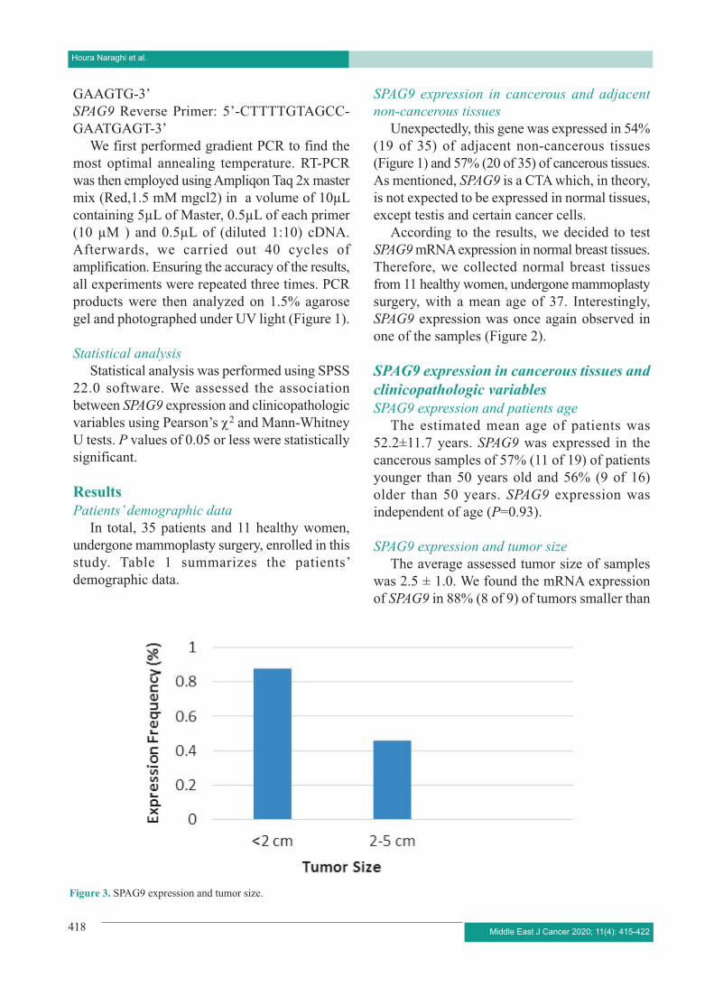
SPAG9 expression and tumor size
The average assessed tumor size of samples was 2.5 ± 1.0. We found the mRNA expression of SPAG9 in 88% (8 of 9) of tumors smaller than
Figure 3. SPAG9 expression and tumor size.

SPAG9 as a Candidate Biomarker in Breast Cancer
Middle East J Cancer 2020; 11(4): 415-422 419
2cm and 46% (12 of 26) of tumors with 2-5cm size. There was a significant association between SPAG9 expression and tumor size (P=0.05) (Figure 3).
SPAG9 expression and cancer stage
SPAG9 expression was observed in 83% (10 of 12) of stage I, 47% (8 of 17) of stage II, and 33% (2 of 6) of stage III. A significant association existed between SPAG9 expression and TNM stage (P=0.05) (Figure 4).
SPAG9 expression and histological type
SPAG9 was expressed in 58% (14 of 24) of invasive ductal carcinoma (IDC), 75% (3 of 4) of invasive lobular carcinoma (ILC), and 42% (3 of 7) of other histological types. We further observed that SPAG9 expression was not associated with the histological type of samples (P=0.61).
SPAG9 expression and histological grade
SPAG9 was expressed in 50% (1 of 2) of grade 1, 60% (14 of 23) of grade 2, and 44% of (4 of 9) grade 3; however, there was no association between expression and histological grade (P=0.69).
SPAG9 expression and lymph node metastasis
We identified SPAG9 mRNA in 81% (13 of 16) of breast cancer tissues with negative lymph
node involvement compared to 33% (6 of 18) tissues with lymph node involvement. There was a significant association between SPAG9 expression and lymph, node, and metastasis (P=0.01) (Figure 5).
SPAG9 expression and ER status
SPAG9 expression was observed in 37% (3 of 8) of ER- and 62% (15 of 24) of ER+ specimens without any significant association between these two factors (P=0.20).
SPAG9 expression and PR status
SPAG9 was expressed in 60% (15 of 25) of PR- and 42% (3 of 7) of PR+ tissues. We found no association between SPAG9 expression and PR status (P=0.35).
SPAG9 expression and HER-2 status
We detected SPAG9 expression in 62% (5 of 8) of HER-2- together with 54% (13 of 24) of HER-2+ samples; however, no association existed between HER-2 status and SPAG9 expression (P=0.50).
SPAG9 expression and necrosis
SPAG9 was expressed in 58% (10 of 17) of breast tissues without necrosis and 60% (6 of 10) of tissues with necrosis. There was no association between these two parameters (P=0.63).
Figure 4. SPAG9 expression and cancer stage.

Houra Naraghi et al.
Middle East J Cancer 2020; 11(4): 415-422420
SPAG9 expression and patients’ age in adjacent non-cancerous tissues
In adjacent non-cancerous samples, SPAG9 was expressed in 50% (9 of 18) of patients younger than 50 years old and 58% (10 of 17) of those older than 50 years. SPAG9 expression was independent of age (P=0.44).
Discussion
Despite the data from previous studies,16,17
our results confirmed that SPAG9 was indeed expressed in adjacent non-cancerous tissues and even the normal breast tissues. This makes the value of this gene as CTA doubtful.
SPAG9 encodes a protein which functions as a scaffolding protein and interacts with c-Jun NH2-terminal kinase subgroup of mitogen-activated protein kinases. Recently, SPAG9 has been reported as a candidate cancer-associated marker in various cancers including colorectal cancer,11 endometrial cancer,12 lung cancer,13
osteosarcoma,14 cervical cancer,15 and renal cell carcinoma.7
To the best of our knowledge, there are only two published papers regarding SPAG9 mRNA expression in breast cancer. The first study was performed by Kanojia et al. in 2009. They reported SPAG9 expression in cancerous tissues, but not in adjacent non-cancerous tissues. Furthermore, SPAG9 expression was independent of tumor
stage, yet significantly associated with the grades.16
The second study was performed in 2013 by Sinha et al. on four breast cancer cell lines, namely MCF-7, BT-474, SK-BR3, and MDA-MB-231 together with normal mammary epithelial cells. Their results confirmed SPAG9 mRNA expression in all examined samples except normal mammary epithelial cells. This obviously shows SPAG9 exclusive expression in cancerous cells.17
Our study showed SPAG9 expression in 54% (19 of 35) of adjacent non-cancerous tissues, 57% (20 of 35) of cancerous tissues, and one of the normal breast tissues. Possible explanations for the attained results are:
As stated earlier, any gene that exhibits an mRNA expression restricted to the testis and neoplastic cells can be called a CT gene. In the literature, the existing definitions of CT genes vary from “genes expressed exclusively in adult testis germ cells and malignant tumors” to “dominant testicular expression, possible additional presence in placenta, ovary and epigenetic regulation, and membership of a gene family and localization on the X chromosome”.6
Therefore, due to the paucity of a distinct definition for CTAs, a growing number of CT candidates have appeared in the literature.
Another classification system for CTAs is based on their expression profile, dividing them
Figure 5. SPAG9 expression and lymph node metastasis.

SPAG9 as a Candidate Biomarker in Breast Cancer
Middle East J Cancer 2020; 11(4): 415-422 421
into three subclasses: (a) testis restricted (found only in the testis), (b) testis-brain restricted (expressed in the testis and central nervous system), (c) testis selective (expressed in the testis and no more than two additional tissues at lower levels than in the testes).18
Based on The Human Protein Atlas (https://www.proteinatlas.org/), SPAG9 RNA and protein expression has been reported in numerous tissues including breast tissue.
Transcriptome analysis of healthy, adjacent non-cancerous, and tumor tissues in 6506 samples from eight tissues including breast tissue showed that adjacent non-cancerous tissues presented a unique intermediate state between healthy and tumor. Also, differential gene expression and protein-protein interaction analyses revealed altered pathways among adjacent non-cancerous tissues across tissue types. A set of 18 genes specifically expressed in adjacent non-cancerous tissues were ultimately characterized.19
Regarding the relationship between the expression and clinicopathologic factors, we found no association between the expression of this gene and the patients’ age, grade, ER, PR, Her-2, and necrosis; however, there existed a significant association between SPAG9 expression and tumor size, cancer stage, and lymph node involvement. Lymph node involvement is particularly related to the risk of metastasis; therefore, it can be proposed that SPAG9-positive expression may be considered as a poor prognostic marker in breast cancer.
This study had certain limitations such as the relatively small number of the patients and the bias towards the middle or high-income patients (often low-income patients refer to the governmental hospitals); Therefore, more investigations are required to obtain more robust results.
Conclusion
Due to the expression of SPAG9 mRNA in adjacent non-cancerous tissues and normal breast tissues, SPAG9 may not be used as a diagnostic biomarker. However, the association of SPAG9
expression with tumor size, lymph node metastasis, and cancer stage suggests the use of this gene as a possible prognostic biomarker in patients diagnosed with breast cancer.
Acknowledgement
The National Institute of Genetic Engineering and Biotechnology (NIGEB) of Iran funded this project.
The authors would like to acknowledge all the people who participated in this study. We are especially grateful to Ms. Samira Shabani and Mr. Jamshid Motalebzadeh for their assistance and comments which greatly improved this experimental study.
Conflict of Interest
None declared.
References 1. Ferlay J, Soerjomataram I, Dikshit R, Eser S, Mathers
C, Rebelo M, et al. Cancer incidence and mortality worldwide: sources, methods and major patterns in GLOBOCAN 2012. Int J Cancer. 2015;136(5):E359-86. doi: 10.1002/ijc.29210.
2. Haghighat S, Akbari ME, Yavari P, Javanbakht M, Ghaffari S. Cost-effectiveness of three rounds of mammography breast cancer screening in Iranian women. Iran J Cancer Prev. 2016;9(1):e5443. doi: 10.17795/ijcp-5443.
3. Otaghvar HA, Hosseini M, Tizmaghz A, Shabestanipour G, Noori H. A review on metastatic breast cancer in Iran. Asian Pac J Trop Biomed. 2015;5(6):429-33.
4. Rahimzadeh M, Pourhoseingholi MA, Kavehie B. Survival rates for breast cancer in Iranian patients: a meta- analysis. Asian Pac J Cancer Prev. 2016;17(4):2223-7.
5. Nicolini A, Ferrari P, Duffy MJ. Prognostic and predictive biomarkers in breast cancer: Past, present and future. Semin Cancer Biol. 2018;52(Pt1):56-73. doi: 10.1016/j.semcancer.2017.08.010.
6. Hofmann O, Caballero OL, Stevenson BJ, Chen YT, Cohen T, Chua R, et al. Genome-wide analysis of cancer/testis gene expression. Proc Natl Acad Sci U S A. 2008;105(51):20422-7. doi: 10.1073/pnas. 0810777105.
7. Garg M, Kanojia D, Khosla A, Dudha N, Sati S, Chaurasiya D, et al. Sperm-associated antigen 9 is associated with tumor growth, migration, and invasion in renal cell carcinoma. Cancer Res. 2008;68(20):8240-8. doi: 10.1158/0008-5472.CAN-08-1708.
Houra Naraghi et al.
Middle East J Cancer 2020; 11(4): 415-422422
8. Ren B, Zou G, He J, Huang Y, Ma G, Xu G, et al. Sperm-associated antigen 9 is upregulated in hepatocellular carcinoma tissue and enhances QGY cell proliferation and invasion in vitro. Oncol Lett. 2018;15(1):415-22. doi: 10.3892/ol.2017.7270.
9. Jagadish N, Fatima R, Sharma A, Devi S, Suri V, Kumar V, et al. Sperm associated antigen 9 (SPAG9) a promising therapeutic target of ovarian carcinoma. Tumour Biol. 2018;40(5):1010428318773652. doi: 10.1177/1010428318773652.
10. Charan J, Biswas T. How to calculate sample size for different study designs in medical research? Indian J Psychol Med. 2013;35(2):121-6. doi: 10.4103/0253-7176.116232.
11. Kanojia D, Garg M, Gupta S, Gupta A, Suri A. Sperm-associated antigen 9 is a novel biomarker for colorectal cancer and is involved in tumor growth and tumorigenicity. Am J Pathol. 2011;178(3):1009-20. doi: 10.1016/j.ajpath.2010.11.047.
12. Zhang L, Yan L, Cao M, Zhang H, Li C, Bai Y, et al. SPAG9 promotes endometrial carcinoma cell invasion through regulation of genes related to the epithelial-mesenchymal transition. Eur J Gynaecol Oncol. 2016;37(3):312-9.
13. Ren B, Wei X, Zou G, He J, Xu G, Xu F, et al. Cancer testis antigen SPAG9 is a promising marker for the diagnosis and treatment of lung cancer. Oncol Rep. 2016;35(5):2599-605. doi: 10.3892/or.2016.4645.
14. Yang X, Zhou W, Liu S. SPAG9 controls the cell motility, invasion and angiogenesis of human osteosarcoma cells. Exp Ther Med. 2016;11(2):637-644.
15. Garg M, Kanojia D, Salhan S, Suri S, Gupta A, Lohiya NK, et al. Sperm-associated antigen 9 is a biomarker for early cervical carcinoma. Cancer. 2009;115(12):2671-83. doi: 10.1002/cncr.24293.
16. Kanojia D, Garg M, Gupta S, Gupta A, Suri A. Sperm-associated antigen 9, a novel biomarker for early detection of breast cancer. Cancer Epidemiol Biomarkers Prev. 2009;18(2):630-9. doi: 10.1158/1055-9965.EPI-08-0629.
17. Sinha A, Agarwal S, Parashar D, Verma A, Saini S, Jagadish N, et al. Down regulation of SPAG9 reduces growth and invasive potential of triple-negative breast cancer cells: possible implications in targeted therapy. J Exp Clin Cancer Res. 2013;32:69. doi: 10.1186/1756-9966-32-69.
18. Whitehurst AW. Cause and consequence of cancer/testis antigen activation in cancer. Annu Rev Pharmacol Toxicol. 2014;54:251-72. doi: 10.1146/annurev-pharmtox-011112-140326.
19. Aran D, Camarda R, Odegaard J, Paik H, Oskotsky B, Krings G, et al. Comprehensive analysis of normal adjacent to tumor transcriptomes. Nat Commun. 2017;8(1):1077. doi: 10.1038/s41467-017-01027-z.