specific lipid binding of membrane proteins in detergent ...€¦ · biochemistry 2016, 55,...
TRANSCRIPT
Specific Lipid Binding of Membrane Proteins in Detergent MicellesCharacterized by NMR and Molecular DynamicsLinlin Zhao,†,⊥ Shuqing Wang,‡,⊥ Changqing Run,† Bo OuYang,*,† and James J. Chou*,†,§
†State Key Laboratory of Molecular Biology, National Center for Protein Science Shanghai, Shanghai Institute of Biochemistry andCell Biology, Shanghai Science Research Center, Chinese Academy of Sciences, Shanghai 200031, China‡School of Pharmacy, Tianjin Medical University, Tianjin 300070, China§Department of Biological Chemistry and Molecular Pharmacology, Harvard Medical School, Boston, Massachusetts 02115, UnitedStates
*S Supporting Information
ABSTRACT: Many membrane proteins bind specificallyto lipids as an integral component of their structures. Theability of detergents to support lipid binding is thus animportant consideration when solubilizing membraneproteins for structural studies. In particular, the zwitter-ionic phosphocholine (PC)-based detergents, which havebeen widely used in solution NMR studies of channels andtransporters, are controversial because of their strongsolubilization power and thus perceived as moredenaturing than nonionic detergents such as the malto-sides. Here, we investigate the ability of the mitochondrialADP/ATP carrier (AAC) to specifically bind cardiolipin, amitochondrial lipid important for the carrier function, indodecylphosphocholine (DPC) micelles. We found that inDPC, the AAC specifically binds cardiolipin in a mannerconsistent with the bound cardiolipins found in the crystalstructures of the AAC determined in n-decyl β-D-maltoside. Our results suggest that PC detergent iscompatible with specific lipid binding and that PCdetergent mixed with the relevant lipid represents a viablesolubilization system for NMR studies of membraneproteins.
The past two decades have seen an increasing frequency ofapplication of solution NMR spectroscopy to de novo
structural characterization of transmembrane (TM) andmembrane-associated protein domains as well as intact full-length integral membrane proteins.1−3 In addition to structuredetermination, solution NMR affords a convenient means ofinvestigating ligand binding and conformational dynamics whenrelevant membrane protein sample systems are beingdeveloped.4,5 Unlike solid-state NMR, solution NMR requiresmembrane proteins to be solubilized in membrane-mimeticmedia, and these media are mostly detergent micelles,5−8 lesscommonly bicelles,9−11 and very rarely lipid nanodiscs.12
The use of PC detergents, however, has been controversial,as these detergents have in some cases performed poorly inpreserving the native activities of membrane proteins, especiallyfor G-protein-coupled receptors.13 While new media forsolution NMR that more closely resemble the natural lipidbilayer are still being developed, a system more suitable thandetergent alone consists of detergent micelles mixed with
lipids.14 In terms of sample preparation, the detergent/lipidmixed micelle is as convenient to use as detergent micelles. Animportant criterion of the mixed micelle application, however,is whether the detergent is compatible with specific lipid−protein interaction that is functionally important to manymembrane proteins.15 Considering this, we sought toinvestigate whether dodecylphosphocholine (DPC), a relativelystrong and the most frequently used detergent in NMR, canpreserve the binding of functionally important lipids to protein.As such, we chose the mitochondrial ADP/ATP carrier (AAC)as a case study because this transporter requires special lipids tomaintain its native activity16 and, when reconstituted in DPCmicelles, exhibited favorable spectroscopic properties.17
The AAC is an essential solute transporter that catalyzes thetrafficking of ATP and ADP between mitochondria andcytosol;18 it has been the model system for structural andmechanistic studies of mitochondrial carriers19 because itremains, to date, the only carrier protein for which high-resolution crystal structures are available.20,21 Earlier functionalstudies showed that cardiolipin (CL), a rich component of themitochondrial membrane and a nonbilayer lipid, is importantfor the transport activity of AAC.22,23 Consistent with thisfinding, high-resolution crystal structures of AAC previouslydetermined in the decyl maltoside (DM) detergent revealedseveral bound CLs on the periphery of the transporter.20,21
Obviously, CL binding was preserved when AAC was extractedfrom the mitochondrial membrane using the nonionicmaltoside detergent. Recently, we developed an NMR sampleof AAC reconstituted in DPC micelles to investigate theexchange dynamics of the transporter.17 As DPC as a detergentis stronger than DM, it is unclear whether its micelle systemsupports specific binding of CL. In this study, we performedthorough NMR measurements of CL−AAC interaction andmolecular dynamics (MD) simulation to address the questionposed above.Full-length yeast AAC carrier 3 (yAAC3) was expressed in
Escherichia coli cells and purified by Ni-NTA affinity, ionexchange, and size-exclusion chromatography as previouslydescribed17 (Figure S1a). During NMR sample preparation,
Received: August 12, 2016Revised: September 11, 2016Published: September 13, 2016
Rapid Report
pubs.acs.org/biochemistry
© 2016 American Chemical Society 5317 DOI: 10.1021/acs.biochem.6b00836Biochemistry 2016, 55, 5317−5320

DPC was systematically reduced to reach the minimalconcentration required to generate a good NMR spectrum(Figure S1b).We first employed the NMR titration approach to examine
CL-induced chemical shift perturbation of AAC. For thispurpose, we used cardiolipin with 18 carbons. High-resolutionthree-dimensional (3D) TROSY-HNCO spectra were recordedat CL:yAAC3 ratios of 0:1, 1:1, 2:1, 3:1, and 4:1. Theapplication of the 3D experiment during NMR titration greatlyimproved the completeness of peak analysis of the otherwisehighly crowded HSQC spectrum (Figure S2a). Addition of CLcaused very substantial chemical shift perturbations; e.g.,residue I25, L92, and A145 showed shifts of as much as 0.15ppm (Figure S2b,c). Moreover, the titration curves showed thatthe majority of the specific changes nearly plateaued after the3:1 CL:yAAC3 ratio (Figure 1a). The estimated apparent
dissociation constants (KD) for A91, A148, and E268 in Figure1a are approximately 100, 100, and 20 μM, respectively (FigureS2d). The AAC architecture resembles an open-top barrelformed by three structurally similar domains (I−III) in aparallel orientation. Each domain consists of two TM helicesseparated by an amphipathic helix. Overall, the chemical shiftperturbation is widespread on the periphery of the carrier(Figure 1b), and the titration data suggest a 3:1 CL:yAAC3binding stoichiometry.
Given the long acyl chains of CL, it is not surprising to seewidespread chemical shift changes because the lipid partition inthe DPC micelle could have indirectly induced chemical shiftchanges by altering the micelle properties. To test whether thechemical shift perturbations are specific, we performed thesame titration using 1-palmitoyl-2-oleoyl-sn-glycero-3-phos-phoethanolamine (POPE), another lipid rich in the mitochon-drial inner membrane.24 The results showed that at a 3:1lipid:yAAC3 ratio, only CL induced obvious chemical shiftperturbations while POPE had essentially no effect (Figure 1c).The combined titration results indicate that CL bindsspecifically to yAAC3.We next applied nuclear Overhauser enhancement (NOE)
spectroscopy to directly identify the CL binding sites onyAAC3 using proteins that are 15N-labeled and perdeuteratedsuch that the NOE between the protein backbone amideprotons and CL protons could be measured unambiguously.DPCs were also perdeuterated to avoid resonance overlap withCL. A 3D 15N-separated NOESY spectrum was recorded with aNOE mixing time of 200 ms at 900 MHz (1H frequency), forassigning residue-specific CL NOE cross-peaks. Consistent withthe NMR titration results, many residues showed distinct CLNOEs, including those of the headgroup (3.90 ppm), themethylene groups of the acyl chain (1.29, 1.40, and 1.61 ppm),and the terminal methyl group (0.84 ppm) (Figure 2a). Inparticular, NOEs to the methylene groups of the hydrocarbonchain are widespread becuse of the large number of thesemethylene protons in each CL molecule.
Figure 1. Characterization of cardiolipin (CL) binding by NMRtitration. (a) Example of residue-specific chemical shift changes uponCL titration as monitored in the 3D HNCO spectrum. (b) Mappingonto the crystal structure of AAC. Sphere colors indicate the following:gray for −0 < Δδ < 0.05, light blue for −0.05 < Δδ < 0.10, and blue forΔδ > 0.10. (c) Comparison between the chemical shift perturbationsof AAC by CL (left) and POPE (right). Red and blue peaks are fromprotein (0.8 mM) in the absence and presence of 2.4 mM lipid,respectively.
Figure 2. Definition of cardiolipin (CL) binding sites by NOEmeasurements. (a) Sample strips from the 3D 15N-edited NOESY-TROSY spectrum (200 ms NOE mixing time) recorded using asample containing 0.8 mM 15N- and 2H-labeled yAAC3 and 2.4 mMCL, showing protein NOEs to three regions of the lipid, including theheadgroup (blue), the bulk acyl chain (orange), and the terminus ofthe acyl chain (red). (b) Mapping CL NOEs onto the yAAC3 crystalstructure for backbone amides that could be unambiguously analyzed(indicated by spheres). Sphere colors indicate the following: gray foramides that showed no lipid NOEs and other colors for amides thatshowed NOEs to lipid regions defined in panel a.
Biochemistry Rapid Report
DOI: 10.1021/acs.biochem.6b00836Biochemistry 2016, 55, 5317−5320
5318
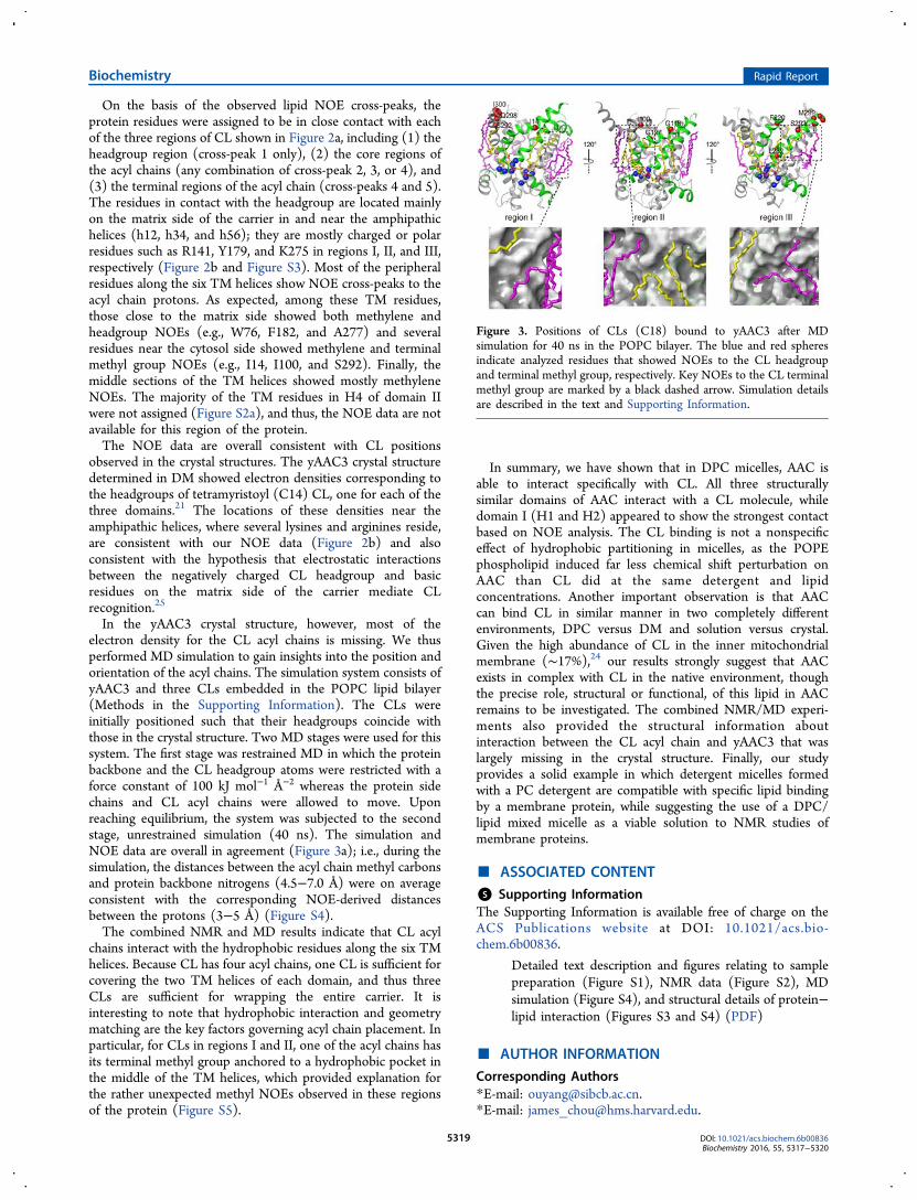
On the basis of the observed lipid NOE cross-peaks, theprotein residues were assigned to be in close contact with eachof the three regions of CL shown in Figure 2a, including (1) theheadgroup region (cross-peak 1 only), (2) the core regions ofthe acyl chains (any combination of cross-peak 2, 3, or 4), and(3) the terminal regions of the acyl chain (cross-peaks 4 and 5).The residues in contact with the headgroup are located mainlyon the matrix side of the carrier in and near the amphipathichelices (h12, h34, and h56); they are mostly charged or polarresidues such as R141, Y179, and K275 in regions I, II, and III,respectively (Figure 2b and Figure S3). Most of the peripheralresidues along the six TM helices show NOE cross-peaks to theacyl chain protons. As expected, among these TM residues,those close to the matrix side showed both methylene andheadgroup NOEs (e.g., W76, F182, and A277) and severalresidues near the cytosol side showed methylene and terminalmethyl group NOEs (e.g., I14, I100, and S292). Finally, themiddle sections of the TM helices showed mostly methyleneNOEs. The majority of the TM residues in H4 of domain IIwere not assigned (Figure S2a), and thus, the NOE data are notavailable for this region of the protein.The NOE data are overall consistent with CL positions
observed in the crystal structures. The yAAC3 crystal structuredetermined in DM showed electron densities corresponding tothe headgroups of tetramyristoyl (C14) CL, one for each of thethree domains.21 The locations of these densities near theamphipathic helices, where several lysines and arginines reside,are consistent with our NOE data (Figure 2b) and alsoconsistent with the hypothesis that electrostatic interactionsbetween the negatively charged CL headgroup and basicresidues on the matrix side of the carrier mediate CLrecognition.25
In the yAAC3 crystal structure, however, most of theelectron density for the CL acyl chains is missing. We thusperformed MD simulation to gain insights into the position andorientation of the acyl chains. The simulation system consists ofyAAC3 and three CLs embedded in the POPC lipid bilayer(Methods in the Supporting Information). The CLs wereinitially positioned such that their headgroups coincide withthose in the crystal structure. Two MD stages were used for thissystem. The first stage was restrained MD in which the proteinbackbone and the CL headgroup atoms were restricted with aforce constant of 100 kJ mol−1 Å−2 whereas the protein sidechains and CL acyl chains were allowed to move. Uponreaching equilibrium, the system was subjected to the secondstage, unrestrained simulation (40 ns). The simulation andNOE data are overall in agreement (Figure 3a); i.e., during thesimulation, the distances between the acyl chain methyl carbonsand protein backbone nitrogens (4.5−7.0 Å) were on averageconsistent with the corresponding NOE-derived distancesbetween the protons (3−5 Å) (Figure S4).The combined NMR and MD results indicate that CL acyl
chains interact with the hydrophobic residues along the six TMhelices. Because CL has four acyl chains, one CL is sufficient forcovering the two TM helices of each domain, and thus threeCLs are sufficient for wrapping the entire carrier. It isinteresting to note that hydrophobic interaction and geometrymatching are the key factors governing acyl chain placement. Inparticular, for CLs in regions I and II, one of the acyl chains hasits terminal methyl group anchored to a hydrophobic pocket inthe middle of the TM helices, which provided explanation forthe rather unexpected methyl NOEs observed in these regionsof the protein (Figure S5).
In summary, we have shown that in DPC micelles, AAC isable to interact specifically with CL. All three structurallysimilar domains of AAC interact with a CL molecule, whiledomain I (H1 and H2) appeared to show the strongest contactbased on NOE analysis. The CL binding is not a nonspecificeffect of hydrophobic partitioning in micelles, as the POPEphospholipid induced far less chemical shift perturbation onAAC than CL did at the same detergent and lipidconcentrations. Another important observation is that AACcan bind CL in similar manner in two completely differentenvironments, DPC versus DM and solution versus crystal.Given the high abundance of CL in the inner mitochondrialmembrane (∼17%),24 our results strongly suggest that AACexists in complex with CL in the native environment, thoughthe precise role, structural or functional, of this lipid in AACremains to be investigated. The combined NMR/MD experi-ments also provided the structural information aboutinteraction between the CL acyl chain and yAAC3 that waslargely missing in the crystal structure. Finally, our studyprovides a solid example in which detergent micelles formedwith a PC detergent are compatible with specific lipid bindingby a membrane protein, while suggesting the use of a DPC/lipid mixed micelle as a viable solution to NMR studies ofmembrane proteins.
■ ASSOCIATED CONTENT
*S Supporting InformationThe Supporting Information is available free of charge on theACS Publications website at DOI: 10.1021/acs.bio-chem.6b00836.
Detailed text description and figures relating to samplepreparation (Figure S1), NMR data (Figure S2), MDsimulation (Figure S4), and structural details of protein−lipid interaction (Figures S3 and S4) (PDF)
■ AUTHOR INFORMATION
Corresponding Authors*E-mail: [email protected].*E-mail: [email protected].
Figure 3. Positions of CLs (C18) bound to yAAC3 after MDsimulation for 40 ns in the POPC bilayer. The blue and red spheresindicate analyzed residues that showed NOEs to the CL headgroupand terminal methyl group, respectively. Key NOEs to the CL terminalmethyl group are marked by a black dashed arrow. Simulation detailsare described in the text and Supporting Information.
Biochemistry Rapid Report
DOI: 10.1021/acs.biochem.6b00836Biochemistry 2016, 55, 5317−5320
5319
Author Contributions⊥L.Z. and S.W. contributed equally to this work. L.Z. and J.J.C.conceived the study. L.Z. and C.R. prepared the samples andcollected NMR data. L.Z. and B.O. analyzed NMR data. S.W.performed MD analysis. J.J.C. and L.Z. wrote the paper, and allauthors contributed to editing of the manuscript.FundingThis work was supported by CAS Grant XDB08030301 andNational Natural Science Foundation of China grant no.31570746 to J.J.C.NotesThe authors declare no competing financial interest.
■ ACKNOWLEDGMENTSWe thank Zhijun Liu and Bin Wu from the NMR facility ofNCPSS for their assistance with NMR data collection.
■ REFERENCES(1) Oxenoid, K., and Chou, J. J. (2013) Curr. Opin. Struct. Biol. 23,547.(2) Kim, H. J., Howell, S. C., Van Horn, W. D., Jeon, Y. H., andSanders, C. R. (2009) Prog. Nucl. Magn. Reson. Spectrosc. 55, 335.(3) Kang, C., and Li, Q. (2011) Curr. Opin. Chem. Biol. 15, 560.(4) Oxenoid, K., and Chou, J. J. (2016) Protein Sci. 25, 959.(5) Jaremko, L., Jaremko, M., Giller, K., Becker, S., and Zweckstetter,M. (2014) Science 343, 1363.(6) Schnell, J. R., and Chou, J. J. (2008) Nature 451, 591.(7) Oxenoid, K., Dong, Y., Cao, C., Cui, T., Sancak, Y., Markhard, A.L., Grabarek, Z., Kong, L., Liu, Z., Ouyang, B., Cong, Y., Mootha, V.K., and Chou, J. J. (2016) Nature 533, 269.(8) Hiller, S., Garces, R. G., Malia, T. J., Orekhov, V. Y., Colombini,M., and Wagner, G. (2008) Science 321, 1206.(9) Fu, Q., Fu, T. M., Cruz, A. C., Sengupta, P., Thomas, S. K., Wang,S., Siegel, R. M., Wu, H., and Chou, J. J. (2016) Mol. Cell 61, 602.(10) Morrison, E. A., DeKoster, G. T., Dutta, S., Vafabakhsh, R.,Clarkson, M. W., Bahl, A., Kern, D., Ha, T., and Henzler-Wildman, K.A. (2011) Nature 481, 45.(11) Lau, T. L., Kim, C., Ginsberg, M. H., and Ulmer, T. S. (2009)EMBO J. 28, 1351.(12) Hagn, F., Etzkorn, M., Raschle, T., and Wagner, G. (2013) J.Am. Chem. Soc. 135, 1919.(13) Serrano-Vega, M. J., Magnani, F., Shibata, Y., and Tate, C. G.(2008) Proc. Natl. Acad. Sci. U. S. A. 105, 877.(14) Seddon, A. M., Curnow, P., and Booth, P. J. (2004) Biochim.Biophys. Acta, Biomembr. 1666, 105.(15) Yeagle, P. L. (2014) Biochim. Biophys. Acta, Biomembr. 1838,1548.(16) Claypool, S. M., Oktay, Y., Boontheung, P., Loo, J. A., andKoehler, C. M. (2008) J. Cell Biol. 182, 937.(17) Bruschweiler, S., Yang, Q., Run, C., and Chou, J. J. (2015) Nat.Struct. Mol. Biol. 22, 636.(18) Klingenberg, M. (2008) Biochim. Biophys. Acta, Biomembr. 1778,1978.(19) Kunji, E. R., and Robinson, A. J. (2010) Curr. Opin. Struct. Biol.20, 440.(20) Pebay-Peyroula, E., Dahout-Gonzalez, C., Kahn, R., Trezeguet,V., Lauquin, G. J., and Brandolin, G. (2003) Nature 426, 39.(21) Ruprecht, J. J., Hellawell, A. M., Harding, M., Crichton, P. G.,McCoy, A. J., and Kunji, E. R. (2014) Proc. Natl. Acad. Sci. U. S. A. 111,E426.(22) Heimpel, S., Basset, G., Odoy, S., and Klingenberg, M. (2001) J.Biol. Chem. 276, 11499.(23) Hoffmann, B., Stockl, A., Schlame, M., Beyer, K., andKlingenberg, M. (1994) J. Biol. Chem. 269, 1940.(24) van Meer, G., Voelker, D. R., and Feigenson, G. W. (2008) Nat.Rev. Mol. Cell Biol. 9, 112.(25) Beyer, K., and Klingenberg, M. (1985) Biochemistry 24, 3821.
Biochemistry Rapid Report
DOI: 10.1021/acs.biochem.6b00836Biochemistry 2016, 55, 5317−5320
5320